Application of FISH technology for microbiological analysis: current state and prospects
10
MINI-REVIEW Application of FISH technology for microbiological analysis: current state and prospects Benedetta Bottari & Danilo Ercolini & Monica Gatti & Erasmo Neviani Received: 24 May 2006 / Revised: 13 July 2006 / Accepted: 8 August 2006 # Springer-Verlag 2006 Abstract In order to identify and quantify the micro- organisms present in a certain ecosystem, it has become necessary to develop molecular methods avoiding cultiva- tion, which allows to characterize only the countable part of the microorganisms in the sample, therefore losing the information related to the microbial component which presents a vitality condition, although it cannot duplicate in culture medium. In this context, one of the most used techniques is fluorescence in situ hybridization (FISH) with ribosomal RNA targeted oligonucleotide probes. Owing to its speed and sensitivity, this technique is considered a powerful tool for phylogenetic, ecological, diagnostic and environmental studies in microbiology. Through the use of species-specific probes, it is possible to identify different microorganisms in complex microbial communities, thus providing a solid support to the understanding of inter- species interaction. The knowledge of the composition and distribution of microorganisms in natural habitats can be interesting for ecological reasons in microbial ecology, and for safety and technological aspects in food microbiology. Methodological aspects, use of different probes and applications of FISH to microbial ecosystems are presented in this review. Introduction The lack of morphological details in many bacteria and microorganisms has usually hampered their identification, making the use of cultivation necessary for identification. This has proven difficult for many environmental or medically important microorganisms (Amann and Kuhl 1998). Traditional methods to determine the composition of microflora require cultivation on selective media, which is laborious, time-consuming and prone to statistical and methodological errors (Moter and Gobel 2000). According to classical microbiology, in order to study a microorgan- ism, it is necessary to isolate it from the original matrix, and this isolation can be done only on the appropriate medium, from the plates showing separately grown colonies, i.e. from the plates corresponding to the so-called countable dilutions. Moreover, it has been demonstrated how any environmental modification during cultivation could affect the structure of microbial communities, thus preventing a complete view of the ecosystem considered. Therefore, isolation and cultivation in agar medium allow to charac- terize only those microorganisms capable of growing, multiplying and forming colonies in the selected medium and condition of growth, with the loss of the information relative to the microbial component which presents a vitality condition despite not being able to duplicate in culture medium. Although new microorganisms are continuously isolated, it is estimated that only a small fraction of the existent microorganisms have been grown in pure culture and characterized. The lack of knowledge is most severe for complex multi-species microbial communities. Even when all bacteria could ultimately be cultured (which is quite unlikely), progress in the understanding of the ecology of complex microbial communities would require studies on Appl Microbiol Biotechnol DOI 10.1007/s00253-006-0615-z B. Bottari (*) : M. Gatti : E. Neviani Department of Genetic, Biology of Microorganisms, Anthropology, Evolution, University of Parma, via Usberti 11/A, 43100 Parma, Italy e-mail: [email protected] D. Ercolini Department of Food Science, University of Naples Federico II, via Università 100, 80055 Portici, Italy
Transcript of Application of FISH technology for microbiological analysis: current state and prospects
MINI-REVIEW
Application of FISH technology for microbiological analysis:current state and prospects
Benedetta Bottari & Danilo Ercolini & Monica Gatti &Erasmo Neviani
Received: 24 May 2006 /Revised: 13 July 2006 /Accepted: 8 August 2006# Springer-Verlag 2006
Abstract In order to identify and quantify the micro-organisms present in a certain ecosystem, it has becomenecessary to develop molecular methods avoiding cultiva-tion, which allows to characterize only the countable part ofthe microorganisms in the sample, therefore losing theinformation related to the microbial component whichpresents a vitality condition, although it cannot duplicatein culture medium. In this context, one of the most usedtechniques is fluorescence in situ hybridization (FISH) withribosomal RNA targeted oligonucleotide probes. Owing toits speed and sensitivity, this technique is considered apowerful tool for phylogenetic, ecological, diagnostic andenvironmental studies in microbiology. Through the use ofspecies-specific probes, it is possible to identify differentmicroorganisms in complex microbial communities, thusproviding a solid support to the understanding of inter-species interaction. The knowledge of the composition anddistribution of microorganisms in natural habitats can beinteresting for ecological reasons in microbial ecology, andfor safety and technological aspects in food microbiology.Methodological aspects, use of different probes andapplications of FISH to microbial ecosystems are presentedin this review.
Introduction
The lack of morphological details in many bacteria andmicroorganisms has usually hampered their identification,making the use of cultivation necessary for identification.This has proven difficult for many environmental or medicallyimportant microorganisms (Amann and Kuhl 1998).
Traditional methods to determine the composition ofmicroflora require cultivation on selective media, which islaborious, time-consuming and prone to statistical andmethodological errors (Moter and Gobel 2000). Accordingto classical microbiology, in order to study a microorgan-ism, it is necessary to isolate it from the original matrix, andthis isolation can be done only on the appropriate medium,from the plates showing separately grown colonies, i.e.from the plates corresponding to the so-called countabledilutions. Moreover, it has been demonstrated how anyenvironmental modification during cultivation could affectthe structure of microbial communities, thus preventing acomplete view of the ecosystem considered. Therefore,isolation and cultivation in agar medium allow to charac-terize only those microorganisms capable of growing,multiplying and forming colonies in the selected mediumand condition of growth, with the loss of the informationrelative to the microbial component which presents avitality condition despite not being able to duplicate inculture medium.
Although new microorganisms are continuously isolated,it is estimated that only a small fraction of the existentmicroorganisms have been grown in pure culture andcharacterized. The lack of knowledge is most severe forcomplex multi-species microbial communities. Even whenall bacteria could ultimately be cultured (which is quiteunlikely), progress in the understanding of the ecology ofcomplex microbial communities would require studies on
Appl Microbiol BiotechnolDOI 10.1007/s00253-006-0615-z
B. Bottari (*) :M. Gatti : E. NevianiDepartment of Genetic, Biology of Microorganisms,Anthropology, Evolution, University of Parma,via Usberti 11/A,43100 Parma, Italye-mail: [email protected]
D. ErcoliniDepartment of Food Science, University of Naples Federico II,via Università 100,80055 Portici, Italy
the activity and distribution of microbes directly inminimally disturbed samples (Amann and Kuhl 1998).
Therefore, in order to identify and quantify the micro-organisms occurring in a certain ecosystem, the develop-ment of molecular methods avoiding cultivation hasbecome necessary. In this context, one of most usedtechniques is fluorescence in situ hybridization (FISH) withribosomal RNA (rRNA)-targeted oligonucleotide probes(Langendijk et al. 1995; Amann et al. 1990). This method isbased on the hybridization of synthetic oligonucleotideprobes to specific regions within the bacterial ribosome anddoes not require cultivation.
FISH originated in medicine and developmental biologyfor the localization of particular DNA sequences inmammalian chromosomes and subsequently has beenapplied primarily in environmental bacteriology (Amannet al. 1995) and to a lesser extent in protist ecology (Lim etal. 1996). Owing to its speed and sensitivity, this techniqueis considered a powerful tool for phylogenetic, ecological,diagnostic and environmental studies in microbiology.
FISH has so far been applied to the study of microbialsymbiosis and microbial diversity in environmental samplesand wastewater treatment (Amann et al. 2001). It is alsoroutinely used in medicine as a diagnostic tool for theidentification of bacteria in complex communities colonis-ing the oral cavity and the respiratory and gastro-intestinaltracts, as well as for the detection of pathogens withinhuman and animal tissues (Moter and Gobel 2000).
FISH combines the precision of molecular genetics withthe visual information from microscopy, allowing visuali-zation and identification of individual cells within theirnatural microhabitat or diseased tissue, so that nucleic acidsequences can be examined inside cells without altering thecell’s morphology or the integrity of its various compart-ments (Moter and Gobel 2000). FISH techniques fordetecting RNAs have been introduced into living cellsusing either fluorophores that can be ‘uncaged’ in vivo orprobes that fluoresce only when hybridized.
One drawback of live-cell in situ hybridization is thatFISH requires mechanical disturbance of the cell in order tointroduce probes. In situ identification of individualmicrobial cells with fluorescently labelled, rRNA-targetedoligonucleotide probes, the so-called phylogenetic stains, isbased on the high cellular content of ribosomes, which canbe found in all living organisms, and consequently, as many16S and 23S rRNA molecules (Amann and Kuhl 1998).rRNAs are the main target molecules because they arerelatively stable and they include both variable and highlyconserved sequence domains (Amann et al. 2001). Theselection of particular regions of the rRNA molecule thenenables phylogenetic specificity to be varied from theuniversal to the subspecies level (DeLong et al. 1989;Amann et al. 1990), even if because of the relatively slow
mutation rate of rRNA, this molecule generally possessesno target sites that differentiate between strains of prokary-otic species (Wagner et al. 2003). Under appropriatereaction conditions, complementary sequences in the probeand target cell anneal, and the site of probe hybridization isdetected by fluorescence microscopy (DeLong et al. 1989;Amann et al. 1990). FISH allowed significant advances inresolution, speed and safety, and later paved the way for thedevelopment of simultaneous detection of multiple targets,quantitative analysis and live-cell imaging (Levsky andSinger 2003). These techniques allow a deeper study of livegene expression in a minimally disturbed context, but mustbe interpreted taking into consideration the possibleartefacts that may result as physiological ramifications ofhybridization (Levsky and Singer 2003).
Methodological aspects
FISH with rRNA target probes has been developed for thein situ identification of single microbial cells and is themost commonly applied among the ‘non-PCR [polymerasechain reaction]-based’ molecular techniques (Amann et al.1990, 2001; Moter and Gobel 2000).
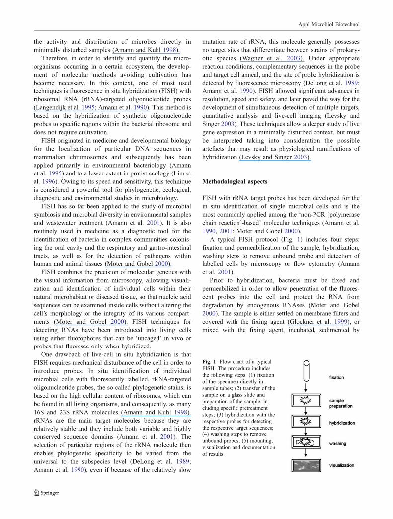
A typical FISH protocol (Fig. 1) includes four steps:fixation and permeabilization of the sample, hybridization,washing steps to remove unbound probe and detection oflabelled cells by microscopy or flow cytometry (Amannet al. 2001).
Prior to hybridization, bacteria must be fixed andpermeabilized in order to allow penetration of the fluores-cent probes into the cell and protect the RNA fromdegradation by endogenous RNAses (Moter and Gobel2000). The sample is either settled on membrane filters andcovered with the fixing agent (Glockner et al. 1999), ormixed with the fixing agent, incubated, sedimented by
Fig. 1 Flow chart of a typicalFISH. The procedure includesthe following steps: (1) fixationof the specimen directly insample tubes; (2) transfer of thesample on a glass slide andpreparation of the sample, in-cluding specific pretreatmentsteps; (3) hybridization with therespective probes for detectingthe respective target sequences;(4) washing steps to removeunbound probes; (5) mounting,visualization and documentationof results
Appl Microbiol Biotechnol
centrifugation, resuspended, transferred to glass slides anddried (Amann et al. 1990). For better attachment ofspecimens to glass slides, some authors suggested firsttreating the surfaces with coating agents such as gelatin(Amann et al. 1990), poly-L-lysine (Lee et al. 1999) orsilanating agents (Moter et al. 1998). If cell suspensions areinvestigated, bacteria are fixed in suspension, spotted ontomicroscopic slides, air-dried and dehydrated in an ethanolseries. In some cases, e.g. for Gram-positive cells, anadditional enzymatic treatment with lysozyme (Schonhuberet al. 1999; Wagner et al. 1998), lysostaphin or an enzymemixture (Krimmer et al. 1999) may be necessary to openthe peptidoglycan layer.
Hybridization must be carried out under stringentconditions for proper annealing of the probe to the targetsequence. For this crucial step of the FISH procedure, apreheated hybridization buffer is applied to the samplecontaining fluorescently labelled probes complementary tothe target RNA. The hybridization takes place in a darkhumid chamber, usually at temperatures between 37 and50°C. Hybridization time varies between 30 min andseveral hours.
The slides are then briefly rinsed with distilled water toremove unbound probe. Finally, the slides are rinsed withwater again, dried, possibly mounted in anti-fading agentsto prevent fluorescence ‘bleaching’ (Moter and Gobel2000), then the results are visualized and documented.
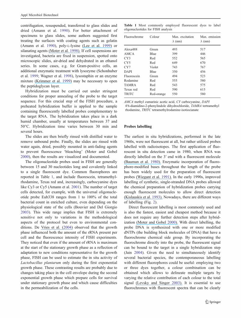
The oligonucleotide probes used in FISH are generallybetween 15 and 30 nucleotides long and covalently linkedto a single fluorescent dye. Common fluorophores arereported in Table 1, and include fluorescein, tetramethyl-rhodamine, Texas red and, increasingly, carbocyanine dyeslike Cy3 or Cy5 (Amann et al. 2001). The number of targetcells detected, for example, with the universal oligonucle-otide probe Eub338 ranges from 1 to 100% of the totalbacterial count in enriched culture, even depending on thephysiological state of the cells (Bouvier and Del Giorgio2003). This wide range implies that FISH is extremelysensitive not only to variations in the methodologicalaspects of the protocol but even to environmental con-ditions. De Vries et al. (2004) observed that the growthphase influenced both the amount of the rRNA present percell and the fluorescence intensity of FISH experiments.They noticed that even if the amount of rRNA is maximumat the start of the stationary growth phase as a reflection ofadaptation to new conditions representative for the growthphase, FISH can be used to estimate the in situ activity ofLactobacillus plantarum only during the first exponentialgrowth phase. These contrasting results are probably due tochanges taking place in the cell envelope during the secondexponential growth phase which prepare cells for survivalunder stationary growth phase and which cause difficultiesin the permeabilization of the cells.
Probes labelling
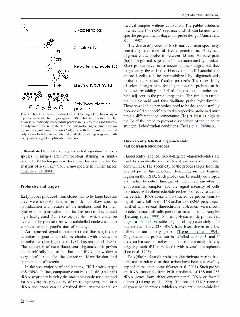
The earliest in situ hybridizations, performed in the late1960s, were not fluorescent at all, but rather utilized probeslabelled with radioisotopes. The first application of fluo-rescent in situ detection came in 1980, when RNA wasdirectly labelled on the 3′ end with a fluorescent molecule(Bauman et al. 1980). Enzymatic incorporation of fluoro-phore-modified bases throughout the length of the probehas been widely used for the preparation of fluorescentprobes (Wiegant et al. 1991). In the early 1990s, improvedlabelling of synthetic, single-stranded DNA probes allowedthe chemical preparation of hybridization probes carryingenough fluorescent molecules to allow direct detection(Kislauskis et al. 1993). Nowadays, there are different waysof labelling (Fig. 2).
Direct fluorescent labelling is most commonly used andis also the fastest, easiest and cheapest method because itdoes not require any further detection steps after hybrid-ization (Moter and Gobel 2000). With direct labelling, theprobe DNA is synthesized with one or more modifieddNTPs (the building block molecules of DNA) that have afluorochrome chemical side group. By incorporating thefluorochrome directly into the probe, the fluorescent signalcan be bound to the target in a single hybridization step(Jain 2004). Given the need to simultaneously identifyseveral bacterial species, the contemporaneous labellingwith different fluorophores could be useful: employing twoor three dyes together, a colour combination can beobtained which allows to delineate multiple targets byvarying the relative contribution of each colour to the totalsignal (Levsky and Singer 2003). It is essential to usefluorochromes with fluorescent spectra that can be clearly
Table 1 Most commonly employed fluorescent dyes to labeloligonucleotides for FISH analysis
Fluorochrome Colour Max. excitationλ (nm)
Max. emissionλ (nm)
Alexa488 Green 493 517AMCA Blue 399 446CY3 Red 552 565CY5 Red 649 670CY7 Violet 743 767DAPI Blue 350 456Fluorescein Green 494 523Rodamine Red 555 580TAMRA Red 543 575Texas red Red 590 615TRITC Red-orange 550 580
AMCA methyl cumarinic acetic acid, CY carbocyanine, DAPI4′6-diamidino-2-phenylindole dihydrochloride, TAMRA tertramethylrhodamine, TRITC tetramethylrodamine-isotiocianate
Appl Microbiol Biotechnol
differentiated to create a unique spectral signature for eachspecies in images after multi-colour staining. A multi-colour FISH technique was developed for example for theanalysis of seven Bifidobacterium species in human faeces(Takada et al. 2004).
Probe size and targets
Early probes produced from clones had to be large becausethey were sparsely labelled in order to allow specifichybridization and because of the methods used for theirsynthesis and purification, and for this reason, they causedhigh background fluorescence, problem which could beovercome by pretreatment with unlabelled nucleic acids tocompete for non-specific sites of binding.
An improved signal-to-noise ratio and thus single-copydetection of genes could also be obtained with a reductionin probe size (Landegent et al. 1987; Lawrence et al. 1988).The utilization of these fluorescent oligonucleotide probesthat specifically bind to the ribosomal RNA is nowadays avery useful tool for the detection, identification andenumeration of bacteria.
In the vast majority of applications, FISH probes target16S rRNA. In fact, comparative analysis of 16S (and 23S)rRNA sequences is today the most commonly used methodfor studying the phylogeny of microorganisms, and suchrRNA sequences can be obtained from environmental or
medical samples without cultivation. The public databasesnow include 16S rRNA sequences, which can be used withspecific programme packages for probe design (Amann andKuhl 1998).
The choice of probes for FISH must consider specificity,sensitivity and ease of tissue penetration. A typicaloligonucleotide probe is between 15 and 30 base pairs(bp) in length and is generated on an automated synthesizer.Short probes have easier access to their target, but theymight carry fewer labels. However, not all bacterial andarchaeal cells can be permeabilized by oligonucleotideprobes using standard fixation protocols. The accessibilityof selected target sites for oligonucleotide probes can beincreased by adding unlabelled oligonucleotide probes thatbind adjacent to the probe target site. The aim is to unfoldthe nucleic acid and thus facilitate probe hybridization.These so-called helper probes need to be designed carefullybecause of their specificity to the respective probe and musthave a differentiation temperature (Td) at least as high asthe Td of the probe to prevent dissociation of the helper atstringent hybridization conditions (Fuchs et al. 2000a,b).
Fluorescently labelled oligonucleotideand polynucleotide probes
Fluorescently labelled, rRNA-targeted oligonucleotides areused to specifically stain different members of microbialcommunities. The specificity of the probes ranges from thephylo-type to the kingdom, depending on the targetedregion on the rRNA. Such probes can be readily developedand tested to detect lineages of uncultured microbes inenvironmental samples, and the signal intensity of cellshybridized with oligonucleotide probes is directly related tothe cellular rRNA content. Polynucleotide probes consist-ing of nearly full-length 16S and/or 23S rRNA genes, eachlabelled with several fluorochrome molecules, were shownto detect almost all cells present in environmental samples(DeLong et al. 1999). Shorter polynucleotide probes thattarget a defined variable region of approximately 250nucleotides of the 23S rRNA have been shown to allowdifferentiation among genera (Trebesius et al. 1994).Oligonucleotide probes can be labelled at both 5′ and 3′ends, and/or several probes applied simultaneously, therebytargeting each rRNA molecule with several fluorophores(Lee et al. 1993).
Poly(ribo)nucleotide probes to discriminate marine bac-teria and uncultured marine archea have been successfullyapplied in the open ocean (Karner et al. 2001). Such probesare RNA transcripts from PCR amplicons of 16S and 23SrRNA genes from either environmental DNA or fosmidclones (DeLong et al. 1999). The use of rRNA-targetedoligonucleotide probes, which are covalently mono-labelled
Fig. 2 Direct (a, b) and indirect (c–e) labelling of probes using areporter molecule like digoxygenin (DIG) that is then detected byfluorescent antibody, horseradish peroxidase (HRP) that used fluores-cein–tyramide as substrate for the enzymatic signal amplification[tyramide signal amplification (TSA)], or with the combined use ofpolyribonucleotide probes, internally labelled with digoxygenin, withthe tyramide signal amplification systems
Appl Microbiol Biotechnol
with fluorescent dye molecules, limits the sensitivity of themethod and aggravates the use of FISH for identification ofprokaryotes with low ribosome content per cell (Wagner etal. 2003). The fluorescently labelled, rRNA-targeted poly-nucleotide probes have been reported to yield higher signalintensities than oligonucleotide probes and may thusrepresent a better means of detecting microbes with a lowribosome content (Trebesius et al. 1994). Due to their bigdimensions, these probes could lead to a reduction inhybridization efficiency because of the inaccessibility ofboth the cell wall and the target site to the longer probes.
FISH with peptide nucleic acid probes
Ribosomal RNA molecules are key functional and struc-tural elements of cells and are highly conserved betweenclosely related species (Perry-O’Keefe et al. 2001). Com-parative study of rRNA sequences has allowed evolutionarymicrobiologists to define the phylogeny and taxonomy ofbacteria, yeasts and fungi.
In addition, as rRNAs are present at a high copy numberin bacteria, they have often been the target of choice foroligonucleotide probe assays (Amann et al. 1995). Variousgroups have noted that the degree of fluorescent signal isdependent on the choice of the probe target region withinthe 16S rRNA molecule, and this variability has beenascribed to poor accessibility of DNA probes to the rRNAtarget because of the highly stable secondary structures inthe rRNA (Amann et al. 1995; Frischer et al. 1996; Fuchs etal. 1998). Furthermore, DNA probes are often limited intheir capability to distinguish single nucleotide changes. Asa result, it is often difficult to design DNA probes thathybridize efficiently within a given stretch of the rRNAdictated by the nucleotide differences found betweenclosely related species (Perry-O’Keefe et al. 2001). Theuse of peptide nucleic acids (PNAs) has been considereduseful in overcoming the variable and sometimes insuffi-cient penetration of probes into bacteria depending on theircell wall characteristics, which is one of the major draw-backs of FISH using oligonucleotide probes, particularly inGram-positive bacteria (Stender et al. 2001).
Peptide nucleic acid molecules are uncharged DNAanalogues that bind to nucleic acids much more stronglythan oligonucleotides because there is no electrostaticrepulsion between the PNA probe and the negativelycharged sugar-phosphate backbone of the target molecule(Ray and Norden 2000).
The unique character of PNA allows these probes tohybridise to target nucleic acid molecules more rapidly andwith higher affinity and specificity compared with DNAprobes (Jain 2004). This highly sensitive approach detectsrRNA in bacterial cells long after cell death (Moter and
Gobel 2000). Moreover, PNA probes hybridize virtuallyindependently of the salt concentration, being ideal fortargeting nucleic acids with high degree of secondarystructure, like rRNA, since hybridization can be performedefficiently in a low salt buffer. Under these conditions, thestability of the secondary structures within the target rRNAis decreased, thereby allowing PNA probes to hybridize toless accessible targets. A standardized PNA FISH proce-dure also increases the possibility of simultaneous identi-fication of different organisms (multiplex identification)using PNA probes labelled with different fluorophores(Perry-O’Keefe et al. 2001). At this time, high prices andspecificity problems are slowing the application of PNAs toFISH (Amann et al. 2001).
Probes accessibility to rRNA
One of the main problems of FISH, besides low cellularribosome content and impermeability of cell walls, is theinaccessibility of probe target sites (Fuchs et al. 1998).Initially, they were supposed to be hindered by rRNA–rRNA interactions, as well as by interactions of the rRNAswith ribosomal proteins, due to the densely packed three-dimensional structure of the ribosome (Beherens et al.2003; Wimberly et al. 2000). Later, Inacio et al. (2003),after studying the effects of different hybridization and cellstorage conditions, observed that, FISH being performed ina strongly denaturing environment, the three-dimensionalstructure of the native small ribosomal subunit is notrelevant to probe accessibility, and that for the same reason,the influence of protein–RNA interactions on target siteaccessibility can be neglected. It was noticed that intra-helix, secondary base interactions are more important thantertiary rRNA–rRNA contacts (Inacio et al. 2003). Fre-quently, probes were intended to be as specific as possible,targeting particular species or genera, or even just particular16S rDNA sequences that were retrieved from the environ-ment. Those probes necessarily target the most variableregions of the 16S rRNA molecule which often are even themost inaccessible sites (Fuchs et al. 1998). Fuchs et al.(1998) found five regions in Escherichia coli whereaccessibility seemed to be very high: positions 38 to 108(except for the terminal loop region of helix 6), positions181 to 215, positions 316 to 359, positions 871 to 933, andpositions 1,383 to 1,427 and 1,473 to 1,517. They foundthat the loop region of helix 6, the loop region and the distal3′ side of helix 18 and the 5′ side of helix 23, which areamong the most important target sites with the evolutionbeing less conserved, are not very accessible. In this study,all probes were arbitrarily grouped according to theirrelative fluorescences into six classes of brightness, wherethe fluorescence of the probes was expressed as a
Appl Microbiol Biotechnol
percentage of the brightest probe (Eco1482). Most probesin the first two classes are directed against five regionswhere accessibility for oligonucleotide probes seems to bevery high, so it is possible to consider the brightest probesas the most accessible ones. In this context, probe Eub338,which is the most widely employed probe for FISHdetection of Bacteria, has been set in the III class, with a58% Eco1482 brightness. The fluorescent signals reportedby Fuchs et al. (1998) should mainly reflect differences in16S rRNA accessibility. Changes in accessibility are oftensteady along the primary structure of the 16S rRNA, butcan also be rapid and are therefore quite unpredictable.Moreover, they noticed that the almost inaccessible targetsites are often in the periphery of the secondary structuremodel, including many loops, whereas regions in the centreof this model seem to be more readily accessible. Furtherstudies investigated the possibility of using the 23S rRNAas a probe target molecule because, despite its length ofapproximately 1,500 nucleotides, it is sometimes impossi-ble to find suitable signature sites on the 16S rRNA for theidentification of the organisms of interest (Fuchs et al.2000b). The 23S rRNA, with its length of approximately3,000 nucleotides, would be the ideal alternative as a probetarget molecule (Fuchs et al. 2000b). The authors demon-strated that in E. coli, the 23S rRNA is more accessible tooligonucleotide probes than the 16S rRNA in the smallsubunit of the ribosome, and for this reason, a 23S rRNA-targeted probe is valuable in increasing the significance ofin situ identification.
Transferability of E. coli accessibility datato other organisms
Due to the high evolutionary conservation of the rRNA, thesefindings on the 23S rRNA and 16S rRNA in situ accessibilityfor E. coli can be extrapolated to other microorganisms. Thetransferability of E. coli accessibility data to other organismshas been studied for organisms of all three domains of life:Bacteria, Archea and Eucarya (Beherens et al. 2002), andthe correlations analysis clearly showed that the in situaccessibility maps are more similar for phylogeneticallymore related organisms. For all three prokaryotes examinedby Beherens et al. (2002), regions of the 16S rRNA withhigh accessibility are positions 285 to 338 (helices 13 and14), positions 871 to 925 (except helix 30 target positions)and positions 1,248 to 1,283. Seven smaller regions of goodaccessibility are located on helices 2, 3, 7, 9, 20, 23, 26, 27and 31. As shown by Inacio et al. (2003), the accessibilityto 26 rRNA in yeasts (Saccharomyces cerevisiae) followsthe same general trend observed for 16S rRNA of E. coli:the most conserved stretches of the region studied aremore accessible, and the most variable areas often show
medium to low accessibility. Otherwise, the in situaccessibility does not depend exclusively on a probetarget site location inside or outside the ribosome, assuggested by Beherens et al. (2003). For example, fixationand hybridization significantly increase the accessibility of16S rRNA target sites to probe, causing massive conforma-tional changes within the ribosome (Beherens et al. 2003).Inaccessible target sites could even be likely opened up byconformational changes introduced by hybridization ofhelper probes (Fuchs et al. 2000a,b).
There is even a link between the type of label andaccessibility. Studies performed by Beherens et al. (2002)and by Fuchs et al. (1998) demonstrated that Cy3-labelledoligonucleotides have higher in situ accessibility in E. coliin comparison with the triphenilmethane derivative car-boxy-fluorescein due to the more linear structure, whichcould reduce steric hindrance and thereby facilitate probebinding to the target.
Hybridization efficiencies of FISH probes
Although the accessibility maps from the studies cited havebeen used for the selection of probe target sites in FISHexperiments, recent studies have demonstrated that it is notworth to eliminate rRNA target regions a priori, even ifthey were previously reported as seemingly inaccessiblebecause these sites could be strongly influenced by thekinetics of the hybridization reaction (Ylmaz and Noguera2004; Ylmaz et al. 2005). In fact, the hybridizationefficiencies of FISH probes can be defined as a functionof not only the accessibility of the target site, but also thethermodynamic affinity of the probe to the target site,defined by the predictable free energy change of the overallreaction (ΔG°overall), which describes the stability of theDNA/RNA hybrid and can be correlated with the brightnessof a hybridized probe. ΔG°overall is obtained from the freeenergy change for the individual reactions (ΔG°i): thebinding of the probe to the complementary site available(reaction 1), the reversible unfolding of the target region(reaction 3) and the folding–unfolding mechanism for theDNA probe (reaction 2). ΔG°i can be calculated as ΔG°i=-RTlnKi, where R is the ideal gas law constant, T is thehybridization temperature and Ki is the equilibrium constantfor each reaction. A more negative ΔG°overall represents agreater concentration of the hybrid and, thus, greaterbrightness from the hybridized probes (Ylmaz and Noguera2004).
For this reason, ΔG°overall is a strong predictor ofhybridization efficiency, and it has been noticed that anaffinity above the theoretical threshold of −13.0 Kcal/molfor maximum hybridization efficiency should be targetedfor rational design of FISH probes to maximize the
Appl Microbiol Biotechnol
possibility of satisfactory sensitivity (Ylmaz et al. 2005).Furthermore, kinetics limitations imposed by structuralrestraints in the ribosome could be overcome by extendingthe incubation period from 3 to 96 h or using formamide, sothat all regions of the 16S rRNA of E. coli can be madeaccessible, thanks to the surmounting of thermodynamicand kinetic barriers (Ylmaz et al. 2005).
Use of FISH in food samples
There is an increasing need for rapid detection and enumer-ation of marker organisms that run to completion within asingle working day in the food and drinking water industries(Ootsubo et al. 2003). A major disadvantage of culturemethods is the time needed to produce results. Generally, theyrequire incubation of culture plates for several days. Anothersignificant disadvantage of these methods is the failure todetect viable but non-culturable organisms. Therefore, itraises doubts that culture-based methods recover sublethallyinjured cells that may occur in heat-treated products such aspasteurized milk. In order to monitor online sanitationpractices, the industry needs rapid and reliable methods toascertain the microbiological quality of foods and ingredients.Fluorescent oligonucleotide probe hybridization is becomingan important tool also in food microbiology to identifyspecific microorganisms in mixed communities without theneed for isolation in pure cultures. The use of FISH allowed,for example, a rapid and accurate enumeration of Pseudomo-nas, facilitating the identification of specific contaminationsources in dairy plants, the accurate validation of pasteuriza-tion treatment and the prediction of shelf life of pasteurizedmilk (Gunasekera et al. 2003).
Moreover, the use of group- and species-specificoligonucleotide probes for in situ hybridization has provid-ed an insight to the microbial composition of Gruyerecheese surface (Kolloffel et al. 1999). Although the FISHanalysis was performed on a cheese suspension and notdirectly in the food matrix , the study allowed the detectionof most bacteria with bacterial probe Eub338, even inmature cheese from which only a smaller proportion of thebacteria were culturable (Kolloffel et al. 1999).
This technique was recently applied for a screening ofthe occurrence of L. plantarum on olives to be used innatural fermentations. However, due to the high detectionlimit of the method, the species could not be detected eventhough a large number of samples were screened (Ercoliniet al. 2006). Moreover, technical problems arose in thisstudy for the optimization of cell permeabilization condi-tion due to the simultaneous occurrence of Gram-positiveand Gram-negative bacteria (Ercolini et al. 2006).
FISH is useful in wine production too, where the earlyknowledge of the microbiological conditions allows the
application of corrective measures before spoiling becomesirreparable. Therefore, rapid and accurate identification ofLAB permits a better monitoring of the fermentation process,preventing the risk of alteration and the probability of theoccurrence of malolactic fermentation, leading therefore to abetter quality of the final wine (Blasco et al. 2003).
The use of FISH and, in particular, the application offluorescently labelled PNA oligomers have also beenreported to be a powerful method for identifying coloniesof Brettanomyces, a well-recognized wine spoilage yeastthat causes an undesirable flavour (Stender et al. 2001).
Despite the huge background of knowledge and appli-cation, the use of FISH in food microbiology has beenrestricted to the identification of bacteria isolated from foodor in food suspension, therefore losing spatial data whichmight provide important information on flora developmentin food ecosystems.
Ercolini et al. (2003a) developed a 16S rRNA FISHmethod for cheese to allow the detection in situ ofmicroorganisms within the dairy matrix. An embeddingprocedure using a plastic resin was applied to Stiltoncheese, providing intact embedded cheese sections with-standing the hybridization reaction. The technique has thepotential to study the spatial distribution of microbialpopulation in situ in foods, especially where the matrix istoo fragile to allow manipulation of cryo-sections. 16SrRNA sequences could be used for species-specific probedesign for the location of specific groups of bacteria withinthe matrix and for the investigation of relationships withinspecific groups of bacteria.
The authors developed probes for the specific detectionof some species of lactobacilli, lactococci and leuconostocsand demonstrated that in Stilton cheese the spatial distri-bution of specific colonies is not homogeneous but dependson the specific site. The availability of such information fordairy products after FISH investigation may play a key rolefor the detection of the sites of actual growth of certainspecies and can be of potential help in detecting zones ofappropriate acidification, aroma development and produc-tion of antagonistic substances in food products. Moreover,the monitoring and analysis of spatial distribution ofcontaminant species might also be achieved by thisapproach, which represents a very important step forward instudying the development of microbial populations in foodwith the ultimate aim of process optimization and qualityassurance of the final product (Ercolini et al. 2003a,b).
Applicability of FISH to biofilms
Although quantitative FISH has provided new insights intothe structure and dynamics of microbial communities, itsuffers from tediousness and limited accuracy for samples
Appl Microbiol Biotechnol
containing densely aggregated cells like biofilms (Daimset al. 2001). Biofilms are formed after rapid attachment andgrowth of microorganisms on a broad range of surfaces incontact with natural fluids. The architecture of biofilms andthe physiological status of the cells contained within thesestructures are of prime interest for clinical, industrial andenvironmental microbiology. For analyses of the spatial ar-rangement of bacteria in a multi-species biofilm, techniqueswhich use specific bacterial cell markers and maintain thebiofilm’s natural architecture are required (Thurnheer et al.2004).
Quantitative data on the abundance of in situ stainedmicroorganisms in complex samples are mostly obtained bytime-consuming manual microscopic counting. The accura-cy of this quantification approach is relatively low indensely colonized biofilms, which probably represent themost common life form of microorganisms on earth(Wagner et al. 2003). This problem can in part beameliorated by using confocal laser scanning microscopy(CLSM) for the detection of probe-labelled cells. However,even if optical CLSM sections are recorded, it is notfeasible to manually count a sufficient number of cells ineach hybridization experiment in a reasonable time periodto obtain statistically reliable results. Even semi-automateddigital image analysis tools are unable to efficiently countcells in dense clusters or biofilms because single-cellrecognition within these structures cannot be automated.This problem can be circumvented by measuring the areasof specifically stained bacteria in randomly acquired opticalCLSM sections. The abundance of a particular populationis then expressed as a fraction of the area occupied by allbacteria (Daims et al. 2001).
The utilization of genus-specific, fluorescently labelledoligonucleotide probes can be a very useful tool for thestudy of particular kinds of biofilms, such as those formedin the gastrointestinal human tract. FISH has been appliedwith this aim for the hybridization of the bacterial rRNAextracted from a faecal sample or fermentation fluids(Langendijk et al. 1995) and has also been used todetermine changes in populations of bifidobacteria, lacto-bacilli, clostridia, bacteroides, streptococci and E. coli inhuman faecal extracts (Rycroft et al. 2001). Moreover, incombining FISH with CLSM, the development of asimultaneous analysis of the spatial distribution of bothGram-negative and Gram-positive bacteria in biofilms hasbecome possible (Thurnheer et al. 2004).
Conclusions and future perspectives
As a consequence of the continuous improvements, FISH isnowadays a very useful tool for studies in microbiology.Through the use of species-specific probes, it is possible to
identify different microorganisms in complex microbialcommunities, giving a solid support to the understanding ofinter-species interaction. The widespread use of FISH as anapplication directed to gaining information on the spatialdistribution of microbial populations will allow the study ofthe contribution of each species to the organization andfunctioning of the multi-species bacterial communities.This is important from different points of view and indifferent fields of application. The knowledge of thecomposition and distribution of microorganisms in naturalhabitats can be interesting for ecological reasons inmicrobial ecology, and for safety and technological aspectsin food microbiology. This technique will be a relativelyrapid method to evaluate the presence and activity ofpathogens, or other undesirable microorganisms, directly inthe sample studied (food, water, wastes, soil), but it willalso be a valid approach to study the activity of bacteriainvolved in technological processes (e.g. food fermenta-tions) or important for human health (e.g. Bifidobacteria).
FISH is a very in situ technique and will keep the potentialto zoom in on the composition of complex microbialassociations in natural ecosystems.
References
Amann RI, Ludwig W, Schleifer KH (1995) Fluorescent-oligo-nucleotide probing of whole cells for determinative, phylogenet-ic, and environmental studies in microbiology. J Bacteriol172:762–770
Amann R, Kuhl M (1998) In situ methods for assessment ofmicroorganisms and their activities. Curr Opin Microbiol1:352–358
Amann R, Krumholz L, Sthal DA (1990) Fluorescent-oligonucleo-tide probing of whole cells for determinative, phylogenetic,and environmental studies in microbiology. J Bacteriol172:762–770
Amann R, Fuchs BM, Beherens S (2001) The identification ofmicroorganisms by fluorescence in situ hybridization. Curr OpinBiotechnol 12:231–236
Bauman JC, Wiegant J, Borst P, van Duijn P (1980) A new method forfluorescence microscopical localization of specific DNA se-quences by in situ hybridization of fluorochrome-labelled RNA.Exp Cell Res 128:485–490
Beherens S, Ruhland C, Inacio J, Huber H, Fonseca A, Spencer-Martins I, Fuchs BM, Amann R (2002) In situ accessibility ofsmall-subunit rRNA of members of the domains Bacteria,Archaea, and Eucarya to Cy3-labeled oligonucleotide probes.Appl Environ Microbiol 69:1748–1758
Beherens S, Fuchs BM, Mueller F, Amann R (2003) Is the in situaccessibility of the 16S rRNA of Escherichia coli for Cy3-labeled oligonucleotide probes predicted by a three-dimensionalstructure model of the 30S ribosomal subunit? Appl EnvironMicrobiol 69:4935–4941
Blasco L, Ferrer S, Pardo I (2003) Development of specificfluorescent oligonucleotide probes for in situ identification ofwine lactic acid bacteria. FEMS Microbiol Lett 225:115–123
Bouvier T, Del Giorgio PA (2003) Factors influencing the detection ofbacterial cells using fluorescence in situ hybridization (FISH).FEMS Microbiol Ecol 44:3–15
Appl Microbiol Biotechnol
Daims H, Ramsing NB, Schleifer KH, Wagner M (2001)Cultivation-independent, semiautomatic determination of abso-lute bacterial cell numbers in environmental samples byfluorescence in situ hybridization. Appl Environ Microbiol67:5810–5818
DeLong EF, Wickham GS, Pace NR (1989) Phylogenetic stains:ribosomal RNA-based probes for the identifications of singlecells. Science 243:1360–1363
DeLong EF, Taylor LT, Marsh TL, Preston CM (1999) Visualizationand enumeration of marine planktonic archaea and bacteria byusing polyribonucleotide probes and fluorescent in situ hybrid-ization. Appl Environ Microbiol 65:5554–5563
De Vries MC, Vaughan EE, Kleerebezem M, de Vos WM (2004)Optimizing single cell activity assessment of Lactobacillusplantarum by fluorescent in situ hybridization as affected bygrowth. J Microbiol Methods 59:109–115
Ercolini D, Hill PJ, Dodd CER (2003a) Development of a fluores-cence in situ hybridization method for cheese using a 16S rRNAprobe. J Microbiol Methods 50:97–100
Ercolini D, Hill PJ, Dodd CER (2003b) Bacterial community structureand location in stilton cheese. Appl Environ Microbiol 69:3540–3548
Ercolini D, Villani F, Aponte M, Mauriello G (2006) Fluorescence insitu hybridization detection of Lactobacillus plantarum group onolives to be used in natural fermentations. Int J Food Microbiol(in press)
Frischer ME, Floriani PJ, Nierzwicki-Bauer SA (1996) Differentialsensitivity of 16S rRNA targeted oligonucleotide probes used forfluorescence in situ hybridization is a result of ribosomal higherorder structure. Can J Microbiol 42:1061–1071
Fuchs BM, Wallner G, Beisker W, Schwippl I, Ludwig W, Amann R(1998) Flow cytometric analysis of the in situ accessibility ofEscherichia coli 16S rRNA for fluorescently labelled oligonu-cleotide probes. Appl Environ Microbiol 64:4973–4982
Fuchs BM, Glockner FO, Wulf J, Amann R (2000a) Unlabeled helperoligonucleotides increase the in situ accessibility to 16S rRNA offluorescently labelled oligonucleotide probes. Appl EnvironMicrobiol 66:3603–3607
Fuchs BM, Syutsubo K, Ludwig W, Amann R (2000b) In situaccessibility of Escherichia coli 23S rRNA to fluorescentlylabeled oligonucleotide probes. Appl Environ Microbiol 67:961–968
Glockner FO, Fuchs BM, Amann R (1999) Bacterioplanktoncomposition of lakes and oceans: a first comparison based onfluorescence in situ hybridization. Appl Environ Microbiol65:3721–3726
Gunasekera TS, Dorsch MR, Slade MB, Veal DA (2003) Specificdetection of Pseudomonas spp. in milk by fluorescence in situhybridization using ribosomal RNA directed probes. J ApplMicrob 94:936–945
Inacio J, Beherens S, Fuchs BM, Fonseca A, Spencer-Martins I,Amann R (2003) In situ accessibility of Saccharomycescerevisiae 26S rRNA to Cy3-labeled oligonucleotide probescomprising the D1 and D2 domains. Appl Environ Microbiol69:2899–2905
Jain KK (2004) Current status of fluorescence in situ hybridization.Med Device Technol 15(4):14–17
Karner M, DeLong EF, Karl DM (2001) Archaeal dominance in themesopelagic zone of the Pacific Ocean. Nature 409:507–509
Kislauskis EH, Li Z, Singer RH, Taneja KL (1993) Isoform-specific3′-untranslated sequences sort alpha-cardiac and beta-cytoplas-mic actin messenger RNAs to different cytoplasmic compart-ments. J Cell Biol 123:165–172
Kolloffel B, Meile L, Teuber M (1999) Analysis of brevibacteria on thesurface of Gruyère cheese detected by in situ hybridization and bycolony hybridization. Lett Appl Microbiol 29:317–322
Krimmer V, Merkert H, von Eiff C, Frosch M, Eulert J, Lohr JF,Hacker J, Ziehbur W (1999) Detection of Staphylococcus aureusand Staphylococcus epidermidis in clinical samples by 16SrRNA-directed in situ hybridization. J Clin Microbiol 37:2667–2673
Landegent JE, Jansen in de Wal N, Dirks RW, Baao F, van der PloegM (1987) Use of whole cosmid cloned genome sequences forchromosomal localization by non-radioactive in situ hybridiza-tion. Hum Genet 77:366–370
Langendijk PS, Schut F, Jansen GJ, Raangs GC, Kamphuis GR,Wilkinson MHF, Welling GW (1995) Quantitative fluorescencein situ hybridization of Bifidobacterium spp. with genus-specific16S rRNA-targeted probes and its application in faecal samples.Appl Environ Microbiol 61:3069–3075
Lawrence JB, Singer RH, Villnave CA, Stein JL, Stein GS (1988)Intracellular distribution of histone mRNAs in human fibroblastsstudied by in situ hybridization. Proc Natl Acad Sci U S A 85:463–467
Lee SH, Malone C, Kemp PF (1993) Use of multiple 16S rRNA-targeted fluorescent probes to increase signal strength andmeasure cellular RNA from natural planktonic bacteria. MarEcol Prog Ser 101:193–201
Lee N, Nielsen PH, Andreasen KH, Jureteschko S, Nielsen JL,Schleifer KH, Wagner M (1999) Combination of fluorescent insitu hybridization and microautoradiography—a new tool forstructure-function analyses in microbial ecology. Appl EnvironMicrobiol 65:1289–1297
Levsky JM, Singer RH (2003) Fluorescence in situ hybridization:past, present and future. J Cell Sci 116:2833–2838
Lim EL, Caron DA, DeLong EF (1996) Development and fieldapplication of a quantitative method for examining naturalassemblages of protists with oligonucleotides probes. ApplEnviron Microbiol 62:1416–1423
Moter A, Gobel UB (2000) Fluorescence in situ hybridization (FISH)for direct visualization of microorganisms. J Microbiol Methods41:85–112
Moter A, Leist G, Rudolph R, Schrank K, Choi BK, Wagner M, GobelUB (1998) Fluorescence in situ hybridization shows spatialdistribution of as yet uncultured treponemes in biopsies fromdigital dermatitis lesions. Microbiol 144:2459–2467
Ootsubo M, Shimizu T, Tanaka R, Sawabe T, Tajima K, Ezura Y(2003) Seven-hours fluorescence in situ hybridization techniquefor enumeration of Enterobacteriaceae in food and environmen-tal water sample. J Appl Microbiol 95:1182–1190
Perry-O’Keefe H, Stender H, Broomer A, Oliveira K, Coull J, Hyldig-Nielsen JJ (2001) Filter-based PNA in situ hybridization for rapiddetection, identification and enumeration of specific microorgan-isms. J Appl Microb 90:180–189
Ray A, Norden B (2000) Peptide nucleic acid (PNA): its medical andbiotechnical applications and promise for the future. FASEB J14:1041–1060
Rycroft CE, Jones MR, Gibson GR, Rastall RA (2001) Fermentationproperties of gentio-oligosaccharides. Lett Appl Microbiol32:156–161
Schonhuber W, Zarda B, Eix S, Rippka R, Herdmann M, Ludwig W,Amann R (1999) In situ identification of Cyano-bacteria withhorseradish peroxidase-labeled, rRNA-targeted oligonucleotideprobes. Appl Environ Microbiol 65:1259–1267
Stender H, Kurtzman C, Hyldig-Nielsen JJ, Sørensen D, Broomer A,Oliveira K, Perry-O’Keefe H, Sage A, Young B, Coull J (2001)Identification of Dekkera bruxellensis (Brettanomyces) fromwine by in situ hybridization using peptide nucleic acid probes.Appl Environ Microbiol 67:938–941
Takada T, Matsumoto K, Nomoto K (2004) Development of multi-colour FISH method for analysis of seven Bifidobacteriumspecies in human faeces. J Microbiol Methods 58:413–421
Appl Microbiol Biotechnol

Thurnheer T, Gmür R, Guggenheim B (2004) Multiplex FISH analysisof a six species bacterial biofilm. J Microbiol Methods 56:37–47
Trebesius K, Amann R, Ludwig W, Muhlegger K, Schleifer KH(1994) Identification of whole fixed bacterial cells with non-radioactive 23S rRNA-targeted polyribonucleotide probes. ApplEnviron Microbiol 60:3228–3235
Wagner M, Horn M, Daims H (2003) Fluorescence in situ hybridiza-tion for the identification and characterisation of prokaryotes.Curr Opin Microbiol 6:302–309
Wagner M, Schmid M, Juretschko S, Trebesius KH, Bubert A, GoebelW, Schleifer KH (1998) In situ detection of a virulence factormRNA and 16S rRNA in Listeria monocytogenes. FEMSMicrobiol Lett 160:159–168
Wiegant J, Ried T, Nederolf PM, van der Ploeg M, Tanke HJ, RaapAK (1991) In situ hybridization with fluoresceinated DNA.Nucleic Acids Res 19:3237–3241
Wimberly BT, Brodersen DE, Clemons WMJ, Morgan-Warren R, vonRhein C, Hartsch T, Ramakrishnan V (2000) Structure of the 30Sribosomal subunit. Nature 407:327–339
Ylmaz LS, Noguera DR (2004) Mechanistic approach to the problemof hybridization efficiency in fluorescent in situ hybridization.Appl Environ Microbiol 70:7126–7139
Ylmaz LS, Hatice EO, Noguera DR (2005) Making all partsof the 16S rRNA of Escherichia coli accessible in situ tosingle DNA oligonucleotides. Appl Environ Microbiol 72:733–744
Appl Microbiol Biotechnol