
The Transmissibility and Control of Pandemic Influenza A (H1N1) Virus
Upload
healthfoodinnovationmanagementCategory
view
4download
0
Epidemiological and Virological Characteristics ofInfluenza Viruses Circulating in Cambodia from 2009 to2011Srey Viseth Horm1., Sek Mardy1,2., Sareth Rith1., Sovann Ly3, Seng Heng3, Sirenda Vong1,
Paul Kitsutani4, Vannra Ieng2, Arnaud Tarantola1, Sowath Ly1, Borann Sar5, Nora Chea5, Buth Sokhal6,
Ian Barr7, Anne Kelso7, Paul F. Horwood1, Ans Timmermans8, Aeron Hurt7, Chanthap Lon8,
David Saunders8, Sam An Ung6, Nima Asgari2, Maria Concepcion Roces2, Sok Touch3, Naomi Komadina7,
Philippe Buchy1*
1 Institut Pasteur du Cambodge, Reseau International des Instituts Pasteur, Phnom Penh, Cambodia, 2 World Health Organization, Phnom Penh, Cambodia,
3 Communicable Disease Control Department, Ministry of Health, Phnom Penh, Cambodia, 4 Influenza Division, National Center for Immunization and Respiratory Disease,
Center for Disease Control and Prevention, Atlanta, Georgia, United States of America, 5 Centers for Disease Control and Prevention, Cambodia Office, Phnom Penh,
Cambodia, 6 National Institute of Public Health, Phnom Penh, Cambodia, 7 WHO Collaborating Centre for Reference and Research on Influenza, Melbourne, Australia,
8 Armed Forces Research Institute of Medical Sciences, Bangkok, Thailand
Abstract
Background: The Cambodian National Influenza Center (NIC) monitored and characterized circulating influenza strains from2009 to 2011.
Methodology/Principal Findings: Sentinel and study sites collected nasopharyngeal specimens for diagnostic detection,virus isolation, antigenic characterization, sequencing and antiviral susceptibility analysis from patients who fulfilled casedefinitions for influenza-like illness, acute lower respiratory infections and event-based surveillance. Each year in Cambodia,influenza viruses were detected mainly from June to November, during the rainy season. Antigenic analysis show that A/H1N1pdm09 isolates belonged to the A/California/7/2009-like group. Circulating A/H3N2 strains were A/Brisbane/10/2007-like in 2009 before drifting to A/Perth/16/2009-like in 2010 and 2011. The Cambodian influenza B isolates from 2009 to 2011all belonged to the B/Victoria lineage represented by the vaccine strains B/Brisbane/60/2008 and B/Malaysia/2506/2004.Sequences of the M2 gene obtained from representative 2009–2011 A/H3N2 and A/H1N1pdm09 strains all contained theS31N mutation associated with adamantanes resistance except for one A/H1N1pdm09 strain isolated in 2011 that lackedthis mutation. No reduction in the susceptibility to neuraminidase inhibitors was observed among the influenza virusescirculating from 2009 to 2011. Phylogenetic analysis revealed that A/H3N2 strains clustered each year to a distinct groupwhile most A/H1N1pdm09 isolates belonged to the S203T clade.
Conclusions/Significance: In Cambodia, from 2009 to 2011, influenza activity occurred throughout the year with peakseasonality during the rainy season from June to November. Seasonal influenza epidemics were due to multiple geneticallydistinct viruses, even though all of the isolates were antigenically similar to the reference vaccine strains. The drugsusceptibility profile of Cambodian influenza strains revealed that neuraminidase inhibitors would be the drug of choice forinfluenza treatment and chemoprophylaxis in Cambodia, as adamantanes are no longer expected to be effective.
Citation: Horm SV, Mardy S, Rith S, Ly S, Heng S, et al. (2014) Epidemiological and Virological Characteristics of Influenza Viruses Circulating in Cambodia from2009 to 2011. PLoS ONE 9(10): e110713. doi:10.1371/journal.pone.0110713
Editor: Florian Krammer, Icahn School of Medicine at Mount Sinai, United States of America
Received July 3, 2014; Accepted September 16, 2014; Published October 23, 2014
This is an open-access article, free of all copyright, and may be freely reproduced, distributed, transmitted, modified, built upon, or otherwise used by anyone forany lawful purpose. The work is made available under the Creative Commons CC0 public domain dedication.
Data Availability: The authors confirm that all data underlying the findings are fully available without restriction. All relevant data are within the paper and itsSupporting Information files.
Funding: The study was funded by the World Health Organization office in Cambodia, the French Agency for Development (SISEA project) and by the Office ofthe Assistant Secretary for Preparedness and Response within the U.S. Department of Health and Human Services. The funders had no role in study design, datacollection and analysis, decision to publish, or preparation of the manuscript.
Competing Interests: The authors have declared that no competing interests exist.
* Email: [email protected]
. These authors contributed equally to this work.
Background
Influenza is a major human pathogen associated with high
morbidity and mortality, both in the temperate and subtropical/
tropical regions. It is characterized by epidemics that occur
seasonally throughout the world every year, with occasional
pandemics arising from novel subtypes of the virus causing a
considerable economic burden and significant cumulative mor-
bidity and mortality [1–3]. Despite a plethora of information on
PLOS ONE | www.plosone.org 1 October 2014 | Volume 9 | Issue 10 | e110713

influenza epidemiology and seasonality, which remains important
in planning prevention and treatment strategies, overall patterns of
infection have not been fully described on broad geographic scales
and for specific types and subtypes of the influenza virus, thus
highlighting the need for more countries to conduct year-round
viral surveillance and report reliable incidence data at the type and
subtype level, especially in the tropics [4].
In temperate regions influenza viruses typically circulate during
the winter period [5]. In tropical areas, influenza activity usually
occurs all year round with annual/biannual peaks in relation with
rainy seasons and/or winter months, but infections can also occur
without a clear seasonality [6–11]. Cambodia is a South-East
Asian tropical country, which lies geographically in the Northern
hemisphere but its influenza season occurs during June-December,
each year [12]. Northern hemisphere countries usually experience
influenza season from November to March/April, whereas the
influenza season of southern hemisphere countries usually occur
from May to September [13,14]. Hence, Cambodia’s influenza
seasonality appears unusual and knowledge of the epidemiological
and virological characteristics of such influenza circulation is
important for public health preparedness.
We have previously reported preliminary data and described
the circulation and seasonality of influenza viruses in Cambodia
during three consecutive years following the establishment of the
Cambodian National Influenza Centre (NIC) in 2006 [12]. In the
present study, we documented the dynamics of influenza activity,
performed antigenic and drug susceptibility analyses of influenza
virus strains and conducted phylogenetic analysis of influenza A
strains isolated between 2009 and 2011, which included the 2009
pandemic.
Materials and Methods
Geographic backgroundCambodia is a tropical country of almost 15 million people, with
a land area of 181,035 square kilometers in the southwestern part
of the Indochina peninsula [15]. International borders are shared
with Thailand and Laos on the West and the North, and Vietnam
on the East and the Southeast. As the country is affected by
monsoon, it is hot and humid with a mean temperature of 27uCand mean relative humidity of 77.5%. There are two distinct
seasons: the dry season runs generally from November to April
and the rainy season starts in May-June and ends in October-
November.
PatientsThe Cambodian NIC was established in August 2006 as a joint
collaboration between the Virology Unit at the Institut Pasteur in
Cambodia (IPC), the Communicable Disease Control Department
of the Ministry of Health (CDC/MoH) and the World Health
Organization (WHO) office in Cambodia for the purpose of
documenting the dynamics of influenza disease and to virologically
characterize the circulating strains. To continuously monitor
influenza activity, an outpatient sentinel surveillance system for
influenza-like illness (ILI) with a weekly reporting and sampling
scheme was initially established in five hospital sites in 2006. In
addition, hospital-based surveillance of acute lower respiratory
infection (ALRI) cases was established in two sites in Takeo and
Kampong Cham provincial hospitals (SISEA project, French
Agency for Development). Three additional ILI sentinel surveil-
lance sites were then opened in 2009 and operated by the National
Institute of Public Health (NIPH) in collaboration with the US
CDC local office in the referral hospitals of Mondulkiri (Eastern
Cambodia), Svayreang (South-East Cambodia) and Kampot
(South-West Cambodia) Provinces. The Armed Forces Research
Institute of Medical Sciences (AFRIMS) operated one sentinel site
starting in 2009 in Western Cambodia in Thmor Kol (Battambang
Province), and opened two additional sentinel sites in 2011 in
Anlong Veng (Oddar Meancheay Province) and Pailin. During the
pandemic of influenza A/H1N1 2009 virus, an event-based
surveillance system was developed by CDC/MoH and WHO.
The system was designed for rapid detection, reporting and
confirmation of clusters of the disease or suspected cases admitted
in both private clinics and hospitals [16].
An ILI case was defined by the sudden onset of fever ($38uCaxillary temperature) and cough or sore throat in the absence of
other diagnosis. For the ALRI study, in children under 5, a suspect
case was defined as an illness of ,10 days duration with cough or
breathing difficulties plus tachypnea. For the 5–14 years age
group, case definition included the above symptoms plus fever
($38uC axillary temperature) on admission. For patients over 15
years old, a case was defined as a person with fever ($38uCaxillary temperature) on admission plus tachypnea or chest pain or
auscultatory crackles. For event-based surveillance of pandemic
influenza A/H1N1 2009 virus (A/H1N1pdm09 virus) infection
conducted from 2009 to 2010, inclusion criteria included any
person with acute febrile respiratory illness (fever $38uC and
respiratory symptoms, e.g. cough, sore throat, difficulty breathing)
with no other apparent diagnosis and one or more of the following
exposures to the risk of A/H1N1pdm09 virus infection within 7
days prior to symptoms onset: a) Close contact with a probable or
confirmed case of A/H1N1pdm09 virus infection; b) Residing in
or travelled to a province or foreign country with confirmed
community transmission of A/H1N1pdm09 virus; c) Is part of a
cluster of ILI cases; d) Handled specimens suspected of containing
A/H1N1pdm09 virus.
An overview of the all of the surveillance and research programs
involved with the collection and testing of strains for this study is
provided in Table S1.
Ethical statementThe ALRI study was approved by the National Ethics
Committee of the Kingdom of Cambodia. All patients or parents
of sick children who participated in the studies provided written
informed consent. The ILI and event-based surveillance systems
are public health activities organized by the Ministry of Health in
Cambodia and as such have a standing authorization from the
National Ethics Committee. Samples were all anonymized for the
purpose of this study.
Specimen collectionThe sentinel sites obtained weekly epidemiological data from
patients who fulfilled the ILI case definition, and collected naso-
pharyngeal specimens from 5 to 10 cases per week. Respiratory
samples together with clinical records were obtained from patients
hospitalized with ALRI during 2009 and 2010 in Takeo and
Kampong Cham hospitals. Naso-pharyngeal and throat specimens
were placed in a vial containing 2 ml of virus transport medium
(VTM). The specimens collected from the initial 5 sites of ILI
sentinel surveillance and the 2 ALRI study sites were then
immediately frozen in liquid nitrogen and the containers were sent
on a weekly basis to the IPC’s Virology Unit where specimens
were stored at 280uC prior to testing. For the specimens collected
from the 6 ILI sentinel surveillance sites newly established in 2009
and onward, the VTM tubes containing the naso-pharyngeal swab
samples were stored at 4uC and shipped to the NIPH and/or
AFRIMS laboratory for testing within 48 to 72 hours. For the A/
H1N1pdm09 event-based surveillance, the specimens were
Influenza Viruses in Cambodia from 2009 to 2011
PLOS ONE | www.plosone.org 2 October 2014 | Volume 9 | Issue 10 | e110713

collected mainly from hospitals and private clinics in and around
the capital, placed into VTM, and immediately sent in a cool box
to IPC’s laboratory for testing.
Laboratory methodsViral RNA was extracted at the participating national
laboratories (NIC/IPC, NIPH and AFRIMS) using commercial
extraction kits (e.g. MagNa Pure LC, Qiagen Viral RNA mini kits)
according to the manufacturer’s instructions. RNA was amplified
using real-time RT-PCR to detect influenza A and B viruses using
standard protocols. Influenza A virus subtypes H1, H1pdm, H3
and H5 were detected using subtype-specific real-time RT-PCR
assays. All samples that tested positive for influenza were sent to
the NIC for confirmation and further characterization.
At the NIC’s lab, viral RNA was extracted from 140 mL of virus
transport medium containing the nasopharyngeal swab by using
the MagNa Pure LC system (Roche), according to the manufac-
turer’s instructions. Seasonal influenza viruses (A/H3N2, A/
H1N1 and influenza B virus) were detected and subtyped by a
multiplexed conventional reverse transcriptase-polymerase chain
reaction (RT-PCR) as described previously [12]. The detection of
A/H1N1pdm09 viruses was performed by using a one-step real-
time RT-PCR according to the CDC protocol for detection and
characterization of influenza A 2009 H1N1, with the use of a 96-
well format IQ5 instrument (BioRad). Primer and probe reagents,
reaction master-mix, and cycling parameters were used as
described in the protocol [17]. The H5N1 virus was detected by
a one-step real-time RT-PCR developed by the laboratory at the
National Institute of Infectious Diseases (NIID), Tokyo, Japan, and
recommended by WHO as the protocol for laboratory procedures
to detect avian influenza A H5N1 virus in specimens from
suspected human cases [18].
All of the influenza strains described in this study were isolated
at the IPC laboratory by inoculation of the specimens that tested
positive by molecular methods onto Madin-Darby canine kidney
(MDCK) cells in a biosafety level 2+ laboratory for seasonal
influenza viruses and in a biosafety level 3 laboratory for pandemic
(H1N1) 2009 virus (until 2010, only) and for highly pathogenic
avian influenza A/H5N1 virus. In brief, two hundred microlitres
of each specimen was inoculated into MDCK cells, with 2 ml per
well of maintenance medium containing TPCK trypsin (except for
H5N1 virus) at a concentration of 2.0 mg/ml, in a 6-well plate.
The plates were incubated at 35uC in a 5% CO2 atmosphere for 1
week to assess cytopathic effects. The influenza isolates were
characterized by a hemagglutination inhibition assay using
reference antigens and anti-sera kindly provided by the WHO
Collaborating Center (WHOCC) for Reference and Research on
Influenza in Melbourne, Australia. A representative number of
influenza isolates collected by the various surveillance and event-
based systems were sent each year to the WHOCC in Melbourne
for confirmation and further analysis (Table 1).
Genome sequencing and phylogenetic analysisAt the NIC’s laboratory, viral RNA was extracted from 200 ml
of MDCK supernatant using the QIAamp Viral RNA Mini Kit,
according to the manufacturer’s recommendations (Qiagen,
Hilden, Germany). RNA was used as template for RT-PCR with
subtype and segment-specific primers (reaction conditions and
primer sequences are available from the authors upon request).
Nucleotide sequencing reactions were performed with BigDye
Terminator Cycle Sequencing kits (Applied Biosystems) following
the protocols supplied by the manufacturer and the products were
sequenced using an ABI 3730XL automatic DNA Analyser (Life
Technologies, Carlsbad, CA, USA) in a commercial facility
Ta
ble
1.
Infl
ue
nza
iso
late
sco
llect
ed
inC
amb
od
iab
yty
pe
and
sub
typ
ein
20
09
-20
11
.
Infl
ue
nz
aT
yp
eS
ub
typ
eo
fIn
flu
en
za
AW
HO
CC
,M
elb
ou
rne
a
Ye
ar
N#
of
spe
cim
en
ste
ste
dN
#o
fp
osi
tiv
es
(%)
A(%
)B
(%)
H1
N1
(%)
H3
N2
(%)
H5
N1
(%)
H1
N1
pd
mb
(%)
H1
N1
pd
mb
H3
N2
IBc
20
09
46
01
74
7(1
6.2
)6
53
(87
.4)
94
(12
.6)
2(0
.3)
26
6(4
0.7
)1
(0.2
)3
84
(58
.8)
10
15
15
20
10
35
07
44
6(1
2.7
)3
43
(76
.9)
10
3(2
3.1
)0
(0)
15
5(4
5.2
)1
(0.3
)1
87
(54
.5)
31
17
41
20
11
27
05
49
1(1
8.2
)1
73
(35
.2)
31
8(6
4.8
)0
(0)
44
(25
.4)
8(4
.6)
12
1(7
0)
36
14
72
aSp
eci
me
ns
anal
yze
db
yth
eW
HO
Co
llab
ora
tin
gC
en
ter
inM
elb
ou
rne
fro
m2
00
9to
20
11
.b
H1
N1
pd
m0
9.
cIn
flu
en
zaB
.d
oi:1
0.1
37
1/j
ou
rnal
.po
ne
.01
10
71
3.t
00
1
Influenza Viruses in Cambodia from 2009 to 2011
PLOS ONE | www.plosone.org 3 October 2014 | Volume 9 | Issue 10 | e110713

(Macrogen, Seoul, Korea). Multiple sequence alignment was
conducted using ClustalW, version 2 [19]. Phylogenetic trees
based on the HA gene sequences were obtained by using the
Neighbor-joining method, HKY model, generated using PAUP
software [20]. Bootstrap analysis (n = 1000) was carried out to
determined the best-fitting tree. Vaccine strain sequences, as well
as sequences from viruses collected in other countries were
obtained from EpiFlu Database available via the GISAID website
(www.gisaid.org), and were included in the analysis.
Nucleotide sequence accession numbersAll Cambodian influenza A/H3N2, A/H1N1pdm09 and
Influenza B virus sequences included in the analysis were
submitted to Genbank and/or to EpiFlu Database and all of
these sequences are available via GISAID website (www.gisaid.
org). Table S2 provides detailed information about all of the
Cambodian strains and sequences analysed in this study.
Neuraminidase inhibitors susceptibility assaysRepresentative isolates, with respect to date of detection and
province of origin of the patient, were randomly selected for
neuraminidase drug testing. The NA-Star kit (Life Technologies,
Carlsbad, CA, USA), a chemiluminescent NA inhibition assay
which utilizes a 1,2-dioxetane derivative of sialic acid as substrate,
was used according to the manufacturer’s instructions. Oseltamivir
carboxylate, zanamivir and 4-amino-4-deoxy-Neu5Ac2en were
provided by the Institute for Glycomics, Griffith University, Gold
Coast, Australia. Compounds were prepared in distilled water and
stored at 220uC until time of use. The concentration of drug
required to inhibit 50% of the NA activity (IC50) was calculated
using the non-linear curve-fitting function in the Graphpad Prism
4 package (GraphPad Software, Inc., La Jolla, CA, USA). The
average IC50 (nM) (6 standard deviation) of two independent
determinations was calculated for each virus. Outliers of more
than 2 standard deviations from the overall mean were retested
twice [21].
Susceptibility to adamantanesThe full M gene segments from representative influenza strains
of the influenza A/H3N2, A/H1N1 and A/H1N1pdm09 subtypes
virus strains isolated from humans were sequenced to detect
mutations associated with resistance to adamantanes. Amino acids
at positions 26, 27, 30 and 31 of the transmembrane region of the
M2 protein are the residues most frequently associated with
amantadine- and rimantadine-resistant strains.
Statistical analysisThe comparisons between percentages and two means were
tested by chi-squared test and Student’s t test respectively. A p
value ,0.05 was considered statistically significant. Proportions,
means and all statistical analyses were performed using STATA
9.0 (StataCorp., College Station, TX, Texas).
Results
Influenza activity in CambodiaThe Cambodian ILI surveillance program collected a total of
7376 samples during 2009–2011. The average age of the ILI
patients was 8.7 years (range, 2 weeks to 84 years) and 51.8% were
male. Of these, 1246 (16.9%) tested positive for influenza virus:
17.6% in 2009, 14.5% in 2010 and 18.7% in 2011. The average
age of influenza virus-infected patients was 7.3 years (range, 1
month to 65 years) and 53.9% were male. There was a statistical
difference between the age of confirmed influenza-positive patients
and those who tested negative (7.3 versus 8.9 years, p,0.001).
During 2009–2010, a total of 2248 patients presenting with
ALRI symptoms were tested. Their mean age was 34.6 years
(range, 3 weeks to 95 years) and 52% were male. Of these, 59
(2.6%) tested positive for influenza virus (3% in 2009 and 1.5% in
2010). The median age of influenza-positive cases among ALRI
was 16.9 years (range, 3 months to 70 years), and 40.7% were
male. The patients with influenza virus infection were significantly
younger than the patients who tested negative in the ALRI study
(16.9 versus 35 years, p,0.001).
During 2009 and 2010, of the 1005 patients identified through
event-based surveillance for suspected A/H1N1pdm09 virus
infection 347 (34.5%) were positive for A/H1N1 2009 virus by
real-time RT-PCR.
From 2009 to 2011, influenza activity was observed mainly
from June to December. The proportion of influenza virus-positive
samples detected per month among ILI specimens between 2009
and 2011 varied from 0 to 21% during the dry seasons and from 3
to 61% during the rainy months, from June to November (Figure
S1). Only 6 to 12% of the ALRI specimens were positive for
influenza virus during the transmission season (Figure S1). The
total number of human influenza strains detected during the
2009–2011 period by ILI, ALRI and A/H1N1pdm09 virus event-
based surveillance are detailed in Table 1 and Figure 1.
The A/H1N1pdm09 virus was first detected on June 23rd 2009
and progressively became, along with H3N2, the most frequent
influenza A subtype circulating in Cambodia. A/H1N1pdm09
activity, detected via event-based surveillance, demonstrated a
peak during the period from September to November 2009,
dropping to a very low level between January and May 2010
before increasing again to a small peak in June-July 2010.
Community transmission of this virus began in August 2009, after
a 2 month-period during which only imported cases were
identified. The peak of incidence corresponded to the spread of
the virus across the country when it started to be detected by both
the ILI and the event-based surveillance systems (Figure 2). A/
H3N2 virus was frequently isolated in 2009 and 2010 but in 2011
the incidence of this virus was lower.
Influenza B virus co-circulated with A/H3N2 and A/
H1N1pdm09 viruses during the 2009–2011 period, with the peak
detection in the rainy season of 2011.
Antigenic analysisAntigenic analyses indicated that A/H3N2 strains belonged to
the A/Brisbane/10/2007-like group in 2009 before drifting to the
A/Perth/16/2009-like group in 2010 and 2011. All A/
H1N1pdm09 strains detected from 2009 to 2011 belonged to
the A/California/7/2009-like group. The influenza B strains
isolated in 2009–2011 demonstrated the antigenic characteristics
of the B/Brisbane/60/2008-like and B/Malaysia/2506/2004
viruses (B/Victoria/2/87 lineage) (Table 2).
Antiviral drug resistanceAmong the influenza strains collected during 2009–2011, all 34
A/H3N2 and 39 of 40 (97.5%) A/H1N1pdm09 isolates showed
genetic markers of resistance to adamantanes (Table S3). All of the
isolated amantadine-resistant viruses contained the amino acid
change from serine to asparagine at position 31 (Ser31Asn) in the
M2 protein. All influenza A viruses that were sequenced indicated
resistance to adamantanes, except for one A/H1N1pdm09 strain
isolated in 2011. Analysis of susceptibility to the neuraminidase
inhibitors demonstrated that all of the tested strains were sensitive
to oseltamivir and zanamivir (Table 3).
Influenza Viruses in Cambodia from 2009 to 2011
PLOS ONE | www.plosone.org 4 October 2014 | Volume 9 | Issue 10 | e110713

Molecular characterization of A/H3N2 isolatesPhylogenetic analysis was carried out for the sequences of the
HA1 domain of 28 representative A/H3N2 strains isolated from
2009 to 2011 in Cambodia. Additional sequences retrieved from
GenBank and/or from EpiFlu Database (via GISAID) and
corresponding to vaccine seed strains, to viruses isolated in
Australia and New Zealand (southern hemisphere), and various
other South-East Asian countries (Vietnam, Laos, Thailand,
Myanmar, and Philippines) were included in the analysis. As
Figure 2. Monthly distribution of H1N1pdm09 virus detected from 2009 to 2011.doi:10.1371/journal.pone.0110713.g002
Figure 1. Monthly distribution of influenza virus detected from 2009 to 2011.doi:10.1371/journal.pone.0110713.g001
Influenza Viruses in Cambodia from 2009 to 2011
PLOS ONE | www.plosone.org 5 October 2014 | Volume 9 | Issue 10 | e110713

shown in Figure 3 (Table S4 and Figure S2), the HA sequences of
the A/H3N2 viruses isolated during the three consecutive seasons
fell into three distinct clusters also corresponding to strains
detected during distinct influenza seasons: Cluster 1 (clade 1) with
virus isolated in 2009 and with one strain detected in August 2010
(A/Cambodia/U0825342/2010); cluster 2 (clade 5) with isolates
obtained in 2010; cluster 3 (clade 3C) with isolates obtained in
2011. The viruses from the cluster 1 belonged to the A/Perth/16/
2009 clade 1 viruses and contained five characteristic amino acid
changes, E62K, N144K, K158N, K173Q and N189K compared
to that of A/Brisbane/10/2007 virus. The viruses from clusters 2
and 3 had multiple amino acid changes in common compared to
that of the A/Brisbane/10/2007 virus, namely K158N, K173Q,
N189K and T212A. The Cambodian A/H3N2 viruses isolated in
2010 and 2011 from these clusters diverged into two major genetic
clades represented by A/Perth/10/2010 and A/Victoria/361/
2011, respectively. The A/Perth/10/2010 clade 5 viruses had
characteristic mutations, Y94H, I230V and E280A. The 2011 A/
H3N2 Cambodian isolates belonged to clade 3C represented by
A/Victoria/361/2011 defined by the mutations S45N, T48I,
A198S, V223I and N312S.
Isolates from each group were interspersed with strains isolated
from other tropical countries throughout the phylogenetic groups
in which the strains from Australia (southern hemisphere) were
clearly positioned basal to each viral clade.
Molecular characterization of A/H1N1pdm09 isolatesIn order to gain insight into the degree of genetic variability of
A/H1N1pdm09 isolates in Cambodia, a phylogenetic tree analysis
was carried out for 33 HA sequences from A/H1N1pdm09 strains
circulating from 2009 to 2011, comprising the region encoding the
HA1 and HA2 domain of the HA protein. The trees were created
with the corresponding regions of 21 HA sequences from strains
isolated from various other South-East Asian countries (Vietnam,
Laos, Thailand, Myanmar, and Singapore). As seen in Figure 4
almost all of the Cambodian isolates fell into the S203T clade.
Although one of the Cambodian strains isolated in mid-July 2009
(A/Cambodia/T021/2009) did not cluster with this clade. The
majority of strains collected in 2010 and all strains collected in
2011 shared an amino acid change at position E374K. Within this
group, one subgroup of strains detected in 2010 shared an amino
acid change at position N125D and another subgroup of strains
collected in 2011 had the characteristic mutations S143G, S185T,
A197T, S451N and E499K. The amino acid substitutions are
outlined in detail in Figure 4 (Table S5 and Figure S3).
Compared to the vaccine strain A/California/07/2009 all of
the Cambodian isolates possessed residues D187 and D222 in the
receptor-binding site. The most common variations compared to
the vaccine strain were P83S, S203T, R223Q and I321V, which
were actually the substitutions defining clades. These mutations
were observed in all Cambodian isolates except for A/Cambodia/
T021/2009, which did not possess the amino acid change at
Table 2. Comparison between vaccine strains and circulating influenza strains in Cambodia from 2009 to 2011.
A/H1N1pdm09 A/H3N2 Influenza B
Season Vaccine strains Cambodian strains Vaccine strains Cambodian strains Vaccine strains Cambodian strains
2009 A/California/7/2009-like
A/California/7/2009-like
A/Brisbane/10/2007-like
A/Brisbane/10/2007-like
B/Florida/4/2006-like
B/Brisbane/60/2008-like
2009–2010 A/California/7/2009-like
A/Brisbane/10/2007-like
B/Brisbane/60/2008-like
2010 A/California/7/2009-like
A/California/7/2009-like
A/Perth/16/2009-like
A/Perth/16/2009-like
B/Brisbane/60/2008-like
B/Brisbane/60/2008-like; B/Malaysia/2506/2004-like
2010–2011 A/California/7/2009-like
A/Perth/16/2009-like
B/Brisbane/60/2008-like
2011 A/California/7/2009-like
A/California/7/2009-like
A/Perth/16/2009-like
A/Perth/16/2009-like
B/Brisbane/60/2008-like
B/Brisbane/60/2008-like; B/Malaysia/2506/2004-like
2011–2012 A/California/7/2009-like
A/Perth/16/2009-like B/Brisbane/60/2008-like
doi:10.1371/journal.pone.0110713.t002
Table 3. Neuraminidase inhibitors resistance in influenza viruses, 2009–2011.
Resistance to neuraminidase inhibitors
Oseltamivir Zanamivir
Virus type and subtype Isolates tested Resistant N# (%) Isolates tested Resistant N# (%)
A/H3N2 12 0 12 0
A/H1N1pdm09 61 0 61 0
B 81 0 81 0
doi:10.1371/journal.pone.0110713.t003
Influenza Viruses in Cambodia from 2009 to 2011
PLOS ONE | www.plosone.org 6 October 2014 | Volume 9 | Issue 10 | e110713

Figure 3. Phylogenetic analysis of the HA1 domains of the HA genes (sequences of 987 nucleotides (nt49–1035)) of influenza A/H3N2 virus isolates collected in Cambodia from 2009 to 2011. The phylogenetic analysis was conducted as a distance-based neighbour-joining phylogenetic tree of influenza using the HKY model and generated using PAUP software with 1,000 bootstrap replicates (values $60 shownon branch) and rooted to A/Brisbane/10/2007. Major amino acid changes are shown in block letters at the appropriate nodes. The vaccine referencestrains are boxed. Scale bar indicates number of nucleotide substitution per site.doi:10.1371/journal.pone.0110713.g003
Influenza Viruses in Cambodia from 2009 to 2011
PLOS ONE | www.plosone.org 7 October 2014 | Volume 9 | Issue 10 | e110713

Influenza Viruses in Cambodia from 2009 to 2011
PLOS ONE | www.plosone.org 8 October 2014 | Volume 9 | Issue 10 | e110713

position S203T. However, this strain possessed the substitution
Q293H. This mutation was also detected in one strain in 2011 (A/
Cambodia/V1019320/2011). Variations were also observed on
viral HA antigenic sites. Two of the Cambodian isolates, A/
Cambodia/T282/2009 and A/Cambodia/T057/2009 possessed
R205K substitutions at the Ca1 antigenic site of the haemagglu-
tinin. Moreover, some of the Cambodian strains isolated in 2010
and one strain in 2011 contained the substitutions N125D
involving the Sa haemagglutinin antigenic site. Substitutions in
the HA2 domain of the HA protein were also observed,
particularly the E374K, in the majority of Cambodian strains
isolated in 2010 and in strains isolated in 2011.
Molecular characterization of influenza B isolatesThe results of the phylogenetic analyses of the HA1 sequence
data generated from 20 representative Cambodian influenza B
viruses obtained from 2009 to 2011 are shown in Figure 5. It can
be clearly seen that Cambodian influenza B isolates in 2009 to
2011 belonged to the B/Victoria-lineage represented by the
vaccine strain B/Brisbane/60/2008 and B/Malaysia/2506/2004.
All of the 2009 isolates, most of the 2010 isolates and more than
two thirds of the 2011 isolates belonged to genetic group 1
(including influenza B vaccine strain B/Brisbane/60/2008),
sharing amino acid changes at positions N75K, N165K and
S172P. There were three subgroups within genetic group 1: one
subgroup with isolates collected in 2009 and 2010, shared amino
acid changes at K56R and V146I in the HA; another subgroup of
strains isolated in 2011 (which had two different subgroups with
mutation N129D and A154E respectively); and the other subgroup
with some of the isolates collected in 2010 and one strain in 2011,
shared an amino acid change at L58P in the HA. Some other
viruses collected in 2011 and one 2010 isolate belonged to genetic
group 2 sharing an amino acid change T37I which was a specific
mutation of genetic group 2. No influenza B viruses isolated
between 2009 and 2011 belonged to the B/Yamagata-lineage
represented by vaccine strain B/Wisconsin/01/2010.
Regions of the Cambodian influenza B virus isolates containing
347 amino acids of HA were compared with those of reference
vaccine strains in order to understand the variations within the
Victoria lineage of influenza B viruses (Table S4). The Victoria
lineage strains were divided into two distinct groups, B/Malaysia/
2506/2004 and B/Brisbane/60/2008 (Table S4). All Cambodian
influenza B viruses isolated between 2009 and 2011 presented in
this study and the vaccine strain B/Brisbane/60/2008 had two
amino acid changes (S134P and A199T) compared to B/
Malaysia/2506/2004. The B viruses isolated from 2009, 2010
and 2011, which belong to genetic group 1 of Victoria lineage, had
three common amino acid changes from B/Malaysia/2506/2004
at N75K, N165K and S172P in their HA genes. In this genetic
group 1 of Cambodian influenza B viruses, the subgroup of isolates
in 2009 and 2010, had amino acid changes at K56R and V146I.
The subgroup of strains isolated in 2011 had two different
subgroups with the mutations N129D and A154E respectively.
The other subgroup, containing some of the isolates collected in
2010 and 1 strain of 2011, shared an amino acid change at L58P
in the HA. The other amino acid changes of this genetic group 1,
compared to the B/Malaysia/2506/2004 strain, occurred at I7V
for the B/Cambodia/T1231151/2009 strain, K162N for the B/
Cambodia/U0707330/2010 strain, S255P for the B/Cambodia/
V1103320/2011 strain and K345R for the B/Cambodia/
U1102389/2010 strain. Some other isolates from 2011 and one
isolate from 2010 were clustered together in genetic group 2 of
Victoria lineage. These strains had one common amino acid
change from B/Malaysia/2506/2004 at T37I; and variation
occurring at P31S and N197S for the B/Cambodia/30/2011
strain; and at V90I for the B/Cambodia/V0316324/2011 strain.
No Cambodian B viruses isolated from 2009–2011 belonged to the
Yamagata lineage (Figure 5, Table S6 and Figure S4).
Discussion
Seasonal influenza virus epidemics occur in temperate regions
of the world each winter, from November to March in the
northern hemisphere and from May to September in the southern
hemisphere [27]. Although influenza virus has long been regarded
as a ‘‘cold-weather’’ pathogen due to its marked winter epidemics
in the temperate zones, recent studies show that tropical regions
can have significant year-round influenza virus activity
[5,8,11,13,28,29]. In contrast, this study combined with our
previous analysis of influenza activity in Cambodia from 2006 to
2008 has demonstrated that the seasonality of influenza in
Cambodia, which is located in the northern hemisphere, has a
consistent pattern characteristic of influenza circulation in the
southern hemisphere, peaking during the rainy season from June
to November as we previously reported [12]. Cambodian
influenza activity reveals a discrete peak for influenza A during
June-December, whereas year round circulation of influenza B
and rare cases of H5N1 and other influenza A viruses occur during
the dry season. This pattern is intermediate between southern
hemisphere and tropical transmission.
We found no statistically significant sex ratio difference between
non-influenza and influenza infected patients. As previously
reported in our study from 2006 to 2008 [12], the proportion of
severe influenza infections detected among hospitalized patients
with ALRI from 2009 to 2010 during the influenza season was
lower compared to the number of patients positive for influenza
virus observed in the ILI surveillance system (Figure S1). During
2009–2010 periods, only 2.6% of the patients recruited in ALRI
surveillance were identified as positive for influenza virus infection,
whereas 16.9% were observed among patients within the NIC’s
ILI surveillance. This low frequency of severe influenza cases,
which was also found in another Cambodian study [22] could be
related to yearly variations in influenza severity [23]; however, the
true burden of severe influenza is likely to be underestimated.
Although the cases detected were from hospital-based surveillance,
there might be an unknown proportion of cases that were missed
due to poor access to hospital care. This is particularly an issue in
developing countries such as Cambodia [23]. Furthermore, some
studies have shown that influenza viruses predispose to secondary
bacterial infections [24,25] and we cannot exclude that bacterial
lung infections diagnosed among patients recruited in our ALRI
study were not associated with initial influenza disease. In
developing countries such as Cambodia poor access to healthcare
may result in late presentation to hospitals and clinics when
influenza is no longer detectable in clinical specimens. To rule out
Figure 4. Phylogenetic analysis of the HA genes (1685nt (nt1–1685)) of influenza A/H1N1pdm09 virus isolates collected inCambodia from 2009 to 2011. The phylogenetic analysis was conducted as a distance-based neighbour-joining phylogenetic tree of influenzausing the HKY model and generated using PAUP software with 1,000 bootstrap replicates (values $60 shown on branch) and rooted to A/California/07/2009. Major amino acid changes are shown in block letter at the appropriate nodes. The vaccine strain is boxed. Scale bar indicates number ofnucleotide substitution per site.doi:10.1371/journal.pone.0110713.g004
Influenza Viruses in Cambodia from 2009 to 2011
PLOS ONE | www.plosone.org 9 October 2014 | Volume 9 | Issue 10 | e110713
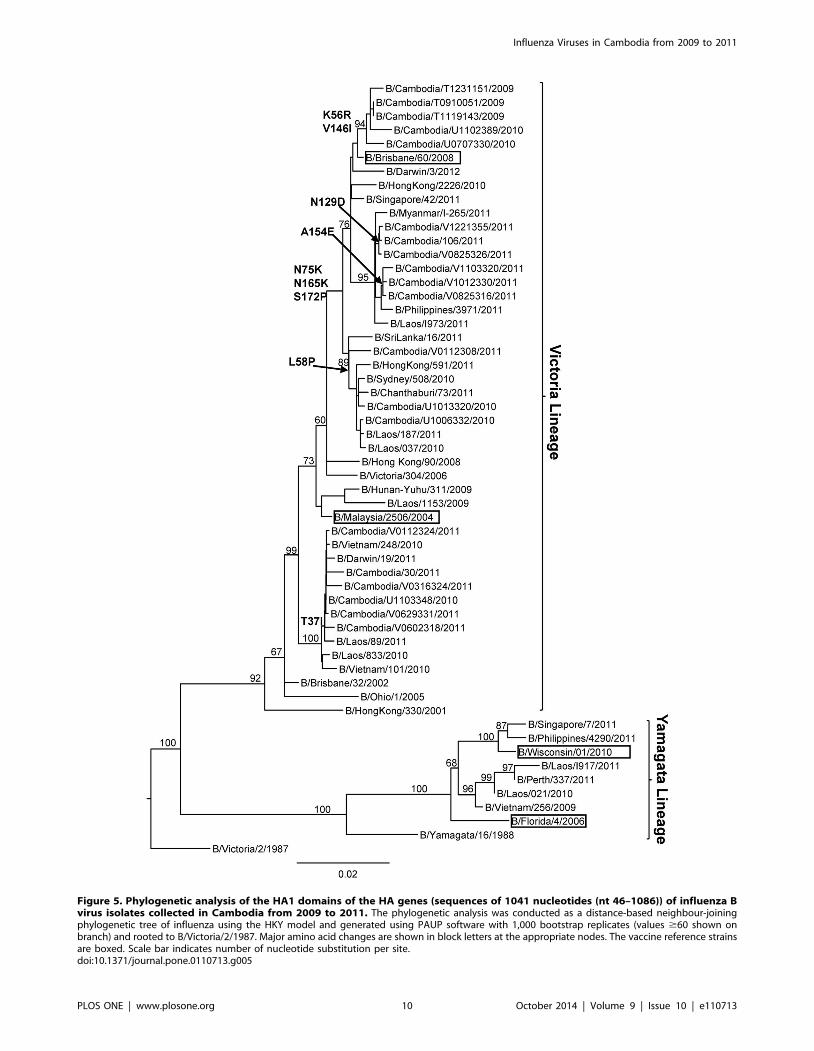
Figure 5. Phylogenetic analysis of the HA1 domains of the HA genes (sequences of 1041 nucleotides (nt 46–1086)) of influenza Bvirus isolates collected in Cambodia from 2009 to 2011. The phylogenetic analysis was conducted as a distance-based neighbour-joiningphylogenetic tree of influenza using the HKY model and generated using PAUP software with 1,000 bootstrap replicates (values $60 shown onbranch) and rooted to B/Victoria/2/1987. Major amino acid changes are shown in block letters at the appropriate nodes. The vaccine reference strainsare boxed. Scale bar indicates number of nucleotide substitution per site.doi:10.1371/journal.pone.0110713.g005
Influenza Viruses in Cambodia from 2009 to 2011
PLOS ONE | www.plosone.org 10 October 2014 | Volume 9 | Issue 10 | e110713

this hypothesis, serological testing for influenza exposure is
required among patients with bacterial respiratory infections
during the seasonal influenza epidemics. The A/H1N1pdm09
virus infection detected both in event-based surveillance and
sentinel-based surveillance systems occurred in all age groups.
However, the median ages of event-based patients tended to be
older than that of influenza positive patients detected in ILI
surveillance. This finding is consistent with the characteristics of
hospitalized cases observed in the USA [26].
The first imported A/H1N1pdm09 cases from Cambodia were
detected from June to July 2009 through event-based surveillance
in two areas where major international border crossings take place:
the international air travel through the Phnom Penh International
Airport and the international overland border crossings from
Poipet, Thailand to Siem Reap Province. Positive cases of this
virus were thereafter detected from communities around the
capital and from the ILI sentinel sites in Siem Reap and
Battambang Province by both event-based and sentinel surveil-
lance from August 2009. In October 2009, the virus spread
throughout the country as indicated by ILI and ALRI surveillance
data. The number of A/H1N1pdm09 virus isolates detected by
the event-based surveillance compared to the number detected by
the sentinel (ILI and ALRI) surveillance systems during the highest
point of the outbreak in October 2009 was 5:1 (Figure 2),
suggesting that when it comes to the timely detection of outbreaks
and important public health events, event-based surveillance is
more sensitive than sentinel surveillance systems, which are not
suited to the detection of rare but high-impact outbreaks of
emerging and unknown diseases [16]. However, at the start of the
influenza season in June 2010, the A/H1N1pdm09 isolates were
first detected by both the event-based and sentinel surveillance
systems before being mostly detected by the sentinel surveillance
system for the rest of the season. This finding is consistent with the
WHO’s declaration on 10 August 2010 that the World was
moving into the post-pandemic period and confirmed that this
virus was taking on the behavior of a seasonal influenza virus [30].
Worldwide, there continued to be co-circulation of influenza A/
H1N1pdm09, A/H3N2 and B viruses in 2010, with the latter two
being predominant [31]. In Cambodia, since September 2009,
there was a sustained circulation of predominantly A/
H1N1pdm09 viruses, but also to a lesser extent, seasonal influenza
H3N2 and B virus, with the latter being the most frequently
detected strain from November and December 2010 through
2011. During this period, limited data from Vietnam indicated
community transmission of influenza there as well, predominantly
influenza type B, but the remainder of southern Asia including
India, Bangladesh, Thailand, Singapore, China Hong Kong
Special Administrative Region, Southern China and Chinese
Taipei reported small numbers of a mixture of all three circulating
types [32]. The circulation of type B virus tends to rise as overall
influenza activity declines. There was a surge of influenza B cases
near the end of each influenza season, usually from November to
December, as shown in our previous report from 2006 to 2008
[12]. There are frequent bottleneck years during which prevalence
of B strains is low, which usually was correlated with high
prevalence of influenza A strains. Indeed, low level of B viruses in
the year 2009 was observed during the pandemic of A/
H1N1pdm09 virus. Once the bottleneck is relieved, there are
usually changes in the prevalence of B strains which could explain
the rise of influenza B virus at the end of 2010 and in 2011 after
the A/H1N1pdm09 subsided (Figure 1). From 2007 to 2011,
except in 2009, the influenza B strains identified in Cambodia
were well matched by the recommended influenza B vaccine virus
(Table 2) [12]. Currently, seasonal influenza vaccines are only
available through private market purchase and Cambodians are
not accustomed to influenza vaccination [33]. However, it could
be problematic if the B strain that was circulating did not match
the one contained in the seasonal influenza vaccine. Mismatches
between influenza B strains circulating in Cambodia and strains
included in the trivalent vaccine should lead to the use of a
quadrivalent vaccine that contains both influenza B lineages.
The emergence of resistance following treatment with amanta-
dine has occurred only transiently and the levels of circulating
resistant viruses were low until 2004 [34,35]. In Cambodia, no
amantadine-resistant strains were found in seasonal influenza
viruses in 2006, but the prevalence increased to 100% in 2007 and
was maintained at this level through 2011 [12]. This finding
supports that of a study conducted by another group reporting that
adamantane resistance in influenza A viruses increased in 2007 in
South East Asia [36]. Surveillance for antiviral susceptibility of
influenza viruses in the Asia-Pacific region during 2011, showed
that .99% of influenza A strains continued to be resistant to the
adamantane drugs [37]. The high levels of amantadine resistance
in influenza A virus strains in the community were not related to
domestic amantadine use, as antiviral drugs for upper respiratory
infection is uncommon among Cambodians. Following the
outbreaks of severe acute respiratory syndrome and avian
influenza, an unusually high prevalence of amantadine resistance
has been observed most prominently in China, Hong Kong, and
Taiwan since 2003, probably due to widespread use of amantadine
[34,38]. However, there was also evidence that the spread of
resistant A/H3N2 virus was related to linked fitness mutations and
not related to drug pressure [39,40]. Cambodia’s rise of resistance
in 2007 may therefore reflect a strong influence from China or
Taiwan in terms of influenza transmission.
A single Cambodian A/H1N1pdm09 strain, isolated from a 10
year old girl patient in Takeo Province in mid-May 2011, did not
contain the mutation Ser31Asn nor any of the transmembrane
region of the M2 protein mutations known to be associated with
adamantane resistance [34].
Alongside neuraminidase inhibitors, oseltamivir and zanamivir
have never been used to any large extent in the country and there
is no evidence that any of the Cambodian patients were exposed to
the drugs before or during influenza infection. The general lack of
primary resistance to this class of antiviral has been previously
reported before introduction of the neuraminidase inhibitors
anywhere in the world [41]. In 2008, a low prevalence of
neuraminidase inhibitors resistance (,1%) was detected in isolates
from untreated patients [42]. During 2011, neuraminidase
inhibitors resistance in A/H1N1pdm09, A/H3N2 and B viruses
in the Asia-Pacific region remained low (,3% and ,1% for
influenza A and B strains respectively) [37]. Therefore, it is not
surprising that all Cambodian isolates tested retained sensitivity to
oseltamivir and zanamivir. However, in our previous study, we
detected two influenza A/H1N1 strains with the H275Y mutation,
associated with a high level of resistance to oseltamivir [43]. One
of these strains detected in October 2007 showed high resistance to
neuraminidase inhibitors by antiviral drug susceptibility assay,
even though the other strain could not be grown in Madin-Darby
canine kidney cells. Following the emergence in Europe in late
2007 of a transmissible human A/H1N1 influenza strain that was
highly resistant to the neuraminidase inhibitor oseltamivir, the
emergence of similar viruses was observed in South Africa and
several countries in Oceania and South East Asia, especially from
May 2008 onwards [44]. However, the mutant H275Y virus found
in Cambodia is not a variant of the emergent H275Y strain
detected elsewhere, but instead has arisen independently, as
occasionally reported previously [42,45].
Influenza Viruses in Cambodia from 2009 to 2011
PLOS ONE | www.plosone.org 11 October 2014 | Volume 9 | Issue 10 | e110713
The phylogenetic analysis of the HA1 genes of A/H3N2 strains
from Cambodia showed three groups corresponding to three
consecutive influenza seasons from 2009 to 2011. Each group,
except the group from 2010–2011, belonged to the phylogenetic
group represented by the corresponding vaccine strain. We
previously demonstrated that there was co-circulation of different
lineages of viruses isolated in 2005 and probable re-emergence of
A/H3N2 viruses in the two consecutive seasons of 2005–2006
[42]. The HA sequences from the A/H3N2 isolates from 2009 to
2011 fell within three distinct phylogenetic clades. These findings
are consistent with the dynamic evolution of A/H3N2 viruses that
present a characteristic co-circulation of several variants for up to
three years, which are subsequently replaced by new emerging
variants with different antigenic features. This constant evolution
of A/H3N2 viruses leads to the emergence of new viral variants
that can elude the human immune response, causing outbreaks
[46,47]. This viral reintroduction plays a key role in the genesis of
new clades and the global spread of these novel influenza virus
variants. The Cambodian isolates collected from each season seem
to have evolved from common ancestor isolates that are also
probably at the origin of Australian srains [43,48]. Additional
research is required to more precisely define factors that contribute
to this influenza seasonality in Cambodia such as the role of
climate in triggering seasonal epidemics and the role for natural
selection and host susceptibility.
The earliest A/H1N1pdm09 isolates from Cambodia (A/
Cambodia/T028/2009, A/Cambodia/T075/2009, A/Cambodia/
T272/2009, A/Cambodia/T282/2009, A/Cambodia/T093/2009
and A/Cambodia/T021/2009) were detected in the period from
the end of June to mid-July 2009 through event-based surveillance
focused on American students visiting Phnom Penh, the capital city
of Cambodia. Positive cases of this virus were thereafter detected
from communities all over the country from August 2009 by both
the event-based and sentinel surveillance systems. Except for A/
Cambodia/T021/2009, all of the other strains detected in that
period clustered into the S203T clade. This finding suggested that
the A/H1N1pdm09 viruses circulating in Cambodia in that period
were introduced into the country, possibly from the USA. Since
August 2009, all strains isolated in 2009, 2010 and 2011 were
assigned to this clade. This is in agreement with recent results
obtained in other regions of the world where it was the most
commonly detected clade [49,50].
All of the Cambodian isolates possessed residues D187 and D222
in the receptor-binding site, which is known to confer binding of H1
viruses to the human receptor, supporting efficient transmission of
these viruses in humans [51]. In other studies the D222G
substitution was detected more frequently in viruses isolated from
patients with fatal outcomes and in isolates from lungs [50,52]. This
residue was well conserved in all Cambodian strains isolated from
patients, but no fatal cases were examined in these studies. The
substitution Q293H had been observed recently in a substantial
proportion of virus strains isolated from postmortem samples from
patients who died from laboratory-confirmed cases of A/
H1N1pdm09 infection [52]. Two Cambodian isolates (A/Cambo-
dia/T021/2009 and A/Cambodia/V1019320/2011) possessed this
mutation and caused pneumonia requiring hospital admission,
although there was no fatal complication. The known antigenic sites
in the HA of the 1918 pandemic virus have been conserved in the
pandemic A/H1N1pdm09 virus [53]. Molecular analysis showed
that these sites were relatively conserved in Cambodian isolates.
Two isolates, A/Cambodia/T282/2009 and A/Cambodia/T057/
2009, which caused mild symptoms, possessed the R205K variation
at the Ca1 antigenic site of the hemagglutinin without altering the
properties of the polar and basic amino acid. The other two isolates
from 2010 (A/Cambodia/U099/2010 and A/Cambodia/U326/
2010) and one isolate from 2011 (A/Cambodia/V0608350/2011),
which caused severe illness, had a substitution N125D at the
hemagglutinin Sa antigenic site, which slightly modified the
properties of the neutral side chain to a basic one. Immunological
pressure may have driven virus evolution as shown by the
displacement of E374E by E374K, a site important for membrane
fusion, and the emergence of the two R205K and N125D amino
acid substitutions involving the antigenic sites Ca1 and Sa
respectively [54]. These antigenic sites contain many amino acids
involved in neutralising epitopes near the receptor-binding pockets
[53]. Although a single amino acid substitution involving one
antigenic site may be sufficient to cause antigenic change, more
commonly antigenic drift variants of epidemiological importance
have resulted from changes of at least four amino acids across two or
more antigenic sites [55]. Thus the variations observed in the
hemagglutinin region of Cambodian A/H1N1pdm09 virus isolates
most likely did not affect the antigenicity of the viruses. Although the
results from antigenic analysis of Cambodian strains support this
hypothesis, in vitro results cannot always predict in vivo effects.
There have been two distinct evolutionary lineages of influenza
B viruses in recent years, represented by the two epidemic strains,
B/Victoria/2/87 and B/Yamagata/16/1988. Phylogenetic anal-
yses of influenza B isolates from Cambodia indicated that the
circulating influenza B viruses in 2009 to 2011 belonged to the
Victoria lineage; and the Yamagata lineage viruses did not
circulate in Cambodia during those years (Figure 5). Some strains
from 2010 and 2011 were closely related to the vaccine reference
strain B/Malaysia/2506/2004, whereas the remainder of strains
from 2009–2011 were closely related to the vaccine strain B/
Brisbane/60/2008 (Figure 5 and Table S4). Our results indicated
that a mismatch between the vaccine and epidemic strains in
Cambodia occurred in the Southern Hemisphere World Health
Organization 2009 influenza B virus vaccine recommendations;
B/Brisbane/60/2008-like should have been the candidate strain
for Cambodia (Table 2). Interestingly, the influenza B viruses that
circulated in 2010 were found to have evolved from different
genetic groups, indicating that the outbreak was caused by
multiple sources. All of the influenza B viruses collected in 2009
and the majority of the 2010 and 2011 viruses possessed amino
acid substitutions N75K, N165K and S172P that defined genetic
group 1 in which the vaccine B/Brisbane/60/2008 strain was
grouped. All of the 2009 and some of the 2010 viruses carried the
amino acid substitution K56R compared with the vaccine B/
Brisbane/60/2008 strain; some strains from 2010–2011 viruses
carried the substitution T37I and L58P; some isolates from 2010
and all strains from 2011 contained amino acid I146V compared
with the vaccine B/Brisbane/60/2008. None of these substitutions
had a marked effect on the antigenicity of the viruses. This is not
unusual, as influenza B virus activity is highly variable world-wide,
with no established pattern [56]. However, this could lead to the
emergence of influenza B reassortants in the next influenza season [57].
This manuscript presents the characterization of influenza
strains collected from numerous surveillance programs and
research studies, and as such has several limitations. As the
collection and initial analysis of samples was conducted in
numerous clinics and laboratories respectively, the influenza
strains included in this study were subject to any sampling or
testing biases that were inherent in the different studies. The
analyses presented in the study were also limited to the data
available to the NIC and did not include epidemiological
information such as detailed admission data and rates of infection.
In addition, samples were not collected throughout 2009–2011 for
some of the studies (such as event-based surveillance for
Influenza Viruses in Cambodia from 2009 to 2011
PLOS ONE | www.plosone.org 12 October 2014 | Volume 9 | Issue 10 | e110713

H1N1pdm09) and some of the study sites did not contribute
samples throughout the entire study period. As a large number of
samples were tested throughout the study (n = 10,813) only a
representative number of isolates could be further analysed for
genetic, antigenic and antiviral drug resistance characteristics.
However, a large proportion of isolates were also sent to
WHOCCs for further characterization (data not shown).
In summary, this study coupled with our previous study of
influenza activity in Cambodia over a six year period from 2006 to
2011 provides strong evidence that influenza was prevalent in
Cambodia and influenza activity occurred throughout the year
with annual peaks during the rainy season from June to
November. Since September 2009, there has been sustained
circulation of predominantly A/H1N1pdm09 viruses. Seasonal
influenza A/H3N2 and B virus have also circulated, although to a
lesser extent. In 2010 there continued to be co-circulation of
influenza A/H1N1pdm09, A/H3N2 and B viruses, with the latter
two being predominant from November and December 2010
through 2011. The influenza virus epidemics in Cambodia
occurred through genetically distinct multiple viruses, even if all
of the isolates had similar antigenicity to the reference vaccine
strain. The drug susceptibility profile of Cambodian influenza
strains showed that neuraminidase inhibitors would be the drug of
choice for influenza treatment and chemoprophylaxis in Cambo-
dia, as adamantanes are no longer expected to be effective and not
recommended for use [58]. Despite the reduced number of
influenza vaccines used in Cambodia, policy makers should
consider the use of a quadrivalent vaccine (when available) to
counter the co-circulation of the two influenza A lineages.
Supporting Information
Figure S1 Monthly proportion of positive influenzasamples among ILI specimens tested from 2009 to2011 and among ALRI specimens tested from 2009 toJuly 2010.(TIFF)
Figure S2 Amino acid alignment of HA1 domain of 28representative A/H3N2 strains isolated from 2009 to2011 in Cambodia with the vaccine strains A/Brisbane/10/2007 and A/Perth/16/2009.(RTF)
Figure S3 Amino acid alignment of 33 HA sequencesfrom A/H1N1pdm09 strains circulating from 2009 to2011 in Cambodia with the vaccine strain A/California/07/2009.(RTF)
Figure S4 Amino acid alignment of 20 HA1 sequencesfrom influenza B strains circulating from 2009 to 2011 inCambodia with the vaccine strain B/Malaysia/2506/2004 and B/Brisbane/60/2008.(RTF)
Table S1 Outline of all of the surveillance programsand research studies that contributed to the presentstudy.
(DOCX)
Table S2 All Cambodian influenza A/H3N2, A/H1N1pdm09 and Influenza B virus sequences includedin the analysis are available via GISAID website (www.gisaid.org).
(DOCX)
Table S3 Resistance to adamantanes predicted follow-ing M gene sequencing of influenza A viruses, 2009–2011.
(DOC)
Table S4 Amino acid substitutions in A/H3N2 virusesisolated in Cambodia from 2009 to 2011.
(DOCX)
Table S5 Amino acid substitutions in A/H1N1pdm09viruses isolated in Cambodia from 2009 to 2011.
(DOCX)
Table S6 Variations in HA1 amino acid sequences ininfluenza B-Victoria lineage viruses isolated in Cambo-dia in 2009, 2010 and 2011 by comparison with vaccinestrain B/Malaysia/2506/2004.
(DOCX)
Acknowledgments
We would like to thank the following Institutions/Organizations/Project
for their contribution: The World Health Organization (WHO) office in
Cambodia, assisting the implementation and supporting the monitoring of
influenza activity and the establishment the sentinel surveillance system for
influenza-like illness (ILI); WHO Collaborating Centre for Reference and
Research on Influenza, Melbourne, Australia; The Office of the Assistant
Secretary for Preparedness and Response within the U.S. Department of
Health and Human Services (DHHS, US); The SISEA project of French
Agency for Development (AFD); Influenza Division, National Center for
immunization and Respiratory Disease. Centers for Disease Control and
Prevention, USA; Centers for Disease Control and Prevention, Cambodia
office; National Institute of Public Health (NIPH), Phnom Penh,
Cambodia; Communicable Disease Control Department, Ministry of
Health, Phnom Penh, Cambodia; The Armed Forces Research Institute of
Medical Sciences (AFRIMS), Bangkok, Thailand. The Melbourne WHO
Collaborating Centre for Reference and Research on Influenza is
supported by the Australian Government Department of Health.
Author Contributions
Conceived and designed the experiments: PB. Performed the experiments:
SM SVH SR. Analyzed the data: SM SVH SR PB NK. Contributed
reagents/materials/analysis tools: SL SH SV PK VI AT SL BS NC BS IB
AK PFH AT AH CL DS SAU NA MCR ST NK. Wrote the paper: SVH
SM SR PFH NK PB.
References
1. Simonsen L, Clarke MJ, Williamson GD, Stroup DF, Arden HA, et al. (1997)
The impact of influenza epidemics on mortality: introducing a severity index.
Am J Public Health 87: 1944–1950.
2. Thompson WW, Shay DK, Weintraub E, Brammer L, Cox N, et al. (2003)
Mortality associated with influenza and respiratory syncytial virus in the United
States. JAMA 289: 179–186.
3. Dawood FS, Luliano AD, Reed C, Meltzer MI, Shay DK, et al. (2012)
Estimated global mortality associated with the first 12 months of 2009 pandemic
influenza A H1N1 virus circulation: a modeling study. Lancet Infect Dis 12:
687–695.
4. Finkelman BS, Viboud C, Koelle K, Ferrari MJ, Bharti N, et al. (2007) Global
Patterns in Seasonal Activity of Influenza A/H3N2, A/H1N1, and B from 1997
to 2005: Viral Coexistence and Latitudinal Gradients. PLos One 12: e1296.
5. Shek LP, Lee BW (2003) Epidemiology and seasonality of respiratory tract
infections in the tropics. Paediatr Respir Rev 4: 104–111.
6. Beckett CG, Hosasih H, Ma’roef C, Listiyonengsih E, Elyazar IRF, et al. (2004)
Influenza surveillance in Indonesia: 1999–2003. Clin Infect Dis 39: 443–449.
7. Doraisingham S, Goh KT, Ling AE, Yu M (1988) Influenza surveillance in
Singapore: 1972–86. Bull World Health Organ 66: 57–63.
Influenza Viruses in Cambodia from 2009 to 2011
PLOS ONE | www.plosone.org 13 October 2014 | Volume 9 | Issue 10 | e110713

8. Nguyen HLK, Saito R, Ngiem HK, Nishikawa M, Shobugawa Y, et al. (2007)
Epidemiology of influenza in Hanoi, Vietnam, from 2001 to 2003. J Infect 55:58–63.
9. Rao BL, Banerjee K (1993) Influenza surveillance in Pune, India. Bull World
Health Organ, 71: 177–181.10. Tsai HP, Kuo PH, Liu CC, Wang JR et al. (2001) Respiratory viral infections
among inpatients and outpatients in Taiwan from 1997 to 1999. J ClinMicrobiol 39: 111–118.
11. Members of the Western Pacific Region Global Influenza Surveillance and
Response System (2012) Epidemiological and Virological Characteristics ofInfluenza in the Western Pacific Region of the World Health Organization,
2006–2010. PLoS ONE 7: e37568.12. Mardy S, Ly S, Heng S, Vong S, Huch C, et al. (2009) Influenza activity in
Cambodia during 2006–2008. BMC Infect Dis 9: 168.13. Viboud C, Alonso WJ, Simonsen L (2006) Influenza in tropical regions. PLoS
Med 3: 0468–0471.
14. Webster RG, Bean WJ, Gorman OT, Chambers TM, Kawaoka Y (1992)Evolution and ecology of influenza A viruses. Microbiol Rev 56: 152–179.
15. National Institute of Statistics, Phnom Penh, Cambodia. Final census results(2nd ed.) (2002) General population census of Cambodia 1998.
16. World Health Organization (2008) A guide to establishing event-based
surveillance. Available: http://www.wpro.who.int/internet/resources.ashx/CSR/Publications/eventbasedsurv.pdf. Accessed 21 March 2011.
17. World Health Organization (2009) CDC protocol of realtime RT-PCR forinfluenza A (H1N1). Available: http://www.who.int/csr/resources/
publications/swineflu/realtimertpcr/en/index/html. Accessed 21 March 2011.18. World Health Organization (2007) Recommendations for laboratory procedures
to detect avian influenza A H5N1 virus in specimens from suspected human
cases. Available: http://www.who.int/csr/disease/avian_influenza/guidelines/RecAIlabtestsAug07.pdf Accessed 21 March 2011.
19. Larkin MA, Blackshields G, Brown NP, Chenna R, McGettigan PA, et al. (2007)Clustal W and Clustal X version 2.0. Bioinformatics, 23: 2947–2948.
20. Swofford DL (2002) PAUP*: Phylogenetic Analysis Using Parsimony (and other
methods) 4.0 Beta. Available: paup.csit.fsu.edu/about.html. Accessed 21 March2011.
21. Naughtin M, Dyason JC, Mardy S, Sorn S, von Itzstein M, et al. (2011)Neuraminidase inhibitor sensitivity and receptor-binding specificity of Cambo-
dian clade 1 highly pathogenic H5N1 virus. Antimicrob Agents Chemother 55:2004–2010.
22. Blair PJ, Wierzba TF, Touch S, Vonthanak S, Xu X, et al. (2010) Influenza
epidemiology and characterization of influenza viruses in patients seekingtreatment for acute fever in Cambodia. Epidemiol Infect 138: 199–209.
23. Nair H, Brooks WA, Katz M, Roca A, Berkley JA, et al. (2011) Global burden ofrespiratory infections due to seasonal influenza in young children: a systematic
review and meta-analysis. Lancet 378: 1917–1930.
24. O’Brien KL, Walters MI, Sellman J (2000) Severe pneumococcal pneumonia inpreviously healthy children: the role of preceding influenza infection. Clin Infect
Dis 30: 784–789.25. McCullers JA, McAuley JL, Browall S, Iverson AR, Boyd KL, et al. (2010)
Influenza enhances susceptibility to natural acquisition of and disease due toStreptococcus pneumoniae in ferrets. J Infect Dis 202: 1287–1295.
26. World Health Organization (2009) Human infection with pandemic A(H1N1)
2009 influenza virus: clinical observations in hospitalized patients, Americas,July 2009 – update. Wkly Epidemiol Rec 84: 301–308.
27. Hope-Simpson RE (1981) The role of season in the epidemiology of influenza.J Hyg 86: 35–47.
28. Chittaganpitch M, Supawat K, Olsen SJ, Waicharoen S, Patthamadilok S, et al.
(2012) Influenza viruses in Thailand: 7 years of sentinel surveillance data, 2004–2010. Influenza Other Respir Viruses 6: 276–283.
29. Jian JW, Chen JW, Lai CT, Hsu LC, Chen PJ, et al. (2008) Genetic andepidemiological analysis of influenza virus epidemics in Taiwan during 2003 to
2006. J Clinic Microbiol 46: 1426–1434.
30. World Health Organization (2011) Influenza A(H1N1) 2009 virus: currentsituation and post-pandemic recommendations. Wkly Epidemiol Rec 86: 61–72.
31. World Health Organization (2010) Recommended viruses of influenza vaccinesfor use in the 2011 influenza season (Southern hemisphere). Wkly Epidemiol Rec
85: 61–72.32. World Health Organization (2010) Global Influenza Program, Influenza update.
Available: http://www.who.int/csr/disease/influenza/2010_12_30_GIP_
surveillance/en/index.html Accessed 22 March 2011.33. Members of the Western Pacific Region Global Influenza Surveillance and
Response System (2013) Seasonal influenza vaccine policies, recommendationsand use in the World Health Organization’s Western Pacific Region. WPSAR 4:
doi: 10.5365.
34. Bright RA, Medina MJ, Xu X, Perez-Oronoz G, Wallis TR, et al. (2005)
Incidence of amantadine resistance among influenza A(H3N2) viruses isolatedworldwide from 1994 to 2005: a cause for concerne. Lancet 366: 1175–1181.
35. Bright R, Shay D, Shu B, Cox N, Klimov A (2006) Adamantane resistance
among influenza A viruses isolated early during the 2005–2006 influenza seasonin the United States. JAMA 295: 891–894.
36. Barr I, Deng Y, Iannello P, Hurt A, Komadina N (2008) Adamantane resistancein influenza A(H1) viruses increased in 2007 in South East Asia but decreased in
Australia and some other countries. Antivir. Res 80: 200–205.
37. Leang SK, Deng YM, Shaw R, Caldwell N, Iannello P, et al. (2013) Influenzaantiviral resistance in the Asia-Pacific region during 2011. Antiviral Res 97: 206–
210.38. He G, Qiao J, Dong C, He C, Zhao L, et al. (2008) Amantadine resistance
among H5N1 avian influenza viruses isolated in Northern China. Antivir Res77: 72–76.
39. Simonsen L, Viboud C, Grenfell BT, Dushoff J, Jennings L, et al. (2007) The
genesis and spread of reassortment human influenza A/H3N2 viruses conferringadamantane resistance. Mol Biol Evol 24: 1811–1820.
40. Ilyushina N, Govorkova E, Webster R (2005) Detection of amantadine-resistantvariants among avian influenza viruses isolated in North America and Asia.
Virology 341: 102–106.
41. McKimm-Breschkin JL, Trivedi T, Hampson A, Hay A, Klimov A, et al. (2003)Neuraminidase sequence analysis and susceptibilities of influenza virus clinical
isolates to zanamivir and oseltamivir. Antimicrob. Agents Chemother 47: 2264–2272.
42. Hurt AC, Barr IG (2008) Influenza viruses with reduced sensitivity to the NAinhibitor drugs in untreated young children. Commun Dis Intell 32: 57–62.
43. Fourment M, Mardy S, Channa M, Buchy P (2010) Evidence for persistence of
and antiviral resistance and reassortment events in seasonal influenza virusstrains circulating in Cambodia. J Clin Microbiol 48: 295–297.
44. Hurt AC, Ernest J, Deng YM, Iannello P, Besselaar TG, et al. (2009) Emergenceand spread of oseltamivir-resistant A(H1N1) influenza viruses in Oceania, South
East Asia and South Africa. Antivir Res 83: 90–93.
45. Monto AS, McKimm-Breschkin JL, Macken C, Hampson AW, Hay A, et al.(2006) Detection of influenza viruses resistant to neuraminidase inhibitors in
global surveillance during the first 3 years of their use. Antimicrob AgentsChemother 50: 2395–2402.
46. Holmes EC, Ghedin E, Miller N, Taylor J, Bao Y, et al. (2005) Whole-genomeanalysis of human influenza A virus reveals multiple persistent lineages and
reassortment among recent H3N2 viruses. PLoS Biol 3: 1579–1588.
47. Pariani E, Fratt ER, Amendola A, Zappa A, Bianchi S, et al. (2009) Molecularcharacterization and phlylogenetic analysis of human influenza A viruses in
three consecutive seasons with different epidemiological profiles. J Prev MedHyg 50: 113–116.
48. Nelson MI, Simonsen L, Viboud C, Miller MA, Holmes EC (2007) Phylogenetic
analysis reveals the global migration of seasonal influenza A viruses. PLosPathogens 3: 1220–1228.
49. Nelson M, Spiro D, Wentworth D, Fan J, Beck E, et al. (2009) The earlydiversification of influenza A/H1N1pdm. PLoS Curr 1: RRN1126.
50. Potdar VA, Chadha MS, Jadhav SM, Mullick J, Cherian SS, et al. (2010)Genetic characterization of the influenza A pandemic (H1N1) 2009 virus isolates
from India. PLoS ONE 3: e9693.
51. Stevens J, Corper AL, Basler CF, Taubenberger JK, Palese P, et al. (2004)Structure of the uncleaved human H1 hemagglutinin from the extinct 1918
influenza virus. Science 303: 1866–1870.52. Glinsky GV (2010) Genomic analysis of pandemic (H1N1) 2009 reveals
association of increasing disease severity with emergence of novel hemagglutinin
mutations. Cell Cycle 9: 958–970.53. Igarashi M, Ito K, Yoshida R, Tomabechi D, Kida H, et al. (2010) Predicting
the antigenic structure of the pandemic (H1N1) 2009 influenza virushemagglutinin. PLoS ONE 1: e8553.
54. Maurer-Stroh S, Lee RT, Eisenhaber F, Cui L, Phuah SP, et al. (2010) A new
common mutation in the hemagglutinin of the 2009 (H1N1) influenza A virus.PLoS Curr 2: RRN1162
55. Ferguson NM, Galvani AP, Bush RM (2003) Ecological and immunologicaldeterminants of influenza evolution. Nature 422: 428–433.
56. McCullers JA, Wang GC, He S, Webster RG (1999) Reassortment andinsertion-deletion are strategies for the evolution of influenza B viruses in nature.
J Virol 73: 7343–7348.
57. Tsai HP, Wang HC, Kiang D, Huang SW, Kuo PH, et al. (2006) Increasingappearance of reassortant influenza B virus in Taiwan from 2002 to 2005. J Clin
Microbiol 44: 2705–2713.58. CDC (2011) Influenza Antiviral Medications: A Summary for Clinicians. Available:
http://www.cdc.gov/flu/professionals/antivirals/summary-clinicians.htm..
Influenza Viruses in Cambodia from 2009 to 2011
PLOS ONE | www.plosone.org 14 October 2014 | Volume 9 | Issue 10 | e110713
Copyright © 2022 FDOKUMEN