Pneumonia and Respiratory Failure from Swine-Origin Influenza A (H1N1) in Mexico
10
The new england journal of medicine n engl j med 10.1056/nejmoa0904252 1 original article Pneumonia and Respiratory Failure from Swine-Origin Influenza A (H1N1) in Mexico Rogelio Perez-Padilla, M.D., Daniela de la Rosa-Zamboni, M.D., Samuel Ponce de Leon, M.D., Mauricio Hernandez, M.D., Francisco Quiñones-Falconi, M.D., Edgar Bautista, M.D., Alejandra Ramirez-Venegas, M.D., Jorge Rojas-Serrano, M.D., Christopher E. Ormsby, M.Sc., Ariel Corrales, M.D., Anjarath Higuera, M.D., Edgar Mondragon, M.D., and Jose Angel Cordova-Villalobos, M.D., for the INER Working Group on Influenza* From the National Institute of Respiratory Diseases (INER) (R.P.-P., D.R.-Z., F.Q.-F., E.B., A.R.-V., J.R.-S., C.E.O., A.C., A.H., E.M.), Biologicals and Reactives of Mexi- co (BIRMEX) (S.P.L.), and the Secretariat of Health (M.H., J.A.C.-V.) — all in Mexi- co City. Address reprint requests to Dr. Perez-Padilla at Instituto Nacional de En- fermedades Respiratorias, Tlalpan 4502, 14080 Mexico, or at [email protected]. *Members of the INER Working Group on Influenza are listed in the Appendix. This article (10.1056/NEJMoa0904252) was published on June 29, 2009, at NEJM.org. N Engl J Med 2009;361. Copyright © 2009 Massachusetts Medical Society. Abstract Background In late March 2009, an outbreak of a respiratory illness later proved to be caused by novel swine-origin influenza A (H1N1) virus (S-OIV) was identified in Mexico. We describe the clinical and epidemiologic characteristics of persons hospitalized for pneumonia at the national tertiary hospital for respiratory illnesses in Mexico City who had laboratory-confirmed S-OIV infection, also known as swine flu. Methods We used retrospective medical chart reviews to collect data on the hospitalized patients. S-OIV infection was confirmed in specimens with the use of a real-time reverse-transcriptase–polymerase-chain-reaction assay. Results From March 24 through April 24, 2009, a total of 18 cases of pneumonia and con- firmed S-OIV infection were identified among 98 patients hospitalized for acute respiratory illness at the National Institute of Respiratory Diseases in Mexico City. More than half of the 18 case patients were between 13 and 47 years of age, and only 8 had preexisting medical conditions. For 16 of the 18 patients, this was the first hospitalization for their illness; the other 2 patients were referred from other hospitals. All patients had fever, cough, dyspnea or respiratory distress, increased serum lactate dehydrogenase levels, and bilateral patchy pneumonia. Other common findings were an increased creatine kinase level (in 62% of patients) and lymphopenia (in 61%). Twelve patients required mechanical ventilation, and seven died. Within 7 days after contact with the initial case patients, a mild or moderate influenza-like illness developed in 22 health care workers; they were treated with oseltamivir, and none were hospitalized. Conclusions S-OIV infection can cause severe illness, the acute respiratory distress syndrome, and death in previously healthy persons who are young to middle-aged. None of the secondary infections among health care workers were severe. Copyright © 2009 Massachusetts Medical Society. All rights reserved. Downloaded from www.nejm.org on July 3, 2009 . For personal use only. No other uses without permission.
-
Upload
independent -
Category
Documents
-
view
0 -
download
0
Transcript of Pneumonia and Respiratory Failure from Swine-Origin Influenza A (H1N1) in Mexico

T h e n e w e ngl a nd j o u r na l o f m e dic i n e
n engl j med 10.1056/nejmoa0904252 1
original article
Pneumonia and Respiratory Failure from Swine-Origin Influenza A (H1N1) in Mexico
Rogelio Perez-Padilla, M.D., Daniela de la Rosa-Zamboni, M.D., Samuel Ponce de Leon, M.D., Mauricio Hernandez, M.D., Francisco Quiñones-Falconi, M.D., Edgar Bautista, M.D.,
Alejandra Ramirez-Venegas, M.D., Jorge Rojas-Serrano, M.D., Christopher E. Ormsby, M.Sc., Ariel Corrales, M.D., Anjarath Higuera, M.D.,
Edgar Mondragon, M.D., and Jose Angel Cordova-Villalobos, M.D., for the INER Working Group on Influenza*
From the National Institute of Respiratory Diseases (INER) (R.P.-P., D.R.-Z., F.Q.-F., E.B., A.R.-V., J.R.-S., C.E.O., A.C., A.H., E.M.), Biologicals and Reactives of Mexi-co (BIRMEX) (S.P.L.), and the Secretariat of Health (M.H., J.A.C.-V.) — all in Mexi-co City. Address reprint requests to Dr. Perez-Padilla at Instituto Nacional de En-fermedades Respiratorias, Tlalpan 4502, 14080 Mexico, or at [email protected].
*Members of the INER Working Group on Influenza are listed in the Appendix.
This article (10.1056/NEJMoa0904252) was published on June 29, 2009, at NEJM.org.
N Engl J Med 2009;361.Copyright © 2009 Massachusetts Medical Society.
A bs tr ac t
Background
In late March 2009, an outbreak of a respiratory illness later proved to be caused by novel swine-origin influenza A (H1N1) virus (S-OIV) was identified in Mexico. We describe the clinical and epidemiologic characteristics of persons hospitalized for pneumonia at the national tertiary hospital for respiratory illnesses in Mexico City who had laboratory-confirmed S-OIV infection, also known as swine flu.
Methods
We used retrospective medical chart reviews to collect data on the hospitalized patients. S-OIV infection was confirmed in specimens with the use of a real-time reverse-transcriptase–polymerase-chain-reaction assay.
Results
From March 24 through April 24, 2009, a total of 18 cases of pneumonia and con-firmed S-OIV infection were identified among 98 patients hospitalized for acute respiratory illness at the National Institute of Respiratory Diseases in Mexico City. More than half of the 18 case patients were between 13 and 47 years of age, and only 8 had preexisting medical conditions. For 16 of the 18 patients, this was the first hospitalization for their illness; the other 2 patients were referred from other hospitals. All patients had fever, cough, dyspnea or respiratory distress, increased serum lactate dehydrogenase levels, and bilateral patchy pneumonia. Other common findings were an increased creatine kinase level (in 62% of patients) and lymphopenia (in 61%). Twelve patients required mechanical ventilation, and seven died. Within 7 days after contact with the initial case patients, a mild or moderate influenza-like illness developed in 22 health care workers; they were treated with oseltamivir, and none were hospitalized.
Conclusions
S-OIV infection can cause severe illness, the acute respiratory distress syndrome, and death in previously healthy persons who are young to middle-aged. None of the secondary infections among health care workers were severe.
Copyright © 2009 Massachusetts Medical Society. All rights reserved. Downloaded from www.nejm.org on July 3, 2009 . For personal use only. No other uses without permission.

T h e n e w e ngl a nd j o u r na l o f m e dic i n e
n engl j med 10.1056/nejmoa09042522
In April 2009, the Mexican Secretariat of Health reported an outbreak of respiratory disease. In the affected patients, a novel swine-
origin influenza A (H1N1) virus (S-OIV) with molecular features of North American and Eur-asian swine, avian, and human influenza viruses1-4 was found. In the same month, the World Health Organization (WHO) classified the global spread of this virus as a public health event of interna-tional concern. After documentation of human-to-human transmission of the virus in at least three countries of two WHO regions, the WHO raised the pandemic level to 6.5
As of May 29, 2009, Mexico had reported 4910 confirmed cases and 85 deaths caused by S-OIV.6 Mexico has reported the greatest number of cases of severe clinical presentations and death,1 where-as other countries have reported predominantly mild cases of influenza-like illness.
This case series describes the clinical and epi-demiologic characteristics of the first 18 persons with pneumonia and laboratory-confirmed S-OIV infection (also known as swine flu) hospitalized at the National Institute of Respiratory Diseases (INER) in Mexico. We also describe apparent trans-mission of this infection to health care workers during the initial days of the outbreak.
Me thods
INER is the Mexican national tertiary care and research center devoted to respiratory diseases. The 178-bed facility provides clinical services pri-marily for the uninsured population of Mexico City and neighboring states. We retrospectively reviewed medical charts and radiologic and labo-ratory findings. This study was determined to be exempt from the requirement of institutional re-view, because it was conducted as part of a public health investigation into retrospective data. All tests and procedures were performed at the re-quest of the physicians in charge of the patients. All study patients had influenza-like illness with opacities found on the chest radiograph (reveal-ing pneumonia) and had laboratory-confirmed S-OIV infection. We also reviewed clinical data from a group of 21 hospitalized patients with influenza-like illness and pneumonia but with a negative result on reverse-transcriptase–polymer-ase-chain-reaction (RT-PCR) testing for influenza A (H1N1).
Microbiologic Studies
Nasopharyngeal-swab specimens were collected at admission, and bronchial-aspirate samples were obtained after tracheal intubation. Specimens were placed in transport medium and kept at a temperature from 2 to 4°C. RT-PCR testing was done in accordance with published guidelines from the U.S. Centers for Disease Control and Prevention (CDC).7 Primers and probes for S-OIV were recently developed and distributed to the Mexican Secretariat of Health and its affiliated national institutions by the CDC. In addition, re-spiratory specimens from all patients were tested with the use of a multiplex PCR assay for respira-tory viral and atypical bacterial panels (Seagene) for the detection of influenza A, influenza B, adeno-virus, respiratory syncytial virus, parainfluenza (types 1, 2, and 3), human metapneumovirus, rhinovirus, Legionella pneumophila, Chlamydophila pneumoniae, and Mycoplasma pneumoniae.
Statistical Analysis
Data analysis was conducted using STATA statis-tical software.8 We compared clinical character-istics on admission between patients positive for S-OIV who died and those who survived and be-tween patients who were positive for S-OIV and those who were negative for S-OIV. The risk of death was analyzed by means of a univariate Cox proportional-hazards model; odds ratios were cal-culated and Fisher’s exact test was performed for dichotomous categorical variables. Continuous data were tested by means of the Wilcoxon rank-sum test. All reported P values are two-sided and were not adjusted for multiple testing.
R esult s
Study Patients
The number of emergency room visits for pneu-monia or influenza-like illness increased consid-erably at the INER in Mexico City during the last week of March 2009, peaking in late April and decreasing during the first week of May (Fig. 1). From March 24 through April 24, 2009, a total of 214 emergency room consultations for cases of pneumonia or influenza-like illness were regis-tered, 98 of which required hospitalization. Of these cases, 18 confirmed cases of S-OIV infec-tion, with pneumonia and influenza-like illness, are the focus of this report.
Copyright © 2009 Massachusetts Medical Society. All rights reserved. Downloaded from www.nejm.org on July 3, 2009 . For personal use only. No other uses without permission.

pneumonia and Respir atory Failure from S-OIV
n engl j med 10.1056/nejmoa0904252 3
Characteristics of the 18 study patients with confirmed S-OIV infection are listed in Table 1, and Table S1 of the Supplementary Appendix (available with the full text of this article at NEJM.org). The ages of the patients ranged from 9 months to 61 years (median, 38 years). More than half the patients were between 13 and 47 years of age, and 90% were less than 52 years of age. Nine patients (50%) were male.
All patients resided in the Mexico City greater metropolitan area. Eight patients had preexisting medical conditions: arterial hypertension (in three patients), non–type 1 diabetes mellitus (in three, one of whom also had hypertension), asthma (in two), and obstructive sleep apnea (in one). Only three of the patients had undergone seasonal influenza vaccination in 2008–2009; all three survived without requiring mechanical ventilation. None of the patients had a history of pneumo-coccal vaccination. Among the 14 patients whose occupation was recorded, 6 were students, 2 were taxi drivers, 3 were housekeepers, 1 was a lock-smith, 1 was an employee of a billiards parlor,
and 1 was a physician who did not have clinical duties and was not an INER employee.
The time between onset of symptoms and ad-mission to the hospital ranged from 4 to 25 days (median, 6) (Fig. 2). All patients had fever, with temperatures higher than 38°C, cough, and dys-pnea or respiratory distress. Four of the five children (all under 14 years of age) had diarrhea, and only two patients (11%) reported wheezing. The median Acute Physiology and Chronic Health Evaluation II score9 was 14 (range, 4 to 32), and the median Sequential Organ Failure Assessment score10 was 6 (range, 1 to 13); both were higher, indicating more severe abnormalities among the patients who died than among those who lived (Table 2).
Twelve patients sought medical care at other institutions as outpatients before hospitalization at INER and were treated with one or more anti-biotics: ceftriaxone (five patients), amikacin (three), azithromycin (one), amoxicillin–clavulanate (two) or other macrolides (three), or another agent (two). Except for two patients transferred from other
33p9
160
No.
of C
onsu
ltatio
ns
80
120
40
0
Januar
y 1
Januar
y 8
Januar
y 19
Januar
y 22
Januar
y 29
Febr
uary 5
Febr
uary 1
2
Febr
uary 1
9
Febr
uary 2
6
Mar
ch 5
Mar
ch 12
Mar
ch 19
Mar
ch 26
April 2
April 9
April 16
April 23
April 30
May
7
May
14
2009
2008
2007
AUTHOR:
FIGURE:
JOB:
4-CH/T
RETAKE
SIZE
ICM
CASE
EMail LineH/TCombo
Revised
AUTHOR, PLEASE NOTE: Figure has been redrawn and type has been reset.
Please check carefully.
REG F
Enon
1st2nd
3rd
Perez-Padilla
1 of 3
08-13-09
ARTIST: ts
360xx ISSUE:
Admission
Figure 1. Emergency Room Consultations for Pneumonia or Respiratory Infection, Including Influenza-like Illness, at the National Institute of Respiratory Diseases of Mexico.
Patients with the reported cases were admitted between March 24 and April 24 (gray vertical lines). The Ministry of Health issued an epidemiologic alert on April 17 and a full sanitary alert, with closing of schools and cancellation of many public activities on April 23, after it was confirmed that the patients were infected with the novel influenza A (H1N1) virus.
Copyright © 2009 Massachusetts Medical Society. All rights reserved. Downloaded from www.nejm.org on July 3, 2009 . For personal use only. No other uses without permission.

T h e n e w e ngl a nd j o u r na l o f m e dic i n e
n engl j med 10.1056/nejmoa09042524
health centers, the reported hospitalization was the first hospitalization related to the disease.
Laboratory Results
At the time of admission, all 16 tested patients had elevated lactate dehydrogenase levels; levels in 10 patients exceeded 1000 IU per liter (range, 1086 to 6309). Ten of the 16 patients had increased creatine kinase levels, which were above 1000 IU per liter (range, 1099 to 5122) in 5 patients. Eleven
of all 18 patients (61%) had lymphopenia (<1000 lymphocytes per cubic millimeter), 2 patients had more than 10,000 leukocytes per cubic millimeter, and 2 patients had mild thrombocytopenia at admission. Patient 3 had myocardial ischemia, as revealed on electrocardiography, with myocardial infarction documented on autopsy. Three patients had elevated creatinine levels (1.8 to 4.6 mg per deciliter [159 to 407 μmol per liter]) at admis-sion. Four patients had d-dimer levels greater
Table 1. Characteristics of the 18 Study Patients Who Had Confirmed Infection with Novel Swine-Origin Influenza A (H1N1) Virus.*
Variable Value
Male sex — no./total no. (%) 9/18 (50)
Age — yr
Median 38
Range 0.75–61
All patients — no./total no. (%)
≤5 yr 3/18 (17)
>5 to ≤10 yr 1/18 (6)
>10 to ≤15 yr 1/18 (6)
>15 to ≤50 yr 11/18 (61)
>50 yr 2/18 (11)
Patients who died — no./total no.
≤5 yr 0/3
>5 to ≤10 yr 1/1
>10 to ≤15 yr 1/1
>15 to ≤50 yr 4/11
>50 yr 1/2
Symptom or outcome — no./total no. (%)
Cough 18/18 (100)
Blood in sputum 6/18 (33)
Rhinorrhea 5/18 (28)
Wheezing 2/18 (11)
Headache 4/18 (22)
Myalgia or arthralgia 8/18 (44)
Fever (temperature >38°C) 18/18 (100)
Dyspnea or respiratory distress 18/18 (100)
Diarrhea 4/18 (22)
Sudden onset of symptoms 13/18 (72)
Hypotension that did not resolve after fluid administration 9/18 (50)
Mechanical ventilation on admission 10/18 (56)
Death 7/18 (39)
Copyright © 2009 Massachusetts Medical Society. All rights reserved. Downloaded from www.nejm.org on July 3, 2009 . For personal use only. No other uses without permission.

pneumonia and Respir atory Failure from S-OIV
n engl j med 10.1056/nejmoa0904252 5
Table 1. (Continued.)
Variable Value
Days from onset of symptoms to emergency room — median (range) 6 (4–13)
Days from onset of symptoms to death — median (range) 14 (10–23)
Days from admission to death — median (range) 9 (4–18)
Laboratory findings — median (range)
Leukocyte count — per mm3 6000 (3100–22,200)
Lymphocyte count — per mm3 850 (200–3700)
Serum creatine kinase — U/liter 366 (58–2156)
Serum lactate dehydrogenase — U/liter 1226 (594–3871)
Abnormal finding — no./total no. (%)
Lymphocyte count <1000 per mm3 11/18 (61)
Creatine kinase >240 U/liter 10/16 (62)
Lactate dehydrogenase >350 U/liter 16/16 (100)
* Coexisting conditions were type 2 diabetes, asthma, high blood pressure, and the obstructive sleep apnea syndrome, as described in the Supplementary Appendix.
33p9
Patie
nt N
o.
1
2
3
4
5
6
7
8
9
10
11
13
12
15
14
16
18
17
Mar
ch 19
, 200
9
Mar
ch 26
, 200
9
April 2,
2009
April 9,
2009
April 16
, 200
9
April 23
, 200
9
April 30
, 200
9
May
7, 20
09
May
14, 2
009
May
21, 2
009
May
28, 2
009
AUTHOR:
FIGURE:
JOB:
4-CH/T
RETAKE
SIZE
ICM
CASE
EMail LineH/TCombo
Revised
AUTHOR, PLEASE NOTE: Figure has been redrawn and type has been reset.
Please check carefully.
REG F
Enon
1st
2nd
3rd
Perez-Padilla
2 of 3
08-13-09
ARTIST: ts
360xx ISSUE:
Hospital discharge
Hospital admissionand concurrentintubation
Endotrachealintubation
Hospital admission
Clinical symptoms
Death
Figure 2. Clinical Courses of the Study Patients.
The median time of presentation to the hospital was 6 days after the onset of symptoms. Most deaths occurred in patients who required mechanical ventilation on admission. Patient 15 was discharged from the hospital on June 8, 2009.
Copyright © 2009 Massachusetts Medical Society. All rights reserved. Downloaded from www.nejm.org on July 3, 2009 . For personal use only. No other uses without permission.

T h e n e w e ngl a nd j o u r na l o f m e dic i n e
n engl j med 10.1056/nejmoa09042526
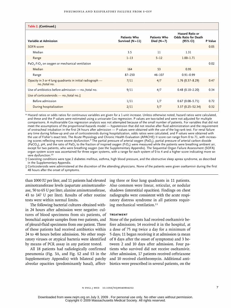
Table 2. Survival and Death among the 18 Study Patients Who Had Confirmed Infection with Novel Swine-Origin Influenza A (H1N1) Virus.*
Variable at AdmissionPatients Who
Survived (N = 11)Patients Who Died (N = 7)
Hazard Ratio or Odds Ratio for Death
(95% CI) P Value
Age — yr 0.81
Median 37 45 1.00
Range 0.75–61 9–52 0.96–1.04
Male sex — no./total no. 4/11 5/7 2.2 (0.4–11.5) 0.34
Hypotension that did not resolve after fluid administration — no./total no.
2/11 7/7 0.02
Orotracheal intubation required within first 24 hr after admission — no./total no.
3/11 7/7 0.06
Renal failure any time during follow-up — no./total no. 1/11 5/7 25 (1.3–1295) 0.01
Coexisting condition — no./total no.† 5/11 3/7 0.78 (0.2–3.6) 0.75
Days from illness onset to admission 0.20
Median 6 6 0.71
Range 4–13 5–8 0.4–1.2
Lactate dehydrogenase — U/liter 0.40
Median 1086 2032 1.00
Range 594–2429 690–3871 0.99–1.01
Creatine kinase — U/liter 0.45
Median 189 514 1.00
Range 58–1249 175–2156 0.99–1.01
Lymphocyte count per mm3 0.25
Median 1000 400 0.99
Range 500–3700 200–1300 0.98–1.00
PaO2 — mm Hg 0.44
Median 55.4 41.5 0.96
Range 33.7–70.1 39.0–51.0 0.89–1.04
PaCO2 — mm Hg 0.91
Median 28.7 36 0.99
Range 23.7–61.7 15–66 0.94–1.05
pH 0.02
Median 7.42 7.35 3.19×10⁻¹²
Range 7.38–7.93 7.19–7.43 3.43×10⁻²²−0.29
Initial PaO2:FiO2 0.71
Median 231 197 0.99
Range 22–334 186–243 0.98–1.01
Acute respiratory distress syndrome — no./total no. 3/10 4/7 1.18 (0.26–5.33) 0.82
APACHE II score 0.02
Median 11 19 1.20
Range 4–20 14–32 1.03–1.40
Copyright © 2009 Massachusetts Medical Society. All rights reserved. Downloaded from www.nejm.org on July 3, 2009 . For personal use only. No other uses without permission.
pneumonia and Respir atory Failure from S-OIV
n engl j med 10.1056/nejmoa0904252 7
than 1000 IU per liter, and 11 patients had elevated aminotransferase levels (aspartate aminotransfer-ase, 50 to 65 U per liter; alanine aminotransferase, 43 to 147 U per liter). Results of other routine tests were within normal limits.
The following bacterial cultures obtained with-in 24 hours after admission were negative: cul-tures of blood specimens from six patients, of bronchial aspirate samples from two patients, and of pleural-fluid specimens from one patient. Three of these patients had received antibiotics within 24 to 48 hours before admission. No other respi-ratory viruses or atypical bacteria were identified by means of PCR assay in any patient tested.
All 18 patients had radiologically confirmed pneumonia (Fig. 3A, and Fig. S2 and S3 in the Supplementary Appendix) with bilateral patchy alveolar opacities (predominantly basal), affect-
ing three or four lung quadrants in 11 patients. Also common were linear, reticular, or nodular shadows (interstitial opacities). Findings on chest radiographs were consistent with the acute respi-ratory distress syndrome in all patients requir-ing mechanical ventilation.11
Treatment
None of the patients had received oseltamivir be-fore admission; 14 received it in the hospital, at a dose of 75 mg twice a day for a minimum of 5 days; 11 began receiving it at admission (a mean of 8 days after the onset of symptoms) and 3 be-tween 2 and 10 days after admission. Four pa-tients who survived did not receive oseltamivir. After admission, 17 patients received ceftriaxone and 10 received clarithromycin. Additional anti-biotics were prescribed in several patients, on the
Table 2. (Continued.)
Variable at AdmissionPatients Who
Survived (N = 11)Patients Who Died (N = 7)
Hazard Ratio or Odds Ratio for Death
(95% CI) P Value
SOFA score 0.05
Median 3.5 11 1.31
Range 1–13 5–12 1.00–1.71
PaO2:FiO2 on oxygen or mechanical ventilation 0.02
Median 164 53 0.95
Range 87–250 46–107 0.91–0.99
Opacity in 3 or 4 lung quadrants in initial radiograph — no./total no.
7/11 4/7 1.76 (0.37–8.29) 0.47
Use of antibiotics before admission — no./total no. 9/11 4/7 0.48 (0.10–2.20) 0.34
Use of corticosteroids — no./total no.‡
Before admission 1/11 1/7 0.67 (0.08–5.71) 0.72
During hospitalization 2/11 3/7 3.37 (0.25–52.34) 0.32
* Hazard ratios or odds ratios for continuous variables are given for a 1-unit increase. Unless otherwise noted, hazard ratios were calculated, and these and the P values were estimated using a univariate Cox regression. P values are two-tailed and were not adjusted for multiple comparisons. A multivariable Cox regression analysis was not attempted because of the small number of patients. For variables that did not meet the assumptions of the proportional-hazards model — hypotension that did not resolve after fluid administration and the requirement of orotracheal intubation in the first 24 hours after admission — P values were obtained with the use of the log-rank test. For renal failure any time during follow-up and use of corticosteroids during hospitalization, odds ratios were calculated, and P values were obtained with the use of Fisher’s exact test. The Acute Physiology and Chronic Health Evaluation (APACHE) II score can range from 0 to 71, with increas-ing scores reflecting more severe dysfunction.9 The partial pressure of arterial oxygen (PaO2), partial pressure of arterial carbon dioxide (PaCO2), pH, and the ratio of PaO2 to the fraction of inspired oxygen (FiO2) were measured while the patients were breathing ambient air, except for two patients, who were breathing oxygen (see the Supplementary Appendix). The Sequential Organ Failure Assessment (SOFA) organ system score was ascertained for three organ systems, with a range for each system of 0 to 4 and a higher score indicating more se-vere dysfunction.10
† Coexisting conditions were type 2 diabetes mellitus, asthma, high blood pressure, and the obstructive sleep apnea syndrome, as described in the Supplementary Appendix.
‡ Corticosteroids were administered at the discretion of the attending physicians. None of the patients were given oseltamivir during the first 48 hours after the onset of symptoms.
Copyright © 2009 Massachusetts Medical Society. All rights reserved. Downloaded from www.nejm.org on July 3, 2009 . For personal use only. No other uses without permission.

T h e n e w e ngl a nd j o u r na l o f m e dic i n e
n engl j med 10.1056/nejmoa09042528
basis of their clinical course: three were given levofloxacin; seven, vancomycin; five, cefepime; five, imipenem; and two, dicloxacillin.
Clinical Course during Hospital Stay
Respiratory distress requiring intubation and me-chanical ventilation developed in 10 patients with-in the first 24 hours after admission. These pa-tients had a median oxygen saturation of 71% (interquartile range, 64 to 77) in the absence of supplementary oxygen (2240 m above sea level), and treatment of eight patients involved positive end-expiratory pressure at or above 16 cm of water. Two additional patients required mechan-ical ventilation during their stay in the hospital (Table S2 in the Supplementary Appendix). Dura-
tion of mechanical ventilation ranged from 7 to 30 days in patients who survived and from 4 to 17 days in patients who died. Norepinephrine infu-sion was begun in 9 of 18 patients (50%) during the period of hospitalization, and 5 patients re-ceived corticosteroids (hydrocortisone at a dose of 300 mg per day or methylprednisolone at a dose of 60 mg per day). Of the six patients in whom renal failure developed, five died. Seven patients had multiorgan system failure. None of the pa-tients had disseminated intravascular coagulation or neurologic complications. Four patients had ventilator-associated pneumonia, each case with a different cause: Acinetobacter baumannii, Achro-mobacter xylosoxidans, methicillin-resistant Staphy-lococcus aureus, or Escherichia coli.
Of the 18 patients, 7 died, and 11 recovered and were discharged from the hospital. Patients died within 10 to 23 days (mean, 14) after the onset of illness and between 4 and 18 days (mean, 9 days) after admission. Figure 3B shows a lung-tissue specimen from the autopsy of Patient 3, a 43-year-old patient who died after a 14-day ill-ness complicated by acute renal failure and myo-cardial infarction. Pathological evaluation of the lung showed diffuse alveolar damage, thick hya-line membranes, and prominent fibroblast pro-liferation.
Patients with confirmed S-OIV infection had more severe disease, including a higher death rate (Table S2 in the Supplementary Appendix), than did hospitalized patients with negative test results. The median time from illness onset to collection of samples for viral testing was 9 days (range, 3 to 46) among the 18 patients who were positive for S-OIV infection and 10 days (range, 3 to 27) among those with negative test results (P = 0.50).
Clinical Infection in Contacts and Health Care Workers
Patients identified a total of 82 family contacts, 20 of whom had respiratory symptoms within a week after the patient was hospitalized. Of the 20, 4 required hospitalization, including 3 con-tacts of Patient 6; 1 contact who had Down’s syn-drome died in another hospital from respiratory failure. Patient 2, in whom severe respiratory fail-ure developed, is the mother of Patient 9, who had milder disease and received early treatment with oseltamivir.
Influenza-like illness or respiratory symptoms developed in 22 of 190 health care workers car-
16p6
AUTHOR Perez-Padilla
FIGURE 3a&b
JOB: ISSUE:
4-CH/T
RETAKE 1st
2nd
SIZE
ICM
CASE
EMail LineH/TCombo
Revised
AUTHOR, PLEASE NOTE: Figure has been redrawn and type has been reset.
Please check carefully.
REG F
FILL
TITLE3rd
Enon ARTIST:
8-13-09
mst
36107
A
B
L
Figure 3. Initial Radiograph of the Lung and Lung- Tissue Sample from Patient 3.
The radiograph (Panel A) shows bilateral alveolar opac-ities in the base of both lungs that progressed and be-came confluent. The specimen (Panel B, hematoxylin and eosin) shows necrosis of bronchiolar walls (top arrow), a neutrophilic infiltrate (middle arrow), and diffuse alveolar damage with prominent hyaline membranes (bottom arrow). Bacterial cultures were negative on admission, and no evidence of bacterial infection of the lungs was found. The patient ultimately died.
Copyright © 2009 Massachusetts Medical Society. All rights reserved. Downloaded from www.nejm.org on July 3, 2009 . For personal use only. No other uses without permission.

pneumonia and Respir atory Failure from S-OIV
n engl j med 10.1056/nejmoa0904252 9
ing for the first three patients with confirmed S-OIV infection in the emergency room and the intensive care unit, including 19 of 104 workers who were within 2 m of a patient or had direct contact. These 22 workers received oseltamivir for 5 days and were sent home for 3 to 7 days. They had mild-to-moderate disease, and none re-quired hospitalization. Three of the 22 workers, who were nurses in the emergency room, had nasopharyngeal-aspirate samples that were posi-tive for S-OIV. After infection-control measures were strictly enforced — with patients confined and isolated in three hospital areas and N95 respirators used in addition to goggles, gowns, and gloves, as well as liberal use of gel-alcohol hand sanitizer — no more health care workers had influenza-like illness, although 26 additional workers received oseltamivir for 5 days because of varied respiratory symptoms.
Discussion
This case series of the first 18 patients hospital-ized in Mexico City with S-OIV infection docu-ments the clinical findings of severe illness or death associated with S-OIV infection that were seen during the beginning of the S-OIV pandemic. The patients, most of them previously healthy, had an influenza-like illness that progressed dur-ing a period of 5 to 7 days, had pneumonia, and had findings during the first day of hospital ad-mission that fulfilled the criteria of acute lung injury or the acute respiratory distress syndrome.12 Seven patients died, all from multiorgan system failure. The most consistent laboratory character-istics were increased lactate dehydrogenase level, a total leukocyte count within normal limits, lymphopenia,13 and increased creatine kinase level, most likely due to myositis (or myocardial ischemia, in one patient).
The patients described were part of an epi-demic of influenza-like illness with pneumonia seen at our institution and other Mexican hospi-tals, and only a fraction of them tested positive for S-OIV. A false negative test in patients who had infection with S-OIV would be more likely if the test were delayed or if patients had limited viral shedding. In general, patients who tested negative for S-OIV had a milder clinical course than those who tested positive but were as much a part of the burden of the epidemic as those who were not tested.
Risk factors for severe S-OIV illness are still unknown, but most of our patients were young to middle-aged and had previously been healthy. The majority of the S-OIV infections reported in other countries have been mild, influenza-like illnesses.2 Mexico has also reported a large num-ber of persons with mild disease, through the national surveillance system for influenza, but the full spectrum of clinical illness has not been determined. Other countries will probably report more severe infections as the pandemic spreads and the number of infected persons increases. One contributing factor for death in our patients may have been delayed admission and delayed initiation of oseltamivir. For seasonal influenza, the elderly and young children are at higher risk for severe disease; however, more than half of our patients were between 13 and 47 years of age, which was similar to the age distribution reported in national data of H1N1 infections in Mexico.6 During the 1918 pandemic, a large number of deaths were associated with bacterial infection,14 but concurrent bacterial infection does not ap-pear to be a major contributing factor to the se-verity of illness in our patients, possibly in part because most received antibiotics before hospi-talization.
Mortality among the patients requiring me-chanical ventilation was 58%, and although four patients had nosocomial pneumonia, in most of our patients, lung damage was most likely due to the primary effect of infection with influenza vi-rus. Possible mechanisms of damage include di-rect injury to the respiratory epithelium15 with a secondary cytokine storm. We do not currently know whether our patients, especially those who died, had viremia, as was reported in association with H5N1 infection, a very aggressive variety of influenza.13,16-18 Coinfection with other respira-tory viruses could also explain the increased pathogenicity among our patients19,20; however, no other common respiratory viruses were found in our patients. Only three of the patients had re-ceived influenza vaccine in fall 2009, since most patients were within the age groups for which vaccine was not recommended in Mexico. It is currently unknown whether seasonal vaccination offered any protection against S-OIV infection, however. We did not find a factor that, before the onset of illness, predicted a worse outcome or death among our patients.
Since 2000, the WHO has prompted countries
Copyright © 2009 Massachusetts Medical Society. All rights reserved. Downloaded from www.nejm.org on July 3, 2009 . For personal use only. No other uses without permission.

n engl j med 10.1056/nejmoa090425210
pneumonia and Respir atory Failure from S-OIV
to prepare for a potential influenza pandemic. In Mexico, pandemic influenza planning began in 2001. Activities included the introduction of year-ly influenza vaccination and a program to develop the country’s national vaccine production. In 2006, a strategic reserve of oseltamivir, antibiotics, and protective items for health care personnel was established. This reserve is the source of the oseltamivir prescribed to our patients and to most hospitalized patients in Mexico. The experience in our institution highlights the need to reinforce precautions and use of personal protective equip-ment to prevent the infection of health care workers.
In conclusion, S-OIV infection can cause seri-ous illness and death in young, previously healthy persons. Future studies should identify predictive factors for severe disease and, especially, the ef-fectiveness of early oseltamivir treatment and protection offered by having undergone seasonal influenza vaccination.
No potential conflict of interest relevant to this article was reported.
We thank Celia Alpuche, M.D., from the Mexican national reference laboratory (Instituto Nacional de Referencia Epidemi-ologica); the CDC for providing training and primers for the real-time RT-PCR assay for the swine influenza; the Canadian National Microbiology Laboratory; Michelle Weinberg for care-ful review of a draft of the manuscript; and all the patients and the personnel of INER who cared for them.
AppendixMembers of the INER Working Group on Influenza were the following: J. Rodriguez, M. Barrales, L. Reveles, M.A. Sanchez-Mecatl, D. Ibarra, L.E. Leon, T. Gutierrez, D. Rodriguez-Parga (Emergency Room); C. Aguilar, F. Rocha, I. Serna, V. Huizar, B. Castañeda, J.G. Garcia (Intensive Care); J.L. Sandoval, R. Hernandez, F. Flores, L. Amaya, C. Espinoza (Influenza Ward); E. Sada, G. Reyes-Teran, J. Romo (Infectious Diseases); A. Osorio, E. Sevilla (Hospital Epidemiology); A. Alejandre (Pediatric Pulmonology); S. Rodriguez-Filigra-na, J. Chavarria (Medical Direction); R. Fernandez, C. Garcia-Sancho, F. Franco, L. Alfaro-Ramos, J. Godinez-Zavala, L. Martinez (Epi-demiology); L. Damian (Nursing); R. Garcia-Torrentera (Respiratory Care); R. Sotelo, L.F. Alba (Chest Imaging); M.E. Vazquez, E. Peña (Pathology); M. Selman-Lama, M.E. Manjarrez, J. Zuñiga (Research Unit); A. Diaz, C. Mora, F. Fernandez, F. de Jesus Contreras, J.P. Villaloz, S. Romero (postgraduate students and residents).
References
Swine influenza A (H1N1) infection 1. in two children — Southern California, March–April 2009. MMWR Morb Mortal Wkly Rep 2009;58:400-2.
Update: infections with a swine-ori-2. gin influenza A (H1N1) virus — United States and other countries, April 28, 2009. MMWR Morb Mortal Wkly Rep 2009;58: 431-3.
Update: swine influenza A (H1N1) in-3. fections — California and Texas, April 2009. MMWR Morb Mortal Wkly Rep 2009; 58:435-7.
Novel Swine-Origin Influenza A (H1N1) 4. Virus Investigation Team. Emergence of a novel swine-origin influenza A (H1N1) virus in humans. N Engl J Med 2009; 360:2605-15.
Influenza A (H1N1) — update 14. Ge-5. neva: World Health Organization, 2009. (Ac cessed June 23, 2009, at http://www.who.int/csr/don/2009_05_04a/en/index.html.)
Brote de Influenza Humana a H1N1 6. México Dirección General Adjunta de Epi-demiología. Dirección General Adjunta de Epidemiología, 2009. (Accessed June 23, 2009, at http://portal.salud.gob.mx/ contenidos/noticias/influenza/estadisticas.html.)
CDC protocol of realtime RTPCR for 7.
inf luenza A (H1N1). Geneva: World Health Organization, April 2009. (Accessed June 23, 2009, at http://www.who.int/ csr/resources/publications/swinef lu/ CDCRealtimeRTPCR_SwineH1Assay-2009_ 20090430.pdf.)
Stata statistical software: release 9.0. 8. College Station, TX: StataCorp LP, 2005.
Knaus WA, Draper EA, Wagner DP, 9. Zimmerman JE. APACHE II: a severity of disease classification system. Crit Care Med 1985;13:818-29.
Vincent JL, Moreno R, Takala J, et al. 10. The SOFA (Sepsis-related Organ Failure As-sessment) score to describe organ dysfunc-tion/failure. Intensive Care Med 1996;22: 707-10.
Qureshi NR, Hien TT, Farrar J, Gleeson 11. FV. The radiologic manifestations of H5N1 avian influenza. J Thorac Imaging 2006; 21:259-64.
Murray JF, Matthay MA, Luce JM, Flick 12. MR. An expanded definition of the adult respiratory distress syndrome. Am Rev Respir Dis 1988;138:720-3. [Erratum, Am Rev Respir Dis 1989;139:1065.]
Hui DS. Review of clinical symptoms 13. and spectrum in humans with influenza A/H5N1 infection. Respirology 2008;13: Suppl 1:S10-S13.
Morens DM, Taubenberger JK, Fauci 14.
AS. Predominant role of bacterial pneu-monia as a cause of death in pandemic influenza: implications for pandemic in-fluenza preparedness. J Infect Dis 2008; 198:962-70.
Ng WF, To KF, Lam WW, Ng TK, Lee 15. KC. The comparative pathology of severe acute respiratory syndrome and avian in-fluenza A subtype H5N1 — a review. Hum Pathol 2006;37:381-90.
Yu H, Gao Z, Feng Z, et al. Clinical 16. characteristics of 26 human cases of high-ly pathogenic avian influenza A (H5N1) virus infection in China. PLoS One 2008; 3(8):e2985.
Wong SS, Yuen KY. Avian influenza 17. virus infections in humans. Chest 2006; 129:156-68.
de Jong MD, Hien TT. Avian influenza 18. A (H5N1). J Clin Virol 2006;35:2-13.
Merler S, Poletti P, Ajelli M, Caprile B, 19. Manfredi P. Coinfection can trigger mul-tiple pandemic waves. J Theor Biol 2008; 254:499-507.
Aberle JH, Aberle SW, Pracher E, Hutter 20. HP, Kundi M, Popow-Kraupp T. Single ver-sus dual respiratory virus infections in hos-pitalized infants: impact on clinical course of disease and interferon-gamma response. Pediatr Infect Dis J 2005;24:605-10.Copyright © 2009 Massachusetts Medical Society.
Copyright © 2009 Massachusetts Medical Society. All rights reserved. Downloaded from www.nejm.org on July 3, 2009 . For personal use only. No other uses without permission.