IMAGE SEGMENTATION BY USING K-MEANS CLUSTERING ALGORITHM FOR PIXEL CLUSTERING
13
IMAGE SEGMENTATION BY USING K-MEANS CLUSTERING ALGORITHM Ramaraj.M *1 , Dr.Antony Selvadoss Thanamani* 2 1 Research Scholar Department of Computer Science NGM College Pollachi India [email protected] . 2 Associate Professor & Head Department of Computer Science NGM College Pollachi India [email protected] Abstract In this paper, we discuss above the medical image segmentation and also comparative study has been made between the conventional k-MEANS and fuzzy c means algorithm. Image segmentation is the first step in image analysis. Segmentation is the process which partitions an image into its constituent regions or objects. The main aim of the image segmentation is the process of analyze and easier to identify the pixel clustering of the image as helpful for the MRI brain images. Segmentation is the process of images which divided into multiple parts and typically used for relevant objects or other relevant object in the digital image for the MRI scan images. Image segmentation is the various or other different application are widely used for the medical images in order to the diagnose disease. An efficient clustering algorithm is used for the medical image for the k-means algorithm in image segmentation.
-
Upload
independent -
Category
Documents
-
view
0 -
download
0
Transcript of IMAGE SEGMENTATION BY USING K-MEANS CLUSTERING ALGORITHM FOR PIXEL CLUSTERING
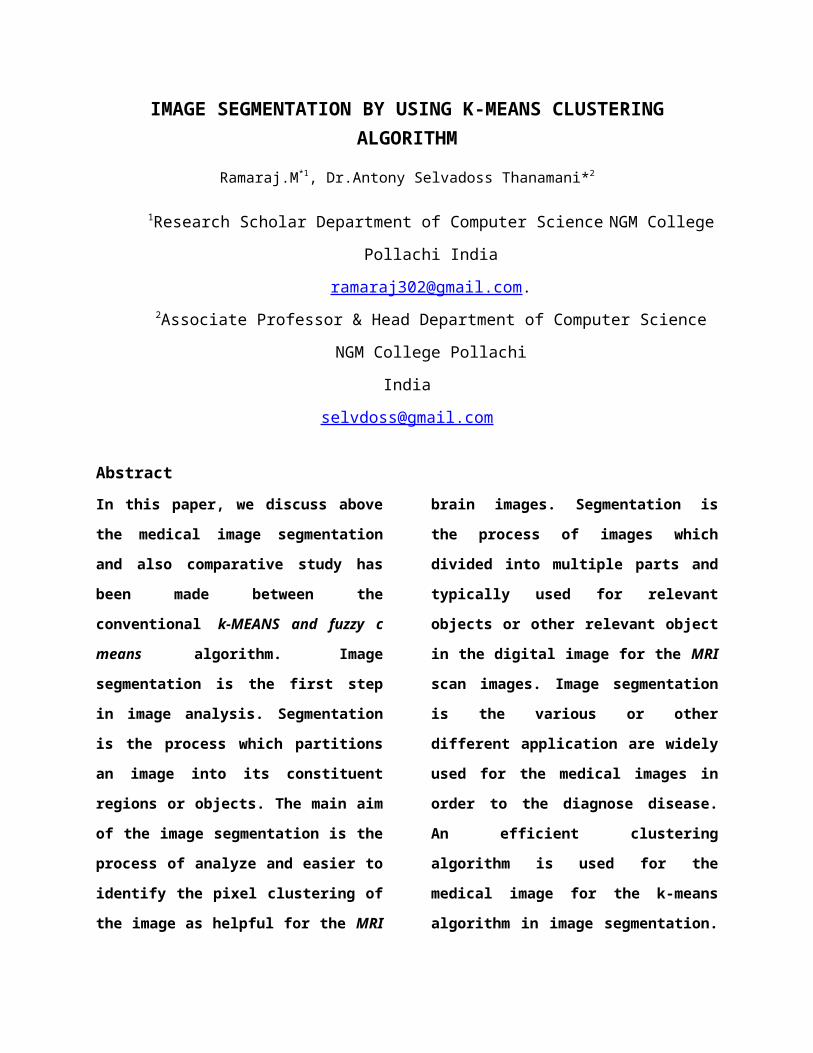
IMAGE SEGMENTATION BY USING K-MEANS CLUSTERINGALGORITHM
Ramaraj.M*1, Dr.Antony Selvadoss Thanamani*2
1Research Scholar Department of Computer Science NGM College
Pollachi India
[email protected] Professor & Head Department of Computer Science
NGM College Pollachi
India
Abstract In this paper, we discuss above
the medical image segmentation
and also comparative study has
been made between the
conventional k-MEANS and fuzzy c
means algorithm. Image
segmentation is the first step
in image analysis. Segmentation
is the process which partitions
an image into its constituent
regions or objects. The main aim
of the image segmentation is the
process of analyze and easier to
identify the pixel clustering of
the image as helpful for the MRI
brain images. Segmentation is
the process of images which
divided into multiple parts and
typically used for relevant
objects or other relevant object
in the digital image for the MRI
scan images. Image segmentation
is the various or other
different application are widely
used for the medical images in
order to the diagnose disease.
An efficient clustering
algorithm is used for the
medical image for the k-means
algorithm in image segmentation.
Medical image segmentation had
been a vital organ of research as
it inherited complex problems
for the proper diagnosis of
incomplete developed mind. In
this research, it provides a
foundation of segmentation and
find out the tumor and
clustering the tumor in the
brain tissue.
Key words: Clustering, K-Means
clustering, Fuzzy c-Means
Clustering, brain tumor, Image
Segmentation.
I. INTRODUCTION Clustering is a method of
grouping data objects into
different groups, such that
similar data objects belong to
the same group and dissimilar
data objects to different
clusters. Current research
increasing interest in digital
image searching, classification,
identification, management and
storage. Some common but
important applications of are
person identification in movie
clips and festive home videos,
recognition in biometric system,
natural scene classification for
robot vision, commercials
filtering, segmentation of
important topics in lectures and
meetings. The image clustering,
an important technology for
image processing, has been
actively researched for a long
period of time and explosive
growth of the Web, image
clustering has even been a
critical technology to help
users digest the large amount of
online visual information.
Clustering approach is widely
used in biomedical image
segmentation and it is
application are used for brain
tumor detection as the normal
and abnormal to find out the
tumor on the brain [4].
The many different segmentation
techniques are used in the image
mining and image segmentation
approaches can be divided into
many parts as: clustering, edge
detection, thresholding and
region extraction. In this
paper, we discuss above the
clustering concept in the image
mining on the image segmentation
process of the clustering and
each object can be have its
place of more than one clusters
to be provisional upon the
degree of relationship
association on it.
Many of things and real
situation, like image,
restricting an object into only
one cluster will be take on the
different tasks.
A technique which is handles the
mining of information,
association of the image data or
additional patterns not
unambiguously stored in the
image data base of the image
segmentation process [11]. The
methods are handling with the
image processing, image stored
and retrieval, data mining,
machine learning, database on
the image to be stored and
artificial intelligence. A huge
amount of image databases have
being implements with as rule
mining concept. It handles with
several features of very huge
image databases which comprises
of indexing methods, image
storages, and image retrieval,
all regarding in an image mining
system.
Image mining is to considerable
patterns without any information
of the image content, and
patterns types are different: it
is classification patterns,
correlation patterns, temporal
patterns and spatial patterns
for the main intention of the
image mining techniques. In this
techniques are implemented with
the good quality of image mining
system provides the end users
with a useful to access the
image on the databases to be
storage area at the time it
recognizing the patterns to be
generates with under the
knowledge of this image
representation. Image

segmentation is the primary
phase in image mining. In other
words, image mining is simply an
expansion of data mining in the
field of image processing. Image
mining handles with the hidden
knowledge extraction, image data
association and additional
patterns which are not clearly
accumulated in the images.
II. RELATED WORK
Clustering is a process of
partitioning or grouping of the
dataset and to a given sector
unlabeled pattern into a number
of clusters in the K-means
clustering and application in
color image segmentation, such
that similar patterns is
assigning to be a group, which
is consideration for a
cluster[9]. It is a brain tumor
segmentation using Fuzzy c-means
clustering to tumor region for
the manual segmentation of brain
tumor from MRI images [2].
Imaging techniques to establish
stage of diagnosis and follow
patients with brain tumors are
central to the clinical
management. MRI is the most
commonly used technique for
injury finding in the definition
of extent detection of spread
and in evaluation of either
residual or recurrent
disease[6]. The existing tumor
detection methods broadly
classified into
Three categories:
Atlas Based Segmentation
Segmentation in MRI images
Segmentation of Brain Tumor
In another definition of the
brain tumor in a growth of
abnormal cells in the tissues of
the brain. Brain tumor can be
begin (not cancer) or malignant
(cancer).
Brain Cancer Statistics:
Every year about 1400 malignant
brain tumors are diagnosed in
Australia and about 100 of these
are in children data about
benign brain tumors are not

collected but an estimated 2000
people and including children on
affected into the every year.
Fig 1: black diagram for
identification of the brain
tumor using image segmentation
method
Fig 1: it’s used for the black
diagram is a brain tumor
identification and the
algorithms of segmenting the
image and using with the k-
medoids (as for the k-means)
algorithms are implemented.
In this study, a new
approach has been discussing of
brain tumor the image, by
applying K-medoids algorithm and
Fuzzy c-means algorithm for
comparison of this algorithms
and its more effective
classification accuracy to find
out the any one of the
clustering algorithm for using
the image [3]. The tumor is
calculated to statistical
parameter are evaluated and
clustering the tumor.
III. METHODOLOGY
K-Means Clustering:
It’s the on the techniques for
the clustering concept in the
data mining process and is very
famous algorithm for the K-means
clustering, because it is
similar or simpler and easier in
computation of an efficient K-
means clustering algorithm it is
the simplest unsupervised
learning algorithms that solve
the well known clustering
problems[12]. It’s the K-means
algorithm is an unsupervised
clustering algorithm that
classified in the input data
points into multiple classes
based on their intrinsic
distance from other dataset
points of his cluster [7].
Image Databases
Pre-processing
Brain Tumor
NomalTissue
Training And Testing Datasets And Both Trained
Segmentation Of The Tumor Using K-medoids
Feature Extraction

Its assume that the
data features from a vector
space and tries to find natural
clustering.
Hierarchical Clustering:
Hierarchical clustering is
any valid measure of distance
can be used for a traditional
clustering method that can be
generates a tree structure or a
dendrograms and it’s a matrix of
distance.
The two types of approaches are:
Bottom- up approaches
To- down approaches
The hierarchical clustering
algorithms are classified into
two stages are given as
Agglomerative hierarchical
clustering
K-means algorithms
Where x, y is a set of two
elements in the cluster and
d(x,y) denote the min-distance
between the two elements of x
and x.
Complete linkage clustering
Its calculate the maximum
distance between the large
set of clusters. Formula as
d(x,y)= maxx∈X,y∈Y
d(x,y) …..
(1)
where x, y is a set of two
elements in the cluster and d(x,
y) denote the max-distance
between the two elements of x
and x.
Its calculate the minimum
distance between the large set
of clusters. Formula as
d(x,y)= minx∈X,y∈Y
d(x,y) …..
(2)
where x, y is a set of two
elements in the cluster and d(x,
y) denote the min-distance
between the two elements of x
and x.
dis (x,y )=∑i=1
d
‖xi−ci‖ ……….. (3)
Presently, the feature vectors
are grouped into k means
clusters using a selected
distance measure such as
Euclidean distance may be
calculated.
Image Segmentation:
Segmenting a MRI images into
homogeneous texture regions
representing disparate tissue
types is often a useful
preprocessing step in the
computer-assisted detection of
brain tumor[11]. That is why we
proposed new algorithm to detect
cancer in mammogram breast
cancer images. The best approach
to image segmentation may vary
between different
applications[1].
The choice between manual,
semiautomatic or fully automatic
methods depends on the image
quality and the number of
objects needs to be matmetric in
the amount of accessible user
time, and the required
classification accuracy of the
images. The segmentation process
is usually based on gray
intensity, color, shape or
texture.
Texture can be
characterized by local
variations of pixel values that
repeat in a regular or random
pattern on the entity or
image[8]. It can also be defined
as a repetitive arrangement of
patterns over a region. A wide
variety of texture segmentation
techniques have been reported in
the literature [13]. We decided
to choose a set of existing
texture features which can
provide us good discriminating
power and are easy to compute as
compare to Segmentation using
gray level co-occurrence matrix
required huge time for tumor
demarcation with less accuracy
[5].
Segmentation by Gray level co-
occurrence matrix basic
watershed algorithm and
LindeBuzo Gray algorithm (LBG)
which are used for comparative
performance of tumor detection.
There are several types of the
segmentation algorithms as
follow
Gray level co-occurrence
matrix.
Watershed algorithm.

K-Means Algorithms:
The principle of the k-
medoids algorithm (its same for
the k-means algorithm) for
cluster the data and k-medoids
algorithm is the one of the best
unsupervised learning clustering
algorithm [10].
Clustering the image is
according to the group of pixels
and the k-medoids algorithm is
initially we have to a number of
cluster k, then k-cluster center
are chosen randomly. The
distance between the each pixel
to each cluster centers are
calculated. The distance may be
of simple Euclidean function.
Single pixel is compared to all
cluster centers using the
distance formula.
∑c (j)=k∨|Xj−Xi|∨22 ……………..
(4)
To calculate the number
clustering in the equation of
the k is medoids point of
cluster.
∑k=1
k
∑C(i)=k
||Xi−Ck||22 …………… (5)
Minimum cover of C
For each i=1….n, to find the
cluster center of ck closest to
xi,
and let c(i)=k
k-medoids algorithm is generate
and returns the higher value is
calculated as
Algorithm:
1. Give the no of cluster value
as k.
2. Randomly choose the k cluster
centers
3. Calculate mean or center of
the cluster
4. Calculate the distance
between each pixel to each
cluster center
5. If the distance is near to
the center then move to that
cluster.
6. Otherwise move to next
cluster.
7. Re-estimate the center.

Fuzzy C Means
Clustering, a major area of
study in the scope of
unsupervised learning, deals
with recognizing meaningful
groups of similar items. Among
all clustering algorithms, the
Fuzzy C-Means (FCM) clustering
algorithm
FCM calculates membership
values for each data point to
all clusters it usually
generates a more consistent
and trust-worthy results. We
applied the FCM into our image
segmentation project to
develop a framework for the
multi-level FCM clustering
Jm=∑i=1
N
∑j=1
Cuij⃦ ⃦m ‖xi−cj❑‖❑2.1≤m<∞....
................................
..............................
6 uy=
1
∑k−1
c¿¿¿
……………... 7
where m, the fuzzification factor, is any
real number greater than 1, uij is
the degree of membership of xi in
the cluster j, xi is the ith of d-
dimensional measured data, cj is
the d-dimension center of the
cluster, and ||*|| is any norm
expressing the similarity
between any measured data and
the center. Fuzzy partitioning
is carried out through an
iterative optimization of the
objective function shown above,
with the update of membership uij
and the cluster centers cj.
IV. EXPREMENTAL RESULTS
MATLAB stands for matrix
laboratory. It’s also a
software program that allows us
to do data manipulation and
visualization, image analysis,
calculations, math and
programming. It can be used to
do very simple as well as very
sophisticated tasks. MATLAB is a
high-performance language for
technical computing. MATLAB is
an interactive system whose

basic data element is an array
that does not require
dimensioning. MATLAB can
import/export several image
formats such as BMP (Microsoft
Windows Bitmap), GIF (Graphics
Interchange Files), HDF
(Hierarchical Data Format), JPEG
(Joint Photographic Experts
Group), PCX (Paintbrush), PNG
(Portable Network Graphics),
TIFF (Tagged Image File Format),
XWD (X Window Dump). MATLAB can
also load raw-data or other
types of image data.
Figure 2: Flowchart represented
by the cluster algorithms to be
implemented with images to
produce the result.
Original image
Fig1: The original image on the
brain
Tumor image on brain
Fig 2&3: Tumor image on brain and
highlight the tumor on affected in
the area of brain.

Fig 3: to cluster the k means
cluster for tumor image
Fig 4: it represent the original
image on the human brain
V. CONCLUSION:
In this paper
described a large scale soft
clustering algorithm that relies
on topic segmentation and
clustering the brain tissue. We
have developed an automatic
image based method to detect
tumor in 2D MRI head scans. The
classification accuracy on image
in 93.74%. In the proposed
method an efficient detection of
brain tumor region from cerebral
image is done using k-medoids
clustering and Fuzzy c means
clustering to be compared with
this algorithm is more effective
and efficient in the extract on
tumor from brain and helpful for
the MRI brain images. In the
future work we can increase
accuracy on the images.
REFERENCES:
[1]
A.R.Kavitha,Dr.C.Chellamuthu,
Ms.Kavin Rupa, “An Efficient
Approach for Brain Tumour
Detection Based on Modified
Region Growing and Network in
MRIImages,”IEEE, 2012.
[2] Wen-Liange, De-Hua Chen,
Mii-shen Yang, “Suppressed
fuzzy-soft learning vector
quantization for MRI
segmentation,”Elsevier ltd,
2011.
[3] Vida Harati, Rasoul
Khayati, Abdolreza Farzan,
“Fully automated tumor
segmentation based on improved
fuzzy connectedness algorithm in
brain MR images,”Elsevier ltd,
2011.

[4] R.B.Dubey, M.Hanmandlu,
Sr.Member, Shantaram Vasikarla,
“Evaluation of ThreeMethods for
MRI Brain Tumor segmentation,”
IEEE, 2011.
[5] Shaheen Ahmed, Khan
M.Iftekharuddin, “Efficacy of
Texture,Shape,and Intensity
Feature Fusion for Posterior-
Foss Tumor Segmentation in
MRI,”IEEE,2011.
[6] T.Logeswari, M.Karnan,
“Hybrid Self Organizing Map for
improved Implementation of Brain
MRI Segmentation,”IEEE, 2010.
[7] P. Dhanalakshmi & T.
Kanimozhi “
“Automated Segmentation of Brain Tumor using K-MeansClustering and its Area Calculation”IJAREEE” 2013
[8] Jayashri Joshi1,
Mrs.A.C.Phadke, “A New Approach
to Image Segmentation for Brain
Tumor detection using Pillar K-
means Algorithm” IJARCCE,vol
3,issue 3, March 2013.
[9] Oyelade, O. J, Oladipupo, O.
O, Obagbuwa, I. C “Application
of k-Means Clustering algorithm
for prediction of Students’
Academic Performance” International
Journal of Computer Science and
Information Security, vol. 7, pp.292-295,
2010.
[10] Meghana Nagori, Shivaji
Mutkule, Praful Sonarkar
“Detection of Brain Tumor by
Mining fMRI Images”IJARCCE,vol
2,issue 4,January 2013.
[11] Hakeem Aejaz Aslam,
Tirumala Ramashri, Mohammed
Imtiaz Ali Ahsan “A New Approach
to Image Segmentation for Brain
Tumor detection using Pillar K-
means Algorithm” IJARCCE,vol
2,issue 3, March 2013.
[12]
Google:http://sites.google.com/s
ite/
dataclusteringalgorithms/k-mean-
clustering-algorithm.

[13] D. Klein, S.D. Kamvar, and
C. Manning, “Feature Extraction
and Texture Classification in
MRI”, International Conference
[ICCT-2010], 3rd-5th December
2010