Branch Transformation Solution NBS Application Rel. 2.xx.xx ...
ORIGINAL ARTICLE
IKKb--I-kBe--c-Rel/p50: a new axis of NF-kB activationin lung epithelial cellsPC Maity, T Ray, B Das and AK Sil
Cigarette smoke (CS), a major risk factor for developing lung cancer, is known to activate transcriptional activator nuclear factorkappa B (NF-kB). However, the underlying mechanism of this activation remains unclear because of conflicting reports. AsNF-kB has a pivotal role in the generation and maintenance of malignancies, efforts were targeted towards understanding itsactivation mechanism using both ex vivo and in vivo studies. The results show that CS-induced NF-kB activation mechanism isdifferent from that of other pro-inflammatory signals such as lipopolysaccharide (LPS). The NF-kB dimer that translocates to thenucleus upon stimulation with CS is predominantly composed of c-Rel/p50 and this translocation involves degradation of I-kBeand not I-kBa. This degradation of I-kBe depends on IKKb activity, which preferentially targets I-kBe. Consistently, CS-activatedform of IKKb was found to be different from that involved in LPS activation as neither Ser177 nor Ser181 of IKKb is crucial forCS-induced NF-kB activation. Thus, unlike other pro-inflammatory stimulations where p65 and I-kBa have a central role, thepredominantly active signaling cascade in CS-induced NF-kB activation in the lung epithelial cells comprises of IKKb -- I-kBe -- c-Rel/p50. Thus, this study uncovers a new axis of NF-kB activation wherein I-kBe and c-Rel have the central role.
Oncogenesis (2012) 1, e8; doi:10.1038/oncsis.2012.8; published online 9 April 2012
Subject Category: molecular oncology
Keywords: NF-kB; I-kB; c-Rel; cigarette smoke
INTRODUCTIONCigarette smoke (CS), a known etiological agent for inflammatoryresponse in the lung, may significantly contribute to the developmentof various inflammatory diseases including lung cancer. CS containshigh levels of reactive oxygen species (ROS)1 ranging from short-livedoxidants to long-lived organic radicals such as semiquinones. Inaddition, exposure to CS causes activation of enzymes such as NADPHoxidase that are involved in intracellular ROS generation.2 As a result,ROS levels increase within the cell and this triggers redox-sensitivepathways. Nuclear factor kappa B (NF-kB) is one such redox-sensitivetranscription factor. It controls several important cellular processesincluding cell survival and inflammation3 and has been shown to beactivated by CS.4,5 Active NF-kB is a hetero/homo dimeric complexconsisting of members of the Rel family (p65 (RelA), RelB, c-Rel, p50and p52), all of which contain the Rel homology domain; however,only p65, c-Rel and RelB possess transcriptional activation domain.6 Inresting cells, NF-kB is sequestered in the cytosol as a result of physicalassociation with a member of a family of inhibitory proteins calledI-kBs, which mask the nuclear localization signal of NF-kB. Theprincipal members of the I-kB family are I-kBa, I-kBb, I-kBe and Bcl-3.Depending on the cell type and the nature of stimulus, different I-kBforms complex with different NF-kB proteins. These differentcombinations greatly contribute to the NF-kB functional diversityobserved in different cells under varied conditions.7 Stimulation ofcells with external stimuli, such as lipopolysaccharide (LPS), elicitsignal across the plasma membrane through specific receptors thatcauses activation of I-kB kinase (IKK) complex.3 The activated IKK inturn phosphorylates I-kB. IKK complex contains two catalyticsubunits, IKKa and IKKb, and one regulatory subunit, IKKg. Previousreports show that IKKb has a vital role in pro-inflammatory stimulus-mediated NF-kB activation, wherein it phosphorylates I-kBa.6
The modified I-kB undergoes proteasomal degradation therebyfreeing NF-kB to translocate into the nucleus and transactivate itstarget genes.
Although CS has been known to cause NF-kB activation for longtime,4 the mechanism of this activation remains unclear as availablereports are conflicting in nature. There are reports that showed theNF-kB activation by CS extract (CSE) in cultured cell lines, includingalveolar epithelial H1299 cells, is mediated by p65 and p50 nucleartranslocation resulting from I-kBa degradation.5,8 Consistent withthis, Rajendrasozhan et al.9 showed the degradation of I-kBa andnuclear entry of p65 in CS-exposed rat lung extract. In contrast,Marwick et al.10 have demonstrated CS-induced NF-kB activation inthe rat lungs, which is independent of I-kBa degradation.
As cigarette smoking is a major etiological agent for severalpulmonary diseases, including lung cancer wherein NF-kB has animportant role, it is vital to understand the underlying mechanism ofCS-induced NF-kB activation. With the aim of elucidating themechanism of CS-induced NF-kB activation, we have performed boththe ex vivo experiments using alveolar epithelial A549 cells and the invivo experiments in guinea pig. On the basis of these experiments wereport that c-Rel/p50 dimer is predominantly involved in CS-inducedNF-kB activation in lung epithelial cells as a result of I-kBe degradationby IKKb. Thus, the present study provides a new axis of NF-kBactivation comprising IKKb-- I-kBe--c-Rel/p50 in lung epithelial cells.
RESULTSCS-induced NF-kB activation predominantly involves nucleartranslocation of c-Rel and p50 in lung epitheliaTo study the mechanism of CSE-induced NF-kB activation in A549alveolar epithelial cells, the optimum condition of NF-kB activation
Received 12 January 2012; revised 1 February 2012; accepted 13 February 2012
Department of Microbiology, University of Calcutta, Kolkata, India. Correspondence: Dr AK Sil, Department of Microbiology, University of Calcutta, 35, Ballygunge Circular Road,Kolkata 700019, India. E-mail: [email protected]
Citation: Oncogenesis (2012) 1, e8; doi:10.1038/oncsis.2012.8& 2012 Macmillan Publishers Limited All rights reserved 2157-9024/12
www.nature.com/oncsis
was standardized by electrophoretic mobility shift assay (EMSA). Itwas observed that the treatment of cells with 2% of CSE for 30 minresulted in considerable NF-kB activation (Supplementary FigureS1a, left panel) and this activation is mediated by ROS aspretreatment with 20 mM N-acetyl cystiene, a known anti-oxidant,before CSE treatment reduces this activation substantially(Supplementary Figure S1a, right panel). The functional transacti-vation property of the nuclear-translocated NF-kB complex,
following CSE treatment, was confirmed by luciferase assay andinduction of known NF-kB target genes (Supplementary FigureS1b and S1c).
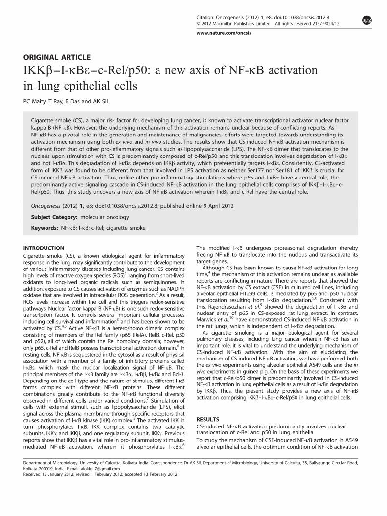
To understand the specific components that constitute activeNF-kB in the nucleus in response to CSE treatment, nuclear --cytosolic fractionation of CSE-treated and -untreated A549 cellswas performed. The results showed the nuclear accumulation ofc-Rel and p50 with time in CSE-treated cells (Figure 1a; upper
Figure 1. CSE-induced NF-kB activation in alveolar epithelial A549 cells predominantly involves nuclear translocation of c-Rel and p50. (a) CSEtreatment predominantly induces nuclear translocation of c-Rel and p50. A549 cells were treated with either 2% CSE (upper panels) or 1mg/mlLPS (lower panels) for different time periods as indicated. Nuclear and cytosolic fractions were prepared and separated by SDS--poly-acrylamide gel electrophoresis. Western blot analysis was performed with anti-c-Rel, anti-RelA, anti-p50 and anti-tubulin antibodies. C and Nindicate cytosolic and nuclear fractions, respectively. (b) Immunolocalization of c-Rel, p65 and p50. A549 cells were treated with 2% CSE for30min, fixed and probed with anti-c-Rel, anti-p65 and anti-p50 primary antibodies. (c) c-Rel downregulation inhibits CSE-induced NF-kBactivation. A549 cells were transfected with pSuper (empty vector), anti-c-Rel (si-c-Rel) and anti-p65 (si-p65) si-RNA constructs. After 24 h oftransfection, cells were harvested and cell extracts were analyzed by western blotting (left panel). Tubulin serves as loading control. Thesetransfectants harboring pSuper, si-c-Rel and si-p65 constructs were treated with 2% CSE for 30min and harvested. Nuclear extracts wereanalyzed by EMSA using radiolabeled NF-kB probe (right panel). The first lane of the gel was loaded with the free probe. (d) Chromatinimmunoprecipitation (ChIP) analysis of p65, c-Rel and p50 recruitment at IL-8 and cyclin D1 upstream promoter sequences. A549 cells weretreated with 2% CSE for 30min and cross-linked with paraformaldehyde. Immunoprecipitations were carried out using anti- p65, c-Rel and p50antibodies. Immunoprecipitated DNA was amplified by PCR primers corresponding to the NF-kB-binding site(s) at IL-8 and cyclin D1 upstreampromoter sequences as indicated in the upper panel and analyzed by agarose gel electrophoresis. DAPI, 4, 6-diamidino-2-phenyl indole; FITC,fluorescein isothiocyanate.
Cigarette smoke-induced NF-kB activationPC Maity et al
2
Oncogenesis (2012), 1 -- 7 & 2012 Macmillan Publishers Limited

panel). While no detectable nuclear accumulation was observedfor RelB and p52, a little p65 was detected in the nucleus(Supplementary Figure S2 and Figure 1a). In contrast, the nuclearaccumulation of p65 and p50 was observed with time in responseto LPS treatment as expected (Figure 1a; lower panel). As anindependent verification, immunolocalization of c-Rel, p65 andp50 was performed in CSE-treated A549 cells. Congruent with cellfractionation results, immunofluorescence experiment demon-strated a substantial nuclear translocation of c-Rel and p50 inresponse to CSE treatment, whereas little nuclear accumulation ofp65 was observed under the same conditions (Figure 1b).
To further confirm, si-RNA-mediated gene knockdown experi-ments for c-Rel and p65 were performed. Cells, which weretransfected with si-c-Rel construct before CSE treatment, showeddownregulation of c-Rel and also exhibited marked reduction inCSE-induced NF-kB activation (Figure 1c). In contrast, cells thatwere transfected with si-p65 construct exhibited little effect onCSE-induced NF-kB activation, although p65 was downregulated.To gain more confidence, the binding of c-Rel, p50 and p65 to theupstream promoter sequence of the two NF-kB target genes IL-8and cyclin D1, which were found to be upregulated by CSE(Supplementary Figure S1c), were examined in CSE-treated cellsby chromatin immunoprecipitation assay. Consistent with the cellfractionation and immunofluorescence results, better DNA bind-ing was observed for c-Rel and p50 compared with p65(Figure 1d). Taken together, these results indicate that consistentwith previously published results, although some p65 does enterthe nucleus in response to CSE induction, c-Rel appears to beprimarily responsible for NF-kB activation under these conditions.Thus, NF-kB activation in response to CSE treatment differs fromthe activation observed after LPS treatment and is predominantlymediated by c-Rel/p50 complex in A549 cells.
These ex vivo results were further bolstered by in vivoexperiments in guinea pig lung. To standardize NF-kB activation,guinea pigs were exposed to CS for 3 -- 6 days and NF-kB DNA-binding activity in lung nuclear extracts was examined by EMSA.The results showed NF-kB activation in lung tissue by 3 days of CSexposure (Figure 2, left panel). As considerable NF-kB activationwas observed by 4 days of CS-exposure, the subcellulardistribution of c-Rel and p65 was examined immunohistochemi-cally using lung tissue sections obtained from the guinea pigsexposed to CS for 4 days. Consistent with the ex vivo results, whilethere was substantial nuclear accumulation of c-Rel, little nuclearaccumulation of p65 was observed (Figure 2, right panel). Asexpected, the p50 distribution pattern was similar to c-Rel(Supplementary Figure S3). These results indicate that c-Rel andp50 form active NF-kB nuclear complex in guinea pig lung inresponse to CS exposure and thus lends support to the ex vivostudies.
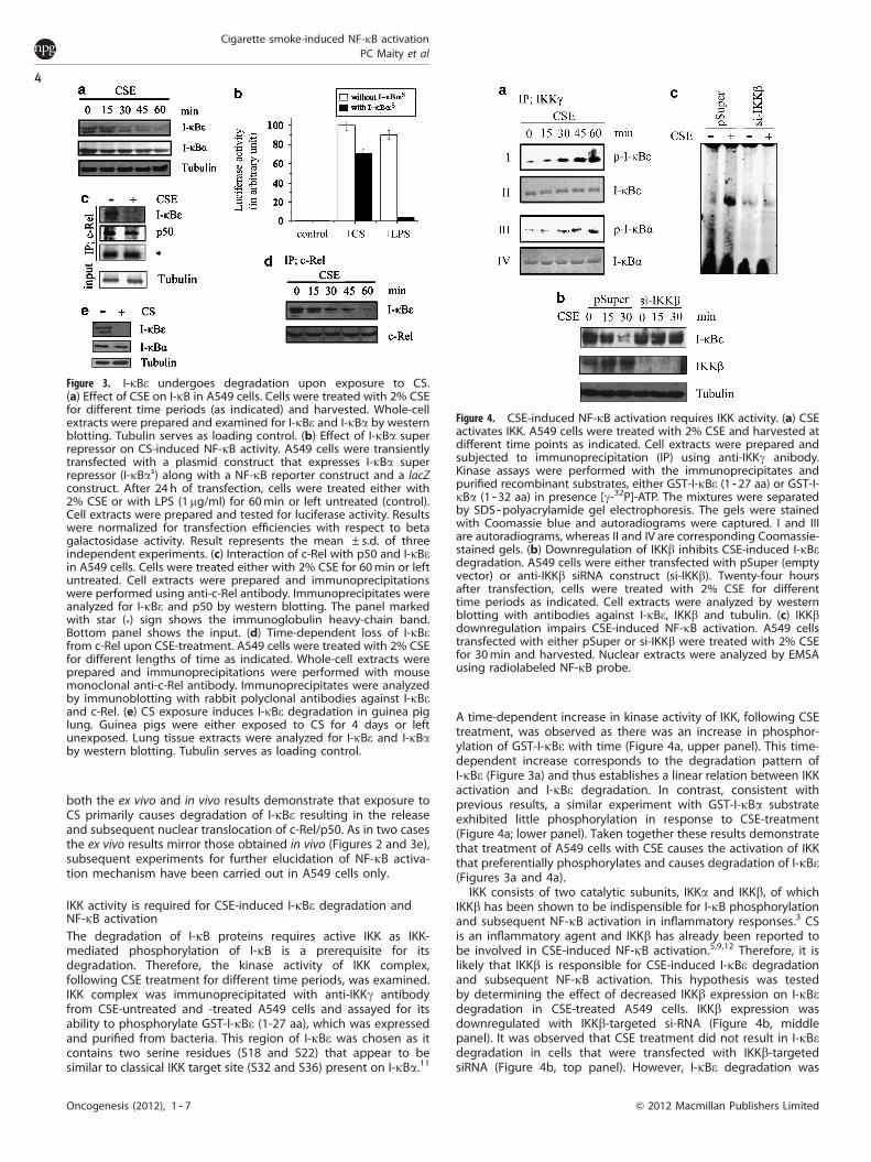
CSE-induced NF-kB activation is predominantly mediated throughthe degradation of I-kBeAs NF-kB dimer is retained in the cytosol by associating with I-kB,the degradation of latter is required for nuclear translocation ofNF-kB components. Anto et al.5 had reported the involvement ofI-kBa in this process. Therefore, the change in the level of I-kBawas monitored in A549 cells following CSE treatment by westernblotting. Although the result showed a reduction in I-kBa levelwith time, the rate was found to be very slow (Figure 3a) and didnot correspond with the substantial translocation of c-Rel and p50that had been observed within the indicated time period(Figure 1a). In contrast substantial I-kBa degradation was observedin control experiments where the cells were treated with LPS(Supplementary Figure S4). Consistent with these results, transienttransfection of A549 cells with I-kBa super repressor resulted in asubstantial reduction of LPS-induced NF-kB activity (Figure 3b). Incontrast, the same I-kBa super repressor exhibited little effect on
CS-induced NF-kB activity (Figure 3b). These results indicate thatI-kBa is unlikely to be the primary I-kB in CSE-induced NF-kBsignaling cascade in A549 cells, which is consistent with the reportof Marwick et al.10
In order to identify the I-kB involved in this signaling cascade,the change in the levels of I-kBe was investigated as this I-kBisoform is expressed highly in lung and has been reported tohave interaction with c-Rel.11 The results showed a substantialreduction of I-kBe with the progression of CSE treatment(Figure 3a), which is consistent with the nuclear translocationof c-Rel.
As CSE treatment induces nuclear translocation of c-Rel andp50, these two NF-kB components must be in a complex withI-kBe in resting A549 cells and this association would be disruptedfollowing treatment with CSE. This hypothesis was tested byperforming co-immunoprecipitation experiment using anti-c-Relantibody followed by blotting for both I-kBe and p50, in CSE-untreated and -treated cells. Figure 3c shows co-immunoprecipi-tation of I-kBe and p50 with c-Rel in CSE-untreated A549 cells. Asexpected, I-kBa was not detected in this experiment. These resultsindicate an association between I-kBe and c-Rel/p50 in restingA549 cells. Following CSE treatment, although p50 remainedassociated with c-Rel, a reduced association between c-Rel andI-kBe was observed (Figure 3c). Consistent with the time-dependent degradation of I-kBe in CSE-treated cells (Figure 3a),a time-dependent loss of I-kBe from the c-Rel complex was alsoobserved in CSE-treated cells (Figure 3d). Thus, these results showthat I-kBe prevents nuclear translocation of c-Rel/p50 in restingA549 cells and following CSE treatment, I-kBe degradation resultsin nuclear entry of c-Rel/p50 complex.
To further validate these ex vivo results in an in vivo system, thelevels of I-kBe and I-kBa were monitored by western blotting inthe lung extracts obtained from guinea pigs that were eitherexposed or not exposed to CS. The results showed that while I-kBewas almost completely degraded following CS exposure, a smalldecrease in the levels of I-kBa was observed (Figure 3e). Therefore,
Figure 2. CS-induced NF-kB activation in guinea pig lung.(a) Exposure of guinea pigs to CS induces alveolar NF-kB activation.Guinea pigs were exposed to CS for 0, 3, 4, 5 and 6 days. Nuclearextracts were prepared from lung tissues, and NF-kB activation wasassayed by EMSA using radiolabeled wild-type NF-kB probe.(b) Immunohistochemistry of c-Rel and p65. Lung tissue from guineapigs that were either exposed to CS for 4 days or left unexposed(0 day) were fixed and paraffin sections were prepared. Thereaftersections were immunostained with anti-c-Rel and anti-p65antibodies. Nuclei were stained with DAPI. FITC, fluorescein iso-thiocyanate.
Cigarette smoke-induced NF-kB activationPC Maity et al
3
Oncogenesis (2012), 1 -- 7& 2012 Macmillan Publishers Limited
both the ex vivo and in vivo results demonstrate that exposure toCS primarily causes degradation of I-kBe resulting in the releaseand subsequent nuclear translocation of c-Rel/p50. As in two casesthe ex vivo results mirror those obtained in vivo (Figures 2 and 3e),subsequent experiments for further elucidation of NF-kB activa-tion mechanism have been carried out in A549 cells only.
IKK activity is required for CSE-induced I-kBe degradation andNF-kB activationThe degradation of I-kB proteins requires active IKK as IKK-mediated phosphorylation of I-kB is a prerequisite for itsdegradation. Therefore, the kinase activity of IKK complex,following CSE treatment for different time periods, was examined.IKK complex was immunoprecipitated with anti-IKKg antibodyfrom CSE-untreated and -treated A549 cells and assayed for itsability to phosphorylate GST-I-kBe (1-27 aa), which was expressedand purified from bacteria. This region of I-kBe was chosen as itcontains two serine residues (S18 and S22) that appear to besimilar to classical IKK target site (S32 and S36) present on I-kBa.11
A time-dependent increase in kinase activity of IKK, following CSEtreatment, was observed as there was an increase in phosphor-ylation of GST-I-kBe with time (Figure 4a, upper panel). This time-dependent increase corresponds to the degradation pattern ofI-kBe (Figure 3a) and thus establishes a linear relation between IKKactivation and I-kBe degradation. In contrast, consistent withprevious results, a similar experiment with GST-I-kBa substrateexhibited little phosphorylation in response to CSE-treatment(Figure 4a; lower panel). Taken together these results demonstratethat treatment of A549 cells with CSE causes the activation of IKKthat preferentially phosphorylates and causes degradation of I-kBe(Figures 3a and 4a).
IKK consists of two catalytic subunits, IKKa and IKKb, of whichIKKb has been shown to be indispensible for I-kB phosphorylationand subsequent NF-kB activation in inflammatory responses.3 CSis an inflammatory agent and IKKb has already been reported tobe involved in CSE-induced NF-kB activation.5,9,12 Therefore, it islikely that IKKb is responsible for CSE-induced I-kBe degradationand subsequent NF-kB activation. This hypothesis was testedby determining the effect of decreased IKKb expression on I-kBedegradation in CSE-treated A549 cells. IKKb expression wasdownregulated with IKKb-targeted si-RNA (Figure 4b, middlepanel). It was observed that CSE treatment did not result in I-kBedegradation in cells that were transfected with IKKb-targetedsiRNA (Figure 4b, top panel). However, I-kBe degradation was
Figure 3. I-kBe undergoes degradation upon exposure to CS.(a) Effect of CSE on I-kB in A549 cells. Cells were treated with 2% CSEfor different time periods (as indicated) and harvested. Whole-cellextracts were prepared and examined for I-kBe and I-kBa by westernblotting. Tubulin serves as loading control. (b) Effect of I-kBa superrepressor on CS-induced NF-kB activity. A549 cells were transientlytransfected with a plasmid construct that expresses I-kBa superrepressor (I-kBas) along with a NF-kB reporter construct and a lacZconstruct. After 24 h of transfection, cells were treated either with2% CSE or with LPS (1mg/ml) for 60min or left untreated (control).Cell extracts were prepared and tested for luciferase activity. Resultswere normalized for transfection efficiencies with respect to betagalactosidase activity. Result represents the mean ±s.d. of threeindependent experiments. (c) Interaction of c-Rel with p50 and I-kBein A549 cells. Cells were treated either with 2% CSE for 60min or leftuntreated. Cell extracts were prepared and immunoprecipitationswere performed using anti-c-Rel antibody. Immunoprecipitates wereanalyzed for I-kBe and p50 by western blotting. The panel markedwith star (*) sign shows the immunoglobulin heavy-chain band.Bottom panel shows the input. (d) Time-dependent loss of I-kBefrom c-Rel upon CSE-treatment. A549 cells were treated with 2% CSEfor different lengths of time as indicated. Whole-cell extracts wereprepared and immunoprecipitations were performed with mousemonoclonal anti-c-Rel antibody. Immunoprecipitates were analyzedby immunoblotting with rabbit polyclonal antibodies against I-kBeand c-Rel. (e) CS exposure induces I-kBe degradation in guinea piglung. Guinea pigs were either exposed to CS for 4 days or leftunexposed. Lung tissue extracts were analyzed for I-kBe and I-kBaby western blotting. Tubulin serves as loading control.
Figure 4. CSE-induced NF-kB activation requires IKK activity. (a) CSEactivates IKK. A549 cells were treated with 2% CSE and harvested atdifferent time points as indicated. Cell extracts were prepared andsubjected to immunoprecipitation (IP) using anti-IKKg anibody.Kinase assays were performed with the immunoprecipitates andpurified recombinant substrates, either GST-I-kBe (1 --27 aa) or GST-I-kBa (1 --32 aa) in presence [g-32P]-ATP. The mixtures were separatedby SDS--polyacrylamide gel electrophoresis. The gels were stainedwith Coomassie blue and autoradiograms were captured. I and IIIare autoradiograms, whereas II and IV are corresponding Coomassie-stained gels. (b) Downregulation of IKKb inhibits CSE-induced I-kBedegradation. A549 cells were either transfected with pSuper (emptyvector) or anti-IKKb siRNA construct (si-IKKb). Twenty-four hoursafter transfection, cells were treated with 2% CSE for differenttime periods as indicated. Cell extracts were analyzed by westernblotting with antibodies against I-kBe, IKKb and tubulin. (c) IKKbdownregulation impairs CSE-induced NF-kB activation. A549 cellstransfected with either pSuper or si-IKKb were treated with 2% CSEfor 30min and harvested. Nuclear extracts were analyzed by EMSAusing radiolabeled NF-kB probe.
Cigarette smoke-induced NF-kB activationPC Maity et al
4
Oncogenesis (2012), 1 -- 7 & 2012 Macmillan Publishers Limited
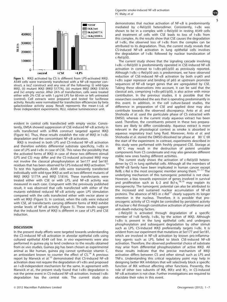
evident in control cells transfected with empty vector. Consis-tently, EMSA showed suppression of CSE-induced NF-kB activity incells transfected with si-RNA construct targeted against IKKb(Figure 4c). Thus, these results establish the role of IKKb in I-kBedegradation and the concomitant NF-kB activation.
IKKb is involved in both LPS and CS-induced NF-kB activationand therefore exhibits differential substrate specificity, I-kBa incase of LPS and I-kBe in case of CSE. This raises the possibility thatthe activated forms of IKKb in case of these two different stimuli(LPS and CS) may differ and the CS-induced activated IKKb maynot involve the classical phosphorylation at Ser177 and Ser181residues that has been observed for LPS-induced IKKb activation.13
To test this hypothesis, A549 cells were transiently transfectedindividually with wild-type IKKb as well as two different mutants ofIKKb (IKKb S177A and IKKb S181A). These transfectants weretreated either with CSE or with LPS, and NF-kB activity wasmeasured in each case. Congruent with the previously publishedresult, it was observed that cells transfected with either of themutants exhibited reduced NF-kB activity upon LPS stimulationcompared with the cells transfected either with vector alone orwith wt IKKb (Figure 5). In contrast, when the cells were inducedwith CSE, all transfectants carrying different forms of IKKb exhibitsimilar levels of NF-kB activity (Figure 5). These results suggestthat the induced form of IKKb is different in case of LPS and CSEinduction.
DISCUSSIONIn the present study efforts were targeted towards understandingthe CS-induced NF-kB activation in alveolar epithelial cells usingcultured epithelial A549 cell line. In vivo experiments were alsoperformed in guinea pig to lend credence to the results obtainedfrom ex vivo studies. Guinea pig has been chosen as experimentalanimal as like human, guinea pig cannot synthesize vitamin C,an antioxidant known to counter the effect of CS.14 A previousreport by Marwick et al.10 demonstrated that CS-induced NF-kBactivation does not require the degradation of I-kBa and proposedan I-kB-independent mechanism for the same. In agreement withMarwick et al., the present study found that I-kBa degradation isnot the prime event in CS-induced NF-kB activation. Instead I-kBedegradation has the central role. The current study also
demonstrates that nuclear activation of NF-kB is predominantlymediated by c-Rel/p50 heterodimer. Consistently, I-kBe wasshown to be in a complex with c-Rel/p50 in resting A549 cellsand treatment of cells with CSE leads to loss of I-kBe fromthis complex. As the results show that CSE causes the degradationof I-kBe, the observed loss of I-kBe from the complex can beattributed to its degradation. Thus, the current study reveals thatCS-induced NF-kB activation in lung epithelial cells involvesthe degradation of I-kBe followed by nuclear translocation ofc-Rel/p50.
The current study shows that the signaling cascade involvingI-kBe -- c-Rel/p50 is predominantly operated in CSE-induced NF-kBactivation in contrast to I-kBa-p65/p50 as previously reported.Although I-kBe -- c-Rel/p50 axis is predominant, we have observedreduction of CSE-induced NF-kB activation by both si-p65 andI-kBa super repressor and binding of p65 at upstream promotersequences of NF-kB target genes that are upregulated by CSE.Taking these observations into account, it can be said that theclassical axis, comprising I-kBa-p65/p50, is also active with minorcontribution. In the previous studies, it is possible that theresearchers overlooked the axis that is operating predominantly inthis event. In addition, in the cell culture-based studies, thedifference in preparation of CSE and applied dose may alsocontribute towards the observed discrepancy. Anto et al. andShishodia et al. used the particulate phase of CS extracted withDMSO, whereas in the current study aqueous extract has beenused. Therefore, the constituents present in these two prepara-tions are likely to differ considerably. Aqueous extract is morerelevant in the physiological context as smoke is absorbed inaqueous respiratory tract lung fluid. Moreover, Anto et al. andShishodia et al. stored the DMSO-dissolved tar phase at �80 1C tillthe start of the experiment. In contrast, experiments described inthis study were performed with freshly prepared CSE. Storage at�80 1C may result in the destruction of potent unstablecomponents from CS condensate and may also generate differentstable new ones having different activities.
The current study shows the activation of c-Rel/p50 hetero-dimer by CS in lung epithelial cells. Although all the members ofRel/NF-kB family have been implicated in human cancer exceptRelB, c-Rel is the most oncogenic member among them.15 -- 17 Theunderlying mechanism of this tumorgenic potential is not clear.However, a bias towards expression of genes involved in survivaland proliferation such as IL-2 and Bcl-xL
18,19 may explain itsoncogenecity. The tumorgenic potential can also be attributed tothe increased and sustained nuclear accumulation of NF-kBproteins. The absence of NES in c-Rel17 clearly supports its longerretention in the nucleus. Therefore, it is possible that pro-oncogenic activity of CS might be controlled by persistent activityof nuclear c-Rel through constitutive activation of proliferative andanti-death-inducing factors.
c-Rel/p50 is activated through degradation of a specificmember of I-kB family, I-kBe, by the action of IKKb. AlthoughI-kBa is present in the lung epithelial cells and undergoesphosphorylation and subsequent degradation by other stimulisuch as LPS, CS-induced IKKb preferentially targets I-kBe. It isevident from our experiment that mutations at Ser177 and Ser181,which are involved in NF-kB activation by known pro-inflamma-tory agents such as LPS, failed to block CSE-induced NF-kBactivation. Therefore, the observed preferential choice of substratemay arise from differential phosphorylation of active IKKb. Allthese results indicate that the precise mechanism of IKKbactivation differs between CS and other stimuli such as LPS andTNFa. Understanding this critical regulatory point may help indesigning better IKK inhibitors that will selectively block a specificfunction of IKK without affecting other functions. Presently, therole of other two subunits of IKK, IKKa and IKg, in CS-inducedNF-kB activation is not clear. Further investigations are required toelucidate their roles in this event.
Figure 5. IKKb activated by CS is different from LPS-activated IKKb.A549 cells were transiently transfected with a NF-kB reporter con-struct, a lacZ construct and any one of the following: (i) wild-typeIKKb, (ii) mutant IKKb (IKKb S177A), (iii) mutant IKKb (IKKb S181A)and (iv) empty vector. After 24 h of transfection, cells were treatedeither with 2% CSE or with 1 mg/ml LPS for 60min or left untreated(control). Cell extracts were prepared and tested for luciferaseactivity. Results were normalized for transfection efficiencies by betagalactosidase activity assay. Result represents the mean±s.d. ofthree independent experiments. RLU, relative luminescence unit.
Cigarette smoke-induced NF-kB activationPC Maity et al
5
Oncogenesis (2012), 1 -- 7& 2012 Macmillan Publishers Limited
Although the mechanism of this selectivity is unknown,functionally it can contribute to a great extent towards thetumorgenic effects of CS in lung. It is known that the different I-kBproteins exhibit different pattern of degradation as well asresynthesis. I-kBa is degraded and re-synthesized rapidly (in 1 h)and is therefore involved in functions associated with transientNF-kB activity. In this study it was found that I-kBe takes 4 h toreappear after the first round of degradation in CSE-treated A549cells (Supplementary Figure S5). The delayed reappearance ofI-kBe significantly contributes towards sustained NF-kB activationby prolonging c-Rel/p50 nuclear activity. Thus, the involvementof c-Rel activity coupled with the dynamics of I-kBe levels inCS-induced NF-kB activation makes CS a potentially strongtumorgenic agent and may explain the strong correlationbetween cigarette smoking and lung carcinogenesis.
Till date several NF-kB complexes, in combination with differentI-kB, have been identified for stimulus-specific activation of NF-kBin different cell lines. This study has identified an NF-kB complexconsisting of I-kBe, c-Rel and p50. Previous studies document theinteraction between c-Rel and I-kBe, and also an active NF-kBcomplex formation between c-Rel and p50. However, the signalingcascade comprising of stimulus (in this case CS), IKKb -- I-kBe -- c-Rel/p50, is neither previously documented nor functionally implicatedelsewhere. In conclusion, a new axis of NF-kB activation in lungepithelial cells is proposed wherein I-kBe and c-Rel, instead ofI-kBa and p65, have the central role.
MATERIALS AND METHODSCell culture and transfectionAll in vitro experiments were performed on Human lung alveolar type IIcell line A549 that were grown in Ham’s F12-nutrient mixture.20
Transient transfections were performed using PolyFect reagent (Qiagen,Hilden, Germany).
Plasmids and protein expressionsiRNA sequences targeted against p65, c-Rel and IKKb were designed usingsiDESIGN- software (Thermo Scientific, Asheville, NC, USA) and clonedinto pSuper Retro Puro vector (Oligo-Engine, Seattle, WA, USA). For theconstruction of GST-I-kBe (1 -- 27 aa) expression plasmid, oligonucleotidescorresponding to 1 -- 27 aa of I-kBe cDNA were annealed and cloned intoEcoRI and XhoI sites of pGEX-4T1 (Amersham Biosciences, Little Chalfont,UK). GST fusion proteins, GST-I-kBa (1 -- 54 aa) and GST-I-kBe (1 -- 27 aa),were expressed in Escherichia coli (BL21 DE3) and purified by glutathione-sepharose beads. The sequences of the oligonucleotide primers that havebeen used for making different constructs are given in SupplementaryTable S1.
Preparation of CSECSE were prepared from filter tipped 69-mm cigarettes from IndianTobacco Company as described by Maity et al.20
Exposure of guinea pigs to CSThree- to four- month-old male guinea pigs (350 -- 400 g) were used. Animalcare procedures were as per NIH (National Institutes of Health) guidelinesand approved by the Institutional Animal Ethics Committee. The guineapigs were fed a vitamin C-free diet for 7 days to minimize the vitamin Clevel in the plasma and tissues as vitamin C is a potential inhibitor ofCS-induced oxidative stress.14 The composition of the diet was as perBanerjee et al.21 After feeding vitamin C-free diet for 7 days, each guineapig was given oral supplement of 1 mg vitamin C/day as maintenance doseand subjected to CS exposure (three cigarettes/animal/day with two puffs/cigarette) in a smoke chamber.14 Guinea pigs were exposed to smokeenvironment for 1 min during each puff and exposed to fresh air forthe next 1 min.
Nuclear -- cytosolic fractionationNuclear -- cytosolic fractionations of differentially treated A549 cells werecarried out as described by Chaturvedi et al.22 For lung tissue, single cellwas prepared by passing tissue homogenate through a micro-sieve(Sigma-Aldrich, St Louis, MO, USA). Thereafter, the procedure, used forA549 cells, was followed to obtain nuclear -- cytosolic fractionations ofthese cells.
Electrophoretic mobility shift assayEMSAs were performed using 32P-labeled oligonucleotide probe contain-ing the consensus sequences for NF-kB according to previously describedmethod by Chaturvedi et al.22 DNA -- protein complexes were resolved on anon-denaturing 5% polyacrylamide gel and subsequently exposed toeither X-ray film (Kodak, Rochester, NY, USA) or phosphor imaging system(Amersham Biosciences).
ImmunoprecipitationLysates were prepared from differentially treated A549 cells and proteinconcentrations were determined. Hundred micrograms of each lysates wasincubated overnight at 4 1C with specific antibody (as per the require-ment). Protein A Sepharose beads were added and kept at 4 1C on arotating platform for 2 h. Thereafter immune complexes were isolated,separated by SDS -- polyacrylamide gel electrophoresis and analyzed byimmunoblotting.
Immunohistochemistry and immunofluorescenceLung tissue from guinea pigs was fixed in formaldehyde. Fixed tissue wasparaffin-embedded, and serially sectioned at 5 mm. These sections werethen deparafinized and made permeable by treating with 0.1% TritonX-100. Thereafter, antigens were unmasked by heating the sections at90 1C for 10 min in 10 mM Na -- citrate buffer, pH 6.0. The sections were thenincubated overnight at 4 1C with specific antibodies as per the require-ment. Sections were then incubated with fluorescein isothiocyanate-conjugated secondary antibody at room temperature for 2 h, washed andstained with 46-diamidino-2-phenyl indole. Fluorescent signals wereviewed under fluorescence microscope (Olympus IX71, Tokyo, Japan).Immunofluorescence was performed as described by Bernard et al.23
Kinase assayThe IKK assay was performed as described by Delhase et al.13 Briefly, IKKcomplex was immunoprecipitated from 300mg of cell extracts obtainedfrom CSE-treated or -untreated A549 cells using IKKg antibody with ProteinA Sepharose beads. The beads were then washed and resuspended in 25 mlof kinase assay mixture containing 3 mg of substrate (GST -- I-kBa or GST --I-kBe) and 8 m moles of [g-32P] ATP in addition to the components of assaybuffer and incubated at 37 1C for 30 min. Finally, the proteins wereseparated by SDS -- polyacrylamide gel electrophoresis, stained withCoomasie blue and exposed to either X-ray film (Kodak) or phosphorimaging system (Amersham Biosciences) to obtain autoradiogram ofthe gel.
Chromatin immunoprecipitation assayA549 cells were treated with 2% CSE for 30 min and thereafter chromatinimmunoprecipitation was performed as described previously by Majumderet al.24 The DNA -- protein complex was immunoprecipitated using anti-p65,p50 or c-Rel antibody in separate reactions. In a separate reaction, a nonspecific anti-rabbit IgG was also added as control. The DNA obtained wasanalyzed by PCR using specific primers (Supplementary Table S1) designedto target the NF-kB-binding sites at IL-8 and cyclin D1 promoters asindicated in the figure.
CONFLICT OF INTERESTThe authors declare no conflict of interests.
Cigarette smoke-induced NF-kB activationPC Maity et al
6
Oncogenesis (2012), 1 -- 7 & 2012 Macmillan Publishers Limited
ACKNOWLEDGEMENTSWe thank Prof DJ Chattopadhyay for his valuable help and support; Dr S Sarkar andMr K Mukherjee for critical reading of the manuscript. We thank Prof M Karin, UCSD,USA for providing IKKb constructs. This work is supported by the Council of Industrialand Scientific Research (CSIR), India (Sanction no. 37(1301)/07/EMR II). PM wassupported by CSIR individual fellowship. TR was supported with a fellowshipsponsored by the University of Calcutta.
REFERENCES1 Pryor WA, Stone K. Oxidants in cigarette smoke. Radicals, hydrogen peroxide,
peroxynitrate, and peroxynitrite. Ann N Y Acad Sci 1993; 686: 12 -- 27 (discussion27 -- 28).
2 Orosz Z, Csiszar A, Labinskyy N, Smith K, Kaminski PM, Ferdinandy P et al.Cigarette smoke-induced proinflammatory alterations in the endothelial pheno-type: role of NAD(P)H oxidase activation. Am J Physiol Heart Circ Physiol 2007; 292:H130 -- H139.
3 Karin M. The I-kB kinase - a bridge between inflammation and cancer. Cell Res2008; 18: 334 -- 342.
4 Nishikawa M, Kakemizu N, Ito T, Kudo M, Kaneko T, Suzuki M et al. Superoxidemediates cigarette smoke-induced infiltration of neutrophils into the airwaysthrough nuclear factor-jB activation and IL-8 mRNA expression in guinea pigs invivo. Am J Respir Cell Mol Biol 1999; 20: 189 -- 198.
5 Anto RJ, Mukhopadhyay A, Shishodia S, Gairola CG, Aggarwal BB. Cigarette smokecondensate activates nuclear transcription factor-jB through phosphorylationand degradation of I-jBa: correlation with induction of cyclooxygenase-2.Carcinogenesis 2002; 23: 1511 -- 1518.
6 Hayden MS, Ghosh S. Shared principles in NF-kB signaling. Cell 2008; 132: 344 -- 362.7 Gloire G, Legrand-Poels S, Piette J. NF-kB activation by reactive oxygen species:
fifteen years later. Biochem Pharmacol 2006; 72: 1493 -- 1505.8 Shishodia S, Potdar P, Gairola CG, Aggarwal BB. Curcumin (diferuloylmethane)
down regulates cigarette smoke-induced NF-kappaB activation through inhibitionof I-kappaB alpha kinase in human lung epithelial cells: correlation withsuppression of COX-2, MMP-9 and cyclin D1. Carcinogenesis 2003; 24: 1269 -- 1279.
9 Rajendrasozhan S, Hwang JW, Yao H, Kishore N, Rahman I. Anti-inflammatoryeffect of a selective I-kB kinase-beta inhibitor in rat lung in response to LPS andcigarette smoke. Pulm Pharmacol Ther. 2010; 23: 172 -- 181.
10 Marwick JA, Kirkham PA, Stevenson CS, Danahay H, Giddings J, Butler K et al.Cigarette smoke alters chromatin remodeling and induces proinflammatorygenes in rat lungs. Am J Respir Cell Mol Biol 2004; 31: 633 -- 642.
11 Whiteside ST, Epinat JC, Rice NR, Israel A. I-kappaB epsilon, a novel member of theI-kappa B family, controls RelA and c-Rel NF-kappa B activity. Embo J 1997; 16:1413 -- 1426.
12 Takahashi H, Ogata H, Nishigaki R, Broide DH, Karin M. Tobacco smoke promoteslung tumorgenesis by triggering IKKb and JNK1 dependent inflammation. CancerCell 2010; 17: 89 -- 97.
13 Delhase M, Hayakawa M, Chen Y, Karin M. Positive and negative regulation ofIkappaB kinase activity through IKKbeta subunit phosphorylation. Science 1999;284: 309 -- 313.
14 Ray T, Maity PC, Banerjee S, Deb S, Dasgupta AK, Sarkar S et al. Vitamin C preventscigarette smoke induced atherosclerosis in guinea pig model. J AtherosclerThromb 2010; 17: 817 -- 827.
15 Fan Y, Rayet B, Gelinas C. Divergent C-terminal transactivation domains of Rel/NF-kappa B proteins are critical determinants of their oncogenic potential inlymphocytes. Oncogene 2004; 23: 1030 -- 1042.
16 Gilmore TD, Cormier C, Jean-Jacques J, Gapuzan ME. Malignant transformation ofprimary chicken spleen cells by human transcription factor c-Rel. Oncogene 2001;20: 7098 -- 7103.
17 Gilmore TD, Kalaitzidis D, Liang MC, Starczynowski DT. The c-Rel transcriptionfactor and B-cell proliferation: a deal with the devil. Oncogene 2004; 23:2275 -- 2286.
18 Chen C, Edelstein LC, Gelinas C. The Rel/NF-kB family directly activates expressionof the apoptosis inhibitor Bcl-xL. Mol Cell Biol 2000; 20: 2687 -- 2695.
19 Liou HC, Jin Z, Tumang J, Andjelic S, Smith KA, Liou ML. c-Rel is crucial forlymphocyte proliferation but dispensable for T cell effector function. Int Immunol1999; 11: 361 -- 371.
20 Maity PC, Bhattacharjee S, Majumdar S, Sil AK. Potentiation by cigarette smoke ofmacrophage function against Leishmania donovani infection. Inflamm Res 2009;58: 22 -- 29.
21 Banerjee S, Maity P, Mukherjee S, Sil AK, Panda K, Chattopadhyay D et al. Blacktea prevents cigarette smoke-induced apoptosis and lung damage. J Inflamm2007; 4: 3.
22 Chaturvedi MM, Mukhopadhyay A, Aggarwal BB. Assay for redox-sensitivetranscription factor. Methods Enzymol 2000; 319: 585 -- 602.
23 Bernard D, Monte D, Vandenbunder B, Abbadie C. The c-Rel transcription factorcan both induce and inhibit apoptosis in the same cells via the upregulation ofMnSOD. Oncogene 2002; 21: 4392 -- 4402.
24 Majumder P, Chattopadhyay B, Sukanya S, Ray T, Banerjee M, Mukhopadhyay Det al. Interaction of HIPPI with putative promoter sequence of caspase-1 in vitroand in vivo. Biochem Biophys Res Commun 2007; 353: 80 -- 85.
Oncogenesis is an open access journal published by Nature PublishingGroup. This work is licensed under the Creative Commons Attribution-
NonCommercial-No Derivative Works 3.0 Unported License. To view a copy of this license,visit http://creativecommons.org/licenses/by-nc-nd/3.0/
Supplementary Information accompanies the paper on the Oncogenesis website (http://www.nature.com/oncsis)
Cigarette smoke-induced NF-kB activationPC Maity et al
7
Oncogenesis (2012), 1 -- 7& 2012 Macmillan Publishers Limited
Copyright © 2022 FDOKUMEN