ijoparb - Indian Society of Perinatology and Reproductive ...
42
IJOPARB Indian Journal of Perinatology and Reproductive Biology ISSN 2249 - 9784 Volume 10, No. 01 January - March, 2020 Official Journal of Indian Society of Perinatology and Reproductive Biology ICV 2017 = 70.99 | ICV 2018 = 69.79
-
Upload
khangminh22 -
Category
Documents
-
view
0 -
download
0
Transcript of ijoparb - Indian Society of Perinatology and Reproductive ...
IJOPARBIndian Journal of Perinatology and Reproductive Biology
ISSN 2249 - 9784Volume 10, No. 01January - March, 2020
Official Journal of Indian Society of Perinatology and Reproductive Biology
If undelivered, return to: Indian Journal of Perinatology and Reproductive Biology
CD-55, Salt Lake City, Sector - 1, Kolkata 700064
ICV 2017 = 70.99 | ICV 2018 = 69.79
IJOPARBIndian Journal of Perinatology and Reproductive Biology
Vol. 10 | No. 01 | January - March, 2020 | ISSN 2249-9784
Official Journal ofINDIAN SOCIETY OF PERINATOLOGY AND
REPRODUCTIVE BIOLOGY (ISOPARB)
IJOPARB appears in the recommended list of Journals of University Grant Commission:
Sl.No: 180; Jr.No. 62671; ISSN: 22499784

2 IJOPARB | Vol. 10 | No. 01 | Jan-Mar 2020
Indian Society of Perinatologyand Reproductive Biology
Estd. 1978
(Reg No. 71 of 1978-1979 under the Societies Registration Act 21 of 1860)
Executive Committee (2018-2019)
President Dr Suchitra N. Pandit 09820416474 Secretary General Dr Meena Samant 09334105945 Immediate Past President Dr Milind R. Shah 09822096280 Editor-in-Chief Dr Hiralal Konar 09433033225 Vice Presidents Dr Arup Kumar Majhi Dr Narayan Jana Dr Gangadhar Sahoo Dr Shanti H. K. Singh Treasurer Dr Pragya Mishra Chaudhary 09835273668 Communicator Dr Supriya Jaiswal 09431207284
Zonal Chairpersons
East Zone Dr Ojaswani Patel West Zone Dr Parul Kotdawala North Zone Dr Anjoo Agarwal South Zone Dr Rooma Singh
Executive Members
Patna Dr Abha Rani Singh Patna Dr Amita Singh Ranchi Dr Archana Kumari Kolkata Dr Biswajyoti Guha Chennai Dr Lakshmi Ravindran New Delhi Dr Mala Srivastava Lucknow Dr Rajul Tyagi Patna Dr Rita Kumari Jha Hyderabad Dr S. Madhumathi Kolkata Dr Sukumar Barik Chennai Dr Vijay Lakshmi Seshadri Lucknow Dr Yashodhara Pradeep
Head Office:IMA Building, Dr A. K. N. Sinha Path, South East Gandhi Maidan, Patna 800004
Web: www.isoparb.org
Secretary General’s Office
Dr Meena Samant, Secretary General, ISOPARB21/D, Road No 10, Rajendranagar, Patna - 800016
Phone: +91 93341 05945 | E-mail: [email protected]
3IJOPARB | Vol. 10 | No. 01 | Jan-Mar 2020
Indian Journal of Perinatologyand Reproductive Biology
CD 55, Sector I, Salt Lake City, Kolkata 700 064E-mail: [email protected]
ISSN 2249-9784 RNI No. WB ENG/2010/39056
EDITORIAL BOARD
Editor-in-Chief
Kolkata Dr Hiralal Konar Emeritus Editor
Kolkata Dr Arup Kumar Majhi
Editorial Advisory Board
Kolkata Dr Gita Ganguly Mukherji Jamshedpur Dr A K Debdas Mumbai Dr Bandana Walvekar New Delhi Dr Sunita Mittal Cuttack Dr S N Tripathy Kolkata Dr Sudip Chakravarti
Associate Editors
Kolkata Dr Picklu Chaudhuri Dr Gita Basu Banerjee Dr Sukumar Barik Dr Subrata Lal Seal
Joint Editors
Kolkata Dr Sajal Datta Kolkata Dr Ramprasad Dey Kolkata Dr Pallab Kumar Mistri Kolkata Dr Sudhir Adhikari
Members of Editorial Board
Patna Dr Abharani Sinha Kolkata Dr Nalini Arora Kolkata Dr Mamtaz Sanghamita Bengaluru Dr Jayanthi Kolkata Dr Aftabuddin Mondal Kolkata Dr Shyamal Banerjee Kolkata Dr Rathindra Nath Roy
Members of Editorial Board Lucknow Dr Anju Agarwal Hyderabad Dr Sampat Kumari Hyderabad Dr Swaraj Lakshmi Kolkata Dr Chaitali Datta Ray Silchar Dr Pranoy Nath Kolkata Dr Somajita Chakraborty Kolkata Dr Sebanti Goswami Puducherry Dr Sayed Habeebullah Kolkata Dr Arindam Halder Vellore Dr Abraham Pedicyle
Members of National Advisory Board Kolkata Dr Narayan Jana Delhi Dr Asoke Kumar Delhi Dr Deepika Deka Cochin Dr V P Paily Cuttack Dr P C Mahapatra Delhi Dr Arun Singh Manipal Dr Murali Dhar Pai Bengaluru Dr Shila Mane Belgaum, Karnataka Dr M B Bellard Dibrugarh Dr Rina Ahmed Lucknow Dr Vinita Das Burla, Odhisa Dr Gangadhar Sahoo
Members of International Advisory Board Kolkata Dr B N Chakravarty UK Dr S Arulkumaran Japan Dr Kiyoko Kato Korea Dr Joong Shin Park Malaysia Dr Ravi Chandran Sri Lanka Dr Rohana Haththotuwa Nepal Dr Gehanath Baral Bangladesh Dr Firoza Begum
Ex-Officio Members President, ISOPARB Dr Suchitra N Pandit Secretary General, ISOPARB Dr Meena Samant
Editorial Office: CD-55, Salt Lake City Sector-I, Kolkata 700064 Email: [email protected]
4 IJOPARB | Vol. 10 | No. 01 | Jan-Mar 2020
Contents
Editor’s Choice
Life or Livelihood? … … … … … … … … … … … … … … … … … … … … … … … … 5Prof (Dr) Hiralal Konar
Views & Reviews
COVID – 19: Care in Gynecology … … … … … … … … … … … … … … … … … … … 7Prof (Dr) Hiralal Konar
Review Article - Care in Perinatology
Novel-19 in Newborns … … … … … … … … … … … … … … … … … … … … … … … 11Prof Shyamal Banerjee
Original Article - Obstetrics
An Analytical study to Evaluate the Prevalence, Screening Methods and Maternal and Perinatal Outcome Associated with Asymptomatic Bacteriuria … … … … … … … … … … … … … 14Vandana N, T. Radha Bai Prabhu, Sahitya Meda, Velayudham Damodaran
Role of Combination of Mifepristone and Misoprostol versus Misoprostol alone in Induction of Labor in Intra Uterine Fetal Death … … … … … … … … … … … … … … … … … … … 20Dr Sweta, Dr Kusum Dash
Case Report - Obstetrics
A Case of Adverse Effect of Warfarin Therapy in Early Pregnancy – A Rare Observation … … 25Dr Abhijit Shil, Dr Kashish Garg, Dr A K Rakshit
Original Article - Gynecology
Evaluation of Platelet Rich Plasma in Infertile Women … … … … … … … … … … … … … 28Dr Vatsal Thakral, Dr Swati Garg, Dr Urvashi Sharma
Case Report - Gynecology
A Rare Case of Choriocarcinoma … … … … … … … … … … … … … … … … … … … … 33Dr Smrity Mool Joshi, Dr Tarun Das, Dr Mamta Dagar, Dr Mala Srivastava, Dr I Ganguli, Dr Rakesh Koul
Instruction to Authors … … … … … … … … … … … … … … … … … … … … … … … … 38
Disclaimer: The Editor/Publisher disclaims any responsibility or legal liability for statements made and opinions or views expressed by the author(s) and contributors and any claims made by the advertisers.
© IJOPARB. All rights reserved. No part of this publication should be reproduced or stored in a retrieval system, or transmitted in any form, by any means: electronic, mechanical or photocopying, recording, or otherwise, without written permission from Editors or Publishers.
5IJOPARB | Vol. 10 | No. 01 | Jan-Mar 2020
In the history, human survival never before faced such challenges, what is presently in the context of Corona virus disease (Covid–19). Corona virus infection, a global pandemic, is the single most issue now, throughout the world. Most of the areas of clinical approach in the management of the disease is still in the dark. Exact laboratory tests for confirmation of diagnosis is awaited. Actual prevention is unknown except some social and behavioral measures. Definitive management is in a state of trial only. Drugs like hydrocholoroquine, remdesivir, ritonavir, lopinavir, oseltamivir, corticosteroid are used in different combinations for the treatment of covid pneumonia. All these are with unproven efficacy and safety. Govt. of India, Ministry of Health and Family Welfare, states no specific antiviral drug have
been proven to be effective as per currently avail data. Hydrocholoroquine and azithromycin are being used for the treatment of covid pneumonia. Vaccines are yet to come. Supportive management is the only option left with us. Many countries are maintaining lockdown either completely or partially to reduce the spread of transmission. This is thought to be protective and life-saving. Aarogya Setu app., launched by Govt. of India, tracks through a Bluetooth and location generated social graph. This is to help us to know, our interaction with some one who could have tested covid -19 positive. It is an essential part of our present day life, as a measure of prevention and protection.
Editor’s Choice
Life or Livelihood?
Table-1: NOVEL CORONA VIRUS (COVID-19)CURRENT STATUS (21.03.2020)
Sl.No. Countries Infected Death1 China 81250 32532 Italy 47021 40323 Spain 20410 10434 Germany 19711 535 Iran 19644 14336 US 14631 2107 France 10891 3718 South Korea 8652 949 Switzerland 4840 54
10 UK 3297 177Overall: • Countries Affected = 150• People Infected Globally = 2,65,495 +• People Died = 11,147 +Source: John Hopkins University, CDC, WHO
Table-2: NOVEL CORONA VIRUS (COVID-19)CURRENT STATUS (01.06.2020)
Sl.No. Countries Infected Death1 USA 1837170 1061952 Brazil 514992 293413 Russia 405843 46934 Spain 286509 271275 UK 274762 384896 Italy 232997 334157 India 190609 54088 France 188882 288029 Germany 183494 8605
10 Peru 164476 450611 Turkey 163942 454012 Iran 151466 7797
Overall: Countries Affected = 215• People Infected Globally = 62,63,901 +• People Died = 3,73,899 +• People Recovered Globally = 28,46,713 +Source: John Hopkins University, CDC, WHO, Worldometers.

6 IJOPARB | Vol. 10 | No. 01 | Jan-Mar 2020
Editor’s Choice
Prof (Dr) Hiralal KonarEditor-in-Chief
MBBS (Cal), MD (PGI), DNB, MNAMS, FACS (USA) FRCOG (London)Member: Oncology Committee of AOFOG
FOGSI Representative to Asia Oceania Federation of Obstetricians and Gynaecologists (AOFOG, 2018-2019)Chairman, Indian College of Obstetricians and Gynaecologists (ICOG), 2013
Prof. & Head, Dept. OB-Gyn Agartala Govt Medical College and G B Pant Hospital, Tripura
Paucity of knowledge is there, in many areas of the virus starting from its nature, virulence, pathogenesis, laboratory testing, prevention and the management. It appears, as if, all the professionals, healthcare specialists and even the scientists are unable to unwrap the mystery of this single stranded RNA virus having reverse transcriptase. We have presented the data since our first observation on 22.03.2020 and is compared the same with the current days (22.05.2020) related to the Covid–19 infection (Table 1 & 2). Comparative data clearly reflects the “unprecedented” aggressiveness of the virus and its fatality. This pandemic with increased severity is now over last 4-6 months. Question remains, are we to live with the virus or we first fight it out, then live normally? The overriding concern worldwide is the societal imbalance with life and livelihood.
The last issue this national journal of ISOPARB, was the first national journal in this country, to
present a full length of discussion with this infection (Covid–19). Detail discussion has been made for the care in pregnancy, labor and the post partum. This was for the members, specialists, pediatricians, post graduates, midwives, nurses and the health care workers. We have come out once again, in this current issue with the Covid–19, for the comprehensive care in obstetrics as well as gynecological practice also (see p. 7).
In this issue, we have got one invited review article for neonatal care by Prof S Banerjee. He is the professor and head of the department and also the unit in-charge of neonatology, Calcutta National Medical College & Hospital, Kolkata. I am sure it would be of immense benefit to all of us. All the other articles in this issue are of special interest for the specialists, postgraduates and the midwives.
The journal, on behalf of the society of ISOPARB, wishes good health for all the members.

7IJOPARB | Vol. 10 | No. 01 | Jan-Mar 2020
COVID – 19: Care in Gynecology
Prof (Dr) Hiralal KonarEditor-in-Chief, IJOPARB
Growing concerns are there regarding the risks of Severe Acute Respiratory Distress Syndrome Corona virus (SARS-CoV)-2 following surgical procedures. Surgical procedures covers all types of Open surgery, Endoscopic and the Robotic surgery. The pandemic (Covid – 19) is mainly due to the paucity of clinical knowledge about the virus. A set of six human corona virus are commonly known. Four of these viruses are common cold and circulate widely. The remaining two are the viruses that cause Middle East Respiratory Syndrome (MERS) and Severe Acute Respiratory Syndrome (SARS – COV - 2). These single stranded RNA viruses are prevalent worldwide. SARS-COV was first noted in China in 2002.
Carlo Urbani, an Italian doctor and microbiologist was first to identify severe acute respiratory distress syndrome (SARS) as a dangerously contagious viral disease in 2002. The case fatality rate was nearly 10% in non pregnant population compared to 25% in pregnant women. He was first to warn WHO about the COVID – 19 pandemic in China. Unfortunately he became infected while in Bangkok, Thailand and died with the disease in 2003.
MERS-COV was detected in 2012. Infection has been reported to cause maternal and perinatal deaths. The main mechanism for transmission of the virus is thought to be by:
1) Direct human to human: when an infected person coughs or exhales droplets that is transferred to another person’s nose, mouth or eyes or to enter the respiratory tract
2) Contaminated surfaces where larger droplets produced from an infected person are spread onto the surrounding surfaces. It is transmitted to another person when the contaminated surfaces are touched and then touching the eyes, nose or mouth.
Recently third mechanism of spreading has been proposed. It suggested that generation of SARS-COV -2 contaminated aerosols from an infected person is an important mode of spread. Thus the procedures that generate aerosols from the respiratory tract (laryngoscopy, bronchoscopy and endoscopy) or abdominal cavity, GI tract (laparoscopy, robotics) and the like are sufficient enough to infect another person.
All the organizations based on individual country, including WHO have published guidance on public health and social measures to protect against the SARS-COV-2 infection.
Added precautions with facemask or respirator: Face mask may be used if respirator is not available before entry into the patient room or care area. N 95 respirator (filter 0.3 micron) or respirators that offer a higher level of protection should be used instead of a facemask when performing or present for an aerosol generating procedure. Disposable respirators or facemasks should be removed and discarded after exiting the patient’s room or the care area. The respirator or the facemask is discarded and hand wash with sanitizer is done.
Eye Protection: To use goggles or a disposable face shield that covers the front and sides of the face, upon entry to the patient room or care area. Personal eye
Views & Reviews
8 IJOPARB | Vol. 10 | No. 01 | Jan-Mar 2020
Views & Reviews
glasses and contact lenses are not considered for eye protection. Reusable eye protection (goggles) must be cleaned and disinfected
Virus and the body fluids: This RNA virus has been detected in the stool, urine and also found in the gastrointestinal mucosa. Till date knowledge about the existence of this deadly virus in seminal fluid is limited. A recent study (Li Diangang et –al), from Shangqiu Municipal Hospital, China, with 38 patients has been reported.1 Results of semen testing found in 6 patients (15.8%), were positive for SARS-CoV-2. More information is awaited as regard the risk of viral replicaton and its infectivity through semen. It is also important to know its duration of persistence in the seminal fluid.
Nothing special medical on actual management of the disease is known. Few patients need ventilator, for the management of complications like covid pneumonia. Mortality rate is high across the globe. Scientists have already declared the availability of vaccine is not a possibility by the next few months if not by the next year.
Immunology and the defense: Question remains with the immunity of a person that he/she has recovered from the disease. “Immunity Passport” or “Risk-free certificate” for an individual to travel or resume work is unknown. Currently there is no evidence that an individual, recovered from Covid–19 with antibodies, are protected from a second infection. Immunological process to develop antibodies is a multi-step complex process. It takes about 2-4 weeks. A non specific innate body response shows progress of viral invasion with the help of macrophages, neutrophils and dendritic cells. Adaptive response produce the antibodies (proteins and immunoglobins) to develop cellular immunity. The combined response may clear the virus from the body.2 Since the body response is strong enough, it prevents the severity of the pathology and at times prevent the re-infection. Most studies showed development of antibodies who have recovered from the infection. Unfortunately as on date of writing this article, no study has confirmed the presence of antibodies SARS-CoV-2 confers immunity to subsequent infection in humans.
Laboratory tests that detect antibodies to SARS-CoV-2 in people including rapid immunodiagnostic tests, need further validation. Question remains
with the test’s accuracy and reliability. Inaccurate immunodiagnostic tests often categorize a patient falsely negative or falsely positive. Any tests need accuracy in distinguishing between post infections from SARS-CoV-2 and those caused by the known set of six human corona viruses.3 Many countries are now testing for SARS-CoV-2 antibodies at the population level or for the health workers. It is now clear that one should not assume he/she is immune to a second infection because he/she has received a positive test result.4
WHO does not currently recommend the use of antigen detecting rapid diagnostic tools for patient care. However, research into their performance and potential diagnostic utility is highly encouraged. Antibody detection tests targeting Covid–19 may also cross react with other pathogens including other human corona viruses.5
Testing: Testing is critical for risk mitigation, data collection and directing critical resources including PPE. Testing is done at the discretion of state and individual clinicians. Pregnant women with suspected Covid–19 or who develop symptoms suggestive of Covid–19 should be prioritized for testing.6
Care in pregnancy: Care related to pregnancy, we have made a detailed discussion in the previous issue of the IJOPARB (2019; Vol.9: Issue No 4). It is available in the website (www.isoparb.org). Intrapartum care needs a multi-disciplinary team approach. There is no contraindication for corticosteroids managing preterm delivery. Woman with Covid-19 in labor, should admitted in the isolated delivery ward. Oxygen saturation to be maintained > 94%. As regard the mode of delivery, vaginal delivery is preferred. Till date Covid-19 has not been detected in vaginal secretions. Stool sample has been detected positive for the virus in 29% of cases. The virus is present in the urine sample of an infected patient. Epidural or spinal anesthesia is preferred to general anesthesia. General anesthesia or use of entonox gas should preferably be avoided due to the risk of increased aerosolization and spread of the virus. Cesarean section should be done with all the staff skilled to use the personal protective equipment (PPE).7 Cesarean delivery is done based on maternal or fetal indications. There should be minimum number of staff in the operating theatre. All the team members must wear PPE. Infants born to
9
COVID – 19: Care in Gynecology
IJOPARB | Vol. 10 | No. 01 | Jan-Mar 2020
mothers with known Covid-19 should be tested and isolated from other healthy infants. ICMR advises, asymptomatic pregnant woman, likely to deliver in next 5 days and residing in cluster or containment zones or large migration gatherings/evacuees, centers from hotspot districts to get tested for Covid-19.
Gynecological Operations:
Gynecological emergencies (ectopic pregnancy, pelvic endometriosis) are not uncommon. Many an elective gynecological operations (cancer) are in the waiting list for the last 3-4 months in most of the hospitals. In the past (1990s) laparoscopic surgery was favoured over open surgery for patients with AIDS. Lately, robotic surgery allow the surgical staff to work from a remote distance from the patient and also from each other in the surgical team. Unfortunately our knowledge as regard the viral transmission in surgical procedure is limited. Moreover, till date our understanding of Covid-19 is further limited. In many cases Covid-19 (SARS-CoV-2) include the SARS-CoV and the Merds-CoV. Covid–19 are highly contagious. The size of these particles ranges from 0.07 to 0.09 microns and are transmitted through droplet particles.8
Aerosolization of viral particles during surgery is a growing concern. Electro surgical smoke produced in surgery and consequent aerosolization raises the question regarding the theoretical risk of aerosolization of virus. HBV was isolated in surgical smoke. Several groups of surgeons found HPV in surgical plumes.9 However there was no evidence that aerosolized HPV-DNA could be transmitted to the surgeon.10 So far the ‘American College of Surgeons’ have stated there are insufficient data to recommend for/against an open versus laparoscopy approach.
Organizations are advocating the use of different modifications to prevent aerosolization due to the release of stagnant heated volume of gas from the peritoneal cavity. This is of equal concern for gynecological surgery when done either as an emergency or elective procedure. Health care delivery cannot be ignored or denied over months for anyone with or without emergency problem, as the duration of current SARS-CoV-2 is unknown.
Amongst surgeons around the world, major concern is the rise of viral transmission with the surgical procedures, as this RNA virus has been detected in
the stool and in the gastrointestinal mucosa. This observation has theorized the threat that the virus can be contracted from the abdominal surgery, be it obstetrics, gynecology or with general surgical procedures. The risk has risen further with the creation of pneumoperitoneum, use of electro surgical devices including harmonic energy sources during laparoscopic or robotic surgery. The concept is based mainly with the hypothesis that the spread of virus through the release of CO2 and contaminated aerosols during and following laparoscopy and robotic surgery.11 Aerosol Generating Procedures (AGP) like pneumoperitoneum with electro surgical smoke increases the risk of aerosol exposure to the operating team.12 It is known that Covid-19 virus is present in the blood of the infected patient. It is also established that surgical smoke contains viral particles (HIV, HPV). Though there is no data till date supporting the presence of Covid-19 in surgical smoke but the possibility cannot be ignored. With this, there is absence of strong evidence to support the safety of endoscopic surgery when compared with open procedure keeping in mind the potential transmission of risk of viral particles.13 Nevertheless it essential that precautions should be taken to minimize any potential risks of viral transmission in this covid –pandemic. Unversal SARS-CoV-2 virology screening in all patients undergoing surgery is to be done. Test negative patients are operated with routine surgical infection control procedures. Surgery in test positive patients should be undertaken with full arrangement of safe surgical procedures.
Modifications for endoscopic surgery has been recommended creating a closed circuit for insufflation and with the use of some sort of smoke evacuator device. This is to avoid any release of pneumoperitoneum into the room. Desufflation at the end of the operation should be through a smoke evacuator device or direct suction. Smoke evacuator filter system for laparoscopy could be used.14 Filter with 0.1 micron is with efficiency of nearly 100%. On the contrary, based on the current scientific research and available knowledge, there is no scientific evidence to support the use of open surgery over laparoscopy or robotic one to reduce the risk of viral transmission (Covid-19).
There is still much to learn about the disease and its transmission.15 There is lack of evidence surrounding the SARS-CoV-2, virus transmission particularly in
10 IJOPARB | Vol. 10 | No. 01 | Jan-Mar 2020
Views & Reviews
endoscopic surgery. Non surgical treatments should be done where possible to reduce the horizontal transmission. Universal virology (SARS-CoV-2) screening should be done before undertaking any surgery. Attempts to minimize the smoke production with electrosurgical procuderes should be the goal. Smoke evacuation filters or smoke evacuator devices should be used. Safety of the patient, all the members
of the surgical team and the health care workers are of utmost importance.
Readers are warned, the views made in this presentation, is based on the national and international guidance (BSGE 2020, ESGE 2020, AAGL2020). It is important for any practioner, to follow the local and national guidance as available and to modify their practice accordingly.
1. Diangeng Li, Meiling Jin, Pengtao Bao et al: https://jamanetwork.com/on05/21/2020
2. Okba NMA, Müller MA, Li W, et al. Severe acute respiratory syndrome coronavirus 2−specific antibody responses in coronavirus disease 2019 patients. Emerg Infect Dis. 2020 doi: 10.3201/eid2607.200841.
3. Liu Y, Liu Y, Diao B, Ren Feifei, et al. Diagnostic indexes of a rapid IgG/IgM combined antibody test for SARS-CoV-2. medRxiv 2020; doi: 10.1101/2020.03.26.20044883.
4. Pan Y, Li X, Yang G, Fan J, et al. Serological immunochromatographic approach in diagnosis with SARS-CoV-2 infected COVID-19 patients. medRxiv. 2020; doi: 10.1101/2020.03.13.20035428
5. Li Z, Yi Y, Luo X, Xion N, et al. Development and clinical application of a rapid IgM-IgG combined antibody test for SARS-CoV-2 infection diagnosis. J Med Virol. 2020 Feb 27. doi: 10.1002/jmv.25727.
6. Novel Corona virus 2019 (covid-19): ACOG Prectice advisory-clinical 2020 (4)
7. Royal College of Obstetricians and Gynaecologists (RCOG). RCOG staffing options for obstetrics and gynaecology services during COVID-19 pandemic. 2020. https://www.rcog.org.uk/en/guidelines-research-services/guidelines/coronavirus-pregnancy/rcog-staffing-options-for-obstetricsand-gynaecology-services-during-covid-19-pandemic/.
8. KimJM, Chung YS, Jo HJ, et al: Identification of corona virus isolated from a Patient in Korea with COVID-19. OsongPublic Health Res Perspect. 2020; 11(1): 3-7.
9. Ferenczy A, Bergeron C, Richart RM. Human papillomavirus DNA in CO2 laser-generated plume of smoke and its consequences to the surgeon. Obstet Gynecol. 1990;75(1):114–118. [PubMed] Google Scholar
10. Manson LT, Damrose EJ. Does exposure to laser plume place the surgeon at high risk for acquiring clinical human papillomavirus infection? Laryngoscope. 2013;123(6):1319–1320. doi: 10.1002/lary.23642. [PubMed] [CrossRef ] [Google Scholar
11. British Society for Gynaecological Endoscopy (BSGE). Joint RCOG BSGE Statement On Gynaecological Laparoscopic Procedures and Covid-19. 2020. https://www.bsge.org.uk/news/joint-rcog-bsge-statement-on-gynaecological-laparoscopic-procedures-and-covid-19/
12. Yalini Vigneswaran, Vivek N Prachand, Mitchell C Posner et al: J Gasrterointestl surgery. 2020 Aprl.13: 1-6.
13. European Society for Gynaecological Endoscopy (ESGE). ESGE Recommendations on Gynaecological Laparoscopic Surgery during COVID-19 outbreak. 2020. https://esge.org/ wp-content/uploads/2020/03/Covid19StatementESGE.pdf
14. Amercian Association of Gynecologic Laparoscopists (AAGL). COVID-19: Joint Statement on Minimally Invasive Gynecologic Surgery. 2020. https://www.aagl.org/category/ covid-19/
15. Royal College of Surgeons (RCS). Updated Intercollegiate General Surgery Guidance on COVID-19. 2020. https://www.rcseng.ac.uk/coronavirus/joint-guidance-forsurgeons-v2/
REFERENCES

11IJOPARB | Vol. 10 | No. 01 | Jan-Mar 2020
Novel-19 in Newborns
Dr Shyamal BanerjeeProfessor and Head, Dept of Pediatrics and Neonatology, Calcutta National Medical Medical College & Chittaranjan Hospital, Kolkata.
Introduction
Novel Coronavirus (COVID-19) is the seventh member of the coronaviridae family that has the potential to infect humans. After a mass breakout in the Wuhan province of China, it has gradually spread to entire world despite every measure to contain virus. On March 11, 2020, WHO declared it as a global pandemic. Though all age groups are susceptible to this deadly virus, children seem to have less clinical symptoms. Data regarding the exact effect of coronavirus on newborn is inadequate till date.1
Mode of transmission in newborn
Whether transmission can occur through mother–infant vertically or via breast milkhas not been clearly established yet. In a recent research article by Dong et al2 speculate the possibility of vertical transmission of the virus in a term infant, from mother with SARS CoV-2 pneumonia at 34 weeks of gestation. They found high level of virus specific IgM and IgG in the newborn two hours after birth. The newborn did not develop any symptoms and nasopharyngeal swab was negative. However, high antibody titre was suggestive of in utero transmission of virus. Since amniotic fluid or cord blood could not be tested, definite conclusion couldnot be drawn.
Data regarding less transmission of virus during delivery by cesarian section as compared to vaginal delivery is inadequate. Hence, choice of delivery method must depend on maternal co-morbidities and
involved antenatal or fetal factors. Delivery should be done in a separate operation theatre with negative pressure air circulation. All health professionals involved in operation must use personal protective equipment.
However, chance of preterm delivery is high in mother with SAR-CoV 2 probably due to chronic hypoxia. Mother may be given antenatal steroid in case of chances of preterm delivery.
Very recently, Zeng et al3 reported aseries of 33 infants from mothers with COVID-19, three of whom were symptomatic (one of which was a preterm with gestational age of 31 weeks) with a radiological picture of pneumonia. The rectal and nasopharyngeal swab was positive in all three newborns. There was no mortality and the swab was negative for all three newborn by 6th or 7th day. Since cord blood or amniotic fluid could not be tested, whether the infection was intrauterine or postnatal transmission could not be determined. The preterm newborn needed mechanical ventilation but there was no mortality.
Transmission through breastmilk could not be confirmed due to lack of adequate data. Chen et al4 reported that all breast milk sample from 9 mothers from SAR Cov-19 positive was negative for virus. Hence, breastfeeding is not contraindicated for Covid positive mother. In all socio-economic setting, breastfeeding improves survival and provides lifelong health and development advantage to newborn and infants. Breastfeeing also improves the health of the mother.
_________________________________________________Corresponding author email: [email protected]
Review Article - Care in Perinatology

12 IJOPARB | Vol. 10 | No. 01 | Jan-Mar 2020
Review Article - Care in Perinatology
Mother should wash hands frequently with soap and water or use alcohol-based hand rub, especially before touching the baby. Mother should wear a medical mask while feeding. It is important that mother must replace masks as soon as they become damp and dispose the mask immediately. Mother must never re-use the mask, nor touch the front of the mask. Mother must sneeze or cough into a tissue, immediately dispose of it and use alcohol-based hand rub or wash hands again with soap and clean water. The health care staff must regularly clean the chamber of the mother and child. All surfaces must be sanitized immediately. Breastmilk pumps, milk storage containers and feeding utensils need to be appropiately cleaned after every use.
Wet-nursing (another woman breastfeeds the child) may be an option depending on acceptability to mothers/families, national guidelines, cultural acceptability, availability of wet-nurses and services to support the mother.5
Symptoms
Clinical features of infected newborn, especially preterm infants, might be non specific and include acute respiratory distress syndrome, temperature instability (fever), gastrointestinal (diarrhea) and cardiovascular dysfunction. Critically ill infant may present as shock. All infants of suspected Covid should be isolated and monitored. Majority of newborn may be asymptomatic which is attributed to immaturity of immune system.6
In the largest case series of Covid-19 to date in mainland China, (72,315 cases, updated to February, 2020), 416 cases (1%) were less than one year but no newborn cases were reported.7 The reason why children are less suspectible to COVID-19 as compared to adults is still unclear. Children are generally more susceptible to viral infection. Otto et al.8 showed that children who recieved combined diphtheria, pertussis, teteanus, Hib and poliomyelitis vaccination within third month of life had significantly less symptomatic infections than those with delayed or partial infection.
Another hypothesis suggests that Covid 19 binds to ACE-2 receptor (angiotensin converting enzyme-2), a membrane–bound aminopeptidase highly expressed in epithelial cells of lung and gastrointestinal tract. It is possible that ACE 2 tissue distribution differs between adults and children and the maturity and
function (e.g. binding ability) of ACE2 in children may be lower than in adult.9
Furthermore, we cannot excludethat pediatrics SARS-CoV2 infections are often unrecognised or underestimated, as they may remain asymptomatic or manifest as mild, non specific symptoms such as hypo reactivity, headache, cough, nasal congestion, runny nose, and expectoration. Most children have only moderate to low grade fever, or even none. Smaller infants can present primarily with gastrointestinal symptoms such as diarrhea, abdominal distension and food aversion.
Newborns, in particular if preterm, need a more close and cautious observation, because they are more likely to present as insiduous and non specific symptoms as lethargy and dehydration.
Need for Testing of COVID -19 in Newborn:
a) If newborn is born to mother with suspected or confirmed COVID -19
b) Related to cluster outbreak or exposed to infected relatives or caregivers.
The virus can be detected by Real time Polymerase Chain Reaction (RT-PCR) in bronchoalveolar lavage fluid, sputum, saliva and in partiular nasopharyngeal swab which are the gold standard for diagnosis. The incubation of Covid virus ranges between 2 to 14 days.10,11
Pulmonary lesions are shown more clearly by chest CT scan than X-ray examination, common findings include ground – glass opacity, multiple bilateral lobular and segmental consolidation, in particular in the peripheral lung.12
The baby should be isolated till COVID status can be accertained.
If Mother is COVID positive but Baby is Covid Negative:
Rooming in of child may be allowed along with breastfeeding if mother is stable enough. Kangaroo mother care may also be practised after mother wears a mask and uses proper hand hygiene. Baby should be monitored with Covid testing from time to time till mother is disease free.
13
Novel-19 in Newborns
IJOPARB | Vol. 10 | No. 01 | Jan-Mar 2020
If Mother is ill and unable to breastfeed, expressed breastmilk may be given to baby. Caregiver may use proper precaution while expressing breast milk and feeding the baby. Feeding from breast should be reestablished as soon as mother is well.
If the breast milk is inadequate or mother is critically ill, top feed or donor milk may be used to feed the new born till feeding can be re-established.
If Baby is Positive:
Baby should be isolated and kept in quarantine for 14 days in negative pressure isolation room. Standard protocol of Covid 19 should be followed in baby with SpO2 monitoring. In case the newborn detioriates, he must be shifted to specially designated NICU for COVID treatment. Newborn with Acute
Respiratory Distress may need high flow oxygenation or mechanical ventilation. Trial of Immunoglobulin may be done in case of critically ill newborn. Role of any antiviral or antibiotic has not yet been implicated in the treatment of Covid -19.13
Newborn could be discharged after resolution of respiratory symptoms, lack of fever for at least 3-5 days and two nasopharyngeal swab negative over 48 hours.14
There is still a lot of research needed to conclude about effect of COVID 19 in newborns and whether in may affect intrauterine growth and development. Effect of Coronavirus on the development of congenital anomaly is yet to be known. There is need for study on a large scale before drawing definite assumptions.
1) Centre for Disease Control: Coronavirus Disease 2019: www.cdc.gov.in
2) Dong L, Tian J, He S, et al. Possible vertical transmission of SARS-CoV-2 from an infected mother to her newborn. JAMA. 2020. https://doi.org/10.1001/jama.2020.462
3) Zeng L, Xia S, Yuan W, et al. Neonatal early-onsetinfection with SARS-CoV-2 in 33 neonates born to mothers with COVID-19in Wuhan. China JAMA Pediatr. 2020. https://doi.org/10.1001/jamapediatrics.2020.0878.
4) Chen H, Guo J, Wang C, et al. Clinical characteristicsand intrauterine vertical transmission potential of COVID-19 infection innine pregnant women: a retrospective review of medical records. Lancet.2020;395:809–815.
5) Frequently asked Questions: Breastfeeding and COVID -19 for health care workers.www.who.int/
6) Li F, Feng ZC, Shi Y. Proposal for prevention and control of the 2019 novel coronavirus disease in newborn infants. Arch Dis Child Fetal Neonatal Ed 2020;0.
7) Wu Z, McGoogan JM. Characteristics of and Important Lessons from the Coronavirus Disease 2019 (COVID-19) Outbreak in China. Summary of a Report of 72,314 Cases From the Chinese
Center for Disease Control and Prevention. JAMA. 2020;323(13):1239–1242.
8) Otto S, Mahner B, Kadow I, et al. General nonspecificmorbidity is reduced after vaccination within the third month of life. The Greifswald study. J Inf Secur. 2000;41:172–175.
9) Turner AJ, Hiscox JA, Hooper NM. ACE2: from vasopeptidase to SARS virusreceptor. Trend Pharmacol Sci. 2004;25:1–4.
10) Zhang H, Kang Z, Gong H, et al. The digestive system isa potential route of 2019-nCov infection: a bioinformatics analysis based onsingle-cell transcriptomes. BioRxiv. 2020.
11) To KK, Tak O, Tsang Y, et al. Consistent detection of 2019 novel coronavirus in saliva. Clin Infect Dis 2020:1–3.
12) Huang C, Wang Y, Li X, et al. Clinical features of patients infected with 2019 novel coronavirus in Wuhan, China. Lancet.2020:1–10.
13) De Rose et al. Italian Journal of Pediatrics (2020) 46:56 https://doi.org/10.1186/s13052-020-0820-x
14) Wang J, Qi H, Bao L, Li F, Shi Y. A contingency plan for the management ofthe 2019 novel coronavirus outbreak in neonatal intensive care units. Lancet Child Adolesc Heal 2020.
REFERENCES
14 IJOPARB | Vol. 10 | No. 01 | Jan-Mar 2020
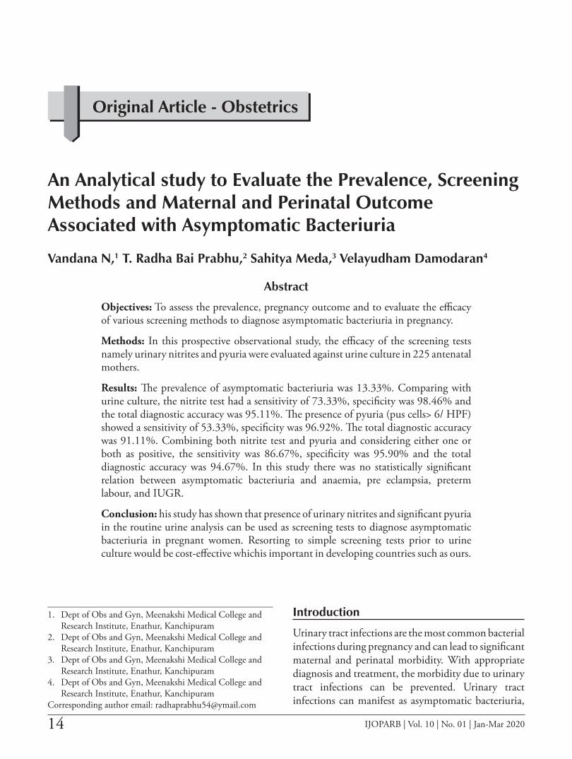
An Analytical study to Evaluate the Prevalence, Screening Methods and Maternal and Perinatal Outcome Associated with Asymptomatic Bacteriuria
Vandana N,1 T. Radha Bai Prabhu,2 Sahitya Meda,3 Velayudham Damodaran4
Abstract
Objectives: To assess the prevalence, pregnancy outcome and to evaluate the efficacy of various screening methods to diagnose asymptomatic bacteriuria in pregnancy.
Methods: In this prospective observational study, the efficacy of the screening tests namely urinary nitrites and pyuria were evaluated against urine culture in 225 antenatal mothers.
Results: The prevalence of asymptomatic bacteriuria was 13.33%. Comparing with urine culture, the nitrite test had a sensitivity of 73.33%, specificity was 98.46% and the total diagnostic accuracy was 95.11%. The presence of pyuria (pus cells> 6/ HPF) showed a sensitivity of 53.33%, specificity was 96.92%. The total diagnostic accuracy was 91.11%. Combining both nitrite test and pyuria and considering either one or both as positive, the sensitivity was 86.67%, specificity was 95.90% and the total diagnostic accuracy was 94.67%. In this study there was no statistically significant relation between asymptomatic bacteriuria and anaemia, pre eclampsia, preterm labour, and IUGR.
Conclusion: his study has shown that presence of urinary nitrites and significant pyuria in the routine urine analysis can be used as screening tests to diagnose asymptomatic bacteriuria in pregnant women. Resorting to simple screening tests prior to urine culture would be cost-effective whichis important in developing countries such as ours.
Introduction
Urinary tract infections are the most common bacterial infections during pregnancy and can lead to significant maternal and perinatal morbidity. With appropriate diagnosis and treatment, the morbidity due to urinary tract infections can be prevented. Urinary tract infections can manifest as asymptomatic bacteriuria,
________________________________________________1. Dept of Obs and Gyn, Meenakshi Medical College and
Research Institute, Enathur, Kanchipuram2. Dept of Obs and Gyn, Meenakshi Medical College and
Research Institute, Enathur, Kanchipuram3. Dept of Obs and Gyn, Meenakshi Medical College and
Research Institute, Enathur, Kanchipuram4. Dept of Obs and Gyn, Meenakshi Medical College and
Research Institute, Enathur, KanchipuramCorresponding author email: [email protected]
Original Article - Obstetrics
15
Analytical study to Evaluate Prevalence, Screening Methods & Maternal & Perinatal Outcome Associated with Asymptomatic Bacteriuria
IJOPARB | Vol. 10 | No. 01 | Jan-Mar 2020
Analytical study to Evaluate Prevalence, Screening Methods & Maternal & Perinatal Outcome Associated with Asymptomatic Bacteriuria
acute urethritis, acute cystitis or pyelonephritis. Asymptomatic bacteriuria refers to the presence of more than 1,00,000 colonies of a single bacterial species per millilitre of urine, cultured from midstream sample, in the absence of symptoms. Worldwide the incidence of asymptomatic bacteriuria varies from 5 to 10% and depends on the age, parity, race and socio-economic status.1 By virtue of short urethra and being in close proximity to the vagina, women are more prone for urinary tract infections, more so during pregnancy. The reduced immunity during pregnancy and thestasis due to ureteral dilatation, not only encourages the growth of organisms, but also allows progression to pyelonephritis. Untreated asymptomatic bacteriuria has been shown to be associated with obstetric problems such as preeclampsia, preterm labour, intra-uterine growth restriction (IUGR) and low birth weight infants.2 The relatively high prevalence of asymptomatic bacteriuria in pregnancy, and its reported adverse pregnancy outcome, justifies screening of pregnant women for the presence of bacteriuria. The gold standard diagnostic test to diagnose bacteriuria is the culture of urine. However, the culture test is relatively expensive and is time consuming and may not be feasible to carry out the test universally on all pregnant mothers especially in low resource settings. The screening methods that are available are: testing of urine for pyuria, nitrites, and dipstick methods and a positive test would indicate the need for diagnostic urine culture. These tests are simple and less expensive. In a developing country like India, it is important to use a screening method which is simple and cost-effective.
Aims and Objectives:
The present study was undertaken to assess the prevalence, pregnancy outcome and to evaluate the efficacy of various screening methods to diagnose asymptomatic bacteriuria in pregnancy.
Materials and Methods
The study was conducted in the department of Obstetrics and Gynaecology, Meenakshi Medical College, Kanchipuram from February 2018 to January 2019. This was a prospective observational study on the prevalence of asymptomatic bacteriuria and its maternal and its perinatal outcome. The efficacy of the screening tests: namely urinary nitrites and urine microscopy in diagnosing asymptomatic bacteriuria
was evaluated against the gold standard test the urine culture. Ethical committee approval was obtained from the Institutional Review Board. Based on the prevalence of asymptomatic bacteriuria in the previous studies and the number of new antenatal registrations in one year, the sample size was calculated. The study included 225 pregnant mothers who were willing to participate in the study. Antenatal mothers with past history of urinary tract infections (UTI) and those who presented with symptoms suggesting UTI in the current pregnancy were excluded from the study.
After explaining the details of the study, informed consent was obtained from each participant. Using a well structured, pretested proforma, information including demographic details, obstetric history, past medical history, examination findings, maternal and perinatal outcome were collected. Midstream urine sample was collected for routine analysis, microscopy and culture.
Investigations:
The investigations were carried out in the department of Microbiology of Meenakshi Medical College & Research Institute.
• For testing for Pyuria, 5 ml of urine sample was centrifuged at 5000 rpm and the sediment was examined under microscope for the number of pus cells present. The presence of >6/ HPF (high power field) was considered positive.
• For testing for Nitrites, biochemical reagent strips (uro color TM10) was used and the colour change was considered positive. (If bacteria is present in the urine it converts endogenous nitrates into nitrites).
• For urine Culture, a semi quantitative, calibrated loop technique was adopted for the primary isolation of organism. An loop full of uncentrifuged urine was streaked on the surface of Cysteine Lactose Electrolyte Deficient Agar plate and Blood Agar plate using aseptic technique. The plates were incubated at 37 degree Celsius for 24 hours. Isolated bacteria were counted and reported as the number of colony forming units per milli litre of urine. The diagnostic criteria for asymptomatic bacteriuria was the growth of a single species of bacteria more than or equal to 105 CFU/ ml of urine in the absence of symptoms.
16 IJOPARB | Vol. 10 | No. 01 | Jan-Mar 2020
Original Article - Obstetrics
Statistical Analysis:
IBM SPSS version 22 was used for statistical analysis.
Descriptive analysis was carried out by mean and standard deviation for quantitative variables, frequency and proportion for categorical variables. Urine culture was considered as the gold standard diagnostic tests. The Nitrite test and significant pyuria (> 6 pus cells /HPF) were considered as screening tests. The specific screening tests: Nitrite test and pyuria were evaluated against urine culture by using bivariate two by two tables. Sensitivity, specificity, Positive Predictive Value (PPV) and Negative Predictive Value (NPV) and the diagnostic accuracy of the screening tests were calculated with 95 % confidence interval.
Results:
The mean age of the study population was 23.71 ± 4.54 years and the range was 18-34 years. Majority of patients (92%) belonged to socio-economic group III and IV. 63.56% were primigravidae and 36.44% were multigravidae. The mean gestational age was 14.38 ± 2.65 weeks and the range was 12-32 weeks. The gestational age at the time of study was between 12 to 20 weeks in nearly 95% of patients. Antenatal problem such as anaemia was seen in 36.4% of patients and majority of them had moderate anaemia. Twenty eight (12.4%) patients suffered from pre-eclampsia. Only four babies showed evidence of IUGR at birth and 6 mothers (2.67%) delivered pre-term and 219 mothers were delivered at term. The mean birth weight in this study was2.81 ± 0.27 and the range was 1.90 kg to 3.40 kg (Table 1).
Among the 225 pregnant mothers screened by Nitrite test and significant pyuria, 11.1% (25) were positive for Nitrite test, 9.78% (22) were positive for significant pyuria and 5.78% (13) were positive for both Nitrites and significant pyuria. Urine culture, the definite diagnostic test was positive in 30 (13.3%) mothers (Table 2). Among the 30 subjects who were positive for urine culture, E.Coli was the most predominant organism grown in 25 cases (83.33%), Klebsiella in 3(10%) mothers, and Staphylococcus aureus and Proteus in one case each.
Evaluation of screening tests:
In order to evaluate the efficacy of screening tests in diagnosing asymptomatic bacteriuria, the results of
Nitrite test and pyuria were evaluated against the gold standard test: the urine culture. The Nitrite test and pyuria, either alone or together were highly specific in predicting the disease; specificity ranging from 95.90% to 98.46%. However, the sensitivity of these tests were moderate ranging from 53% to 86%, the Nitrite test and the combined test showing better sensitivity. The negative predictive value of Nitrite test was 96%, pyuria was 93.1% and that of combined test was 97.9%. The total diagnostic accuracy of screening tests ranged from 91% to 95% (Table 3). On analysing the association between asymptomatic bacteriuria and maternal and perinatal complications such as anaemia, pre- eclampsia, IUGR and preterm births, the difference in the proportion between the positive and negative culture test was statistically not significant (Table 4).
Follow-up: Women who were positive by urine culture were treated as per the sensitivityprofile. As 215 mothers (95.5%) were screened in early pregnancy, screening was repeated between 32 to 34 weeks of gestation. Except two mothers, all the others were negative by urine culture.
Discussion:
Asymptomatic bacteriuria is defined as persistently and actively multiplying bacteria more than or equal to 105 bacteria per millilitre (ml) within the urinary tract without any obvious symptoms. Women are more susceptible to UTI than men. Females have short urethra and the anatomical closeproximity to the vagina predisposes them to urinary tract infections. During pregnancy the prevalence of UTI is further increased. Number of factors contributes to the increased prevalence of UTI in pregnancy. During pregnancy the dilated ureters, glycosuria, and aminoaciduria give an excellent culture medium for the growth of bacteria.3 Associated co-morbid conditions like Gestational Diabetes Mellitus also add to the burden. There is also decreased levels of serum interleukin and serum antibody responses to E Coli antigens.4 The prevalence of asymptomatic bacteriuria in our study was 13.3%. Globally the prevalence of asymptomatic bacteriuria in pregnancy is reported to vary from 4% - 23.9%.5 Various Indian studies have shown the prevalence of asymptomatic bacteriuria in pregnancy to vary from 7.4% to 11.8%.6 In rural areas, the prevalence of asymptomatic bacteriuria is

17
Analytical study to Evaluate Prevalence, Screening Methods & Maternal & Perinatal Outcome Associated with Asymptomatic Bacteriuria
IJOPARB | Vol. 10 | No. 01 | Jan-Mar 2020
more due to their low socioeconomic status, poor hygiene, sanitation and lack of antenatal clinical visits.2 Untreated asymptomatic bacteriuria has adverse effect on maternal, fetal and neonatal health. If asymptomatic bacteriuria is untreated, it can lead to acute cystitis in 40% of cases and pyelonephritis in 20-30% of cases during the pregnancy It can also cause septicaemia and acute respiratory distress syndrome in 2% of cases.7 It can also lead to adverse obstetric events such as anaemia, pre-eclampsia, abortions, pre-term labour, IUGR and low birth weight infants and puerperal sepsis. In Byna et al study, compared to healthy pregnant women, those diagnosed with asymptomatic bacteriuria showed higher incidence of complications; anaemia in 35%, PROM in 14%, preterm labour in 18% and pre-eclampsia in 14%. The postpartum complications were wound infection in 5% and puerperal fever in 14%. The low APGAR, low birth weight and neonatal infections were observed in 19%, 20% and 8% respectively.8 Both asymptomatic bacteriuria and pyelonephritis are associated with the maternal anemia and hypertension, which are important causesof maternal and perinatal morbidity and mortality. Our study has not shown significant association between asymptomatic bacteriuria and adverse maternal and perinatal outcome. The possible reason could be, nearly 95% of women were screened early in pregnancy, were treated as per the sensitivity and were followed up with repeat sampling of urine. None of the 225 mothers were symptomatic and only in two mothersasymptomatic bacteriuria was detected late in pregnancy.
The high prevalence, as well as the adverse maternal and perinatal outcome associated with untreated asymptomatic bacteriuria, screening and treatment of asymptomatic bacteriuria is mandatory to avoid maternal and fetal complications. It is also cost beneficial when compared to not performing screening test.9
Various screening methods are used for the identification of asymptomatic bacteriuria during pregnancy. Urine culture is the gold standard method for the detection of bacteriuria in pregnancy. It is considered as the most reliable method for the diagnosis of urinary tract infection.10 Various International organisations such as The Infectious Disease Society of America and United States Preventive Services Task Force have recommended screening by culture in early
pregnancy.11,12 The American Academy of Family Physicians have recommended that pregnant women should be screened for asymptomatic bacteriuria in the first trimester of pregnancy.13 Studies have shown that routine screening for asymptomatic bacteriuria should be carried out in the first trimester itself as it can reduce the risk of adverse maternal and fetal outcome later inpregnancy.14 In our study, 95.5% of women were screened in early pregnancy less than 20 weeks of gestation. Jain, V., et al. study compared the detection and treatment of asymptomatic bacteriuria in early and late pregnancy, and showed that there was increased incidence of preeclampsia, preterm premature rupture of membrane, preterm labour, intrauterine growth restriction (IUGR), low birth weight (LBW) in late detected women (32-34 weeks) as compared to healthypregnant women. Whereas was no significant difference in obstetric outcome between asymptomatic bacteriuria detected early in pregnancy (<20weeks) and healthy pregnant women.15
Though the definitive test to diagnose bacteriuria is culture of urine., universal screening with culture during pregnancy may not be feasible in low resource settings, as the test is expensive and the necessary laboratory support and staffs may not be available. Moreover, as the prevalence of asymptomatic bateriuria during pregnancy is high, there is also a need for repetitive screening for UTI during pregnancy. Therefore, in developing countries like India, there is a need to use screening methods which can be repeated, not expensive as well as effective in identifying asymptomatic bacteriuria in pregnancy. The screening tests that are available are: pus cell count, nitrite test and Leucocyte esterase test and dipstick methods. In our study we have taken pus cell count and Nitrite test as screening tests and their efficacy was evaluated against the gold standard test: the urine culture.
On analysingthe efficacy of urine Nitrite test, the specificity was high at 98.46%, sensitivity was 73.3% and the negative predictive value was 96%. The specificity reported by other authors were 88.8%, 99.6% & 97.05% respectively.16,17,18 Similar to our study, other authors have also reported a sensitivity of 75.4%, 74.4% & 79.31%.16,17,18 The negative predictive value of 96.00% is also comparable to other authors’study; 95.5% reported by jayalakshmi et al and 97.77% by Sushma et al.16,18
18 IJOPARB | Vol. 10 | No. 01 | Jan-Mar 2020
Original Article - Obstetrics
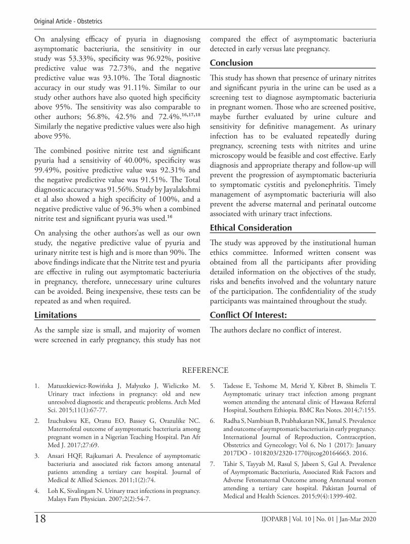
On analysing efficacy of pyuria in diagnosisng asymptomatic bacteriuria, the sensitivity in our study was 53.33%, specificity was 96.92%, positive predictive value was 72.73%, and the negative predictive value was 93.10%. The Total diagnostic accuracy in our study was 91.11%. Similar to our study other authors have also quoted high specificity above 95%. The sensitivity was also comparable to other authors; 56.8%, 42.5% and 72.4%.16,17,18 Similarly the negative predictive values were also high above 95%.
The combined positive nitrite test and significant pyuria had a sensitivity of 40.00%, specificity was 99.49%, positive predictive value was 92.31% and the negative predictive value was 91.51%. The Total diagnostic accuracy was 91.56%. Study by Jayalakshmi et al also showed a high specificity of 100%, and a negative predictive value of 96.3% when a combined nitrite test and significant pyuria was used.16
On analysing the other authors’as well as our own study, the negative predictive value of pyuria and urinary nitrite test is high and is more than 90%. The above findings indicate that the Nitrite test and pyuria are effective in ruling out asymptomatic bacteriuria in pregnancy, therefore, unnecessary urine cultures can be avoided. Being inexpensive, these tests can be repeated as and when required.
Limitations
As the sample size is small, and majority of women were screened in early pregnancy, this study has not
compared the effect of asymptomatic bacteriuria detected in early versus late pregnancy.
Conclusion
This study has shown that presence of urinary nitrites and significant pyuria in the urine can be used as a screening test to diagnose asymptomatic bacteriuria in pregnant women. Those who are screened positive, maybe further evaluated by urine culture and sensitivity for definitive management. As urinary infection has to be evaluated repeatedly during pregnancy, screening tests with nitrites and urine microscopy would be feasible and cost effective. Early diagnosis and appropriate therapy and follow-up will prevent the progression of asymptomatic bacteriuria to symptomatic cystitis and pyelonephritis. Timely management of asymptomatic bacteriuria will also prevent the adverse maternal and perinatal outcome associated with urinary tract infections.
Ethical Consideration
The study was approved by the institutional human ethics committee. Informed written consent was obtained from all the participants after providing detailed information on the objectives of the study, risks and benefits involved and the voluntary nature of the participation. The confidentiality of the study participants was maintained throughout the study.
Conflict Of Interest:
The authors declare no conflict of interest.
REFERENCE
1. Matuszkiewicz-Rowińska J, Małyszko J, Wieliczko M. Urinary tract infections in pregnancy: old and new unresolved diagnostic and therapeutic problems. Arch Med Sci. 2015;11(1):67-77.
2. Izuchukwu KE, Oranu EO, Bassey G, Orazulike NC. Maternofetal outcome of asymptomatic bacteriuria among pregnant women in a Nigerian Teaching Hospital. Pan Afr Med J. 2017;27:69.
3. Ansari HQF, Rajkumari A. Prevalence of asymptomatic bacteriuria and associated risk factors among antenatal patients attending a tertiary care hospital. Journal of Medical & Allied Sciences. 2011;1(2):74.
4. Loh K, Sivalingam N. Urinary tract infections in pregnancy. Malays Fam Physician. 2007;2(2):54-7.
5. Tadesse E, Teshome M, Merid Y, Kibret B, Shimelis T. Asymptomatic urinary tract infection among pregnant women attending the antenatal clinic of Hawassa Referral Hospital, Southern Ethiopia. BMC Res Notes. 2014;7:155.
6. Radha S, Nambisan B, Prabhakaran NK, Jamal S. Prevalence and outcome of asymptomatic bacteriuria in early pregnancy. International Journal of Reproduction, Contraception, Obstetrics and Gynecology; Vol 6, No 1 (2017): January 2017DO - 1018203/2320-1770ijrcog20164663. 2016.
7. Tahir S, Tayyab M, Rasul S, Jabeen S, Gul A. Prevalence of Asymptomatic Bacteriuria, Associated Risk Factors and Adverse Fetomaternal Outcome among Antenatal women attending a tertiary care hospital. Pakistan Journal of Medical and Health Sciences. 2015;9(4):1399-402.
19
Analytical study to Evaluate Prevalence, Screening Methods & Maternal & Perinatal Outcome Associated with Asymptomatic Bacteriuria
IJOPARB | Vol. 10 | No. 01 | Jan-Mar 2020
8. Byna P, Muvva N, Kolli S, Shaik MV. A study of risk factors and consequences of asymptomatic bacteriuria in pregnant women and feto-maternal outcome. Int J Reprod Contracept Obstet Gynecol. 2015;4(5):1300-5.
9. Ansari HQF, Rajkumari A. Prevalence of asymptomatic bacteriuria and associated risk factors among antenatal patients attending a tertiary care hospital. Journal of Medical & Allied Sciences. 2011;1(2):74.
10. Abdullah AA, Al-Moslih MI. Prevalence of asymptomatic bacteriuria in pregnant women in Sharjah, United Arab Emirates. East Mediterr Health J. 2005;11(5-6):1045-52.
11. Nicolle LE, Bradley S, Colgan R, Rice JC, Schaeffer A, Hooton TM. Infectious Diseases Society of America guidelines for the diagnosis and treatment of asymptomatic bacteriuria in adults. Clinical infectious diseases. 2005:643-54.
12. Screening for asymptomatic bacteriuria in adults: U.S. Preventive Services Task Force reaffirmation recommendation statement. Ann Intern Med. 2008;149(1):43-7.
13. American Academy of Family Physicians. Summary of recommendations for clinical preventive services. Revision 6.4 August 2007 Accesssed at www.aafp.org/online/en/home/clinical/exam.html.
14. Kasinathan A, Thirumal P. Prevalence of asymptomatic bacteriuria in antenatal women attending a tertiary care hospital. International Journal of Reproduction, Contraception, Obstetrics and Gynecology. 2014;3(2):437-41.
15. Jain V, Das V, Agarwal A, Pandey A. Asymptomatic bacteriuria & obstetric outcome following treatment in early versus late pregnancy in north Indian women. Indian J Med Res 2013;137(4):753-8.
16. K.V. Leela, Thyagarajan Ravinder, Shemalatha et al Evaluation of screening tests to detect asymptomatic bacteriuria in antenatal women. Int. J. Curr. Microbio. App. Sci (2017)6(1):168-174
17. J. Jayalakshmi, V.S. Jayaraman, Evaluation of screening tests to detect asymptomatic bacteriuria in pregnant women. Indian Journal of Pathology and Microbiology 51(3) July- September 2008 379-381
18. Sushama S. Thakre, Supriya S. Dhakne and Priya Kale, Can the Griess Nitrite test and urine pus cells count in pregnant women be used for screening or early detection of Urinary Tract Infection in Rural India. J Clin Diag Res. 2012 Nov; 6(9):1518-1522
20 IJOPARB | Vol. 10 | No. 01 | Jan-Mar 2020
Role of Combination of Mifepristone and Misoprostol versus Misoprostol alone in Induction of Labor in Intra Uterine Fetal Death
Dr Sweta,1 Dr Kusum Dash2
Abstract
Objective: To compare efficacy, safety and toleranceof combination of mifepristone and misoprostol versus misoprostol alone in induction of labour in late intra uterine foetal death (IUFD).
Materials and Methods: This randomized prospective study included 60 women gravid up to fourth with IUFD after 24 weeks of gestation. Women were divided into two groups. Group 1 received a single dose of 200mg of mifepristone; and after 24 hours 100µgm of intra-vaginal misoprostol administered, followed by intra-vaginal misoprostol at four hourly intervals if required. Group 2 received 100µgm misoprostol at four hourly intervals intra-vaginally. Misoprostol was given for maximum of six doses in either group. Primary outcome measures were achievement of successful delivery and induction delivery interval (IDI). Women who did not deliver after six doses of misoprostol were considered as failure. In all the women, induction delivery interval, total doses of misoprostol and adverse effect of the drug were noted. Data were analysed by using Student t test and Chi-square test.
Result: Successful delivery was 93.3% and 90.0% in group 1 and 2 respectively. IDI was significantly less in the Group I (10.53±5.13) compared with Group II (18.43±4.63) with p<0.001. Parity, gestation, and bishop score did not affect the IDI in the two groups. Complications were experienced more in misoprostol group due to higher doses of misoprostol.
Conclusion: Pre-treatment with mifepristone is more effective in terms of reducing induction delivery interval, requirement of lesser dose of misoprostol and its related side effects.
Keywords: IUFD, mifepristone, misoprostol, induction of labor
Original Article - Obstetrics
_________________________________________________1. MBBS, DNB Obstetrics & Gynaecology, Junior Consultant, Mahavir Vatsalya Aspatal, Patna, Bihar2. MBBS, DNB Obstetrics & Gynaecology, Joint Director & Unit Head, Bokaro General Hospital, Bokaro Steel CityCorresponding author: [email protected]
21
Role of Combination of Mifepristone and Misoprostol versus Misoprostol alone in Induction of Labor in Intra Uterine Fetal Death
IJOPARB | Vol. 10 | No. 01 | Jan-Mar 2020
Introduction:
Motherhood is an all-encompassing and life altering experience that changes a women’s life forever. Motherhood is achieved through the journey of labour pain, which is described many bones breaking simultaneously.
Scenario in case of intra uterine foetal death (IUFD) is entirely different. Besides being emotionally challenging, late foetal demise raises a host of questions and increases an obstetrician’s medico legal risk. The assessment and management of IUFD is unique because such losses differ significantly from those occurring in early pregnancy. The patient and her family will ask “why”, seeking answers from the theological to the medical.
IUFD is encountered in 1% of all pregnancies.1-3 90% of mothers with IUFD deliver spontaneously within 34 weeks, If the dead foetus is retained for more than 4 weeks, it can lead to intra uterine infection, consumptive coagulopathy and disseminated intravascular coagulation.5,6 Thus, social and maternal desires and a moderate risk of maternal complications compel the caregiver to induce labour soon after diagnosis, aiming for a safe and speedy delivery.
Various methods have been tried in the management of intrauterine deaths. Before the introduction of the prostaglandins, women with intrauterine death were managed by giving repeated high dosage of estrogens,7 intra amniotic injections of hypertonic solutions8 use of hygroscopic tents9 boogies, catheter and balloon10,11 or more frequently with repeated high dose infusion of oxytocin.12,13 Oral Misoprostol administration for labour induction with an IUFD was first described in Sao Paulo, Brazil in 1987. Thereafter misoprostol were widely used for induction of labour in IUFD.14,15 But repeated dose requirements and side-effects such as uterine over activity (hyper stimulation, hyper tonicity and tachysystole) and systemic response (nausea, vomiting, diarrhoea and shivering) always remain area of concern.
The role of mifepristonefor uterine priming was first reported by Cabrol et al.16 The advantages of mifepristone pre-treatment prior to misoprostol administration on the basis of its pharmacodynamics17 have been demonstrated without a doubt. It has an accepted role in the first and second trimester
termination of pregnancy.18-21 Mifeprestone blocks progesterone receptors, thus inhibiting the influence of progesterone. This leads to the softening and dilatation of the uterine cervix and increased sensitivity of the uterus to prostaglandins. Thus lower doses of misoprostol are required and hence the associated side effects and the induction delivery interval in IUFD which is of utmost importance. However, the evidence related to the use of such a combination for induction of labour in 2nd and 3rd trimester IUFD is still deficient in the absence of well-designed randomized controlled trials (RCT). Aim of my study is to evaluate safety, efficacy and tolerance of combination regimen of mifepristone and misoprostol with conventional use of misoprostol alone in IUFD whose POG is >24 weeks.
Material and Methods
This randomized prospective study included 60 women with IUFD after 24 weeks of gestation attending department of Obstetrics and Gynaecology of Bokaro General Hospital from June 2016 to December 2018
INCLUSION CRITERIA were as follows; i) women who understand the procedure and give consent, ii) not in labor, iii) with singleton fetus, iv) with gestational age ≥24 weeks
Exclusion criteriawere; i) Women in active labour, ii) previous trans mural uterine incision, iii) with multiple pregnancies, iv) with grand multipara ≥4, v) with evidence of coagulopathy, vi) with serious medical or obstetrical condition which needs immediate delivery, vii) with Known allergy / contra-indication to use of Mifepristone or Misoprostol, viii) With known case of epilepsy, heart disease, asthma, glaucoma
Thewomen were randomized into two groups by lottery method, Assessment of eligibility, enrolment and procurement of informed consent were performed by the researchers, while assignment to intervention, administration of drugs and maintenance of confidential records were performed by residents not involved with the study. Women in group 1 (combination group) were given a single dose of 200mg of mifepristone and after 24 hours, 100µgm of intra vaginal misoprostol administered, followed by intra vaginal 100µgm misoprostol at 4 hourly intervals if required. Women in group 2 (misoprostol only group) were given 100µgm of misoprostol at 4hrly interval
22 IJOPARB | Vol. 10 | No. 01 | Jan-Mar 2020
Original Article - Obstetrics
for maximum of six doses. The time, date of induction and total number of doses of misoprostol was noted carefully. After 1st dose of misoprostol, vital signs, any side effects, uterine contraction, systemic symptoms, labour progress (in modified WHO partograph), and blood loss were recorded. Oxytocin in active labour was used if required. Active management of third stage of labour performed. Any adverse events, such as postpartum haemorrhage or retained placenta, were recorded. If the placenta was retained for more than 30m after delivery of the foetus, additional intervention was performed. In the event of failure to deliver the foetus and/or placenta within 24hr after the first dose of misoprostol, an alternative protocol was followed i.e. augmentation of labour was done by oxytocin infusion.
The rate of successful delivery (within 24h of commencement of the first dose of misoprostol) and induction to delivery interval from the first dose of misoprostol to complete delivery of foetus and placenta noted carefully. The procedure was failed if delivery not occurred in 24 hour of first dose of misoprostol in both groups.
Statistical analysis was done by student, s T test and “chi-square test”.
A “p-value” should be considered to be non-significant if > 0.05 and significant if <0.05.
Results
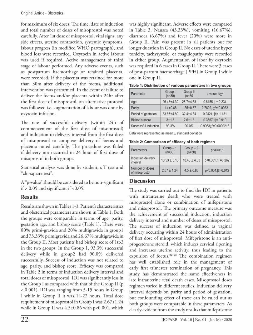
Results are shown in Tables 1-3. Patient’s characteristics and obstetrical parameters are shown in Table 1. Both the groups were comparable in terms of age, parity, gestation age, and bishop score (Table 1). There were 80% primi-gravida and 20% multigravida in group1 and 73.33% primigravida and 26.67% multigravida in the Group II. Most patients had bishop score of 1to3 in the two groups. In the Group 1, 93.3% successful delivery while in group2 had 90.0% delivered successfully. Success of induction was not related to age, parity, and bishop score. Efficacy was compared in Table 2 in terms of induction delivery interval and total doses of misoprostol. IDI was significantly less in the Group I as compared with that of the Group II (p < 0.001). IDI was ranging from 5-15 hours in Group I while in Group II it was 14-22 hours. Total dose requirement of misoprostol in Group I was 2.67±1.24 while in Group II was 4.5±0.86 with p<0.001, which
was highly significant. Adverse effects were compared in Table 3. Nausea (43.33%), vomiting (16.67%), diarrhoea (6.67%) and fever (20%) were more in Group II. Pain was present in all patients but for longer duration in Group II. No cases of uterine hyper tonicity, tachysystole, or coagulopathy were recorded in either group. Augmentation of labor by oxytocin was required in 6 cases in Group II. There were 3 cases of post-partum haemorrhage (PPH) in Group I while one in Group II.Table 1: Distribution of various parameters in two groups
Parameter Group I(n=30)
Group II(n=30 p value, t/χ2
Age 26.43±4.39 26.7±4.53 0.8155|t| = 0.234Parity 1.4±0.68 1.35±0.67 0.7602, χ2= 0.0932Period of gestation 33.87±4.80 32.4±4.84 0.2424, |t|= 1.181Bishop’s score 3±1.6 2.6±1.8 0.3667,|t|= 0.910Successful induction 93.3% 90.0% 0.9963χ2=0.0000218
Data were represented as mean ± standard deviation
Table 2: Comparison of efficacy of both regimen
Parameters Group – 1 (n=30)
Group – 2 (n=30) p value, t
Induction delivery interval 10.53 ± 5.13 18.43 ± 4.63 p<0.001,|t| =6.262
Number of doses of misoprostol 2.67 ± 1.24 4.5 ± 0.86 p<0.001,|t|=6.642
Discussion
The study was carried out to find the IDI in patients with intrauterine death who were treated with misoprostol alone or combination of mifepristone and misoprostol. The primary outcome measure was the achievement of successful induction, induction delivery interval and number of doses of misoprostol. The success of induction was defined as vaginal delivery occurring within 24 hours of administration of first dose of misoprostol. Mifepristone is an anti-progesterone steroid, which induces cervical ripening and increases uterine activity, thus leading to the expulsion of foetus.22,23 The combination regimen has well established role in the management of early first trimester termination of pregnancy. This study has demonstrated the same effectiveness in late intrauterine fetal death cases. Misoprostol doses regimen varied in different studies. Induction delivery interval depends on parity and period of gestation, but confounding effect of these can be ruled out as both groups were comparable in these parameters. As clearly evident from the study results that mifepristone

23
Role of Combination of Mifepristone and Misoprostol versus Misoprostol alone in Induction of Labor in Intra Uterine Fetal Death
IJOPARB | Vol. 10 | No. 01 | Jan-Mar 2020
and misoprostol combination had shorter induction delivery interval. This is in agreement with study done by Wagaarachchi et al. (2002),24 Panda et al (2008-2011)25 and Sharma et al (2011).26
Dose of misoprostol required was significantly higher in misoprostol group which can be explained on the basis pharmacodynamics of mifepristone as mentioned earlier. The result of our study is not comparable to the study of Vayrynen et al,27 who studied 130 womenwith intrauterine fetal death (21-42 weeks of gestation). In their study, 82 women received 100µg misoprostol at 4-hour interval and 48 women received 200 mg mifepristone followed 19 hours later by misoprostol 25 µg at 4-hour interval and found that induction to delivery interval did not differ between the two groups (13.3 hours vs. 12.8 hours). The reason for the similar induction delivery interval in both groups may be the lower dose of misoprostol as they have used only 25 µg misoprostol in group which was pretreated with mifepristone, whereas 100µ g misoprostol in another group which was not pretreated with mifepristone. More cases required analgesia in misoprostol group as compared to combination group which can be directly correlated with length of contraction or duration of
labor. Although there is possibility of confounding factor of patient’s perception of pain. Gastrointestinal side effects were noticed in 16% of oral misoprostol group. Similar result was seen by Fairley et al28 (15%). We prefer to keep patient in hospital but they can be sent home after giving mifepristone with proper counseling and to return after 24hr/SOS.
Conclusion
Pretreatment with mifepristone is more effective as it shortens the duration of labor without any increase in adverse effect. In intrauterine foetal death case, mifepristone plus misoprostol is an effective regimen to cut short the fruitless journey of labor pain. It is safe, easily tolerable, and more efficacious than conventional regimen of misoprostol alone. However, conventional regimen with misoprostol alone may be appropriate in settings where cost is a prohibitive factor because successful delivery rate was almost similar in both group
Acknowledgments There is no financial support from any institution or company for this study. There is no conflict of interest with this study
Table 3: Comparison of side effect between two groups
Study ParameterGroup – I (n=30) Group –II (n=30)
|Z| cal d.f P valueNO. % NO. %
Pain 30 100% 30 100% Not appliedFever 2 6.67% 6 20% 1.297 1 0.2547Nausea 8 26.67% 13 43.33% 1.171 1 0.2792Vomiting 3 10% 5 16.67% 0.145 1 0.7038Diarrhoea 1 3.33% 2 6.67% 14x10-6 1 0.9991Analgesia requirement 10 33.3% 17 56.67% 4.310 1 0.1193Oxytocin in active labour 3 10% 6 20% 0.523 1 0.4694PPH 3 10% 1 3.33% 0.268 1 0.6044
For Test of Significance, Here we use “Proportion test |z| - test” at 95% confidence limit
1. National Institute for Health and Clinical Excellence (2008) Induction of labour. NICE clinical guideline 70
2. RCOG late intra uterine fetal death, and still birth Green–top Guideline No. 55 October 2010
3. Gomez Ponce de León R, Wing D, Fiala C. Misoprostol for intrauterine fetal death. Int J GynaecolObstet 2007; 99: S190–S193.
4. Silver TM. Fetal death. ObstetGynecol 2007; 109: 153 –167.
5. Rådestad I, Steineck G, Nordin C, Sjögren B. Psychological complications after stillbirth – influence of memories and immediate management: Population based study. BMJ 1996; 312: 1505–1508
6. Parasnis H, Raje B, HindujaI N. Relevance of plasma fibrinogen estimation in obstetric complications. J Postgrad Med 1992; 38: 183–185.
7. Menzies DN. Oestrogen therapy in missed abortion and labor. J ObstetGynecol Br Empire 1955;62:256e8.
REFERENCE

24 IJOPARB | Vol. 10 | No. 01 | Jan-Mar 2020
Original Article - Obstetrics
Journal Subscription Rates for ISOPARB Non Members / Institutions /
Medical Libraries / Corporates
Single Issue Subscription 4500.00One Year Subscription (4 issues) 6000.00Two Year Subscription (8 issues) 10000.00Five Year Subscription (20 issues) 25000.00
Contact Editorial Office or mail to [email protected]
8. Courtney LD, Boxall RR, Child P. Permeability of membranes of dead fetus. Br Med J 1971;8:492e3.
9. Lackritz R, Gibson M, Frigoletto FD. Preinduction use of laminaria for the unripe cervix. Am J ObstetGynecol 1979;134:349e50.
10. Lewis GJ. Cervical ripening before induction of labor with prostaglandin E2 pessaries or a Foley’s catheter. J ObstetGynecol 1983;3: 173e6.
11. Obed JY, Adewole IF. The unfavourable cervix: improving the Bishop score with the Foley’s catheter. West Afr J Med 1994;13:209e12.
12. Loudon JDO. The use of high concentration oxytocin intravenous drips in the management of missed abortion. J ObstetGynecol Br Empire 1959;66:271e81.
13. Liggins GG. The treatment of missed abortion by high dosage syntocinon intravenous infusion. J ObstetGynecol Br Empire 1962;69:227e81.
14. Gomez Ponce de León R, Wing DA. Misoprostol for termination of pregnancy with intra-uterine fetal demise in the second and third trimester of pregnancy - a systematic review. Contraception 2009; 79: 259 –271.
15. Gawron LM, Kiley JW. Labor induction outcomes in thirdtrimester stillbirths. Int J GynecolObstet 2013; 123: 203
16. Cabrol D, Bouvier D, Mermet E. Induction of labour with mifepristone after intra uterine death lancet 1985:15:1019-20
17. Robbins A, Splitz IM. Mifepristone: Clinical pharmacology. ClinObstetGynecol 1996; 39: 436-450
18. Gemzell –Danielsson k and Lalitkumar s. second trimester medical abortion with mifepristone and misoprostol and misoprostol alone: a review of methods and management Reprod Health Matters 2008: 16:162-72
19. Bartz D and Goldberg A. medication abortion. ClinObstetGynecol 2009:52;140-501.
20. Ngoc NT, Shochet T, Raghavan S et al. Mifepristone and misoprostol compared with misoprostol alone for second-trimester abortion: A randomized controlled trial. ObstetGynecol 2011; 118: 601 –608.
21. Nagaria T, Sirmor N. Misoprostol vs mifepristone and misoprostol in second trimester termination of pregnancy. J ObstetGynaecol India 2011; 61: 659
22. Loose DS, Stancel GM. Estrogens and progestins. In: Goodman & Gilman’s the pharmacological basis of therapeutics. 11th ed. New York: McGraw Hill; 2006. p. 1541e72. [23] Jang GR, Benet LZ. Antiprogestin pharmacodynamics, pharmacokinetics and metabolism: implications for their long term use. J PharmacokinetBiopharm 1997;25:647e72.
24. Wagaarachchi PT, Ashok PW, Narvekar NN, Smith NC, Templeton A. Medical management of late intrauterine death using a combination of mifepristone and misoprostol. BJOG 2002;109:443-7
25. Subrat Panda; M.D., VandanaJha; M.D., Santa Singh; M.D.: Role of Combination OF Mifepristone and Misoprostol Verses Misoprostol alone in Induction of Labour in Late Intrauterin Fetal Death: A Prospective Study. Journal of Family and Reproductive Health;
26. Damyanti Sharma, Savita Rani Singhal*, Poonam, Anshu Paul, Kunika. Comparison of mifepristone combination with misoprostol and misoprostol alone in the management of intrauterine death. Taiwanese Journal of Obstetrics & Gynecology 50 (2011) 322-325
27. Vayrynen W, Heikinheimo O, Nuutila M. Misoprostol-only versus mifepristone plus misoprostol in induction of labor following intrauterine fetal death. ActaObstetriciaetGynaecologicaScandinavica. 2007;86:701-5.
28. Tara E. Fairley, Maureen Mackenzie, Philip Owen, Fiona Mackenzie. Management of late intrauterine death using a combination of mifepristone and misoprostol—experience of two regimens. European Journal of Obstetrics & Gynecology and Reproductive Biology 118 (2005) 28–31
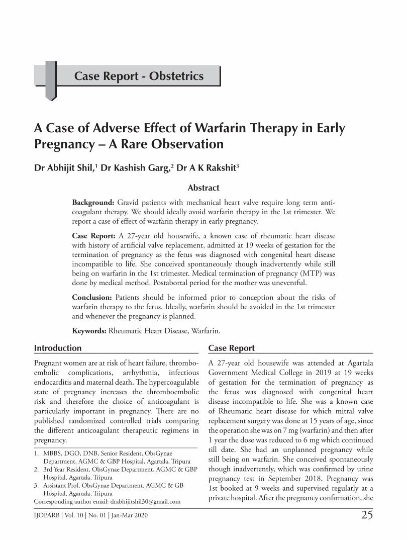
25IJOPARB | Vol. 10 | No. 01 | Jan-Mar 2020
A Case of Adverse Effect of Warfarin Therapy in Early Pregnancy – A Rare Observation
Dr Abhijit Shil,1 Dr Kashish Garg,2 Dr A K Rakshit3
Abstract
Background: Gravid patients with mechanical heart valve require long term anti-coagulant therapy. We should ideally avoid warfarin therapy in the 1st trimester. We report a case of effect of warfarin therapy in early pregnancy.
Case Report: A 27-year old housewife, a known case of rheumatic heart disease with history of artificial valve replacement, admitted at 19 weeks of gestation for the termination of pregnancy as the fetus was diagnosed with congenital heart disease incompatible to life. She conceived spontaneously though inadvertently while still being on warfarin in the 1st trimester. Medical termination of pregnancy (MTP) was done by medical method. Postabortal period for the mother was uneventful.
Conclusion: Patients should be informed prior to conception about the risks of warfarin therapy to the fetus. Ideally, warfarin should be avoided in the 1st trimester and whenever the pregnancy is planned.
Keywords: Rheumatic Heart Disease, Warfarin.
Introduction
Pregnant women are at risk of heart failure, thrombo-embolic complications, arrhythmia, infectious endocarditis and maternal death. The hypercoagulable state of pregnancy increases the thromboembolic risk and therefore the choice of anticoagulant is particularly important in pregnancy. There are no published randomized controlled trials comparing the different anticoagulant therapeutic regimens in pregnancy.
Case Report
A 27-year old housewife was attended at Agartala Government Medical College in 2019 at 19 weeks of gestation for the termination of pregnancy as the fetus was diagnosed with congenital heart disease incompatible to life. She was a known case of Rheumatic heart disease for which mitral valve replacement surgery was done at 15 years of age, since the operation she was on 7 mg (warfarin) and then after 1 year the dose was reduced to 6 mg which continued till date. She had an unplanned pregnancy while still being on warfarin. She conceived spontaneously though inadvertently, which was confirmed by urine pregnancy test in September 2018. Pregnancy was 1st booked at 9 weeks and supervised regularly at a private hospital. After the pregnancy confirmation, she
_________________________________________________1. MBBS, DGO, DNB, Senior Resident, ObsGynae
Department, AGMC & GBP Hospital, Agartala, Tripura2. 3rd Year Resident, ObsGynae Department, AGMC & GBP
Hospital, Agartala, Tripura3. Assistant Prof, ObsGynae Department, AGMC & GB
Hospital, Agartala, TripuraCorresponding author email: [email protected]
Case Report - Obstetrics
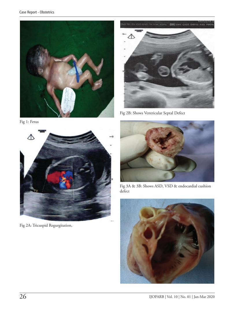
26 IJOPARB | Vol. 10 | No. 01 | Jan-Mar 2020
Case Report - Obstetrics
Fig 1: Fetus
Fig 2A: Tricuspid Regurgitation,
Fig 2B: Shows Ventricular Septal Defect
Fig 3A & 3B: Shows ASD, VSD & endocardial cushion defect
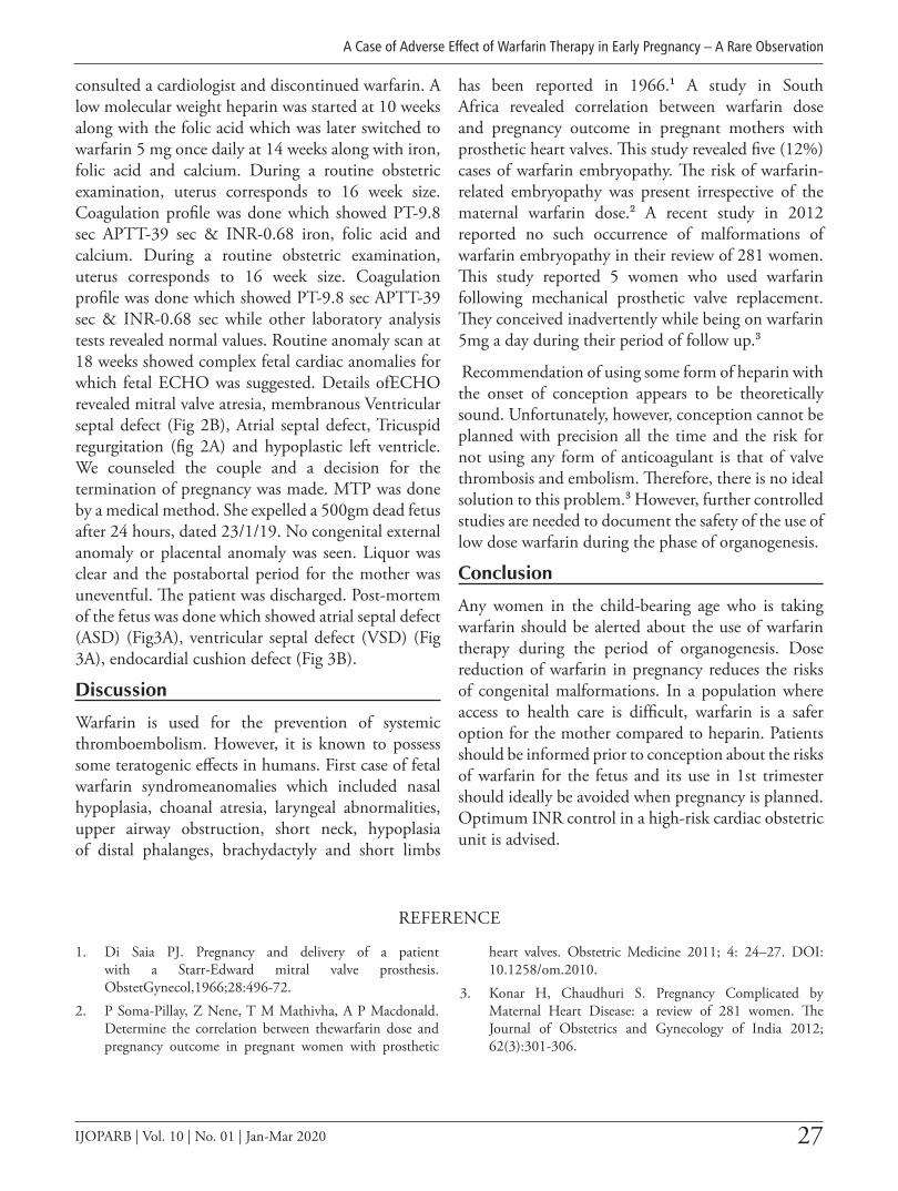
27
A Case of Adverse Effect of Warfarin Therapy in Early Pregnancy – A Rare Observation
IJOPARB | Vol. 10 | No. 01 | Jan-Mar 2020
consulted a cardiologist and discontinued warfarin. A low molecular weight heparin was started at 10 weeks along with the folic acid which was later switched to warfarin 5 mg once daily at 14 weeks along with iron, folic acid and calcium. During a routine obstetric examination, uterus corresponds to 16 week size. Coagulation profile was done which showed PT-9.8 sec APTT-39 sec & INR-0.68 iron, folic acid and calcium. During a routine obstetric examination, uterus corresponds to 16 week size. Coagulation profile was done which showed PT-9.8 sec APTT-39 sec & INR-0.68 sec while other laboratory analysis tests revealed normal values. Routine anomaly scan at 18 weeks showed complex fetal cardiac anomalies for which fetal ECHO was suggested. Details ofECHO revealed mitral valve atresia, membranous Ventricular septal defect (Fig 2B), Atrial septal defect, Tricuspid regurgitation (fig 2A) and hypoplastic left ventricle. We counseled the couple and a decision for the termination of pregnancy was made. MTP was done by a medical method. She expelled a 500gm dead fetus after 24 hours, dated 23/1/19. No congenital external anomaly or placental anomaly was seen. Liquor was clear and the postabortal period for the mother was uneventful. The patient was discharged. Post-mortem of the fetus was done which showed atrial septal defect (ASD) (Fig3A), ventricular septal defect (VSD) (Fig 3A), endocardial cushion defect (Fig 3B).
Discussion
Warfarin is used for the prevention of systemic thromboembolism. However, it is known to possess some teratogenic effects in humans. First case of fetal warfarin syndromeanomalies which included nasal hypoplasia, choanal atresia, laryngeal abnormalities, upper airway obstruction, short neck, hypoplasia of distal phalanges, brachydactyly and short limbs
has been reported in 1966.1 A study in South Africa revealed correlation between warfarin dose and pregnancy outcome in pregnant mothers with prosthetic heart valves. This study revealed five (12%) cases of warfarin embryopathy. The risk of warfarin-related embryopathy was present irrespective of the maternal warfarin dose.2 A recent study in 2012 reported no such occurrence of malformations of warfarin embryopathy in their review of 281 women. This study reported 5 women who used warfarin following mechanical prosthetic valve replacement. They conceived inadvertently while being on warfarin 5mg a day during their period of follow up.3
Recommendation of using some form of heparin with the onset of conception appears to be theoretically sound. Unfortunately, however, conception cannot be planned with precision all the time and the risk for not using any form of anticoagulant is that of valve thrombosis and embolism. Therefore, there is no ideal solution to this problem.3 However, further controlled studies are needed to document the safety of the use of low dose warfarin during the phase of organogenesis.
Conclusion
Any women in the child-bearing age who is taking warfarin should be alerted about the use of warfarin therapy during the period of organogenesis. Dose reduction of warfarin in pregnancy reduces the risks of congenital malformations. In a population where access to health care is difficult, warfarin is a safer option for the mother compared to heparin. Patients should be informed prior to conception about the risks of warfarin for the fetus and its use in 1st trimester should ideally be avoided when pregnancy is planned. Optimum INR control in a high-risk cardiac obstetric unit is advised.
REFERENCE
1. Di Saia PJ. Pregnancy and delivery of a patient with a Starr-Edward mitral valve prosthesis. ObstetGynecol,1966;28:496-72.
2. P Soma-Pillay, Z Nene, T M Mathivha, A P Macdonald. Determine the correlation between thewarfarin dose and pregnancy outcome in pregnant women with prosthetic
heart valves. Obstetric Medicine 2011; 4: 24–27. DOI: 10.1258/om.2010.
3. Konar H, Chaudhuri S. Pregnancy Complicated by Maternal Heart Disease: a review of 281 women. The Journal of Obstetrics and Gynecology of India 2012; 62(3):301-306.
28 IJOPARB | Vol. 10 | No. 01 | Jan-Mar 2020
Evaluation of Platelet Rich Plasma in Infertile Women
Dr Vatsal Thakral,1 Dr Swati Garg,2 Dr Urvashi Sharma3
Abstract
Objective: Platelet rich plasma (PRP), a promising regenerative therapeutic application can offer benefits withoutdetrimental side effects as it is a direct product of own blood sample. PRP has been employed in several fields of medicine like plastic surgery, maxilla-facial surgery, dental surgery, orthopaedics, eye surgery and gynaecology. Recently clinical trials have shown its beneficial effects in the field of infertility through its regenerative property on the endometrium. It is also being used in patients with poor ovarian reserve and Asherman’ssyndrome. Endometrial thickness (ET) is one of the main factors for implantation. PRP treatment could result in successful management of poor responders, patients with failed IVF attempts, poor oocyte yield and poor embryo quality.
Methodolgy: Our study includes 50 infertile patients (24-46 years) who have undergone PRP application. Patient selection criteria include suboptimal endometrium, IVF failure and poor oocyte yield.
Result: The mean pre-PRP ET was 5.9mm which significantly increased to 7.3mm post-PRP. There was a significant increase in vascularity seen on power Doppler.
Conclusion: This study suggests that the use of autologous PRP holds a promising rolr in the treatment of women with suboptimal ET and vascularity for embryo transfer.
Keywords: PRP, IVF Failure, Suboptimal endometrium.
Original Article - Gynecology
Introduction
PRP is a new promising regenerative therapeutic application which can offer therapeutic benefits without detrimental side effects as it is a direct product of own blood sample. PRP is blood plasma prepared from fresh whole blood that has been enriched with platelets. PRP is highly rich in several growth factors
hence it showcases many proliferative as well as anti-inflammatory effects while working on tissue repair. PRP has been employed in several fields of medicine like plastic surgery, maxillo-facial surgery, dental surgery, orthopaedics, eye surgery and gynaecology.1 Recently clinical trials have demonstrated that PRP can have many beneficial effects in the field of infertility through its regenerative property on the endometrium. It is also being used in patients with poor ovarian reserve and Asherman’s syndrome.
Clinical pregnancies have been described in endometrium from 5 to15 mm.2 In a meta-analysis by Kasius et al in 2014, the probability of clinical
_________________________________________________1. Resident 2nd Year, Dept of Obstetrics & Gynaecology,
Mahatma Gandhi Medical College & Hospital, Jaipur2. Professor & Head, Dept of Obstetrics & Gynaecology,
Mahatma Gandhi Medical College & Hospital, Jaipur3. Assistant Professor, Dept of Obstetrics & Gynaecology,
Mahatma Gandhi Medical College & Hospital, Jaipur.Corresponding Author: [email protected]
29
Evaluation of Platelet Rich Plasma in Infertile Women
IJOPARB | Vol. 10 | No. 01 | Jan-Mar 2020
pregnancy for an ET ≤ 7 mm was significantly lower compared with cases with ET > 7 mm [23.3 versus 48.1%].3
Thin refractory endometrium is still a challenge in ART. PRP is one of the novel therapies tried in such patients. Intrauterine instillation of PRP to improve endometrial quality in terms of thickness has been first used by Chang et al in 2015.4 The ET Increased at 48 to 72 hours after PRP infusion in all the patients and reached >7mm on day 4 of progesterone administration. ET is measured by transvaginal ultrasound as the maximal distance between the echogenic interfaces of the myometrium and endometrium in the plane through the central longitudinal axis of the uterine body. Several reports state that for predicting implantation vascularity plays a more important role than the endometrial thickness or pattern of endometrium.5,6,7,8 Conception rates are almost doubled when vascularity was seen in Zone 3 & 4 of the endometrium than when it reached only Zone 1 & 2 with low abortion rates.7
Various conventional therapies are used for suboptimal endometrium. These include higher doses of estrogen (Estradiolvalerate upto 16 mg), low dose aspirin, oral or vaginal sildenafil (Viagra 25 mg 8hourly), pentoxifylline 400 mg BD / vitamin - E 500 IU BD and acupuncture.
These techniques are active only on the vascular side of the uterine functional network, so most of the techniques often fail, since they require a healthy endometrium to work.
Failure of endometrium to grow results in cycle cancellation and cryopreservation of embryos or in cases of refractory endometrium in repeated cycles only option left is surrogacy that has many medico-legal issues.
Material and Methods
Our study included 50 infertile female patients who presented in Departmentof Obstetrics and Gynaecology, Mahatma Gandhi Medical College and Hospital between the age group of 24 and 46 years.
• Primary objective of the study was to evaluate the role of intrauterine fusion of autologous PRP on the ET and vascularity of women undergoing frozen embryo transfer with suboptimal
endometrial pattern assessed by Transvaginal sonography.
• Secondary objective was to determine percentage of pregnancy rates.
Inclusion criteria of my study were Suboptimal endometrium, IVF failure, poor oocyte yield.
Exclusion criteria of my study were Asherman’s syndrome, congenital anomaly, patients who didn’t give consent.
Patients were started on Estradiol Valerate from Day 1 of their Menses in a dose of 6-8mg per day which was gradually increased upto 12mg per day. Serial TVS was done using Transvaginal Probe of 5-9 MHz starting from day 7/8 and repeated as and when required. ET was measured in the Median longitudinal plane of uterus as maximum distance from one basal endometrium interface across endometrial canal to opposite endometrial-myometrial interface after patient had completely emptied her bladder.
Using Doppler different endometrial vascularity zones were identified-
Zone 1- Myometrium surrounding the endometrium
Zone 2-Hyperechoiec endometrial edge
Zone 3-Inner hyper echogenic edge
Zone 4-Endometrial cavity
Hence, more than 5 signals in zone 3 and 4means that there is excellent vascularity, upto 4 signals in zone 3 and 4 means modest vascularity and <1 signal in zone 3 and 4 concludes sparse vascularity.
PRP infusion was done after explaining the procedure in detail to the patient and after obtaining due written and informed consent. Under all aseptic precautions, 0.5 to 1ml of PRP was instilled gently by IUI catheter. Catheter was withdrawn slowly after 30 seconds of instillation. Patient was kept in lying position for next 10-15 minutes.
PRP was prepared from autologous blood in our blood bank. 10ml of patients own blood sample was taken in 2 ACDA vials and was kept at room temperature for thirty to forty five minutes. Sample was then centrifuged with soft spin at 1400rpm for 10 minutes. Plasma was separated and placed in a conical

30 IJOPARB | Vol. 10 | No. 01 | Jan-Mar 2020
Original Article - Gynecology
vial and was centrifuged with hard spin at 3200rpm for 10minutes.
PRP is now ready for infusionand subsequently 1cc of PRP was infused in the uterine cavity on the 11th day of menstrual cycle. Repeat USG was done 72 hrs later
to note ET and vascularity. Second sitting of PRP was performed in patients who failed to show desired results. Frozen embryo transfer was done in patients who achieved a satisfactory endometrium (ET of 7mm with modest to excellent vascularity pattern). Appropriate luteal Phase support was provided and
7
5.9
7.3
0
1
2
3
4
5
6
7
8
Average Endometrial Thickness
Average Endometrial Thickness
Pre PRP ET Post PRP ET
RESULT AND OBSERVATION
• The age group of the women included in the study was 24-46 years.
• A total of 50 women were included in the study in which frozen embryo transfer was
done.
• A total of 18 women required single sitting while 32 women required 2 sittings of PRP
infusion.
• The mean Pre PRP ET was 5.9 mm and Post PRP was 7.3mm. ( p < 0.0001 at
95%confidence interval)
8
• 12 patients showing sparse to modest vascularity Pre PRP instillation had an excellent vascularity pattern.
• In 36 patients vascularity pattern improved to modest from sparse.
β-hCG was positive in 30 out of 50 patients who were subjected to frozen embryo transfer.
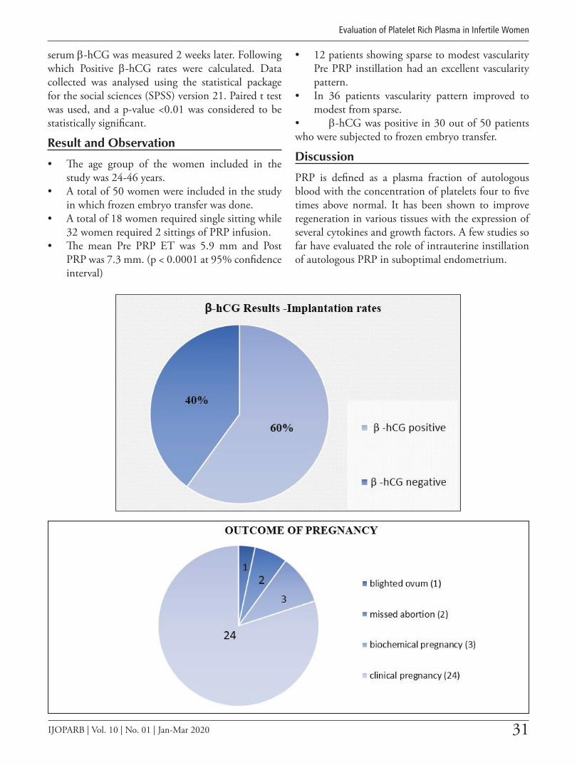
31
Evaluation of Platelet Rich Plasma in Infertile Women
IJOPARB | Vol. 10 | No. 01 | Jan-Mar 2020
8
• 12 patients showing sparse to modest vascularity Pre PRP instillation had an excellent vascularity pattern.
• In 36 patients vascularity pattern improved to modest from sparse.
β-hCG was positive in 30 out of 50 patients who were subjected to frozen embryo transfer.
serum β-hCG was measured 2 weeks later. Following which Positive β-hCG rates were calculated. Data collected was analysed using the statistical package for the social sciences (SPSS) version 21. Paired t test was used, and a p-value <0.01 was considered to be statistically significant.
Result and Observation
• The age group of the women included in the study was 24-46 years.
• A total of 50 women were included in the study in which frozen embryo transfer was done.
• A total of 18 women required single sitting while 32 women required 2 sittings of PRP infusion.
• The mean Pre PRP ET was 5.9 mm and Post PRP was 7.3 mm. (p < 0.0001 at 95% confidence interval)
• 12 patients showing sparse to modest vascularity Pre PRP instillation had an excellent vascularity pattern.
• In 36 patients vascularity pattern improved to modest from sparse.
• β-hCG was positive in 30 out of 50 patients who were subjected to frozen embryo transfer.
Discussion
PRP is defined as a plasma fraction of autologous blood with the concentration of platelets four to five times above normal. It has been shown to improve regeneration in various tissues with the expression of several cytokines and growth factors. A few studies so far have evaluated the role of intrauterine instillation of autologous PRP in suboptimal endometrium.
9
32 IJOPARB | Vol. 10 | No. 01 | Jan-Mar 2020
Original Article - Gynecology
A single study by Chang et al. published in 2015 evaluated the role of autologous PRP in thin endometrium in five patients undergoing frozen embryo transfer cycles. The ET increased at 48 to 72 h after PRP infusion in all the patients and reached >7 mm on the day of progesterone administration. All the five patients were pregnant. One of the patients had a missed miscarriage secondary to a chromosomally abnormal foetus, whereas the other four had viable intrauterine pregnancies, followed up till the first trimester.4
A review article by Garcia-Velasco et al. published in 2016 also cites the use of autologous PRP as a potential method of improving ET in women with refractory endometrium.2
Another important parameter to assess the implantation potential of the endometrium is Endometrial vascularity. A retrospective study of 500 ovum donation-embryo transfer cycles published by Nagori and Panchal in 2012 demonstrated that conception rates were almost doubled when vascularity was seen in zone 3 and 4 of the endometrium than when it reached only zones 1 and 2, with low abortion rates.7 Another recent study in 2014 by Sardana et al. also evaluated 165 women undergoing frozen embryo transfer cycles and concluded that the presence of
endometrial vascularity significantly improves the outcome in frozen embryo transfer cycles.8
Our study has a sample size of 50 patients and we analysed both the endometrial thickness and vascularity after intrauterine PRP infusion and further also evaluated the positive b-hCG rates. Autologous PRP is safe and easily available and also inexpensive treatment for infertile women with refractory endometrium. If this procedure is used routinely, it would reduce the financial and psychological burden of such patients who otherwise face the risk of repeated cycle cancellation.
Further research in the form of large randomized control trial is still needed to use this modality so that clinical practioners can use this more efficiently and it would also help to strengthen our observations and results.
Conclusion
Our study showcases that use of autologous PRP has a promising role in treatment of infertile women with suboptimal endometrial thickness and vascularity for embryo transfer. It helps to reduce the risk of cycle cancellation and decreases the financial and psychological trauma due to repeated cancelled cycle.
No conflict of interest.
1. Yamaguchi R, Terashima H, Yoneyama S, Tadano S, Ohkohchi N. Effects of platelet-rich plasma on intestinal anastomotic healing in rats: PRP concentration is a key factor. J Surg Res. 2012; 173:258–266
2. Garcia-Velasco JA, Acevedo B, Alvarez C, Alvarez M, Bellver J, Fontes J Et al. Strategies to manage refractory endometrium: state of the art in 2016. Reprod Biomed Online 2016; 32:474-89.
3. Kasius A, Smit JG, Torrance HL, Eijkemans MJ, Mol BW, Opmeer BC et al. Endometrial thickness and pregnancy rates after IVF: a systematic review and meta-analysis. Hum Reprod Update 2014; 20:530-41.
4. Chang Y, Li J, Chen Y, Wei L, Yang X, Shi Y Et al. Autologous platelet-rich plasma promotes endometrial growth and improves pregnancy outcome during in vitro fertilization. Int J Clin Exp Med 2015; 8:1286-90.
5. Gingold J, Lee JA, Rodriguez-Purata J, Whitehouse MC, Sandler B, Grunfeld L Et al. Endometrial pattern, but not endometrial thickness, affects implantation rates in euploid embryo transfers. Fertil Steril 2015; 104:620-8.
6. Mahajan N, Sharma S. The endometrium in Assisted reproductive technology: how thin is thin? J Hum Reprod Sci 2016; 9:3-8.
7. Nagori C, Panchal S. Endometrial vascularity: its relation to implantation rates. Int J Infertil Fetal Med 2012; 3:48-50.
8. Sardana D, Upadhyay AJ, Deepika K, Pranesh GT, Rao K. Correlation of subendometrial-endometrial blood flow assessment by two-dimensional power Doppler with pregnancy outcome in frozen-thawed embryo transfer cycles. J Hum Reprod Sci 2014; 7:130-5.
REFERENCES
33IJOPARB | Vol. 10 | No. 01 | Jan-Mar 2020
A Rare Case of Choriocarcinoma
Dr Smrity Mool Joshi,1 Dr Tarun Das,2 Dr Mamta Dagar,3 Dr Mala Srivastava,4 Dr I Ganguli,5 Dr Rakesh Koul6
Abstract
Choriocarcinoma is a tumor originating in the trophoblastic tissue. It may develop anytime following term pregnancy, spontaneous abortion, and even after ectopic pregnancy. Howeverchoriocarcinoma in a postmenopausal female is very rare. This was a rare case when a 52 year old lady presented with vague complain of pain abdomen for 5 days. Her MRI pelvis reported presence of large mass on posterior wall of uterus with possibility of mitotic activity. Hence Staging laparotomy with frozen section, was planned. After hysterectomy and bilateral salpingo oophorectomy, the frozen section of the uterus was sent for evaluation. This was reported to be poorly differentiated carcinoma. Staging laparotomy with pelvic and para aortic lymphadectomyand omentectomy was done. Postoperative period was uneventful. Histopathology report was suggestive of Choriocarcinoma, involving more than half of the myometrium, with involvement of cervical stroma. Bilateral parametrial margin and vaginal cuff margin were free of tumor (pT2pN0pMx). Thus, the final diagnosis of choriocarcinoma with FIGO stage > 7 was made. The patient was then started with chemotherapy (EMACO) and was kept on follow up. Choriocarcinoma in postmenopausal patient presents a diagnostic dilemma. In this case, we could only diagnose it postoperatively after the histopathology report and provide appropriate treatment.
Key words: Choriocarcinoma, Staging laparotomy, Serum beta HCG
Introduction
Choriocarcinoma is a highly malignant tumor originating in the trophoblastic tissue. The incidence of Choriocarcinoma in Europe and North Americais 1 per 40,000 pregnancies and 1 per 40 hydatidiformmoles while it is more in Southeast Asia and Japan where rates are at 9.2 and 3.3 per 40,000 pregnancies, respectively.1 About50% of the choriocarcinomaare diagnosed following a term pregnancy, and 25% after a hydatidiformmole.2 About
_________________________________________________1. Fellow in Gynecologic Oncology, Sir Ganga Ram Hospital
(SRGH), Delhi2. Associate Consultant in Department of Obstetrics and
Gynecology, SGRH, Delhi3. Senior Consultant in Department of Obstetrics and
Gynecology, SGRH, Delhi4. Senior Consultant in Department of Obstetrics and
Gynecology, SGRH, Delhi5. Emeritus Consultant Department of Obstetrics and
Gynecology, SGRH, Delhi6. Senior Consultant in Department of Surgical Oncology,
SGRH, DelhiCorresponding author email: [email protected]
Case Report - Gynecology
34 IJOPARB | Vol. 10 | No. 01 | Jan-Mar 2020
Case Report - Gynecology
90% of these tumor have complete cure following chemotherapy.3 There are reports of Choriocarcinoma following term pregnancy, spontaneous abortion, and even after ectopic pregnancy. It can develop anytime between 5 weeks and 15 years after gestation or even aftermenopause. Hence this is a case report of such an unusual case of choriocarcinoma in a lady after one year of menopause and 19 years after the birth of her last child.
Case:
A 52 year, postmenopausal, P2L2A1, lady presented at the hospital with complaints of, pain abdomen for one week. The pain was severe in nature and non-radiating. It was not associated with vaginal bleeding. She had menopause for one year. Her last child birth was 19 years back and her last abortion was 23 years back. There was nohistory of use of contraceptives. On examination, her general condition was normal, ECOG 0. Her abdominal examination revealed mass of 14 weekssize in the suprapubic region. The mass was mobile and nontender. On per speculumexamination cervix was healthy and on per vaginal examination uterus was 14 weeks size, mobile, non tender and bilateral fornices were free.
Her USG reported a large heterogenous mass measuring 9.3 x 8.5 x 7.3cm within the uterine cavity. Mass was extending up to the cervix and associated with multiple internal cystic area. MRI showed a large heterogenously intense enhancing mass lesion in posterior wall of the uterus causing indentation of the endometrial cavity with possibility of locally aggressive lesion with mitotic activity. Her base line investigationwere all normal. Her CA125 was 39.3IU/ml.
Since there was suspicion of malignancy, she was planned for Staging Laparotomy with frozen section and proceed. Per operatively, there was 70ml of hemorrhagic fluid in the peritoneal cavity. Uterus was 14 weeks size, soft in consistency and highly vascular. Both ovaries were grossly normal. Pelvic lymph nodes were palpable. Omentum and rest of the abdominal cavity was grossly normal. Hysterectomy with bilateral salpingo-oophorectomy was done. Specimen was send for frozen section which revealed poorly differentiated carcinoma of uterus. So furtherpelvic and paraaorticlymphadectomy withomentectomy was
done. The postoperative period was uneventful and she was discharged on fifth postoperative day.
The histopathologic finding revealed microscopically the tumor cells largely to be composed of mononucleatedcytotrophoblast, intermediate trophoblastand many multinucleated syncytiotrophoblast. These mononucleatedcytotrophoblast and intermediate trophoblast have moderately pleomorphic vesicular nuclei with prominent nucleoli with abundant eosinophilic to clear cytoplasm. Some cell had bizarre hyperchromaticnuclei. Mitosis was high. There was large area of hemorrhage and necrosis within the tumor. Prominent lymphovascular permeation was seen. The tumor had not breached the serosa of the uterus. However cervical stromal involvement was seen. Other tissue involvement was absent. Vaginal cuff and parametrial margin was free of tumor and the lymph nodes were negative for tumor.
On IHC, tumor cell were diffusely positive for beta HCG. Synncytiotrophoblastwere positive for PLAP, Ki67. The finding was suggestive of Choriocarcinoma pT2pN0pMx, FIGO anatomical staging stage I.
Post operatively the patient’s serum beta HCG was 510852 IU/ml, alpha fetoprotein was 3.4 IU/ml. As the FIGO score was > 7 she was thenplanned for multi-agent chemotherapy.
Two weeks after surgery when she came for follow up with per vaginal bleeding. On examination with speculum there was bleeding from the vault. On pervaginal examination there was a soft mass of 3 x 3 cm. PET scan showed, metabolically active heterogeneously enhancing lobulated mass along the vaginal vault and along the left posterolateralpelvic wall. There was also mild ascites. There was also metabolically active bilateral pulmonary nodule, which was highly suspicious of metastasis. Hence the patient was then planned for chemotherapy by EMACO.
The patient then after diagnosis has received five cycle of chemotherapy. At present the patient serum Beta HCG following her fifth cycle of chemotherapy has reduced to 215 IU/ml. She has been planned for sixth cycle of chemotherapy.
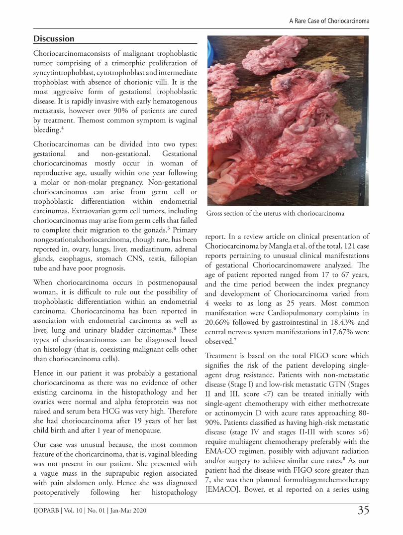
35
A Rare Case of Choriocarcinoma
IJOPARB | Vol. 10 | No. 01 | Jan-Mar 2020
Discussion
Choriocarcinomaconsists of malignant trophoblastic tumor comprising of a trimorphic proliferation of syncytiotrophoblast, cytotrophoblast and intermediate trophoblast with absence of chorionic villi. It is the most aggressive form of gestational trophoblastic disease. It is rapidly invasive with early hematogenous metastasis, however over 90% of patients are cured by treatment. Themost common symptom is vaginal bleeding.4
Choriocarcinomas can be divided into two types: gestational and non-gestational. Gestational choriocarcinomas mostly occur in woman of reproductive age, usually within one year following a molar or non-molar pregnancy. Non-gestational choriocarcinomas can arise from germ cell or trophoblastic differentiation within endometrial carcinomas. Extraovarian germ cell tumors, including choriocarcinomas may arise from germ cells that failed to complete their migration to the gonads.5 Primary nongestationalchoriocarcinoma, though rare, has been reported in, ovary, lungs, liver, mediastinum, adrenal glands, esophagus, stomach CNS, testis, fallopian tube and have poor prognosis.
When choriocarcinoma occurs in postmenopausal woman, it is difficult to rule out the possibility of trophoblastic differentiation within an endometrial carcinoma. Choriocarcinoma has been reported in association with endometrial carcinoma as well as liver, lung and urinary bladder carcinomas.6 These types of choriocarcinomas can be diagnosed based on histology (that is, coexisting malignant cells other than choriocarcinoma cells).
Hence in our patient it was probably a gestational choriocarcinoma as there was no evidence of other existing carcinoma in the histopathology and her ovaries were normal and alpha fetoprotein was not raised and serum beta HCG was very high. Therefore she had choriocarcinoma after 19 years of her last child birth and after 1 year of menopause.
Our case was unusual because, the most common feature of the choricarcinoma, that is, vaginal bleeding was not present in our patient. She presented with a vague mass in the suprapubic region associated with pain abdomen only. Hence she was diagnosed postoperatively following her histopathology
report. In a review article on clinical presentation of Choriocarcinoma by Mangla et al, of the total, 121 case reports pertaining to unusual clinical manifestations of gestational Choriocarcinomawere analyzed. The age of patient reported ranged from 17 to 67 years, and the time period between the index pregnancy and development of Choriocarcinoma varied from 4 weeks to as long as 25 years. Most common manifestation were Cardiopulmonary complaints in 20.66% followed by gastrointestinal in 18.43% and central nervous system manifestations in17.67% were observed.7
Treatment is based on the total FIGO score which signifies the risk of the patient developing single-agent drug resistance. Patients with non-metastatic disease (Stage I) and low-risk metastatic GTN (Stages II and III, score <7) can be treated initially with single-agent chemotherapy with either methotrexate or actinomycin D with acure rates approaching 80-90%. Patients classified as having high-risk metastatic disease (stage IV and stages II-III with scores >6) require multiagent chemotherapy preferably with the EMA-CO regimen, possibly with adjuvant radiation and/or surgery to achieve similar cure rates.8 As our patient had the disease with FIGO score greater than 7, she was then planned formultiagentchemotherapy [EMACO]. Bower, et al reported on a series using
Gross section of the uterus with choriocarcinoma
36 IJOPARB | Vol. 10 | No. 01 | Jan-Mar 2020
Case Report - Gynecology
EMA-CO; the 5-year survival rate was 86.2%; in this series, patients with brain metastases received an increased dose of MTX to 1 g/m2 IV infused over 24 hours, followed by folinic acid.9
All patients with GTN are followed with weekly HCG values until undetectable for 3 consecutive weeks, then monthly until undetectable for 12 months. All patients must be encouraged to use effective contraception during the entire interval of monitoring.
Relapse rates range from 3 to 9 percent for stages I to IV and the mean time of recurrence from the last non-detectable HCG level is usually 6 months.10
Conclusion
Choriocarinomahave a good prognosis however, their variable presentation and symptoms may sometime cause diagnostic dilemma. This was a rare case of postmenopausal choriocarcinoma in female with menopause for 1year and last child birth 19 years back without any history of per vaginal bleeding. Hence, the diagnosis was possible postoperatively after histopathology report. Following the diagnosis our patient has been kept under chemotherapy[EMACO]and weekly follow up.
REFERENCE
1. Lurain JR. Gestational trophoblastic disease I: epidemiology, pathology, clinical presentation and diagnosis of gestational trophoblastic disease, and management of hydatidiform mole. American journal of obstetrics and gynecology. 2010 Dec 1;203(6):531-539.
2. Soper JT. Gestational trophoblastic disease. Obstetrics & Gynecology. 2006 Jul 1;108(1):176-187.
3. Seckl MJ, Fisher RA, Salerno G, Rees H, Paradinas FJ, Foskett M, Newlands ES. Choriocarcinoma and partial hydatidiform moles. The Lancet. 2000 Jul 1;356(9223):36-9,2019.
4. Placenta Gestational Trophoblastic disease Placental site trophoblastic tumor (PSTT). 2003:2019: Availble at http://www.pathologyoutlines.com/topic/placentaPSTT.html. Accessed September 9th
5. Weiss S, Amit A, Schwartz MR, Kaplan AL. Primary choriocarcinoma of the vulva. International Journal of Gynecologic Cancer. 2001 May 1;11(3):251-4.
6. Khuu HM, Crisco CP, Kilgore L, Rodgers WH, Conner MG. Carcinosarcoma of the uterus associated with a
nongestationalchoriocarcinoma. Southern medical journal. 2000 Feb;93(2):226-228.
7. Mangla M, Singla D, Kaur H, Sharma S. Unusual clinical presentations of choriocarcinoma: A systematic review of case reports. Taiwanese Journal of Obstetrics and Gynecology. 2017 Feb 1;56(1):1-8.
8. Gestational Trophoblastic Neoplasia [internet] 2014 Review of Cancer Medicine on the WHO list of Essential Medicine: Available at: https://www.who.int/selection_medicines/committees/expert/20/applications/GestationalTrophoblasticNeoplasia.pdf
9. Bower M, Newlands ES, Holden L, Short D, Brock C, Rustin GJ, Begent RH, Bagshawe KD. EMA/CO for high-risk gestational trophoblastic tumors: results from a cohort of 272 patients. Journal of Clinical Oncology. 1997 Sep;15(9):3168.
10. Goldstein DP, Berkowitz RS. Current management of gestational trophoblastic neoplasia. HematolOncolClin North Am 2012;26:111-131

37
A Rare Case of Choriocarcinoma
IJOPARB | Vol. 10 | No. 01 | Jan-Mar 2020
BE A LIFE MEMBERof
Indian Society of Perinatology and
Reproductive Biology (ISOPARB)
[Affiliated to the Federation of Asia and Oceania Perinatal Societies (FAOPS)]
Website: www.isoparb.org
Why to Submit in IJOPARB?
• National and International recognition, readership and acceptance.
• Not to wait too long for publication.• Quality feedback from the expert editors for your improvement.
Support to prepare your paper
For any support of composition before submitting article you may contact the
IJOPARB Editorial Board.
Email: [email protected]
How to Submit:
Instruction to authros IJOPARB in each issue. Page No. 94
For any more information, please contact Editorial Board
Email: [email protected]
Members of ISOPARB interested to work in the editorial Board as reviewers, are requested to submit their names with their updated Curriculum Vite to the Editor in Chief.
Kindly mention your interest in the subspeciality that you need to be involved (e.g. fetal medicine, oncology, general gynaecology, general obstetrics etc.)
AN APPEAL !
DONATE GENEROUSELY FOR IJOPARB JOURNAL
Cheques / Demand Draft to be drawn in favour of Indian Journal of Perinatology and Reproductive Biology, payable at Kolkata

38 IJOPARB | Vol. 10 | No. 01 | Jan-Mar 2020
The Journal of Indian Society of Perinatology and Reproductive Biology which is the official publication of the Indian Society of Perinatology and Reproductive Biology (ISOPARB) invites original research articles in gynaecology / obstetrics / related subjects in the following category: Clinical Articles; Review Articles; and Brief Communications (including Case Reports).
All manuscripts should be prepared according to the guidelines detailed below. Any manuscript that has not been formatted as per the ISOPARB requirements will be returned to the author for correction. All manuscripts should be created and submitted in Word format.
1. SUBMISSION
Authors must submit manuscripts by Email ID – [email protected] dr_gita [email protected] [email protected]
Hard-copy submissions will not be considered.
Please submit a cover letter to the Editor-in-Chief mentioning the following:
• Each author’s name, address, and email address.
• Each author’s affiliation and qualifications.
• The name of the author who is to deal with correspondence and proofs.
Once submitted, manuscripts undergo initial screening by the editorial staff and editors and then papers will undergo peer review.
2. Authors must give a separate “Author Guarantee” document mentioning the following:
(1) that all authors have met the criteria for authorship and have participated sufficiently in the work to take responsibility for it;
(2) that all authors have reviewed the final version of the manuscript and approve it for submission to the ISOPARB journal.
(3) that neither this manuscript nor one with substantially similar content by the authors has been published elsewhere or is being considered for publication elsewhere;
(4) that the manuscript has been submitted with the full knowledge and approval of the institutions or organizations given as the affiliation(s) of the author(s);
(5) that the authors have informed the editor in a cover letter and in the manuscript itself of any conflicts of interest; and
(6) that the corresponding author affirms the manuscript to be an honest and transparent account of the study being reported.
In line with ICMJE standards, the criteria for authorship are as follows:
(1) Substantial contributions to the conception or design of the work; or the acquisition, analysis, or interpretation of data for the work; AND
(2) Drafting the work or revising it critically for important intellectual content; AND
(3) Final approval of the version to be published; AND
(4) Agreement to be accountable for all aspects of the work in ensuring that questions related to the accuracy or integrity of any part of the work are appropriately investigated and resolved.
3. CLINICAL TRIALS AND REVIEW ARTICLES
Clinical trials
Submission of clinical trials must include reference to ethics approval (or explanation of why ethics approval was not received). Authors must consult the CONSORT statement and checklist and submit a CONSORT flow chart as an editable figure in Word/PowerPoi ntformat.
The clinical trial registration is preferable and information should be included at the end of the abstract of the submitted manuscript.
Review articles
Reviews based on the recent and relevant subjects of clinical interest should be considered.
4. LAYOUT OF MANUSCRIPTS
Manuscript text should be in English (US spelling), double-spaced, font size 12, in Arialmmes New Roman font.
First page
The first page of the manuscript should contain the following: (1) title; (2) full names of authors (6 maximum, although listing more authors may be considered on an individual basis if authorship requirements have been met and a request has been included in the cover letter); (3) affiliations of authors (i.e. department, section or unit of
Instruction to Authors
Instruction to Authors

39
Instruction to Authors
IJOPARB | Vol. 10 | No. 01 | Jan-Mar 2020
an institution, hospital or organization, city, and country (4) full contact details (postal address. email address) of the corresponding author; (5) a list of up to 8 keywords for indexing and retrieval:
Footnotes linking author names to affiliations should be listed as 1,2,3 etc..
The first page should also list the type of article: Clinical Article; Brief Communication: or Review Article.
Abstract
Clinical Articles
A structured abstract not exceeding 200 words is required for all full-length clinical articles. It should contain all and only the following headings: Objective; Methods; Results; and Conclusion.
The Objective reflects the purpose of the study: that is, the hypothesis that is being tested. The Methods should include the setting for the study, the participants (number and type), the treatment or intervention, and the type of statistical analysis. The Results include the outcome of the study and statistical significance, if appropriate. The Conclusion states the significance of the results.
Review articles
An abstract not exceeding 200 words is required for all review articles.
Narrative reviews require an unstructured abstract. Systematic review articles should have a structured abstract with the headings; Background; Objectives; Search strategy; Selection criteria; Data collection and analysis; Main results; and Conclusions.
Brief communications
Brief communications should not include an abstract.
Main text
In full-length articles, subject matter should be organized under the following headings, with no subheadings: Introduction; Materials and methods; Results; Discussion; Acknowl edgments; Conflicts of interest; and References.
Brief communications should not have any headings separating the text.
Clinical articles
The main text of clinical articles should not exceed 2500 words, excluding the first-page information, abstract (no more than 200 words), author contributions, acknowledgments, Conflicts of interest, references (no more than 25), figure legends, and tables and figures. Please include the word count in the cover letter and on the first page of the manuscript.
Review articles
Review articles should have no more than 3000-3500 words in the main text and 40 references. Please include the
word count in the cover letter and on the first page of the manuscript.
Brief communications
Brief communications should be no more than 400 words, excluding the first-page information, synopsis, keywords, author contributions, acknowledgments, conflicts of interest, references, figure legends, and tables and figures. There should be no more than 4 references and no more than 1table or 1figure.
Power calculations, statistics, and reporting of numbers.
Power calculations
Where appropriate (e.g. for clinical trials), power calculations should be performed as part of the study design, and a statement providing the power of the study should be included in the Materials and Methods. Authors should state how the power calculation was determined, including what type of difference the calculation was powered to detect and on what studies the numbers are based.
Statistics
The statistical tests used and the significance level set should be listed in the methods for all studies that employed statistical analysis. Information regarding the statistical software programs used should be included in the methods: for example, “SPSS version 20 (IBM, Armonk, NY, USA).” This information should not be included in the reference list.
P values should be provided where calculated. The largest P value that should be expressed is P>0.99. The smallest P value that should be expressed is P<0.001.
For measures of effect (e.g. relative risks, risk ratios, odds ratios), authors should also report confidence intervals (e.g. 95%) so that the precision of the effect estimate can be assessed.
5. Ethics approval and informed consent
Studies of patients, patient records, or volunteers require Ethics Committee approval and informed consent.
Ethics approval
Include a statement in the methods that the research protocol was approved by the relevant Institutional Review Board or Ethics Committee before the study began; if such approval was not needed/obtained, include an explanation. Authors must provide copies of the appropriate documentation if requested.
Informed consent
Include confirmation in the methods that all human participants gave written informed consent before the study began; if consent was not needed/obtained, include an explanation. Authors must provide copies of the appropriate documentation if requested.
40 IJOPARB | Vol. 10 | No. 01 | Jan-Mar 2020
Instruction to Authors
6. Acknowledgments
Sources of funding should be acknowledged by the author(s), along with the names of individuals who do not fulfil the criteria for authorshi p, but who have made a substantial contribution to the manuscript.
7. Confllcts of Interest
A conflict-of-interest statement must be included in the cover letter and before the reference list in the manuscript. It should list any relationships (for any author) that may be deemed to Influence the objectivity of the paper and its review, or state that no such relationships exist. Commercial associations, either directly or through immediate family, in areas such as expert testimony, consulting, honoraria, stock holdings, equity interest, ownership, patent-licensing situations or employment that might pose a Conflicts of interest should be stated. Conflicts for other reasons, such as personal relationships or academic competition, should also be stated.
8. References
The number of references should not exceed 15 for clinical articles, 25 for review articles, and 4 for brief communications; in general, they should be limited to the past decade. They must be numbered and listed as they are cited in the article, using Index Medicus abbreviations for journal titles. Cite the names of all authors when there are six or fewer; when there are seven or more, list the first three authors followed by “et al.” Include the volume number.
Journal article
[1] Vellacott ID, Cooke EJ, James CE. Nausea and vomiting in early pregnancy. Int J Gynecol Obstet. 1988;27:57-59.
Book
[2] Speroff L, Glass BH, Kase NG. Clinical Gynecologic Endocrinology and Infertility. Baltimore: Williams and Wilkins; 1982.
Chapter in a book
[3] Disaia PJ, Creasman WT. Invasive Cancer of the Vulva. In: Disaia PJ, Creasman WT, eds. Clinical Gynecologic Oncology. St Louis: C.V. Mosby; 1984:214-219.
Web reference
[4] World Health Organization. WHO Recommended Surveillance Standards, Second Edition [WHO website). 1999. http://www.who.int/csr/resources/publications/surveillance/whocdscsrisr992.pdf.
Text references can be indicated by Arabic numerals in superscript. abc1
Tables
Each table should be titled, numbered (with Arabic numerals), and placed on a separate page after the reference list (not embedded within the main text).
All tables must be cited in numeric order in the main text as “Table 1” etc.
Footnotes to tables should be listed as a, b, c etc.
9. Figures and photographs
Figures and photographs should be submitted as jpg format. CONSORT flow charts should be created and submitted as editable Word/ Power Point files. All figures must be cited in numeric order in the main text as “Figure 1” etc.
Figure permission
All authors wishing to use figures (or any material) that have already been published must first obtain the permission of the original author and publisher and/or copyright holders, in addition to giving precise reference to the original work. This permission must include the right to publish in electronic media. Confirmation should be included in the cover letter (the actual permission correspondence from the copyright holder does not need to be submitted).
Photograph/video consent
If photographs or videos of identifiable people are used, authors must obtain and submit a signed statement of informed consent from the identifiable person(s) or their next of kin. Authors should not try to conceal identity with black bars over eyes etc.
9. Drugs
Give generic names of all pharmaceutical preparations and, where appropriate, include the trade name and manufacturer’s name and address. Review drug names and dosages with care. The author is responsible for all recommended dosages.
10. Plagiarism
Plagiarism entails the “use or close imitation of the language and thoughts of another author and the representation of them as one’s own original work.” Self-plagiarism, a form of misconduct in which an author reuses his/her previously written text, data, or ideas, wholly or in part, without indicating previous dissemination, will also be considered plagiarism. Verbatim copying of sentences, even if a citation is provided (unless the sentence appears in quotation marks), is considered to be plagiarism.
11. ON ACCEPTANCE
If your paper is accepted for publication, you will receive an email informing you of this decision.
12. Copyright
Once accepted and published, all copyright will belong to ISOPARB. No part of the article could be published without permission. All disputes are subjected to Indian Jurisdiction.
13. It is desirable that the, author(s) submitting article in IJOPARB be a member of ISOPARB.

IJOPARBIndian Journal of Perinatology and Reproductive Biology
ISSN 2249 - 9784Volume 10, No. 01January - March, 2020
Official Journal of Indian Society of Perinatology and Reproductive Biology
If undelivered, return to: Indian Journal of Perinatology and Reproductive Biology
CD-55, Salt Lake City, Sector - 1, Kolkata 700064
ICV 2017 = 70.99 | ICV 2018 = 69.79