IIflI fllfllflfflfflf - Defense Technical Information Center
533
AD-AIOb 234 NORTHEASTERN UMIV BOSTON MASS DEPTOF BIOPHYSICS AN-ETC F/B 6/18 BIOLOGICAL EFFECTS OF LASER RADIATION, VOLUME 1. REVIEW OF THE !TC(U) OCT 78 S FINE, E KLEIN 0A-'49193-MD-243 UNCLASSIFIED NB IIflI fllfllflfflfflf IIIEIIIEEIIIEE IIIIIIIIIIIIIu EEEEEEEEIIEEEE IEEEIIEIIIIII
-
Upload
khangminh22 -
Category
Documents
-
view
0 -
download
0
Transcript of IIflI fllfllflfflfflf - Defense Technical Information Center

AD-AIOb 234 NORTHEASTERN UMIV BOSTON MASS DEPT OF BIOPHYSICS AN-ETC F/B 6/18BIOLOGICAL EFFECTS OF LASER RADIATION, VOLUME 1. REVIEW OF THE !TC(U)OCT 78 S FINE, E KLEIN 0A-'49193-MD-243
UNCLASSIFIED NBIIflI fllfllflfflfflf
IIIEIIIEEIIIEEIIIIIIIIIIIIIuEEEEEEEEIIEEEEIEEEIIEIIIIII
ORIGINAL__BIOLOGICAL EFFECTS OF LASER RADIATION
Final Scientific Report - Volume I(Review of the Literature on Biological Effects of Laser
Radiation-to 1965)
Samuel Fine
Edmund Klein
..'-).,TIC
OCT 8 1981
ASubmitted October 17, 1978
(1 July 1963 to 30 September 1971)
Supported By
U.S. ARMY MEDICAL RESEARCH AND DEVELOPMENT COMMAND
Fort Detrick, Frederick, Maryland 21701
Contract No. DA-49-193-MD2436
Department of Biophysics and Biomedical EngineeringNortheastern University
Boston, Massachusetts 02115
Approved For Public Release;Distribution Unlimited
81 10 27 289

SECURITY CLASSIFICATION OF THIS PAGE (When Data Entered)
READ NSTRUCTIONSREPORT DOCUMENTATION PAGE BEFORE COMPLETING FORM
I. REPORT NUMBER 2. GOVT ACCESSION NO. 3. RECIPIENT'S CATAI.OG NUMBER
Li . TT4._ ,t . .. .... -.. .,. T P)EO," REPj i~ r-joo COVERED
BIOLOGICAL EFFECTS OF LASER RADIATION. - Volume I- jFinal I 1 July 1963-.(Review of the Literature on Biological Effects 30 Septmxfr 1-971of Laser Radiation-to 1965), of. R PORT NUMBER
7. AUTHOR(#) 6 6. CONTRACT OR GRANT NUMBER(@),,e S u li ine-.. . i _
. Edmund/Klein / DA-49-193-MD436
9. PERFORMING ORGANIZATION NAME AND ADDRESS 10. PROGRAM ELEMENT. PROJECT, TASKAREA & WORK UNIT NUMBERSDepartment of Biophysics and Biomedical
EngineeringNortheastern University, Boston, MA 02115 61102A43S161102BS05
". CONTROLLING OFFICE NAME AND ADDRESS 12. REPORT DATE
US Army Medical Research and Development Command Vjt October 47., 1978Fort Detrick 13. NUMBER oF PAGES
Frederick, MD 21701 526 pages14. MONITORING AGENCY NAME & AODRESS(I different from Contro lUnyOfflce) 1S. SECURITY CLASS. (of thil report)
-Unclassified
So. DECLASSIFICATION/DOWNGRADINGSCH EDULE
IS. OISTRIBUTION STATEMENT (of this Report)
Approved for Public Release; Distribution Unlimited
17. DISTRIBUTION STArEMENT (of the abstrat entered in Block 20, if dlfterent from Report)
18. SUPPLEMENTARY NOTES
19. KEY WORDS (Continue on reverese ide if necessay end Identify by block number)
Biological Effects of Laser Radiation; Electron Spin Resonance; Spectroscopy;
Microscopy: Tissue and Cell Culture Interaction; Biochemical Studies and
Macromolecular Transformation; Normal Animals; Tumors; Mechanisms; Laser
Radiation Hazards; Flash Lamp Hazards; Electrical Hazards; Scattering;
Polarization; Non-Linear Effects.20. AUT'RAC? fo.tlu a revere af rnm'eeary - Identify by block number)
Research on biological effects of laser radiation to 1965 is reviewed. Laser
production of free radicals, use in microscopy and holography, and tissue and
cell culture effects are discussed. Possible beneficial transformations in
macromolecular preparations are detailed. Effects on normal animals together
with gross and microscopic studies are described. Benefits and hazards of
applicability and relationship to animal tumor models are presented.
Ophthalmological uses and hazards are covered. Protective systems, limitations
in applicability of systems current at that time to ophthalmology and in
DD , 1473 EoTom OF I NOV GS IS OBSOLETE
SECURITY CLASSIFICATION OF THIS PAGE 'Whie Dara Entered)

20 (continued)
experiments, are indicated. Possible use in dentistry and in entomology arereviewed. Mechanisms include consideration of thermal and non-thermal models,pigmentation, interfaces, steam production, pulse duration, non-linear andsonic waves, bubble production and collapse, charged particle production,hypersonic waves, and radiation scattering. Problems associated with measurementof temperature and pressure and methods for carrying out such measurementsare presented. A section on non-linear effects discusses second harmonicgeneration in laser systems and production in target sites, Raman scatteringand possible production of ultraviolet radiation. Effects and hazards areconsidered. Sections on gas lasers, flash lamps, electrical hazards andscattering are included.

The findings in this report are not to be construed as an
official Department of the Army position unless so designated
by other authorized documents.
AD
BIOLOGICAL EFFECTS OF LASER RADIATIONFinal Scientific Report - Volume I
(Review of the Literature on Biological Effects of Laser Radiation--to 1965)
Samuel FineEdmund Klein
Submitted October 17, 1978
(1 July 1963 to 30 September 1971)
Supported By
US ARMY MEDICAL RESEARCH AND DEVELOPMENT COMMANDFort Detrick, Frederick, MD 21701
Contract No. DA-49-193-MD2436
Department of Biophysics and Biomedical EngineeringNortheastern University
Boston, MA 02115
-Accesoo ror
Approved For Public Release; j.
Distribution Unlimited
s tf
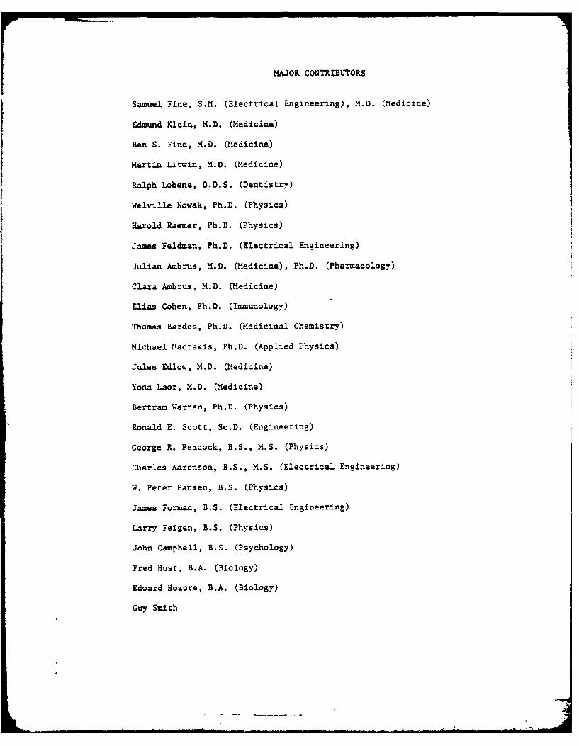
MAJOR CONTRIBUTORS
Samuel Fine, S.M. (Electrical Engineering), M.D. (Medicine)
Edmund Klein, M.D. (Medicine)
Ben S. Fine, M.D. (Medicine)
Martin Litwin, M.D. (Medicine)
Ralph Lobene, D.D.S. (Dentistry)
Welville Nowak, Ph.D. (Physics)
Harold Raemer, Ph.D. (Physics)
James Feldman, Ph.D. (Electrical Engineering)
Julian Ambrus, M.D. (Medicine), Ph.D. (Pharmacology)
Clara Ambrus, M.D. (Medicine)
Elias Cohen, Ph.D. (Immunology)
Thomas Bardos, Ph.D. (Medicinal Chemistry)
Michael Macrakis, Ph.D. (Applied Physics)
Jules Edlow, M.D. (Medicine)
Yona Laor, M.D. (Medicine)
Bertram Warren, Ph.D. (Physics)
Ronald E. Scott, Sc.D. (Engineering)
George R. Peacock, B.S., M.S. (Physics)
Charles Aaronson, B.S., M.S. (Electrical Engineering)
W. Peter Hansen, B.S. (Physics)
James Forman, B.S. (Electrical Engineering)
Larry Feigen, B.S. (Physics)
John Campbell, B.S. (Psychology)
Fred Hust, B.A. (Biology)
Edward Hozore, B.A. (Biology)
Guy Smith

MAJOR CONTRIBUTORS TO SPECIFIC CHAPTERS
Chapter: 1. Samuel Fine and Edmund Klein
2. Guy Smith, Samuel Fine and Edmund Klein
3. Samuel Fine and W. Peter Hansen
4. Samuel Fine and Edmund Klein
5. Edmund Klein, Julian Ambrus, Clara Ambrus, Elias Cohen,Thomas Bardos and Samuel Fine
6. Samuel Fine, Edmund Klein and Guy Smith
7. Samuel Fine, Edward Hozore and Jules Edlow
8. Samuel Fine, Edmund Klein, Yona Laor and Fred Hust
9. Edmund Klein and Samuel Fine
10. Edmund Klein, Samuel Fine and Martin Litwin
11. Ben S. Fine and Samuel Fine
12. Ralph Lobene and Samuel Fine
13. Samuel Fine and Edmund Klein
14. Samuel Fine and W. Peter Hansen
15. George R. Peacock, Samuel Fine and Larry Feigen
16. W. Peter Hansen, Samuel Fine and Ben S. Fine
17. W. Peter Hansen and Samuel Fine
18. George R. Peacock and Samuel Fine
19. Larry Feigen and Samuel Fine
20. James Forman, Harold Raemer and Samuel Fine
21. James Forman, Samuel Fine and Welville Nowak
22. George R. Peacock and Samuel Fine
23. George R. Peacock, Samuel Fine and Ronald E. Scott
24. Charles Aaronson and Samuel Fine
Addendum James Feldman, Samuel Fine, W. Peter Hansen, James Formanand Harold Raemer

TABLE OF CONTETS
Chapter Page
1. Free Radicals and Electron Spin Resonance Studies 1
2. Lasers in Spectroscopy 8
3. Microscopy and Holography 16
4. Tissues and Cell Cultures 20
5. Studies on Interaction with Macromolecular BiochemicalPreparations 26
6. The Laser Microbeam 51
7. Embryology 82
8. Studies on Normal Animals 91
9. Tumor Studies 159
10. Clinical Studies 188
11. Ophthalmology 232
12. Dental Studies 263
13. Entomology 271
14. Mechanisms of Interaction of Laser Radiation withBiological Systems 274
15. Radiometric and Photometric Units 318
16. Laser Eye Protection 338
17. Laser Dosimetry - Biological Systems 346
18. Polarization of Lasers - Effects on Energy Measurements 362
19. Discussion of Some Laser Output Detectors 365
20. Hazards due to Light Scattering 381
21. Non-Linear Effects 416
22. Gas Lasers 425
23. Flash Lamps - Associated Hazards 435
24. Electrical Hazards 457
Addendum 471

The report presented in the following pages constitutes the efforts
of a group consisting of individuals with training in the various sci-
entific disciplines. Although major contributions to the various sec-
tions have been credited on the previous page, it should be recognized
that many contributions have been made by these individuals to chapters
Focher than those to which they are major contributors. A number of
individuals who have contributed to various sections may have been
inadvertently omitted. An attempt will be made to rectify this omis-
sion in the final report.
Due to the time duration from initiation of the contract to pre-
paration of this report, the mathematical derivations in various sec-
tions, particularly if they are original, should be considered as not
being in final for. Some of the models developed for biological
systems will require refinement.
The authors wish to thank the many individuals who have contributed
their time and effort towards this report. in particular, they wish
to thank Miss Angela Reid, Mrs. Patricia Broun, Miss Grace Wood, Miss
Joan Liquori, Miss Sandra Taylor, miss Avril Gozna, and Miss Linda Law
for their devoted secretarial assistance.
$

Note on Content
This review of the literature on biological effects of laser
radiation was submitted to the U. S. Air Force Missile Development
Center, Air Force Systems Command, as part of a management study. It
contains a review of our studies up to and including 1965, which were
supported by the U. S. Army Medical Research and Development Command.
In addition, it includes a review of work by others.
It is being submitted as Volume 1 of the final scientific report
to provide information concerning our research under the contract to
1965. It should be noted that this volume was completed at that time.
Chapter 20, Hazards Due to Light Scattering, was extensively
revised and corrected in subsequent volumes. Since it is not of major
significance in regard to review of our work to 1965, the chapter was
not revised for purposes of this report. A revised version, as submitted
in Part III of the original report in 1966, is included in the addendum.

FREE RADIC.LS PND ELECTRON SIN ESOyANCE STUDIES
Several methods have been used to study free radicals. Potentiometric
titration was discussed by Michaelis (1,2). Masnetic susceptibility tech-
niques are reviewed by Brill (3). The appearance of the radical can often
be detected colorimetrically or spectroscopically (2,4).
Electron spin resonance techniques are presently the most common methods
of investigation (5,6 - 9,11). The method is based on exploitation of the
characteristic feature of the free radical: the unpaired valency electron.
The unpaired valency electron has both angular momentum and a magnetic
moment. A free electron, itself, should give a single, sharp absorption
line. However, the unpaired electron in free radicals will 'a associated
with one or more nuclei. The effect of these nuclei is to perturb the local
magnetic field in which the electron moves. For example, the observed ef-
fect of a proton coupled with the electron is to produce splitting of the
single resonance line; two protons will result in a triplet spectrum; a
nucleus of higher order in a more complex spectr-. Other factors modify
the spectrum obtained. The nature of the free radical consequently cannot
be deduced from its ESR stectrum alone.
Free radicals are norally present in many types of tissue. They are
considered as essential intermediates in many metabolic pathways (12,10)
and have been implicated in carcinogenesis and agin- (7). They occur during
a nu.mber of enzymatically catalyzed reactions and are of significance in
photosynthesis (13).
Electron spin resonance signals have been obtained from liver, kidney,
heart, lung and adrenal of several species (14). Although not universally
002001
4 -
2
valid, metabolically active tissue such as liver appears to possess a higher
free radical content than less metabolically active tissue. A relationship
of free radical content to mitochondral concentration has been considered.
The variation in free radical concentration in the rat liver with age has
been studied. lfelanin containing tissues yield an ESR signal, due to the
melanin. The melanin has been considared as a semiconductor with bound
protons serving as electron traps. It has been suggested that melanin acts
as a trap for otherwise harmful free radicals produced.
A reversible threefold increase in unpaired electron concentration, on
irradiation by visible light of aqueous suspensions of eye malanin, indicates
that melanin =ay be of importance in the photoreceptive processes although
not specifically in image for.-ations. Artificial and dried melanin did not
show this effect.
In studies on heart muscle mitochondria the definite oxygen and tempera-
ture sensitive ESR signal observed is considered as d&e to succinic dehydro-
genase. Signals have also been measured in ozher f' vonrotein enzymes,
including dehydro-orotic dehydrogenase and Dh:-cytczhrome reductase.
Free radicals are of significance in photosynthesis. The literature in
this field is vast, and is reviewed by Blois and Ueaver in Photoohvsiolonv,
edited by Giese (13). One ESR signal observed on illumination decays rapidly
after the light is turned off, a second residual signal may remain for minutes
or hours. The rapid signal is considered as associated with chlorophyll, and
probably coincides with the primary process of photosynthesis. The origin
of the residual or slowly decaying signal is not well defined.
In some cases, as in the study of some flavoprotein enzymes, the ap-
pearance of an ESR signal does appear to be correlated with the appearance
002

3
of certain optical absorptions that are apparently characteristic of flavin
free radicals (12). This hs been considered as due to the fact that the
riboflavin may have accepted an electron, but is still in close contact with
the donor molecule. Observations of this ty.?e suggest that complete reliance
should not be placed on ESR studied for da:er-mination of the presence of free
radicals
The purpose of the foregoing discussion is to indicate that numerous
studies have been carried out on free radicals, particularly using electron
spin techniques. Elucidation of the presence of free radicals, their chara-
cterization and quantitation required considerable study.
The studies by Derr, Klein and Fine (15,16) on detection of free radi-
cals by electron spin resonance following laser irradiation were carried out
to determine whether free radicals could be produced on laser irradiation.
Characterization of the free radicals was left to future investigation.
A.n ESR signal was obtained in black =ouse skin and an enzyme prepara-
tion, fibrinolysin, following laser izradiation at 6943 R. The g value of
approximately 2.002 indicated that the signal was probably due to the presence
of free radicals. Me continued presence without deterioration of signal in
irradiated black skin warmed to room temperature for 24 hours, would indicate
that the free radicals produced were stable free radicals. The essential
difference between the white mouse skin, in which no ESR signal was obtained,
and black mouse skin is the presence of melanin and a melanin precursor
system. Consequently, the signal observed may have been due to the production
of free radicals in melanin (or in the melanin precursor system). Since all
tissues that contain melanin can yield an ESR signal (12), the undetecta-
bilit-y of free radicals in the unirradiated black mouse skin requires further
003

4
explanation. It may be due to the fact that the concentration of melanin
free radicals in unirradiated black skin was below the threshold detecta-
bility of the ESR system used.
The actual frequency of the spectrometer used by Derr et a!. appears
to have been of the order of 30 ZiZacycles (15). For biological studies,
this frequency may prove -referable to the usual 10 gigacycle frequency
unit. It may have been desirable o orovide more information concerning
the BS. spectra shown of the various tissues, such as whether the figure
L reprasented tissue that was warmed previous to m.easurement or not. Further
characterization of the free radicals produced is desirable, and comparison
with those observed on heating and ult_-aviolet irradiation indicated.
Plucking of hair in the black mouse skin previous to irradiation may assist
in determining whether melanin is of significance in the production of
the free radical. The melanin granules may act as an energy absorption or
transfer agent with production of f-ee radicals in adjacent tissue. If
separation of melanL from the remainder of the tissue can be achieved/
with little trauma, this may assist in determining the site or production
of the free radicals.
Further studies on smaller molecules may assist in determining the
n-t-ure of the free radical produced on laser irradiation, and possible the
site of photon interaction with the molecule. It is possible that ESR
spectra will be obtained on laser irradiation, whereas spectra may not be
observed on low level irradiation at the same wavelength. This can occur
for several reasons. A certain number of free radicals must be present to
be detectable. If the decay time is rapid, then high intensity radiation
may provide a num.ber which are detectable, whereas low intensity radiation
004

would not provide the same conc.-z-tion. T:o ?hoton processes dependent
o-. -;-ntens i, .ay result in I.ee r.:ica! fo:-.tion, other-:ise unobservable.
zt low radiation intens-ity levels, the effects are due to photon-nolecule
interactions. At high intensity levels, the effec-s may be due to the
production of ultrasonic or hypersonic frecuancies, which can resul-, in
the production of free radicals. So-e .ree radicals may be due to locali-
zed elevation of temperature. The affect of general elevation of tempera-
ture of the medium can be reduced by cooling the sample prior to and during
irradiation.
005

1. 7.ic*-zeli., L., '.r, Rz-dic--lz as ~azeStaps of Oxidation Reduction,
2. C-.ace, S. , 7F7ee Radic--ls a=C _zyzs-Subs trat Con-ounds : A Tribute to
-,,o r __ ntelis, in 7ree RaC.~a-,i- - -c-::~ -,' e edited by
:s.Blis, Jr. .U. Brown, R.to . Ln-c:'!, and !.Weissbiuth
(:wYork : Aczdem'ic Press, 1961) pp. 1-16.
3. ~ A The Detection of Free' RaiZ s:-daa in Biochemnical
* 2azctions by Their la-netic S"z:aitibility, izz 'Free Radicals in Biological
Svset~, ei~a by~!.. Bois ~. jI.. Brown, R21i. Lenmon, R.O. Lindblom
and 11. iobk(NaW York: Ac~demic Pr-ess , 1931) pp. 53-74.
4. Bainert H. , and ',an.s , R.H. , Sae.nicuinone 7o=_ticn of Flavins and 'Flavo-
Droceins, in: free ~aiasi- 3_ oiogicz S-szams, edited by M.S. Blo-is, Jr.,
BzzW* Brzn, R.*.Lenon, R.O. a~~~n nd . tliskh(New York:
.-z-demic ?-ress , 1961) ?p 17-52.
5. ':-,ram, D.j.S., aze~c1 s Studied *by, :E:ec~zn, -,; fescace,
C:.aw Yorit: kcadetic Press , 193.3.
-, :eRc s in o oica1 l te~ edited by H.'!. Blois, Jr., -.*W* Bzown,
'---on, R..Ln~oand ~.Welss'luth (U-ew York: Academic Press -961).
7. So-o, Tol., Toart, B..,., Ylaclear and Electron ?arC=Sretic Resonance
-nd its 2_,)nicat4ion to Biolozy, Advan. 3Siol. "..d. Ps.5: 1-32, 1957.
S. S7.aller, .,Electron Paramagnetic Rescna-,ces Studies of Biological
--terast, Advan. Biol. I:ed. ?hvs. 9:225-269, 1963.
9. :.-sohn, 3., Electron Parzagnetic Resonance of Organic 1folecules, in
*:~Z: iont of Orzanic Structu-res by Physi-cal 7Methods. Vol. 2,
edited V - F.C. '"achod and tZ.D. ?hillips (New. Yor:: -- ader.-ic Press, 1962)
~p533-616.
006

10. Weissman, S.I.,"Electron Paramanecic Resonance' Comprehensive
Biochemistry III, Florkin, M., and Stocz, E.H. (Ed.), (Amsterdam:
Elsevier, 1962), pp. 189-208.
11. Boag, J.W., "Electron Spin Resonance in Biology," Radiation Effects
in Physics, Chemistry and Biology, Ebert, M., and Howard, A.,(Ed.)
(Chicago, Ill.: Year Book Medical, 1963), pp. 194-214.
12. Isenberg, I., "Free Radicals in Tissue, "Physiol.Rev., 44:487, 1964.
13. Blois, M.S., Jr., and Weaver, E.C., "Electron Spin Resonance and
Its Application to Photophysiology," Pho onhysiology, 1, Ciese, A.
C. (ed.), (New York: Academic Press, 1964) pp 35-63.
14. Co=noner, B., and Ternberg, J.L., "Free R.Idicals in Surviving
Tissue," Proc. Natl. Acad. Sci.. U.S., 47:1374, 1961.
15. Derr, V.E., Klein, E., and Fine, S., "Presence of Free Radicais
in Laser Irradiated Biological Specimens by Electron Spin
Resonance," AovL. Ootics, 3:786, 1964.
16. Derr, V.E., Klein, E., and Fine, S., "Free Radical Occurrence
in Some Laser Irradiated Biologic Materials,"Federation Proc,
24 (1) Pt III, Suppl. 14:S-99, 1965.
007
-

Lasers in Spectroscopy
Two distinct spectroscopic techniques have been used by spectroscopists (1).
The high power densities obtained with a focused Q-switched laser are being
used in emission spectroscopy to vaporize microscopic amounts of samples for
elemental analysis. On the other hand, the intense, highly monochromatic
laser beam is being employed as a light source for Raman spectroscopy to study
molecular structure. These two uses of lasers in spectroscopic analysis have
been sucessful because of the rapid progress made in laser technology to date.
The use of the Laser Microprobe in spectrochemical analysis of the elements
is based on the ability of a laser to vaporize material on which it is focused
(2,9).This was observed by W.S. Boyle (3), who demonstrated the formation of
a luminous plume when a graphite target was irradiated with a focused ruby laser.
Similar plumes have been reported following the laser radiation of cetals (4)
and biological systems (5), and many studies have been, and are being conducted
on the nature of this plume by investigators representing several and varied
points of view (5.7,8).
in 1962 Brech and Cross (9) developed the "Laser M.iroprobe", one of me
first practical laser applica:ions. The instrucent ,esiinea by 3rec'h e: al.
used a small ruby laser focused through a microscope .:zn a Z:x, 0. 25 .IA
objective. A sample placed at the focal point o. the laser -as ;:raciaced,
the hiGh energy density vaporizing a small portion o: ta, ;nnoun material,
the vaporized material forming the a:aracteristic pl.:e. The plame, :ontaln-
ing atomic gases of the sample, was :hen further exc--ed by a l :o 2 kv art
between cross firing carbon electrodes placed sIL.ht, above :he sample (1).
The resultant spectrum was recorded phocographcally by a spectrograph. This
original microprobe has gone through several ahanges in design and operational
008

improvements including the incorporation of a 2-3 n':, electro-optically, C-
switched laser (10) and more recently the substitution of a 10 M1W passive Q-
switched laser.* The latter change was accompanied by the substitution of
air spaced complex lenses for cemented ones (11).
This system has been used in its various evolutionary phases for a number
of spectrochemical analysis of various tissues (11,15), in addition to metal-
lurgical and geochemical studies (10). The first report of tissue analysis
with the laser microprobe unit was by Rosan et al. (12). This group analyzed
rat brain tissue, sliced to 100L in a cryocone, dried stained 100 L sections
of elastin, and two types of calculus with the microprobe. Four sections of
rat brain were analyzed and showed to contain various amounts of xn, Fe, Cu,
zn and Ca (12). Three l004 sections of pancreas were analyzed after stain-
ing and produced similar spectra, except the pancreatic septum which contained
Be in addition to the other five elements (The Fe line and a single faint Zn
line in pancreatic acini is very similar to that in the background spectrm).
A 12L section of ligamentum nuchaz elastin was analyzed and probably contained
Cu, Ca and small amounts of Fe and Zn, while unsectioned, unrounted, unfixed
pearly and hemorrhagic periocoital calculus produced strong lines of Ca, as
well as lines of Cu, Zn and Fe, attributed to blood by the authors (12). No
at:em=n was made to obtain quantitative data; although, the authors believe
relative concentrations can be "etermined to + 20".. The authors state that
a crater size of 504 and 10-7 gm. sample,can be obtained. This seems to be
a very small diameter in view of the results obtained by researchers vorking
with the laser microbeam (see Laser ':icrobeam Section of this report). %:any
spectral lines were, of course, not anayzed. information concerning the
6rating used, the spectroBraph speed and the slit widths enploved would have
been of interest. In a later report spectrograph speed is given as f. 22 (13),
009

Q. .
in a Wadsworth mount. Reproducibility presents a problem, parcicularly sinze
the laser pulse is difficult to reproduce (14). The advantages of this laser
spectrograph over standard emission spectroscopy for in vitro sections, parti-
cularly prepared sections requires further consideration.
Rosan, Brech, and Click (14,15,16) have discussed some of the problems
encountered in the evolution of this new technique and possible solutions to
* these problems. A commercial plastic for the sample holder is now in use to
avoid contamination encountered with glass slides (11). The recent develop-
ment of internal standard methods for quantitative analysis of histologic
sections has been attempted (14). The method consists of monitoring the Ag
in Kodak 6.+9-GH spectrographic film, which has a uniform distribution of
Ag (1.5%) in a gelatin matrix (13). Sansicivicies of the order of 10- UM
(Mg'+ ) to 10"9 M (Co++) can be obtained. This method was required to measure
Ca and Mg in frozen dried sections of human pylorus, 30 in thickness (14).
However, the 10% to 20% variance obtained for curves of Ca and Mg in con-
centrations of 10-7 to 10- 12M or 0.02 meg/l would seem to indicate that
further development is required before the laser microprobe can be considered
a quantitative tool (14). It is possible that better quantitative results may
be obtained using photoelectric detection, rather than photographic methods.
The laser microprobe has been used for a limited number of ;.n vivo spectro-
chemical analyses (14,15). Rosan, ec al. gave no indication what elements
were detected in their studies (14). Preliminary in vivo spectrochemical
studies by-'Fine and Klein on the skin of normal intact black and 2hite mice
and on melanomas in mice, indicated the presence of Fe, Mg, Si, Cu, Ti, Ag
and Ca (15). The relative position of the corresponding carbon electrodes
had to be modified to obtain satisfactory results. Detection of various
010

elements in vivo on a clinical basis is possible, but the long term effects
of Q-switched laser radiation must be considered before implementation of the
method (17).
The laser microprobe may be of value in studies on calcified tissues, bone
and teeth (13,18). Since the laser can volatilize a micro area (504 in dia-
meter by 254 in depth) of bone or tooth without difficulty (18), determination
of inorganic elements can be carried out without previous preparation. Studies
v have been initiated by Sherman et a!. on the concentration of inorganic elements
present in various microregions of normal and carious teeth, of supragingival
and subgingival calculus, and cortical and trabecular bone (18,1,20).
The comparative study of enamel, dentin, carious dentin, supragingival
calculus, subgingival calculus, cervical cementum and apical cementum showed
both a qualitative and a relative quantitative difference in the elements present
in the various regions (18,20). Of interest was the higher concentration of
P and Mg found in carious dentin than in normal dentin. The findings of
relatively high concentration (1-10%) in one sample of carious dentin and at
lower concentrations in all samples of apical cementum is not well understood.
This was also observed in in vivo spectroscopy (15). The capability of analysis
of miaroquantity samples of calculus which previously required sample pooling
was of interest. Zinc was present in all subgingival calculus analyzed but
in onlyJ.sample of supragingival calculus. However, information as to the number
of samples analyzed from each area to establish the significance of these
findings would be desirable. Included in these studies were analyses of the rat
mandible, which discloses a variation in the phosphorus and magnesium con-
caentracion between cortical and trabecular bone. Variations in the aluminum
content of the gingiva, and in the magnesium and silica concentrations in the
osseous tissues were found during wound healing in dogs (18,19,20). That
011

these results, obtained from zpecific samples arc representative of the
average amount of the elemcnts found in the tissue requires further study.
Studies by Lithwick, Healy and Cohen (21,22) on 1001 thick, undecalcified
cross sections of dog femur and human costochondral junction are of interest
in evaluating the laser microprobe. Ca, P, Mg, Al, Cu, Si, Ti, Zn, F, N,
and trace amounts of Y were detected (21,22).
Old and new osteons, interscitial la=ellae, hypertrophic cell zone and
proliferating cell zones of cartilage were analyzed. Sixteen to thirzy deter-
L minations per bone section were made on the average, and the data were analyzed
with reference co Scandh"s wet microche=ical analysis of Haversian Systems for
phosphorus, nitrogen, and calcium to Haumont' s analysis of bone for zinc.
The elements Mg, Al, Cu, Ti, Y and Si were consistently found in higher
concentration in young bone than in old bone (21). The authors were unable
to explain the absence of Pb or the low concentrations of P. They considered
the high concentration of the elements Al, Si, Ti, Cu and Zn in young osteons
as compared either to old ones or to interstitial lamellas. To be of interest,
the authors indicated that extrame care in handling the sample was necessary
to prevent contamination, which of course affects the accuracy of the technique.
These authors indicate that established standards are necessary for cali-
bration, if the technique is to be considered quantizative. Some of the bio-
logical studies reported would have benefited by further information conc erning
the spectrograph used. The problems associated with obtaining reproducable
Q-switching require investigation.
The spectra published by Ferguson and Nicholls (23) indicate that a suf-
ficiently intense spectrum can be obtained from the plume of a single
012

OaOb
( witched pulse, z fast, high quality spectrograph, such as a Hilger f/4
is used (24). Spectrochemical analysis, using a Q-switched ruby laser as
the only source of excitation (withoutocross firing electrodes) has been
reported. A degree of reproducibility high enough to place laser excita-
tion on a par with A-X or D-C arc methods for quantitative analysis were
obtained. Ferguson (2,3,7,25) and Howe (26) have observer a number of
molecular bands in spectra using only laser excitation.
0
613

REFERENCES
1. Brech, F., Applications of Lasers to Analytical Chemistry (a) Atomic"Spark Emission" Spectroscopy (b) Ranan Spectroscopy, Abstracts I49thmeeting, Am. Che. Soc., Detroit, Mich.
2. Levine, Albert, K., Lasers, American Scientist 51 (1):14-31, Ilarch 1963.
3. Schawlow, A.L., Optical Nlasers, Sci. Ann. 204:52, 1961.
4. Linlor, W.I., Some properties of plasma produced by laser giant pulse,Phys. Rev. Letters, 12: 383-385, 1964.
5. Fine, S., Klein, E., Scott, R., Seed, R., Laser Radiation in the SyrianHamster, Skin, I1:43, Feb. 1963.
6. Ready, J.F., Effects due to absorption of laser radiation, J. Appl.Physics, 36:462, 1965.
7. Mentall, J.E., and Nicholls, R.W., Temperature M-easurements on Laser- Produced Flames, presented at spring meeting Am. Phys. Soc. meeting=,W-.ash. D.C., April 27, 1965.
S. Fine, S., Nowak, W., Hansen, W., Her=.enrother, K., Scott, R.E., Donoghue, J.and Klein, E., .Measurements and Hazards on Interaction of Laser Radiationand Biological Systems, NER- Record, 1964.
9. Brech, F. and Cross, L., Optical Microemission Stimulated by a Ruby Maser,Appl. Spectroscopy 16:59, 1962.
10. Maxwell, J.A., The Laser: As a Source in Emission Spectroscopy, Chem.in Canada, April 1963.
11. Rosan, R.C., Brech, F. and Glick, D., Spectrographic Analysis of NanogramSam les by Improved Laser .11icroprobe Technique, Fed. Proc. 23:174 (1964).
12. Rosan, R., Healy, M,, Mc.Nary, W., Jr., Spectroscopic Ultramicroanalysiswith a Laser, Sci. 143 (3589):236-237, 1963.
13. lfcNary, W.F., Rosan, R.C. and Fealy, M.K., Abstr. 3rd Boston Laser Conf.1964.
14. Rosan, R., 3rech, F., Glick, D., Current ?roblems in Laser xicrcprobeAnalysis, Fed. Proc. 24 (!):126-128, 1965.
15. Fine, S. and Klein, E., 3iological Effects of Laser Radiation, in Advancesin Biological and Nedical Physics (in press).
014
,... ,3x
16. Rosan, R., Glick, D. and Brech, F., Progress in Laser Microprobe, EmissionSpectroscopy, Fed. Proc. 24:542, 1965.
17. Fine, S., Klein, E., et. a!., Inceraction of laser radiation with biologicsystems, I. Studies on interaction with tissues, 24, 1, (suppl. 14) pt. 3,s35-45, 1965.
18. Sherman, D.B., Ruben, M.P., Goldman, H.M., The Application of Laser forthe Spectrochemical Analysis of Calcified Tissues, Ann. N.Y. Acad. Sciences,122:767, 1965,
19. Goldman, H.M., Ruben, M.P., and Sherman, D.B., Application of laser Spec-troscopy for Qualitative and Quantiatative Analysis of Calcified Tissues,Oral Surg., Oral Med., Oral Pathol. 17, 102 (1964).
20. Sherman, D.B., Ruben, M.P., and Goldman, H.M., Abstr. 3rd Boston LaserConf. 1964.
21. Lithwick, N., Healy, M., Cohen, J., Microanalysis of Bone by Laser MIicro-probe, Surg. Forum 15:439-41, 1964.
22. Lithwick, N.H., Cohen, J., and Healy, M.K., Microanalysis of Bone byLaser Probe, Biomedical Laser Conf., Laser Mled. Res. Found., Boston, Mass.,June 1965.
23. Ferguson, H.I.S. and Nicholls, R.W., Some Further Aspects of LaboratoryAtrophysics and Space Science, Can. Aeronautics and Space J., 10: 1964.
24. M:entall, J.E. (Personal communication)
25. Ferguson, H.I.S., Mentall, J.E. and Nicholls, R.W., Laser Excitationof Powdered Solids, Nature 204:1295 (1964).
26. Howe, J.A., J. Chem. Phys. 39, 1362, 1963.
27. Scribner, B., Metallurgical Applications of Laser Probe Analysis, J. Am.Chem. Soc. Abstracts of 149th Meeting, Ar. 5-9, 1965.
015
Microscopy and Holography
The advantages of using monochromatic, coherent light to
obtain improved contrast in interference fringes in micro-
scopy was proposed by Townes (1). Barnes (2) discussed the pos-
sibility of developing a microscope which will allow one to do
matched spatial filtering in a microscopic system. References
given include that of Vander Lught (3-6). Experiments were
performed by Barnes on phase microscopic systems, using a helium
neon beam as a source. Previous studies by Fine and Kein (unpub-
lished data), using 6328 A illumination indicated that this
wavelength appeared to be relatively unsatisfactory for direct
visualization in microscopy probably because of the decreased.
sensitivity of the eye at this avelength. However, with the
increasing number of wavelengths available, possible improvements
on direct viewing as well as in the photographic improvement in
contrast iscu-sed by Townes may be obtained.
Applications of Holography
The advent of laser beams possessing high temporal and
spatial coherency has given increasing impetus to holography
studies, originally described by Gabor in 1949 (T). The basis
of this "lenseless photography" is the reproduction of a 3
dimensional image of the original object in two steps. The
first stage is the formation of an interference pattern on a
photogaphic film, due to the interference of reflected (or
diffracted) rays from an object, illuminated by a coherent
source with the reflected reference beam obtained from a mirror.
In the second stage, when a coherent beam is passed through this
016

2
photographic film, and observed, an image of the scene similar
to that viewed by binocular vision is observed.
Mgnification can be measured in two ways. It can be
obtained by geometrical magnification. A portion of a fly's wing
magnified by geometrical techniques is shown by Stroke (10). A
second method is by irradiation of the scene at one wavelength, and
then irradiating the interference pattern on the film at a longer
wavelength. In the studies by Gabor (7) a mercury vapor lamp was
used as a source, and holograms obtained at these frequencies. Gabor
intended to use the technique for izproving the resolving power of
electron microscopes. A diverging electron bean would be used to pro-
duce the diffraction pattern on the film. TIne fLlm would then be
viewed with visible light resulting in magnification. He suggested
its application to X-ray microscopy. X-ray microscopy has not been
feasible in practice because of the unavailability of practical foc-
using systems at these wavelengths. The systeris proposed using
holography were, however, incapable of resolving points less than
10,000 1, as two fringe patterns of the two points are too closely
spaced for the X-ray plate to resol7e. (X-ray wavelengths are of the
order of 1 1.)- Stroke and Falconer (8, 9) suggested that this could
be overcome by deflecting all the waves diffracted by the object into
a direction where they would make a zero (or very s:.-al angle)u-ith the
reference wave, and thus maintain separability of the various waves.
Techniques for achieving this effect, and initial experiments are dis-
cussed by Stroke (10). The technique of holography applied in the X-ray
region, may permit three dimensional visu.alization of macromolecules,
such as myoglobin, to be achieved in much shorter time than heretofore
possible.
017

An excellent, non-mathematical presentation of principles of
holography is given by Leith and Upatnieks (11). Further studies on
holography were dicouseed at the Optica.l Society of k.erioa 1965 Spring
meeting (12-15). Since considerable energy density is required for
suffici-nt exposure of the photographic plate, ti.e periods of the
order of minutes are required with present day HeNe lasers. Hola-
graphy utilizing low power gas lasers are consequently of little value
for motion studies. Holograms mrade with pulsed lasers (11',, may prove
of interest in biology.
/-
018

References - 'acroscony and Holo;raDhy
1. Oct. Ii'sers and Poss. Aplications to Biol., C.H. Townes, Biophys.J. 2:325-329, ir. 1962.
2. Frank Barnes - Personal Corwnications.
3. "Signal Detection by Conlex Spatial Filtering", A. Vander Lught,I.Z.E.Z. Transactions on information Theory, pp. 139-145, Apr. 1964.
4. "Optical Filters: Their Equivalence to and Differences from Elect-rical Network", T.P. Cheatham, Ir., and A. Kohlenberg, 1954 IRZ Con-vention Record, pt. 4, pp. 6-12.
5. "Spatial Filtering in O:tics", Z. O'Neill, ME Trans. on informationTheory, Vol. IT-2, pp. 56-65, June 1956.
6. "Optical Data Processing a.d Filtering Systems", L.J. Curtona,et al, MRE Trans. on Information Theory, Vol. IT-6, pp. 386-400,June 1960.
7. D. Gabor, Proc. Roy. Soc. (London) A 197, 454, 1949.
S. G.W. Stroke and D.G. Falconer, Phys. Letters I_: 306, 1964.
9. G.W. Stroke and D.G. Falconer, in Syposium on Ontical and Electro-Optical Information Processing Technology (Eov. 9-10, 1964), J.T.Tippett et al (Eds.) (IUT Press, Cmbridge, I.ss., to be published).
10. Lensless Photography, G.W. Stroke, Internation Science and Technol-ogy, pp. 52-60, Nay 1965.
11. Fhotography by Laser, E.N. Leith and J. Upatnicks, Scientific Amer-ican 212 (6): 24-35, June 1965.
12. Abstract # WB12. Optical Soc. of America, 1965 Spring 2.eeting, SomeEffects of Coherence on the Wavefront Reconstruction Process, G.O.Reynolds.
13. Abstract # tW13 Optical Soc. of A.'erica, 1965 Soring Meeting, Holo-gram a1croscopy and Lens Aberration Compensation by the Use ofHolograms, EVI Leith, J. Upatnieks, and A.V. Lugt.
14. Abstract # 1014 Optical Soc. of America, 1965 Spring MKeeting, Mag-nification and Third-Order Aberrations in Holography, R.W. :eier.
15. Abstract # WB15 Optical Soc. df America, 1965 Spring Meeting,Attainment of High Resolutions in Wavefront Reconstruction imaging--II., G.W. Stroke and D.G. Falconer.
019

Tissue Culture Studies
Most of the investigations on effects of laser radiation
on tissue culture have been carried out by Rounds et al (1,2,3).0 0
These studies were conducted at 6943 A, at 3471 A by frequency0
doubling 6943 A with an ammonium dihydrogen phosphate crystal and at
0 05300 A by frequency doubling 10,600 A. Effects of both non-Q-
switched and P.-switched irradiation were investigated. Studies at0
10,600 A were not reported. Methods for determining energy and
power require documentation, to the extent possible.
The importance of pigment and dyes is indicated by the follow-
ing experiments. At 25 joules/cm 2, I millisecond pulse duration0
(at 6943 A), tissue cultures of pigmented cells (Negro skin,
pigmented rabbit retinal epithelium, pigmented mouse melanoma),
ware i-mmediately destroyed, while unpigmented counterparts (skin
from a Caucasian donor, retinal epithelium from an albino rabbit,
unpigmented mouse fibroblastoid elements) showed no morphological
changes for atzleast 24 hours post irradiation (1). In a second
report (2), studies at 1,000 joules/cm resulted in immediate cellular
death of pigmented cells, without evident injury to non pigmented
cells at this energy level. Prior staining with a vital dye, Janus
Green, increased the sensitivity of nonpigmented cells to laser
injury. Chick retinal epithelium cells containing more than eight
pigment granules were destroyed, while those with less than eight
survived.
0 2 0
0
At 3471 A, obtained by frequency doubling from a Q-switched
laser, cytoplasmic blebbing within tan minutes, followed by death
within seventy-two hours, occurred on exposure of unpigmented cells
at 0.2 joules/cm 2. There was no gross evidence of injury on irradi-0 2 a
ation ac 6943 A at 0.8 joules/cm . In these studies, at 3471 A, and0
6943 A, cellular absorption measurements are required to determine
the relative absorptivity and consequently, the sensitivity of the cell
at the two wavelengths, Since the energy absorption is heterogeneous
F throughout the cell, and probably differs in distribution as a function
V of wavelength, the problem is complex.
Chick retinal pigmented epithelium cells were destroyed ato 0
5300 A , frequency doubled from 10,600 A. The energy and power
levels associated with destruction of these cells would be of interest.
Excised hearts from 3-6 day chick embryos showed slowing of
rate and irregularity of contraction following irradiation at 2.5
2 0joules/cm at 6943 A (2). The contractile activity of all three
types of muscle (cardiac, smooth and skeletal) was inhibited by
laser irradiation (2). It would be of interest to note whether these
effects are similar to, or differ from that produced by heat. The
temperature gradient, particularly as a function of time, will be
dependent upon the power levels at which the energy is supplied.
The DNA synthesis of Hela cells was slowed, the proplase duration
of human adenocarcinoma cells delayed, and mitosis of salamander lung
cells stopped without apparent chromosomal injury. The synergistic
011
effect on a line of human adenocarcinoma cells of laser radiation
at 6943 R in conjunction with cobald gamoa irradiation was explored.
Further studies will probably be required to determine whether the
effects are synergistic. Should synergism be shown, it will have
been shown only for the system studied.
Chromosomal alterations have been obtained. Dicentric chromosomes,
chromatid breaks and shift of the modal value for a line of rabbit
aortal endothelium from 42 chromosomes to 40 were produced. This
F, indicates that genetic transformations may result from laser irradiation0
at 6943 L.
Other studies were oriented towards determining the site and
mode of interaction of laser radiation within the cell. Mitochondia
were irradiated at 5300 X (Q-switched) without significant alteration.
Alteration may have resulted if more energy were used - the energy of
the radiation consequently requires documentation. Irradiation of
0DPNH together with lactic dehydrogenase at 3471 A caused a 60%
reduction in DPNH formation, whereas irradiation of either DPNH or
lactic dehydrogenase did not affect the subsequent reaction rate.
The effect on reaction rate of combining separately irradiated DPNH
and LDH was not reported. This may be of significance, as it would
indicate whether separate irradiations of both would result in end
products, which in themselves affect the reactions.
Inhibition of ATPase activity was proposed as a working hypothesis.
The basis for this is presented but may required further documentation.
022

-4-
Although the author considers that the cytotoxic effect
produced by laser radiation is wavelength specific, he probably
means wavelength dependent. He is aware of the physical as well
as the biological complexities of the system undergoing irradiation,
and the necessity of further study to elucidate the parameters of
importance. Studies on threshold determinations would be desirable.
It would be of interest to compare thresholds with in vivo threshold
studies on the eye such as those by Ham et al, at the various wavelengths,
[F and to determine the relative effect of Q-switched radiation.
r
Effects due to irradiation with a Mercury arc lamp at 17.9
watts/cm 2 for 10 minutes were carried out and may be significant
per se, but cannot be compared directly with effects of laser
radiation, because of the total energy density (in excess of
10,000 joules) applied with the arc, the lower power level at
which it is applied, and the wavelength band used.
Determination of the relative and absolute absorption of
energy by the cells would be of significance, but is of course,
difficult. This, in conjunction with studies at various energy levels
would permit a determination of two factors (),the relative sensi-
tivity of pigmented and unpigmented cells to radiation at that
wavelength and (2) the thresholds at which destruction occurs. The
potential cytotoxic effect on unirradiated cells in the vicinity
requires further consideration.
023

In summary, chromosomal abnormalities have been obtained.
Mitosis of salamander lung cells has been stopped without apparent
chromosomal injury. DNA synrhesis has been slowed. Physiological
processes, particularly of the heart and of the intestine have been
affected. The importance of pigmentation has been studied.
Many of the extensive studies discussed were considered by
the author as preliminary at the time of publication, or when
presented in a progress report. It is probable that many of these
studies will be further refined, threshold measurements made, and
the effects compared with those found on in vivo studies. The
author is aware of the in vivo studies being carried out, and
correlates the results of his studies with in vivo effects
observed. Further attention should be directed to radiation
measurement, within the limits feasible.
Knowledge of the normal, unirradiated, development of the
cell lines used, is of course, necessary in order to meaningfully
assess the effects of laser radiation on tissue culture, which
are being intensively and scientifically pursued by Dr. Rounds
and his colleagues.
024t

References
1. Rounds, D.E., Federation Proc., 24, supp. 14, (1), Pt.III, 1965.
2. Rounds, D.E., t al, Conf. Lasers, N.Y. Acad. Sci., 1964.
3. Rounds, D.E., et al, Boston Laser Conference, Aug. 1964.
4. Rounds, D.E., personal Communication.
[0
/
025

STUDIS ON ITRACTIONS WITH MACROMOLECULAR BIOCKaCAL PREPARATIONS
026

Interactions of in vitro biologic systems with radiation in the
ultraviolet, visible, and infrared regions of the electromagnetic spectrum
have been studied at low-power levels (1). The effects of pulsed
irradiation on biological preparations were studied at relatively
high energy density (of the order of 10 joules/cm2 ). These studies
employed primarily flash photolysis, utilizing the discharge of energy
stored in a capacitor bank through a flash tube. The purpose of the
flash photolysis experiments was to investigate free radical formation,
[absorption spectra of excited states, and products formed immediately after
irradiation. Although most flash photolysis studies were carried out in
the gaseous phase, studies on solutions of biochemical preparations have
been reported by Gibson and Ainsworth (2) and Grossweiner and Mulac (3).
In studies on pulsed photoexcitation of ovalbumin by Grossweiner (4) and
by Grossweiner and Mulac (3), broad transient absorption apectra of both
helium-saturated and air-saturated oval bumin were determined. The
transient absorption spectra of smaller molecules were studied to elucidate
the sites of photon interaction on the macromolecule.
The development of lasers made available sources with high-power
densities as well as high energy densities at relatively narrow band widths
as compared with those obtained from flashtube systems. The objectives
of studies on the effects of laser radiation on macromolecular preparations
of biologic origin include: 1. Determinations of changes in biological
activities or in physical-chemical properties and elucidation of para-
meters affecting such alterations. 2. Exploration of effects of energy
transfer agents on inducing or altering the interaction. 3. Investigation
027
2.
of differential interactions with diverse biochemical preparations.
Initial studies were carried out by Klein, Fine, et. al. ( 5 ) to
determine whether specific effects of biologic significance could be
demonstrated after laser irradiation of in vitro biologic systems. The
observations made in these exploratory studies provided a basis for further
studies of the interactions of laser radiation with biologic systems.
Studies on the effects of laser radiation on macromlecular prep-
t" arations of biological origin were concerned particularly with components
of enzyme systems and immuno-chemical systems. Enzymatic or immunological
* reactions can be detected and characterized at considerably higher levels
of sensitivity than usually attained by chemical or physical-chemical
studies. Subtle changes in molecular confor=mation masy thus be reflected
by enzymatic and iunologic methods. The purpose of these studies was
to determine whether laser radiation could produce functional or structural
changes in macr-omolecular preparations and whether these could be demon-
strated to bear a relationship to each other. While functional changes
were demonstrable by enzymatic and i=unchemical techniques in various
macromolecular preparations fillowing laser irradiation, chemical afid
physical-chemical techniques used so far have not been sensitive enough
to indicate the specific associated structural changes. it has been pos-
sible, however, to demonstrate that the functional changes (and presumably,
the structural changes) induced by laser radiation differ from those pro-
duced by-conductive heat. This suggests that the interaction of laser
radiation is not due to ordinary thermal factors alone. It further suggests
that high intensity radiation such as available through lasers, may offer
028
i i nn I . . . 1 NI i -- &... . ...... . . . . . .. . n. . . . i . .. .. .. ...

3.
an additional tool for studying properties of macromolecules.
Enzymatic Studies
One of the preparations chosen for these studies (5 ) was a lipase
system, since the activity of this enzyme can be affected by several
categ~ies of macromolecules, including the lipase itself, the lipo-
proteins, and albumin or other proteins acting as fatty acid acceptors.
The action of lipase, furthermore, can be assessed by analytical u;tra-
centrifugation in terms of changes in the distribution of the lipoproteins
which provides a sensitive means for studying 'some aspects of the functional
V as well as structural characteristics of thesc macromolecules.
Pancreatic lipase preparations in several stages of purification were
studied. In the less purified form, this lipase preparation contains
other enzymes such as proteolytic enzymes (trypsin, chymotrypsin),amylases,
phosphatases, and various nucleases. These relatively impure preparations
are suitable for assessing differential interaction of laser radiation with
various enzymes present in the same preparation. Such differential inter-
action between the effects of laser radiation on lipase activity and
proteolytic activity, respectively, was demonstrated.
The conditions of irradiation described below for the studies on io4ase
(5 ) were essentially the sa-e as those used by the same authors and their
associates for the investigation of other macroolecules. Such modifi-
cations as were made are indicated in the su-aries of the respective
studies.
Most of the studies reported were carried out with a ruby laser operating
at a wavelength of 6,943 A and energ levels ranging from 3 to 100 Joules
029

4.
per pulse with a pulse duration of the order of 1 msec. Exploratory
studies were carried out with pulsed laser units employing neodymium-
doped glass, operating at a wavelength of 10,600 R, at energy levels
exceeding SO Joules per pulse. Radiation was unfocused or defocused by
simple lenses to provide spot sizes of 8 to 14 --. in diameter unless
otherwise stated in the experimental section. These spot sizes were
usually sufficient to be equal to or larger than the maximuM cross
section of the irradiated s-pecimen.
Aliquots of macromolecular solutions or suspensions were usually
irradiated in 7-mm. test tubes or cu-Vettes. The volumes ranged from
0.1 to 0.5 ml. per alJiquot. Mhen larger voluces were required, the
samples were irradiated successively and pooled prior to determinntio.ns
of biologic activities or structural charocteristics. To some of the
preparations a solution of methylene blue was added to provide final
concentrations of the dye rangirZ from 0.1, -o 0.0001. Aliquots con-
tainirZ methylene blue were compa:'ed with aliquots of the same prepar-
ation to which the dye had not been added. Controls included nonirradiated
s.ecimens, with or without methylene blue, and specimens irradiated In
the absence of methylene blue, to which the dye was added imediately
followinr irradiation.
Lipase preparations were obtained from hog pancreas purified up to
stage V of the =ethod of Bashys et al. (6 ). The lyophilized prepar-
ation was suspended in a 0.15 M solution of sodium phosphate, pH 8.6, to
gi-e a concentration of 0.0014 of the dye. Preparations containing
methylene blue and control preparations were irradiated with pulses at
030
at eneray levels raning from 3 to 100 joules; the number of exposures
per sample varied from 1 to 20 at intervals raning from 5 to 10 min.
between successive exposures. The radiation was delivered unfocused at
a distance of 20 cm. from the face of the ruby.
After irradiation the lipase preparation was diluted with 9 volumes
of buffer to a concentration of 2 ma. of lipase preparation per ml. of
buffer; aliquots of the latter concentration were serially diluted to a
F level of 2 rarma of lipase preparation permilliliter of buffer.
The activity of the lipase at the various dilutions was tested for
deturbidification and release of free fatty acids as previously des-
cribed (6). The activity of the lipase decreased as the total energy
of the radiation was increased in the presence of the dye, while in the
absence of methvlene blue changes in lipase activity were not significant.
Less highly purified lipase preparations which contained appreciable
levels of proteolytic activity were irradiated to determine whether
different effects would be produced on the respective enzyme activities.
Proteolytic activity was not altered by laser radiation whether methylene
blue was present or not, while the lipase activity was reduced or
inated in the presence of the dye.
Lipase contains two essential components, each of which is inactive
without the presence of the other, while recombination of the separate
componentsresults in reconstitution of the origiiwl level of activity.
One of these components is labile when kept at 56C for 10 minutes, or
more, while the other component is stable to heat at 100 C. for an hour
or longer. Following laser irradiation, it was found the lipase component-.
which is stable to heat at 1000 C. had been de-activated, while the component
labile at 560 C. did not lose activity. These observations suggest that
factors other than tzhe usual thcr.ml effects may be involved in the
interactions of lasar radiation with lipcsc preparations.
Studies on a n.ber of enzyme proparations were rcported by ITelman
et. al. ( 7 ). The authors found that tyrosinase was not activated by
radiation at 6,943 in vivo or in vitro, while the activity of this
enzyme is Rknown to be increased by X-rays or ultraviolet radiation. The
other preparations w-hich were studied ( 8 ) in vitro, included trypsin,
- lysozyne, catalase, pencidase, alcohol dehydroGenase and serum amylase.
Laser radiation at 6,943 A and at 10,600 R. was used. The energy levels
of the ruby laser radiation was 45-85 joules per pulse in a non-Q
switched mode, and 0.5 Joules per pulse at a peak power level of 10 Mw
Q switched. Neodymium radiation was non-Q switched at 9 Joules per
pulse. The spot sizes or the encrgy and power densities are not stated.
From 1 to 7 exposures were employed, which persur ably were successive,
although the intervals between e.-posures are not stated.
The only effect noted was a 30-50% reduction in peroxidase activity,
which is known to be photolabile. The other enzyme* activities remained
unaltered. No attepnts were made to study the effects of laser radiation
in the presence of dyes or--other enerzj absorbing or transfer agents.
The authors conclude that "if laser radiation of the type used in
these e-Derizents and under these e..oerinental conditions has a significant
ionizing potential, it could be e:-pected to affect all enzymes in a fashion
similar to X-ray irradiation" Tey state, "that this is not the case as
indicated by the results" (i.e. lack of efects),"and it appears that this
laser radiation has no unsuspected property that is deleterious to enzymes
in general."
032

7.
Although the authors did not find alterations in specific enzymes
under the conditions stated, their conculsions cannot be extrapolated to
all enzymes, nor to laser radiation (at 6943 X) at all energy and power
density levels. At high peak power densities, ionization is produced in
air. Free radical formation in biological systems including enzyme prep.-
arations has been demonstrated with a high degree of probability following
laser irradiation (9). Charged particles have been produced (10) by
Flaser irradiation of biological material. The production of hyper-
sonic frequencies on irradiation of liquids may be of significance in
Faqueous enzyme preparations. Since the actions of ultraviolet radiation
(which is ionizing) differs from that of X-irradiation, the effects should
not have been compared to "ionizing" radiation in general.
The enzymatic activity of plasmin (or firinolysin) is demonstrable
on a number of different substrates, such as fibrin, other proteins,
(i.e. casein), plasminogen (or profibrinolysin) which is converted (activated)
to plasmin (fibrinolysin), and tosyl argenine methyl ester (TA e) or other
synthetic substrates, in which an ester linkage is cleaved. The action on
these substrates is referred to as fibrinolytic, caseinolytic, activator
and esterase activities, respectively.
The various parameters of plasmin activity were investigated (5)
following laser irradiation at 6943 in order to determine whether the
activities of an enzyme preparation could be differentially altered by laser
irradiation in respect to the diverse substrates upon which it acts. in
addition to being a suitable preparation for these studies, the general
effects of laser radiation on fibrinolysin were of interest because of the
033

8.
increasizZ reco-nition of the physiolo-ic siCnificance and therapeutic
applications of this enzyme system. One of the advances in hematology
in recent years is the realization that the blood-clotting system is not
along responsible for maintaininZ hemostasis and hemofluidity, but that
other systems such as the retractozyme system and the fibrinolysin system
have to work in harmony with the clotting system to maintain a homeostatic
balance. Since some!of the studies on laser irradiation of fibrinolysin
preparations bear on the coplexity of this system, the current status
of the fibrinolytic mechanism is briefly outlined.
It was suGested by several investigators ( 13 ) that the clotting
of fibrinogen is a continuous process which is in dynamic balance with
the continuous lysis of fibrin. As a result of this balance, a thin,
possibly mono-molecular layer of fibrin is continuously present on the
luminal aspect of the blood vessel wall. Hyperactivity of the fibrinolytic
system may lead to hemorrhagic tendencies; overbalance of the clotting
componentsand relative hypoactivity of the fibrinolysin mechanism may
result in a thickened fibrin meshwork which may then tend to trap large
=olecules of lipoproteins, thus initiating the formation of atheromatous
plaques ( 12). The fibrinolysin system is not only of physiologic impor-
tance, but more recently several of its dcmbers have entered the thera-
peutic armaentarium. Most human tissues contain activators which are
able to convert plasminogen, a normal plasma component, to the active en-
zyme, plasmin. Activators of this system are also found in some bacteria
and fungi. Pilasmin is able to digest fibrin and other proteins, including
such nonspecific proteins as casein. There are other noral plasma com-
ponents, called anti-plasmins, which are able to form a complex with plasmin.
034

Fibrin can compete for plasmin with antiplasmins, absorbing plasmin to
its surface. (13) This phenomenon explains the specificity of plasmin
for fibrin and the fact that fibrin clots can be dissolved with doses
of plasmin which will produce little change in other clotting factors
and plasma proteins.
Astrup (14) in explaining the mechanism whereby SK (streptokinase),
a bacterial enzyme, can activate plasminogen, suggested that another plasm
protein call pro-activator is involved, which is changed by SK to an
activator, which is then able to act on plasminogen. Human blood seems
to contain a great deal of this pro-activator, while plasma of a number
of ma-als contains much less and some species, for example, cattle,
seem to contain practically none.
A hypothesis has been developed on the significance of the fibrinolysin
system in hemostasis and wound healing. UThen tissues are injured, thrmbo-
plastins are liberated, partly from the injured tissues and partly from
the platelets which disintegrate on the rough surface of the wound; they
will produce rapid clotting and thus cesaation of hemorrhage. At the same
time, tissue activators, which will activate plasminogen, are liberated
from the tissues. Plasmin is then adsorbed to the fibrin clot as it Is
for-ed, while in turn any free plasmin will be neutralized by antiplasmins.
'Endothelial regeneration will comence, and soon the continuity of the blood
vessels will be re-established. The blood clot will become intravascular
and is responsible for preventing the re-establishment of the circulatn.
in the meantime, however, the slow fibrinol -tic effect of plasmIn will
result in recanalization and possibly the complete elimination of the clot.
Teleologically the rapid action of the clotting system and slow action of'
the fibrinolysin system is readily understandable.
035

In addition to its fibrinolytic and caseinolytic activity, plasmin
also has an esteratic effect and is able to split such synthetic esters
as TAM (tosyl-arginine methylester). If SK is added to human plasmin-
ogen, maximal fibrinolytic and caseinolytic activity develops very
rapidly. As time passes, the enzyme acquires an esteratic activity as
well (15).
If increasing amounts of SIC are added to a certain amount of plas-
minogen, the fibrinolytic activity first increases until a maximum is
obtained, and further anounts of SC will then be inhibitory. The same
phenomenon is observed as far as the caseinolytic effect of plasmin is
concerned: Namely, with the increase in MC concentration we get first
increased and then decreased activity. However, if plasmin is measured by
its TAMe esteratic activity, the inhibiting effect of high SIC concen-
trations does not manifest itself.
Plasmin is not considered to be a single enzyme; there are several
molecular species of plasmin which possibly transform continuously into
each other. At least three related enzymes - alpha, beta, and gama
plasmin - have been characterized (16). This may be an analoous situation
to the activation of chymotjpsinogen into chymotrypsin through a
series of molecular forms.
Aliquots of a 10% solution of plasmin in distilled water were exposed
to laser radiation in the presence and absence of methylene blue (final
concentration of 0.05%). The energy per pulse varied from 32 to T2 joules,
and from I to 75 successive pulses -were delivered at intervals of 5-10 miO
as unfocussed radiat on at 6914.3 A with a pulse duration of the
order of 1 msec. The fibrinolytic activity, the plasminogen
036

f activator activity, the caseino)ytic activity and the TA 4e esterase activity
were determined as previously described (1T, 18,19).
In the absence of methylene blue the above parameters of plasmin
activity remained unchanged after laser irradiation. In the presence
of methylene blue the activities of the plasmin preparations were decreased
(5,20). After 40 successive exposures at 50 joules per pulse, fibrinolytic
activity was reduced to insignificant levels, while appreciable activities
were apparent in determinations of plasminogen activator activity, caseino-
lytic activity, and TAI e eaterase. activity.
To determine whether these phenomena reflected the effect on the plas-
minogen molecule or extended to the activator, urokinase was irradiated in
parallel studies by Ambrus et al. (in preparation). In the presence of
methylene blue as the energy transfer agent, the activity of urokinase in
the activation of plasminogen was markedly reduced by laser irradiation.
In parallel studies (9), it was observed that laser irradiation of
plasmin preparations was followed by the appearance of free radicals, while
radiation of the same quality did not produce.free radicals in a'-number of
other enzymenpreparations. The relation between alteration of enzyme activity
and free radical formation after laser irradiation has not been investigated.
Calf thymus DAIA (125 Pg/ml) in the presence of methylene blue (0.125 g/ml)o
was exposed to laser radiation at 6,943 A under the conditions described
above for studies on lipase preparations. Approximately 20% reduction of
priming activity in a regenerating rat liver-DNA-polymerase system (21.) was
observed. No reduction was found in the absence of methylene blue. These
were preliminary findings and require further stply.
.037

Studies on blood group substances
Studies on blood Group substances were carried out in order to ex-
plore whether laser irradiation would alter the specific reaction of an antigen
with a corresponding antibody ( 5 ). The activity of the blood group sub-
stances was assayed by inhibition of hemo-aglutination (22 ). Coamercial
blood group substances A and B were diluted to 1:8 or 1:32 with 0.85%
saline buffered to pH 7.2 with M/150 phosphate. The diluted substance was
divided into 2 aliquots, one of which was exposed to laser radiation. The
conditions of irradiation were as described above for studies on l1pase
preparations, except that dyes were not added; the other aliqilot was re-.
tained at 4 0C. as a control.
The degree of inhibition of hemagglutination was determined by incubating
0.5 ml. of blood group solution with 0.5 ml. of commercial anti-A or anti-B
(dilution 1:1o) for 1 hour at 250 C. The control for this consisted of
0.5 ml. of bulfered saline incubated with 0.5 ml. of dilute antiseruz for
1 hour at 250 C. After incubation, the mixtures were serially diluted in
10 x 75 =. test tubes. To each tube 0.5 ml. of 2% fresh group A or B
erythrocytes were added. Tubes were shaken and the cell-polysacchride mix-
ture incubated for 1 hour at 25 C. The tubes were centrifuged for 1 min-
ute at 1,000 RPMI in an International Model Clinical Centiiifuge. Aggutiznation
was read zacroscopically after shasirZ tubes 5 times manually. The degree
of inhibition was manifested by reduction of hemagglutination titers and/or
the agglutination scores from those of the controls.
Following laser irradiation, the activities of the blood group sub-
stances were found to be increased. The activity of blood group substance B

wrs more markedly affected, then that of blood Group substance A. Heating
of preparations of the blood Group substances at 100 C. for 10 minutes
did not alter their activities. The inhibitory activity of irradiated
(and control) specimens in the bem-agGlutination t~st was not significantly
altered by dialysis. The electrophoretic mobilities and the boundaries
observed on analytic ultracentrifugation did not reveal differences between
irradiated and control specimens.
The irradiation of blood group substances which resulted in an increase
in imuno-chemical activity suggests thot the number of. functional groups
( required for the specific interaction of aaglutinogen with iso-agglucinin
may have been increased. This could have occurred due to fragmentation of
the molecule resulting in the liberation of active groups. Alternately,
the molecular conformation of the blood group substances may have been altered
in such a manner that previously inaccessible functional groups were relocated
into positions in which they were available for reaction. interaction with
laser radiation may also have produced functional groups de novo or des-
troyed an inhibitory activity. Since dialysis of the irradiated specimens
did not alter their activities, relatively large molecules appear to be asso-
ciated with the increase in the agglutinogen activity. Failure to reveal
differences between the electrophoretic and ultracentrifugal patterns of the
controls and of the irradiated preparations does not exdude Involvement of
molecules, which may have been too large to be dialyzable, but tot large
enough to be reflected by changes in the moving boundaries.
039
.1

Studies on ga.ia a1obulin
Studies on gama globulins were carried out to investigate the
effects of laser radiation on proteins in regard to protein-protein
interactions (5). Serial dilutions of the laser treated, heat-
treated, or control HGG were prepared in 0.85% saline containing 1:10000
parts of l1rthiolate (Eli Lilly). The initial dilution was L 125, based
on the total concentration of protein of the particular serum. Activity
of gaa globulin was determined by several methods. The percipitin test
was carried out by placing 2.T ml. of each dilution of serum in a
haemoscope curvette and recording the photoelectric reading of turbidity.
To each curvette was added 0.4 ml. of rabbit anti-GG serum. Each
curvette, as well as the control were incubated at 37.5 0 C. Readings were
made photoelectrically at 20, 40 and 60 minutes, respectively. Correction
was made for turbidity contributed by dilutions of globulin alone, as
well as by the rabbit anti-serum alone. Sumation of points of the
precipitin curve were proportional and representative of the curve areas
of the precipitin reactions. The peak of each curve occurred at the
opti=al proportions (23) of antigen and antibody. To the left of the peak
-was antigen excess, to the right antibody excess. It was possible to
evaluate shifts to left or right as demonstrations of effect of irradiation,
or heat treatment upon HGG antigen.
Laser treated heat-treated and control EGG preparations were studied
by imunodiffusion. These preparations were added to 0.3 ml. capacity
wells molded into 1% Difco Agar in saline. The reactions wells were
located in a circle surrounding a 0.3 ml. capacity well containing rabbit

15.
anti-HGG. The formation, number, and identity of precipitin arcs was
noted at 16, 24, 48, and 72 hours. Photographic record and analysis of
precipitin arcs followed each experiment.
The microimmunoelectrophoresis technique of Scheidegger (24) -was
employed on standard microscope slides, using Agafor (National lstrument
Company) equipment. A veronal buffer (pH 8.6) was used in the buffer
compartments. Power used was 6 volts per cm. Separation -was done at
[ 45 minutes. The goat anti-humn serum ( I..P. Hyland) was placed in a
center trough on each diffusion slide after electrophoretic separation of
r HGG in the micro-reaction wells, that bracket the trough. After 16 hours
of incubation in a moist diffusion chamber, the slides were visually in-1.
spected. Slides were dried and the protein arcs stained with light green
SF dye. Position, shape, and dentity of protein arcs were analyzed macro-
scopically.
n the reaction of antiserum with gamma globulin, maximum precipi-
tation occu.-red at lower concentrations of antigen (i.e. hu=an gamma
globulin) when it had been irradiated, then with the untreated controls.
The curves for the reactions at serial dilutions of zhe irradiated speci-
mens at constant levels of antibody were shi fted to the right of the con-
trol curves. The irradiated gaa globulin preparations reacted less
actively with rheumatoid serum than the untreated controls while heated
gamma globulin showed a more intensive reaction. On gel diffusion and
iun o-electrophoresis there -were no apparent differences between ir-
radiated and control gama globulin precipittton as.
Discussion
The data presented suggest that interactions of relatively co-
herent, monochromatic electromaanetic radiation at high-peak power with
biologic systems in vitro can result in charges of so-a of the properties
of these systems. The data are preliminary in that only some qualitative
aspects of the biologic properties of macrozolecular preparations were
eroplored. The desirability of further quantitation of parameters of the
radiation, of the interactions, and of the effects on biologic activities
is indicated by these initial e:-lorations of the availability of suitable
systems and the feasibility of the reciiisite studies.
In the studies on partially purified lipase preparations, a reduction
in the rate of lipolysis was evident. The initial rates of enzymwtic
activity were considerably reduced follovlng irradiation in the presence
of methylene blue, as compared with the non-irradiated controls, whether
the latter contained methylene blue or not. The addition of methylene blue
resulted in increased absorption of the incident radiation. At pulses of
35 joules, approi=mately 14 Joules were transmitted in the absence of
methylene blue, ard no transmission of energy was detected in the presence
of the dye. T.erefore, a ma.cimum of 35 joules could have been absorbed,
less the energy reflected or scattered by the system under these e:-peri-
ental conditions. rra-iation at highcr energj levels, at w1hich the
difference between incident and transmitted energy was greater than 35
joules in the absence of methylene blue, did not resu!t ia reduction in
rate of l 4 polytic activity. This would suggest that mthylene blue acts
as an energy transfer agent, which may sensitize the lipase system to the
02
17.
absorbed eneray in addition to increasing the amount of cner~j absorbed.
Another possibility is that the rezultants of the interaction of methylene
blue with hi~h-rea -power radiation at 6,943 1 are different from those
of the components of the lipase preparation.
The lack of overt effects on the proteolytic activity, present as an
impurity in the lipase preparation, at energy levels at which irradiation
reduces the rate of lipase activity indicates differential e'ffects on dif-
ferent .enzymes. -It has previously been observed ( 6) that the activty of
pancreatic limase is heat labile (1000 C. for 5 w.h.), wile the proteolic
activity (trypsinchymotrypsin ) is not reduced under those conditions.
The amount of heat that would be produced by the radiation, even if all theV
ener p had been converted to heat (assuing uniform temperatures, i.e.,
C., throughout the aqueous enzyme preparation) would not have been
sufficient to reduce lipase activity to the sae degree as brouGht about
by laser irradiation. Transient temperature elevations, however, may have
occurred without beinz demonstrable under the conditions of the e:.peri-ents.
These considerations, as well as observations made previously ( 25,26 )
and concurrently (20), suggest the possibility that other than the u'sunl
thermal effects may be involved in producing changes in the biologic
activities of nacrTmolecular preparations.
Since the lipase preparations were not pure, some of the effects ob-
served may have been due to changes in one or more of the impurities. Thus
contaminating prteins, which act as acceptor proteins for the triglycerdes
or free fatty acids, may have been altered with resultant reduction of
their capacity for lipid acceptance. Alternately one or more of the impurities
18.
may have been converted into inhibitors of lipase activity; such chances
would then be indirectly responsible for reduction of lipolytic activity.
The observations on plasmin indicate that laser radiation may differ-
entially aifect the activities of an enzyme on its different substrates.
This suggests that different activities of the same enzyme may be affected
to different degrees or in different ways by electromagnetic radiation at
relaltively high .energy and power density. These effects may be due to
differential primary interactions with the respective functional groups
on the enzyme molecule, or to differential secondary effects of the sme
primary interaction. It may also be related to the heterogeneity of the
enzyme preparation, laser radiation acting more effectively on some members
of a popiulation of closely related enzymes.
The initial rates of plasmin activation were considerably lower than
those of the corregponding control preparations. TMe rate of the second
phase of activation, which represents a relative decrease in fibrinolytic
activity, however, was not altered by laser irradiation. This may further
indicate a differential effect on enzyme activity at stages at which it is
the limiting factor in the rate of the reaction, as compared with stages
at which other rate-li.miting factors predom-inate.
Since the plasmin preparations studied were not pure substances, it
is possible that one or more of the activities were associated with com-
ponents other than the fibrinolytic enzymes. Thus, laser radiation may
have resulted in differential interaction vith different molecules rather
than with different functional moieties on the enzyme molecule. The inter-
action may also have resulted in changes in configuration of molecular
0644
19.
structure, altering the intramolecular spatial relations rather than altering
the structures of the active sites on the enzyme.
The changes in the izunochemical properties of -he blood group
substances and of the ga-a globulin prep--ations -,ere produced at 6,943 A
without the addition of an energy transfer agent such as methylene blue.
At the low-power levels of standard spectrophotometers, radiation at 6, 943
is transmitted without any 2etectable absorption by these preparations.
SAt the relatively higher power levels (10-100 kw. rwnge) of laser radiation,
absorption of energy at 6,943 M occurs. The fraction of trans-itted
energy does not appear to be a !:iear function of the energy of the inci-
dent radiation. Thus considerable additional work is needed to determine
the relationship between incident und absorbed energy at a given wave-
length for these cacromolecular preparations. Since it apears that the
promo.tions of transmitted energy vary with different macromolecular prep-
arations, it would be interesting to deterine whether absorption of electro-
=aZnetic radiation at high-peak powers can be correlated with structural
characteristics of the molecule. This nay provide further indications of
the relation of structure to f-nction, in addition to the infor-_ation that
may be obtained from the effects of selective interaction with funct-ional
groups or the effects of confornational changes on biologic properties.
The non-linearity of the relation of transmitted to incident radiation
was further indicated by solutions of =ethylene blue and other dyes.
Standard spectrophoto-etric determinations showed various levels of erans-
mission by the solutions of the dyes in the spectral region of 6,943 A.
The same solutions showed considerably lower proportions of transmitted
045

20.
energy when the incident radiation was at a power level in the 10 to
100 w. range.
Irradiation of preparationz of human gama globulin indicated
differential effects on mcmbers of this molecular zpecies. Irradiation
resulted in loss of rcacti,-vty with serum of..patients with rheumatoid
arthritis, while energy of the same order of m=g.nitude, cpplied by
raising the temperature, did-not increase the reactivity of the gamma
globulin prepar.ation in this system. In the reaction with heterologous
antibodies to human Z-a globulin, the ir'radiated antigen shoved macinmum
reactivity at lover concentrations (of the antigen) than the untreated
controls. Some of the i-radiated specimens not only reacted raxinally
at lover concentrations, but also showed higher levels of maximum re-
activity than the controls. Irradiation of heterologous antibodies to
limulus serum appeared to increase the area of the precipitin curve, quanti-
tated photoelectrically, in comparison with control specimens. Further
studies are needed to elucidate the nature of this change. These obser-
vations fu.rther suggest that the interaction of laser radiation with these
biologic systems was not entirely due to the usual thermal factors.
Interaction of the radiation with aqueous systems of macroolecules
may primarily involve soltite (or suspensoid) molecules rather than the
-water molecules. Similar considerations may pert-ain to dyes in aqueous
solution. Taus, thermal effects at high intensities may be relatively
sharply localized to the interacting molecules in an aqueous system. The
usual reactions to generalized heating of the entire system may, therefore,
046 _____ ___ ____ _ -

21.
differ from the reactions to molecularly localized interactions.
Although the actual energy was not measured in these studies,
relative absorption of energy was estimated on the basis of calor-
imetric measurements with and without interposition of the sa=ple.
Consideration of some of the physical factors involved in the inter-
action is suggested below.
The energy per quantum at 6,943 is about 2 ev., equivalent to
3.2 x 10⁻¹² ergs., equivalent to 3.2 x 10 joules. Il' every mole-
cule in a gram molecule absorbed a photon at 6,943 %. the total amount-23
of energy absorbed would be of the order of 3.2 x 10"19 x 6.0 x 10
joules per gram molecular weight or 1.9 x l05 joules, which would ap-
proximate 40,000 cal. per gram molecule.
o 18Since a 1-joule pulse at 6,943D A contains 3.1 x 10- quanta, a
100-joule pulse at 6,943 A would contain 3.1 x 10²⁰ quanta. The macro-
molecules under consideration have rolecular -weights of the order of l05.
A solution containing 10 =g/ml will, therefore, represent an order of-4lO1610 4, and consequently cont-in of the order of 6.0 x 10 molecu.ies/ml.
If the quantum efficiency approached 1, then all the atoms would be
excited by a 100-Joule pulse at 6,943 3.
If the quantum efficiency of the interaction is O.oOCI, (0.01%) for
a specific reaction, then the per cent of molecules altered will be of
the order of (3.1 x 10 104 )/(6 x 0 6 ) x 1005 or 50 assuming single
phot-A dngle molecule interaction. If the lifetime of the excited state
was of the same order of magnitude as the duration of the pulse, theh
interaction between excited molecules could be considerable. 'Even if
4
22.
the lifetime of the excited state is several orders of =znitude less
than that of the pulse duration, a considerable number of molecules
will still be in the excited state at any one time and interactions,
normally not detected within the limits of chemical determination, may
become apparent and measurabla by endproduct analysis. .olecular changes
due to interactions between excited states may consequently be obtained
beyond those that could be recoGnized if- the equivalent energy were
delivered at low-pover levels. Because of possible transient absorption
spectra, the possibility of interaction of quanta with excited states
of the aom also exist and may result in a detectable number of altered
molecules not otherwise =easurable.
048

References
1. Giese, A.C. (editor) Photophysiology, (New York: Academic, 1964.)
2. Gibson, Q.H. and S. Ainsworth, Photosensitivity of Haem Compounds,Nature 180: 1416, 1957.
3. Grossweiner, L.D. and W. A. Mulac. Primary Processes- in the FlashPhotolysis of Ovalbumin and Constituents, Radiation Res. 10: 515,1959.
4. Grossweiner, L.D., Metastable States of Fnotoexcited OvaJbumin andConstituents, J. Chem. Phys. 24: 1255, 1956.
5. E. Klein, S. Fine, J. Ambrus, 2. Cohen, 2. Neter, C. Ambrus, T. Bardos,and R. Lymanj, Interaction of laser radiation withbiologic systems. 17I.Studies on biologic systems in vitro, Fed. Proc. 24 (1 pt.3) suppl.14: S104 - S 3.10, 1965.
6. Baskys, B., and E. Klein, and 'N. F. Lever. Lipases.- Blood endT issues. III. Purification and Properties of Pancreatic Lipase.Arc1h. Biochem. Biophys. 102: 201, 1963.
7. Igelman, J.M. and T. Rotte, Effects of lzr radiation on Tyrosinase,V Fed. Proc. 24 (1 Pt. 3) Suppl.-4: S 94-S 96, L965.
8. Igelmar, J. M., T.C. Rotte, E. Schecter, and D. J. Blaney, Exposureof Enzyme to Laser Radiation, Ann. N.Y. Acad. Sci 122: 790-801, 1965.
9. Derr, V., Z. Klein and S. Fine Free radical occurrence in somelaser-irradiated biological materials, Fed. Proc. 24 (1.Zt. 3)suppl. 14: S 99 - S 104. 1965.
10. Fine, S., W. Norak, W. Hansen, K. Hergenrother, R. Z. Scott, J. Donoghueand E. Klein, '*:easureents and Hazards on Interaction of Laser Radiationon Biological Systems, 1P4 Record 6: 158-159, 1964.
11. Ambrus, C. M. Clot Formation and Dissolution: Basic Ccrniderations, N.Y.State J. Med. 62: 3738-3744, 1962.
12. Duguid, J.B.: Thrombosis as a factor in the pathogenesis of coronax7Atherosclerosis, J. Path & Bact. 58: 207 (1946).
13. Ambrus, C.M. and Markus, G: Flasmin-antipasmin complex as a reservoir
of fibrinolytic enzyme, Am. J. Physiol. 199: 391 (Sept.).1960.
14. Astrap,T: Fibrinolysis in the organism, Blood 11: 781 (1956).
15. 'Markus, G. and Ambrus, C.M.: On the for--ation of different rnes ofplasmin oy strepto-kAnase activation, J. Biol. Chem 235: 1673-1960.
0 4l1- ~-. ----- - .
16. Markus, G. and Ambrus, C.M. Characterization of enzymes resultingfrom plasminogen activation. Fed. Proc., 24: 98, 1959.
17. Ambrus, J.L., C.M. Ambrus, N. Back, J.Z. Sokal, and G.L. Collins.Clinical and experimental studies on fibrinolytic enzymes. N.7.Acad. Sci. 68:97, 1957
18. Ambrus, J.L., C.M. Ambrus, J.!l. Sokal, G. Markus, N. Back, L. Stutznau,D. Razio, C.A. Ross, D.I. Smith, A.C. Rahati, G.L. Collins, D.L. 31ineand J.B. Bishman. J. Cardiol. 6: 462 -475.
19. Ambrus, J.L., et al:, Clinical and experimental studies with fibrinolyticenzymes. A progress report, In: Anticoagulants and Fibrinolysins. (Phila-delphia: Lea & Febiger, 1961) p.4.13.
20. Fine, S. and E. Klein, Biological Effects of Laser Radiation, Advan.Biol. Med. Phys. 1965 (in press).
21. Fine, S., E. Xlein and Y. Laor, Modifications of Effects of LaserRadiation by Light Absorbing Chemicals, Boston Laser Conf. 3rd,Boston, Mass., 1964.
22. Klien, S. and S. Fine, The Biological Aspects of Laser Radiation, Absts.149th Meeting An. Chem. Socl, Detroit, Mlichigan, April 1965. p 41.
23. Cohen, E., E. Netar, and B.M. Norcorss. Detection rhumatiod factors by meansof photoelectric measurement without hemaglutination. Am. M. Clin. Path.31: 507, 1959.
24. Scheidegger, 3.J., Une micro-methode de l'umruno-electrophorese, Int. Arch.Allergy a: 103-110, 1955.
25. Fine, S., E. Klein, R. Scott, Laser irradiation of Biological Systems,I.2.E2. Spectrum .1: 31, 1964.
26. Fine, S., S. Klein, J. Ambrus, E. Cohen, C. Ambrus, V.E. Derr & W.Nowak, Interaction of relatively coherent laser radiation and bio-
logical systems. Fed. Proc. 3_: 442, 1964.
050
THE LASER MICROBEAM
-51-

VI. T L_S a ?- CHUB-'..
The concept of combining a source of radiation with a nicroscope
to produce a microbeam to injure or destroy -icroscopic structures was
first renorted by Tchakhoti-ne in 1912 (1). Folloing the active lead-
ershiz of Tchakhotir.e a number of investigators have e:.1orcd the effects
of artial cell irradiation on cellular Ihyiolog-. This .,or!,. has been
done on various biological systens using ultra-violet, soft X-ray, and
high enerZ particle =icrobeas w ith beam diseters doim to 1 p. The
sm-al beam diameter was obt-ained -ith a w ide variety of optics, in-
cluding magnetic focusing of electrons, conventional, and reflecting
nndcroscope optics (2, 3).
T.,hen lasers became available one of the first applications con-
sidered for the laser was microsurgery (4). This was suggested because
in theory the beam emitted by a laser could be coherent and very direc-
tional, having an angle of divergence approaching the theoretical diff-
raction limit, I/D, where / is the wavelength and D is the diareter
of the end olate of the laser. Such directional coherent energy can be
focused to an e.trer.ely sr--.1 snot. The spot size obtainable ith the
coherent ben- is li-itec only by diffraction. To a good appro:--ztion,
this linit is /)/2, or for a ruby laser with a wavelength of 6943 t
theoretical i-4t is anproxirately 1/3 :icron (4).
im practice, this li:it is very difficult to achieve, as lasers
are not completely coherent and the area to be irradiated .must be lo-
cated izn the exact focal plane of the lens system to achieve dnimum
snot size. Soot sizes of I},i diameter have been reported (5). A more
conplete discussion of the onticZ.l principles involved in a laser-micro-
-52-

2
scope system can be foind in: -crs by Ua.t )=d by eppeers _6)nd Fina et al (25).
The advantage of a laser r icrobeam systom is the very high enerar
density which can be obtained over a snal rejion. The power
density at the focal plane of a laser-microscope system comprised of a
ruby Laser with 100 mj.. output with a oulse duration of 0.5 milliseconds,
focused through a -icroscope to a spot 2.5 microns in dia;eter would be
apporoxiately 4 : 10 9 2/cm. The rower density for this sa-e systen
focused to a snot 25 u in dia.T-eter would be appro:.ately 4 x 10 /cm2.
. 2To,,es (4) points out that a oower density of 1 0'./cm would have anL 6electric field strength at the focus of about 10 volts/n. He i-ndicates
V that ionization can be produced at these poier levels at the focus of
a laser beam. A laser microbean is conpasble in some respects to other
ty-pes of icrobeas, bu- differs because of the possible hig:h electrical
field strength. However, the field strength considered is that in
vacuum and not the field strength in the target.. For e:z_-ple, for a
perfect conductor the field strength ithin the conductor will be zero.
Also, the configuration of the time varying field, even in vacuiu-, is
e:trerzely complex, and will not be discussed further at this point.
instru -entation
Bessis and N-omrsHi, workizn in Paris, had developed an efficient
uv microbea= systom (7) wh:ich they altored to acco.odate a ruby (S9,1 0)
or neodymium (11) laser rod. The ricroscope (fig. 1) used embodies a
vertical illuminator. The object is placed on a dielectric mirror ().
The aligrent is achieved by observing a second i-age of the object by
autocollination on the exit surface (.) of the laser (I), the brightness
of which is controlled by two polarizers and (I-I and by a quartcr
-53-

S
wave plate (Q) that is ithirawn during operation. It is then possible to
localize the spot for the ali-,nment of th. apparatus by means of a very weak
image resulting from the reflection on the anterior face of the laser. The
laser used by Besis e:.ployz L iazcr rod or.ly 3 r-1. in diaoeter and 50 mm.
long; thus the bcam *.noduccd is smller in dia:neter than the pupil of the
objectives of the microscope. This enables the .:hole .icroscope to be used
to advantage, an-d the laser is placed in front of the eye piece. The eye ring
is the only factor limiting the beam under these circumstances. The diameter
of the eye ring used by Bessis is do = 500/6 (IXD.) co.bined with a 6X ocular
and a IOOX objective. This system diaphrams the laser beam by 1/3 to give a
calculated snot size of 2.5 u. .L6other advantage of Bessis' system is the use
of -lzsed circuit TV to observe the specimen while it is being irradiated. A
16 . motion picture camera is also used to provide a permanent, dynanic
record of the eAeriment, although the camera tube is blocked when the laser
is fired. This system has incorporated in it precautionary measures to nro-
tect the investigators from the hazards of laser radiation. 'he oculars used
for focusing are equipped with filters to protect the experi-.enter in case of
accidental firing of the laser. A similar filtor is placed in front of the 1T1
camera to protect the vidicon tube from dmagre. Da:---ge to the phosnhorescent
coating of the vidicon tube has been encountered by other investigators using
closed circuit 77 in laser research (12).
The laser microbe-am system described b-- Bessis (8,10) stanrds as the Most
complete system yet to be reported in the literature. Ur-fort';ately, there is
no mention of the energy levels or energy densities produced b- tis system;
consequently, it is difficult to compare the work done in Sessis' laboratory
ith results obtained by workers in other laboratories.
-54-

4
Another, much simpler, system which has been used by several diff-
erent groups (15, 13, 14) has been developed on a co .ercial basis by
T.R.G. This system consists of a triocular Leitz Ortholux microscope
combined with a ruby laser mounted vertically so the laser beam is
aligpned with the optical path of the microscope. The laser uses a
ruby crystal 6.25 z. in diameter and 37.5 rm. in length. bmimim out-
put of this laser is a noinal 100 =. joules per pulse, and the duration
is 10- 4 seconds. Using various combinations of oculars and objectives,
Saks has been able to obtain beam diameters from 500,u to 4.3.,u (16).
Others using the TRG. system have reported bea--m diametars as si-all as
i'u (5).
In a recent paper (6), N.A. Peppers of Optics Technology, Inc.f
discussed some of the theoretical and practical aspects to be consid-
ered in the designing of a laser .icrobe =- system for bio-optical res-
earch. One of the practical aspects considered.was the damage thres-
hold of the objective. Damage thresholds of some optical cemonts for
a 200 u sec ruby laser is about 1 J/cm2 . Zn this case, a. cemented ele-
ment objective with a 3 =2 cross section would be damajed by a pulse
of only 30 mj. Therefore, with very powerful high energy laser beams,
a good quality f/i simple thin lens should be used to obtain a spot of
about 25 =icrons diameter. On tho other hand, if a radiation spot of a
few _icrons or less is required, a very intense spot can be obtained
with a compound microscope and a low energy laser. A typical laser
beam of 5 m red. divergence can be focused by a 10X eyepiece to a pot
diameter of about 125 microns at the focal plane of the objective. A
-55-

5
IC2,Z objective ..-ith a high na:ric2 e.-eue c. f-2.=-er ''-ce t.e
snot to ap .roxir.ate.ly 2 ,. Such a system with itS enerTr restricted
to 30 -j would give an energy density at the 7r-ir.ry focus of
approx i.ztely 106 j/cm2 . The theoretical diffraction ! it can be
essentially re .lized in such a system, if one places an auadlia-jr dia-
p iaam in thc focal plane of thc eyepiece, such that the relationship
D <:.H iis observed; w:here D = size of aperture, = orking magni-
fication of the objective, dn = theoretical diffraction lizmit. Ho'wever,
J in this case the aperture reduces the spot size and Z to relativelyp
si.l values. 1lternativelv, if 2 is selected such zh.t D>Mdzl!, the
diffraction effects are negliGible, and both the snot sizo .nd are-p
considcred by the authors as being large. "t is however probable that
the encr: 7 density in this case will be greater than .;hen E << :ii"
However, the actual enercr density -ay not be lar-e with the sane laser.
The author ap-Parently does recognize this fact, since he states that a
co.nrozse -st be achieved for which relatively high exerry densities
can be achieved for relatively s-all spot sizes.
.. ith these principles in -mind, a laser ::.icroboan syste.- was con-
st'mcted erloyizia a pulsed ruby laser zournted on a diffraction lin.ized
:.crcscome. The laser uses a raby cr--stal 6.35 -. x 76.2 =. and has
a pulse duration of 0.2 milliseconds and a :--zra encr-y output of
j.5 joules per pulse. The =_icroscope is an ordinary nonocular -icro-
scope vith a 15 layer dielectric li.itinrg aperture at the secondary
focus of the objective. A variable iris diaphrag_, is placed in the
focal plane of the eyepiece as an adjustable aperture to eli:1-.inate un-
necessary radiation from being focused on the limiting dielectric aner-
-56-

6
ture. This is accomplished by adjusting the diaphragm for each objec--
tive used, such that the back aperture of the objective is just fully
ill--inted by the laser beam. This insures realization of the full
resolving power of the objective, while preventing unnecessary radia-
tion from damaging the dielectric aperture or the objective. For most
of the experiments reported by Peppers, a 1O eyepiece was combined with
a 50)a limiting aperture and a 40X objective (N.A. = 0.65) to obtain
a spot size approximately 2,u in diameter. T-his system only allows
about 2.8% of the original laser beam energy to pass completely through
the system (first aperture with nominal diameter of 1 =. passes ap rox-
imately 41 of energy, objectives are assumed to transmit '70% of incid-
ent energy). However, on the basis of the damage to samples, the energy
density of tae laser microbeam nas estimated to be of the order of
102j/ra2 . This is sufficient to hemolyze red blood cells, and to punc-
ture cell membranes of various other tissues.
Attempts to combine a laser with a microscope were carried out by
Malt (', 17) where both a ow gas laser and a pulsed ruby laser were
focused through one ocular of a binocular microscope. The second ocular
-as used for focusing. Lang, et al (18) also tried this system; appar-
ently this arrangement is not very satisfactory, as both have abandoned
it. Various ivestigators, in their studies, hoped to take advan-
tage of the high degree of coherence, that can be obtained with a cw 6328 2
laser in order to obtain a spot size essentially equal to the diffraction
limit (0/2=/3,u) Unfortunately, the relatively low power output of
available cw gas lasers was insufficient to produce any discernable
changes on the various biological samples tested (5, 17, 18).
-57-

Tomaberg (I'l, 20) used a system consisting- of a lascr -,ounted Ver-
,icall y on t oo of a .icroscope tu,be, to whn c- are connected t-w.o oadi4tional
side tubes, one containing a viewing ocular, the oth er a dosizeter. After
focusing is conoleted, a bear- snlitter in the nain tube is rotated, so that
an undefined par-t of the energy is n-onitored by the dosizeter. Thne e~d t
enercy, however, is not monitored. ':he results obtained with this system,
such as crzater zroduction in- a s'eci:.on and clotting of blood componcnts
are attributed to "e chanical effocts zi to that observed on nOaddle
trneels of radiometers", or high electric fields. -~o nastoloGical data is
presen~ted. ::o iform'ation is given is to why the llcoagJation" resuts
are deto high electric fields. M~eusuci. clotting factors are not dis-
cussed.
Cor-lex, high electric fields ca-n be -roducod in vacuc at the
focuz of a laser be=r, but may not be 2n..an .itrin tissue, partic-
ularly --4ncc scattorineg is _-ked, arnd thie attenuatin, constant m-ay be
hi;h. The complex-ty of the cor-ug.ti :n systc= precludes its use for test-
ing this hypothesis. Insofar as ki4netic effacts du,:e to lihradi-ometer
7addle ' a'ee s r not turned by radiation pressure, which i~n general is
low.. Tie ;-Yothec~s prczentod, the-ref o:e, bcoz n eatcsi to the ex-
n eri_-ental results obtai4ned.
A la-ser -.o4robewn systzn ezloyinr, a ruby laser- an" a closed circuit
WTsstm avail:able in Europe, asdescribed by lzcc'auin(z.
n1965 Ilzmon Sz-pur, of Space-a;,s, 7Ino., developed a focusing system
for use .,it.h either nulzed or ow lasers, based on reflection rather than
reffi-action. Two highly reflective mi'rrors to focus the bea.r Jinstead of
2 systoars of lenses are used (22). Tvo s-)ecially formulated stainless
steel :aiors :provide an average transfer efficiency exceeding 60,,", accord-
agto the mnufacturer. The laser bear strike s a conic al primary ir~rror
--nd i-s reflected onto 2 pa~rabolic secomdary u-irrors %.hi-ch focusees the rad-
.~in t a 00 dismeter s-rot. Z-.is sYstem hs an adv7antage over a

8
refracting systen in that it is not i-itd to the low energy or
power levels that are below the dazage threshold of refractive
=terials, According to the company that tested the instrument (23),
it is also relatively free from chroatic aberrations which are
cor-:on to reflective focusing systems.-- of these laser vicrobeam syst,_as have one co.on feature,
that is, they were designed and developed for use in biological
research. Another Lnterestin observation is that 5 of the 8 systems
'-'entioned were developed by coizercial corporations. This is some
indication of the interest which has been -ho-n in laser microbeam
research in the biological sciences.
Aications:
tit the advent of the laser, microbeam techniques, originally
conceived as a tool for the study-ng of cellular physiologF by
partial cell irradiation (1), have become of interest to researchers
in many different areas of Biology and Medicine.
Probably the simplest system used in what might be considered
as a laser microbem study, -,,as reported by l-itt, 'eed and Tittel (24).
They used a pulsed ruby laser, output 1 joule per pulse and pulse w,,idth
of 1 nillisecond, focused through a 32 . Bausch and Lozb
microscope objective to obtain a spot 100 microns in diameter with an
est-in--ted power density of 107 U /cm2 . T.is was used to irradiate 20
adult fez.2e s-piders. The behavior of the irradiated spiders was
then followed by periodically photographing and measuring the webs
constructed by these spiders and copa-ring the webs constructed after
irra-diation with those constructed before irradiation and with the webs
-59-
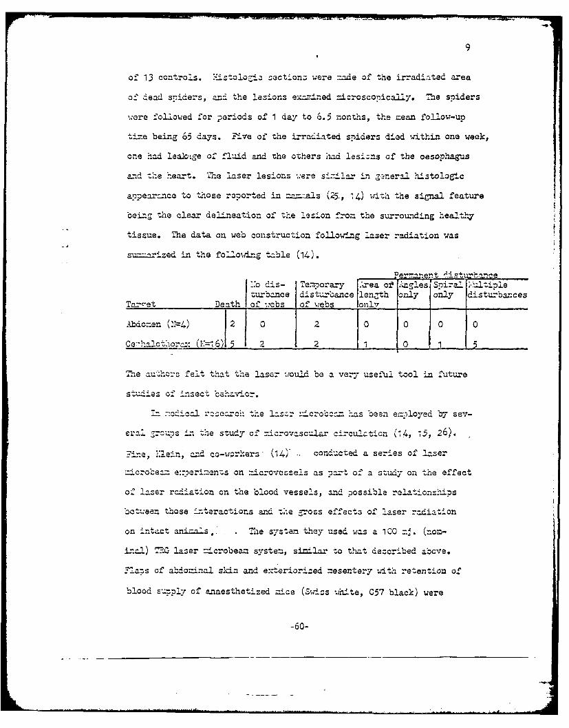
-4
9
of 13 controls. M.istolo~ia sectionz were made of the irradiated area
of dead spiders, and the lesions examined microscopicaly. The spiders
were followed for ?eriods of 1 day to 6.5 months, the mean follow-up
ti.e being 65 days. Five of the irradiated spiders died within one week,
one had leak.ge of fluid and the ozhers had lesisns of the oesophagus
and the heart. The laser lesions ;ere sii.!ar zn ge-neral histologic
appear-nce to those reoorted in mam_:.als (25., 14) with the signal feature
being the clear delineation of the lesion from the surrounding healthy
tissue. The data on web construction following laser radiation was
su.-arized in the follo,,Lng table (14).
Perr~t it'bn.e,:o dis- Te.mporary rea of .AglesI ira. ;J-tipleturbance disturbance length only on.ly disturban-es
Tar_et Death of wcbs of webs only__
Abdomen (.4) 2 0 2 0 0 0 0
Ce7Tha!otaor.: (,= 6) 5 1 2 2 0 5
1Te autho-s felt that the laser wouId be a very useful tool in future
studies of insect behavior.
..... dicl .e--c -he L. :'cro-c .as been eaployed by sev-
erl groups in the study of ricrovascuiar circulation (14, -5, 26).
Fne, _ein, aid co-workers (14) conducted a series of laser
-icrobe= e::'eri--ents on ::icrovessels as ta-t of a study on the effect
of laser radiation on the blood vessels, and possible relationships
bct:een those Literactions and the gross effects of laser radiation
on intact animals.. The system they used was a 100 zj. (emo-
in.-) R-?. laser microbeam system, similar to that described above.
Flazs of abdominal skin and exteriorized mesentery with retention of
blood supnly of enaesthetized mice (Swiss white, C57 black) were
-60-
10
:rrCiatcd" - at various energy levels. The smallest spot size obtained
at the focal :lane was appro-imately 5 microns in diameter. Some of
the factors affecting the i-nteract.ioz were the energy level of the rad-
_ation, power density at the focal .1 a-e, spot size, and the diameter
and shape of the blood vessel. At low energy levels, constriction of
the blood vessel occurred, persisting for several -inutes. At higher
energy levels, blood flow within the vessel was arrested by the form-
ation of an intravascular clot; although the interity of the blood
vessel wall did not appear to be altered. A further increase in ener-
ZJ caused aneu-ysr-al dilatation at the site of the clot forMation.
interstitial e::travasation of blood was observed from the smaller
vessels, and at sites of bifurcation. -n ore.i_-nazy studies on sin
flaps, a -switched laser di-ected through the microscope produced hem-
or-ha a, rather than thrombosis (27).
H.ochen and 3aez also used a -. G laser microbeaan system to conduct
a series of e-ceriments on n_crocircula ion (i5,2.6,2SZ9). These studies
were o..e wit a 55X water a.'zersizn objective, ,,hich produced a spot
5 u in dix.aeter at the focal plane. The intensity of the laser be_ was
controlled ith neutral density filters of _ rnoi transmission, a" was
aeasured with a TG ballistic thermopile zounted on the substage of the
microscope. Ln order to obtain a dynanmic record of the phenonena ob-
served following laser irradation, iochen and Baez incorporated 16 -m.
ciJnemato a ~hic fac--ties into the s-ysten (Z). This series of exper-
inerts included st"udios on e:- osed, rat maso-an-endIx (26), and studies
ezsloying 2 vascular. model systems (2S).
-61-
wii
11
The findingz of i.oclhen and Baez :e-e in general agreement with
those rwported by Fine and Uein (14). Kochen and Baez (26) found that
single laser puJses (0.5 up to 7.5 mj.) resulted in a progressive in-
crease in vasomotion and a vascuIar hyperreactivity to epinephrine.
The hyperreactivity to epinephrine sti=ulus increased with increasing
laser enor7 output. A single microvessel showed a progressive increase
in reactivity reaching a peak hyperreactivity in about 25 minutes after
receiving a single 7.5 =j. laser pulse. These changes in vascular res-
ponse were no" accompanied by any disce -ible vascular or intravascular
mor.hologic changes or thrombus for:mation. However, the injection of
colloidal carbon partic:es into the blood stream rosulted in the ad-
herence of the carbon particles to the endothelial lining of the vesselwall in the region exposed to laser injury. Laser pulses of !tig1er en-
ergy (7.5 - 15 mj.) resulted in transient "stickin-1 " of platelets and
leukocytes at the site of lasing,azd the accumulation of carbon Parti-
cles as much as 100 microns un s- rea- and don,m stream from the lasing
site. The depcsition of carbon tarticles in this 7.5 - 15 mj range
e::tonded from the inner endothelial surface of the vessel through the
perivascular snace to the basement membrane and perivascular sheath.
The extent and degree of carbon accumulation an7eared to be indepen-
dent of the rate of blood flow.
Laser pulses of 15-30 =j. resulted in ma..mally enhanced vaso-
motion and was consistently associa ted ith formation of a-zooc._te at<t the fo1aio of inuy U!.-
trombus at the site of injury. Z:cosure of venu es (30-60 u in diam-
ater) to single laser pulses in the 15-30 mj. range resulted in the
formation of a thrombus composed predoinantly of platelets and leulco-
-62-

12
cytes, which often occluded the lumien of the venurae. A progressive
recanalization of the t.'ombus and subsequent dislodgenent of fraements
of cell 'm--s and embolism followed within 2 minutes of zt-mum thrombus
formation. L.zosure of arterioles (30-45 u1 diameter) to a laser pulse
of 15-30 nz. inteznsity, resulted in a sinilar but -iore transient adher-
ence -nd a-g egation of platelets and leu':ocytes. Consequently, the
throbas had a tendency to be swe- t away as a -icroembolus. in neither
arterioles nor venUes did laser intensities under 15 mj. result in
interstitial-- henorrhage or distortion of the no:re!J morphology of the
outer vascular wnl. or siurour.dinG tissue.
3ocusing of a laser pulse of -=eater than 30 mj. intensity on the
-;_ll o-: an arteriole or venule resulted in the doosition of a fused
red cell mass on the endothelial surface at the site of lasing, the
evolution of a Zas bubble which quickly disappeared, and the forr.ztion
of an extensive and stable thrombus. Single laser pulses of 3C-60 mj.
intansity frequently resulted in localized regions of coz~plete vessel
uall bre-,=o-a and varying degrees of interstitial hemorrhage.
in -a atte:.-ot to evaluate the effects of laser radiation on the
vessel waiI, -er se, and on the elements of the blood, experizents wer
conducted oz tw;o -:ode! vascula systems (28):
(I) glass capillary tubing (60 u diameter) perf ased -th
0.9- saline solution colored sly blue and with heparinized
rat blood.
(2) isolated, acutely denervated, rat mesoappendix vascul-
ature erfused with -.alian Ringer's 3.7' albu=in solution
and hepar--nized rat blood.
-63-

13
eueof t he ls cz:.'illary -erfuzod wit,' -.hy blue za-i-ne solution
to za --Lr "zU-1:o of 30 mi. intcncity did not produce any obzervable
cnanr.Co in thIe flow ch~actarictics of the saline solution. Class cap-
ilay perf%.,zed %-_t,,t heiparir.ized- rat Uoood e;.-oscd to a laser pulse of
silrintensity (30 mi.) resulted in the dcoosition of a f'used red
cell mzz or. the cglass ca',illary and evolution o4f au'ttl as bubbles.
Zxiosure of isolated esa'ni:vessels to a 30 -.J. laser pulse pro-
duced no a-.)arent :.ornholo-4c a2 ch4ne i vessels -oerfused with color-
less I~izrls ab.' soluation; lnow evcr, subseauent :-erfusion xith
hen:--ini~ed rat blood rcsul.ted in thl-e transient adherence of lew~o-
cytes and platelets to the site of prev--ous irradiatiLon. ' si4-:7e 30 mij.
laser riu-se -)roduced a stable tlzro.bus in iSolated --e sow-,.)endi'_C vessels
oerfused with rat blood at thie tize of irradiation. inen the Isoae
vessels .:ere -oerfused ith colored 2.inger's album.'in solution, la-ser
irradiation- -roduced various de -ees of dr-.*a, ran,-ing fron. discreet
cell adherence to coaz-%eta brea-ccoia of the Vessel wall.
'oc'e - d Baez corclae fro-u: their- studies that the vessel wallI
-rinoled in ti'zo:-.borenesds, but tic alteration initiating; the ad-
herence of blood elements to th'e -vessel wall is not a change in wett-Za-
bility. T'hese concliisions seen, Justif ied but not of i-znediate signifi-
cance. Thne authnors d.o point out th-e interesting fact that- th'e changes
in va-sc-ular reactivity th-ey observed after laser irradiation of --icrc-
v-essels were s4-1=a to those observed following _zicroelectri;cal sti-2
ulation of the *-az-stcr cheek pouca vasculIature, whlich also was followed
Cy thzo::;bus f-ornation (30). This observation is of interest, sin-ce, in
-64-

14
some cases the electric field my be of some iportance. However,
dCtermination of the fields in tissue are not easily carried out, and
may not correspond to those obtained by simple calculation for electric
fields in vacuum. Zhis is Particularly true since the tissue i6 not
at the focus, =zd rapid defocusing occurs. On the other hand, the
opcosite reaction, relaxation of vessel sr.:ooth muscle folloiring ex-
posure of vessels to i-ght between 250 and 430 -,)z in wavelength, has
been reported (31).
L It has been reported (32) that a group of doctors at Montefiore
Hosnital. have been investicaCing the possible use of a laser bea.- to
create anasto-noses in sn.-I-l vessels. hey use a CuSO solution to4
stain t:he area of the vessels where the vessel union is desired; then
they irrzdiate th-s spot Iith a neodymiium laser. Hlowever, suff-
icient ifor-_ation for evaluating this technique is not available at this
time, except as reported below.
A report by Stru.13y et al on these studies has been prescnted (33).
d-.or --n. acceptor vessel ..,ere -lued side to side. The co,-on wa
.. s t.en ez.ose. to l rdiation by focusing th-ourh the open end
of the donor vessel. There _-e obvious nroblems with this -ethod.
Since the donor vessel -aust be opened to per.it penetration of the beam,
a rlfe or fine cautery could have been ust as readily used to incise
the vessel wll. At the region of impact, ternerature elevation occurs.
This wi: result in thrombus foration and blood coagulation. The -
erial whic'h is removed by the irradiation wil result in emboli. Blood
coagulation at the site of irradiation ,il.l also result in emboli.
-65-
15
'I2thou:; the tac.hnique has been i resented in the medica~l pres's (32) as
successfuld, no indication of tlais is eviden~t.
.hile t he -i crobcan is beine used for the -irst tine in entom-
o2.ogy and microcia-oulatory research, iicrobeam: have been used for
nar-tial cell irradiation (?CI) since 1912 (1). How~ever, the intro-
duction of tWho lasr icrobam has ztizalated an unorecedented itor-
est in ?C1.
Bessis and co-wor!:ers have nzde zan e::tansive study of cell in-
;,ury and cell death (911). Using- lethal doses o' laser radiat ion, then
followin- the Lr-Adiated cell with tine-lapse cinematoaaphy, a phen-
omenon referred to as 'Inecrotaxsl (11) lw:as observea. "f a hu=an red
or whiite blood cell is i:-radiated, as soon as the irradiated: cell shows
an,' si5--. of alteration, the surrounding leu.I-ocytes travel toward it,
attack.ng and thagocytiz-in.- the affected cell. Hiowever, on occasion
the pha cyizir.& leukzocytes w-.ill nnsune only part of the dead cell,
abandioring the re~aininS portion of cell for some izr2laoiM reason (10,
on a bOCieic"I basis, as it =L__ -rie"d 7ertinent infoznation on the
general pheno:..ena of cheraotaxiss. Bessis has indicated that he has been
e:.111oriLng the '2ossibilities of selective or-anelle destruc tion. v'ia diff-6'
arential stainz. to contrIol absorntion of las-er radiation (10). H~ow-
ever, no data 1ave been prezented to indicate successful differential1
St-;n-;nrg -LO- destructiLon of s-oecific cellula- organelles.
Saks and co-wor2lers at N.I.U. have ex-olored the areas ofl :icro-
surrr- and oartial cell irradiation with a Z.-G 'iZillilaser" ±icro-
be=n syste=. Zarly in 1963,Salcs and Roth (13) reported their initial
-66-
16
studies ,,ith a laser iaicrobeam. They successfully made opezings of
a ppro:d aely 25 ricrons in the cell walls of the alga, Soiroga
without causing other gross irreversible damage. This facilitated
microsurgery to be perfor-ed on soze internal structures of the cell
without doin& irrepai-able daz-age to the e:xterior cell wall. The laserused for these early s-udies had a .nazjuu enerr output of 2Omj
per pulse nd a pulse .idth of 5 z 10- 4 seconds; however, even with
these energy levels it %.as -possible, because of the encrar and power
Fdensitics, to sever the chloroplast helix, disturb intracellular crys-
tals, and to produce localized coagvlation of the ci'!oroplasts and
cytoplasm. Leppard, has also been able to remove chloroplasts from
a variety of plant cells, usi g a similar system (5). Salis and Roth
e.re)orted (13) that irradiation of the cell nucleus with an incident
be--m enerr of 0.08 mj per pulse produced no discernible effects. The
focusing of higher energy laser pulses on the nucleus resulted in
coagulation an2d a reduction of nuclear volume. A grossly similar
nuclear reaction is caused by ultraviolet ight (34). Sz.s and Roth
(13) also found that addition of a low concontration (0.O3,j) of ::eth-
ylene blue chloride, which localizes on the cell w-all, appreciably
reduces the energy required to produce alterations 15-25,u in diameter
on the cell wall. Siilrly, irr csiation of the ch-loroplast heli. with
0.3-0.6 =j resulted in coaglation of chloroplasts and cytoplasm, loss
of turgor precsure, ad a rounding of the nucleus.
,ditioral studies conducted by SakIcs et al (16, 35) have re-
sulted in some -z.,rcvementc in technique, and have ptoduced some in-
-67-

* 17
""'ue-, an .... Ji~oved versicn of';h
IZar: crob_ _-: ;zz ... : ±wit'-azz: out-ut 0 1 0 j
3~ ~ nuiz.5 'Ia 'o: a 1": ocu2l-.r and a 10::, 32X or
~'~~Cobcctve(~,,or -a 20~o~' .~: a 5Z;-- ob~ectie 3).2
f*-o of -:-0 ' to -4 e a s5-oz 4.~
zcrons india-eter. le e--alevS deare oontrolJle -:zith r~etali.zed,
tich-10r're out a:2 T s- zr'_ oc:uzato j~tl1:~i
the oztic_-. a--Jz of the -ficrosco--e (16). thin f-:!- of &recn dye
i ' to dr-; on -= cro-oc sliCe . _'s -he s"Iae dries, vrz"
cra-ch!s :--e fonez.: in the sur...ace o th-o C.'e; th2z-e craclhs c=n 'be used
to th'l zicroscona to Tzze tlzc szlda -1is in the focal" 7)lane.
T'he lIssr I's fi'ad-rdAi~ a hoc' i-n the dyc. Th,,e &i-er ececs-
wr o obt-'_ ;4ti2 stti tha -:rcer '-osition can be don~e.
Th e ali=_rLent is checllecl aS-I .sin- ':h~ ca=;ocexa~re, tith
---------------------------- z :~~a s-;stcz over: ot'L~e
m :a t tZ. -';Laye or *--' o t*-.r-C 0=
u::z ~ ~ ~ ~ ~ ~ ~ ~ t tacus stide oro3o a:: stecah h
ozct--. '1is Is if.~:za one 'azsto obtain the ±ez
S: c'L oszol for a givae ocu._- o*- joctive.
~t:izoi ita-Ila =i)dC. - httelsrncrooez
an '" ezotiv-,e n-4croVz-r-4cal t co I-. _h-ic- co:::ented the r-
-uOl- -nd rnicroinjeCton sySteia. radat of: the( iter-
c~alcll t.a.neart ncoQ,. "he ies,-at a 15C'j --4cobezn
-68-

with incident enar=r of 12.5 mj per pulse considerably altered the
cell1 will at the site of irra=diation. This area zrovi;ded a "ivndowl" in
the cell wall thr oug.h w&,.hii czipettes cou2l be inserted to injiect va-
ious nari-s or -nerfor= other micros-.:,7cal nonipul1ations. Previous-
ly, the call wallI had Oreatly hindecred microsur-ical e.xoeriments on
plant cell1s. :~rradiation with 5-1,' obj ectiVe with a 20X ocla (35)
produced holes in the cell walll of 'Titela thus -)ernitti=g the inser-
tion of a :ncroaizette. (The nehnia ithdrawal of the -- icro-
[7 riflette -r.oduced more cdanae thann the laser bexn.) The la-ser bea=
ni-ified by a factor of 1450X in th.is syste= fused. 8-16, chloroplastsr --- caused only restricted* local daage.
The effect of las-er irradiation on litela growth rates after re-
ceiving 1, 3, 5, *or 7 pulses of 12.5 rnj from the in.cident bean, with
a ben diameter of 156 u (i9.5,n, in e::perLnent, 2) was studied. A single
poulse produced brownming and blackeni G of cell wall in the target area
si-ilar to that seen in S'io~a(i3), and chioronlasts were disoersed
or coagulated in an area aboutc 64 u in diameter around the targSet site.
Ovlosis was stopped for a period of tine thIat increased with the nu=nber
of tine '-te 'I±tel>a was irrc.diated. Ln tlhose -!ieia receivigol
a sirngle pulsa' the vclclosis -esu--ed~:. 2 to 5 -ainutes 'ter irrad-
iati on, find in thiose recei.ving 7 successive pulse-- of lasG.- radifation,
cyclosiz resun-ed -Athin 12 to 15 =inutac following; irradiation. -1he
-grwt rate of- all the irradiated olwontc was less than t.hat of the con-
trocls; however, the pe.- cent decrease did not corresoond to thle nu=n er
Of, irradiation~s.
:zzer n:icrobean stadies on .'zoeba -,roteus (16, 335) are of part-
icular i.nterest S4 nce a n-uno-er of ultraviolet aza hivn enercy nacrobea=
-69-

19
studies of .I..oeba have been reported in the literature (2, 3). Saks
and his associates irradiated ,.oeba i'ith laser pulses of 20 mj and
100 mj per pulse at p.ulse repetition rates of 1 pulse per minute and
1 nulse per 3 minutes, urith bea-- diaeters of 50 0 ,u, 116 u and about
5 ).1 (16, 15). hey fou.nd_ obc. to be very resistant to laser radia-
tion (16); this alzo occurred with other ty.Tes of radiation (3). Saks
reports that follo.C repetitive ir adiation of the cytoplasm at 1
-pse er zinute at 20 =j per pulse, the .,oebae go through a cycle
following each pulse:
1. Brief latent or unditubed period.2. Zhanced foria-d ctoplasric strea.ing.3. -Zh.2n ced reverse cytopZasmic strea=z.nC.4. Local shift in sol-gel equilibrium toward solution, referred
to as "be-llooning:?.5. recovery.
..ver..ea recovery tine was S-9 seconds (16). This description of
the A.-oeba's rezzonse to laser zicrobea. irradiation is similar to
that reported by Tclsahotine in 1935, after izradiation of Aoeba with
his 2S0Q . microbeam (6). Acciuulative changes in the sol-gel state
of -oebae reculted from repetitive 20 .4 pulses of laser radiation
(16). Thi-s "bTllooA-g" was detactable by the progressive loss of the
ability to form and =intain nseudo:podia; in al' cases the Aoebae were
able to recover from this llballooning" phenomenon. Sa;s suggests that
this accu=---ltive da,=.ge may indicate thit the lipoproteins and proteins
of the sol-gel system are srvactIcally denatured and accumulate with
rometitive irradiation.
-70-

20
~rr~iatof o=!ioeba cytorl~asm wit h a100 :7, incident laser bea=
in:dZtely --roduc-ed rapid solution, violent contraction of tche whole
:.,zal, and recovery. 7*' th113 cyto-plasra was Jirradiataed repeatedly wt
100 mi pulses there was accu.=_ative darnaSe. Frequontly, the cy-Loplasm
in tha target =ea was inched of'2, eznd occazionallr fracgnen-6azin o f
the -ccb- 'o-lo!. d. in ad,.:ition, iraa-t~ o" -- eba cytoz:lisa w4t
100 mi pulses caused the specific aj-c*tion of f'ood vacuolas, es-pecia.Uy
the i-ngested, food cozntai_-ned cihl"ororhy2ll (16). R. 7Zolo and Sas
have renortad (35) that if~ a:oebaa are injected ,.ith -ic =o dr o zs o f n
era! oil, olive oil-, or olive oil cont_-*:1 ..i1 _eBlue Sulf'ate and th e
o-;- crons -r=adiatcd -with the_ 116, -irolaser-bear, the oil d ops are
f:equantly ejected'. Hiowever, iJrradiation ol' intracytoZ1lasric oi drotps
-a= the 20. oc"a and 53:. objective -o ob-.,_n- a- =nifactior. factor
o_-' 14.50X (I.e., be_=' di=3tr annro:.aly 5/,u) _ id not elicit such
drastic affects. There Was only an acc'.zu~aticn of c'-tZlmc debris
at the oil-cvto;,_' -a interface, whutejactio n of~ thie oil dr-t.
tz'e aoeoe wre ily itane vth Zthylene 31ue, or 'ile blue... e atoe wer niriljatdit h yols rteoldo a
drc (5) Te :ichg o t ofn ir rdiated cfteras th s ilso beenV-1
akcws nd i co- wckcrs 1 6 35. _als tde feto h
~~~~~~~5ulaser --;crobeam onusouali oly thenules as rrdite . :.
~~~eated- fl o irradiatio fnce ih2 ad i cidetobeas: witha _1so
snjot size -,roduced behavior distinctly diffarent fron those a:zoes-aa
whose c-ytonlasm, was i.-radiatod. rradiation of thea nuclaus resul~ted

21
cto0 p a Zz'c Z. z _ . The innain. t.on of cyvtor~laznic stroar...'eZ was
"oilolwd by a revorse :rzunul_- flow (cyto92.aci'.ic st oposita
ttatin non-ira&diatcd o:- rccovc.rc-* =:oo bQa). This was f 11 -wed
s.'ioty 0 y e~-c e c yto 11 c tz -"J .in he ori_; 4'a& diection
or in a ' f-a';n d; zC c k.t- on -n iio, -:0.ton ana .,SoLC
cytoplas=zic ste.nas eVla.enz, an- th~e rate a:tac the azoebae
-dvancod -~ez..oiw e ~uce-a. .. e -ad-;aa: on w.as d-ieconti-nued,
tha -_eba %are a-ble to recover =::i rarro_-uce, if' d~v-etotenc
:eus tI-'s
Trzadiation. of =uclei with !00 mi froz the incid ent be z esu ed
-na sudcem sto:?oa:e o-' cyto-2.as:.c 'oea~g oo..ed by 'riol.ant r:.-
i_, solation zrnd- contraction of'th whiole anceba, afte: which the a=-
oebca -:ecover-ed. aa-,etitiv;.r -::.r- __;aon of zhe nucleuS -wath th.e 1CC :-J
in~.en O~Z cuzz iroeribednae.Tis re-.:cinaaCro-
sc~clye.fec:ive;:-y e:4 ucleated anoeba, although the n;uclsuE-, waI o:
elTected by th-e =c..z~ea. The =,oobaa bacaze_ mr2Or snhei-ical, cvto- zszzic
stranig bcae Jite, ad se-onoaawcra not forned. Th-e effects
zo:vr h zoel.zae w.ei-e vit4-a:.y Z;ane wtr .. ta '-ne ',nce
arradiation wi-_th the ICO :-j laser :zcocncaused t'he nuclaus to be
~'oowiigsuccessiv;e irradiations (1 to 7) -~ Iul.aac-~ols
of -ocebae at incident ener 5- levels of' about. 103 :j a=d
ui i-n a.:azer .ra~iitad aoebae were c.loned wihnon-irradi--ated
-72.-

22
controls, in order to det.c:z'.ie th~e effact of a lz r e on
tre nioia d -ot ae o ze .In al: cases heizra(ated
a:-oooae solwed a decrease in -nactic r-ate as comn -ad to thea control
colonies. The ma:Lx decrease appec-od 2after 5 :)ulses. of laser rad-
-ation. Five -:Ul5se also- resulted innz d=cecr:easa a the Erowth
rate of ".italla (16).
±nitial nicroben studies on the formed.- elenents of the blood were
carad out by Tine, *Clae2n, and cc-;wcre:s 4.).- The general morph-
Fo 1 ogy and enoeboid movenant, o--: isolated, vibeh=.an granulocyteas
,.ere not affectC,7 by the 6943 .; laser radiation, using a MrG ni-,croscoze
l~aser system. 'Hoever, t..Le addition of: ...ethylene blue (final concen-
tr-ion mz-I) ,rh2edly altered the effects ofP irradiation. Ie~end4=g
on- the site and energy of irait.n leulocytes in 'ethylane blue
solutio-. waere oartialIly o:- entirely fraZ:en.ted, ---d e-ven- whe-n a semb-Iace
of stutrlitgiywsret.Znead, azeoi ove=ent was lost.
~lrlplatalets zhowe6. no an:eteffects f; lase:r -. daf-
noe.ever, in the przaance of .znln blue., laser irradiation rez-lted
an fZarzanntation of thie -ola-telets accozz: ant ed by the release or for-
ation of raznU.laz 7zeiI :, ot-tioaedIaehu~~~
rocytas contracted into :g .ar aedbodies 2-5,u ir.- ,tdt n
zi1=O Sne olwn Izzradi~ataon wat.the laSer Marba~. nse
obcarvations a.-e -4i. -greement w-jith ot.e:-s whohae reorted laser
:z.zrooea= studies on the for:-ed elznoof the bl).ood (3, 6).
Other data retportad by in,:IalLn et a! (14) was the- effect of
the laser a-icro bea.- irradizazion on alli.7:tor ez--"thzloc,.tes. ~iao
ar-yth--ocytes ara eII4lt-cal, azr~taly 10,,z x 310u sie -a d
-73-

23
contain a nuc2.cuz. TMhey fo-xnd that ir:m-a-tion of or].v the cytoplasm
co*.icd affect only the cyto-)Li:. - : t- -- 'th ~ e the nrnz;-rad-
ja-ted partS of t-'a c.Cl rc~c 2t'~yuc~nc i. n contrazs-c
irradia-tion of th uc2.cuz w.ith- t*,2z io-bcj cauz:-cd the alli-
rator eryt =zocytaG to contzac"' J to a ruloo z~cz-urc about 1/3
the a ao size. 2Mis r,'~cv:tlom i_ of PaculZ- intarest'
-n ie!w of as re;,,ort (16) thtthe rss:ncnse o _ 'ae is :z:c*, -or!e
vi~ole±2l w.:ean thez nuclaus is i~~aeaz o-)ose& to i~a~tzof
the cytc:pJl-= on.11.
.sdoubtfn.2. th t theesuisihcztet.tncla tert,
sini:'icant for =aintaining cellualar inti ty. le 6.ifferenze
thne 'ffects =rOduzced is naro'ba'2.' 7u to di er aenes int-, bsr~
ticr: chan-actaristics of the1 tisus i __ ticular, r:if^_erences 3 ;s
th ytpasiC and rraclear coanstituants. .ho 0U. vriu colnstituents
::-,y have s.rnso~inC :e~tcter2:tv ocnrto
of theca ,;ould a fect the to-:al enr- bcorbed nar Init o~r:.~
shor1 .', be noe ta atouh = ener -. ,szeet wre :.
sooze anstan:ces, actua,_-l absorption aszcco zenot c-a-red Out.
-"-'~-"--'~-cnt foU be Ctc: ly;iclt to c arrZy Out
at igh~ea zoerdensities on cigecells, since such- actors aS
refl'ectior. and scte=ust be consideareld. :?artheznora, the beam
-_eczet_-y is not constant, but vszied =x :edly, both1 w.ith d i Stlmc a f.-on,
tac -foca-l z:oint, and w-itlh scatter -ad aoton The s3et size s-
cussed =ust be consid~ered as a~o~ntSince it is based or. effects
Ca S',ecific n7azarial tha-t is used as a :rdai*t robably rep-
rasents an= axea- of rclativol7 hivh0owr cdensi'tv-.
-74-

1,; -robable that a oesohisticatad system w;oul arcv,_'do a
m or e satizsfacto.f --Jicrobeam" r-eom9-try 'or Zom pro--es. The syste=
Would_ consizt of' a set'. fse whi~ch would convergZe the bcam, i.'--to a
relatively par-nllel bean much sm:.ller in dixaneter th- thre ori,, nal
'.e=z, and would bec inderendent of th3 o; ticc' systa:m of the -_, zo-
scope. A.systcm of -, .is tyzte co- 'ld :-acorzo:aze eithe;:r =as, sem-7con-
ductor or solid state lasers. it would have the disadvantage that th"^e
ener -:r den--ity woul_4 be constcant, whae-eas -,ith a focused system, tne
onor,-r density c2.- be low rala-zivz to t*he energy -ensitl~ in the region
o-F' interest. How-ever, sir-ce the f'ocal zoint of the microscone lae
system _-d i*h o loc=tion of the tC.0e do nt coincide exactly, thei
raoion of m-.i- intensity of the' ber- is not at thae point in focus
throuzh -.he mi4croscope, and is difficult to co:;cenzate for, ra=pidly
and easily. . reaieyparallel bam of' very srall cross section
would also be difficult to n7-4tai= in. air, over an-y diiszt=.e =ndtie
because of thec higJh field str-azg ths and consequent irrita4on trOduced.
7-arS '=ae been successfuLLly coxhim-ed .t Ihnicrosco-pes 'or use
in othear arcas of biological research chdn the study of cell
--- mssion szcectr-ocot07 (A'O,. 'h-ese
an lczntionz are covered :in lra.:det ail in other s-ecti--ons c-' S
r:-c:ort. L...ser-r.aicros-coie =ziz s aIave aliso been c~eveloaed for such- i-4-
ds tra a-) ,li c nti-' onS a s --i cro - weIdi: anc zi_4c:- c-dr -;i ng 421 .2)
Mhece =-nits are oierat4on. no-w vocvesch ap-lica-ti ons, thou:71
inta-erOSting-, are not of z-uffcient biloicl ortancea to be Con-
sared in detail *-ere. Hazards associated wi4th these units -will re-
c-aize frhrstudy.
-75-
25
ito~~ czci t.; --t t- jct -..O-T )c ~'is an 'ie too! ci
er--t :c.az ofbo-eu~~ ~c. o;v there ::.zzny :obJ.::s
of:t~. rt ::-on-ftorin 2 syzte. 2'-e ivt.zor.z :- cord-
-)x= te s, -e ..l a,-: -c
the tot-- ener -- :ncii---nt on the s~m1e, an the to~enszr : .sorozza
~-t~--e ooo o tae-z s btzri te__-mc
-4.n s feasible. Sam-olin- of t-H bei7.~~u~ thea obj ective =,- ta=-
e . t s ~ S d.O :Oc4 ~ f!e't f cutuaaS- rc used. e
ener,-y tra.-=:-.tted co-,!,- ezti--'a o -, '-ooj ci
ector on the Subst-M. 7- ecass-nry t'at a standard~
mc~-uemetsysvtcn be u::ad in :-. c:-oc=zsu c so that data c-- be
coz.,-a.rcd with that of oth,-er inc aos.~.z'~hlaser --4cro-
azed: tr:' an -.7raio~ zad az non-e acrocea z -'t;.
=ornoraw-no closedai _-2 t 0rotez-t~ suc -.' 3assis
(10). . :.ould enable Cbz or-:e oez.aa
... ~.eiatly olloiuin-' nir7ad-*.,t4in, to so-ce:tct with^out e: onOCgc
w~zslvsto the azca: e&asa. Z-i:a the r, -zseuatz
-;7Short, r&. zouta-om ~ated, he -CIIZaJes l. not -za:: -. o-
o-uncod as e:-:occtad. 7!idoo ta'e raccrd=n; a =., of' ccurse, '-e included.
.a*:-s oif tHe i:dv&. ._-sssi z i h ae aro-
stu.dies th-at 1z-v 2ben ra tOrted, t.:ey ha-e --.L. iizate Pd areas
c ea deal- oil 2 or ae " '.:e studiUes

26
of~ jtt hi is :-ssociat:Q (;-4,) on S b.C.Or b z-rizo: t'n
:.r z.C=Ie~ c o.n vzaJ.zl tool, no ; cn y inl e.t.ooy t or
(10, 11i) on cell _4=4,=- cel- oL:r'iI nozt:: on
w"" *orl: 1-th I~r~a~ §~ 6 by ce:.on-
st"a'a. the uzefunesc of the Iz.zosr crbmni c -o=ery. Z'ic
studies on. alligator ort~o~~~ i~ ei (14) and"~z st,--
Fies ona c.-zebaq 0~6) h'aave g.va.. t lez s4in
rCall Lisezr ir d.tin toStty ;': z)Grt2cne of Vzricus S--ce_-J22.
organellIes, is ''zil.Holwever, no one ha-s yet Q:o2tac.a me-
Ofc;'' ou~t selIect"-ive dest:7uczicn o. secL' orZe 1 es. Th uce
o f s -,ci e aiCCyes nas been su~t-stc (17', bu't data has no t been ra-
7ortad. BEessiZ atated heat*he -s vaccessfu'- - dc-troyred, a sel-
_o..sde z:.__Le buc; n c eav 0r, ~o tc ta Z.re .4 3t a~ 1.z~ :,07 eln.-
uton. Other- ao~n~ Mea of selective destruction of cell or:.n-
elles izlcet' c UZe of 1 ac: wit o,t "utsats'cicfzu.is
corr.:zozdinz, to soz-.e Ybo~inbco x::ia t3 ... I
one 0riua e~ 1fo~~ce '- 7:-; 4.......~~
c= xdrod I's :ncz uc 0~ lozzc 1nr nicro'be-z '=.- ;r Izcs
of nhotoselecticn (4-3) to tcahe advatt e of the ienec CS
t:e orce't of selectiv iet'c it -wou '-e of zores
out f'u:ther c tutcie E to con.Vir::_ ' Sa:sl zvtz t*=. .vzy =-"zzz~s,
or less, nrcducc only cor"2Led loc'-'C t-e %.:7ca of'th
-77-

27
-"::-et Site.
mb..~2A3col~l e- 0-: value in Cetar-±ning the ~ca~s.
syst.Zs. 3Since t-'er~ isz a ccdbo od o±' microbe=- reSearch
totz* ot=- so-=ces- Of rdiati"on (2, 3~it =ay be =ossible to
co7-z:-c tha efffec ts off othe:r ty-.ss off radiati4on rith the efcts
cs'used by, laser microbeas oz selected or.garnIss or coonetts.
it ioujld also ba relativaly; easy to do co:7rarative studies at a
cl'lrlevel. Mhe value off icrol=--~ studies in eJlucidating
efffeotZ on thae intact ar42 ; : rtially indicated by the
=icrovasculax studies off Koch=~ and Baez (20', 23) and ine, Mlein,
et al (14 ), ..hich deonstrated the a-bili-ty off lasers to produce
vanscular changes, icd 4 g eo-raeand thxonbosis.
The cost off laser i--cro'cea= uni ts ffor research would vary
between $-3,000 and $25,000 de-Pending on the conpledty off the
=icroscope laser system, and the associated equinent iniclu-ring
nicro-.ninlato"rs, T7 viw. facilities, nd raco;.,d!: canera-s.
-78-

2. Zi -2.e-, R.. ?U1-Cell lI if-tion, Aac.Bi.ol.. Fays. 5:j!C!---.46 (2195T).
3. C'..1. =i~b~a d Pcxtial. Call l==dia.tion, Imtez-at. ?.ev.Cytcol. 16:133-153 (19641).
14. Tci-~es, C..H., Cmtica. =dc thaio- ibl A--nlicztions '6o
3iol , 3iozs. J. 2(t )325-329(.G)
r ~5. :=It, R.: afcts of I-sez Raffzt±eJUa C~j s
J.3~1y.------r3)7771 (196).8. Y.sz, ., A 1 -7, ~'l. U ., :-zzi 1,d.-, '8t~~ e
255:1010 (r 55552).5)
9.7 ss 4.G.Be . ~~s~zin pe etuto des ur-lesCJ.ul Bic eYz M -c'== o C'eis .z' :77-791,C. S"c6. o.1819 16)
1. BZ-ssiz, 14 c z e B.o~ss, iro~-4uzct= Ao Cellscio by eo
Lsr Bacza, -i=-N. 1r. Aoc",;. Eel. 1;2:689-694 (1.0,05).
11. 3zssizo, M., Studies =1 Call AtZLoy =* D --:: i'tte-=4t CIZssificz-tion,Cib. Fz ninSym::oi=, Callul.= nj- y edite-. by A.V.S. de R.euc-,
12. Rina, S., flein,' Ea., Hay--*, G., SzztR.E., KZn;, W.=adAan ,C,T~aa U~se cf loe Cirvz"t T L-s"Z~naor T j.
13. Sz2.os, I1173==, I. Rots, C~zlez : ly Laeser as a c-suiazstrentScience- 141: 2:6.2!. (1363)).
Criscay, J., Doo::,J. P-J Barr, V.:S., Ted. Proc. 2L(,t 3Isumml. !L: s35-45 (1965).
15. 22, S. =dn&~~z J.A., =cts of ?Used La-ser --mr-ano th~e.cr cu.t i ?-crc. 23:253 (19,5).
079

Cel. 2- ?. y Lz-sa IT.tzn Y. l * Si. 122:695-712 (2.965).
1.7. MZ2t .. ~C,-3., C--ticzl '.--3- imn Biolc1:Zr =d 1.zdicize,
18.~~~~~~~~~~ L=,K.2---s TSJC C., Laar a
TVl 1-<1c-.-4r. sn:675-677 (195")
2 36. - , o?2., r C.4z~. ~ ,.osodSie e
2500 iS.V. Bzd Klica E., Sioz~ ,ef of:c Laser ztio-- t -r t ~~' al -- -- -
728-737 (965))
T 2,7 ' W .ra . -
25. P4-.:e, S. =-d Xocfe, of'. Laser
Z,~~. S.,T--the
26. s. u....e. ar o'-z Sa J s
of ~ ~ ~ i Rzt~~s2 f -, J.ka. zdyio. si41=227192.).7(295)
27.-7.= Ti:2,S., Zz-ec oo emoizu

_____ -aze 0 c'"caso, 1.(1964.); as
. , 6, .sS--
zzz~~z___ 2 --zozz:~, c. 3izzed. LasZer C ='
T.as 'P-Ca. 7-- -.1-C-:--' , 'r, B,-om, Mass. (A .296")
37. GzzyZ., -Azta. "3±C. zczov- SE-=. Z=01. L: 47-57 (2196.).
33.=-A ,; Tez.3roth, B., rTz--e 0 S-itched aas aTool iz
39. 2Rns, FP.S., 2 A:ctzsof~ 1-,--sZs to azb: d 321o -lilca.
~ ~it~a L -c e~e 142:23 6-237 (1963).
42. ?c;Z. i. as:Cedz3.-cricng mithTe~r~cs, XY
mp. -. ,croccy Ohio
4,5. - S. * ~ demi, S., A-- s of flTA 4: Livng~ Sora: AB~z-::yi Lnm. -lysis, S=ioze .6222VO(12)
081
Embryoloxy
The blastoderm of unincubated Zertile hicken eggs were
irradiated with a ruby laser rated at 0.1 joule output laser
energy by Lang et al (1). The beam was focussed to blastoderm
size by a lens, the focal length of which terminated at embryonic
depths within the eggs. Irradiation of the blastoderm in these
cases required penetration of the shall, shell membrane and
several millimeters of albumin. Following incubation for
22 days, deformities including splay legs, a club foot and
visceral protuberance were obtained. Further experiments were
carried out by windowing the eggs with a sterile cover slip
to facilitate direct focussing of the laser on the blastoderm.
All controls that survived to 72 hours were normal. None of
those receiving 2 shots of the "low energy" beam appeared normal.
Only the heart and associated vessels were identifiable as a
tissue organ system following irradiation, the other embryonic
structures could be identified only in traces.
These results indicated that teratogenic effects could be
produced at 6943 A, at laser power and energy levels. The input
energy to the laser at which the irradiations were carried out
are listed. However, since lasers have a threshold, and are essent--
ially non-linear insofar as output versus input is concerned, the
energy of irradiation requires documentation, for more meaningful
future studies. There is no ifidication that the refractive
index of the media was taken into account on focusing. The
measurement of scattering in this heterogeneous system is difficult.
Thse factors would affect the energy and energy density incident
on the blastoderm. Since the number of eggs used was relatively
082
small, the statement that the number of deformities (expressed
as percentage of eggs irradiated) is roughly proportional to
the energy received by the eggs requires further data and study.
It. is interesting that at high energies the number of eggs
hatching (45.4%) was almost equivalent to that of the control
50%. At much lower energy output levels the number hatching
was significantly less. Its significance relative to the
deformities produced required further consideration. This
[ was essentially an excellent preliminary report and it is
n- presumed that more extensive studies will be carried out.
/
083
In the 19th century two concepts formed an important back-
ground for work in the field of experimental embryology. These
concepts were the concept of mosaic development as advanced by
Roux in 1888 and the concept of totipotency as advanced by
Dueseh in 1894. The first of these led to the studies on
localization and mapping of prospective areas by Vogt 1925 (et al)
while the second led to the production of postulates defining
totipotency. Experimental evidence led to the realization that
a series ranging from absolute mosaic development to absolute
totipotency existed rather than either extreme.
Morgan in 1895 found that inversion of the amphibian egg
or removal of the damaged blastomere would preclude mosaic
development. Studies were conducted by Spemann in 1938 on the
potentialities and interrelationships of the two halfs of an
egg. The emphasis of the studies thus shifted from the potency
to the mechanics of organization during development. Nicholas
and Hall and Tarkawski, carried out studies on isolation of
mammalian blastomeres.
Laser studies were carried out by Daniel and Tajahashi (2)
Fertilized ova from rabbits were flushed from the ova ducts
with F-10 culture medium and collected in sterile watchglasses.
A 2-cell state was obtained at 22 to 26 hours postcoittu a
4-cell stage at 30 to 34 hours postcoitum, and 8-cell state
at 38 to 42 hours postcoitum and a 16-cell stage at 46 to 52
hours postcoitum. (Cleavage was irregular after the 4-cell stage).
084

-4-
The ova were stained with methylene blue in a concentration of
0.05 mg. per cc. which was previously determined to be nontoxic
to the ova insofar ascleavage is concerned. Following staining,
the ova were washed once in stain-free medium, supported in a
hanging drop of medium, positioned under a microscope, and a
single blascomere brought into focus. Followimg lasing of the
blastomere, (at 3 millijoules output from the microscope) it
collapsed within minutes into a amorphous mass. Generally,
each blastomere was lased separately. As the zona was not broken,V.both the destroyed and the surviving normal blastomeres were
retained within it. Following irradiation, the ova were
cultured in vitro.
Cleavage of the surviving blastomeres were noted, and
followed until controls of unlased ova reaches the 32-to64-cell
stage. At 8-and-16-cell stages, it was difficult to obtain
sufficient isolation between the cells on lasing. In three cases
they did manage to destroy all but one cell in the 16-cell stage.
The output energy was measured with a ballistic thermocouple.
Following irradiation at exit energies from the microscope
of 3 millijoules, the unlased cell assumed a more spherical
shape, considered as possibly due to decreased pressure on it
by its coblastomeres. (However, thealteration in the chemical,
as well as physical environment must be considered in explaining
this effect).
Cleavage of the surviving blastomeres occurred. The surviving
blastomere in the 2-cell stage cleaved regularly to produce 16 cells
r~-1

-5-
in the 4-cell stage to produce 8 cells, and the 8-cell stage to
produce16 cells, but not regularly. In the 16-cell stage, the
surviving blastomere cleaved once in one case, and in the ocher
two came&, twice. With decreased staining or irradiation
energy, a general slowing of the rate of cleavage occurred. In-
hibition of cleavare by visible lighc has previously been
reported by Daniel (3).
Attempts were made to calculate the temperature pattern
within the cell, assuming a homogeneous energy absorption. The
effects were considered as primarily thea..;al, rather than due
to electric fields.
The result indicated that a surviving rabbit blastomere
may grow and divide as well in vitro as intact ova. The authors
indicate that it does not show independent.cotopotency of one
of the first eight or of the first sixteen blastomeres of the
rabbit. Reference is made to Seidel who has obtained a
narmal viable young rabbit following.transplantation of an
ovum after destruction of one of the first two blastomeres with
a hot needle (4).
The possible use of a laser as a tool in embryology has been
described. It may possess advantages over a hot needle which
has been used for destroying blastomeres. Although alpha
particle irradiation can be attempted, its penetrability is
poor. Ultrasonic radiation probably cannot be easily focussed
to irradiate a small region without damage to adjacent areas.
Since low energy is required, the use of gas lasers may provide
a more satisfactory method, since they can be focussed to a smaller
086

7AD-A106 234 NORTHEASTERN UNIV BOSTON MASS DEPT OF BIOPHYSICS AN-ETC F/6 6/18BIOLOGICAL EFFECTS OF LASER RADIATION. VOLUME I- REVIEW OF THE -ETC(U)O IECT 78 S FINE, E KLEIN DA-N9-193-MD-2436
UNCLASSIFIED 7NL
lommhhhmhhhhhlmEohEohEEmhEEEEohhhEEEEEEEE.

-6-
spot size, and permit the use of other wavelengths. This may
permit selective destruction without staining, or with less
staining than used in the above studies.
Studies by Edlow et al (5,6) were undertaken to determine
whether laser induced alterations could be produced in
mammalian embryos and fetuses directly in utero without rupture
r of the uterus or leakage of the amniotic fluid, and if this
should be possible, to characterize the nature of fetal tissue
F response to localized injury.
Localized lesions were produced in the mammalian rat
embryo and the fetus following focussed laser irradiation
(69439, millisecond, pulse duration) through the intact
uterus which was delivered through a ventral abdominal incision.
An intrauterine fetal lesion was produced, although not in all
cases, without rupture of the uterus and amniotic sac, and
without gross leakage of amniotic fluid. In some cases, the
uterus was opened following irradiation and the fetal lesion
examined grossly and microscopically. In other instances, following
irradiation late in gestation, it was possible to produce a
localized lesion in a fetus in utero which was similar to a
lesion observed in one of the litter-mates when the pregnant
animal delivered normally per vaginum 36 hours later. In
some of the embryos, resorption was observed several days
following irradiation.
Generally, fetal lesions were significantly larger and more
intense than those produced earlier in gestation. Microscopic
examinations of the fetal lesions showed dilation of vascular
087

-7-
channels with marked congestion and extensive disruption and
fragmentation of fetal tissue. These changes were especially
prominent at the boundaries or surfaces between adjacent and
frequently dissimilar tissues. These included the interface
between cartilage and muscle, muscle and skin, muscle and
subcutaneous tissue, and akin and subcutaneous tissue. The
latter tissues were most frequently involved. Maximum disruptive
lesions appeared in the plane of the laser beam. Fetal skin
showed no evidence of carbonization. The skin of one fetus,
however, showed epidermal coagulation. The skin of several
others showed intra-epidermal edema-with marked pyknosis ,f
the nuclei of reserve cell layers. All of these lesions were
in the tract of the laser beam. On several occasions, marked
histopathologic alterations occurred in internal viscera in the
path of the laser beam. This involved necrosis of hepatic
parenchymal cells in two fetuses and intense but focal pulmonary
congestion in another.
The importance of factors such as tissue and organ interfaces
of differing physical and chemical properties and of possible
pressure waves associated with changes in phase within the
tissue were considered.
Localization of the site of irradiation presented a
problem. The possibilities of obtaining better localization
especially in embryos, with the use of normal pulse duration
or Q switched lasers coupled throughnicroscopes, with suitable
focal length objectives was indicated.
088
-8-
These studies by Edlow et al. indicated that production of
localized lesions in a maummalian fetus through the intact
uterus, by laser radiation could be acheived. The comparative
response to local injury by fetal, neonatal and adult animals
is of basic interest, and is being currently pursued.
It is not possible to well localize and delineate the
r interaction, because of problems of visualization and the
inherent properties of the radiation and target. Measurement
of actual energy and energy densities within the tissues
will continue to present problems. The effect of anaesthesia
must be considered. Localized irradiation and surgical procedures
on the rat embryo in early pregnancy is probably not feasible.
Other mammalian species may provide a better basis fdr production
of localized superficial lesions in the embryo and fetus with
minimal injury to the uterus, ammion and other tissue.
The method may prove superior to that of the hot needle,
if techniques for localization can be achieve. It will have to
be compared with results obtained with ultrasonic irradiation/
of fetal tissue.
089

1. Lang, K. R., Barnes, F. S., Daniel, J. C., Maisel, J. C.,
"Lasers as Tools for Embryology and Cytology",
Nature, 01, (4920):675, 1964.
2. Daniel, J. C. and Tajahash,, A. "SaleQatCve Laser De ,trueaton
of Rabbit Blastomeres and Continued Cleavage of Survivors
in Vivo" (to be published in Experimental Cell Research).
3. Daniel, J. C. Nature (London). 201, 316 (1964).
4. Seidel, F. Naturwissenshaft. 39, 355 (1952).
5. Edlow, J., Fine, S., Vawter, G. F., Jockin H., Klein, E.
"Laser Irradiation: Effect on Rat Embryo and Fetus
in Utero" Life Sciences 4 (5):615:623, 1965.
6. Edlow, 3., Farber, S., Fine. S., Klein, E. "Prenatal
and Neonatal Effects of Laser Radiation", Abstracts
2f Biological Sessions, Boston Laser Conf., August, 1964.
090

091

Studies on insact animazl are required for an understanding
of the in vivo interaction of any a;ent, whether it be
physical, chnical or biological in nature. Animal studies
provide data which, in addition to their inherent significance,
Zorn the basis for studies in humans. Further information
concerning the phenomena observed is obtained by studies
on biological systems in vitro, such as isolated molecules of
biochemical significance, separated tissue components, or
tissue preparations, and model systems. Studies on e :perizent-
ally induced pathological states, such as transplanted tumors
4n animals become meaningful when compared to studies on che
normal state, as determined by the vivo and in vitro investiga-
zion. Some of the information gained ray then provide a
backgrcund for application to clinical problems in man. h en
new =odalities bec-me available for Dio-redical research,
nrooie=zs arise due to the lack of background information such
as is va4_:-' .e in established fields of research. This lack
of information =ay be more readily replaced by unfounded
hypozhases than can be introduced into areas which have 'ean
c,.cu...scriba by serious scienzific efforts. At the same tine,
it is more difficult to evaluate invastigacionai approaches
in a new field because specific and definitive background data
is lacking.
The clinical scientist is usually unfamiliar with the
general aspects of the interactions of ohysical agents with
biological systems. The physical scientist, in turn, who has
092

2
,-ad limited contact wizb. bi'co icz! st-zdies is not conizant of
the probleinas i-nvolved in bico ;ical anad cl'nicaj. research.
Some of tha litaa:ure directed towards the cl.inical
aspects of laser raiation discuss p1-' results, in whi.ch
r2nalnsidaaton is -givazn to pracaeding or concurrent, aiial
studies. Verbal prasentazions, either giver, at maetings or
trough the dmof news ralasas without the benefit of'
scienctic eioilreview ha-_ve resulted in the dissemi-nation
of so=e but nort all factors associaced with the observations,
and ths ave _)rovidad a basis lor misinterpretation. Even
whenasera hog accaole)I ch=als of: scaent"-;c.
c=-maicatiamas, a =ore or.derly sequenca of studies would b e
desirable. Th'us a nu of0 reroorts or. studies of- laser effects
on ae- :;ri=man a.l cumors uould hlave banefited from preceding or
accocna-y'w.-in studieas on nor-'' animals. Since many ex'3eriments
-ere oerformed: with prototype, unreaablja equipmenz '.sorne
studlies were not well cor.,trolle-d, and cannot be statistically
evaluated. Inepeainax:r aoI~a:ec Zrom. few observations
=ay be lacking in scienzific info-azion. The overall-2 e'ffct,
t.aezora, i.s n~ot only a failu~e to provida scientif-ic info0rmation,
'Dur ca-, result in -~~~nainof future work, including-
axzansions to clinical applicarions which =ay not be as -aI f l
nusuae as would otherwise be possible and whmich may lead to
unjustifiable hazards to patients..
0903
Por these resos :ic~h azo bcing docvz::nteci t.,uourhout1
t;.iJ rcporzt, stuijcs on nor" nf. zais rqiepaxticuJ..r
att~tan 2-e rorali our~ose o'i these stu -ea may be sum-
-..nized as f.olos:
1 . To invecti gate tile chort te= ~'on; 4-'
eff'ects of the2 inlteraction f lase J -nO -dtiom
wth sIg.n a -. ."derlying n-o.2.- and tuinor
(discissed sepa-o.eely) tissue.
2. To deter:-.ze thz=esholds f~or Gross and. histo-
2o~icalJ cn~ges.
3. To er a.te the hazards asociated i.ith laser
:adiation.
4. To orient subsecuont studies of the biological
intarctiJon of lascer radiation as available t,
presen~t and in thIe futuure wit", bio.ozica2.
syst._, both in vivo and- in -7iro.
5. -To nzov-.de a "basIs fo_- cLinical arz-,lcetions oiL
Co:~iveStudias wo.re caz-ried cut by Pine, Klein
=d co-c: Zs ( 3. Stu-dicz ifncluded an netaic
on 'w"NO -.-icc in addition to rc',irsd Control znizals. Z-7-2.ora-
o yz-,r- as by these Zui r c caO seare'-" Zpa.insecles,
=uhasrt, h~stars, rabbits ---: M~'v'~ eze studies were
intoc'rated wihin vitro studies on bioogical,. Orz-naratiozls
(7,91,l,~,l~,l-l9 2l,24 and i stuc~is on the p..'siczI

4
theze ctU.ci_4s wzzac: ou' ,--t'h iu.zed lz-zc-rs a- 694
a ' o0 : . zI1oratzry (a= jCt .zzre-ortcd) ~tz~ on -,:e
thz1C ~nz.. .=0e cz=.'ri oat uth Ia 4iorizc (con tLuous)
ztz .s;a~e ca='-ied out .'i~ co~z"-cus G. Is saicrnductor
azasrs '- thz- 1 :0 ='w, _; 'a.~ rz e waz uare cozst- cted cu
aog-.cf, stucies .ith r~dllatiom obtained from C.Y. Ha-17e (I w.,
. 32' _) =di loir power 7 ulsed Iitrco-an -as lzsers (3477 A
did not show hze in the in vivo s:;:cnCs tndI in;-esti,:-ation.
. -.e studies wit.i pulsee solid state !aoers, th a paa-
zers of' the radilation t~~varied to the e:tnt1eazible at
~e .... ~ 7C s~~'o ..~e r**' ___ level to
7.0=3 th OO 1000 Res ncr -o-Uls. .0~soni~~ower lev7 s
i-anjd '%c't, att rz31_ion t'.o =orc thn~ 500 :.
-O~ t-a 500 '-~th~ orvod emzzEy and.
2o;~z dnztjs c~n'ad on t--ie laser crystcl siz, on;hte
C-: at i on w.as 'o cu zS 0e-, ie'cuzzd or unfocuszzd. ?o:
dn::4.Iae :'--- in e;xcess o1' 1 jez.:ttor =n2 could 1-e at-
-Czi( rad -in zzrirlz; at -c"u2 (oter mznites a aze '
t studies, no att-e:nt w.as zneto control laser sop-z-4,
zo1arizatio= 01' the ouztbe= or oulze s.,.2oe. ?~oreneti-
0ng

zz~z~ ~ ::z~c ~z 1~r zcor.z. to. one
ez~o~zeoer wee.- --o:- va2. wel2 oft.c = c-oz=z t~o
z _L=ro cotizcuz -.oc,,ces rZe .z orde of one
secondto one ho=~z. Of~ de-
fcu:zzed w3.iat-r Z;erzCen
nto accont ( _a tt or; s .~ .t..ebtntin
al stuas),e by pai;te ni1 ct a ;'A:e ad st~ze fzon
-.,a len-sas or the las-e', cl- t2±i;aSY:-._- o.- lenses a~
~zo~. Ttenityvazatioz tc~§. te bz~zcross section
weze notcozz2..
S-_d_3son Stn. =d ~~~o: isz
Zaa iztzjtuaies by Mine, Z7'e_ - at c-. ( 1-5,7,8 ') ..are
occrto'y an&. ccernc-Jai w.ith the affects of lasar
of t-1,1 Z!::-, MUCo-las ''~ '~n~..~l
strctu:zes in h= stars 1-3 ), -.ice ( 5,7,8), az.rabbits ( 7,11,16).
~tho~ Lzhzi ".az OZae n he ohZ7 sa~:i sitea of
an ~e nnc.~o~ ~ ~ . "c~ se viscera
was7 3bovaa io(-, 5, 7, 1,2. tfurther-
zore waCS ~~et~o these es-les O-,)rcs I135) ra n
addftic:nto nat, othzr f'actors b-_d to -a zon ~acz a
vtand4-- the =e - 0-, -:-a itractions 0- 1.ear =- 02ai
inhLtact za.an orz:.niSn a c ~ *, 0h :zalzrity theszu ies
-:3 aX-4 - 01-C Om --cd~l-- r ---- 1
.-.e rac-Ots for condacting stuaadez -4- -,:~a~n~ ere that
t-s7renresented cae-o.Yzdlsysz-,s, unzs o
zu',n;- C- fects of 1--Z'= raainho...cz ot h "c een
09-16
L -shadow

6
- - 0 novcr ti2:eat di-et I.to f-h3dz
obt~ir.~ in " of -. =trz on i:'sat2o2 a c. -; :7 Z-. c;
cor--3ntatjo= tava --f __"0sa az-ac, x,-zic: raJ' Zzhr. ,Ior-
V tinas thle -'- ccant-o
o~- cr-.7'c= -ractica1 of s-3--ce --dea.:e of
:z=;a of~ coztrou.nAQ -folr~c czzz. e-4sic -I,- ~ a
='.baer of 3r---fi~z.=lt os-.is c t'.:s ~~ z. o~
Line -, . ,"s the
- * -0 J-'* t- -
743

-7
10-: Lav.:r is n-- - o
r ~on to be'_-ve tlzt Czt t :c",~ce a-, t.ie Laz:aa nz Z7
'*'3 to d- -c-ar to -
nor,.-s~tcz to t2 -yat7
751, o -'1, :iu: - - 2
-o-. - -- s,
~ , .. ~ - c.. --za: iD z.: ea
S01J98

0C-
Sor tlrJ z.2~-~r~o z: Zc=3
:~t~ri--of Z,13 )aio2 Or - lz 03eam.bo~rt.i~c
c~z~.e ~:tct . zO~ Z Ot 7 _Z 3:';
e-f -r ~cs:a laer o ti.zsu zacz~e -ass
3a r~.ie to prod-ce e.,ts~zt ef'et I~ f-e -4--S
-- n a - z o ,13 jrZ -- =SS zzon
bC -c -0 f d- - z -
fo C1O3.Of foCl3Z. 2 Clas, ZZ cZ.B.
z:l-.6- =.d _-Itcrati c2 of~~i of coliaem. --t Ce.LlS
099
9
srouce-a ov' zhC 2.2.e of a sr:-;ca o: hezz to :ha surface. (11,62)
.n'a 4 arnia3. e.ffeczs :).oduoead bv laser radia:ion with d ant tissue
comzponencs ware conside-rable =ca a:-;kd t'-an those due to the. :aoplica:ion
of -)owra conductive haa:. 7,he di fference b3etween lasi4:ns Produced by
conduczive heat (or ale-roauary) zand lasear rad'iation, zspe:ively,
suzesze6 zhat: the critical factors might.- be associated w*.ich the short Pulse
.Urazioa, -ip ea- poWer a-.d pea, po-war 6arnSity, as wall as cherre. properties
and di-athernancy of the zissue. Phase traa-sfor-az-A*ovs and secondary inter-
actions &,:a to clhanges in wavelength or ec~ission of particulate =attar also
require consideration in tais rspecZ.(ll,_, 16, 17) in discussin, their
.indings, F7ine at a! (1) suGgested zthac for purposa of an.alysis, the energy could
be =egardead as baing delivered in the fonof an impulse.
~:e. relative mrildness of the sunerficial skin lesions did not correlate
w~a ae nrhd sv .it o: ;:-e asotaddaeler lesi;ons foi;laser
~rrad iacion, '1, 15) a s lesons odadby conductCive heat Znd electrocau-cery
swc e~aaveun~on~yof thea de--r-e o:: cra:.aaz ir. the var-ious tissues to
which- it w:as apsd . hso-. a nz~uaz on wih dept-. Th-e obsazvacions ca
che eiz?ar izy ewen:cda. raes :)f injury produced. in ditfferent ti ssues by
laser i2:izzon were sussecuenzly zeontrte ano :tandad by stcudieLs at
aihe nar:y Z11,16) and zeak power levels (5) and at relatively high; pulse
rape: oin fraquencies ().Thea aucho"rs retrczn various le-velz of total
energy, b-ut do moc indicat-e the esiasi bactween the energy used and the
specifia -ype of Cffeact observad. Studie: on tissue reflectivity, w:hich may be
perzinenz, wera noL discussed.
100

no-. -~~ to 1; ::.:C L-.- t~ m:L o-' - .'
> ,rias it'a:.c as.,ocianted ti:ewlzv- .sions of th itr.
o"slt: rio raYzts Lt a e~y:z leves ~n Z soswr
evrid'=t ;-I t"I aJlbir' --m1zit. ntlla shin of the ec-z of~ b0' -c!
abizccz~lete tis. Ictuto .=a tnroducad. Sections of,
th.e ear tac 1: f~-s.taze::z' tcz t'e lzse'- 'bet Showed. a
deae. =assec Jze ro~hci.:xi,~z'~ zc.to the
c 0-0n~. Z-.e dc:% so;:zc co= ozcn =d n~z~ ~n~t
of co .nz '--i" a .. acytic*.-'-ra7o
:.raC Z-: C.V n~z.n
c~zz~z zoicdeC.:a *a -- faz tration of , ays
and. tcctc :!-cci'.zi .jctto..*
ed a lo ss ofn.~n Tr=c t~ deSo nt-'cn ji ven
- Oz C- 'L. . u- ti.4!..-Y - i -u
dczd mot e::cced O.C4 ,c:-~ psa. Sizca u 2ocuzsad it.n
:.tt~.ison:27Qn-sit la~val (frcn au- L daee c 1 s&
101

wculd z-e -unli.-.ay to I-av p-zLt-a :1 su.a describled above (i.e.
":hou.;and C~~ issue st-cin)it is -probabl.e thnat the
ba '.,as focused. Thae pachological changies in a one hour specinen
are cascribed. v~urther infor=:ion would be cdesirable regarding
the variations in d;.ffazenz anrinals, and -u:*eohIer the skin was
shaved or depilated. TIhe gross or nicrosconic studies of the lesion
izadiately following irradi'ation and at inevl h~:rwould
bocf~o interest. The description of t--- one hour soec:'Meza
d:ecls with the =icrosconic anermnce of the lesion. The statement
'Ithroush and throush destruction" dascribes one aspect of the gross
aceaance wnicn cnooars to idn-'-cace that =arlcad changes were present.
:n the dascripticn of the -zicroscopic examinacion the changes in
the blood vessels or c*he carti4l:a and perichondriun would be
o-: incerest. These early observatioas (1962) arppear to be
prelininary to --ore conplete studies.
Kalwi3, et al (.10) -Zenorj-e detaiZ4leden zlh~~lgc
al studies on laser induced changes of sarucz-_ra aznd anzyma
activitieas in. the skin of pig;s ovar a range of enar-y levels.
- i:-o ws carried aut ac levels of to 40 -cules at a
pulse duration of the order af 1.5 m--1aisecoad. Energy at each
e::osrewas monitored by bea= s~licting devices, the fraction
102-

-"-. - C-- -
- .:-C ... 0--
c.:1:Cz~~~:.'O -'rn zz*nc o- . .' 4
slt 4z-z not-:~. st- .7
sv ~ ~ in tt;2.S of * .az. -- ZCl C21 O d
U-oCZ in t..a =U3i~j~r,~j- .. aC ~ i
2oi~2.5:tZ lava" iCzf notz
Ofa f-~ .",B - **,- Of
Cz.-i 'z ~ ch.iz; :co:? e:atic 7= a~'odcaa~
103

'- --Sz-' u-- -z losZ Of' Uo s i.
Z-l: zo 0: oC 0 > .!C SZz-.' s oz Of~ ""e'p t..A-ct&0 c4 C
(Otze tc "7:zn y ~ ~ 'Of''-oe
s-c roz v-.,z
fz'o -',C o t'-':o. Cat ::o~~--.~ ~ zs~e
sac : z to th - - - ~ S ~C~ e
":,!z-. of t'- vasl -
* -~ az-.C .4Oz S.~
~ C ~5 ~ ~ t .~.-J~ ~ :e:

=3.--,- ate CnL)
c 0~ 2 -1 f-'-
ad -7,a zr .'C. .e3 -- r
TZie cl Sn~C3S Obsa~-Vcd.b tz-z ~Z2~ hzv.3 ba= a,, to0
oc2 0_-ir. : t--S- =:..i:.& zce s co'--'n ce b tahe
-7~o czsz c--:-*; *-anig in~ se ZctCZ
=.e cu a 6 2V
ac- -- * I:= -G - c:za
ZX t--va of ,,, ---
a:"; 4- -c- -Z of..a ~ ~ *
ed0

7 f I f - 7 1
va c-:.02zt o -L.. a Z ~ 1Z~ G Z.OWz. ltet ,-z
ai~z. Z~b--:-- caliz l::bi~ e=: of c.~ct-cc ora. -
=e -- "-od c c.e.z:s;c.,-r~d cne or .ore
8 t t-zcs 0rsz 0-d ~i.te-13 vc~, c o,;ath itLhe at zr-s~
-c= a~ tn o~~ Ca~~n'e L:r 0 1'A-- :; u
r e -c in:~ zteaa. t t ~~ic. n caniiJaraswaa n e ano-nt
an-leci'..1 of, wz the noICle -ara theo CLcuf o2 Cncd c a
co: r cr-= latnanth th o t-. vzsfc heo: .c..~ ..
not ...- ~-
.a' a-. f an
vc~~cL~c an ~ e zoof of t.:e -"~n~on o hi
106

Of z. .o2 C zzC --!s-
ca.L C.-so 0:-z =0e~
2:i&er 016 Ct :Ils ca nh.'r frcs s ~te~ tlath7=1 r
o ~ ~ ~ 'h.i.aJ c*-'z zt tl b- se o:, the %v,-;- - a-
obJ~t~ra.~zI~C~oytss Z~s hWO--,':oZ -- f7eSt tC t.hO
tz*vieulayriz .tct 3 lJa 0=0Of the vesicl.e e
:- - C1 -z;- c-7~s, do.z.-Zey by cz
lcv~coc-te a~iC2 a~~ th a c e... .'e '---a of'th
I..-..'7-C= azs .v
107

ox ~ ~ ~ ~ O at-,( t:C oa~ :'.l o 1~ 3E. aOZ CeL c1fl. a
of080

~~ c~~~ zz3 :nc. -. :.CL.Lco-.z ~. s ~
red a-c . :-- sd ti ~ d ~ ~ - z~ r~ciz to ir z
a ~t Lat:t o ---I.r the zc- Mlate~ s C" =a
nl ::: th o:z2. m z 3 iz stcn . c.a~~ c orfl:se c iit -. I s 0
"; m - -la e='sc l~t:,;C-iaSl- r
ul n--, to -
.. a~.ztiSMO2 ezzyz tat t~0r0i ~ . r
t.,*Z:=.: aza. ;, r avo %rc:o Co-.,' 2r:, :;.ae zosze
a109h

19
3 Joules focused on an _-- 2 -. 4n dia--etcr of skin surface re-
suited in a marked decrease of SDH, IDH, D.="-diaphorase, and T'A.-da-
phorase activities at both the 30 -i and 24 hr intervals that was con-
'ned to the area of anatomicalJly altered epiderr.a. cells and ad-
jacent stractlra.y nor---a] cells. The sane ener y focuses on an
area 4 mm in diameter resulted in a broader area of decreased enzyme
activity; however, the decrease in activity was not apparent until
the 24 hr Lnterval and no structural changes were found. The delay
in loss of heat labile e.nzyatic activities would indicate to the
reviewers that this was due to secondary effects, rather tha to
, pri-'_-y ther.-l effects resulting in heat denaturation, w"ich should
have been apparent much earlier.
An e::posure of 11-12 joules resulted in a loss of enzyme
activity in the epiderm-is for SD, 'H, DP'-diaphorase, and Ti-dia-
phorase at both time Lntervals. I..rhen .the area of e:nosure was 2m
in diameter, the decreased enz--.e activ-ity was confined to a small
area of the enidermis w ,th an abrupt tr_.siton bet;:een the adiacent
area showing relatively stron; activity and the zone of di'.-_sished
enzyme activity. U-.hen the area of e: osure was I -= in diar.eter
there was a focal loss of SD:I, z, DP::-diaphorase, and T:::-diaphorase
actiVities in the epidermis. Zones of structurally norral cells
hav-ng strong enzyme activity were interspersed with the -eas of
decreased or absent acti-ity. .ilterations of -MH and -6?-d activities
in adne-al structures were not encotutered with any of the energies
used up to and including 12 joules.
110

20
The authors (30 ) believe the laser .esions are coaroie
to thcr7al inu_- in pig shin. 7ris co--.arissn and the
equations quoted vill be discussed by the revi-e,.ers in a-nother
section of this renort.
The authors suzgest 60 ) that the laser induced lesions in
the shli can be ex.lained without brin:i..; other physical effects
into play. Mhe magnitude of the predicted thernal effects are
further considered as limitir- the detectability of other .hysical
F. or biologic effects. This conclusion is based or. studies at t'.e
ener!-y and power levels used in the szecific system examined
(pigskin). At other energy and power levels, or in other bioloSi-
cal systens (i.e. frontal area of the head of the mouse) other
physical and biological effects become detectable, ,,hich annot
be e.-lained on the basis of the usual ther..l factors w-ithout
change of phase. Consecuently, it "ould not be nossible to
ex-tend the the=-,! h.othesis (even if correct) aplied to these
studies to genera! conclusions w-hich w.?ould be applicable to
biological effects of laser radiation under varied e;.erinental
conditions.
lll

DTi~. 01thlo, ',-.a zt~ fte, effcts of lzar
rad4 a--*on onl ta sl-~ - - -- bry:n& stzuctueC of. t h
alxmi.aJ wc2.were rcn)orted by Iaor, Sinpson, I'Zein and
~-' 'e (25 ).Their studies =re in-c-ldec: in futlI, siLnce tne
nub2.±cat4 on which is cuz:'ent> ;nrU_1il not be avail-
abl.e for soze ti-e.
:lst of tae stuies (dezcr"Ibed.4 be2ow) were ca~rried ott
-4th sin!-Ie zul1 ses obtaied fro= b cr-fsta. lazers (9.5 ~
rod di-aoeter)' ooeratina at a wave length of 60943 A, at enersy
levels of 3-100 peconr zulze and at nuisa durati'.ons of the
order of 1 hilllisecord. Coesondizn;ly larc-er ruby laser
units were used at hi, ner ener7. levels. SomeG stUd-Ies were
carried o,", iwith neodym:iumz in glass =nits o-peratir~zgat a wave
leangth of 10,603 M. and at enery coutputs rari infT from 300 to
00 4jou_,7s Der Dizise. Teracfiation was unfocussed or focussed
to a --pot size of 1-2 at t!"e slhir surface by a sizule_ lens
syota=. Thszsot size iwas detarmned by asilz~to
th!-rcu,:h ch r--stal, and Is no'. mecessr_'7 th7-e spot size
ata7e ih e laser radiatio n. S i _4s s (.wa te=;and C 5 7('b"-c_) mice of -rious azos an' both_ sexes w4ere used. ze
,animals were anesth-etized, thea hai4 clined and the-- _-ea aze:_-
atod che-_ically prior to ir*radi-ati4on. The anmlwasten
mounted on an- optical boclc to fa-cili,-tate lzst;t
t,.e l.ens syste=- =d the di-rectiJon of radifation. lit
22
oe=z w OZs o0ectec 1zoj tho a3. of the e =YZ c: 410.t
l ocz.i-=4n, 'he irrcdcintion z te on -lic; zioue. T.he
laser to tarzet distance ,:ra--ezc.
An~'~£ zaoscrnf.ccd --odi-tey -o2.2o-,rng e:-.poZ,:e
and a.t L-ite-v-s '-e-.cu~:~~ :-z to 2$ .s
;Thc.re was a -- ~ of -I znis In C -- zgo (Table
.;zcoaps 1, 4, 5). Mie wi..£;ere cuton~sfed, c:: e-.t -o
irr-:diatcc- :rice oz-.- ron-iracZited, cont.-ols ch '%7ee ':art
for lilfc-t-i-e foi-low uz. Zone of the obser~ations (Table 1:
Group-s 3, 7, 9) coul-d not be fuxtte te a hywr
oot--ied fr-on e:.2.oratory studies car.ried cut 4th la-ser
devicos at early protot-:71e sta:se-s of kdevalolpnent. Th ese-
devi-ces were -y*' -iitended for develoonental' usas other
than biomedic&- i;nvesticationS. Their a7--labil , y -,.as
thierefore uie.Deszite the sn-alJ ntlnber of azi-,s
in thaese ~ru ,the incor;ation not. ot'us vailable
was coertinent anod ils .n-cluded.
Since da-ta on to.injury zz mice --ao not readily
-v2~ain tha ZZerte,2 les4ions ,iare 4induced b.Y
=onor- bi-ol:r alectccoa i::awion for cozozasi-son with
the effects of laser radiatiLon. The cautery needle,
thxou.rh th'-e shin4 to the rl n;tisue, was arn-),Zed
-. an17l for th-e nin os -- be tiz-C. Ten other lesions
ware induced by a szeal rod !;hich had b-een kept in boiling
_,tr rior to anr-lication or zy Dieti3 brwae ot
tbe z-urface of' zhe sl'in. z,- additional a oup o- - :32 zce were
e::moed to a meu .ryI=. ('Tae-- cooled G-;-6 1000 vatt h
.pressure =:rcury- -!=.- uzinG tha opti cl syzten of a ZScomic on m ojector
SZ:±-in lesion~s -,.-r3 visua"'y anoin d dptojaed erdc.2.
Sk-in and i~enllesions iere recorded at autopsy. M:aterial for
histoloZgical e.:xa..nation wa fxe edntl in 1-0 u~ee
formalin, ermbedded in -~a~and sta .ned wit laetrato::yJlim n eosin.
Selected slides were stained *ih ontanz stain for melanin, retic2.i-
tr-crore =nd Ma yers henLato~y.In. Trozen sections w,.ere cut f Sm soze--
of the material. and staine=d -with 0411 red 0. Serial sections were not.
made routinely, but n'u titple levalz -ere sectioned in, the absernce of
a Gross lesion.
0O-e haun,ded and ninety nice iwere included in studi4es on ira'=ussedi-
radatin.~rrdiael t'~own~irradiation a sli~ht di-scoloration
w,.as seen over an axea of' up to 10 Ln ndeeecorso~i&t
-0 -. i c.-ee r, cceresmendinato
TO-1.O Joules. Below 70 .Joules, ianedia"te zrrozs lesi-on-s were zeen "
a decreaoins :proportion of nfce. _'To indtelesions .er-e rac;:Zed
on zr1osz obsarvatinn belov 20 ,joules. :%.r 3C0-.cCC Joulcs, the l-esiozns
,neax,=rd abo:ut 15 =.in dinne=ter, ccrreszorni to the aerintr
of tihe rs, and c::ncioted ad' a 'ealez outer zon-:e surroundinz a rdi
bro-wn- area -wit'a zent-_' des'er':ctior- =_ the eTar~i Ce ...eS..ons were
o 1-an t different amatomical sites e:xcept that r'en-ture 01 teshi
6t1
24
of -.he foehen&d with e:-Oosuze of the s.-ij. occ'zred int-ce ira2.0
receivin- r::ntl 360 joulez at .0,61000 -A to this area.
After ref'lect-Lor o' t'.-e ~Z a f'o-i7,c7 a were -eer. occasionally
on the deem surf'ace ±'olow!ir 4i=rdiat'on at, 20 Joul.es. 71hese -er3
nresent more fecquantly as the c-r;y was i.nczeased to TO zoules.
Abo-ve TO .5oul.es, di. -,uce henorrho.-es occurred. Above '300 jou2.es,
1ie:orrhaCge was absent fro the centrai 3-4 =.of the lesion on the
[irmer suzrfacc of the reflected s.1:4n. 32eyond this arca, to-erz a a
cir-cular reg-ion o±f necrosis 5 =~. in widtlh, ze-parated f~rommnona tissue
by a hemorrha~ic 2. 1 nn. in width.
PL -ue enorxhe1a s w..ere occar-Jona.Jly seen In= the aboi. nd
Chest 1-m-l nuscles at enerzy levels o--7 less than T0 Joules and were
st re sent above thi--s energy Tve1 . The heora ie nvol"Ved
areas *a? to 20 =. in dineo er. 7 -ve the =re severe ezrasdi
not result in rumture of the nuoscle Ahat. A enarrgy levels absve
300 Joules the lesion of the rmuzcular Ia7e:r coino- ca :-ncorrsdodin
-.ones cn th-e sur=--'ace of the shin. r-e deZ::ee =f' henorrha7e was not
constant at a Siven enerofy level. Lesio*nz in the u eynzviscera,
eszecia2lly the.ier were m-eizszoducod at. 20 -osannid r
c2s alay oesent at 3 olsand abcove. Tjryto -.he
'intestines az2d thce brain -,ere u'.'r 1 ly ze soo-nsibl 1 ;Or deat ocouri
wi4thin th iist f-zdy fe
115
25
7:-=c*-, ~ ~ ~ ~ ~ ~ - ..--. a=. f- z- C2 lZ-- : -m IZY a.C_ :3,2 t
i - -te. r ..odcy: !, c , ca;
-. ,cx vrea t:: antire z*.:-2a-_e 4n -Aiyo _Zl
Sev Ae._ZZve 70>_,th ~ e:o -,as -ze:t o a
=c :: _ t shdff nan ,,3c in t::.e
::fnaty-two ---ce ..ers st-_'ie-d fo11 i-4ns -fcuzzed iain
A yih-wh-e -- b"-" cated 3,=".a le2. i, 1-2 -._ La -.iz~, :as
-or-sen 2 , ate7y cf-zr :-oue to ener-, 2.evelz rni:'z
10-100 Olz _--e Severity 0.-: thn le~1in _e___.. was Mlszr_" ona.
to the deli-;er~d ane=Zy. El:nosi--e to ae:er4ies above 3-00 ,Jo-_*es
b-.,a -,a-'- cin::--.;'- Loe reddsz d;o.o_"atio-n waoreseaww.nt
w.e centc'-.cz.-e= *Le siOns f,_= S00-9,30 ti~ues were i.ioi;.za~
:z ce rvea t ar ::czc'-' ':0.:, jAzoveC0 leziez e:e cffect
dcn: t.:eIie zzuas e::Lcn_.ed s-:: ov az axan .e -.a -e s-
7 4 v- ~e c c3c n ; .-: f _ cu a . Ja 0n -.a
-0 -*. c 3 7 --3 1 -t cI Z,
116

2oiz~:'~recovc:7, the =ajor-fy s .f :~
la,, =ca zcettered -'-a:~u z Cerou-r), temnc=..y fa_2X-Irc :
'Zfe .sdurir~z tbhe rez'ye- nai z :b-erved,? OLZ t:=, flin
c'z-n-.4c ul~cer is - ear -o7 :2 i or. So--
onee~ -Xn. z
dL-e-eeces zie-taeo,,-7.a.i: -.,are r~2 e~e~te rSt1f
to th e ririain in~ bla:: a: whit =cce.
T'.et--3of the s-tysi- -(~be I kor25-) hh
to 10" -mn.I :ne --. i:-.- *o - £
Tih the L-:ocs zp-eaxc=ices, tlhe Bzdfae m4 cr-cz-1c c*.anr is
~:2~ C.Z.nunfocusacd =-e- Zna Cn nz
lcl;-als below 100 jjo-es. Tlere f.s cc:Zietz .le r..
2.esiz, "but n:)t in the -tc :-"~.eznz~.ce a~~
e:=czad ande= the -,a. zcdtos
t-e e cimeiz zarii'e ==, z 2e4ately or w~~~ a
cell:s an~d of' thn2iz nutclef in. a zon~e 17-2 i. 2 dfi-zetecr. :ceiaxe
17

27
. -)d zc ' - C.a C
.z _4ncrc 2.cuole f:CS aro-' tO..~z. ~ T.. f-'=.' ~
foc C1 ltn- c=:;A by 2-3 of. : c-oidra s -,-e
occaionaay seen. below 70 oz2az ccur- ,ith -:7e (jur e cy
C:'-va :Ia el. -: ce L.U2.c- cl..ar.es a=~ :zicx bt c ce 2z
c andtC d ist o-ted =rd t1c- c7 .w y lbe incraaczad~r
t~c~~ez ver =r. czea 3-41 in. 1 1 the cen~ter of t.:e
't s4 on. Sta.-- s-.aces c-e =dz~~ itiicc bcr.eeathr tihe e-.)dc-S.
z"2-ico, xe=o-'V0 to a' -a7yn e-~. ... ,-l f t'- lso
cyx:cnCds d-m~ to thea hna4i rzzs amd Zi-=ul '.2es re -t-ae-ent, -',e
Z:7a.-J'e: axe scattc~cd -.*:.th- the haiz and, laar roo, and socretin.es in
t~ z'rud.Ot~~ ~v 0 ~stee ch=rZez e:t.Cnd fozr
0.'.O-~an h ~ 4 ~isi =teyzsaated in the! center of'
= a * f' i% z~f I~t a Ct' - c
~a~c.. -~hc isCrc~' :1tZ thz 0~z2 ,inr~z e enez' 2e,
'a rasd 3Belol 70 is--,Is, citera-tionz axe feranaraly restricte-d

22
L zter evi; _e: ce Of e t-1 zIhiz, is Z-:01in o t'l- iw~
inz~tdL ze but rz:t alL, Of^ tlzese c--_e iz =o -c-:
.zyt~cez2 C~Z~Z 2.: o rcd cls-=t
for ztzc -cetochic in h l~r~ L:'. cnr.'r !__vas. Oftz
Svae'ns 's rfsowi7 : th-e t cz 2z ' -e ~
C_-t mo z". Atza the-a G~~-.
'---f -. 1t on U I -... f "*- o z ! :
I~-y -- :.f thee cc-- _-- _:,a tr -~ s
119

11~~ y a 3 7a = a
Zah s Ccrzmns to tc. =arc iZ. .:z4C" ~---- -2' ZLc:
-'--c1e ~a;e .7as scen fol owimZuzz:;c :7acJi--o at ar
-Zf 7 'Oez U. i-tzi~. becn rat a tlh,
zcr~ izzy sct~na L_-.., - -joc--'e3, -ule-s-*=
the a=zznz2 ~ch-st .;allis -*._I'edt thecete of.
of ti'e z=.zc2e laci= Lcreazes,r~cit in2- - __ -ter 860
ju2.e s. Vati a 2:t.-id of c, rtopazz 's~ i he cente
::=Z 3s: t"-- =zcle. 3-':-4.~s z o h ~so.c
=2e: ze :a cstizzmz -x:,cre tile :1:4-: =add wzZc.~all -e:-2 e-' sc

tooether,, attachment of the :pl-tyzla by looze connective tizzu3 zo
the sheeth of the urnderlyina vizcles apocars -.o be loct. Occasionall-'y
-platyzma cells are abnormalJ1y elorZated or a:Taxently cabsent -.n the
center of the lesion.
7The 4nediate skin lesion followirz focuzSed irnitini, as
ex-ected, snaller but more severe than thatfoJo:n the cso~arable
urf'ocuzsed bear. A zone of flattened epider-nis is recoZn_-zaole
followoing tradiation at T joul~es. Between. 40 and TO Jou.les, total'
enoidermnl necrost ad coagulation of the full thiJ'ckness of -.he dern.is
becomnes increasingly cco--on. Between 70 and 100 Joul.es, thte lesion
is 3-4 =. in diameter, with a central area of total csagu2.ation 2-3-
across. In a specimen following irradiation at over 300C Jo-,]ez, th
area of total coagulation of the sldn 1-c about 3 =. inr dianeter.
Other changes -were not satisfactorily ident4ified because of 1post-
mortem chanzGes.
In Zeneral, the deemer lesions rcsen-bie thoise -prcduced b:*1 th*e.
corresponding unfocussed berams, A miLnute lesionr in The.:bc,.rmaaouz
tissue ad muscle was reco-nized at 7 Joules. -:Zo lesoa~s -,.ere foun_'
at thslevel ez.ter unfocussed irradiation.
7he "heal ing process is si -I I ar inlesions 3--oduc' y Zoused
and unfocussed ir-radiation with cormpcarable tizz,_ da-'-e. ",:e day:
after laser L-radition, healing is ardyocc=ring. In hezi2ist
lesions the only evi-dence of la~ser =jur may be h.rprplastic ei'
cells with a thin layer of dead cells on T.he szrface. It i nr-obabole
t',at t;-cza lczion.- arc not rocor-n~zCabl czczinlya~
~--'~ L~adiation as ch=n;ez prosent at- 7-10 'Ju:es.Athhc
c..erZies the ]esion e,tenidz over a !--Zer arca r-n i=col
zcin__,ns e::anincd inediately -oon. diation t 7_10 joul.es.
There _z a sli~ht diffuse po rphonuclear inL'&itratfion in thw dermnis
associatcd wiheena -ndvascular dGiJiatation in the-1 nlder lezsio:ns
such2 as occ= a 20-1* joujles, of unf'ocu~sed irzadi~oton. "."acoi
z=111l blood vessels in the dee-ner dermis wita meri-vaszculcr.natr
cell's --s :re sent i:n =-a severe lesions. Aheieila r
iltrate z==.d s cxccas of coaG.,;:tcd dermis. Mde!-fui n a
scanity cell,'1 "- exudate may be re sent between the polctysma,, and
underlying, abdo=4i-nal =Inscle.
Three days 4_l~zi. rradicatisn, nucleaz pylno sis and 1:yolyrsis.
is less maxz':d at the epoidernal l6evel, but =ay still bie zeen a~tn7
dee'--- ncr'otic tissue. 7There =-- more maroacsepecially in-
adimosetue and uscle. Hcmatoyrhi1 materialszs e o: f~~
cerozsit, ~soften mrcze-nt in tectisszucs. Vezzezn~h ~i~
necrzszw... o-intrmi.DZnrcts sn' e o::a~
ne;e *et".:een the muscle Jl-Ters. S'*& e -~t*,he-lic2 clls' a=d some
_Z;.-i4=uZ- c.xuza are seen over the mari-tones2. zurface in scome cases.
Scabs3 hav :=rcd and -a =s-tially sortd pihla ~ t
-z ac-.iver at teedges oif the Iesion and -s= the ncks of' te air
.Actr ~icdays, _;, an ul'cer is sti:L n:7cscn,jtebaei n..
tis:sue . Z7 it I .-* G egnr =at ion .*s very a ctive -.,4 ptlh albnra. thc:
~demi =d larce cdnaxa. 1n th-e deort!.Ze M=7;e Q--e

7cocnt =Czum dead~ nuZzc_'c c~; At 7'TC-Z .40ucz, ::zcz
acroosz,' l '_'h" to 9 s ' ot1y .ecz-otic. S'
zc oze--ant -;n necrotic vc-zzcl wa.2z, in L )-oz c..oz =Tin d a-
=c=d t. e *ve:;zeJ'-, a n -- '.adacent d.cc2 =:nez. Tz:zr= t*, od:,;a o:'t
lezn ' red~ c02.2.s ce seen vJ*thi-_n vcz-ze~ - z'ith ar- C:c:.y ncc~otic
-- aU. Froch into ch -tinssuc 4s co0on 4= t--ese =az*
ada znt ~es vezc l: t~ y Cp a=a rsa" .A t:e e'_ Zes z' -e
dead mu:zclle tlie stz-.irn axeib =.-cn~ovz~t2bz icct~~
Fr-i cerntrc-.ihy-ecla-onat ic r.-,cei.'
ao:nc=-s or- microsco.!ic e::aniztio. tL'.
7ne cns rthen Uac zce, tanna toin be~ dce_ -
oaeof t'.e a_-T cycle,.
in a :'a!! Ic'Onz studicd 1ntor tha= 5 .:ys, - rZn zear-- o ;e
p:-:zezse(d to _-4101'ouS rcc-z as z~ be e:.-,_cted f'or any naze
inu. Cccac.-ona_.l7 cc-11z, fn -."-a~ :
n :a -. va been seenm at' he -. sit toc:.s::.
azc3 a2.-o Z.-n 4r va-4 otts t-2s o:f ciz-ns. sethv .. L
tosand scatt -rc: of' "ne7c-nin tt' be c::oe'zonz
d:c:e to ~e ts ?oCin .sc zZutini -ooione
::'_z. is.~ a stecie-s dferencce Z=c czos~ riezb o~
123
ecf cz 7rc Z r..~ fz -
de s~ cr e'tsu tae -i, rit l-tre-e
c=_Plate- etr.iOf tlae ej=z. .:e= zheze con-A;*tizn.m, daze,
Ci r d~t . -i 5 4
-- =s--s no zr at
C.- L the- :tt_.cs on.h- =4: ztrad iant ene:y . z.z ,t,
ra-t, Shze--Ine a=' cO-wor:ez3 (32)f z"' a co::ra' c; t~ol be- -an t:,e cno~nt
czid de-ptl Of --;zu.e ~n;yadt-,2 e:tezt c:, __,ace c-aae. rc-ey
al.so 7'0=d tlac: thc scycrity Of' zu",--acc ~n s~c:- c a z ed tie enzrQy
:as dei~rd~i~na shcr-tezr r!n:iod (33). ic' e~ ~~c
4- -n'led f'or a-z ztyI .cc , z'3~ dc--a 3toozte::=-
nacrosi:z~e a czrre-S-s-,-v-- e 0d ~i~ ut n ~~ =a to
-ue, 71s s. --D-- d ue to0=c-a
r~atOn at lrariou raecza n-_z. .zeze 3mc=_s
azOrm-tIon of e'e-v?! and cO:, zcn.tc r.i di o~ib-0-
tsuas.
cze -- :,0 LJe-,enaent On -..e eftze z.~a rize Of t-e.-:atnc __t~: e

-V' ~ so-- ..- , -: - 7: O.--= -- I
prodcaiy b y z-cY z-~ 0"~- 't'-' e a_ a ~cC d :i c
ze0f fz.Cz" jae: =2Z Z df-r r, -- aZO
er 1 :..1. -2~ - -
7 i~'i~en~e ze di ss a-tizn Of'-eat in. the deeper t -Dfz~2t3ZsZC
me sOf ta --neratuxe 'a ft-ib 0-4 ~ziijca-, :yst a -,s -Ou7ced by :~
lac be=z is .sicu.t s :ze the t '.erial daetz:a zzha. Oezcup..s
-?u.4be ~ re -oth .3' "a radZt*.z:, and -z.ebczouz~d
eceby --e-zzin
Heat - -,,io is-a b fer- -- -- ----
Z" C 27 r I 10
=ce -'e 1rt: esz Cffectcd:.tb-a z-en:z z~~s
tot'.CZ-vc-evacula l 0 o: 25 n

bean- - - - -Z -
-7n S:7z~ z
-~2 z ah-.-
e vc Z- c---=2
ac 26Z

Z-~~~ ~ ~ ~ ~ e. s -.- D or: 0 a. .--- f.Zz, ad
1;e-," a~s thae ',a:L- Cycle, z :chay t3e~ '- ' - t: --ZZ-
az'e ciz--2ty unde:7 zt-ay.
susceo:±-b2le to.0.
vezzcls 'descricd noe -~ -- -- "'-'-- -,
Zn -- ay be d-:: to z:--- --- a se
va--orization Cn.d :CSCzLze Czto. : z& of ~ n~ of
-- e -- z a -' -
or1 2?

9-.2- C- C4
b om --- r szv~ les c c T.- - .. 44 *-.- .4..9.9
acco,,z- for thea~~ tozZ' . ~ ~
ccz -i:va t-'.z:c !z,=ds --E'-'--'- --
.49.z9-. c I~ f---- eZ"f
- - 9- - .-.. o.9. -,.. ,............--
f c=2499-.:Z 999 C,99*24* 29. c9 9 9
::f ccu=:.-* - . '
foZzzc -' nA.-
ic c)=,7 - -.- , LZ' _ - - - - a:.l-
ncre =c 7=o=~ 9, to t5 .~:zt50~ ±t~
,.a of cnc ,'a-. a- ) c a
0,"-"-2 of t h inc ohen
e ~ZC::)7 CCC-.- t t2 t'--= V~t*..L th , ~e
S- -- o .- a -*--:: :. --z
-'--'~c-: 0.,a c1-A I -
wuuld be cZ cu t 'o etzcz"t'az~~ c:L tc e-t t-3 ~ c '-2 e2f'ec-,
199

-, - *CZ*34 -. . .--
bhc L-4czy ands faooz :=:- .. y~ t e a -L*za-e.
atiznicaio oit tlheC 7,CZi --f lcaue;=72~ ~ 2ze
of the . -
caanze t-zsuce ratzy2 ::u~e- ~-b -ez
biIIic IJ.erets i-Zd or- iJu~~n o- c ten-.n -,-d ~ ofd th
lasertzfl 4-rdito ls r, deatizn t.eo-t-c r
of - c t 4 -wa n, o -e in t w*, e : ::T d --- :.-*X * -4 - ' - t l0u s

i 'ie et PI. (1)bscr-ved iz~ le::iDr: 'n --.-e -,rc z:ce
i .tct hamsters and =i.ce. rTurt:cr studies &"these vatl'zrr
(T', U., _6 ) ..ere dizecteC. _~zd~s a more detc-Ied irsiair
of eff'ects on dee-o zea-:zec tism.ues.
7-. s-.des o- mice at enerz:7 levelz 'zTe5 z2erv7';e
mer nul.ze obta-_'ed --:1- a d./C.! di.et;e:re laser7.
The a~zityof -ice sutivcd focized and ~ue zaitz
of .,e enterior a-bdomi.:2. :a.i. 7-- neri cdically cacriff iced
xn~ls, c:aenes in. leIc n of c't~~del~; scera (Iver,
i-itestirne) 7-ere Thl;d J.Zz~ee::enzi-e L~rh
an,, zavere to>.ie S cciaet uctures, :'rce blood wa.s no':
present in. the -peritoner- cc'; t-waen. some ;. dtissues,
tIhere -ucre ieii c-ers -. hi--ch e-vnzred to h-e~r~t
less aavaa:e. Sue~ca.S'ir. ICSicrs were welde- a-ted
folbcwi-- 'ocuscd or ozeiraitt. rcrlz;lcsocrns
of The iv--er wecre also --el" 12fle~d~lci~h~ oct, Sed an,13
unoc_.zed rediaz:7 irece ttec;l'. t:-
:neetita _,1 !cin var'ed yn': y and de.:re ofQ~cait
to cr-oss vasculax dace, tersse~,occeasszall:, ru -aL :f
t'he vessel iall. :r. atic- lesicts a zh~tr~'cn r-
observed. :n scre coses, c 'c:tldeZnera,-o.Ic e:neded -
.h~e i.enatic iessels beyc thi's lit*e of tr=_zztion. 7.e eneral

chi4tec-.-z',e cf'- live= Twns nrze-~ for at-,~z 5 th c
cctal . --- :o. t:,-e hel.oz '' e ~iz and iito-tono
cells and ran-c2.e4i was -:e sent. --- enze con:-estion zcredin
thle Cirnsoids of the outcrnmost ",art o' the Iesi=nmditl
after i-rradiation, bnf-r.: nrae was not obzerved.
::eroiso--' thze iesnl .a2 uu*aa2.l'., i-Volved a eacrt Of -- e
cirzc'z--ererce, -nd az f'recaently associated with "aemorrne. e i-nto
th'-e ,,all. ea izuen. ..ore severe ntsie.Z i caused zerfo-
ation and -oeritoninis 74*-"ch- Zes'"ted in death. Mi-croscomic
leios!ere seen in the pcrastzonach and other abdonin1-al
and -pelric =rgansa
As indicated in other smections. of this ramorn., th-e irrediz-
tion of tlhe intact aninal is accsn'=nied by a sudden eshria
distension within the d=cinal -allI wh-;ich w,;as monitored *X. 17.-nh
seed htgnyct '.CO . rzz.0 e second (7) it is diat
t o dete=-i.-e the ert ent to wihssore Of th.'e gross and micro scz:ic
c' ances are dnae to thermcl :'ectors or other f'actors, such es
thne m-szductio. :zTi' transi ent 7thr.c cags.
At ener::- levels e::ceedlng ZO 0 onles at d
at t-einacaldominal saeof mfice, thle effccts= -;Cre :-Sre
narh'.ed thran i-n the 7,5 jc rnve' 7', 1,1,5. S~vival time
raried from~dit deat-h to acath -w.-thin to da'. s f'ollorrin7g
irradiation. 7ree bclood ;as nrc senlt i-n the 7aritone el cav-ty.
C3ross and nicr-osconic lesions w..ere eresent in -.-e .zidney, s-pleen.
and -maCcreas as w7ell as in the l;ier and i*ntestine. At enerzy

L2
levels e-::Ceedi-n:: 500 jo-2-C, c- C!-- ^-A ii
elthou:zh nore Severc effects --ere 0*;=--C(7, 11, :6)7
:-c ation of t-e 7.iors:.:
Radiation, at enerzy levels 'oelov., 1C 0.2,oules direc-.ed at
th!-e chest, .hether focused or umfoc':sed, did not. result i
death T; 7Z11 6) ne:= L.evels e;:ceedin, --0zue at
69,1 A resulted in i-eiate death to death - thin tive da-o,
(7, i1, 16), derandine- onm the exact locati=. of the iat
j Cn ato-psy, Cross lesions i-are =-esemt in thie lun :s and bat
Shareo demarcation be-eernczn~~ and relatively morma
l=n ras evi~dent on ---oss i.nsn-cion. Or. ni-croscomic e::oinati;on,
con~estion of alveolcx -.allsz, franh eorh- into alvreolar s-ppces,
end a-lmzst con---,1ete lo-sc th,-e stai--'ning:: mnro-,2rties o' the cells
im th-e alveolaz al was observed. So-::e of' the 7essels -. I-thin
the af-eted zon-e =ontaineed d lo.At enr levels
evceedi-n:g 500 -joules, at both 059--1 d 1C 03 Ap, da-rced at the
chest or dbdonen of no~al nice, survival3 tiucsed:-rs
~-edateto two day-s nost-irradiation. -ne de erar losios in
the th1o7Coi.c or:ans, a~s i-n th ~C700na-l Visa= zat these enr7r'
leel wre rno:-e e:n-ensive han at lo e rsc-aoon ener- -n.
i-eneral, the focused bea,:* -.:s better tolerated then the un-'ocus a--d
benat ecuaj. outnuts of0 en:'-, and the e:reant c-- :he lac~cn
caused 1-y the focused beam =zs cocine'md to a zonaller arec.
Sof -7 b d 7:nal. ?s..C an,! 7horacic C-sac=-
.6n v-wof the =a:-::cd ef-ects on, ',,-cera -.?"en intact cninels
%33

*~e ~ r-a .. cre e: df~crectl!-, to las~r
Swis- and blec. C57 'cze w.,,s c:!z:.4_e o-, at ecL 3 .az ~ te
levels ran,,-inZ 4, 1.o 7~5 joules. Be.unfocused and foc'~ed
-redaio -..as emmlc'ed. The e:;nosed crznzc2,ded 1 iver,
s-3leem, -3anczeas, 'bader, t.est es, !un-: sz~c, ~ , heart
emd brin ollo-ir.: irr itin h sio=, thr .; wichlt'e
re:saece.=ee:-texC) hci h-ecn -:o~.:.- lzd
_-rsdiztion oV-F th*e ztc:azh -intestina eeez' * evel of
75 joles prizduced tezi-ncl raiz and dect. 7 a~a-
t on --' the other a-nzi-a izea rduced n-fat.a lesizs
wic Cw ere e ecb ' ~:~ :LOZ 'zrn iei.~
7-e ir=z: a e f2e c c - zz d r a d a on at . 75 c ul es
rer -n-se (s~tsize - £ n d~ater) o re iiver wams a iwell
cir-.ccrzed QK:red les..on. On: sra_=4iflce twO ~a'D ost-_
irriaior, his lasion ~-das awl cir:'7--cr ze! w.faie
=ea t a nalerrzwred 7=in. -Tocnsei aiai at 75 joues
a .ce~~e tiau -Silz~t, e iZSUav M crater>i~2
olerZcc cuzred in : SO3a cses, b.Ut ceased c~el'~hr
sevaral.-r~e. zlw: unfocuzed i:sitc at. Lrcr evels
belzw 12 j~ue (srt sizz - 9 m" .. t9 lesions 7:ere
evident on .-ross e:nrti.AteneirGy levals above 18 l-ou,,"c
lesions in tae livrer -,ere obser-ved czrszcal?

eer= l.zrez of b 3 -Da: ofz~ t . - -
or zc.--....--c- or
strctu~.es ad.ace to th 'radiatio site -FlLr-* re-11i
of' the abd=41 ni ?as.2 s*y.t2. t'-e e:--:zscd axea h 'd een. ir=-ited,
did not Z-fiffer :'== conwzols. 7hese i~srai ndicz~ated
thate:~~oraorystud..es of -."e am -icat ion of '~~tz
Similar' observations -z- zde f'c lzoin:7 -a ofo
otherz a&odo7_inc!2. cn o-s -tez- s--ica2 a :cse,
iniaiZfuft-er t'he =ntentie.a val-..e of laser r d-'tion for
7?artla2 resection 0-7 so0zrc~rs(,1,2
reatof o the s-._ean at =ore thI'.-n 50jole a~r
.plse resulted in Zross ef'ectZ sJ*.=f2.r to those o.hser-7ed on
irrna,_-aiorn of' t.he liver. 2edi; as sone;na -oe severe
than in the IIIver, 'but al~so sto-ned s tacousl>.
Microsc-oc c"-es z.oted in the livear and s-pieen ThowiO7n:
i::ect ;:radito d:a.- -_' c in deZree from thcZe --oan 4in t::ese
or-an on t.rrcue t! --n7 . n Son
in=dce te: ezosea - o:,. z~e yee~zn:If
r4z7,'er Ihn2CZ.O~e -oe:: Zc. c=. consisted of sutzzar=ficfia
"cers, vihin ~reacei ie-n -ho increas-_ - ener-? en7
7-e- iee o t*he acrshz=ed ccolate destru~ction of la'Ver
tissue. ~ ~ ~ ~ -f -ei~J t~tzsietfale as ce' 1 s, -,.-re eo-..;ated,
di-st.orted -,,-d su.ror.died 'y cn ica ~,ces 7 ,~,2 )a

Umfocuzead and f'zzuzed --~i'on of ::idn resulted in
interstita2.I and sucZlrh~~e ihu ross '-.1edinz
bezn t-e conf-'ine3 of'th ::-idne:'. -- r-odiaticn of t:,e f-ull
bladder did n~ot result in izz2diate z~erforation or hemorrhna-,e.
_a - - cases, sb~-of th-e bladIder occurred ±'olJloved byv
:'ydrcne,h-rsis and death ( 7,16). cfa.tinz
t m :osed heart res,-'ed .n instamtaneous death. nrac
lesions foJ2.o-Ted i'L_:7adiZtjor of' the un:;s. zin ea
.esti4cul1a= and ovzrian ir-ndition, the aniz.=_z -;rerted.
17lo zref.-ncies occurred. (16)
L46=-7tn et al (a 2 studied t.1e ez'':ccts of lase::r ad-7-ation at
693Roz. the su~c2ye:mozed c:-'-e ~'e at enzrr-y levels
of 060 an 500 2ole. th urzs~irraiation Cnd redia-tion
f'ocused on the liver surface an- de3x to t..-e ivrsur*_ ace -.as
e--loyed. w7-ese studies era rrie o-Ut to cde-e--ne the
feasibil"ty o'clinifcal use of IaSaer racliation. -for the tree "ent
of Snaciiic -thlicZci-*-in iZeaatic tors.
::izra:b2eadins occu= d, _n c=t .:a ncso~
t-e 12.-ver. Th e lesizns -:ea nitial>!_:. ~ie f 4iable I ut
-acrese in-osoecyo a 7:r4.:z o- -.-a.s t."Ila th.ey de-
orwesd in sj=2. C;iam livr bz-)zier, ,erearr: i ite
~oJz~.: rraiatonand a t interv- s '-stc.As c*hzer,.red
rriod of' tin (7dasinteze stis, lhu eatccl
necroseiz -.;z ==r,:ed. Alterations in thie =,ic=:~czni a*meaxalce
vei-e nantover the re-t scnveral =o:nt*.-. Fiborcnlasia, of e~i
3 6
on c-.n-- ,,,.-t,:n ar!
-- ; -J - - c*~- -- ::
raz.Lit7:!r. e . al ( aS, 35 :: zc;e n wes -r "--cd
c":e--szrr. an live- zr ~-.~ could no'. ae t-rzibuted to
nortio-. of t*,e liver, as co=re. e. u aerrdt.
could accout f'or c:-=r;e s blood che-1 -t-'y v.:srecuirC.
f"'-',"-er considercvi om. ofth -~ poe -*-e:
.~e do- wlas -hol;,ed agezernt r-: av~te t'-e
temmorery caz -- c: s xudie d=c_-*- -nc
course of t-_ do,-s du-_= c:ta d folo-i- *:radistion ,sil
c-.nliat o o laser :7adiatim to s=:r.JLca1 rr-ocedures
a. dditio-n to'sie orm the di;rect e+' ec~s :: laser
~.radatinconsidantion -.as L17n '; -in- et~ al '-
of adzios - rsz0e o.:i erd zcts du-e
o scattering; o- scm Of:. ~ ti:Sue in t-e ni--'c o-
d: at -o. ?uhe studfcis Cat -'7.eme5 y le':als!, at S
and at 10, "'00 and -wifth Ihif, =Z-,= o -as IZsera U cre~ z.c
The fi: n- al-i tc S 5 aese~at:
subtatiaedb't*-:sc-p:ezcntzd endizs~:z-o 0
7-e li-vers cf se-versl 3b'.esus 7o:es ae sric:eewoe zna

li.- 73re =t ':enrtd A= _ attcz_-z -.o i=-,a~e a
=orznc.1,e in the -'ans zu d-e to S=
and elect:.on
ef-Tccts c'& ru~b-, 2.se:: :,_'laz-;4: _t 3 to 15 ~ z
The beam~ '-s eith'er fc-a-sed. to a :4o zze of = at, t-e lfiVelr
s=ac or 7ras 6) =r -:nto.~ht~ zer of ca2a
an azea of '-'i i:~o s nozae -' i: es,:a~ft:a t-
b-eo:m 77-s aiezcc,=3d as the .i~''sed --a rn'catl. '/B" 'daneter.
Mhe elte:zative of, u.in' . st-O-o to 714=4 a~e zea7 v2-tz' r ;o
c-'---:st cer~i -j--cs.O--
eo nZ3-ilie an of~i oe- s bsrvd t arov ee
0=coszi 'onisz z-- csz ae-rdai~z
i~-er t~sz-~e. : The ~z~e tzat a m-' er :--OSCC;-
C~m';e = na~s O I C-t-_Zic53_llc

=ans fcr, --- 7 - x
c-- '-
*-at a
-e~~~~a a z-_ 3no
. & )Cr~ay
On ~ ~ 39a s
o49
eztc:-,' z '--e - -.z.-e to z-d
bsez y c i-rb'al.2e ~ocitY, c--dr a at ~
S'-2 Uze of cortz~z: --S laz-z~ '-o-"7ver
be cons ',7aed as a:,. nt.-- to z=zo ccztery .
-fet of n*VIa -- fova~..........
of-, Ce ao crr o =-d
C~ ~2.-~-o~ ~~.zo~at enrylevals less th 700Z joules,
32- di,-d 7rith- n 2L ~oof~a~o. ~ co zaie
nCZ2 Cr -0
u r e~.:;orni -,:IS notC;- -'~etzuzzs.' :~wsoule
e~;eas~s~os, ~zhe-~-ncri.clez cnd t*he cz .
140

50
2.? ;Ju Ofe _n: s-.. :edte-' 1 :nsa a :
cc~t, ~.. .z toz~z Z-.- ~ ~-1-a c c
-as tl ~ tasc.-zt-:-al =d Z -fthin t*Ce
te~rauz -,e z. -.. be.-.. -- C-.-unte*--'
f=7-. th -it; OfC ,'
s.- -Z a .- C-Z.:zC -'n e -- :e C of
of - t~e -....-
to .- Zfat 141:,

-0c:f: ".c:Z alilr a= l-aCanoccuz, tl~
t- -c-' :-,- - (%.5". ,eref - -aJ
S.= ---.--- Z.-- to -he
vo2.t:ze. IF-cze C*-=Z7 rayczO=
be L Ze to t"-e - 1.v~s . e . t'e ---=fce of :
C2f', 0 .tvLtr n .r 3: z %--.
.2 . J.. Z. .2Z. Z3 *~-- ~ -. , -.- ' 7.* - ce
... ~~ . I -- .Zs : : 2, :
- , . -11 .... ......
=02 7:z2z , Z-. Z-mZ
of~. 72' of
-7ce nt l--*,"a . .- t z to li. C.Z...Cz e-. vs ay. s
and ti-a .2- 3. . ;a2t.*--:fa.'72 f -
o-;^ t43

. PER F-V 9 -" M". .~ -~ .
7-2 e CC. a' * '
3--*,.- =-y .-.------ d-ua -o -.
nz-
s. - a:. -- c
* M -- e-- - '- --- -. 7:7 c .--sZ=
c:-' tlea~da fct f-rc -e
3:: c c z . r ,-'S 3 e , ,2 t
.,cs ---Le = llcic a d-*Zz-- f-,n-. -a ,-.a sf a :Zz s
-- t4

-Z~ -'.a cc
:. =, ';' -. 7 --- ' I-f:
onbLa=-.~
- '77- -*'o=
i-n-'145

55
:ns X:r z i za - sura~, "he
.. sszzic, ultrasouaic c.: ;zzby~s~ ~ es =zzSLt
ba consdee.
Dys
7 - ej.- - -~ -, - .
-.- - --- -r,...- - -
1--tha=! = C. '.e
sJ ez=z3z to- t ~ - -fn;'-S- ~-es~
oft~e aisZs vialeef-ecs n46a-D

___ *. Ict
Su--t _ - --- " - f ac C' -- __
notz*- *; lZ .C. - - - 0a
c:- ca- --.- 2 .. __
-n f ~ -cue da~-Zz c t as a . e:i
-~ ~~ 7~' ~

57.
-_ -. . t = a."r _3 --- = ...... . . _ - " - =- =" _ = -- -
-'C-i -- ....°* *---------- ,*--*-.,.......
s,- = - "-- = -- -- -....... . - .d.... ..'"-
-- s, z as
... ,) s o-f t-.-3 =3 - a .. -
-[-----e
-zz .an -- A- *
- - - - - - - - -- s sf -
c:.- sz -
U----------------
.0 3 Z . . -
- .- . , ------ - - - -..- a -.-...
- -S.-. -.- . ,- -.- - -S- ' ?
Z 74779

--ar to .---- - - ---.- -O" fc
5- --- 60 mi tLs-.d C
c'- .-- -ce c-.-=Z -- vclus TL-a.;*
c~~z~~e-- Zt h ; a zr
~-. 4-
150

6o
i-nadequaze ant i~emic resnornse to inhibit the ramaining tuzzrs.
Sinilax phenomena have been reported f oJ Zxn'_ sizgicaJ re~oval
cr :x-ixradiation of a t'-~r in aninals with nuJ-Itiple (tranzsplated)
r.aomlas--s. There is no evidence that laser radiation alterz a
snDecific t.i--r protein to induce a selective antigenic response.
Ccr--ative Studies.
I-- view of the rclative mild-ness of sumnerficial lasions in
tbe ;resence of saevere deoep-seated le-sions folloving lase= irraditation
of the intact and in viev of the discontin7:Ity of the lesions
along the direction in- which injtzy had tak~cen place, cczpearaTive
studies on the direct anmlicatio fc-utv etwr
carried out (16,25). - Sa EtUd es were initiated since data
on therna). trau- :_n --ce were not available. lesions; ware induced
by =onopolar or b-ipolar electrocoalation current. 7mTe cauter-y
needle was annp ied -'aually tbhrouGh tha skin to the underlying
tissues for the shortest tine- Pozsible * Lesions were also induced
by a steal rod which 1-ad been k--ept in boilin water prior to
a~lication or by pi-patting of boillnZ water onto the s*rface of
t:ha s':n. Lesions were further induccd by flash tubec iada
and ex-posure to a nercury =.c. Mhe i-njury produoed by electro-
cautery was relatively *"'i'oxr th.rotz7hout the affected tissues
as represented by a conti==",~ co--ulut along the math of the
61
cautery needle. The distortion of the epide,-ris was siilo- to
that found in severe laser injur. The dermis showed hcmzGer-ous
coagulation. Swelling of the adipose cells in the subcutneous
layer was maxked. The muscle had lost its striations and vacuoles
were found within the muscle fibers. The lim'.in" of the blood
vessels were contracted and empty and thus hardly recognizable.
Me changes follwing injuries with boiing water or hot
steel showed separation of the epidermis from the der=is without
empty spaces or spongiosus within the epide-mis. The superficial
lesions v-rc relatively mild --d the muscle layer ap-peared to be
unchanged.
The animals which had been exposed to flash tube irradiation
at energy levels up to 5,000 joules per pulse su-vived these
exposures ithout apparent internal injuries. ?ae3o=th of the
hair .;as acc-p.naied by diffuse loos of pig-- ntation. This was
in contrast to vm-"l which had been exposed to laser radiation
in which, at considerably l wer energy levels, internal injuries
were produced and depigwantatis was sharply local'i1zed.
.mosure of the m-_rc-I arc at 100 watts for 60 seconds (53)
produced a continuous zone of "--rst total coagulation wlhich extended
tb-ouGh the muscle. The distortion of the epidermis a emide-a
a. endaes did not seem to be as marked as at cor-aable levels of
laser energy. uptured blood vessels in the scle layer which were
produced by laser radiation, were not observed follrwing -rc-
arc i.radiation, although the chanaes in the muscle fibers were co--rable
in sevwrity.
152

62
The v3_ious coparative studies failed to induce the differa..ial
effects on certain types of tissuss, which were produced by laser
radiation.
Conclusicns
Further studies on -nimajns are required to investigate the
mechanisns of the interaction of laser radiation and to delineate
p hazards. Studies on small animals present data on "scaled dora n
bmode. systems, Which were suitable for exploration of the effects
K of laser devices iith relatively low energy and power output avail-
able at that time. Direct e7-trapolation of these observations to
effects of radiation on larger arAmas or nan at higher ener.y
and power levels is difficult. Since technological advances have
made high energy and zower levels available, studies on larger
an=ls have been initiated. Co-op:ative studies with Q suitched
a-v nornal =ode laser systems should -e intensively prsued. .L
vivo effects ard hazards associated -with gas laser systems, scme
of which are currently operating at power levels exceed-g 20
watts, require exploration. These st-'dies will be of significn-ce
in regard to the effects of laser radiation on na.

- I 3 *
I I*.
~C 0 IC. -~ -. -
) 0 0
-. .1
C) I .- , -~
I 0
*~.. "I - I
70 _ I - _
I ..- -
I I I
-0 ~ -- *0 '0~ I-.' I..I
I - ~ 0 0
£ . Q
* * -
- - C~) . - -.
-' I -~
~-'~ I~0 0-. 0 ON I
-' -, _______________________--
-. I I . -
-~ -~ IN) \1 -~ -
0 0 0)* .1 ~ 0 0~ 0 0
C? 00 '. S
-I j .I
'00
1 -~ 0
I-' I -)
'.0 '0 -
-~ ~o.0 ()
U.. -~
*0 ~
'.'I ~ ZN *~0IN) ~AQ
-. ~ ~< ) ~
-. 7IZC1 -~
* 0
- ~.4 - C, ~. *~ -.
Cl - -
-'-'C-Ii
~ cZ~ ,(1
II.) *,) -J I. 1N) C
\~~< \
I -~
**~\J~ '~~<
C-i'
I.. ~
* 0 C~
*-~:~ -* :-
I -~
.1-p

1. Fine, S., Klein, E., Scott, R.E., Roy, A., So=e effects of laserradiation on the skin of the Syrian ha-ster, Life Sciences,1:.30-35, 1963.
2. Fine, S., Klein, E., Scott, R.M., Seed, R., Laser rzdiatoin '- theSyrian haster, Skin, 2:43-48, 1963.
3. Fine, S., Klein, E., Faber, S., Scott, R.E., Roy, A., Seed, R.,in vivo effects of laser radiation on the skin of the Syrianh--ster, J. I=vest. Dot-at. "0:123-124, 1963.
4. Fine, S., and Klein, E., -ffects of vulsed laser irradiation of the[ forehead in -ice, Life Sciences, 3:199-207, 1964.
5. Fine, S., '. ", T.H., Klein, E. and Scott, R.E., Biological effectsof highpeack poer radiation, Life Sciences, 3:209-2, 1964.
6. Fine, S., Klein, E., Hardway, G., Scott, R.E., King, W., Aca.nson, C.,
Use of closed circuit television in laser iznestigations, j. Invest.flernat. 4:289-291, 19641.
7. Fine, S., Klein, Z., W.'.a, W., Scott, R.Z., Laor, Y., Sison, L.,Crissey, J., -nohu, J. and Defr, V.:., interaction of laserradiation vith biologic systes. I. Studies on interaction withtissues, Fed. _7roc. 24 (1, i..3), supv 14:s35-45, 1965.
8. Mein, E., Fine, S., L=r, Y., Si.son, L., P b---s, J., Richter, U.,Snith, G..C., Anronson, C., interaction of laser radiation-withbiologic systens. !I . erinenta1 tuors, Fed. Proc. 24 (1 9t. 3),suppi 1,:s143-149, 1965.
9. Klein, E., Fie, S., A.rus, J., Cohen, E., .i:ter, E., Anrus, C.,a--dos, T., Lyman, R.: Zntercction of laser radiation vith biologicsystems in vitro, Fed. Froc. 24(1, pt3), suppi 14:slc4-1O, 195.
10. Dcrr, V.E., Mlein, E., Fine, S.: Free radical occurrence inso=laser irradiated biologic naterials, Fed. Proc. supp2l 14: s99-103, 1965.(24, (lPt. 3).
11. Fine, S., Klein, E. and Scott, R.E.: Laser Irradiation of b:olo-gical
systens, Spectrur, 4:81-95, 1964.
12. Nowvak, W.B., Fine, S., "{le4in, B., Hergenrother, K., Hansen, W.P,On the use of ther-couvles for te--Trature neasureen.t during laseri.-adiation, Life Sciences, 3:1475-1481, 1964.
i 55

13. Fine, S., iToaak, W., Eansen, I;., Herge- other, K., Scott, R.E.,Donochue, J. an-d Kein, E.: 1asurnarnts end hazards on interactionof laser radiation and biological systes, I -'4 Re-cord, 6:158-9, 1964.
14. DerT, V.:., Klein, E. and Fine, S.: .PIresence of free r-dicals inlaser irradiated biological specinens by electron spin resonance.App. Opt. 3:786-787, 1964.
15. Edl , J., Fine, S., Vater, G.F., Jockin, H. and Klein, E.: Laserirradiation: effect on rat enbryo and fetus in utero, Life Sciences,4:651-623, 1965.
16. Fin, S. and Klein, E: Biolsoical e.fects of laser radiation, Advan.Biol. Mad. Fays. (in press).
17. Fine, S., Klein, E., Fine, B.S., Lithin, M., N-smk, W., Hansem, W.?.,Caron, J. and For-.an, j.: 1ch ns and control of 2aser hazardsand -amage~nt of accidents. i bnograph of the L.d Ntional Conferenceon Laser Techologr (in press).
18. Fine, S., Klein, E., P=brus, J.L., Cohen, E., A-brus, C., Derr, V.E.,NNowak., W.: interaction of realati'Ve2Zy coherent laser ra-diation andbiological systes. Fed.Proc. 23:442, c64.
19. Klein, E., Fine, S., Scott, R.Z., Ferber, S.: Observations of laserirradiation on e rp-ariz-ntal ti-ors, presented at the AmricanAssociation for Cancer Research, April 1964, Chicago, Illinois.
20. Klein, 1., Fine, S., _ 2fects of laser radiation on ei-_a tissues,presented at Conf. Laser, N.Y. Acad. Sc!, Ney York, May 4-5, 1964.
21. Fine, S., Klein, I., Liaor, Y., 'Modificatfon of effects of laserradiation by light abszrbin ch_Zca.s, rresented at BostonLaser Conf. 3rd, Boston, ihzssachuzstts, Au'-ust 5-7, 1964.
22. Klen, Z. nd Fine, S., Laser i.radiation in bioo,'Z Znd -dlcine,_presented at .Ann. Meting .adiol. Soc. N..A., A ec. 4, 1964.
23. Klein, Z. a Fine, S., Laser radiation in Dpreztolo ,, presentedat Ann. t m. Acad. Chito±., Chicago, . Dec. T, 1964.
24. Klein, E., Fine, S.:The biological asnects of laser radiation.Absts. Meeting Am. Che=. Soc., 149th, Datroit, :Zchigan, April 9, 1965.
25. Laor, Y., Sinpson, L.C., Klein, E. and Fine, S., Mae patholoey oflaser irradiation of th. skin a body wal of the ouse, Am. J.Pathol. (in press).
26. Lit-rin, M.S., Fine, S., MeCo Es, f.L., Klein, E., Mcts of laserradiation on the surgicallr exposed camine liver, Fed. Proc., 24:556, 2.965.

27. Klein, .,Fine, S., Lz~r, Y., T-tw-*i, .S., Dzou3e, J. andSi-oo, L., L-radcatio of the ski, J. e~.~nao.
43:565-570, 1964.
28. Edlco,, J., Fine, S., Vawter, G .P., T,--2 izradiation: effect onliver of neonatal rat, Fed. Proc. 224 556", 1965.
PatholoVr of the effect of the laser ber- on the skin , 7-re
197:912-914, 1963.
30. HeLt'ig, E., Jones, W., Hayes, 3., Zeitler, E., atncandhistoccc chanzes in skcin 2"'ter l.aser i-;radiation. Fed. Proc.24 (1, Pt 3) s-a--n. 1,:50-3-591, 25.5
31. 1-:oritz, A.R., The patlaollog-y nt:a~si of cutzneous b*,=ns anexrer!--ntal study, Ae.Jouzn. of .!o~~,239591 94T.
32. She2.ine, G., Allem, E.,, A., Zffects of h~intensity radiant ener ; -a -:- n. eOf inj-ury ='d itz relationto emergy delivezry rate, .2Arch. Pathol. 55:2055-70, 1953.
33. A.4-1en, .,Sheline, G. -l., , zokas, A., affects of highint ensity rad-iant energy' on sZn1. Qucntitativ~e ~eot.issue Jinjury on dx-ration o-f e:-.-osurxe, k.2.iA. A.rch. ?athol,55:230-285, 1953.
34. Bell., Zu~erne, Action of utzosund on adt*'-t an-d efzbr'oic organsystes, A--. J. Phys. -!!d. 37:-l *--_91, 1958.
35. Lit-win, IVM. S. (in reaation).
36u. Il 'ins, F.X. and ,L~no-- 3.2., '---e effe'-ct of multii~le *aigh'- e.-er7laser or. es on te zrizate liver, --roc. 3izL.Laser Cormf.Laser ',.d. R~es. Foun=d, June2, 2.565.
37. Fai.th, G.C., Eayes, J.a. and Stefin, :.: zdaeeffects oflaser =a~onu o zza lie : li7ktand electron n-icrcscoo-4cobservations-, Fed. Proc. 124:2323, 1565.
38. PasnR.L., Peiiaystudy of =rolo~fic amzuli1cations of thelaser: effect of =-ahy laser ra-diation ona th-e o-= dor bader?-roc. Biz=edical Lase= Conf., Laser '--ed. Pes. Found., June 1965.
339. Ezarle, KISte=!lingt, C., o arU., Ross, E.., vaes, j.R.,-and Zeitler,' E.Z., Central er!' syzsten effects of laser aa=Fed. Pr-oc. 224 (1 pOt 3) suz-0l 149 5-129, 2.965.
157
40. Zcxl.e, tY.J. an --yes J.,~Z -bt.3d BostZm La.zar C., 1964.
z27:7: , ZY.,Z f cf ec-cs of~ 2zser rad,.-ation,J. TeUro-Oatholo~r =;I ~ atr C~r1 2.: 164-165, 195.
42. Fox, J.L., Hayes, j.R. and Stefin, Z-1.. T*:e eff~ects of 2--lerr~liat on ot* - zcaia ztructar-es, :t-cc. 3i--zzodic a. LasearCon.,Lasar mz .e. wA.., 2.n 965.
43. Stellar, S., Zfects of izze- e--ezr on .;ain and na-ze tissues,?--cc. 3io-ciaJ T; wCx' %--Xd. * s. Fon. June 165.
144. A=-,x, L, Bru-nn, I. D s,, LelbJen-, M., Fennel, G.,Veighe, 'I., nataction of e2cstic la-.cs (uJ-z;:aso-ni-c) in tneoccipital bone in-duced by 2.a-sa= -2ulses in thne eye of a :-=-bit,C.R. Aca. Sc. Pari;s 259 (Za-.3543-3565, 1964.
45. Rosonoff, H., Helistron, R., ~-~,J., Ccrroll, F., M-fact of lmse=on carcironz i-n -a, .1 j n . Assoc. 192(2):167, 19"5.
46. Hels-'a=, J., Fhamrp,1 G., i' as H., Fi--ste=, H., bie og caleffect of~ 2.as-e. eme=r on hz o'znCancer 7: 1239,19.
4T. c-oldnan, L., iloR., Z-anto,-' b-ssal call emithelioo. byla-ser raitoJ. A--. ,,--d. 4soc., 189:73, 1564.
4:8. Go,- L, j~lnn ., 11i-chfied, D., z[=-act of' the 1 '-en onnevri and, neooas -h'-t1 0:71, 21964.
49. Minton, Jr.p. --- I KetC-,, A.S. ccz-sno. h ~c ~microsocond =nd =3anasecond 1ruby raaia ti-~-on on rat tissues and=ouse :zalanoa, J. Surs. RFos. = :281-235' 94
50. Strully, K.j., Yzr -.-. Z 'z=it E.S. c~ivascular laser sry=: -a f=hd 'rs:o bl=od vcascel ztazi
uti~zin :ar an~ ~ Prc. 3edi-' -~'Conf., :Z;. 2
51. Paraz, C., Cnoiuzoulon, G., Zas j., a=d 'P-fol-t, J.: Z;es effetsbiolo-iquas du 1=onr~n aer. 1. e:1oanonc±'ozctiOms chaz 11=4-1'2 C.i. So-- la 31ol. 25?lll2.l 96'.
52. Bard, fl.S. and JZtn .?., 'Z inoa-ca ez'acts o±' the laser=~d cautery oz ora axnd maonplactic tissue-s in infbrad rats:mre1i~n-j ra~zrt, ?roc. B3ianadical Las-er Co-nLf., Las= .-:ad. Res.Found., June 1965.
53. Hust, F., I -, .., Klein, B., Fine, S. ad aoY. (in ecrto)
158
Tumor Studies
Studies on the effects of laser radiation of malignant tumors trans-
planted to the cheek pouch of the hamster were carried out as part of
initial explatory experiments by Fine, Klein et al (1,2,3) of the inter-
actions of coherent electromagnetic radiation with biologic systems. The
earliest studies (1) indicated pronounced destruccive effects on tumors,
while irradiation of the normal cheek pouch, under otherwise the same
experimental conditions, produced relatively minor changes. The differences
in effects on tumors and normal tissues, respectively, were related to
differences in architecture, the nature of the blood supply, and the presence
of pigments acting as absorbents of the radiation. These studies provided
no evidence that the marked changes in tumors were due to characteristics
of malignant cells per se, as compared to normal cells.
These exploratory studies (1,2) suggested, however, that some properties
of tumors were more suitable than normal structures for the investigation of
the interaction of laser radiation with tissues. The relatively large cell
population of tumors is more readily accessible in small laboratory animals
than normal cell populations of comparable size. Inhibition of growth is
an easily measurable parameter of biologic activity. Even a few tumor cells
surviving exposure to radiation, furthermore, would manifest themselves by
growing out to appreciable dimensions. Some components of tumors, such as
pigment in the melanomas, may behave as energy absorption and transfer agents
thus intdu=e differential interaction with the radiation (4). Subsequent
studies (5,6,7,8) indicated that extensive damage to neoplastic tissues could
be produced by laser radiation in several transplanted mouse tumors as had
been previoinuly observed in tumors in the hamster (1).
i59

2
Since these initial studies (1,2,3) the effects of various types of
laser radiation on experimental tumors and on cancer in patints has drawn
increasing interest. As in any new area of investigation, the effects of
laser radiation on tumors require intensive investigation for a clearer
understanding of the physical and biological factors underlying them. The
possibility of meaningful clinical application of lasers to the manageent
of cancer in humans in particular, will depend on defining indications and
contra-indications. Extensive studies on experimental tumors in animals will
furnish some of the information for guiding a justifiable approach to clinical
investigation.
In most of the studies by Klein, Fine et al (8) pulsed laser radiation
at 6943 9 and 10600 a was used. Energy levels ranged from 10 to 900 joules
per pulse and up to 2000 joules in total energy. Most studies were carrried
out at power levels of the order of 10-100 kw. Successive exposures were
carried out at intervals ranging from 1 sec to several days. Radiation was
focused by simple lens systems or was unfocused. Spot size ranged from
3 to 14 = in diameter. Exploratory studies were carried out at 6943
with Q-switched radiation at energy levels of 4 joules per pulse and peak
power levels ranging from 50 to 500 17. The spot sizes were approximat ely
10 m in diameter, whether the Q-switched radiation was unfocused or de-
focused. Studies were also carried out with an argon gas laser (5100 )
and a helium neon gas laser operating at 6328 a and a nitrogen gas laser
operating at 3371 i.
Studies by Klein, Fine, et al (8) were carried out on the effects of-[. - __
laser radiation in more than 1,000 tumor-bearing animals in addition to nor-al
controls and tu=or-bearing controls which were not irradiated.
60

Although Fine, Klein, et al (1) had observed significant effects of
laser radiation on tumors in the hamste: :heek pouch, most of their subse-
quent work was carried out on tumor-bear 5 mice. The reasons for emphasi-
zing studies in mice were the availabili of a large number of inbred strains
and established tumor lines in that spe,... Defined biological character-
istics and considerable background information from the oncological litera-
ture could therefore be utilized. The tumors under study were Harding
Passey and Cloudman S melanomas, Ridgway-.- osteogenic sarcoma, and Lewis91
bladder carcinoma. Transplants were carried out in the following strains
of mice: Swiss (Harding Passey, Ridgway); DBA/l (Cloudman S91); and
C57/6 (Lewis bladder carcinoma). Irradiation was carried out at various
intervals after transplantation. In several groups, multiple transplants
were symmetrically located to provide control systems. Irradiation of
t=umors was carried out through the intact skin, except for some studies
in which the skin and subcutaneous tissues were incised and retracted to
expose neoplastic tissue directly to the radiation.
In an early study (5), four groups consisting of 50 Harding Passey
melanoma-bearing mice were irradiated. The diameter of the cross sections
of the tumor nodules varied from 7 to 25 mm. Group 1 consisted of 18
animals exposed to a single exposure ranging from 350 to 1,200 joules.
After two weeks, regression of the tumor was noted in 13 animals. Regres-
sions were complete within six weeks, Two of the animals, which showed no re-
gression, had considerably larger tumors prior to irradiation than the
average (25am. in diameter); three animals died within 24 hours, presumably
due to the trauma of the irradiation.
Group 2 consisted of seven animals exposed to high pulse repetition
frequencies at energy levels ranging from 1,000 to 2,000 joules. Three of
__ - - 161

these animals died within 24 hours after irradiation, and all seven were
dead within six days, presumably due to the trauma of irradiation. Necrosis
was more severe in the irradiated parts of the tumors in this group than in
groups I or 3. At autopsy, five of the mice irradiated at high pulse
repetition frequencies had extensive hemorrhages and other injuries in
viscira located deep to the irradiated tumors.
Group 3, consisting of 20 animals, received multiple exposures at
intervals of 3 minutes; the total energy varied from 10 to 620 joules.
Regressions were observed in three animals which had received a total of
t" I more than 300 joules. The cross sections of the tumors were less than
10 m. in diameter. The estimated depth, to which the tumors extended,
ranged from 3 to 7 m. The course of irradiated tumor-bearing animals
which did not show regressions was essentially similar to the course of
non-irradiated, tumor-bearing controls.
Group 4 consisted of five animals that received multiple exposures
at energy levels of 160 to 220 joules. Four exposures were carried out
on each animal at intervals of 24 hours. Regression occurred in two
Mice in this group; the remaining three animals died due to extensive tumor
growth.
A total of 16 animals had been implanted with two or more tumors;
one tumor in each animal was irradiated. Six of these animals shcwed
regressions in both the irradiated and non-irradiated tumor. In the
reaining ten animals, both tumors continued to grow and killed the host.
Regression of multiple implants is known to occur following surgical removal
or X-ray exposure of one of the tumors, and is therefore not unique for
laser irradiation.
- -62

In animals with one or more tumors in which no regression occurred,
irradiation was followed immediately by a lesion which involved the skin
and underlying tumor tissue. These lesions were 3-7 mm. in diameter and
depth, with a narrower extension for another 5 mm. or more, depending on
energy and direction of the radiation. For the next 24-48 hours, the size
of the necrotic area of the tumor increased, accompanied by progressive
breakdown of the overlying skin and subcutaneous tissues. The necrotic
area then became covered by an eschar, which remained stationary or
gradually increased in size until death. The residual, viable part of
the t=uor, not involved by the primary effects or by secondary necrosis,
continued to grow at the periphery of the laser-induced necrotic area.
icroscopically the central area of the laser-induced necrosis
differed from the necrosis in control non-irradiated tumors by its relatively
smooth outline due to the absence of the usual perivascular extensions of
surviving cells; superficially, changes similar to those in epidermis after
laser irradiation sometimes occurred. Distorted cells surrounding small
esicular spaces were seen and these retained nuclear staining longer than
the deeper necrotic areas, which appeared to be due to spontaneous necrosis.
The initial local reaction to the radiation appeared to be similar
whether or not it' was followed by regression. However, after 2-3 weeks,
regression of the ti-or was suggested by lack of extension of necrosis and
decrease in size of palpable tumor. Healing with scar formation followed.
By the time healing was apparently complete, the tumors were no longer
palpable. Regressions persisted during observation periods of more than
24 months. The non-irradiated controls and the irradiated tumor-bearing
animals, which had not shown regression, died during the expected period
of t4ime (i.e. 3 months following implantation of Harding-Passey melanoma).
...... .. .. ... 1 6 3 -

6
Biopsies were taken from irradiated sites in 16 animals with regressed
i.arding-Passey melanomas, 1-6 months after irradiation. These animals
re=ained alive except for one ani.al, which , cd under anesthesia. 3iop-
sies of the 16 animals showed microscopic evl nee of a previous lesion, but
no viable tumor cells. In the sections of si;- animals, groups of melanin-
containing histiocyres were present in the subcutaneous tissue. In seven
animals, mild, chronic inflammatory changes were found while in two animals
the only evidence of a pre-existing lesion were areas of dense collagen in
the dermis and subcutaneous tissue, and absence of the normal appendages of
-he epidermis. In three: animals, hyperplastic changes were present in the
K epithelial cells of the skin appendages.
A group of 35 animals with Cloudman S91 melanomas received multiple
exposures to unfocused radiation of 10-75 joules per pulse. Exposures were
at intervals of 24-48. hours, and total energy levels ranged from 30 to 250
joules. In one of these animals, the irradiated tumor nodule appeared to
decrease in size; however, metastatic lesions developed in areas adjacent
to the irradiation site as well as at more distant locations. The course
of the other 34 animals did not substantially differ from that of the non-
irradiated controls. These observations differ from those of Minton et al.
(9) and from the results of later studies by Klein, Fine et al. (10). The
lack of regression in the earlier studies on S (Cloudman) melanoma is due91 (lumn eaoai u
to the relatively low energy and power levels of the radiation and the large
sizes of the rumors. The advanced stages, at which S9 1 tumors were irradi-
ated in the early studies by Klein, Fine, er al. (4) furthermore indicated
that metastatic extension is likely to have taken place prior to irradiation.
Therefc.re, even complete resolution of the primary implant following laser
irradiation would not have effectively altered the course of the disease.
____X 6 _______ _____

7
A group of 22 animals with Ridgway osteogenic sarcoma were irradiated
:our weeks after implantation. Eleven animals were exposed to a single irradi-
ation at 100 joules aimed at the center of the tumor. Regression was observed
in one animal. The other 10 animals died due to the continued growth of the
tumor. Seven animals were exposed to single exposures at 350 to 900 joules.
Regressions were noted in four animals. Four animals received 10 exposures
at 100 joules per pulse at five minute intervals. Each exposure was directed
to the same area of the tumor. No regressions were noted in this group.
The gross effects of laser irradiation of Ridgway osteogenic sarcoma
were similar to those observed in progression and regression, respectively,
of irradiated Harding-Passey melanoma. Biopsies taken from five sites, at
which regression of Ridgway sarcoma had taken place, showed scar formation,
chronic inflammation, and histiocytes containing hemosiderin.
Lewis bladder carcinoma was studied in 23 animals. Irradiation was
carried out four weeks after transplantation. Three animals were exposed to
single pulses varying from 900 to 1,200 joules. No regressions were observed
in these animals. Thirteen irradiated animals received multiple exposures
with total energy levels ranging from 40 to 200 joules. Regressions occurred
.n two animals. The other irradiated tumors in this group continued to grow
as in non-irradiated controls until death of the animals. Three animals were
exposed to high pulse repetition frequencies at low energy levels per pulse
(zotal energy 300 to 1,500 joules). The animals died within 43 hours, pro-
bably due to trauma of the irradiation. Autopsy revealed extensive hemor-
rhage and injuries to underlying viscera as had been seetiin the normal and
U.arding-?assey-bearing mice irradiated at high pulse repetition frequencies.
In these studies, except where death was irmediate, a primary lesion
was seen i-rediately after irradiation, followed by more extensive necrosis
16 ___

8
wi:hin 24 hours. The survival time of irradiated animals in which no re-
gression occurred was shorter than, or fell into the same range as the survival
-me of non-irradiaced controls.
Although information regarding the irradiation energy is given, wave-
.engch, energy density and size of tumor for each group irradiated is not well
defined. Some consideration is, however, given to these factors in the author's
discussion. Since the method of energy measurement is not given, it is not
possible to determine the accuracy of the energy values stared. Although beam
splittars were used to measure the energy, particularly at high energy levels,
inaccuracies in measurement of high energy levels may have been considerable.
Since high energy irradiation were carried out at other laboratories than
*those of the investigators, often with prototype equipment, the exact values
given are questionable. The data are, however, stated by the authors as being
preliminary.
These observations (4) were confirmed and extended in subsequent
studies by Klein, Fine, and their associates (8,10) on more than 500 tumor-
bearing animals. It was apparent from these studies t.at energy and power
density, size of irradiation field, size of tumor, capacity for local or
-etastatic tumor growth, stage of tumor development, presence of oigment,
nature of blood supply and anatomical location of tumor were interrelatedfators in regard to the effects of laser radiation on normal tissues or
on tumors.
in recent exploratory studies by Klein, Fine' Paananen, et a!. (10),
* the effects of continuous radiation in the blue-green region of the spectrum
obtained from an ar7-)n gas laser were investigated in normal mice and tu-:mor-
bearing animals ('arding-Passey melanoma, S Cloudman melanoma). Zxposure91
to a power output of 5"4 for 5-20 seconds resulted in regression in 5 out of
166______ __

9
10 Hardin-?ssey -elanoma-bearing -ice. No regressions were obserned in
10 animals wit-h S91 Cloudman melanoma under these conditions of irradiation.
Following the initial report by Fine et al. (1,11), XcGuff et al.(12)
reoorted studies on experimental tumors transplanted to the cheek pouch of
-he ha-ster. These tumors included carcino- of the thyroid and breast, and
melanoma of hu.man origin. Znergy levels obtained from a ruby laser with an
output of up to 900 Joules per pulse were employed. Complete regression of
the irradia:ed tu.ors were reported to follow exposure to single pulses.
Control studies are not presented in this report (12). These would have
been particularly indicated, since heterologous tumors (human) in the hamster
have a high spontaneous regression rate due to immunogical incompa-abilities.
A few huran tumor lines have become established in the hamster, but no state-
ment was made that the tumors used by McGuff et al.(12) were obtained fro=
established lines. In a subsequent report, McGuff et Al.(13> present observa-
tions on tuor-bearing animals including non-irradiated controls. in these
studies, the irradiated t.=ors underwent regressions, while the non-irradi-
ated control tu-ors continued to grow. Transplanted tumors of hamster origin
initially induced with chemical carcinogens, were included, presumably to
avoid im.-unological incompatibilities. Since Syrian hamsters (unlike mice)
are not available as inbred lines, transplantation of tumors of hamster
origin reduces, but does not exclude the role of iz-unological factors in
t=or regression. MicGuff et al.(14) subsequently compared the effects of
laser irradiation with those of x-rays (1000%) alone and of x-rays in combina-
tion with laser radiation. The laser irradiated tumors were reported to have
s-cwn earlier regressions than tumors exposed to a cobination of x-radiation
and laser radiation. Tu-ors exposed to x-rays alone did not regress, b-ut
S67

10
continued to grow at a slower rate than the non-irradiated control tunors,
as expected, since a tumoricidal dose was not used. It is of in:erest in
this connection that Rounds et al.(15) concluded that gamma radiation and
laser radiation acted synergistically in damaging cells in tissue culture.
In the studies on t-ors in the hamster cheek pouch by :ocGuff et al
(12) no reference was made to the characteristics of the blood supply. The
blood supply to the cheek pouch, and therefore to the tumors implanted in it,
arises from the blood vessels in the base of the membrane. The major blood
j vessels are exposed and as shown in previotus studies on the normal cheek
pouch by Fine et al. (1) are subject to serious damage by laser irradi-
ation. It would therefore be i=portant to determine whether the effects of
laser radiation on the tumors implanted in the cheek pouch is due to direct
action on the tumor or to damage of the blood supply which would secondarily
result in tumor death, or to both. There is no evidence of control studies
in the:re6orts.byMcGuff dealing with the normal (i.e. free of tumor) ha=ster
c.eek oouch. The resultspresented by McGuf et al. (12,13,14) do not relate
the parameters of the radiation, such as energy per pulse, total energy, spot
size, etc., with size of the irradiated t-or. The sizes of the tumors irradi-
ated, furthermore are not indicated. It is therefore difficult to assess
whether only part of the tumor was irradiated, and the remainder regressed
although it had not been directly irradiated, or whether regression zook
place as a result of the irradiation covering the entire area involved by
tumor. No indications are given furtherm-ore of the depth to which the t=or
had penetrated. It should also be noted that a majority of the tumors i-
olanted in the cheek pouch, particularly those of h-an origin, do not
.etastasize but grow as expanding lesions. These studies therefore do not
present indications of the effects of laser radiation in regard to advancing
1 68

or inhiti:in; the rate of metastatic invc .-ent. It is possible that laser
irradiation may result in the separacior viable tumor cells, which may then
lead to dissemination of the tumor. Si .:cGuff et al. (16,17,18) carried
cut concurrent studies on malignant tur. in nan, it would have been import-
ant to ootain this type of information animal stu.-...
Statistical analyses of this data are neither presented nor mentioned.
Since the data obtained by McGuff et al. in several of their studies (12,18)
are close to the all-or-none type, statistical analysis would have been
particularly meaningful as well as impressive. Presumably the lack of stati-
stical evaluation may be related to the paucity of control studies presented.
Further data presented by "Mc~uff et al. (19,20) indicate that complete
regression of the tumor is identified as a lack of residual tumor cells on
excisional biopsy. After excisional biopsy has been performed, it is obviously
impossible to determine recurrence rates of non-nietastasizing tumors (as were
the majority of those studied); since recurrence would have to be local. I:
is well known that particularly tumors of human origin can remain dormant in
the hamster for up to one year or longer. T1hus the lack of apparent tumor
calls does not exclude the possibility of their continued presence and even:ual
reactivation into a growing tumor. Since, furthermore, McGuff's data do not
indicate that serial sections or serial blocks were examined, it is d-fficult
to exculde the possibility of residual tu-or cells even at the time at which
the biopsy was obtained, regardless of whether eventual recurrence would have
taken olace or not.
The paper by McGuff et al. in the Annals of Surgery (18) includes the
;a-e -a:erial as ?resented in the Canadian Medical Association Journal by the- ) , , although in the former paper irradiation of carcinoma of the
_n :-e -amster cheek pouch was not mentioned. Since the methods and
-I 9
12
the discussion in the two papers are virtually the same, as well as the data,
the critique of one of these papers (19) which is presented in another section
of this report will not be repeated here. In brief, however, the statement
is made that 20 different types of tumors were studied in 700 animals in one
or two anatomical locations. Assuming uniform distribution, an average of
35 :=.ors of each type (and of unnoted size) were treated with from "60 to
1SO joules per burst" with one or more exposures. Thus at least three vari-
ables would have been studied in 20 animals assuming 15 controls were used.
Ti-s would leave approximately six animals per parameter studied. it would
L] be difficult to see how six animals would be sufficient to obtain signifi-
cant data on radiation parameter and tumor characteristics. Should more than
one parameter have been varied (no statement was made as to which of these
iight have been kept constant), the number of combinations of variables would
reduce the number of data to observations on one or two animals for each
exner imental condition.
Of 20 different types of tumors in 700 animals, two groups of animals
are presented in specific numbers. In the section dealing with "adenocarcinoma
of human origin" the nature of the adenocarcinoma is not stated. The range of
energy is stated as "required" without indication how this level of energy
was determined nor for what effect it was required. Since the number of
ani-ais available for this study was 16, it would clearly have been impossible
to -e:er.-ine sequentially the response to increasing amounts of energy over
that wide a range. The statement is furthermore made that the average amount
of energy "required" was 250 joules per tumor. It is difficult to reconcile
-hese statements with the number of exposures specifically stated as having
zeen carried out.
In the section on malignant melanoma 3 of iS irradiated animals died
by the seventh post irradiation day and two more animals died by the third
170 ____2_

13
post irradiation week. The causes of death are not indicated. Again the
physical parameter (energy per pulse, number of exposures, cross-section of
the spot size, etc.) and the biological characteristics of the tumor (such as
cross-section, depth, time following implantation, etc.) are not stated. Of
17 animals referred to as controls, five animals were kept for "long time
study" (8h months), but no statement is made as -to the fate of the other
12 "control" tumors.
Preliminary analysis of the data is stated to indicate that tempera-
tures probably do not exceed 460 C one second after the laser '"burst". No
indication is given how these temperature-measurements were carried out.
Several of the observations mentioned in the papers by McGuff et al
had previously been reported by a number of other authors, but are not
referenced in regard to their source. Other observations made by different
groups, which bear directly on the work by McGuff et al, are not referred to
by the authors.
Ketcham, Minton, and their collaborators (21) reported studies on
several experimental tumor systems, particularly Cloudman S91 melanoma and
T-241 sarcoma. Their studies explored the possibilities of correlating
probability of inducing regression with the absorption characteristics of
the tumor tissue and the energy density of the radiation. An attempt was
made to predict the energy required to destroy tumor. Minton et al (22)
have attempted to relate the energy required to destroy a tumor to tumor
diameter. The relation between tumor diameter and tumor volume is not
discussed. Since the extent to which the tumor is below the surface has
not been related to tumor diameter, the relation of tumor diameter and tumor
volume has consequently not been established. From the data, therefore, it
is not possible to determine the energy required to destroy a tumor of a
specific volume (size).
-171-

14
Although Minton et al (23) emphasize the significance of the con-
sistency of the tumor in regard to the effects of laser radiation, the model
developed for predicting tumor destruction does not appear to take into
account the location of the tumor, particularly in regard to whether the under-
lying structures are hard or soft. Since Minton et al (23) indicated that
other than thermal effects per se, such as pressure waves, are of significance
for tumor destruction,, in agreement with studies by others (4,24,25), the
site of tumor location and the consistency of the surrounding tissue must be
considered in formulation of an adequate model for prediction of the require-
ments for tumor destruction.
Power and power density, and energy density are not mentioned in the
development of the model for tumor destruction. Since previous studies (25)
had shown that power (and power density) is of importance in the interaction
of laser radiation with biological spystems, these parameters should be con-
sidered in the formulation of an adequate model. Based on relatively few
tumors destroyed (54 tumors) at various energy levels it is concluded that
"it is evident there is tendency for the ruby laser to destroy a tumor more
effectively than the neodymium laser source". Since the tumors were located
at some depth below the skin, it is necessary to take into account such
factors as reflection from the surface and interfaces, absorption and scatter-
ing within superficial tissue layers and refractive indices at 6943 X and
10600 X respectively to provide a basis for the above statement. In regard
to the graph comparing the energy absorbed at the ruby and neodymium wave-
lengths by the tissue suspensions, no information is provided as to how these
measurements were made, except that a DK-2 spectrophotometer was used. If
spectrophotometric determinations were carried out in the usual manner,
measurements of transmission rather than absorption were made. Since a high
degree of scatter is produced by cellular homogenates, no direct relationship
172

15
exists between transmission and absorption measurements. Furthermore spectro-
photometric measurements on an essentially linear time invariant system at
low power levels cannot be extrapolated to absorption of radiation at high power
densities, as provided by lasers, by time varying systems in which phase trans-
formations are produced.
It is probable that the temperatures quoted were due to direct irradi-
ation of the thermocouples. This problem is discussed in the paper by Nowak
et al (26). Minton et al (22) state that the temperature elevation, as
measured by the thermocouple persisted for less than 1 millisecond. The
established data for thermal conductivity and heat capacity of tissue indicate
that the temperature elevations of tissues would persist for considerably
longer periods. The rapid decay in the temperature elevation would therefore
probably be due to temperature changes limited to the thermocouples. It is
therefore likely that the temperature of the tissue in the region of the
thermocouple junction was considerably lower than stated. It cannot be con-
cluded, therefore, that evidence for phase transformation was presented.
The authors (27) suggest that the depth of the penetration of the
laser beam can be controlled more effectively than that of either X- or
gamma radiation. Factors such as scattering and absorption of energy by
tissues at the various wavelengths require consideration in this respect.
The depth to which the tumor has extended requires further considera-
tion. The extent of a tumor in the animal or in man is frequently difficult
to determine, particularly if penetration into the soft tissues or into a
body cavity has taken place. The depth and total volume of the tumor have to
be taken into account, as well as the cross section, which was considered by
the authors in order to establish a relationship between the energy required
and tumor destruction. Predictions of tumoricidal effects is attempted (23)
S73

16
on the basis of the nature of the immediate reaction of the tumor to the
radiation. The conclusions were that completely flattened tumors following
irradiation would regress, but tumors in which a crater had been produced
would not regress. In agreement with others (4), Minton et a] (23) did not
find that survival of irradiated tumor-bearing animals was increased when
the tumor did not regress. In the paper "The Laser, a Unique Oncolytic
Entity" (28), previous studies on experimental tumors are reviewed.
In an investigation entitled, "A Comparison of the Effect of Micro-
second and Nanosecond Ruby Laser Radiation on Rat Tissue and Mouse Melanoma:
A Preliminary Report" (29), Minton and Ketcham studied the effect of ruby
laser radiation on normal rat tissue and mouse melanoma at pulse durationso
of the order of several hundred microseconds non-Q-switched and 100 nano-
seconds Q-switched. The maximum non-Q-switched energy of the unit was 30
joules, which was probably the non-Q-switched energy used in the studies.
The Q-switched energy used was probably of the order of 1 joule. Normal
rat tissue and S91 melanoma-bearing mice were irradiated. The effects on
skin, exposed liver and intrahepatic tumor implants were studied.
The non-Q-switched irradiation focused deep to the skin into the
melanoma proper produced perforation of the skin and partial destruction
of the underlying melanoma. The focused Q-switched laser beam produced no
apparent effects on the overlying skin. These differences r.ay have been
due to the differences in irradiation energies. Seven days following irradi-
ation, microscopic examination revealed coagulation necrosis in the tumor,
*. but no skin changes following Q-switched irradiation.
The microscopic sections discussed were obtained five and seven days,
respectively, following irradiation. Since this is ample time for partial
or complete regeneration of damaged epithelial tissue, examination of sections

17
at earlier stages in regard to lack of damage to epithelium is indicated.
Little tissue destruction was observed on single irradiation of two rat
livers,non-q-switched. Microscopic sections taker through the liver showed
cellular cloudy swelling. No effects were observed grossly or microscopi-
cally on irradiaion of the muscles of the abdominal wall. Single Q-switched
laser irradiation of two rat livers resulted in defects. None were observed
on muscle tissue irradiation.
Exposure to single pulses of non-Q-switched radiation of surgically
exposed S melanoma liver implants resulted i partial tumor destruction.91
The effects of Q-switched laser radiation appeared to differ from the non-
Q-switched irradiation. Defects were produced in the tumor mass, similar
to those in the liver, and repeated Q-switched exposure was capable of
"completely disintegrating small tumor implants". If the irradiated tumor
implants in the liver were small, repeated unfocused irradiation would
have been expected to injure normal surrounding liver tissue. Information
regarding the surrounding liver tissue, or focusing of the beam would there-
fore be of interest. Repeated non-Q-switched irradiation of tumor implants
was not attempted. The relative size and geometry of the tumor implants
is significant; the relative size and geometry of the tumor implants ex-
posed to the non-Q-switched and Q-switched irradiation was not discussed.
Further studies appear warranted regarding the subsequent course of the
small tumor implants, which had undergone disintegration.
The use of fiber optics for destruction of selected primary tumors
and metastatic tumor implants is mentioned. That the decreased effect on
the melanoma beneath the skin on nanosecond laser irradiation "may be due
to a decrease in the velocity of the laser beam as it passes through the
tissue" is not understood by the reviewers.
175
18
Since these studies were preliminary an attempt was not made to keep
energy (one of the parameters) constant. Tn order to compare the effects
due to varying peak power levels, it would be necessary to keep the other
various parameters constant. Further studies on larger numbers of animals
are warranted.
Attempts at utilizing animal tumors for models for the quantitation
of energy requirements in regard to destruction of human tumors on the basis
of laser wavelengths absorption of human and animal tumors respectively were
presented by Minton (30). The author indicates that the equations presented
for the prediction of energy requirements for destruction of experimental
tumors may be applicable to human tumors. These equations are based on
spectrographic determinations carried out on homogenates. Problems associ-
ated with spectrographic studies on tissue homogenates are discussed above.
Several biological factors in addition to those discussed by the author
would warrant consideration in the development of equations for the predict-
ion of the energy requirements necessary for tumor destruction. The
absorption of energy at a given wavelength by a tissue or tumor homogenate
would be different from the absorption of the same amount of energy at that
wavelength by an organized cellular system. Reflection, scattering, dif-
faction, refraction and other factors affecting the interaction with the
primary radiation would be dependent upon the properties and anatomical
location of the various tissue components. The characteristics of a highly
complex cellular system such as tumor or normal tissues which require con-
sideration include a large number of surfaces of different properties such as
cell membranes, nuclear membranes, cytoplasmic constituents and subcellular
organelles as well as differences between various structures such as blood
vessels, connective tissue fibers, interfaces between different tissue layers
and between cells within the same tissue layers. Similar considerations
176
pertain to secondary effects which are known to be generated as a result
of the interaction of the primary radiation with the tissues. Thus, the
dissipation and transmission of the various forms of energy associated
with heat, pressure, acoustic waves, secondary electromagnetic radi-
ation and particular matter, (charged or uncharged) would differ in an
oaranied tissue sysem as comparad cc a homoGenaed auspension. The
extrapolation from a single parameter to a number of unrelated para-
meters as represented by the absorption properties of a homogenate and
interaction with an organized tissue system, respectively, consequently,
does not seem to be justified.
Further considerations inherent to the problem of relating in-
formation obtained from homogenates to intact tissue or animal systems,
have to be considered. These in essence are the multiple interrelations
which are thought to bear on a specific biological system within the
intact host, which cannot be reproduced in the experimental conditions
imposed on an in vitro system. In particular regard to the study of
tumors, these involve the blood supply to the tumor, the anatomical
location of the tumor, and the relationship of the tumor to the host.
Discussion
The observations presented in the various studies on experimental
tumors provide some orientation for further investigation of the inter-
actions of laser radiation with biologic systems. Definite conclusions
can not be drawn from this data which is preliminary for several reasons.
The numbers of animals in the several groups studied so far is too small
for meaningful statistical analysis. Laser devices are still in develop-
mental stages; therefore, the practical difficulties of reproducing radi-
I 177
.20
ation qualities coincident with specific stages of tumor development are
considerable. Equipment and procedures have not been adequately deve-
loped for measurement of the various parameters of the interaction of laser
radiation with tissues. Studies in normal animals which may serve as a
basis for further investigation of interactions of laser radiation with
normal and neoplatic tsasuae indicate the considerable complexity of the
interaction of the incident primary radiation with the surface and with
deeper structures
Irradiation of the intact skin overlying the tumor resulted in a
plume of backscattered material and radiation not confined to the quali-
ties of the primary radiation as had been previously observed in normal
animals (3,8), and was subsequently studied in further detail.
The gross and microscopic examination of early and delayed
effects of laser radiation on transplanted tumors suggested that some
neoplastic tissue may be more suitable than normal tissues for the study
of several biologic factors. The interaction of the radiation varies
with different tissues as shown in normal animals (3,4,6,7,8,11,25).
It was observed after irradiation of the intact abdominal wall that the
reaction did not appear uniform hroughout the affected region. There
was obvious severe damage to the skin while changes in the subcutaneous
tissue appeared to be less marked. The underlying muscle was severely
affected. Extensive hemorrhages occurred in and around the muscle, in
the mesentary, and in affected areas of intestine, but free blood was not
seen in the peritoneal cavity. Hemorrhage was not seen in the liver after
irradiation through the abdominal wall, although subcapsular areas of
necrosis several millimeters in diameter occurred frequently.
FO78

21
Some of the variables, which are inherent to the succession of
tissue layers within the area involved by the primary interaction or
secondary effects, are eliminated in neoplastic tissue. It may there-
fore be possible to investigate such factors as attenuation with distanceo
from the site of primary interaction more adequately in neoplastic tissues
than ia normal struetures.
The interactions of laser irradiation with cell populations of
normal and neoplastic tissue were different in several respects. In
relatively homogeneous normal tissues such as liver, the initially induced
lesion did not appear to increase in size and was no, followed by second-
ary necrosis of other parts of the same tissue or 6f adjacent structures.
Repair became evident within 36 hours. Necrosis after irradiation of
some tumors was progressive, proceeded usually to liquefaction, and in-
volved tumor that had probably been outside the track of the primary
radiation. It is however difficult to exclude the role of infection in
ulcerated tumors.
The reactions of neoplastic'and normal tissues to laser irradi-
ation were similar in that early lesions were sharply demarcated from
the non-irradiated adjacent areas, the architecture and outlines of
cellular components were partially retained, the area immediately af-
fected was not determined by the blood supply, and interaction with
pigmented structures (i.e., melanin-containing tissue, muscle, liver,
and highly vascular organs) was more marked.
in view of the differences in the reactions of normal and neo-
plastic cell populations, several types of tumors were investigated.
Melanoma was selected to study the effects of interaction with pigment
1 7 9ii

22
over larger areas than was possible in normal animals in which melanin
is confined to very small areas. Cloudman S-91 melanoma was included
to study the effects of irradiation on rates and extent of metastatic
dissemination. Osteogenic sarcoma and bladder carcinoma were selected
for studies of interactions with cell populations of connective tissue
and epithelial origin, respectivaly. Tho data, if survival 6f tumor-
bearing animals is used as a criterion, suggested differences in the
interactions of the various types of tumor with laser irradiation. The
information available at present is insufficient to relate these dif-
[ ferences to specific biologic characteristics.
FSome relations of the parameters of laser radiation to the inter-
action with neoplastic cells are apparent. The total energy of the
radiation is an important factor. The rate at which the energy is
delivered is also important. In studies on Harding-Passey melanoma,
regressions were not observed with single pulses in the 30-60 joule
range. Radiation at a nominal output of 350 joules or more per pulse,
delivered as single pulses, was followed by a considerable incidence
0or regression. Radiation at 6943 R and 10600 A appeared to have similar
effects on regression of Harding-Passey melanoma at single exposures of
900 joules or more per pulse. Tumors of small size (10 mm. or less in
diameter) showed a considerably higher incidence of regression than
tumors with larger cross-sections. Important factors in determining
the effects of the interaction include energy density and tumor para-
meters, in addition to the energy per pulse, peak power, and the total
energy delivered.
Delivery of total energy from 350 joules to more than 2,000
joules as 30-50 pulses at intervals of 1 second (high pulse repetition
~i P0
frequency) resulted in severe trauma followed by death in less than 24
hours in the majority of normal and tumor-bearing animals. Death ap-
peared to be associated with severe lesions to adjacent normal structures
rather than to effects on or of the tumor. It had been observed (4,14)
that the effects of irradiation do not appear quantitatively similar
when the same amount of energy is delivered at pulse-repetition fre-
quencies in excess of I pulse/second as compared with delivery of the
total energy as a single pulse of one millisecond duration.
Some regressions were observed with total energies over 150
joules delivered in doses of approximately 40 joules fractionated at
intervals of 3-5 days. It appears from these observations (4) that the
frequency at which successive pulses are delivered is significant in
addition to the total energy and energy density. It had previously
been observed (10) that the power levels and power density are of con-
siderable importance in the interaction of laser radiation with normal
and neoplastic tissues.
Averaging the survival time of irradiated tumor bearing animals
in which complete regression was not observed, indicated reduction in
the expected survival time as compared to non-irradiated tumor-bearing
controls. However, this must be qualified by the difficulties of
disassociating the effects of radiati.on on the general state of the
animal from those on the tumor. Early fatalities usually followed laser
irradiation of animals with large, far-advanced tumors. The reaction
to injury induced by radiation, and consequent exposure of considerable
areas to infection, would be poorly tolerated by a debilitated animal.
Irradiation effects on other organs, particularly the intestine, may
have contributed to the increased death rate. Lesions of internal
181
24
organs of an extent similar to those occurring in tumor-bearing animals
were however well tolerated by normal mice. The pressure wave produced
by laser irradiation, furthermore, may result in dissemination of residual
viable tumor cells by direct extension or by metastasis through the blood
and lymph.
The lack of regression following irradiation of animals with S-91
melanoma and of Lewis bladder carcinoma observed in one group of studies
(4) may haveubeen due to the low levels of energy per pulse available at
the time of studying these tumors. In these studies, the animals with
Lewis bladder carcinoma and with S-91 melanoma were irradiated further-
more at a relatively advanced stage of tumor development. The studies
by Minton and Ketcham (21,22) on S-91 melanoma-bearing mice were car-
ried out with radiation at considerably higher levels of energy density
and at earlier stages of tumor development than those of Klein, Fine
et al (4). The differences in the characteristics of the radiation
and the state of the tumors probably account for the high incidence of
regression of S-91 melanoma observed by Minton and Ketcham (22).
The microscopic appearance of specimens taken from the sites at
which regression of Harding-Passey melanoma (4) had taken place suggested
that tumor had been present. The presence of melanin in the histiocytes
was compatible with the preceeding presence of melanoia in Swiss (white)
mice. The regression of osteogenic sarcoma (Ridgway) induced by laser
radiation was also followed by protracted chronic inflammatory changes,
including histiocytes containing hemcsiderin. It is of interest that
intact epidermal appendages are present in close proximity to the sites
of chronic inflammatory changes associated with the sequelae of tumor

AD-AI06 234 NORTHEASTERN UNIV BOSTON MASS DEPT OF BIOPHYSICS AN-ETC F/S 6/1SBIOLOGICAL EFFECTS OF LASER RADIATION. VOLUME 1. REVIEW OF THE -ETC(U)OCT 78 S FINE, E KLEIN DA-49-193-MD-2436
UNCLASSIFIED NL3l//I//III///f/mE hEh
EEEEEEEEllllIEEIIIIIEEEEEEEIEEIIEEEEIIII
IIIIIIIIIIIIIInIIIIIIIIIIIIIInlmll/Il/Ill///, •

regression. The continued chronic inflannation and epidermal hyper-
plasia in some animals several months after the gross appearance had
suggested complete healing, however, requires consideration of the pos-
sibility of delayed effects of the laser radiation (4). In the studies
by McGuff et al (18) residual tumor cells vere not found on excisional
biopsy following laser irradiation of a number of experimental tumors.
These authors do not indicate whether other histological changes were
present.
Neoplastic changes attributable to laser irradiation have not
been observed so far in normal or tumor-bearing animals in which re-
gression permitted survival for protracted periods of observation.
The observation of possible production of free radicals by laser ir-
radiation in normal tissues (31,32) requires further study. Initial
studies of the relationship of free-radical production and laser ir-
radiation were inconclusive in Harding-Passey melanoma, since appreci-
able levels of free radicals were found in the tumor prior to, as well
as after, laser irradiation.
Experimental studies of the interactions of laser radiation with
transplanted tumors indicate that neoplastic cell populations have pro-
perties that warrant the use of tumors in future investigations of the
interactions of laser radiation with tissue. The propensity for growth
of residual viable cells, the consequent ease of end-point determina-
tion, the presence of pigment in some cell lines, and a high degree of
vascularization facilitate the investigation of the differential effects
of laser radiation in biologic systems. Considerable additional studies
in experimental tumors are required to elucidate mechanisms of inter-
actions of laser radiation with neoplastic cell populations. This
183
26
information will provide a basis for establishing indications and
contra-indications to guide clinical investigation of laser radiation
in malignant disease.
/7"
/
16
pI
REFERENCES
1. Fine, S., Klein, E., Scott, R., Seed, R., Laser Radiation in theSyrain Hamster, Skin 2:43, 1963.
2. Klein, E., Fine, S., Scott, R., Farber, S., Observations on Laserirradiation of Experimental Tumors, Proc. Am. Assoc. Cancer Res.,1964.
3. Fine, S., Klein, E., Scott, R., Laser Irradiation of BiologicalSystems, I...E. Spectrum 1:81, 1964.
4. Fine, S., Klein, E., Nowak, W., Scott, R., Laor, Y., Simpson, L.,Crissey, J., Donoghue, J., Derr, V., Interaction of Laser Radiationwith Biological Systems. I. Studies on Interaction with Tissues,Fed. Proc. 24 (1), Pt 3, suppl. 14: S-35; 1965.
5. Klein, E., Fine, S., Laor, Y., Simpson, L., Ambrus, J., Richter, W.,Smith, G.K., Aronson, C., Interaction of Laser Radiation with Bio-logical systems. II. Experimental Tumors, Fed. Proc. 24 (1. Pt. 3)suppl. 14: S-143, 1965.
6. Klein, E., and S. Fine, The Effects of Laser Radiation on Experi-mental Tumors, Ann. Meeting Am. Acad. Dermatol., Chicago, 1964.
7. Klein, E., and S. Fine, The Biological Aspects of Laser Radiation,Absts. 149th Meeting Am. Chem. Soc., Detroit, Mich., April 1965,p. 14.
8. Fine, S., and E. Klein, Biological Effects of Laser Radiation,Advan. Biol. Med. Phys. 1965 (in press).
9. Minton, J., Absorption Capabilities of Experimental Malignant Tumors,Life Sciences 3:1007, 1964.
10. E. Klein, et.al. (in preparatloh).
11. Seed, R.G., S. Fine, and E. Klein, Biological Effects of LaserRadiation, Boston Laser Conf., 2nd, 1963.
-' 12. McGuff, P., Bushnell, D., Soroff, H., Deterling, R.,Jr., Studiesof Surgical Applications of Laser, Sur&. Forum 14, 143, 1963.
13. McGuff, P.E., Deterling, R., Levy, C.K., Bushnell, D., Roeber, F.,A comparative study of the effects of Laser and/or X-ray Radiationtherapy on experimental adenocarcinoma, Boston Laser Conf. 3rd,Boston, Mass., 1964.
14. McGuff, P., Deterling, R., Jr., Levy, C., Bushnell, D., Roeber, F.,A comparative study of the effects of Laser and/or X-ray Radiationtherapy on experimental adenocarcinoma, Boston Laser Conf., 3rdBoston, Mass., 1964.
185
i4

15. Rounds, D.E., Olson, R.S., Johnson, R.M., The Application of the* Laser to Cytology, Proc. Ann. Biomedical Laser Conf., 1st,*Boston, Mass., June 1965.
16. McGuff, P., Effects of Laser R diaton on Tumor Transplants, Fed.Proc. 24 (1, pt. 3) suppl. 14: S-150, 1965.
17. McGuff, R., Deterling, D., Jr., Gottleib, L., Bushnell, D., Roeber,P.,1' Laser Radiation of Malignancies, Ann. N.Y. Acad. Sci. 122:747-757, 1965.
18. McGuff, R., Deterling, R., Cottlieb, L., Fahimi, D., Bushnell, D.,
Surgical Applications of Laser, Annal. of Sur&. 160 4:765, 1964.
19. McGuff, P., Deterling, R., Gottlieb, L., GahLmi, D., Bushnell, D.,Koeber, F., The laser treatment of experimental malignant tumors,Canad. Med. Assoc. J. 91, 1089, 1964.
20. McGuff, P., Comparative effects of laser and/or ionizing radiationon experimental malignant tumors with report of synergistic tumor-cidial effect of combined laser and ionizing radiation, Ann. Bio-
medical Laser Conf., lst., Boston, Mass., June 1965.
21. Minton, J., Ketcham, A., Dearman, J., Tumoricidal Factor in LaserRadiation, Sur&. Forum 15:335, 1964.
22. Minton J.,-Some factors affecting tumor response after laser radi-ation, Fed. Proc. 24 (1. pt.3) suppl. 14: S-155, 1965.
23. Minton, J., Ketcham, A., Dearman, J., McNight, W., The effect ofneodymium laser radiation on two experimental malignant tumorsystems, Surg.,,Gyn, Obst., 120:481, 1965.
24. Klein, E., S. Fine, E, Cohen, J. Ambrus, E. Neter, R. Lyman, R. Scott,Effects of Laser Radiation on Biological Systems, presented at Ameri-can College of Physicians Meeting, Atlantic City, N.J., April 1964.
25. Fine, S., Maiman, T., Klein, E., Scott, R., Biological effects ofhigh peak power radiation, Life Science 3:209, 1964.
26. Nowak, W.B., S. Fine, E. Klein, K. Hergenrother, W.P. Hansen, Onthe use of Thermocouples for Temperature Measurement during LaserIrradiation, Life Sciences 3: 1481, 1964.
27. Minton, J., Ketcham, A., The effect of ruby laser radiation on theCloudman S-91 melanoma in the DCBA/2F, Hybrid mouse, Cancer 17:1305, 1964.
28. Minton, J., Ketcham, A., The laser, a unique oncolytic entity, Am.J. Sur&. 108:843, 1964.

29. Minton, J., Ketcham, A., A comparison of the effect of micro-second and nanosecond ruby laser radiation on rat tissues andmouse melanoma: A preliminary report, J. Surg. Res. 4:281, 1964.
30. Hinton, J., Pulsed laser - an adjunct to cancer surgery, Ann.Biomedical Laser Conf., lst, Boston, Mass., June 1965.
31. Derr, N.E., E. Klein, S. Fine, Presence of Free Radicals inLaser Irradiated Biological Specimens by Electron Spin Resonance,Appi. Optics 3:786, 1964.
32. Derr, N.E., E. Klein, S. Fine, Free Radical Occurrence in someLaser Irradiated Biologic Materials, Fed. Proc. ;4 (1 pc. 3)Suppl 14:599 S-103, 1965.
187
CI 2~a= STMDE
i88

This section deals ith studies on hwu-ans other than those on
ophthalmoloZy and dentistry which are revieved else,:,here in this
report. Litwinnd Glew ( 1 ) suzmarized the potential applications
of laser technoloy to clinical'medicine, such as the possible use
of fiber optics, the application of laser devices to gross and
microsurgical procedures and the exploitation of effects at spec-
ific wavelengths. i!.lt and Tot.,nes ( 2 ) have discussed clinical
exploration of laser radiation. Fine, Klein, and Scott ( 3 ) re-
viewed early e:xperimental findigs in normal and tumor-bearing
aninals as a basis for orientating eventual clinical studies. The
possible hazards were discussed. The authors e.phasze that studies
on the long term effects of laser radiation in animals and considerable
additional understanding of the mechanisms of interactions are needed
as prerequisites for clinical investigation. The exploration of
laser radiation in the margerent of malign.ant disease was also dis-
cussed by Ketcham and M-Lnton ( 4 ).
Studies on tutors in anials were oriented towards establishing
guidelines for deterining the conditions recuired for managerent of
tumors in patients. Devitalization of the tumor with rnultiple
appropriately placed laser pulses prior to removal of part of an
organ which had been replaced by tumor was suggested. The possibility
of removal of malignant tissue by lase= radiation as a palliative
measure, when definitive cure is not possible, was discussed. These
189
----------------

-2-
generall considerations (1, 2, 3, 4) were based on aninal and in
vitro nvestigat-ons rather than on direct mxoerience with clinical
studies on laser radiation.
i*'ost of the published renorts dealing directly with laser
irradiation of hu=ans would be benefited by documentatioh of the
ph-rsical and, frequently, the biological principles involved. To
some extent this may have been due to the lack of participation in
clinical] studies by a physical scientist with e:p.erience in biologi-
cal research. Quantitation and intarpretation of such results as
were obtained, were consequently lacking. This, together with the
fact that only prelininar7 comparative animal studies had been
carried out, makes it difficult for other investigators to derive
n benefit from the investigation in order to orient their own
studies in man.
Considerations of inadequate background information also applied
to natients with malignant turors, who were irradiated. AUlthough
exploratory laser irradiation of inoperable terminal cancer may be
J:stified by appropriate indications, adequate definitions of these
indications for laser irradiation requires seriour consideration.
A cuestionable basis for studies in - may be presented by ma].-
ignant tors excessively rich in melarin which absorbs electronag-
netic radiation over wide ranges of the spectrui. Since destraction
of vascula_ iztegrity by laser radiation may also account for tunor
regression, zone rationale can be developed for irradiation of
highly vascular tu--ors, such as herorrhagic sarcomas. However,
190

-3-
these tT'es of tu-:ors are available as experizenta neoplazms in animals
aund should be ::ore extensively investiCatcd before studies in =an are
initiated. These considerations are even zore aznolicable to studies
on tumors in =an for which adeouate routine ranagement (w.:th cure rates
of 9*-100 ) is available (i.e., basal cell carcinoza, squaous cell car-
cinoma .n situ). oviously the e:zloration of benign diseases, much as
a number of sllin diseases including benign tu-ors, should also await
a full Inowledge of the effects .hich laser r-adiation can cause on
a long tern as well as on a short term basis. :N-rthermore, specific
clinical indications for investigating laser7ixradiation in benign dis-
eases in man have not been presented. Specific indications have not
been adduced as yet which would lead one to expect a sufficiently fav-
orable, unique resnonse or important information, not otherwise obtain-
able to be demonstrable, which would justify taking risks in volun-
teers or patients with relatively inconsequential diseases.
A number of the reports on laser irradiation of man are largely
inconclusive. In general the observations are too few in numbers to
be suitable for objective or statistical evaluation. They consist
almost entirely of case reports on ver few patients with different
diseases. Generalized conclusions, therefore, cannot be dran,. from
these reports, on which further studies in man could be based. A
nurber of the disease entities (i.e., psoriasis, melanoma) which were
studied have spontaneously variable courses thus rendering any inter-
pretation of the finding difficult.
in general, it has been postulated that lasdr radiation has
"selective" action on human tumors, presumably as comred to normal
tissues, for which evidence is not available. Some of the biological
191]

-4-
characteriStics w.hich are nore rromi4nent in certin ty.es of ttu-r
(such as pigment in zel -icza or a rich blood supply in Kaposi's sar-
co'a),=ay be responsible for differential interactions and subsequent
course. I'o evidence, however, has been presented that ralist cells
per se differ from normal cells in their resnective -eactions to laser
radiation. -a intensive i-avestigation of the reactions of normal ---
-malignazt cell populationz to laser irradiation iould be recuired for 4
demonstrating that a difference e:ists.
[V ' _ a! d's.ge to norma.J. structures in the presence of total
destrUction of tumors was reported. These statements were based on
observations obtained follo--ng irradiation of the tumor, (.hinch was
consequently damaged), hile normal surrounding tissues, which had
not been irradiated were not damaged. Direct irradiation of morma.
skin and other tissues has been shoim to result in injuries to them
(5 ).
Only lim .ited conssideration was given to the fact that tumors
spontaneously undergo partial necrosis. Any traumatic agent can in-
duce necrosis more readily in a tumor, whnich is about to exceed its
-recarious blood supply, th- in normal tissues, w.hich usually have
a more adecuate circulatory reserve. hile there is general agree-
ment that on!y superficially located t'uors would be a-enabie -.o
laser irradiation, inti-ations are presented that .idely metastatic
cancer --y eventuaiVy be benefited. Unsubstantiated speculaticns
conce.r-in; he echan.ism of tumor destruction were entertained, such
as: the induction of auto-inzzune processes (sometimes considered to
be specific for a hypothetic tu'nmor protein whiich has become anti-
1 92

zenic); the selcctive alte-rations of an enzyze specifically essential
for the turor, (which has not been dezonstratcd in oncoloGical studies),
or, the release of cytotoyic acents from darged tuzor, which in tu.rn
are selectively toic to the tumor and not to normal tissue (such a
substance has not been deonstrated).
In some cases, it is difficult to follow or to deternine the se-
cuence expected to underlie the presentation. !ethodology is at tines
inadequately presented, which would prevent repetition and, therefore,
confirmation by other laboratories; the sections on results usually
include references to future plans or incomplete studies in progress,
,which are then referred to (in subsequent reports) as though they had
been authenticated data. However, it must be recognized that several
of the renorts are considered by the authors as preliminary, and, in
some cases, were carried out with prototype laser units which were, at
that tire, not highly reliable _insofar as :p.ysical parameters are con-
cerned. Since these were the only units available at that time, ex-
tensive meazurement of physical parameters wac not faasible.
n the light of the nature of some of the investigative mater-
ial, which should be considered as exploratory, the evaluation given
below could not be oresented in as critical a mner as was considered
desirable in other sections of this retort.
193
-6-
Studies on both Caucazi and :.arro -,olun'-3crs w;ere :?resented
by Ooli0 7 dZ ).<;7 S7-* =-,--on-ly zhiow.ed --Ii lesions, the
nauction of wnich -.as aco:cidby a sliw: ht 7-ric~lin- sensation at a
-eve! 7,res-=ned to be 0.5 Joab zer zilse at '0943 r. "'t 5 joules per
,ulse more e%-tcnsive c-.zncS .,ere noted in ::esro s",:n ..o esons
-- a re'.orted to nebeer. detectable under sinlr odiinso
e~~oiz n Caucasian skzim. Var-ious attez-ots -..ere made to i-crease
the absor--tion of zadia tion by the skin., includin- stripping of the
-caratin layer, to-pical ampnlicati;on of soot obt,-ied from a candle,
a~. anlictio o th aaydr~yacetone to Caucasian sltii. The use
of difhy.1droxy acetone Tresents ausefif a-.)roach to relate effects to
Uo dezee of -pijmentazaon. zHcwever, studies to indicate that absor-
zntion of tiae rzadiation by k~eratin is sifZnificat i= relation to
)tnn; wul-beo:itret Ind eed, the presence of a relati elY
.rans- arent surerf'icmal layer :c* nsz aeresttiinrae
Jzjury -o the =z0dclY:Ing absorbing lay er.
in the nrosence of increased color- content a clear -.whitve zone
davalo-oed in slki-n followingr laser radia;_ti-on (presuzably at botih
o.5 .Joule =nd 5.0 joules -er nulSe). '-h'e reaction i.s ret-ortad to
haVe been ace ntanse an t-3 zskin of Iegro voluniteers. :_ark airl
c-7ra. rr- diation wa accomaied by a 11-uff of saokze". hi s
-ay have been associated wt" bac':zca'--zred matori al at a velocity
-uCh that it-~ at : recent be considr; au:e n atient. with
=rr--a did not4 azzecr to react to laser radiation. 2ef"e:rence was
194I
a~lzo =do to a varie-ty of Studies in nro-rs such as burns produced in
bazo-squanouo -anoiiaor~s a-nd studies of the effects of laser irradiat,6.ion
on red cells, cz:)UIlari as, old hemorrha-e in the skIn, pign-ented nevi,
pi eted basal cell carcinoma, tattoo narks, exci sed flas of huan
and anizal ski--n, and calIlosit;Wias colored Azith various dy'es. Zata on
these studies as to the =ethods which were used, the nun-ber of sann-les
studied, the directions whnich the proposed studies were taing, and
wh:at their obj ectives wuere would have been of interest. :coc~
lenses were dar-geri by laser radiation of tha characteristics referred
to above (presumably 5 joules per pulse, with a -.ulse duration of the
oraar of milli~secon~ds). Although, is suggested by the authors, the
coati-nz or the cement =air contribute to daaZe, inter.nosition of a
cover sl_'O w.ould elirminate backscattered interia as a cause for so:ne
len-s damage. ;teuion of the enerVr by these lenses on irradiatio
of tis-sues reqct;.re evaluation since thiis would affect the ener, r del-
ivered to the tissue. The authors also indicate that -- asurements of
rad and inifrared reflection and transmission are being rade without
indicatuingi how these measureu-ents ar-e beingcaridot nor -.hat e
result of the neasurenents a-re. ::easurement of these .:arazetors at,
-h ommr levels and c7eoz.3r- used is in-ncrtant. :,%.ither references
are -=-e to e. =erinents waich had been ccarried out on fiberoiotics
!'conbined w-ith laser baxns" to :)ovide deener zenetraticon. - would
be of interest to :mo,. how th:is conbinati-on was achieved, -nd -,mat
the reaie ef-facts with and without transmission through fico tics
.:are. Al2though pr& l "'inr studios were ca-rried cut by the re'yiewrers
a:,,te!' associates in 1962 on transmission of laser radiation through1
fiber optics bundles the relatively poor results at that tineina
195

-C-
catod-- that considerable thcuught :ould have to be iven to bundle
coaerency end dama--e. Conzsiderable eff~ort has been e:~ended in the
7at several year-s in attczmpting to couple lazors wi74th fiber optics
stems. (25) Sco-. biological. studies i.ere r---o ted (26).
.a'czosconi-c e:-uination revealed a uefic l cer and if--
atory e::date in la-ser induced lesions; the hiStoc*hen-iCaJ s tudies
fa~dto s-how chanrges in the sulfide or disulfide 6.ot s of the ker-
atiz -'Yers. it, =us t be concluded that the lack of ch'--zes in the
V sul-3hydry! group, haowever, does not in1dicate that the keratin layrer
was not da aed, but' thet the sil-, containing amino acids do not
;atcinate in the interaction.
Goldman et a1 ( 7 ) fur-ther re'norted cytologicalI studies cn
cutaneous cells and on blood cells. iLn t!-e section headed 'TecILnic-ues"
of this nublication the authors indicate thast they~ w'=e -'so stozdy=ing
fwngi and bacteria in various mediaa and containers. -'2thou'4 the
no.7.inaI ener-r outru*-its o4' the various lasers used a given, the chqar-
acteri~tics of th radiation (such as e.-erT.) and eners~ aens: tY) are
not weldezfino.d. To sonea of the cutaneous and blood cell 7renera-
tions various edyes were added (-Zi=cce blach T, vnsbu, ra
bleN. Sn o-' th cell -zre-paxat:ons ;c so st;.iied by r-outine
histologiatchaizoues and zono ware e-xaized in I.=e unstai-ned state
-- 't-r th'ey had been finzed and embedded in paraffin.
.he ob~actives of' tlhese e::renta=t -re not readily ann.-rent.
The .uthors ~~~~sta.te that hemorrhage in vivo asareutolse re-
iat~bn irezonts a hazard, and therafore e-fects on sta-ined redblo
cells -. erea ilvesti.-ated. The rejlat-onship of effects on stained or
1 96

-9-
pzaffin enbedded preparations to investigations in vivo is not dis-
cussed. The cutaneous cells included keratinizing and non-keratinizing
cells of the epide-mis and cells from various disorders such as non-
pigrmented basal cell carcinomas, warts and molluscu= contagiosixm-
Zpithelial cells included zcrapings from the buccal and vaginal =cosa
and Hela cells obtained fro= tissue culture preparations. ;'ile there
was no recognizable effect reported in these preparations as such,
complete disinteg-ration %.as observed when the cells imere vitally
stained. The debris was reported not to be susceptible to counter
staining. Descrintion or details to indicate more clearly the results
of laser radiation, whether any differences bet-een these various tTes
of tissue were apparent or i-ether any indication of secific signifi-
cance were obtained would have been desirable. Vary-4ng the concentra-
tions of the dyes to determine whether different thres. hold levels for
selective destruction of various cellular components or subcellular
orranelles e-dcst would have been of interest. M-folated cells of
uilcerated alano=a showed no melanin arnn-_ules following irradiation.
nuaclear structure hoever ancnears to have been preserved. On the
other :"nd unstained zaterial obtained from melanoma is re-oorted to
have dergsne call-' ai .- on. .t is difficult to reconcile
these observations on melanora cells. If nuclear structure pas pre-
z vec, thaen cell--- fr_ entation could not have been comlete.
.ha2l- demarcated areas, izdicating presuvably the site :-f ir.adia-
tion, were observed in paraffin sections of pigmented and non-pimented
basal cell carcino-."as, warts, baso-s.uanous acanthomas and molluscu
contageosum. The sigzificc.nce of thc observations is not discussed.
197
.41

Lcocuzir.- rczv-tctd in L.)ott,7 dictribution o-f d.ectructior. and
re~ua =--:al. sz o-f vnai'ecz'ed cy-toloZica]. =atzrial. -t -pe
to tc roaetrr tat those results nay h=,z been due to dii'ferences
in, tho. cytolog-ic-lnatril but =re Probably are due to non-uniform
dtrbtion of energy writ'in the boea-.
St'rH es on blood s;:ears are 4-ndicatad, as nrevtcusly stated, to
be izrortant becaus-e of in-jurie6.s to blood vessels aond blood cellsfollot_.4 ira2ato b h itniyLasars" of rabbit ears and
angiomas in na.-t is difficC~t to daternine whethaer the e-PI'ects of
laser radiation on blood =nars cea be extra-olted to th,.e inductio
of' hemorrhagze. The afectz on nor--' blood smears are described as
a ux-itaned. area of disanicearaace of the rad cells. A descri-ntion of
L. ctolo~ical ermnges associated w-t' thle effeact observed bwy the authors
as "an exilosion ..ith a forn-ation o--: a suoerficial. bcb crater"Z woua
hwae been of interest as it migat have indicated elffects other than
heat oer se. 'at cells weare not deag,ed. This has been stated ina
a number of reports by other auth-ors. Stucdies in )ros=(ss on a:-oaboid
movement and 'ali=2-na nhos-,-hata.*se of whaite celIls a listed ;,-I-thout
4-aication of the nethodolo or results. Sinlar effects on the red
cells and w,-ftc ccoils, rez:-ectavely7, a -e-.-orted on snear obtained
nro ca atent urith iy-zhatic leZni.Te reasons for this e:vnri.nent
--,e not ao.eant or Stated. Buff'y coatz or pir' 'ant exudatar, were
founz-d to =,der 'o undefined charZes fcsllowinZ laser irradiation. DI-iec
ore'narations of cell a-r-gra-tes obtain*ed ra a==ubar of skcin lecions
are rapozzted to show "a grossly visible area" r'--king thei site of
arra~Aatiot Zzc:SjVe cellula- destrucztion wa,-s present. 4ois ore
parations vere significa-tly less da~aaed then dry pre-
'atirs. 1t ulo'd t~c~ "ht thiZ MnayI have been due to the therrmal
-'ro'nertios of wao.Vital Stczi:Z of &J. allatcrls si:nficantly i-
crcaz ;d the decr.ee of uncefincd des-truction. 71 his3tological chan--ges
had ~ ~ ~ ~ S been deciati ih ave Provided cr;.fr'on az to w.hether
difeent-iall affects onl cellular co:. oents occurred. Sore e:--larnation
of this could have been derived s'rnec ozrn studies.
The authors in~di-cate that they-,; ccnsider "the z.-ost, prctica- a-d
si~etast for studying- attei-.ted -orotection of,5 *biological =aterial
a~ai~zt the laser be:-,- to be the use of hair mrma iosnoned on
glass slides. -~t appears queStionable that an essentiall~y inactive
ticture su,:ch as hair would lend n-uch infornation in ret-ard to
activelyr metabolizinr ,tcs The effects on hair would be la-rely
due to its Dicguent content, whil4e most tissucs do not contaJ- sigif
icant or a-ny azonsof --elanin. i-1, would therefore be dif ficult to
e::traj olzte from, the effectz on -=4n-ented na-tcrial to no-pgen' ed
structures. Zie differences *b-etween- thle low water cortcnt of hair
and hizh 1:.'ar content of most ti-zsuas -ould al so :eco..:naxison of
th esretv efrfects difficult . Parhe=,ore t-e StMc'tUreo hair
is zm.rhedly different that of ::o-t or-anized l~~tsus
JOU-'c C wo~.-revent correlatio. of tersetv neatos
therforea:Dears th.at the di-feee tewe= n az a the :-a, orat y
of other tissUes are such that the h.air ceannot b-e considersd as a
celfor the study of laser hazar-ds. :? =ther data .- th rpagard to
the references :-ade to detailed histocherical st'dies on iroo-
ing irradiation have not, been described in the -:ubl-cations re-,r.ewe,-.

-12-
ICUtrht~ authors a, cc. :tto .:erc st'y;rn a2
erof Aniandi~corn h o.ly -,efarencco to io-rnss
a:ttears to be to t' -*nci verolor. n the abzence of -'v=. s blue no
ciln-eS were observed, '..hile folloi-d vital stainin'j, (.e --truc Zion
of bocth- the et ri1cells and fu~lele.-..entc was seen. Charo=.-
so:-..e ~r~a~n- f-== "Chror-osozse cul ,"sS, (not o thezise
identific-d in ze~ard to nat-,=e of oriEin or method of pretaration)
ar-e eioedto Show no effect ueS t:nined with orcein which
:e!slt--:d in e:z-tansiva destruction lor'lasar izadiation. There
is no i-dication of w: -- . raant b~r llcezt ction" in regard to hist-
olo~c~2.or cytolo~rical deScr"iztions.
It i's difficult to be cl'e= abdout t- esiz-7ts observed, part-
icularlv s:-rce mathocoloay and sources of r~trasare not adecuately
described. --resizzably the ob~ectives of the investi±Z atior. were to
aeneral].y screen the effects of lazaer radiat ion on a ni~zber of avail-
a~z2le :xrezaticns. Thae conclUsion wou-1d c.,:ear to be that cclorin-
w:ch rasu'z ~ abzorption of the rad- ntion. (e.cmresseu by t7h e authors
as 11increased thec anera)7 of absorption of the ase beams") recuits
in incra-e& dzzna-ae to celluazr z ub-cellul-&'ens
The )a-er reviewad c. ove -,!s a sn;-ort rot.Sin-ce the ni.~rof
Ianis st diod in each case are not gieand since a '-~Vze:
of :natarials =--- ctudiod, it =ust be orcs~nd thtver- 'ew s=trles
cza-nt 1:e considered a-s of statistical s~ii~e
Gocznet :1(0) renortad ~aallstudies on oralhuzz:n s:=4-
-aan e1------ s. n-,,ie induced lwy .-aser:o
ccl.ored =hn :ae oe -r:o.nouzcd than in e.hi-ta shi"n in r-egard to Zross
90

~-13-
a:.d nicrosconic ajoeaance of the lesions. GoI.=n ot al(8, 1o.) .report
the presence of aca:thosis and bizzare nuclei v'hich resenble carcinoma
in situ in epider-.al cells folloi.ing in vivo laser irradiation in
hu'-=-s. ,1ot vithstc.uding the observation of histologic Ql changes
sugZestive of formation of sidn cancer foLloiAwng laser irradiaition,
studies on norv-l hu.an volunteers and on patients with benign skin
diseases were continued -without intervening studies on anina.Ls.
Io marked changes t:ere found in psoriatic plaques followoring
laser irradiation Vhether or not tar or dye had been applied to the
lesions, in contradistinction to previous findings by the authors and
others, in which the application of dyes 14-e found to alter r-arkedly
the effects of laser radiation on skza. Angio=as in infants (33)
and children, port wine angiomas and senile angicnas were also eqosed
to laser irradiation at 6943 A. .=gionmas in infants and older patients
underwent resorption. Since angiomas in children have a high spontan-
eous regression rate, it is difficult to evaluate the e:tent to which
laser irradiation contributed to their resolution, as comn-ared to the
natural course of the disease. it should be noted, however, that
regrcssion of amsios as a srontccnecus -ihenonenon occurs gradual l y
ikle that induced by laser radiat'on -resumably 'Droceeded more ran-
idly. bvf-ously a great de- of additional studies on the safety of
the procedure in regard to long-tern hazrds are indicated, before
laser radi-a-tIon c=n be considered as a nossilble approach to the treat-
sent of a spontaneously rcsolvng, benign, rather insignificant siin
lesion in early life. Following observations on long tern results,
it will then be necessary to evaluate statistically the notential
therapeutic advantages which laser irradiation nay offer in co.z.parison
to established procedures in the zanagement of this disease.
201

ze v' _ zoi-;- of c ;, c=,io~.. --.n-"- =7- u-- cc:,. ca
* yt..-ee met-',c.z t'he cizre rate iz- in c::cess of oE6 hs e
ty-'os of cancer.- r-rcczn-. Sariou.s t.*---eut.ic o-zs::enahy zeccne
dee-r1y in-vasive (baz, ". cell carci-nc:z. or -- tastatic ~sioscell
carcino:.a). ~ a -~ctnz.7, "cn..s is a rzre occurrenice, sinc1.ete aer
fi~iclsions ar-e usu2.J.y r.eco,-izc-,e i4n tin fo eitive ae:r.
aview. of theS 111* ,--', aaeC'"t-e eSTa blished met'-Ods of ain rz-
enith-Il tunors of the sl:.n, it is difficut to assess the clinical
indications for e: omorincg laser radiaction in these tu.norS. This be-
co~nss a mttter of articuiaz co.acern, since thelanf effoc-zs of
laser radiation, z) .ic"'-"'ly thc lo-:-tern:- res-ats , have not been
a(dequa-'C ly e:mloreo. Lven f:o- an Leit oitof View, sor~e
osSbla dan-ers are anpoarent w:hich re.-%ie e::cl-,:sion before laser
r--, iaion is a'mlie' to cacar .'Ic:ft2*as C=-able by present methods.
?ozcia ir=-ediate souxcec of d~c-e:- have to 'oe considered i.n view of"
ouzr lac.- of :mzowl-d~v of the ofhnss ---- of l:, o radia-
-on ;i-Zstes. The enery =r~so-tosad ocibe ress'ure
waveS~~~~~~ i .t tmaaa.atera-ctior. could rcs.t ina- -s
lO-aticnZ 0" vibl '.c c2z hchny then Dc snrcad frnthc
oriZ4 naJ. a of the t,.=Or by locazl extension into deaoer 1- ers of
tis -c ~terr.atcly, ice.yntc of t,.zor :ells na co'.r, if te
-r -r'elled by t.1e s~dz~nrz of 7:ecz~ewvs into -.he blood
stenor the ~h iochannel-s frcan which they could be fte
ds Zenann at e d. Ti,-s could rcsul t In motastatic s-rcad, earlier than
-wzuia occu.r In the nat:raj co-.;rse of the disease.

T'he nrcbl~m of local c::tzn-sion a to bo considerd in re,-urd to
aslcc""' carcino:. as ~!~1as squa:.ouzc Ccll carcinozn. 14oc _- en
zion zyresult in a- buizcd =Yc~o whc narolifera-te without bcnn
a-.-arent until it is no lo-ncer curable. in regar-d to scuarous cell
car-cimoza thge illd-uction of a -'etastatic - roceS s by blood or 1.=ph born
~ss~it.on ould. obviously -:1ace t:'ais t;7- o--: tu:nor beyond the
reachzeo of curative -orccedures. zlrconsiderations annly to other
t.- ez of :;izyc~~cr uch, as -.:z~i:nt -:zalom:a, which Lare eizell
I~otn for their -.rooensity to disser.-inate as a result. of supe-.iz:-osed
tra-'~.Zo date, ani:L studies hee ot been co. nclsve in denon-
statngwhthrlascr i:a.aJ~ Ccf zrnna t==os ---y result i n
aincrease in t'he do:-roe of locale.::-c or in th-e r-at,- of cecvel-
oazzent of :;tzazz o':eva:., therzo f_ soa indication tha-t local-
e.-ten-sion can occur. .-. ja Clearly be iicated that oertin e Mt
dca-. f;o= e.-erizental tzorrc 2n zan:Jza2 be obtained before clin~ical
stu=ies are undertalCen. s ince laser irradiation of ~r-aycataneous
turzors ini ran aenot baszed on -4ny s-pcci fcanl y uanique izdiccatiors,
studies on othnrise curable tune.rs in=nn would aznearz i-nadvisable
on the ba-Sis of inadccuate Icwlad:e of idteas wall as- !cn:--
In.' itil studies by Goldman and asscciates (,S) on "Laser rdaio
of bas-al cell ccarcin:ona -le to show. eradication of the tunmor.
2Iesidual.7 t==or cells were fau-nda nd ea:.-Iy rac,=rece -..as report ed.
Go I . _..; lscn (9) subsequently recportod laser irdaonof 8
lesions in a -)atlent -with mltiple nodular boasal ceLl carcinomas at
eerz~ dencities ran:;ingI frozi 20 to 13,000 jou-'es -ier c=.2. One of
these 0' lesions was irrsdiated Ath 4 successive Pulses at 100 Joules
2 03
2.z~'~ rz. of' zu~or 1- 5u~ 1 Zon a )'ex
Zr'cti4o but r ot dia-370n- ooz ervcw. on -,eriod-s fr~i; om
I to 5 .. ont. 7nQ 1n -Jcn t h it t1ichc~lt
r-es.s.on ba enzsv~ on e:=4~zn. a basc.J. cell C=-
, o . _1 -)Z7:.L-
-ite...ona :.o " coc- Zluously 'Cvre~- ma-.. lezicnzs ,ztot2e
'Z.aa2 cell.- inr fo'und adac-nt to tCe ze ofirr'Uair h ad
.ezde novo or zrsntc arcurcce. Z'e-osit ttis
oriz"= n7ay be Zzsccia*te'" with lazer rac.ato c--- also not be e;z-
c...ded. Thi.s -csc.~ty sh-culd. ba ct Lnin , :7ti_>'---
74 of 0 > ZU rs Z±.- c':st -~cal c-h:n.es irn cu.aneous
e-')th __ lo-! lZae_= -rzd .= 'jhict corsi-sted oi' aIcanthoosis
-. :" e '4"'o -
ra;y oan out ti:.t no concl-,.;sions bc~z e rn o=. teir
st'fa s--c t'-- n z-er of leslonn abser'frea il-as *;o-o Li-7ited to -er-
mitev~iazcr.o2' -- a ata an'd thc. obs eriaticn - erioa z to re
- ~ ai-tccnsions. W'Zahc:s, :lc;:37er 0ceav2
caat' 8b tl~o rs i=radia.teod coe--d be vie~re-_ as a- control Stvuy r
=;azzzsn- tsannt of 2a zer radi-tion (i.e., dIfferences
=7 :-7 i~~ .. saeta t '.astssiaze to obServre t*.her-
a-ea.c racZtz. -hey rcffsr to thl-e effact on th tc hah a
o= z tzate.:-cnt thtneithor stttical ncor defL-iitiVe biolO;i cal.
s- '4ioacc o l be azsi E-ned to these st'is :' oc:e
-17-
ateir c.ta- 4 -di c.to arelztiornshiz) between tno enerzy densit' and
the eventual re::-_crse of the tuneX-r to irradi.tlion. Docunentations of~
this conclusion requir3 f-ther stunly -at various -,ower and ener.7y levels.
The autrhors stLte that although no sigmificant einidernma. or vascular
chen--1-e were noted ui-ich w ould suCest yreaneos h es in the skin
or --'.e o the blood vess-els, longer periods of obsezrvations are re-
cired to detezmine the late effects of la-ser radiation on the tissues
of man. They further advocate that ccn'entional z:ethods ol' surgery
an radi~ati.on s~zould be used in- the treatnent of basal cell eoitheliomas.
1.-CGff et, al (11j re'norted studi.es on 3 patients with ain t
=elanon-a and one on a coatient with a sc'uzzous cell carcizor-a of the
orbit. The inti-,1 -)a:rt of the coaner is concerned wihthe h-istoryr
of the develonnent of lasers and the physical zsiects thereof. T1he
statem-ent thnat "the laser"i ha.s the unique characteristics of being
coherent, =onochro~tic, amd capable of high Lntansity should have been
=odified, since laser radiation, while it -cossesses a High degree of
coherency =nd a relatively narrow bazndWidth, -Is not coimletely coher-
ent nor m:onochroia-Lei. 7n ccnj,=tcon iwith2 the .-tuciies on -an, xnimal,
studies are ren.ortaad. ..aanant neSlanora of :=.= oril -- n wna _- 'ad
1iia -d een grt1in c'h-eek n)ouc-es o+' h-asters, *..as t r=n:zlarted
beneath thne =ucous -. ~baeof t.he cheek nouch of a second series of
..as'ers. -=hce ~ r:- of -anstated 'size were irradia-ted, -and 17
-..ere used as cortrols. Anzongst this groun of tuzors bo-,h- amelanctic
n-.relazotic tuorz were fo,,d, al though they ,:ere deri-ved fro:= a
sing~cle source. Both the size a;z,.CL ga-,ent cor.teat of' the tuior on
Arradiat",ion *would be of zi.-ificance since these factorrs 74y affect
thle resuts obtained. In the studies on thyroid carci-nc: ofhue
origin treonzzlan-ted to the cheek pouch of thae hamster, th3 size of the
020 5
..... 1.

or h.~~i~i . n.or. i -7. ;:o rz2t.r
~tor c: ~r~~ -n IrcL7on S recu ''e asou
ita~ioiz.of the~ o--it, W-~ iz*-t,!azasa2 :.el o az7d S=-zicall.
a:rjosad sbllcu~taeous z2et.. ,.-e' o --:' ~ ' Cc=-,Dete reg-resoz.n ;
not ol-Zarved2 in a-nv of .V -zrd-zO-;lei~s
7Tie autiior t s st .tel:ent "it in -ke tha~t a ::ore -Jro-fotud "o-ol-
ocoJ.z effect nay 00 oobtaancda with. a lz~rof' lowez w ,e-'en7,1.h than
~ b~ '~v~nu.d enL- sbsartain -I t is pres~ed
vhat 2.owaz -.-,7e le .t eamnn a sh~orter -.c~ao1n
::'ethods -for ;.easurememt o-,*- t=,ei-ature a:-:3crc. Ai thermo-
c:i3 rcbe --as ne:rtod ".8h t-Lnor, ana ~- eztza :azuze-
Of t: t cea-n o.ortcd, v~ah ac 710 at 2 ---,an 2010,F
at'.~. are-a to die~ raiz of o: "- "! =Y-'
not cornodto ta -lctUa t- at-z of t'n -Ze tis at t hose
~ :L-~ o:.t ~ Of ter:noz rc-S7onsa ili
....... :a-a-a as to t=.= t:c~-t:era'-zes -:
"c.ze it jz d-ff4inult to ectabtzsh acuratnly th: ',oniticnzinr ofa
~rnthe dae.-th3 gvan -2 =di 5 - st'n-e coniasroa a
osiatz Joug te s c- cL Ie d "t- arz d, ac ay t e" o r c c.- ne e
20 6

tuJor du~z:crin: =nd folJlowinrG Lsr adlation was co-: "-cd wt
therzal, cl=znces oroduccd by cautery, certain orobl.3zs arise in co.=_arZzg
tho-se =ces of injury, since the initial cnerCZ~ distribution ,ith.in
the tun.or diffei:s in both cases.
The discuss-6on cnnnot, be easily revi-ewed. The headinE of -.he
first -Dart ofi the discussi-on is 11aelationshio of Laser -ner=. to Tine
and rlelativityll. Some persoznna hazards wh,'ich care discussed were
nreviously deccribcd more =tansively by various athors (5).
cGuff et al (11) state tha.t the effect of laser etergy or. norn-aJ
tissue is nizland healing is rarid, but present no ev:!a-ence r o
siu.-1ort, thi1s statement. This is in ddxect contradiction to t-a 3u,--
ished fidings (discussed in another section) of the variou= invest-
i,-ators wh=o have in-tensively studied efifects on no-r.a.1 tissu,.es. The
effects or- nor-' tissue depoend on the properties of the tis~sue and
of teradiatkion. 'f the r-orz tissze, such as the h=-stor chze::
7:ouch, is relatively transparent to the radiation, the effect4s w.ill
ba lis-tad to t, - -olood vessels. Howo ver, if the taiss-,e is rclativcly
o-,-acue to t:he radiation (such as pia-mented shnor 2.er ahen a
defnitvoeffac.il be observed.
:n discuscirr the effects of laszer e.-erZ.. on tumo=, the authors
chaactrisics inlu~rgeitlier encry density or '?ower den~sityr.
3tli aner-' dan-ty a'-e :owor de:sit..Y are of consequence -1) 1e
total ener-, and the total owras -,-e not cors-iderc-d as pa:7=.-tOes,
'est1=s is 1-- ed as the ratio of the- dose and the tax,,ct a:ea to
tunor, size. 1 t i4s s tated thiat "laser has a mizli=ed-ate ef, foct"L1
=nd "an intermediate effLect of pzro gres-sive regression" of the t.,=or.
207
Z~27.LCZ' n :.r 7 Z 17. 3 v z0: Z3 :o-iai~
a7:::zt ir.a ot.:oer i:r:z' n tne fiael, have bcva zrt
~.~ZZS .'ian t'.-,s S-c-ac;ce of d:Tes d~ not Occz r~ r-a-nd
?~~u~1tz'Ce17- fcund -;n a7 3:z ezozed to laser'
ir2.da-aZAcf (j 11 'he auth.ol-s imc:.i ccta S.1v; -
t~tt-a wsrot :resent. 7n a' t*at thie )fh total.
r-esoluti on in:.=S ras ue to imadequttz dosar*a. *,'he wu- .hcrz
_=ict J n tao szcaio :r-7-1~ '~
lesi ons,, n le-sions and e;sti rtse stUd> b the:. In tit.
:Ztz coneraan- re-ults of these stu"dies '-as not seen fou:7d by 't*h-e re-
Sauth o-_S (ii ou:- est anbrof cri-teri a fo 11-- usel
it a: "th. ~ Z :0--- T-J stae .t
of leztinzra:- .ust *z nb sdat
f 3:uater arerach" n . ct5:s '------zc"rac on--c
zr:-iar~~~~~~entsrion~~~~1 on -0~-'-- ot: t;asrossoc asial
for _rsitn' 'sutsaounts Of laser racatas to350
7 e ' a ,
ther snar~ te aut::o:'s ( 'o r ta ha o- 'e ctiv las aserCC
.tu.::Orz n eure.- ofns tun:' -scuti e=as, _atuc ts
208
- ~ ~ ~ ~ ~ -U "e 't~ ui,~ aveS to ce v L:;d
z7._ca re-.na to rc - o Iiec, 'z eures -Z effccive
a -~r-si,)rccess w:hich thIen conti4nueas to re;;ult .n uzc:- rcosolution.
-~zsiztz~tonis made al=zc, cordiin.g to the :.uth:ors' own obs-e-
vation (lJ.:, co:nlate tl--or resolution ddnoz occur in any o-f the
patens roae -hZe rea2inder of:. tah- sunzar infers tie -;nactiva
tion of a hy-pothetic_-;.al enz- necezszar "fo th ne oliSsi o. the
tu..or'l -. hich is -)ostul:-ated to 'ander-7o inactiva-tion or chn~rige in specifi-
ci t';. as a result of las or i=rraaantion. ~tog hi.ghI electri-czal
fLids,' ma:7 chsn= -a:olecula bon-do, the -,rsoernce of h-ifgh electric
fIel'-d in tisu as aae i icult to esalsh.
GCc'-" (13) re-ortead on thc eff-zctS of laser radi.ation at 6943 9L
c-nd =n aeer-r ou-otut of Q.&,A. joulas per -,UlIse (PuiLso durati-on of the
ordcer of a :.illsecond) ina pner entitled, Y11atholor ov thc ;fk.
of tihe lzser Benon tila 1-. this report gr~ots and .. icroscopic
a-O C==zcas of t.Iio lesions prod!uco .d by lase-r radc"iation In Caucasian1
ana~r vol-=zteers are renortad in addition to c' ,:-L=ber of, insects
(bL.?ck Ir o; snider, coc-oaches, oaan riilarz), snears from .)Uccal and
vam-a.. zr coz, -pi--, nto'd =d nz-a-nnoibslccl. cs--rcano---sn.-
ano .. and te trcue 'ouio those authors refs.:,r in several o.e
31bl icztions. "o -detail"s of their -ehdoo- acgvo nzhs'es-
enta-tion. 2soldmmnan et al. (13) re.;,Ort Lfzryche=naes in --iJ 7ented
szn ~o~ ~ laser irradiation ,n~c era not a--)1arent In acsn
shinj~ S4- ilarly e:-,.o --d. ~sdercriboi.- 4- another report bcy the authors
(~O, ~rc~n~of the koCrati;n layear uitn -r-- w~tizout ap.plication of
209
-22-
soo did not -produce 3ztholc-ical. chas-Zes at this ensro/ level1. 76n
colored -.hill (nresunabl~oy :.ecro sikin rather thzxn e-ificial.2y colored)
laser irradiation -prcduced zurporficia. lesions uiith fraa-zzntat Ion =d
splittinZ of~ the strat-t= cormce=. TIn :;o= lesions the, cells of the
uvpper !aye= were eosizo, 4; ilic d pyh.notic, ildicatins that they had
uzidarzome deseroratioa. 3as.' cc'",, sho--ad hy.oic degoneration with
decrease in cell u -x size, pyirn'otic nuclei and eosins- ilic sta inn.
7Tere -,.as considerable varia-tior. in the degree of injury at the cellulax
V 2evel!. 'The area adiacent to the site of irradiation showad edean in
thea e-oithelial cells as vell as in the der-is. A par-ivascu2.ar inf iltroze
vas also mi-esent. The capill-ories .;aza dila-ted and the en~dothelial cells
vere distended. No changes ware observed in the eccrine sweat glands
at the low1er le-mel of the dens. * aeuthsrs do not report on cha=Ses
in the nln granules a-lthough one w-o" d e:7--ct that these would have
teen altered. Since the autlaors sugg-est that thte difference in the
reaction of colored am-1 Caucasian skia - es-e etively is due t o t'he
incre-ased abso.-t*;on of ene-rzy by the colored -- "e=ial, the canGesi
=elanin in :u:-i~ s oki d be of s i-:ficaace as one of theinott
distintiv - istological effects.
!a- ths bsarvationz on the affects o-f laser radiation on albino
and black rabbit cars, the aizthors (213) indicate that lo-ss o1f =2la:-4m
pignent had occurred. Skin of the black rc-1-bit had undergone destruction
a e:-sended throuzh the dernis nt , t --e criase.
A deon reddish discolored zone amoesred following z ito o-*
the hecd aea of the ca=2i'lla. Histologically, a sharp--ly dearcaza4d
zone of coaZ*ulation necrosis wasc cobserved. The blac-kis:--br-.;n sataze of

the c- ollar azssued a golden-yellow appearance following laser
irra-iation. The signific.nce of this study is not clear.
Studies on e.perisnta) tumors in animals (5, 14) as well as
nozr-zl aminals (15, 16) have indicated that pignoted stractures
(-_lnin, hemglobin) appeared to be differentially affected by laser0 0
radiation at 6943 A and 10,600 A, as compared to -on-pig-- nted
structures. Pim-nted tumors inman, both benign (nevus, h-emio-a)
and XnUg~w '- natm c.zn aposi' s hamzrrbagic sarcomnZ) were~
included in clinical e=lorations of laser radiation in hurans (8, 10,
il T, 18).
Gold=a et al reoorted observtio=s (8, 10, ! ) on irradiation
of benign navi and Tralign-a"t =elanoma in -atients. Some of the lesions
;ere excised following irradiation while others were left in place for
-v-ryin teriods. The authors report a superficial coagulation necrosis
distinsuished from that produced with electrocoagulation or heat
caterization by the sharp line of dearcaltion, as previously reported
in animal studies by Fine, Mein et a). (3, 5, 14, 15, 16, 20).
Goldman et - (19) further rem-ked on the resistance of h__ follicles
to laser riation. In this report they state that tissue necrosis
continues to s-przad for some tizm after irradiation. This reaction
ann.=s to differ from that reported by the authors (9) in irradiated
basal cell carcinoma, in which delayed reactions -rere not observed.
Reference is made in this reort (19) to focussed radiation vithout
statimn the beam convergence, thus rendering it difficult to assess the
distribution of the energy delivered in relation to the effects
obsezr;ed. Following an interval of 21 days after iraitin icro-
211

scozpic secti:ons zho,.ed an a-:-c-ic apidcrmtiz wihu oa.cells
az- -Ply~ cenarcated c'-- scr desr, iono h
3ediata s'pacif c c.w-es and of the zubs-ecuent courze od
na-ve been of i~nterest'.
lae observation for i'-tzncc, tlhat hadr fo11icle-v are "resistant'
to laszer r-diation is no or ted b. o~nain sfiin
doc'u-entation relatin~c to 0gross a- ..ell. as micros conoic evidence -was
oresent~ed in regard- to: hether hair =.rowth continued; the deSgree of
=:6-mentation of the hair; vhere the hair follicle k as Icca-ted i_'
relati on to the irradizti4on site, or imn relation to lesion; what
changes occurred in other tissues (normal or pathological) -which did
not. occur in te '_Jr follicle, thus incidicz~ting its llresistalce;
whether 'resistance" vas indi cated by an isolated observation, or
w.as notcd over a nleriod of ti~e, how many -.air follcl~es w~ere so
o'-served; etc.
Si:ni-'!r conside--rations apnly to the proposed central topics,
such as nevi an-d =3lanomas, whi,-ch were Irradiated. Sone o-f the
4=zora-t a snects of- these studies reoure "llzther dcc-et.Ptio=.
nz o:rnticn resgadi-n. the lesicons (i.e., siz;-e, shape) would pe==it
these obServato.s to be co-ae-A wth other stadies. The sequence
of cli'_ns-s in.duced by laser radiatio*n warnsconsi-derati4on. F'urther
st =isae Indicated to e::tend observations, wihare presented, and
to SIubstantiate the interrretlaticns. * laports on the follow-up studies
over =cre nrotracted obscrvatic'. ocracas ill zrovide addltional
in=fo.rra-tion regard "na theze pr~~find!-.
2129

irradiation of the ralignant relanomas was reported (19) to have
been studied in four patients (although only 3 patients are enumer--
ated. One of the 3 patients was apparently studied at two energy
levels). Excised nodules of melanoma were irradiated . The mature
of the destructive changes produced are not described. Ped cells in
the area f the irradiation are also reported to have absorbed
"considerable laser energy" and to "show changes". Further information
on the nature of the chan4es or the ==er in which the absorption of
erergy was demorstrated, would be of interest.
in a second patient a =!-eanma which had develoed fr= a lentigo
=aligma, was excised and irradiated with unfocussed and focussed
radiation at a level giv n as 125 joules per pulse. Mhe focussing
characteristics of the lens system are not presented. Superficial
destruction and deep necrosis were produced by the unfocussed
radiation in this in vitro soacinen. The effects of the focussed
beam wvere stated as nmc-rosis extznding to approxi-tely 4-5 =. in
devth. Whnether the effects on the relaoma differed from those on
no -mal structures would be of interest, as nuld a description of the
gross nd tiicroscopic effects. The lesion, which was -rradiated,
apparently uas a primary melanoma (there is no statermnt on whether
it had e-tered). It would be of interest to deterzim whether and
how the effects of laser radiation on primary !manoma differ from
those on =mtastatic =mlanomza, in the one or ore patients included in
this report.
In a third patient, eaom3o . of the back was irradlated in vivo.
213

Both focussed and unfocussed radiation at a pulse level of 50 joules
vere em.2oyed (19). Tae lesion was excieod -vithin 24 hours following
irradiation. It would be of interest to Io whether this was a
solitary lesion or -hether at the tir! of irradiation rtastatic
disease was present. This would be of significance, since trauma to
primary =eroma before or dt-uring surgery is contra-indicated, in order
to avoid the possible dissa i=nstion of =lanoma cells. Blood vessels
in the area of irradiation we-.e reported to have been thro-.osedo
Since the sreci4en, however, w s obtained 24 hours following irradi-
ation, it would not be possible to determine whether thrombosis
occurred 4-diately =r wh-ether it arose e.t so-e tie folloving
i diotion as a secomdary paenvzena. in the latter event, there
would have been enough ti - for viable cells of malignant elano -a
to have eztered the blood (or ly-._h) vessels and thus be able to
faitiate m-tastases. Mie force p-oduced durin irradiation dislodges
cells and injures blood vas els (5). Thus the conditions are provided
for mossible dissei-f-tion of -lig n-t cells. A studies are not
conclusive but 'ndAicate that eatension of aligmant tuors may result,
daeending on the cor.itioms of lazer i=Cdiation. it .zuld therefore
be of i==ortan a to st--te what precattins ware taken to prevent the
possibility of disc-ndn:tion of _aignont cells.
The otatient listed as the fo-th is stated to have the s--
n-her of -tastatic nodules (i.e., 53) of =mra'-a as the patient
listed as the first. Three =zdules vaere exposed to "the ruby laser
214

_2T_
and to the neodymim laser". It is not clear fr= this state-ent
urhethlr e..osure was concurrent, successive, superizposed, or
-hther one or more nsuyes wara exposed to only one typ of radiation.
Conceivably several different areas on each nodule were exposed. This
asect of the m-thodology, houever., is not. presented, it dif-
ficult to interpret such description as is givan in the obsarvations.
This consists of a state=ant that "deep necrotic zones" were produced
by a ruby ler at 110 Joules pr pZe where the radiation srs focus-
sed. Te effects prcduced by the .aodyn-_- laser (at an e=argy level
of 42 joules par = 2 obtained by focussing at 10 joule outmut), are
described as having boen "deep dest.-ructive chamges". in the absence of
a clear description of gross or histologic apme_-a ce it is not clear,
-L.hether there were ay differences betumen th effects nor -hat these
differences =iht have been. At a level of 10 joules or less "super-
ficial" ch'-"ses wmre observed.
in two patie-ts suspected of having =lanoma, -aser irradiation
m was ccried out. Cze of thesa umt-_ants was found to have angickera-0
tA -which was irndiated at 6943 A with (pres. mbly a sin-Gle pulse)
at. 0.5 jous3 per zllsa. ,his les4i -Lfoud to h-re undergone
suj._rficial. cl'a's. The rature of the gross or microscopic changes
,is not stated. in the second vatient ith suspected mols.om., the
lesion was found to be a nodular hidrazden=a which sho,d ectemsive
dest-active changes folloming irradiation at enargy levels of 110
jouLes -er pulse obtainod from a ruby laser. Although a microscopic
section -as made to provide the diagnosis no data are presented on
such microscopic changes as might have been produced by the radiation.
215.1.
9,n

-23-
TMe authors state that they atterted to provide controls for
laser ex-erinmnts on living tissue by irradiatin. a tattoo =ark on
the forearm of presumably a no-zl individual in vivo at 100 joules.
0Radiation was unfocussed end orestbly at 6943 A. Th-e emider-is in
this imrivid-I vas found to have been lifted by the in.act. ExtensLve
bazorhilic changes were noted along a "wide broad area". Der-abrasion
rior to irradiation increazed the area of destruction. Te sizes of
[ the areas involved by irradiation or the reaction to it, are not
stated and therefore, can not be cc-seed. Azother portion of the
tattoo was ex-osed to radiation at 10,600 A at an energy density of
42 joules par = 2 , probably at 10 joules. Bestruction was resorted to
be much deeper t!=n that oroduced by the unf4 ocussed radiation at 100
joules -cr pulse obtained fro= the ruby laser. Although these studies
.e_-e c-xried out at -fercnt va7;ole=ths, they were not carried out
at s,--ilar ernrQr lemels nor at i_ -n' detees of focussing -and
conseuently do not provide a na-"ingful basis of cozcrison. This
attenpt to provide control studies for observations ade on neoplastic
lesiors thus introduced further v-riables both in regard to the ph-sic--
characteristics of the radiation and the biological ch-racteristics of
the systam urder study.
Reference is -_de to studies in progress -- th ,-s.:itchin
techniques apnlied to u.zby :a neod,-iu lasers, respecti'ly. :. is
stated that the areas of imact are sa-ler but that the dest.action
is deeper, in sono undefined tissues while in other the chr-es are
216

-29-
ore s=7.rfticial. These presum-bly refer to differences observed on
non-Q-sxritched and q-mrltched irradiation. It is probable that the
Q-switched and non-Q-s.witched energies were not equal. The type of
destructive changes, furthermore, are undefined. The staterent is
also made that _re' 4 ' a.-y e.ne-ri-ants with "the laser of an exit
ermery of a 1000 joules" at n uzdefirad wavelenG h bave zhz much
ore extensive changes in tissues, which hvever are undefined.
The authors fu-ither refer (19) to studies in progress on t.orz
in hamsters induced by polyo-.= virus and mthylcholanthrene induced
tIors in mice. AlthouGh the relationship of these tz.or systems
to Pigrnted tunors in --- is not clear, the basis for these studies
a*sears to relate to "changes in i'=u.ological =chanism.s which may be
produced by the effect of the laser bea .-hich would be more specific
for the turor tissue". _astructi;e chai es -ere ra.orted in polyc- a
viruas tunor at enar.- levels of 1200 Joules at an undefined wavelergth.
Since only superficial changes were observed in m.thylcholantbrene
induced tu-ors, the authors conclude that hyparkeratoses =ay exert a
.rotective action, since hy: erkeratotic =asses were present in the
=9thylcholthre"=- induced tzors.
The azu*hors conclude by stating that at the tine of the study,
the laser should be considered as a laIatory tool and not as a
practical- surgical instru-ant. They indicate that little pain was
associated irith the interaction.
Eelsper et al (18) reported observations folloving irradiation
of nevi and nalignant =lan=--a in man. Energy levels varied from
217

-30-
0.2.1z2e to 2 joz--z --- r rul~z f onzzcd to a 0.25 .- t the tzo-,
Sin:Ce the 'UJ--a I~io "42-l±i--Ccond at 6)43 paahe -c. p7.ier
O~u asU .. robazJ, 2.ezz -ths tho 20,0100 vn-tt--zi~van. M~a -- thoda of
es-tinztir., t --t acsvt no0 r-J ni the locz throu~h the
=3~ as 8 iz not in ex~cised mavi - :;--'I =re-. f necrociz
-- ,c~a int.'-a o 'w_-- leszion. (2-Zstulti.9 was obsred
to ere~to the s Z--2-t baz'z lc~e-r of tha'-
eat' cha.es in th=.sinoe s--szrtins tisz-aes.
Ii o natic::ts .- -d-ssma 2cz-t 3-ea-e eat,- e d -- e uutsous nodu-les lihich lmre selected for
raintin wret=_ated vith adrealim to arvent blead-!ng aed with
a l.ocal =czsthetic to cawoid pai4n. Uhe lesicns iwere suzC;iecally
e:::o-sed zcnior to "-=::s. heaiber=:oue varied frou-
1 to2.5, fo~lowilag wzcx szesiraical .;c,,d was clo-se.d. The irrndiatad
aa s.a rez szL~~ns but no srul-,ence. or dlrainage. Two
w.-eeks fo~ig L-.-ato it ,7zs moted that th2 snzIl treated
netastatic mcdulles borzaz- to dccrese in size:- and eve2ntually disar-ed.
Lnrge:- rnodu2.s did not unrozre eeszsbUt shzs..-ed dcressisa7.
d ec~ ze oez Cccu~ecd at 6 to 8 ek flzi adia-
tion. Th-e nouls hcad not conpl.etely d.i*Sa-ssear-d by then,
a-=carently ba to inmcrae "--- It woull '-ve be en- of int.'eres-
to havev =ae~'-~~~cne~ the-_ z'ysc.2 sczezters of the
ailsur-der stu-dy. Y'c~zcro-p c e:zuto s- zsne nodules -whi ch
Iw r su-sbl: irracdiated revealed sho. -cyes c -,t-,i~lrg nut
o-. -- "at ae thzan vale tune calls ei3'- wee2ks ps-rn so

-31-
A suggestion that. irradiation ith lower laser doses over longer
periods applied to larger areas resulted in more marked tuor necrosis
at an earlier post treatment time does not appear to be substantiated
since irradiation data is lacking, Although the authors conclude that
the effects could not have been due to thermal factors per se, alter-
native mechanisms for affecting the course of the tumors were not discussed.
The authors (18) refer to studies on adenocarcinoma which was not
affected by laser radiation, unless dyes were introduced into the
irradiation site. Even the dyed specimens were found to be less altered
by laser radiation than malignant -elanoma. The type of adenocarcinma
and a description of the effects would be of interest.
The authors further rafer to studies in progress on tissues
cultures in order to detercine uhether a cytotodc product may be
prcduced. They suggest that an en.zyme or a new compound might be
formed aald indicate that their studies suggest that such a material
emists. Although such an observation would be of very considerable
significance, no details are presented that would indicate the nature
of the preliminary indications for its edcstence. Te authors conclude
that no significant therapeutic effect has "yet been demonstrated
although some possibilities are suggested l for the management of
mlignant disease. It is not clear from their report uhat specific
therapeutic possibilities were suggested.
Rosomoff^ etal (28) described effects following laser irradiation
of carcinoma of the laryn and carcincma of the lung.
219
_____
-~a~on ws obaine at 9143 A, at energy levels of 1-3 Joules
":2= nlse at a mul2se duration of 5 miII 3econ.ds. The tcar~et smot size
on focussinZ was estinad as 1 __ 2 . e le ryn~eaJ. t'-:=r which had
e:-*tended to the sl:in- of the lef't zba -yarea resented =n exposed
=rea of 4 X 1.0 cant-intears. Tae be- was focussad with a cl'zrtz len:s
with a foa e~hof 9 c=en;tctas. Fiive Pres.'- liy sue-_ri=_osed
expsurs ,rzbblyfocuzzed to a dc th of 5, S =. d 8 centl atars
Lth_=cuzh tha ccnter of the lesion were deiverad. The half value
F, denth for the ra-iation -is, of coursa, =nh less than this. Mhe
-- tient =ecorted no sifze tiva senoatio= durin- the irdain
..o reainat the t==o sur-face was detected -follmws z ixradiation.
7t wotd e of inter st to pnwhwte tients eyes we7-re orotected.
?azi2.ncrosis of the'1_ tizor wuas noted over a 6 week period
:o2.lowing ~ 7= L-=ito.TenCrotic area w..s debridead A-'d a second
course o f e:xnosures 7wacs cad st. Cn3 side of the lesion was
Mnainted w-ith 1 7=ns blue. I-oia=io wsCarried out in a fch.eck-er-
boar-d ditiuinZt 1 cazntineter ita--I"s. TLe e~:Oosures weree
carried otton eahsida :focussed to an esti~T-ed de-th of 1, 2 and 3
centinetars, ras7Zcivaly. kaetJyno c=roS sceatio= a noted.
Other_2 charactearistics of the nocrosis we-re ascribable that wo-u2ld
t, is leio nf the evi1dent snonteneous areas~ of e .
Si--~no ndctin of burn=ah~~ we7ra obsezrved, the 2az=hors,
therefore, consider p;uraaly thrc1affects as "'' Lely to be rsos
ible for t=2 changeas obse-red, (they do not refer to t'-e- c0nsidercble
l-iterature decling -vith this que stion). Taey furthar exclude analogy
n~

- -33-
to X-rvy by the statenent that with the power output utilized in the
present study, lamser is not ionizinzg, bout present no data. to support
thifz view. The rechoism of action is considered as- possibly sirnilar
to that acssociated with X-rvys in the.t the inte~rit7 of the cell
re_-abrana systen =Zr be disrupted. Mhe patient died 3 wvec-ks fol o;wiz
te second course of la-ser redieztton. On =icrosco-gic ezvir-ation of
post.-r~ten crns oi of =necrosis ware found tb=onZ'hout the
tor. WI-thin these =ecrotic fohowevar, rouzded areas spaced 1
centire-ter ar__t vera noted a-1 -e=a cozsidered as being associated
-with the secorad course of ercdiation. The other necrotic areas
whicha were ir~~in dietributior- uere assund to be due to
s-ont.---ous rnecrosis * The authiors stated that therea uere no charc te=-
istics by wfaich one fo'= e- ncrosls could be distimmaished fr= the
other. The iradiated sites, hoi-iever, -ere sharlcfcsrie
and show:ed &2=e.t tot-' cellular- deztruction as"ei bazoohilic staining
of the residual- debris. Aparent viable tunox cells rare 1ir-zdiately
adjacen:t to the s*_,rolpy circzcribed mecrotic, areas =alated to lacer
radi-t iom.
Ax'lgous studies sere carried out on an &apzmantly subcut~zeous
lesion rhich bad zxisen in a retastasis of carcinor-a of. the lung.
Mhe irradiated rass a-jraared to liav decreased in size within ona
week off laser irradiation. INurther observat ions during- the eight
week period which oreceded the death of this patient, would have been
of interest. Zistological studies were cax-ried out on the neost-
ina -ii:tion to a "br-i of mcrozis e.zta::i-n- the demths of the t~o"
The zhazo2.y cfzcu-cribo-d borde~s of this barnd .2re intarnreted as
a~r~this area to, be roeoa~---: othe "azert. The
zuthors further su.zast t'-.at t-a basic effect =:---, be a -:b'otojptic effect
on en~esystans du2 to aozt o f enargy at t"_e r-ota wavea length
by enoynes or co-enszes, cu.zJythoce ccntaiz-- haavy net" s,
[7 as w~J.as a- nizer o:f other 7ossib22' =ehnsewihae not too
weJJ.l defi=ned The authors c:orc.-c.:! thatr of tis-ue destro:yed
zy be- t , the eaer-,ies usdby tlh-Zn is 2lzited. In the s=_r-y,
. ~ ~ ~ ~ 7 ,- . ~ cr~o~
s, , itis anrrntly we ..?32 t'olerated by zro='ain; =or~l 'issue.
:-o evidence is eneccncerni:;_, irradiation of no-" ts sue to
jaiate that it idaed is ola tc by ao~struct're s. This
.7ould have beean iz--zrtant since dcZs~e to norzzj. tsues due to lase=
4 - 2~tonhs baei en ~o::ed by those aoha-,e stude effects o
:::a st-teac:! io -- ,!a that one lezi on wh_ ich had racuz-e w.as treated
were ossr.ved (on b~~yZtuie3s oe zznth foLlowi-~~oa:ln)a
the sites inLvolved by tl~r az czpsx)aed to control sites which had
not been irradiated.
1-- the fourth pcatient a sulperficial r-alammoa was exposed to
laser radiation. Biomsies ovar an 8 nouth ireriod fe8i1led to show
twar ce"l excemt for =- -a fa ich hcad received a lowear enrgyr
density. At tVhat site anir-edea. reciur-rene of .elanoaP
w.as obsaerved. Sinx additional ezpostuzes elt T0 Joules par cm uare
follwedby cearng f this lesion.
in the fifth zatient, lesiorns of iXaposi' s henorrhagic saxrco=
w.ere irr.adiated. One of the 'L-sionzs -- 'ich -w;as bionsied vas in the
anciou zs Vhase. Response to previous treat.nzt with X-ray therapy
had -esuJ:ted in par-tial rel-ession, -%hiach -was t;e=porary. Laser radiation
resulted in "ldephrrn and subsequent f'adl-3 of the target ar-ca t .
Pex-iohea3L zones of the resid-ja tuors *. en-re sent aftear t:he nodulzx
p;ortion, whi11ch vrs'~l as -;- the center of the iraa~nfield
had disma aed. MOL thIe irradiat-ion field covered the entire nodule,
telesions a=-eaxed to have uz4ergone colete resolution. Lesions
which vere -ore ezzansive, such as ex-t onatou- m-laqes shwe fd
w-hich vas 1ite to t-he 4rraiation field. N--du -- lasion-s nolus
whfich had not received X-ray or laser ra-diation continued to s-read. It
-is not clear from= the descrintion w-hether the ozrtia. effect on ery~the-
= t ou S ola-ues 1=s ten---r a7 or sted for the 8 nont~hs of the obser-
vat.ion ;period. Desions iwhich had disanpoeed entirely, did not recur.
di:ring- this observation -zeriod.
It is not clear fro= the descrimltion whether the evaluation of
the effect.s ar-e based on Gro-ss -insmnoctiom as well. as microscomic
exam4 nation. It appears that microsco-pic st-udies -were carried out,
2234

-~~ -ac .. ivi-ty in thze zei zone of~ le.sions
(not c:::: e.- cvre y h rac.atioa field) would -n obably have
been nad~e on the bazis of istololsicaul exa-tion. It, ~~hoe
would ha-ve been difficul t to ccme to the conclusion that the tt=ors
had clesared ccp-letely end that rec-xences were not present in those
lesio=3 whfich h-ad dspeadunJless biopsies were carried o11t.
Che3 sZta. maie't w;az a whte -enale w-ith a 5 Year history Of
p Xoci' hzra"Ic saxco-= ;--a~ntly the natient had responded
t o X-oiainin the 'oast, but this nodality was not used a-Gain.
laser rad-iation was Sivan to recuri-inz nodIules and to a nodule whnich
h:ad arisen de =novo. ±ti charring an d crusting faich was f owexd
byj fla-tteninz and tha nre-sence of a brown discoloration owe
laser=raiton.Ewvr a biopsy obtained 4 =='th-s later Of
one of the ncduleas TL,2icr. hz2 bean irradiated, continued to show the
znic;natous ct-- f~ t~e disease. The continued mrasence of ti-zr is
described as ivvigscatterad areas i which fibrosis - a-d taken
ulcze. it is diffi.,P- cult to a-ar~ th-e sircnificance of these obser-va-
tions, zince lesions *-a-h d no't recaive-d treaatraent during the per-iod
of cbsavion did not anneear to have unroech'anes. In, view of
the consi-ral vcrblit 'o the- cour=Se of -6:dilvidual lesions in
the sa=ne m-tient, it is difcjtto deta7-ine w-hat thce effects of
lase= radiatWion had been.* Since annrn La erann bio-psy
of~ the leinhad not beer. obtained, dire-ct carparison wihthe nost
-diation status is not zossibl-o.
Case i was a naiat w*o had adv.anced ulccrativ aci*ao
the breazstL A heavy fibrin-ous e:.da e:moooed to laser radiation

sho.-ed no gr-oss eidence of chaa-es. After debridement, however,
laser radiation zroduced deeoa chezred areas ia the tuor-z. These
studies could not be continued due to death of the patient within
two i~ehs of laser irradiation.
Apporentmly a biopsy was not obtained f-'ro the site of irredia-
t.ion in this pati,--ent. Thea nture of the chamtes uould have been of
int erest, in order to dete=.ne wether the effects of laszer radia-
tior oz acrn of the breast differed from= trose _zrodu-ced in the
other tye;s of t*tors Iresented in. tlhis rapzzt.
in theI eighth pain nintra-eidernaJ. saa-~az cell car.cinoma
(3ow:)-s;--ease) located on tha fore-head was irreadiated. "Pour days
-owi az.; = to laser radiation the entire 2-esion vas excised.
Althoufh the tatient had Izeen c)rotected w.ith heavy glasses and a
black cloth covering his eyes, 'he reported a briCht flash of lieht.
Asolar cell ifhiclh had been laced ova= his eye2~d did notindcate
enetrtionof laser radiaticn. Mhe -aossibility t.hat the ctet
observation. of a bright f2--s of 1-iGht -.asc due to tran-sL 1-4 mto
of '.he tizzues Twas- raiszd. Optamcstudi'es .m"" be of iztexest.
The authors further cc2Zt on th act t1a no recurren=e ha been
noted in tlhis lesion. it-ovided e.-cision- was a~dequate, a recurrence
wrmldq not be ez:"Zcted, hthror not laszr rcdiation had been ep--loyed.
Pres'--"ly th-e la-ck of r-ecurrence is enwhasized in order to iniiaete
that lzar irradiation of this type of lesion, whic-, is usually con-
f0-1 d to th':e s*uCerf~cic-l epithelial layrers, 'had not. resulted in di*s-
er-ination or elxten-sion of themeifan cells to the deeper layers.

- tis ossfbi2.it -Inze deanl2y b io u-_.r can '?o~'rte~r
;:=2 otacedzricds than six =znths ;,rith:zut becz_--. 3ross1lj
broson clin4c-' e=,-n3tcn. Mhe o-b=orations !-3e on CerI_,.2
bizi13-Ls to eou the ofsiilt -i ~.r~ e==ces of the
tt=zr, wcuiJd Ica of intcres.
lhe aut-os inict tattey .11Ve Carried out studies on lasers
camaol s of en outzut of 1500 joules i. nf-1 tors. Zzcy fter
ii indicate tha-t ;e levels of energy and large: dianewts oil the
cross saction o--: - a radlation are recred for thIe trea _sn-t of
4,--n di*sease, tli- tbose .?_~c1 iwere c-=Doyed in the studies
recoded Itis nt cearf~cnther =a:-n theay did not use
concavea lEZSes, since thie' eacoress their ait;naenesz that su;c;. lenses
'.:zli ern t n icrease of thie raiat-ion fi eld_. T!he reduction in
enar.- dans: ty 1:aul be cou-neawed for 'b the-- hz.-ha*r energyor tu
of the uni t wihan outmat of; 1.50 jo-ules.
Teauthors reze:*r to tirst, ais ;.i.th ga rods camabola of
t=ansnittin e-nergy desi-ties iz;xcceding 1ZC jules, in order to
reach OtI.er.Asa l. S-r.S also refer. studies
cold -_rrorz of rh reflive t7,Y :3s~yfr the sez
zurzse. 2 ecitoof the .-. reulsobtace cn t.zorz: .ith z;h-ese
~en ~Z~:ccof i =t33s
-- a auth-ors3 refer to cocntrcu Ztdi
e-:'fectz of 2.=_e_" ratio'n are not-.u to sim-c1e the~zll factors.
2 2 6
r -39-
They refer to studies using cryotherapy, X-ray and grenz ray radiation
as the controls. It is unlil"ely that the levels of energy and power
density attained with X-rays or with Grenz rays, were of the same
order as those provided by laser radiation. it is difficult, further-
more, to connare cryotherapy to laser radiation. it .:ould furthermore
be necessary to give consideration to the rate at which the energy was
delivered. Other pa.-raeters of the quantitative aspect or qualitative
characteristics of the various forms of physical agents employed would
have to be taken into account to permit adequate comparison.
The authors further refer to chromosoral changes in tissues
cultures as evidence that the cellular changes induced by laser
radiation are more than ther__l reactions. Presumably this is in
reference to the studies of Rounds (28).
The authors emphasize that the data and the studies are
prelimi=ry and that the period of observations are too short, the
nubers are too few and the studies have been too limited to permit
long term evaluation. They s=ggest uthat it is necessary now to
e:lore in a critical nvestigative fashicn the effects of laser
radiation sn t-ors. _.ey endorse the sugges-cion made byI Ketcbh =d
L.Zto= (i) to institute a cooperative study I-oup comsistimg of
scientists drawn from the several discipli-es involved, as previously
suggestad by Fine, Maein and Scott for stadies on biological effects
of laser irradiation (3).
227

Conclusions
It is vidnt om the attentW -,t presenting a critical ravicw
of ta studies rozorted on the ectX of laser :rradiation -a.,
tt these are inclu;ded zr,*Inaw y 17 t -idcate the hihl relininary
stages Of thLcse studies. Zn st.ary, =ea;ningful st-adies in na
be '--sad on a better =~dest-din,- of the mecha-sms, of th-e inte=-
actions of laser radiation with biolog7ical systa=s :L- veneral. 7-s
V of the rapi:_dly ex'di atcllnology of laser devices it is difficult
for the stuadies on the lb:ological z~ffccs 4.0 I-ep 0--ce with the rate of
tacbaological ad-ace. it. is ;=n-ortrizt to recognize, howeva=, that,
such, o.servati-ons as have been nadc in naare not sufficien-t to 'nrovide
Soecific -4--cations of a fu.nd_,an'nta] di-*ffereance in the in-teractions of
lazai- ila-ton of any t~ce with th.3 tissues of nan as compared to those
of other ==Ials. It. is further i~*or*tant-' to recognize for thae 7uur"oses
of thsreport, that data on the poSsile hazards to na ising from
a-,zsuwre to .iaser radiation are insufficiant to z~taeut
svaluation. Such obServations as 'have baen made innndo not e.=:2ae
the noZzible hazards ta are -Jd catcd b studeas, or. Znj ni.s
3")aObiou- ly=7 G' czt 1as ='de cii 44 'a]. studies to avoid
Zerious effect--, w-e-eas ai 'a s"-- is the o,,-cite as the obj-c-
Zia. TeSt~ies in" ware ririyoricanted to-Ward the :ossa le
thraati ~u.iaicsof laser radi-ation. The- studies --- an ias
-'r e t, .-y Incl6u1ed anto to te possible traei asnects of

lascr mdiation, were directed to'ward assessing the possible hazards
• hicl g~h. arise. .e, the rapidly i-creasinz scope ad voltme
of "--aal and biological in vitro studies on the effects of laser
iadiation may provide the basis for delineating specific medical
Sindicat"--s for the application of laser radiation and for assessing
the hazards. At that poiat a =ore n "eaningful analysis of effects
which may be specific for h='--n tissues might be made.
229
:47, 1A
4. 7tc:".z, . a~tx' - -- -: IS-
5.'.-+ +j~~r~~ of' Lazei' --- iain
- - . .- - o f t ' - - 7 a e - 5 - i - ] l
247-251, 1 3.
C- C- =- n _fatso n
7 C9-'a'==-.1to an

15. F.L=o, S., Klein, E., Scott, R.Z.., Se:2d, R., Laser Pdiation in theC. ol-n H-'ster, Skin 2:42i3, 1963.
16. Fine, S., Klein, E., 1I-zwok, W., Scott, .. ,Laor, Y., Sizmson, L,Cr2.zZey, J., Donoghue, ., .. , V., interaction of Laser Radiatioflvith Biological Systen-s. I. Studies on Zznteraction urith Tissue;Fed. Pr-oc. 24 (1), Pt. 3, su-,Pp. 14:s3% 2.965.
1T. Cold~aa, L., W-ilson, R., Bzzmby, P., !.::y--., R., Laser Radiationof 1.I Lignnc-my in 1.an, canr, 13: 533-45,, 1965.
Mie Biolc~ica1 Efect of La-serl Energy -on Fir-cmanna Cancer,!7:1299-13C4 ), 15614.
19. Gold---, L., Ig~eJ2naa 5T.I2., -Rich-b-:ie.1d, D.F., :1 =pact of the Laser onXevi4 an. Malanconzs, Arh. -rLeol. 90:72-75, 1964.
20. Klein, E. and Fine, S., Taa 3iological Asmacts of Laser Radiation,Abztz. 149-6h 1De-etinI An Cr-z:. S=c,, fLztroit, ichApril 1965, p. 41.
21. -Rosono-ff, H., Helistron, R., BruJ., Carroll, F., E=Iect ofLaser on Carei-n In, *-;:, j. -l-Zd. ,Assoc. 192:16T-168, 1965.
22. Rvunds, D.Z., 02--o=7 -R.S. and Johnson, R.I.., The "aml2ication o:fthe Laser to Cytology, ?otCe. Bio. 3 . -T_ 1 ser Conf., 1st, Boston,
~zsJun-e 1965.
23. 11-Unton, J. ad X~etcha, I -nt'Reslts :L7on '-ost-e ofCloZu-1-n S-91 1ao=-- in "t.:e. C3ak/22F, Hybrid Mo.':use to Xao-dy=.unor Ruby-Laser- Rciaticn, 3presontzd at Coni'. Laszer, N.Y. Acad.
24. Fi:ne, S., M.ei , Utin ill ~ 1 -E~ , .*,Carom, J. en-d Jonn .: =abnsrsad Control of Lasear Hac.zards and
25. Xaay .S., FbeC-tics =!d the Laser, An.I..Accd. Sci,1":0515-637, 11s:a 1965.
26. C-oe-', J., Ho=by, P., Locn, C., ='fectz of the L"aer on the Skin* ~no~ssonof LaerB nz .ah Fibzar Optics, J. :,nVest.
27. ?Rc=onoff, H ., eitr,-R., Ero~m, J., Carroll, r., Effects of-12a=e on= Carcinona- in J~, . I=. 31-d. As-soc. 192:175-176,
Amrii 1965.2B. Rou~s, D.Z., Chnelan .C., cr Oki.gaki, T., Laser Radiation of
Tissue Cult-ure-, Coof. Leasaxl, N.Y. Azad. Sci.,. 196k.
231
OphthalmoloMy
The Purposes of a Study of the Interaction of Laser Beams
with the Eye (1)
A. Military:
1. Ocular protection from:
a. Accidental injury - laboratory, field
b. Deliberate injury - field
2. Development of weapons to produce:
a. Temporary blindness (i.e., dazzle or glare)
b.. Permanent blindness (i.e., burn or tissue destruction--scar)
1. Immediate
2. Delayed (also possibility that repeated dazzle might
lead to delayed permanent effects - thresholds of
such possibilities not yet evaluated).
Delayed - (i) direct or indirect retinal damage
(ii) to other ocular media such as the cornea and lens..
B. Clinical:
1. Development of a useful clinical tool in ophthalmology
(especially in regard to those aspects where the laser may
improve on existing photocoagulators such as the xenon arc
instrument made by the Carl Zeiss Co.).
t23

2. Ocular protection from accidental injury. Needs differ
here from Section A. 1. because of the great difference
in power. Clinical instruments are of low power ( a few
joules output at most). Military instruments are of high
power (hundreds to even thousands of joules).
3. Functional studies using attenuated laser beams. Very
useful because of their extremely narrow band wave lengths.
Such studies are not only necessary for basis understanding
[I of retinal function but are necessary for section A. 2. a.
From the point of view of the eye, there is little question that
damage can be produced if sufficient energy reaches the retina and
its pigment epithelium (2). Because of direct and indirect
(e.g., reflections) exposures which may lie far below those obvious
levels of damage, knowledge of a "threshold" for damage to the human
eye has been sought (3-6).
The exposure necessary for threshold injury varies in accord
with the meaning of the word injury (i.e., temporary or permanent,
immediate or dalayed), and in accord with the method used to detect
this "minimum injury."
Studies that involve detection of necessary intensities to
produce temporary blindness in both light-adapted and dark-adapted
individuals are being carried out (6). Such studies have been
facilitated by the availability of a reliable, controlled light
source capable of producing a suitable range of intensities, the
Meyer-Schwickerath xenon lamp device manufactured by Carl Zeiss Co.-
in Germany, or a modification thereof.
Availability of reliable laser sources of various intensities
and wave lengths, suitably attenuated, might also be used along
similar lines. Approaching the problem of "minimum irreversible
damage" from another direction, Sperling (7) is investigating flash
blindness as a function of wave length specificity on the human
fovea. At narrow band wave lengths (2-10 m u) he is able to obtain
sufficient intensities for spectral light adaptation, whereas
previous workers required wider spectral bands (15-50 m u) to
obtain satisfactory adaptation. He found that where very narrow
adapting bands in the upper range of intensities of normal vision
are used, extreme changes in the shape of the function result.
This suggested that after an intense flash sensitivity might be
preserved in some parts of the spectrum permitting continuous
viewing through special narrow band eye protective filters.
Studies were made using adaptation to white light and narrow
band green, yellow and red-lights. Further studies are planned
to extend these findings to other wave lengths and varying durations
of adapting stimuli in an attempt to cover the range up to those
intensities that produce irreversible retinal damage.
Flash blindness studies are currently under investigation in
a number of laboratories, using white light, attenuated lasers,
and modified groupings of lasers ("stacked blinking system".) (6).
234

-4-
In these studies as in those for permanent injuries, "minimum"
standards are of importance, exposure vs. recovery time and
exposure necessary to initiate irreversible damage.
It is this crucial area of "minimum irreversible damage" that
is of greatest importance.
Various methods have been used to determine this level of
minimum detectable damage. Ophthalmoscopy was the earliest and
most obvious method, but this method varies with the competence
Vof the observer and with wave length of light used to make his
observations (generally a red free light source for ophthalmo-
scopic observation may give lower threshold, even for the less
experienced observer).
Histologic methods have been used, including conventional
histology, enzyme histochemistry by Geeraets et al. (8), electro-
retinography by Tengroth et al. and McNeer et al. (9,10) and electron
microscopy by B. S. Fine and W. Geeraets (11) in an attempt to 1)
pinpoint the site of initial tissue response to a currently accept-
able "minimal" exposure; and 2) to determine if a lower threshold
can be detected by these or other means, or a combination thereof.
Histologic studies have been carried out to determine clinical
thrusholds for laser and xenon arc photocoagulation, but the
parameters of retinal irradiance and exposure time could not be
well controlled in the instruments used (12, 13). (These instruments
were primarily designed for clinical use.) Some qualitative
differences were, however, observed in that pigmentation appeared
earlier (by both ophthalmoscopic and histologic examination) in
235

P!
the laser-produced lesion than in the lesion produced by xenon
lamp coagulatiom. Such observations are of questionable
significance because of lack of control of exposure time and energies
used. Similar criticism may be advanced against conclusions which
state that the laser lesions occur at a relatively more external
level of the retina, as well as other supposed differences, such
as subretinal hemorrhages, etc. Conclusions to the effect that
the final scar produced by the two instruments is similar appears
quite reasonable in view of the fact that tissue reaction in this
region is relatively nonspecific.
Other investifations by Ham and Ceeraets using instruments
(laser and xenon) in which both exposure time and retinal dosage
could be more accurately controlled (14), calculated and varied,
indicate that under similar conditions, similar changes are produced (3).
These investigators compared the lesion produced by a 175
microsec. exposure from a light coagulation with the lesion produced
by a 200 microsec, exposure from a pulsed (non Q switched) ruby
laser and concluded there was n6 difference in effect if time and
energy were equal. The retinal doses required to produce a minimal
2lesion were calculated to be 0.67 joules/cm for light coagulation
and 0.72 joules/cu 2 for a pulsed (Non Q switched) ruby laser.
Preliminary studies were carried out using a Q switched ruby laser
with an exposure time of 27.5 nanosec. The minimum dosage required
236
book8

-6-
to produce a detectable lesion was found to be 0.07 joules/cm2
The lesions prodtaced by this high power density (2.3 Mw/cm.2)
were considered to differ from those produced by a non Q switched
laser. They felt that the light coagulation and non Q switched
laser lesions coild be explained on the basis of calculated temperature
rise and thermal conduction, but that q switched lesions in the
nanosecond exposure range could not be easily explained by these
mechanisms.
Experiments were also carried out to determine the sites within
the eye which were responsible for energy absorption (15). In this
study by Geeraets and Ham et al. absorption of light energy by 3
pigmented structures, the retinal pigment epithelium, the randomly
distributed pigment cells in the choroid and the intravascular
pigments were studied. They found that pigment granule distrib-
ution varied in the cells of the retinal pigment epithelium but an
even greater variation in pigmentation was present in the choroid,
an observation in full agreement with existing anatomic knowledge.
They noted, by using the albino rabbit, that the blood pigments
could only account for 10 per cent of the light incident on the
cornea. They concluded that the most important layer for absorption
was that of the retinal pigment epithelium, and that in mild lesions
this layer may even play the entire role in production of such a
lesion. These latter conclusions are also in agreement with previous
work by Verhoeff (2) and more recent investigations by B. S. Fine
and W. Geeraets (11).

This great importance of the pigment epithelial layer in mild
lesons, was considered to be due to the concentration of such a
highly absorbing layer in a thin film (approximately lp in man)
as contrasted with a layer of similar absorbing ability (the choroid)
wherein the absorbing cells are distributed throughout a layer
100 to 200 g thick (in man).
This calculation based on conventional anatomic studies, is
strengthened by more recent electron microscopic studies (4,11)
that narrow the pigment layer to as little as 1/3, or no more
than 1/2 of the thickness of the pigment epithelial cell.
More recently Pomerantzeff (5) pointed out that this variability
of pigmentation in both pigment epithelium and choroid would result
in greater variability in absorption for a (clinical) laser output
than for a (clinical) xenon lamp discharge.
Such biologic variability would handicap determination of an
accurate or pinpoint minimum *detectable biologic response. A range
of exposure for a variety of minimal detectable changes would there-
fore be more likely. He also noted (5) that if a very light
pigment epithelium were to be superimposed on a very darkly pigmented
choroid, the penetration of the laser beam would be much greater
than anticipated for "average" calculations.
Previous experiments by Geeraets et al (16) iJadicated that for
exposure times greater than 0.3 seconds heat loss due to blood flow (choroid)
238__________________I

-8-
and tissue conduction increase substantially the amount of energy
required to produce a threshold chorioretinal lesion.
Later work indicated that for short exposure times in the milli-
second rang, mild retinal lesions were similarly produced by both
laser and white light sources and could be adequate explained on
the basis of thermal damage in the pigment epithelium and in the
retinal regions immediately adjacent to the pigment epithelium.
They felt that lesions produced by higher power densities and
shorter exposure times (Q-switched pulses in the nanosecond range)(3)
could no longer ba explained on the basis of thermal injury. Fur-
ther investigation of these short duration high density exposures is
therefore necessary.
Many of these observations for pulsed white light and pulsed (non
Q-switched) ruby laser radiation were supported by more recent light
and electron microscopic observations (11), This latter study indicated
that the primary site of reaction to a mild or minimal insult lay in
the retinal pigment epithelial cell, more specifically, in the system
of smooth-surfaced endoplasmic . reticulum which surrounds the pigment
(melanin) granules. The thickness of the melanin granule layer of the
pigment epithelium was noted to be even thinner (and therefore even
more highly concentrated in a single plane) than previously estimated
from the conventional anatomic literature.
Evaluation of the shorter duration higher power density (Q-switched
ruby laser) threshold lesion is planned.
Threshold lesions demonstrable by conventional histologic techniques
were found to be ophthalmoscopically detectable. (8). Therefore, attempts
239
were made to detect morphologic changes below these histologic methods
by demonstrating enzyme inactivation by histochemical studies, using
precipitated nitroblue tetrazolium to form insoluble formazan salts from
reintal diphosphopyridine nucleotide diaphorase activity (DPNH) (an oxida-
tive enzyme function present in mitochondria) before and after insult.
These enzymes are therefore encountered in high concentrations in the
ganglion cell and outer plexiform retinal layers, sites where mitochondria
abound. They are also present in the pigment epithelium layer, but
the insoluble salts produced by the tetrazolium reaction are easily
obscurred by the pigment granules. These investigators found that by
such enzymatic inactivation studies, there was minimal spread of thermal
conduction (and so damage) at short exposure times (175 microsec.) but
with longer exposure times (30 mic rosec.) there was significant "spread"
of inactivation (and so damage) from the edge of the irradiated zone,
depending on the intensity of the energy delivered. They were able
to detect changes in the sensory retina (i.e., photoreceptor ellipsoids)
by such impairment of enzymatic activity when the energy of the
exposure light beam had been reduced to 10 to 15 per cent below
threshold for an ophthalmoscopically-visible or histologically detect-
able lesion. Changes in the pigment epithelium were apparently
not satisfactorily detected by these methods.
All of these observations and experiments it must be noted, have
been carried out mainly on the rabbit eye, using a pulsed non Q-svitched
ruby laser. Preliminary observations by Geeraets and Ham indicate that
use of a Q-svitched ruby laser lowers the retinal threshold of damage
to a- tenth (3) i.e. 0.07 joules/cm2 . Other varieties of laser, both
pulsed and continuous at the various wave lengths becoming available
20i
-10-
may have differing "threshold" values for retinal injury.
The electrorecinogram (ERG) has been used by Tengroth et al. and
McNeer et al. in an attempt to detect lower thresholds of damage.(9,10).
Unfortunately, current interpretation of the E R G is controversial
(10, 49), and its value for such a study may be seveze!ylimited as
has been pointed out (41). They caution that objective reduction in
B wave amplitude after light exposure may not necessarily equate with
functional disturbance, a relationship difficult to determine in
a rabbit.
McNeer et al. (10) have shown that B wave changes can be
demonstrated after light exposure, with energies 50 per cent below that
required for ophthalmoscopically visible lesions if a sufficient area
of the retina were involved. This area of retinal exposure was found.
to be approximately 40 m.m. (2) They used a delay period of three
days before ERG evaluation to avoid various artifacts. A single lesion
of 0.75 mm. diameter (detectable by conventional light microscopic
methods) (3) was insufficient to obtain an ERG change and therefore
was not detectable by this method. These authors point out the work
of others (Aronson and Garoutte) (50) who showed that the amplitude
of the B wave could be reduced in linear fashion with visible retinal
burns when 12 to 49 per cent of the working retina was involved.
Other complicating factors pointed out were those of the normal day-
to-day variation in ERG amplitudes (51).
Tengroth et al. (9) used a human volunteer prior to enucleation
of an eye for carcinoma of the orbit and tested the ERG with b oth ruby
laser flalshes and white light photostimulation. With laser stimulation
__ - 241

alone there was absence of the A wave and the B wave was of slightly
greater amplitude but shorter duration than those present after normal
white light stimulation. No change could be detected using the usual
white light photostimulation to evoke the ERG after a laser impact.
They felt that the slight laser flash induced ERG changes were unlikely
to be a local response from the retina because of the small area (i.e.
approximately 0.07 m 2) of the retina exposed. They concluded that
this slight response was a photopic effect from the whole retina
due to scattering of the light.
Tengroth -et al (9) also noted that in their human subjects no real
dazzling was obtained with the flash from a ruby rod laser, but exposure
to a Karpe photostimulator did produce a dazzling effect. This effect
0they thought might be due to the peculiar (red) and narrow band (6943A)
wavelength which might fail to stimulate a large number of relatively
red Insensitive retinal rods.
These observations point up a potential serious flaw in all the
experimental models using the rabbit retina. Although most investi-
gators felt that data obtained from the rabbit retina may be easily
extrapolated to the human, some doubt must exist if this is true for the
macula and foveal areas, for the rabbit does not'possess this critical area.
The rabbit retina, unlike the primate, is also, for all practical purposes
nonvascularized. If damage to the human eye is the primary concern then
damage to the critical areas of macula and fovea are of primary concern.
Small lesions in the peripheral retina are unlikely to cause any functional
di turbance (other than possibly temporary dazzling effects discussed
previously or intraocular hemorrhages, to be noted later).
iT ' ( n) ,rb~h, - '-•--
-1.2-
Preliminary investigations have been carried out by Jones and
McCartney (52) using primate (i.e. monkey) eyes exposed to laser pulses
of 5 to 250 joules using a Maxwellian view. By this means they illumin-
ated approximately 24 percent of the total retinal area (78.5 cm2). At
outputs above 5 joules they produced total disruption of the lens and
gas bubbles in the anterior chamber. Considerable intraocular damage
also resulted but is difficult to evaluate from their published data.
Investigations of similar effects on the rhesus monkey macula and
fovealaareas are unfprtunately accompanied by a number of serious techni-
cal difficulties that are not present in the rabbit. For example, only a
single fovea and macula is present in each eye so that no great number of
experiments can be conducted in any single primate eye. These problems will
make "threshold" damage detection in the primate macula and fovea extremely
difficult.
Although there are many points of anatomic interest in the region
of the fovea (53), such as a consideration of the structure of the
rodlike cones, and the possible difference in height of the pigment
epithelium, the ease of formation of artifactitious "edema" or swelling
in these retinal layers, and detchment of this area makes interpretation
of experimental "threshold" lesions very difficult. Generally,
only small fragments of foveal photoreceptor outer segments can be
obtained for such studies as electron microscopy, although character-
istic inner segments and synaptic areas are readily preserved (54).
Very few electron microscopic studies have yet been made on this area (55).
Protective devices are basically of two kinds: (1) A device to
attenuate the entire exposure to below levels of injury either temporarily
or permanent; (2) Development of a narrow band eye protective filter
which might permit continuous viewing through some parts of the spectrum
243
-LJ
where sensitivity is preserved after an inense flash. b
Knowledge of a "minimum" threshold of damage therefore becomes
of considerable importance.
Straub, using the previous data of Ham and Geeraets based on their
non-q-switched ruby laser studies in the rabbit retina, and taking a
safety factor of 100, constructed a protective glass of Schott type BG-18
filter glass suitable for the wavelength of a ruby laser. Unfortunately,-
with development of Q-switching devices raising the power levels of
the laser, as well as proliferation of lasers of various wavelengths,
the usefulness of this device becomes severely limited.
Because variation in fundus pigmentation appears to determine
susceptibility to retinal damage, a means of estimating this suscepti-
bility in vivo by use of light reflectance from the ocular fundus was
attempted by Geeraets et al (18). They used a light meter bridge and
a vacuum phototube (peak sensitivity 400 qi) attached to a Zeiss fundus
camera. A later modification used a photomultiplier tube in lieu of
the phototube, in order to be able to measure narrow spectral bands
as well as to permit measurements in human eyes where the amount
reflected is lower than in the rabbit, and stronger illumination would
not be well tolerated by the subject.
Fundus light reflectance was therefore measured arbitrarily in
microamperes and this was charted against transmission through the
various ocular layers determined by rapid dissection of the eye following
exposure, and determination of transmission coefficients using a DK-l
spectrophotometer. They found that exposure time for the production of
minimal lesions did not increase linearly with increase in per cent
transmission, e:pecially beyond transmissions of 50 per cent. When
244
-14-
exposure time in milliseconds was plotted against light reflectance in
arbitrary (microamperes) units they found that useful correlation did
exist and was also applicable to the human Bye.
Although this technique may be of considerable value in choosing
rabbits (or other experimental animals) with approximately uniformly
pigmented fundi for experimental purposes, this technique appears, at
the:moment, to be of limited value in the clinical (either military
or civilian) situation.
Search for suitable protective devices is of greatest importance.
Accidental permanent injury due to a ruby rod laser flash has already
been reported (in the unclassified literature) from at least one
laboratory (19).
Damage from exposure to atomic flash is well documented by Byrnes
et al and Rose et al (20-22).and this problem is further aggravated
by the greater and longer intensity flash of high altitude nuclear
explosion.
If current protective devices under development should succeed
in preventing permanent ocular damage, they may only attenuate the
flash with temporary incapacitation as a persisting hazard. Such a
hazard is of great importance to the pilot of high-speed aircraft.
Suggestions have been made (6) that if there is failure to protect the
pilot in full, the instrument panel might be flooded with white light
so thatithe dazzled pilot might see enough information by extra-foveal
vision to protect himself through this temporary period of disability.
In all of these studies it is probable that the macular and foveal
(cone or photopic) vision of the pilot is of greatest importance.
Peripheral (rod or scotopic) vision may be 9f7 much leiser: importance,
. 245

whether flying is by day or night. It is highly unlikely that damage
to peripheral vision alone either by permanent injury or by dazzle
will incapacitate the pilot unless perhaps he suffers an intraocular
hemorrhage. Focal damage or dazzle to the fovea or macula will ......
incapacitate him greatly.
The ocular media, however, are stated by Pomerantzeff to absorb
25 per cent of the light from a xenon lamp source as compared with 5
per cent of the light from a pulsed ruby rod laser source (5,56) probab-
ly due to the higher content of infrared in the xenon source.
Until recently, absorption by the ocular (transmitting) media of
wavelengths from approximately 3,000 X to 8,000 or 9,OOC 9 at low
intensities has been minimal and nondamaging. Wavelengths above and
below these limits are increasingly absorbed by the ocular media. This
is especially true of the infrared. To reach the retina their energies
must be great. Recent developments in laser research (e.g., neodymium
0doped glass, wavelength 10,600 A) are making available these wavelengths
with very high energy density levels such that immediate and delayed
effects on the cornea, lens and even the retina (either primary or
secondary) may be anticipated.
Although certain nonlinear effects such as the Raman effect, secondary
harmonic generation, acoustic wave generapion ind damage to transparent
media are possible with the power densities of lasers of great output,
only acoustic wave generation seems likely to be produced by a clinical
laser coagulator (40). Such acoustic effects may therefore result from
the lower intensities that may be expected from accidental reflectians
from the target on irradiation with high intensity sources.
246

-16-
Clinicallk, lasers are ruby rod devices of small output (12,25,
27,34a) and retinal vessels were relatively unharmed, presumably due to-
their reflectance of the red light. Higher energy laser sources may
increase the hazard of intraocular hemorrhage especially those devices
generating wavelengths easily absorbed by the blood in the retinal
vessels. With this method long-term incapacity may be produced even
by irradiance (accidental or deliberate) of the peripheral retina.
In threshold studies with a ruby laser, Campbell et al Aave
obtained preliminary results that indicate the energy for a threshold
lesion obtained with a laser beam are consistent with Ham's Xenon arc
data ( 59 ).
Some consideration must be given in threshold determinations
to various factors. The non-uniformity of the irradiating beam will
result in "hot spots." The resultant energy density distribution
throughout the beam will not be constant. The power density distribution
throughout the beam will also vary as a function of time. The output
pulse from the ruby laser is not continuous, but consists of a series
of spikes. The effect, therefore, may differ from that produced by a
non-spiking system whose parameters are otherwise similar. The signifi-
cance of this factor may be dependent on the thermal parameters of
the primary absorbing site, which is probably the pigment epithelial
cell. Furthermore, the threshold in joules/cm may vary as a function
of the retinal area irradiated.
2 47

-17-
Other studies on the interaction of laser radiation with the eye
have been directed towards determining effects at energy levels in
excess of those required to produce threshold injury. The first reported
studies on biological effects of laser radiation, the production of
ocular lesions were initiated by Zarec et al C 57 ). The maximally dilated
.pupils of adult pipented rabbits were irradiated using a ruby laser
(0.1 joules per pulse 0.5 millisecond pulse duration). On irradiation
of a region containing medullated nerve fibers proximal to the optic
nerve head, a site where energy absorption is least efficient, the visible
lesion was minimal, but bubbles were produced in the vitreous. When
directed towards pigmented regions, the lesion consiited of blanched
coagulated retina, elevated in crater fashion, and containing a small,
centrally placed hemorrhage. Five days later, the appearance of the
lesion was that of a flat white scar with pigment clumping in and around
the area. Following multiple exposures the hemorrhage extended into the
vitreous. Focused irradiation of the pigmented rabbit iris by means of
a short-focus lens, resulted in a characteristically dark brown and
irregularly shaped lesion. Several days later the-pupil constricted in
a grossly eccentric fashion. In further studies on eyes of brown and
chinchilla grey rabbits, a central zone, which contained a black deposit,
together with a chorioretinal hole, occasionally obscured by a small gas
bubble or hemorrhage extending into the vitreous was observed (34a).
248

° 18-
An annular region of blanched, coagulated retina began to evolve 1-3 seconds
after exposure. A region of retinal edema which had the appearance of
a halo surrounded the coagulated zone. The lesion in the brown and
grey rabbit was similar. Reduction of the beam energy resulted in smaller
areas of coagulation. Light microscopic studies were carried out, and
were well described. (The resultant cicatricial chorioretinal adhesion
was similar to that produced by other light sources).
Initial experimental studies by Koester, Snitzer, Campbell and
Fl Rittler were reported, using a 0.04 joule, 0.8 millisecond ruby unit (58).
Further studies carried out by Campbell, Rittler and Koester included a
large series of studies on eyes of adult pigmented rabbits using a laser
and Xenon-arc photocoagulator ( 12). In these animal studies, the
lesions produced by the laser appeared to be at a more external level of
the retina but were qualitatively similar to those produced by the Xenon
arc unit. On microscopic examination, pigmentary changes appeared to
occur earlier after laser irradiation.
The occurrence of vitreous bubbles may consequently be of import-
ance in considering the laser for a clinical tool. Follow-up of the
lesion over a period of years is desirable.
Trans-scleral application of laser techniques by Campbell et al
(28) has been attempted using a fiber optic bundle of neodymium-doped
glass as the laser source. This approach was developed because of clinical
difficulties in treating lesions of high reflectance or lesions located
in the extreme periphery of the globe where location and optical inhomo-
geneities limit the transpupillary approach. Although clinical application
2)49

-19-
of this technique is limited due to technical difficulties of maintaining
integrity of the fiber bundle with repeated exposure, these experiments
point up the possible hazards of trans-scleral injury from directed or - "
misdirected powerful laser sources. Deep intraocular damage (without
transmission through the pupil) is a possibility that is currently
under investigation by Neidlinger (29).
With development of high intensity laser the possibility of
cataract formation, either immediate or delayed, has been considered by
Zaret (56). It was once felt that truc c-caracts could be produced by
direct absorption of infrared by the lens (Vogc,30), but'subsequent
work of Goldman (20) and Langley et al (30), indicate that the cataracts
produced are dependent upon absorption of light by the overlying pigment
epithelium of the iris with conversion of the energy to heat. The
underlying lens epithelium is damaged by the heat with formation of a
focal anterior subcapsular cataract (swelling and even necrosis of the
epithelial cells). Subsequent faulty proliferation and posterior sub-
capsular migrationudf the injured cells produced a posterior sub-
capsular cataract of clinical significance, the latent period being
of the order of 60 to,90 days. This latent period was cdnsidered to be
variable depending on the amount, duration and distribution of the irradi-
ating dose. Radiation of the peripheral iris which does not make
contact with the lens, or the albino iris which has no.pigment, failed
to produce lens changes.
250

- 20 -
Very long term cataractogenesis as in x-radiation is not to be expected
from white light photocoagulation, especially those of clinical usefulness.
Recent evidence indicatei that the higher density output of laser devices
cause ionization 3l)or free radical formation in tissue not unlike the effects
of x- and gamma radiation. If true, delayed cataractogenesis is possible
and must be considered. Once again the medicolegal implications may become
exceedingly complex because of the natural incidence of small peripheral
lens changes in older individuals. These peripheral, generally spoke-like
cataractous changes differ slightly in their clinical appearance from the
more globular pre-equatorial changes that might be observed following ioni-
zing radiation in the early stages. Later ionization changes may be in-
distinguishable from senile cataractous change.
Although such damage which may produce an iritis may be of lesser
importance in the acute military situation (i.e., pilot, etc) because the
pupillary portion of the lens is not involved, these delayed effects are
definate hazards in the laboratory and clinical situation where higher
powered laser sources are being used experimentally, or in the attempts
to produce optical or filtering peripheral iridectomes% -3-) .Even
iritir, if recurrent can be a hazard in producing a chronic situation which
may ground a highly trained pilot.
In the clinical situation use of moderate to intense radiation may
provoke an intraocular hemorrhage from the choroid or retraction of the col-
lagenous vitreous framework with the added complications of delayed retinal
detachment and the formation of fixed retinal folds. In the experimental
and military situation where sources of greater power are being used, these
immediate and delayed complications are an even greater potential hazard.
Although heating of the vitreous body has been considered to be minimal'(35-38)
the effect of cu-ulative heating on the vitreous framework collagen is not
251
-21-
known" Thermal summation in the vitreous body has been considered more
likely with xenon arc photocoagulation devices than the pulsed ruby rod
because of the greater duration of the irradiation and greater absorption
by the ocular media (5). It may be anticipated that this discrepancy will
disappear with prolonged exposures to a continuous laser source.
Direct thermocouple measurements as a means of accurate temperature
recording in biological structures gives rise to many artifacts of reflect-
ance, absorption and conduction at the very site in the tissue where
measurements are being made. The thermocouple is generally large in size
in relation to the illuminating beam and its response time too slow for
the rapidity of the temperature rise (36,38,41).
Because of the greater attachment of vitreous to retina in the peri--
phery and because of increasing (physiologic) degeneration of the retina
(cystoid degeneration) eccentric (peripheral) exposure to repeated low
elevations of temperature may be a distinct hazard in regard to accentuating
retinal tears and detachment. Heavy coagulation damage to the peripheral
retina which does not impair vision has been known in clinical photo-
coagulation to produce cystoid changes in the distant critical macular area
with drop in vision. These possibilities conjure up a nightmare of medico-
legal possibilities in retinal detachments and decreased vision occurring
in personnel engaged in laser activities.
An earlier, well done study of photocoagulation effects on the dog
eye by Okun et al (39)(Zeiss-Meyer-Schwickerath clinical apparatus) indicated
that mild lesions might be more damaging in the long run because of a
proliferative response of the glial cells which remain viable, together
with a condensation effect on the adjacent vitreous to produce vitro-retinal
adhesions and potential traction bands.
252
[. .
-22-
Other complications such as choroidal rupture, hemorrhage, irido--
cyclitis were attributed to excessive photocoagulation.
These observations followed up for as long as eight months after
exposure, support a cautious acceptance of the safety conclusions arrived
at by such means as. temperature measurements made in the vitreous body
immediately after a few impacts. Much long-term clinical observation and
experience supports the experimental findings of Okun and Collins (39).
Cibis (42) and Zaret (56) have pointed out many of the hazards in
clinical photocoagulation such as overheating of the anterior chamber by
inadvertently striking the iris, the production of thermal cataracts,
vitreous hemorrhages from retinal or choroidal vessels, secondary retinal
tears and eventual retinal detachment at the atrophic periphery of old
chorioretinal scars, retinal holes due to necrosis of retinal tissue,
transient exudative retinal detachment and macular deterioration by
traction from coagulation scars. Cibis also points out that albinotic
eyes are not immune to high energy injury and that there is a reduction
in viability of ocular tissues with aging.
The retina has long been known to be susceptible to a variety of
chemicals, a number of which have been widely used for therapeutic purposes.
Chloroquine retinopathy has been known for approximately five or six years
(43).
In addition to these antimalarial drugs, others such as the pheno-
thiazine (and other polycyclic compounds) are being widely used in high
dosages. Recent experimental work by Potts (44,45,45a) has shown that
these latter categories of medications are sequestered into the melanin
granules of the choroid and retinal pigment epithelium, the sites where
253

-23-
the light energy is believed to undergo., transformation Into damaging
heat. The potential retinotoxic effect of these drugs, like other drug
hazards (46,47), might be potentiated by exposure to excessive photic
energy (i.e., the retina under medication might become more susceptible
to photie injury -- a lowering of che threshold for damage). A wide
variety of therapeutic agents are known to be photosensitizing (47), at
least to the skin. The lens of the eye, embryologically a derivative of
the surface ectoderm, should be suspect of similar responses.
Exposure to high oxygen pressures (hyperbaric oxygen), widely used
for therapy and en-'Ponmental research, has been shown by Beehler to
produce retinal hemorrhage, retinal detachment, hypocony and iritis
in dogs in as little as 48 hours (48). Although these changes were in
part reversible, they may potentiate retinal susceptibility to photic injury.
These possible threshold lowering agents may be of greater importance
when one is concerned with a potentially more susceptible human macula or
retina.
254

- 24 -
LASER PHOTOCOACULATORS: (Clinical devices commercially available)
1. American Optical Company (U.S.)
- water cooled ruby rod device - floor mounted device.
- dingle telescopic viewing; indirect ophthalmoscope with erect
image by optical inversion - continuous fundus view during firing.
Approximate cost $13,000.
2. Optics Technology Incorporated (U.S.)
- ruby rod device in hand held direct ophthalmoscope
- special adaptor available for iddirect ophthalmoscopic viewing
- mechanical shutter obstructs view during firing
- 2 models available with costs ranging approximately $4500 to $6000.
3. Vernon-Ingram Laser Ophthalmoscope by Keeler-Nelas (England)
- hand held ruby device in direct ophthalmoscope - continuous fundus
view during firing.
Approximate cost $7,000 before customs duty.
4. Maser Optics Incorporated (Boston, Mass. U.S.)
- floor model ruby device using a built-in indirect binocular ophthal-
moscope - continuous viewing during firing.
Approximate cost $7,000. /
5. Experimental Unit - not yet commercially available:
Minneapolis-Honeywell, hand held direct ophthalmoscope - in trial
use at Mayo Clinic, Rochester, Minnesota
No price.
6. Clinical xenon lamp ohotocoaxulators - commercial
1. Carl Zeiss (Germany) - Meyer-Schwickerath apparatus - direct
ophthalmoscopic view. Approximate cost $13,000
21 Zeiss, Jena (Germany - east) - binocular viewing. Approximate cost $13,OOC
• -- _255

REFERENCEq
1. Fine, S., Klein, E.: Biological effects of laser radiation.
Advances in Biomedical Physics, Academic Press, in press, 1965.
2. Verhoeff, F.H., Bell, L., Walker, C.G.:The pathological effects of
radiant energy on the eye. Proceedings of the American
Academy of Arts and Sciences. 51: No. 13, 603, July 1916.
3. Geeraecs, W.J., Ham, W.T., Jr., Williams, R.C., Mueller, H.A., Burkhart, J.,
Dupont Guerry, I1, Vos, J.J.: Laser versus light
coagulator: a funduscopic and histologic study of
chorioretinal injury as a function of exposure time.
Federacion Proc. Suppl. 14, Vol. 24 #1, Pt. III,
S-48, Jan-Feb, 1965.
4. Fine, B.S.: Limiting membranes of the sensory retina and pigment
epithelium: an electron microscopic study. Arch. Ophth.
66: 847-860, 1961.
5. Pomerantzeff, 0.: A comparison of the physical aspects of xenon-arc
and laser photocoagulation. International symposium on new
and controversial aspects of retinal detachment. Houston, Texas
June 3-5, 1965.
6. RACIC report. State of the art study on visual impairment by high
intensity flash of visible, infrared or ultraviolet light.
Report No. BAT-171-9 under contract SQ-171 by ,-
Christner, C.A., Cress, R.J., Drumheller, R.A.
Hassfurther,M.E., McFarland, R.R., Bugbee, N.M.
Remote area conflict information center, Battelle Memorial
Institute, Columbus, Ohio.
256
4

7. Sperling, H.G.: Flash blindness as a function of wavelength
specificity. Federation Proceedings 24: Suppl. 14, #1, Pt. III
5-73, Jan-Feb 1965.
8. Geeraets, W.J., Burkhart, J., Guerry, D., III: Enzyme activity in the
coagulated retina: A means of studying thermal conduction as
a function of exposure time. Acta Ophthalmologica, 76:
79, 1963.
9. Tengroth, B., Karlberg, B., Bergqvist, T., Adelhed, T.: Laser actionr on the human eye. Acta Ophthalmologica, 41; 595, 1963.
10. McNeer, K., Ghosh, M., Geeraets, W.J., Guerry, D., III.: Electroretinography after light coagulation. Acta Ophthalmologica
Suppl. 76: 94, 1963.
11. Fine, B.S., Geeraets, W.J.: Observations on early pathologic effects
of photic injury to the rabbit retina: a light and electron
microscopic study. Acta Ophthalmologica, in press, 1965.
12. Campbell, C.J., Rittler, M.C., Koester, C.J.: The optical maser
as a retinal coagulator: an evaluation. Trans. Am. Acad.
Ophthal. 67: 58-67, Jan-Feb, 1963.
13. Noyori, K.S., Campbell, C.J., Rittler, C., Koester, C.J.: The
characteristics of experimental laser coagulation of the
retina. 72: 254, 1964.
14. Ham, W.T. Jr., Williams, R.C., Ruffin, R.S., Schmidt, F.M., Mueller, H.A.,
Guerry, D. III., Geeraets, W.J.: Electronically pulsed light
source for the production of retinal burns. Acta Ophth.
Suppl. 76: 59, 1963. (Abstr. of Med. Electr. article)
15. Geeraets, W.J., Williams, R.C., Chan, G., Ham, W.T. Jr., Guerry, D.II1',
Schmidt, P.R.: The relative absorption of thermal energy in
retina and choroid. Invest. Ophth. 1: 340, 1962.
257

16. Geeraets, W.J., Williams, R.C., Ham, W.T., Guerry, D. III,
Rate of blood flow and its effect on chorioretinal burns.
Arch. Ophth. 68: 88, 1962.
17. Ocular effects of laser radiation Part I. Dep. Biophysics and
Ophthalmology, Medical College of Virginia. DASA-1574,
Contract DA-49-146-X2-tO2, Defense Atomic Support
Agency, Washington, D.E.: Ham,W.T., Williams, R.C.,-
Mueller, H.A., Ruffin, R.S., Schmidt, P.R., Clarke, A.M. AnO
Geeraets, W.J.
18. Geeraets, W.J., Williams, R.C., Ghosh, M., Ham, W.T. Jr., Guerry, D.III,
Schmidt, F., Ruffin,R.,: Light reflectance from the ocular
fundus - a means of estimating susceptibility to retinal
burns. Arch. Ophth. 69: 612, 1963.
19. Blancard, P., Sorato, M., Blonk, K., Iris, L., Liotet, S.: A propos
D'une Photocoagulation maculaire par laser, accidentelle.
Ann. O'culist. 198: 263, March 1965.
20. Byrnes, V.A., Brown, D.V.L., Rose, H.W., Cibis, P.A.: Chorioretinal
burns produced by an atomic flash. Arch. Ophth. 53: 351, 1955.
21. Byrnes, V.A., Brown, D.V.L., Rose, H.W., Cibis, P.A.: Retinal burns
new hazard of the atomic bomb. J.A.M.A. 157: 21, 1955.
22. Rose, H.W., Brown, D.V.L., Byrnes, V.A., Cibis, P.A.: Human
chorioretinal burns from atomic fireballs. Arch. Ophth.
55: 205, 1956.
23. Whiteside, T.C.D.: Dazzle from nuclear weapons - Vision Research
Rports, 111. Papers presented at 39th Meeting,
April 4-5, 1960, 79-95 (AD-252513) quoted in RACIC
-report, page 32.
258

24. Culver, J.F., Newton, N.L., Penner, R., Neidlinger, R.W.:
Human chorioretinal burns following high altitude
nuclear detonations. Aerospace Medicine 35: 1217, 1964.
25. Flocks, X., Zwenrg, H.C.: Laser coagulation of ocular tissues.
Arch. Ophth. 72: 604, i964.
26. Zweng, H.C., Flocks, M., Kapany, N.A., Silbartrust, N., Peppers, N.A.:
Experimental laser photocoagulation. AJO 58: 353, 1964.
27. Havener, W.H.: Technical aspects of laser coagulation. AJO 58:
38, 1964.
28. Campbell, C.J., Noyori, K.S., Rittler, M.C., Innis, R.E., Koester, C.J.:
The application of fiber laser techniques to retinal surgery.
Arch. Ophth. 72: 850, 1964.
29. Neidlinger, R.W.: Walter Reed General Hospital, Department of
Ophthalmology - personal communication
30. Langley, R.K., Mortimer, G.B., McCulloch, C.: The experimental
production of cataracts by exposure to heat and light.
Arch. Ophth. 63: 473, 1960.
31. Fine, S., Klein, E., Fine, B.S., Litwin, M., Nowak, W., Hansen, W.P.,
Caron, J., Forman, J.: Mechanisms and control of laser
hazards and management of accidents. Proc. 2nd classified
laser conference, Chicago 1965.
32. Meyer-Schwickerath, G.: Light coagulation. Tr. S.M. Drance, C.V.
Mosby Company - St. Louis, MD., 1960.
33. McDonald, J.E., Light, A.: Photocoagulation of iris and retina.
Arch. Ophth. 60: 384, 1958.
259
34. Hogan, .J., Schwartz, A.: Experimental photocoagulation of the iris
of guinea pigs. AJO 49: 629, 1960.
34a Zaret, M.M., Ripps, H., Siegel, I.M., Breinin, G.M.: Laser photo-
coagulation of the eye. Arch. Ophth. 69: 97, 1963.
35. Noyori, K.S., Campbell, C.J., Rittler, M.C., Koester, C.: Ocular
thermo effects produced by photocoagulation.
Arch. Ophth. 70: 817, 1963.
36. Najac, H., Cooper, B., Jacobson, J.H., Shamos, M., Breitfeller, M.:
Direct thermocouple measurements of temperature rise and-
heat conduction in the rabbit retina.
Invest. Ophth. 2: 32, 1963.
37. Campbell, C.J., Noyori, K.S., Rittler, M.C., Koester, C.J.:
Intraocular temperature changes produced by laser
coagulation. Acta. Ophth. Suppl. 76: 22, 1963.
38. Crowder, J.: Measurements of the vitreous temperature during
photocoagulation in the rabbit eye.
Acta. Ophth. Suppl. 76, 32, 1963.
39. Okum, E., Collins, E.M.: Histopathology of experimental photo-
coagulation in the dog eye: Part 1, Graded lesions,
vitreous effect and complications. AJO 54: 3, 1962.
40. Koester, C.Z.: Energy density considerations in laser photo-
coagulation. International symposium on new and
contfoversial aspects of retinal detachment.
Houston, Texas, June 3-5, 1965.
41. Geersets, W.J., Ridgeway, D.: Retinal damage from high intensity
light, Acta Ophth. Suppl. 76: 109-112, 1963.
260

42. Cibis, P.A.: Limits and hazards of photocoagulation. Symposium:
Photocoagulation, Trans. Am. Acad. Ophth. & Otol. 66:
71-87, 1962.
43. Bernstein, R.N., Ginsberg, J.: Pathology of chloroquine retinopathy.
Arch. Ophth. 71: 223, 1964.
44. Potts, A.M.: The concentration of phenothizanes in the eye of experi-
mental animals. Invest. Ophth. 1: 522-530, 1962.
45. Potts, A.M.: Further studies concerning the accumulation of poly-
clinic compounds on uveal melanin. Invest. Ophth. 3:
[1 399-404, 1964.
45a Potts, A.M.: The reaction of uveal pigment in vitro with poly-
clinic compounds. Invest. Ophth. 3: 405-416, 1964.
46. Cutting, W.C.: Guide to drug hazards in aviation medicine.
Fed. Aviation Agency, Aviation Medical Service, 1962.
47. Young, J.W.: A list of phocosensitizing agents of interest to the
dermatologist, Bull. Assoc. Military Dermatologists. 13:
33, March 1964.
48. Beehler, C.C., Newton, N.L., Culver, J.F., Tredici, T.J.: Retinal
detachment in adult dogs resulting from oxygen toxicity.
Arch. Ophth. 71: 665, 1964.
49. Jacobson, J.H., Najac, H.T., Stephens, G., Kara, G.B., Gideon, F.G.:
The role of the macula in the electroretinogram of monkey
and man. AJO 50: 5, Pt. I, Nov 1960.
50. Aronson, S.B., Garoutte, W.: The effects of retinal lesions on the
ERG. Invest. Ophth. I: 3, 416, June 1962.
51. Spivey, B.E., Pearlman, J.T.: Day to day variations in the ERG of
huans and rabbits. Invest. Ophth. I: 3, 432, June 1962.
261

52. Jones, A.E., McCartney, A.J.: The effect of high energy ruby laser
pulses on primate ocular structures. Abstr. - Biological
sessions - Boston laser conference, Northeastern University,
Boston, Mass. Aug 5-7, 1964.
53. Polyak, S.L.: The retina. Univ. of Chicago Press, Chicago, Ill, 1948.
54. Fine, B.S.: personal observation
55. Dowling, J.E.: roveal receptors of the monkey retina: Fine structure.
Science 147: 57-59, 1965.
56. Zaret, M.M., Fed. Proc., 24(l) Pt III, Suppl 14,:S-62, 1965.
57. Zaret, M., Breinin, G.M., Schmidt, H., Ripps, H., Siegel, IM., and
Solon, L.R., Science, 134, :1525, 1961.
58. Koester, C.J., Snitzer, E., Campbell, C.J., and Rittler, M.C., J.
Opt. Soc. Amer., 52:607, 1962.
59. Swope, C.H., and Koester, C.J., paper presented at Opt. Soc. Am. Spring
Meeting, Washington, D.C., 1964.
26

Dentistry
Interaction of laser radiation with oral hard tissues was
studied by Lobene et al (1). Extracted human teeth were exposed
to laser radiation at a wave length of 6934X at energy levels of
12-25 joules. Unfocused, defocused and focused beams were used.
A lens system provided spot sizes less than 1mm. in diameter at
the point of impaet, resulting In estimated power densities
2exceeding 1 Mw/cm . Duration of impact was of the order of 1
millisecond per exposure. The penetration of intact enamel was
approximately 1mm. An area of enamel with a fused glass-like
appearance 4-B mm. in diameter surrounded each opening. Root
comentum and adjacent dentine were penetrated with charring of
the surface cementu. Undecalcified sections revealed an amorphous
zone of enamel immediately adjacent to the cavity and disruption
of the rod structure from the apex of the cavity to the D.E. junction.
Changes were not observed in the adjacent dentine. When examined
under polarizedlight, reduced birefringence was seen in the
amorphous zone and the disrupted enamel rods adjacent to the
cavity. Enamel exposed to defocused laser radiation at an
energy density less than 100-joules/cm2 showed no gross surface
changes. Sections examined showed disruption of enamel rods
similar to that observed in the cavity area produced at higher
energy densities.
Xray diffraction patterns, microhardness, and solubility
of irradiated enamel were also studied. The specimens were
examined microradiographically.
263

-2-
Dental studies were reported by T. Kinersly et al. (2).
The introduction gives a review of the application of lasers to
biological systems as well as the principles of operation of
laser systems. In this experiment a microscope and a system of
lenses aligned on an optical bench were used to produce focused
ruhy laser beams.
The following oral specimens and dental materials were
tested: hard tissues; powdered, sectioned and intact tooth
enamel; calcified and decalcified dentine; cementum calculus
and bone, carious and noncarious teeth that were naturally and
artificially stained; soft tissues of the lip, tongue and oral
mucosa in vivo and biopsy specimens of oral mucosa. Dental
materials tested were amalgum, gold alloy, solder, matrix bands,
orthodontic wire and acrylic plastic.
Chalky spots, craters or holes appeared in the impact
areas of intact and powdered samples of dental tissues. Stains
were easily removed from the surface of teeth. With impact
spots of 20 -25 p holes 25. -:30 u in diameter resulted in
noncarious enamel and increased in diameter to 50 - 75 4 in carious
areas.
An unfocused laser pulse of 0.5 joules to the lip produced
no sensation of heat or pain. When such an area was stained,
sensations of heat were reported. Herpes simplex of the lip
was unaffected by a single exposure to the laser beam.
Dental materials were cratered. Attempts to weld metals
were unsuccessful. The setting time of freshly mixed batches of
pink acrylic were retarded.
The applications of laser beams to dentistry are discussed.

Extracted teeth and a molar tooth in vivo, were exposed to
laser energy by L. Goldman et al (3). The energy density ranged
from 4,000 - 13,000 joules/sq. cm. The teeth were sectioned and
examined in normal light, polarized light and microradiographically.
Normal enamel showed pitting and charring in the impact area
but microradiographic examination showed no changes in the
subsurface enamel and dentine at energy densities of 13,400 joules/sq.
cm. Enamel waspenetrated 2.5 m. with no changes seen in the
enamel adjacent to the hole. At an energy density of 4,000
joules/sq. cm. a carious lesion was not deeply penetrated. Two
impacts of 13,400 joules/sq. cm. to a superficial carious area
produced deep penetration. Microradiographic and polarized light
examination of sections indicatud the carious area was not affected
by the laser impact. Color increases absorption of laser energy
by tooth structure.
Using fiber optics a molar was exposed to laser energy
in vivo. The temperature rise as measured oy thermistor placed
along the side of the tooth was only 100C and the patient reported
nol discomfort. On the bench this tooth recorded a temperature
rise of 290C from an exposure to laser beams of 4,000 joules/sq.
cm./millisecond.
A microbeam probe capable of vap-orizing biological materials
is formed by focusing through a microscope. The sample is
vaporized by the laser at 5,000 K. A crater about 50g in diameter '
is produced and corresponds to a sample size of about 10 7g.
265

-4-
The report by Rosan et al (4) discusses a spectral analysis
of brain, pancreas, elastin, pearly calculus and hemorrhagic
calculus. There was more iron found in the sample of hemorrhagic
calculus compared to the pearly calculus.
A ruby laser was used by H. M. Goldman et al (5) to vaporize
minute amounts of calcified tissues. The laser beam produces
craters 50 g in depth and 50 4 in diameter. Spectrographic
analysis of supragingival and subgingival calculus and mandibular
cortical bone is reported. These were analyzed for Mg. Al, Si,
Ca, P, Fe, Cii and Zn. Theumst noteworthy finding was the greater
Ca content of supragingival calculus compared to subgingival
calculus.
Carious teeth were exposed to the impact of a pulsed ruby
laser with exit energy of 90 joules and focused energy densities
of 9,000 joules/sq. cm., by L. Goldman et al (6) The teeth did
not become hot on the outside even after repeated impacts. The
effect on caries varied from the production of small 2mm. deep holes
to the complete disappearance of the carious tissue. Areas of
destruction were smaller but deeper with focused beams.
Laser beam energy of 5 - 20 joules and 2 Q 5 joules was used
in studies by Stern et al (7). Intact anamel showed a glass-like
fusion with reduced birefringence under polarized light. Dentin
was exposed to comparable energies which caused charring due to
its higher organic content. The reflecting property of enamel causes
considerable reflection of the laser beam. Restoration of silver
and gold absorbed more of the laser beam than did adjacent enamel
resulting in destruction of the restoration. Efforts to fuse
266

-5-
powdered substances to enamal with laser beams were haddicapped
by differentials in energy dissipation of the structures and by
the impact power of the beam.
Studies on rat tissues were carried out by Taylor et al (8).
The introduction reviews briefly studies of the interaction
of laser radiation with biological tissues. With the interest
in the application of lasers as an effective microsurgical instrument,
the teeth and oral mucosa of small animals were studied grossly
and microscopically after exposure tovarying intensities of laser
radiation.
The authors describe in some detail the ruby laser used in
their experiments which emitted laser energy at a wavelength
6,943 i units. Twelve Syrian hamsters were used, six receiving
35 joules laser radiation and six receiving 55 joules of laser
radiation directed at the anterior teeth and tongue. The spot
size was 0.5 m. the pulse duration 3 milliseconds. Animals
were sacrificed at three and seven days and the tissues stained
with hematOxylin and eosin, mallory and periodic acid - Schiff
stain. Jaws were decalcified, sectioned at 12 4 and stained with
hematoxylin and eosin.
The gross changes in tooth surface were the same as described
by other authors. They were more severe foll2ving laser
irradiation at 55 joules than at 35 joules. The tongue, exposed
to 35 joules initially showed a reddish spot. 23y3 days a large ulcer
appeared. By seven days healing became evident. The changes in
the tongue exposed to 55 joules were similar but more severe.
24 6 7

-6-
Microscopic findings in the pulp at three days following
exposure to 35 joules of radiation showed necrosis of the pulp at the
site of impact and hemorrhagic necrosis some distance along the
incisor pulp. The pulpal changes were more severe following 55 joule
irradiation. By seven days, signs of healing were evident in the
group irradiated at 35 joules but not in the group irradiated at
55 joules. The tongue showed no specific inflammation at three days in.
the 35 joule group. After seven days there was evidence of granulation
17~tissue and epithelization. There was reduced odontablastic activity
in the molar pulp and gingival inflsmmat, in the molar region some
distance from the impact area. The interdental septum showed decreased
osteoblastic activity.
The preliminary findings appear to indicate there is scattering
of the beam to the adjacent tissues.
The literature reviewed on the interaction of laser radiation
with oral hard and soft tissues is of a preliminary nature, dealing
with gross findings of short term experiments. The microscopic
findings presented are also preliminary in nature in those cases
dealing with human extracted teeth. There have been several in vivo
experiments dealing with oral mucous membranes and teeth. In these
cases the experiments were premature and cngerous since the interaction
of laser radiation with oral tissues is not well understood. The
only precaution taken was protection of the eyes. Since the possible
side effects from exposure to laser radiation are not known, human
experimentation at this time seems ill advised.
Although some in vivo information is presented in the paper by
Taylor et al, this could have benefited from associated in vitro studies.
The findings from the Mallory connective tissue stain and the periodic
268
acid - Schiff stain for mucopolysaccharides are not presented.
The entire question of protective shielding and controlled therapeutic
dosage has not ;yet been investigated. There are obvious hazards
from the material scattered in the plume, which may be embedded in
or injurious to adjacent oral tissues. Production of a crater during
irradiation may tend to scatter the plume radiation through a small
angle, thus causing a high energy density of scattered radiation
within a specific oral region. Scattering of radiation and particulate
matter through the hard palate to other anatomical regions probably
presents a minimal hazard at these energy and power levels. Scattering
of particulate matter through the pharynx and bronchi and into the
lungs is presently a problem in high speed restorative dentistry.
Lung abscesses are possible. Laser radiation may consequently present
a greater hazard where the scattered material (in the plume) has a
finite velocity. At higher energy and power levels, these problems
may be of greater significance.
It must be recognized that the long term effects of trauma to
oral tissues are similar to those in other anatomical sites. Should
healing not readily occur, continued irritation with resultant
pathological consequences may arise.
9269
-8-
References
1. Lobene, R. R. and Fine, S. Interaction of Laser Radiation withOral Hard Tissues. Presented at the International AssociationFor Dental Research, July, 1965.
2. Kinersly, T., Jarabad, J. P., Phatak, N. M., and DeMent, J.,"Laser Effects on Tissue and Materials Related to Dentistry,!.J.A.D.A., 70:593 , March 1965.
3. Goldman, L., Gray, J. A., Goldman, J., Goldman, B., and Meyer, R.,"Effect of Laser Beam Impacts on Teeth," J. A. D. A., 70:601, March,1965.
4. Rosan, R. C., Healy, M. K., and McNary, J. F., Jr., "SpectroscopicUltramicroanalysis with a Laser," Science, 142:236, Oct. 1963.
5. Goldman, H. M., Ruben, M. P.:, and Sherman, D., "The Applicationof Laser Spectroscopy for the Qualitative and QuantitativeAnalyses of the Inorganic Components of Calcified Tissues,"Oral Surg, Oral Med. and Oral Path., 17:102, Jan. 1964.
6. Goldman, L., Hornby, P., Meyer, R., and Goldman, B., "Impact ofthe Laser on Dental Caries," Nature, 203:417, July 1964.
7. Stern, R. H., and Sognnaes, R. F., "Laser Beam Effect on HardTissue," J. Dent. Res., 43:873, Sept-Oct, 1964 (Abstract).
8. Taylor, R., Shklar, G., and Roeber, F., "The Effect of LaserRadiation on Teeth, Dental Pulp and Oral Mucosa of ExperimentalAnimala," Oral Sur%.. Oral Med. and Oral Path., 19:786, June 1965.
/2
270
Entomology
In studies by Wilde ( 1 a non-Q-switched ruby laser (pulse
duration 2 milliseconds, output energy 0.57 joules) and a Q-switched
ruby laser (pulse duration 10-7 sac. energy 0.06 t-O.04 joules) was
used. The variability in pulse duration was obviously high. The
impact areas were 1.5 millimeter; irradiation distances, 38 milli-
meters to 30 centimeters. Larvae of crogoderma versicolor species
of dermestid beetles were irradiated. Three were radiated behind the
head region and died within 3 to 48 hours. Irradiation of the dorsal
and ventral abdominal areas resulted in loss of negative phocotaxis,
cessation of feeding and death of all larvae within 18 days. When
directed at the posterior dorsal surface no loss of coordination,
mobility, or negative phocotaxi3 occurred but it did affect the epidermal
layer as shown by succeeding molts. Epidermal layer damage was trans-
mitted through 3 =olts in 4 of the treated larvae. Pupae or adults
did not exhibit this type of injury. Cockroaches were irradiated in
various regions. Life duration was reduced. The author concludes
irradiation may be considered for insect control, particularly where
monolayering of food deposits occur.
The approach considered is expensive compared to possible chemical
approaches, or the use of incoherent radiation. No immediate, obvious
value in pest control is evident, unless sterility can be easily
produced in some insects. The technique of release of insects made
sterile by other types of radiation among the nor=al insects has been
attempted as a means of insect control. Although there is no justifi-

cation for the use of pulsed ruby laser radiation, the advent of gas
lasers operating at shorter wavelengths may deserve investigation in
this respect.
Other studies by Witt, Reed, and Tittel on the behavior of
irradiated spiders, particularly with respect to web construction are
discussed in "The Laser Microbeam" section of this report. In the
report by Goldman et al, "Pathology of the Effect of the Laser Beam
L on the Skin," one black widow spider, two cockroaches and three
caterpillars were irradiated with a pulsed ruby laser at an (undefined)
energy level. Hematoxylin and eosin stained sections of the caterpillar
showed sharply demarcated areas.
272

References
1. W~ilde, W..H.A., "Laser Effects on Two Inisects. II.e alignj
Encomlogist, 97:88, 1965.
2. Witt, P.N., Reed, C.F., and Tittel, F.K., "Laser Lesions and
Spider Web Construction," Nature 201 (4915):150, 1964.
3. Goldman, L., e al, Nature 197 (4870):912, Mar. 1963.
273

Mechanisms for Laser Interaction with Biological Systems
Elucidation of the interaction of laser radiation with biological
systems is a complex problem depending on many physical and biological
parameters. Knowledge is required of the characteristics of the laser
radiation itself (wavelength, powar, energy, energy density, polarization,
and some aspects of coherency), the physical characteristics of the
biological system (absorption coefficients, scattering coefficients,
thermal diffusivity, specific heat etc.), biological characteristics
and reactions of the system (metabolism, age, pathological character-
istics, anatomical structure, biochemical structure, etc.) and the
possible variation of both physical and biological characteristics
during laser irradiation (bleaching, coagulation, volatilization,
physical rupture, alteration of function, etc.). The mechanisms by which
electromagnetic laser energy is transformed to thermal energy, mechanical
energy, or secondary radiant energy (wavelength alterations, scattering)
require at least partial knowledge of physical and biological character-
istics listed above. In this section, a review will be presented of the
studies carried out to characterize and elucidate mechanisms of laser
ener;y transformation in biological systems. It must be noted that
while the transformation to thermal energy is most commonly observed
and is a prerequisite for some secondary effects, it is not the only
transformation that occurs nor is it always of primary importance as to
the resulting biological effects on a short or long term basis.
274=
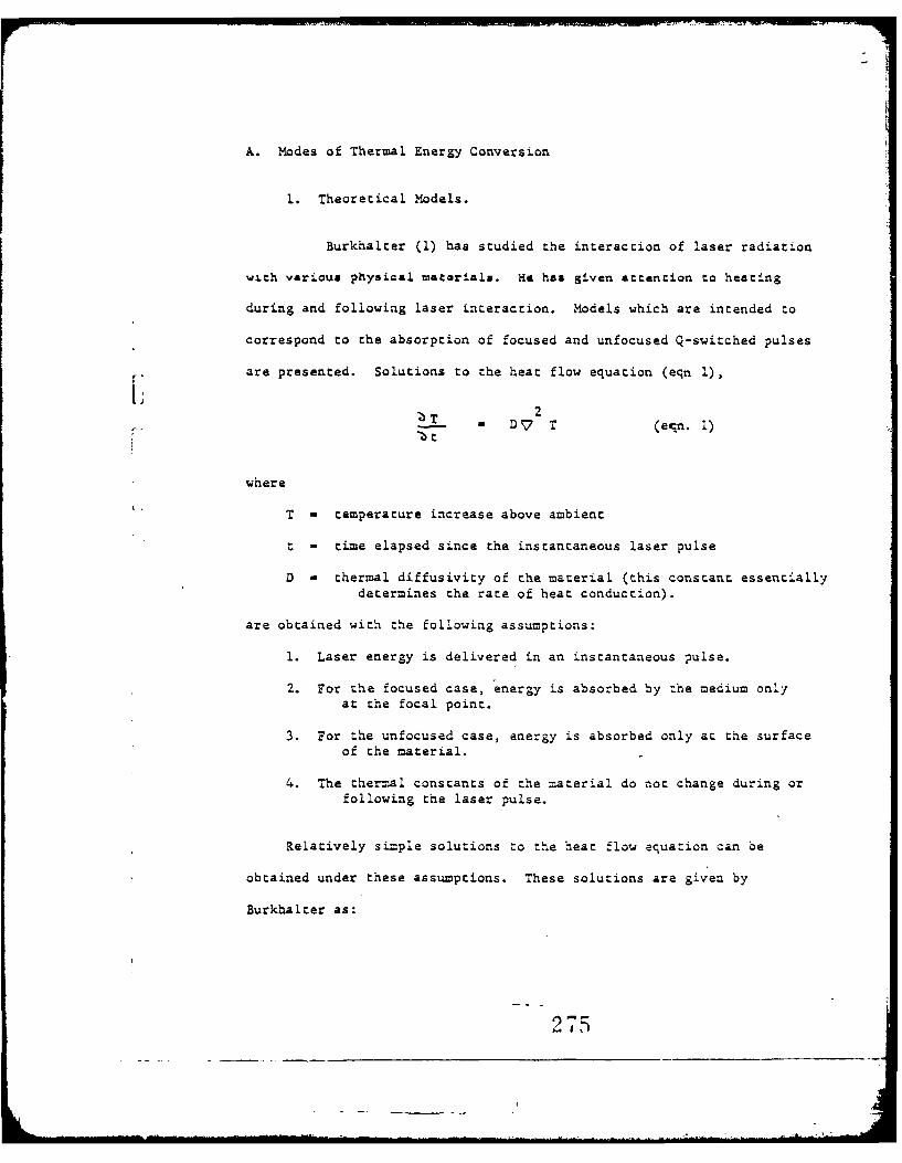
A. Modes of Thermal Energy Conversion
1. Theoretical Models.
Burkhalter (1) has studied the interaction of laser radiation
with various physical macariala. He has given attention to heating
during and following laser interaction. Models which are intended to
correspond to the absorption of focused and unfocused Q-switched pulses
are presented. Solutions to the heat flow equation (eqn 1),
T D 7 T (e-qn. 1)
where
T - temperature increase above ambient
t - time elapsed since the instantaneous laser pulse
D - thermal diffusivity of the material (this constant essentiallydetermines the rate of heat conduction).
are obtained with the following assumptions:
1. Laser energy is delivered in an instantaneous pulse.
2. For the focused case, energy is absorbed by the medium onlyat the focal point.
3. For the unfocused case, energy is absorbed only at the surfaceof the material.
4. The thermal constants of the material do not change during orfollowing the laser pulse.
Relatively simple solutions to the heat flow equation can be
obtained under these assumptions. These solutions are given by
Burkhalter as:
275

J
r2r
T (r,-) - e (focused case)
ps (4Dt) 3/2
2z
T (z,=) - e 4D (unfocused case)Aps (430t) 1/2
where
r - radial distance from laser focal point
z - depth below absorbing surface
A - cross sectional area of beam at absorbing surface (noteBurkhalter does not discuss the fact that equation 3holds only when,
P - mass density of material
s - specific heat of material
Q - number of calories absorbed at surface or at focal point.
3urkhalter states that these assumptions correspond to a rough
model for absorption of focused and unfocused Q-switched laser pulses.
Burkhalter does not present data justifying the applicability of this
model to biological systems. One can, however, show from simple consider-
ations that it is improbable that pigmentation in biological tissue
occurs in layers so thin and opaque as to warrant the assumption that
extremely short, (nanosecond range) laser pulses are required to avoid
heat conduction during the laser pulse.
Ham has noted that the darkly pignented human pigment epithelium
absorbs approximately 40 of incident ruby laser radiation in a layer
276
10 microns thick. If one assumes such tissue to be composed of densely
packed, non-scattering melanin granules, then the absorption of light in
pigment epithelium should obey the following relation
E absorbed -e " x
E incident
where
x - thickness of pigment epithelium (cm)
o< - absorption coefficient (cm'1).
If human pigment epithelium is assumed to be 10 microns thick then
0.40 - 1.00 (l-e )
This relation yields an approximate absorption coefficient for melanin
-lof C< - 500 cm This result indicates that a layer of melanin 20
microns thick is required to absorb a large fraction ( 70) of collimated
incident laser radiation. Assuming that such a layer exists in biological
material and that all incident laser radiation is absorbed in this layer,
it becomes a simple matter to calculate approximately how short a laser
pulse must be for negligible heat conduction to occur during the pulse.2
The laser pulse duration,' " , must be less than - for negligible40
20uheat conductions to occur during the laser pulse. If x - -= and
0-3D is approximately the same as the diffusivity of water (1.4 x 10 3m
per second) thenl' must be less than 0.2 msec. Therefore, in most
biological tissue layers, absorption coefficients are not high enough
to warrant the assumption that Q-sw.tched (nanosecond range) laser pulses
are necessary for negligible heat conduction to occur during the pulse.
However, Q-switched (nanosec) laser pulses may be necessary for negligible
heat conduction to occur within the pigment granule during the pulse. This
2??

may be significant in elucidating the difference between threshold values for
Q-switched and non-Q-switched irradiation.
For focused laser radiation, spot diameters of one micron are
achievable. However, insufficient light abosrption ( - 5%) will occur
in a one micron diameter, spherical volume to warrant use of the
Burkhalter model. Heat flow will not take place in these focal diameters
for laser pulse durations of less than 0.4 Lsec.
In summary, experimental evidence shows that absorption in biological
materials is normally too low to require "the use of a Q-switchedz model
for laser radiation absorption. However, as Burkhalter points out (1)
at high peak power densities, absorption may become non-linear and
increase. It may be significant in pigment granule absorption.
Studies by Yura (2) on laser interaction with metals, particularly
the transition metals, have given evidence for long electron recombination
lifetimes zas' discussed!- by Burkhalter. In these studies, it is suggesad
that excited electrons are trapped in metastable quantum states and
subsequently recombine emitting light and/or imparting vibrational energy
(phonon production) to the metal lattice. Phonon production is the
mechanism by which heat is generated within the metal. Electron recombin-
ation lifetimes from such metastable states are predicted to be of the
order of one nanosecond, which is comparable to the pulse duration of a
Q-switched laser. Burkhalter states "in this manner, a material may
absorb much more energy per unit volume from shoit pulses such as
Q-switched pulses than from long ones," One can see, however, that lower
electron energy states may be depopulated by a Q-switched pulse, and
relatively few electrons will decay back to these states during the pulse.
278- j

7 A0A106 234 NORTHEASTERN WLONY BOSTON MASS DEPT OP BIOPHYSICS AN-ETC FIB 611SBIOLOGICAL EFFECTS OF LASIR RADIATION. VLUME 1. REVIEW OF TIE -ETCCU)OCT 7B S FINE, E KLEIN DA-49-193-MO-2436
WdNCLASSIFIEO0 NI.

In this case, radiation absorption may actually decrease rather than
increase due to the depopulation of lower elect:ron energy states.
Other quantum state configuratiozis exist where increased absorption
may occur for high photon densities and/or short laser pulse durations.
It is possible, however, that Q-switching may result in a non-linearly
decreasing absorption coefficient rather than a non-linearly increasing
absorption coefficient. The electron trapping mechanism discussed by
the author is an energy storage process. A comparison between this
1: type of energy storage and classical thermal energy storage is deve-
loped in the following paragraphs for biological systems.
Consider a highly pigmented tissue layer (eg. the retinal pigmented
epithelium) which is heated by a laser pulse with uniform energy density
across a collimated beam. Assume that one wishes to raise the temperature
of the adjacent retinal tissue (photoreceptors) to some critical tempera-
ture, Tc, WithIiut considering quantum processes, it is apparent that V
laser pulses of duration shorter than the time required for heat to conduct
across a single layer of melanin granules in the pigment epithelium (assume
melanin granules 0.3p in diameter) will serve only to store thermal energy
in the melanin. Therefore, to raise the temperature of the adjacent
retinal tissue to Tc will require that more radiant energy be supplied
as the laser pulse duration is shortened. Conversely, if the laser pulse
is too long, heat will conduct away from the pigment epithelium too rapidly
to raise the temperature of the adjacent retinal tissue to TC . 1f thermal
energy is stored in a single layer of melanin for a time approximately
equal to the laser pulse duration then the laser energy that must be
279
supplied to result in steam production is a minimum compared to longer
or shorter pulses (4). The time for heat to conduct across a single
melanin granule may be of the order of nanoseconds, based on the thermal
diffusivity of water. Hence, thermal energy can be stored in a melanin
granule, wich relatively litla loss, during a Q-switched laser pulse
based only upon classical heat flow considerations. If the time
required for phonon production in biological material, such as melanin
is also of the order of nanoseconds, then it will become difficult to
separate classical heat conduction effects from the quantum effects
(electron trapping) that Burkhalter has cited. Q-switcnea.pulses to
the retinal pigment epithelium should also be the most efficient pulses
for producing high temperatures in the retina from the standpoint of heat
conduction alone. Hence, some of the drastic effects noted following
Q-switched laser irradiation of biological systems may be exlained in
terms of classical thermal conduction. Further experimental and theoretical
work is required before.the importance of quantum mechanical interactions
per se during Q-switched laser impact is known.
Fine et al (5) have included the effects of interfaces on laser radi-
ation absorption in biological systems.
"Biological systems may be considered to consist of strata ofdiffering composition and properties, separated by interfaces. Forexample, in the abdominal region, the strata can be grossly consideredas skin and discontinuous subcutaneous tissue, muscle, peritoneallayers, and viscera. Interaction with this multilayered system willdiffer from interaction with a homogeneous system. Multiple reflec-tions can occur from the various interfaces, on both a gross andmicroscopic level, resulting in trapping of radiation energy withina specific gross or microscopic region, particularly if a changein wavelength occurs. The energy density within this region canconsequently be greater than if the region were not part of amulti-layered system and if reflections, particularly from theinterface deep to the layer, did not occur."
280

Calculations are performed on infinite, three-layer material (App. 1)(5)
with partially reflecting interfaces and a given percent absorption in the
central layer. It is shown that the radiant energy absorbed in the
central layer is increased in the presence of partially reflecting inter-
faces although the energy absorbed in the central layer cannot be greater
than twice the energy absorbed without boundaries. Greater absorption
can occur in the central layer if there is an alteration in wavelength
with an associated alteration of absorption coefficient. If there is an
alteration of wavelength because of interaction at the interfaces, then
considerable trapping of energy within the central region could occur
(the greenhouse effect).
Fine et al suggest that increased absorption at abdominal inter-
faces may result in steam production and possibly cause the skin distention
that has been observed in mice (5). An extension of the interface model
shboild include the effect of tissue curvature during the distention.
Reflective focusing of trapped radiation will increase the energy density
of the radiation in zone 2. Since the curvature of the interfaces changes
during laser impact, the problem of reflective focusing in skin distention
is both non-linear (due to non-uniform steam production) and time varying.
Further modification of the simple model presented by Fine et al should
include the effects of refractive focusing of incident laser light by
both external lens systems and by the curvature and refractive indices of
the various tissue layers. Models are in preparation, by the authors of
this review, which include the effects of refractive and reflective
focusing of laser radiation in biological tissue (6).
201
Variations in refractive index on a cellular scale introduces
radiation scatter. More refined models of laser radiation absorption
should include this effect.
In the excellent gross and histological studies by Heiwig It a1 7 )
the epidermal temperature elevations for various beam diameters and incident
energies delivered by a non-Q-switched ruby laser have been calculated.
Pertinent parameters for skin are taken from studies done at low power
levels such as that of a spectrophotometer or carbon arc and on various
animals and in vitro human systems by Hardy e al (8), Davis (9), Henriques
and Moritz (10), and Kupenheim et al (11). For beam diameters of the
order of 1 cm and an epidermal thickness of about l00L, heat conduction
can be ignored during a laser heating pulse of 1.6 msec duration in
order to obtain a rough estimate of the temperature elevation of the
epidermis. The equation used by Helwig e._.t al is:
Eo t (1 - r)T. -
4 c(p /C
where E is energy of the incident laser beam in calories; o< , the
0 -1absorption coefficient at 7,000 A in cm ; t, the epidermal thickness;
(l-r) the fraction of the beam not reflected; jd2 .; the exposure area4
or image size; /0, the epidermal density in g/cm 3 ; and C, the epidermal
specific heat in cal/g per °C. The factor o t in the denominator is'
probably an approximation to the expression (1 - e- n) for c t small.
For o( 4 20 cm' ~and t Z 1004, the error in using this approximation is
about 107.. Helwig's calculations indicate that the heat content per
gram ( ,, 5 x 10- 2 cal/Sm) necessary for protein denaturation is easily
282

achieved in the epidermis by laser irradiation in the ten joule range
for beam diameters of about 1 cm, (Fig. 23 of (7)). The authors conclude
that thermal processes, such as protein denaturation, are sufficient to
explain laser induced lesions. Furthermore, they feel that the magnitude
of the predicted thermal effects per se would tend to limit the detectability
of ocher physical or biologic effects. Further attention should perhaps
be directed to the photographs shown by Helwig et al of histological
slides taken of porcine skin following laser irradiation in the 12 joule
range. These photographs show possible marked mechanical rupture (perhaps
due to steam) of tLAe epidermal layers which may be different from thermal
effects per se. However, other possible explanations such as formation
of an exudate are possible. Whether one should include pressure transients
cratering, change of phase, and ablation as "thermal effects" requires
further consideration.
In the course of their comprehensive well documented irradiation
studies on the eye, Ham et al (12) have developed a model for laser
radiation absorption and heat conduction in the human ocular fundus. Accord-
ing to this model, significant absorption begins at the pigment epithelium
where approximately 40 of the laser radiation is absorbed exponentially
in passing through 10 microns of tissue, and approximately the remaining
60% is absorbed exponentially in passing through 100-150 microns of
chorsid.
In Ham's model, absorption in the retina and anterior ocular media
is considered as negligible. This is a reasonable assumption since absorption0
and/or scattering in these regions is only about 10 at 6943 A. Other
283
I ......

'I
highly absorbing tissues, such as the iris, are not specifically included.
It is also reasonably assumed, only for the purposes of this model, that
pigmentation in uniform in the pigment epithelium and choroid. Scattering
is mentioned as a possible attenuation mechanism in the fundus, but is
not considered in the model, because of its complexity. In the lightly
pigmented choroid, some thought can be given to back-scattered radiation
from the sclera which may yield higher radiation energy densities at
the pigment epithelium.
Ham et al note that the highest temperature gradients will occur
between the pigment epithelium and retina, due to the relative transparency
of the retina. Although the retina is transparent to visible wavelengths,
it may be relatively opaque to those associated with heat. This high
temperature gradient will cause heat to be conducted preferentially to
the retina rather than to the choroid. A more complex model than that
presented may include the effects of choroidal blood supply on radiation
absorption and heat dissipation. Although the velocity of flow in the
choriocapillaris is low, the blo(,d volume may be high, and merit consider-
ation. The highest temperature will, as indicated in the model, occur
initially in the pigment epithelium and spread via heat conduction to the
photoreceptors in the retina.
A reasonable model is assumed by Ham et al in which the maximum
temperature of the irradiated portion of the pigmented epithelium is
estimated by neglecting thermal conduction and considering a "pill box"
toen microns thick and having a diameter the same as the laser beam incident
at the pigment epithelium, If 40% of the radiant energy incident on the
2e

I,-
surface of the pill box is absorbed, then the authors conclude that a
radiation dose of 0.85 J/cm 2 (delivered in 200 4sec) yields a threshold
lesion in the rabbit retina and will produce a temperature rise of
approximately 81°Centigrade in the pigment epithelium (12).
Examination of this calculation shows that Ham has assumed a
relation for temperature increase of the form:
L) T -
where H is the number of calories absorbed in the pill box per unit
cross-sectional area of the incident laser beam (at the pigment epithelium),
and t is the thickness of the pill box. The specific heat and density of
the pigment epithelium are assumed to be unity (the same as that of water).
Ham et al have considered that radiation absorption in the pigment
epithelium and choroid would probably occur exponentially. Another
possibility, consequently, is to assume exponential absorption (diathermancy
of the tissue). This would chen yield a somewhat higher expected maximum
temperature in the pill box.
The following relation assumes adiabatic walls (no heat conduction
out of the pill box), zero reflectivity at the pigment epithelium, no
scattering and zero conductivity within the pill box based on che model
by Ham et al. If the diathermancy of the tissue is included via the
absorption coefficient, c<
T (x)max - E 0 O 5e
4.18 DC Ap

T (x) WAX is the maximum temperature rise at a depth, x, within the pill
box; E. is the incident energy from a collimated beam at the retina;
O( is the absorption coefficient for the pigment epithelium (approximately
500 cx "1); A is the cross-sectional area of the beam; D and C are thep
density and specific heat of the pigment epithelium and are assumed to be
approximately unity. For E/A - 0.85 joules/cm 2 , T (0), the maximum
surface temperature rise of the pigment epithelium nearest the retina
is 1020 C, of the same order of magnitude as the 810 C calculated by
Ham. For the Q-switched case of 0.07 joules/ca 2 incident on the retina
T(0) is 8.40 C, and varies little from the 6.70 C as Ham has calculated,
if the assumption is made that radiation absorption in the pigent epithelium
and choroid will probably occur exponentially, as discussed by Ham et a].
The maximum temperature of the--pigment epithelium under adiabatic
conditions (no heat flow) will therefore be somewhat higher than that
obtained for the model where uniform absorption occurs. The assumption
of exponential decay leads to temperatures above the steam point for
water when ambient temperature (approx. 370 C) is added to the 1020 C
temperature rise for the case of, 0.85 J/cm 2 incident at the retina. In
the extensive investigations by Ham et a)l, this energy density, when
delivered in a 200 gsec ruby laser pulse, resulted in an ophthalmoscopically
visible lesion in the fundi of pigmented rabbits five minutes after
exposure. This type of lesion was termed a "threshold lesion" by Ham.
Ham points out that such a lesion may, however, not represent minimum
irreversible damage to the rabbit retina. Further study is required
before the importance of steam production in retinal damage can be
ascertained.
2 P6

Additional data is presented byHam et al., based on extensive studies
for exposure times longer than 200 .sec (using Xenon or carbon arc light
sources) which tabulates the energy density required for a threshold lesion
and the total temperature rise of the pigment epithelium at the end of the
pulse. Temperature calculations are presented for cases where first, heat
conduction is neglected and second, where heat conduction from the faces
and sides of the "pill box" absorber in the pigment epithelium is taken
into account. Calculations were carried out by Ham et al on the model and
are presented in Table 1.
TABLE I
Maximum temperatures in the PE for difftr.,t exposure times as estimated onthe basis of the pill box model. An electrical analog has been used to takeaccount of heat conduction in the model.
Exposure time Radiation dose Max. temp. rise in Max. temp rise inin psec in j/cm 2 PE neglecting PE correcting
conduction for conduction
0.030 .07 6.7 6.7200.0 .85 81.0 52.8500.0 1.10 105.0 50.51,000.0 1.38 132.0 55.02,000.0 1.72 165.0 53.55,000.0 2.41 230.0 49.310,000.0 3.07 294.0 47.320,000.0 3.90 373.0 46.8
In the Q-switched case, temperatures of approximately 450C can be ex-
pected (assuming exponential or uniform absorption). The relatively low
temperatures produced by Q-switched laser irradiation and the appearance of
choroidal hemorrhage extending into the subretinal spaces when only relati-
vely mild retinal damage is seen lends credence to the hypothesis that other
than thermal processes per se may account for Q-switched laser damage to
the rabbit eye and other biological tissue (12,5).
2)F

In development of the model, uniform absorption in the subretinal
pipented tissues was assumed by bogh Ham (12) and Vos (13). They have
not included the problems of intra-layer and interlayer inhomogeneities
of pipentation, possible laser emission inhomogenieties across the end
surface of the rod, and the effects of radiation scatter. It should be
noted that these inhomogeneities have been considered by Ham et al in
their comprehensive studies. Attention to the problems of interlayer
variations in radiation absorption by biological tissue has been given
by T.P. Davis (9) and J.M. Davies (15) in investigations of in vivo
burn production in pcr-cine skin from broad-baud radiation. Davies (15)
has given some consideration to the problems of scatter in tissue.
Felstead and Cobbold (16) have developed an electrical analog for thermal
conduction in the human eye. They assume rotational symmetry of the eye
and coagulating beam and neglect effects due to vaporization and pressure.
Some consideration of the relative absorption of light in the various
tissue layers is given, but, diathermancy is neglected insofar as these
layers a-e assumed to absorb uniformly as a function of depth. Radial
variations in beam intensity can be included in the Felstead-Cobbold
analog if these variations are ,invariant under rotations about the optical
axis of the incident beam. Such variations are not considered by
Felstead and Cobbold in using the analog.
A number of attempts have been made to measure temperatures at
various sites in rabbit eyes. These studies, in some cases, have been
carried out in an attempt to verify various theoretical.models for thermal
alterations in the rabbit fundus and to elucidate mechanisms of laser eye
damage and clinical photocoagulation of the in vivo human eye.
288

Since direct temperature measurements are extremely difficult to perform
during and following laser impact, and are probably not reliable, expected
temperature elevations in the ocular fundus must be calculated, (as they
have been done by Hrm et a1) from measurements of laser energy, laser
pulse duration, bea= diameter at the pigment epithelium, absorption of
various ocular tiss=es, and the physical dimensions of the tissues involved.
Beam splitter techniques for measuring laser output energy and pulse
duration must be carefully designed to avoid the problems of inconsistent
beam polarization. The measurement of laser beam diameter within the
eye required accurate knowledge of the optical properties of the particular
eye under investigation. For example, considerable differences may be
present between the human and rabbit eye. The absorption properties of
various ocular tissues have been considered by Geeraets et al. The figures
0of 40% and 60% attenuation at 6943A in the pigment epithelium (PE) and
choriod, respectively, must take into account the fraction of the light
absorbed and the fraction scattered. Variations in pigment epithelium and
choroidal thickness and density of pigmentation are important, but, difficult
to include in a model. All of these factors make it very difficult to
predict an accurate temperature distribution within the eye following laser
irradiation. An error analysis, accompanying the calculations which are
intended to predict these temperature distributions would be of interest.
Further correlative efforts between more accurate-temperature measurement
and theoretical predictionv-are necessary before a model for thermal inter-
action in the eye can be developed.
Vos has performed a theoretical analysis of experimental data obtained
by Ham al (14) in studies of retinal flash injuries produced by white
light in rabbits (13). In the Vos analysis, a model for steam production
in the pigment epithelium is developed and compared to a model where
temperature elevation per se is considered as the mechanism for irreversible
289
threshold lesion production'in the rabbit pigment epithelium. Temperatures
well above the steam point for water, as calculated for the energy densities
and irradiance pulse durations used in the studies by Ham. Based on these
results, curves of constant steam production (by weight) are drawn and
found to coincide reasonable well with Ham's experimental results for
exposure times between 20 msec and 250 msec. Vos interprets these results
in the hypothesis:
"A threshold lesion as defined by Ham is produced when a steamexplosion takes place which is sufficient to give a marked disruptionof the retinal tissue. In very small images, this may be expectedto happen when steam production reaches an absolute minimum. Inlarge images, the production of steam per unit of area should be thedetermining factor." (13)
This "y-,3thesis should be subject to review, based on the excellent experi-
mental results of Ham et al. using relatively monochromatic ruby laser radi-
ation in the one millisecond and 30 nanosecond range.
Vos' calculations of temperature elevations are indirect and rely on
experimental data and associated variation similar to those that were pointed
out in conjunction with the Ham model of the rabbit fundus. Vos also
neglects the diathermancy of the pigmented tissues posterior to the non-
pigmented retinal layers. It is assumed that all of the radiation incident
at the retina is uniformly absorbed in a volume of pigmented tissue termed
"the thermal image" by Vos. The depth of the thermal image is neglected for
exposure times longer than 300 sec whereas the cross-sectional area is
assumed to be that of the incident beam at the retina. However, for exposure
times greater than I psec and less than 300 isec, the depth of the thermal
image must be included. Therefore, if the Vos model is to be used co pre-
dict the temperature distribution within the "thermal image", all dimensions
of the thermal image should be considered. Should this task be pursued, a
model for the ocular fundus including radiation scattering and tissue dia-
thermancy is desirable.
290
A129

In development of the model, uniform absorption in the subretinal
pigmented tissues was assumed by both Ham (12) %nd Vos (13). o.onsidered
h1areare the problems of intra-layer and interlayer inhomogeneities of
pigmentation, possible laser emission inhomogenieties across the end
surface of the rod, and the effects of radiation scatter. It should be
noted thatthese inhomogeneities have been considered by Ham et al in
their experimental studies. Attention to the problems of interlayer
variations in radiation absorption by biological tissue has been given
by T.P. Davis (9) and J.M. Davies (15) in investigations of in vivo
burn production in porcine skin from broad-band radiation. Davies (15)
has given some consideration to the problems of scatter in tissue.
Felstead and Cobbold (16) have developed an electrical analog for thermal
conduction in the human eye. They assume rotational symmetry of the eye
and coagulating beam and neglect effects due to vaporization and pressure.
Some consideration of the relative absorption of light in the various
tissue layers is given, but, diathermancy is neglected insofar as these
layers are assumed to absorb uniformly as a function of depth. Radial
variations in beam intensity can be included in the Felstead-Cobbold
analog if these variations are invariant under rotations about the optical
axis of the incident beam.. Such variations are nbt considered by
Felstead and Cobbold in using the analog.
A number of attempts have been made to measure temperatures at
various sites in rabbit eyes. These studies, in some cases, have been
carried out in an attempt to verify various theoretical models for thermal
alterations in the rabbit fundus and to elucidate mechanisms of laser eye
damage and clinical photocoagulation of hhe in vivo human eye.
21

Campbell, Noyori, and Rittler have studied the temperature elevation
at the interior surface of the retinae of in vivo grey chinchilla rabbits
during and following laser retinal coagulation. The laser photocoagulator
( 17 ) utilized a ruby crystal and suitable viewing system for the
observation of coagulations and thermocouple positioning. Laser output
was maintained at 0.065 joules for pulse durations of one millisecond or
less. Baldwin-Lima-Hamilton, micro-miniature, exposed junction, chromel-
alumel thermocouples were inserted through small scleral incisions and
positioned with the active junction assumed to be in thermal contact with
the interior surface of the retina. For temperature measurements at the
site of the coagulation a curved thermocouple was inserted through the
sclera behind the area to be irradiated thus eliminating any shadowing
of the retina by the thermocouple during irradiation. Temperature measure-
ments outside the coagulated area were made by inserting the thermocouple
in a position tangential to the surface of the retina. Thermocouple output
was displayed on an oscilloscope.
Results indicated that temperature changes of less than 0.5 degrees
centigrade occurred in the plane of the retina at distances greater than
1 millimeter from the coagulation site. At the site of the coagulation
itself temperature elevations of 30 degrees centigrade were recorded.
Temperature changes of about 0.5 degrees centigrade were obtained with
the thermocouple tip positioned in the middle of the vitreous and
centered in the laser beam.

There are errors associated with thermocouple measurements that are
made when the temperature sensing junction is exposed to direct or
scattered laser radiation (20,21). The oscilloscope traces that are
illustrated in the report by Campbell at al were recorded on a very slow
time scale. Thermocouples of this type have been shown to respond in
approximately one millisecond when exposed to direct or scattered laser
radiation (20). Therefore, much important fine structure such as
L thermocouple irtadiation is lost in oscilloscope traces where the time scale
is of the order of seconds per centimeter. It is doubtful that Campbell
et al could observe the effects of direct thermocouple irradiation from
direct or scattered light in their oscilloscope traces. It should be
noted that an irradiated thermocouple junction (and insulating sheath)
can heat surrounding tissue. The temperature of the heated tissue is
relatively persistent, having a thermal time constant of several seconds.
The thermocouple outputs observed on slow oscilloscope traces by Campbell
et al are probably a result of both direct laser heating and thermocouple
heating of retinal tissue adjacent to the thermocouple junction. These
two mechanisms of tissue heating requirc further consideration.
Measurements of the relative transmission characteristics of the
in vitro human fundus have indicated that little energy is absorbed or
scattered by the non-pigmented retina itself (22). Most of the absorption
or scattering of ruby laser radiation takes place in the pigment epithelium
and choroid posterior to the retina. Ham (12) assumes that the highest
temperature elevations would occur when the retinal pigment epithelium
absorbs and does not scatter incident radiation. Ham has also suggested
that, from a practical standpoint, a temperature of close to or perhaps
293

at lO0°Cin the piLment epithelium is required to coagulate retinal
tissue to the point where the relative reflectance of light from the
pigment epithelium through the normal retina and the retina with lesions
canabe distinguished by ophthalmoscopic observation. Vos (13) has sug-
gested that temperatures achieved in the pigment epithelium reach the
steam point and steam production accounts for lesions observed in the
rabbit retina by Ham and co-workers. In either the Ham or Vos model,
it is not necessary that temperature at the inner surface (anterior
surface) of the retina will probably produce ophthalmoscopically visible
retinal lesions. Further experimental study is required before the
"temperatures" measured by Campbell at al. at the anterior retinal surface
can be correlated to temperatures in the pigment epithelium. Measure-
ments of this latter type should provide insight toward elucidating the
mechanisms involved in clinical retinal coagulation and retinal damage
from laser radiation.
Small temperature changes were noted by Campbell et al. outside the
coagulated area. The mechanisms for these changes require further con-
sideration. Some of the mechanismz may include scattering of light or
thermal conduction. The thermocouple responses shown for points i m
outside the coagulated retinal site show rise times of much less than one
second and decays of the order of a half second. if the retina is assumed
to have the thermal diffusivity of water (1.4 x 10⁻³ cm2/sec), then the
time for conduction from the coagulated area to a point 1 mm from the edge
may be longer than the time delay observed. However, analysis Is diffi-
cult since the relationship between temperature distribution and coagula-
tion of tissue is not well understood.
2

If thermal conduction is to account for temperature elevation at these
sites, the thermocouple trace should show rather slow rise times (of the
order of seconds) and a relatively persistent d.c. thermocouple voltage
output at subsequent times. The very fast, transient thermocouple out-
put noted by Campbell et al. could be due to scattered laser light reach-
ing the thermocouple or to specific monitor.-ng,-circuitry whereby the
thermocouple oupput was differentiated.
The author concludes that thermal summation does not result with
repeated coagulation, derived from his experimental studies of rapid
temperature decay. Further temperature measurements are necessary in
the pigment epithelium. Temperature measurements may be perturbed by
direct or scattered laser radiation to the thermocouple. Further
investigation where thermocogples are not exposed to direct or scat-
tered laser radiation is necessary.
Najac et al (23) have carried out extensive thermocouple measure-
ments in the moderately pigmented retina of gray chinchilla rabbit eyes
(in vitro) during irradiation by a modified leyer-Schwickerath light
coagulator in the spectral ranges 380-750g and 380-1,350g. This work
was a light coagulation study. The sensing junction was placed anterior
to the pigment epithelium. Information as to the exact location of the
thermocouple would be desirable, but, of course, is limited by biological
factors. The temperature elevations measured for a threshold lesion (small
amount of coagulation in the center of the lesion) ranged from 12.50 to
20.60 centigrade. These temperatures are cited as being below the steam
point but high enough to produce albumin coagulation. This conclusion
295

may be correct at the point of temperature measurement but does not exdiude
steam production within the pigment epithelium itself.
The time delay between thermocouples placed at various 'locations'
within the retina were correlated with times for thermal conduction.
For identical thermocouples, this technique would yield valid results.
Data as to the response characteristics of the individual thermocouples
and circuitry would have been of interest. Interchanging thermocouple
positions between experimental runs and comparing the results might
assist in decreasing effects due to dissimilarities between thermo-
couples. The usual problem of effects of direct and scattered light-
on thermocouple functions must be considered.
The problems of direct thermocouple irradiation have been discussed
by Geeraets and Ridgeway (21), Nowak ec a (20), Davis (28) and Crowder (24).
Geersecs and Ridgeway have noted that the insertion of a thermocouple
into biological tissues gives rise to artifacts of reflectance, absorp-
tion, and conduction, at the site in the tissue where measurements are
being carried out. The relative-size of the thermocouples with respect
to the image size of the incident light beam on the retina and with
regard to the dimensions of the biological structures is of great
importance (21).
Crowder (24) has noted that a large artifact was introduced into
measurements of the temperature of the vitreous at various points along
the path of the beam during photocoagulation if a thermistor probe was
placed directly in the photocoagulator beam. The detector absorbed a
296
significant amount of the light energy even when a fine mirror surface
was used. To overcome this difficulty, Crowder has introduced a device
whereby the thermistor probe is mounted at the tip of a fine needle and
injected into the vitreous at various times following photocoagulation.
While no calculations were shown, Crowder feels that the temperature of
the vitreous during photocoagulation could be calculated from the decay
curve of the thermistor. The photocoagulations used by Crowder were
either one or five seconds in duration. The thermistor probe had a rise
r time of 0.085 msec and was injected in less than 10 milliseconds following
the end of the photocoagulator pulse. When the coagulating beam diameter
is less than one millimeter then appreciable heat flow can occur during the
irradiance pulse. Unless the temperature at the point of measurement
varies monotonically in time during the irradiance pulse and during the
time before the thermocouple is injected, it is not possible to reconstruct
a time temperature history from the decay curve of the thermocouple.
Attempts to reconstruct time-temperature histories from thermocouple
decay curves have also been made by McCartney and Hall (25). Electronic,
time-delay circuitry was thougiz sufficient to allow thermocouples implanted
in adenocarcinoma tumors (in black mice) to come to thermal equilibrium
and allow extrapolation of the thermocouples responses to yield tempera-
tures during laser impact (2.5). Effects of direct thermocouple irradiation
require evaluation. The results of extrapolations to zero time require
further study.
297

A theory for reconstructing initial temperature distributions from
data obtained at later times and at various positions has been presented
by Maskec (26) using a modified Green's function approach to the solution
of the heat flow equation with time reversal. One must also consider the
faet that if thermal injury or Qhange oQcurs within a biologisal system
during radiant insult, the chances are in favor of the system becoming
non-linear as biological change proceeds. This further complicates the
problem of reconstructing the time-temperature history of a biological
system from data taken at times following thermal change.
Nowak et al (20) have shown, using the energy-specific heat balance
equation, that a chromel-alumel thermocouple positioned in air and in the
path of a 1.2J, 1.0 cm diameter, I msec laser pulse will indicate tempera-
ture rise of approximately 8000C. The thermocouple bead is assumed to
have an effective thickness of 34 x 10-4 cm (4/3 the radius of the thermo-
couple bead) and a spectral absorptivity of 0.75.
To test these calculations, experiments were car-ied out wherebyA
chromel-alumel thermocouples (30) with junction-tip diameters of ap-
proximately 0.002 inch and sheath diameters of 0.014 inch were exposed
to unfocused radiation from a non-Q-switched ruby laser. The thermo-
couple was connected directly to the d.c. input of a Fairchild 704
oscilloscope. The response of the unshielded thermocouple is shown
(20) with tip exposed in air perpendicular to a 12. beam. Also shown
is the output of the thermocouple with exposed tip, but shielded sheath.
A peak pemperature of 7900C was obtained in a good agreement with the
previous calculation. Shielding the tip, or equivalently, shadowing the
tip by the thermocouple sheath from the incident radiation, resulted in
a modified response. _ .

To evaluate further the response of the thermocouple, experiments
were carried out at 18J in air, and at 12J in a 1% saline solution and
in. tap water. The solutions were contained in a plexiglas box 5 inches
X 2 inches'. The thermocouple was centered within the box.
The ratio of the peak temperatures, 12600C and 7900C, obtained for
18 and 12 joule exposed-tip irradiationsiin air is in reasonable agree-
ment with the ratio of the energies and with the calculations. The
V temperature of the thermocouples (12600C and 790 0C) is obviously of a
different order of magnitude than the actual air temperature in the
laser beam without the thermocouples present.
Thermocouple temperatures were measured within the solutions
irradiated at 12 joules. No differences in response were noted between
water and saline solution. With either the whole thermocouple exposed,
or tip alone exposed, the peak temperature rise recorded was approximately
5000C. The peak of shielded-tip response in solutions, was 900C. This
is apparently due to scattered radiation reaching the tip; the irregula-A
rity of this peak should be noted. The expected rapid quenching of the
tip when in solution was observeO, i.e. 2 msec compared with 16 msec to
l/e of peak. The delayed peak in the shielded-tip response is due to
the time required for the heat absorbed by the sheath to diffuse to the
tip. This diffusion probably takes place along the thermocouple, rather
than through the air or solution, since, the thermocouple is the path of
highest thermal conductance and since the 25-30 msec delay occurs both
in air and in solution.
299

1_7
Davis (28) has outlined the following criteria for valid temperature
measurements in tn vitro biological systems based on the fundamental
condition that temperature is defined only when a system is in thermal
equilibrium (no thermal energy exchange with environment.-,4r other parts
of the system). A thermometer is inteoduced as an auxiliary. thermo-
dynamic system when the biological system in question exhibit& no
useful thermometric characteristic of its own. The thermometer must
not perturb the thermodynamic state of the system whose temperature is
to be measured. The measurement is ., valid only when the system and
thermometer achieve and maintain thermal equilibrium (no thermal energy .
exchange between thermometer and system). Thermometers with low heat
capacity and high heat transfer coefficient relative to the system can
follow the temperature excursions of the system closely but never
exactly. In those cases where both the system and thermometer are In
contact during a heating period, the thermometer and system must respond
identically for a valid temperature to be registered. In the case where
an opaque thermometer is immersed in a translucent biological medium
and the two systems are heated together by a radiant energy input thermal
equilibrium is almost impossible to maintain between the thermocouple
and biological system. In a biological system where transient heat
flow occurs, temperature is undefined except within an infinitesjimal
volume where no temperature variation is assumed to occur within this
", volume. If arbitrarily small volume elements are assumed to lie in
equilibrium temperature states,then a small thermometer can measure the
temperature of a volume without experiencing a temperature gradient across
the .sensing element (27).
300

B. Modes of Mechanical Interaction
At low energy density and power density levels, the gross interactions
of laser radiation with biological systems are due mainly to temperature
elevation per as. As the p~wer and energy density is increased, mechanical
interactions due to phase transformations, tissue volatilization, thermal
expansion, and quantum processes involving phonons (quanta of vibrational
energy) become important. The effects of mechanical interactions may
Vexhibit characteristics different from the effects of temperature-le-ation per se.
Burkhalter (1) and Fine et al (5) have investigated the mechanical
effects 6f laser interaction with torsion and ballistic pendulums. Fine
et al have reported that when the blackened surface of a ballistic
pendulum was exposed to a laser pulse, oscillations were observed. This
occurred when the pendulum was mounted in air or in a vacuum. Oscillations
were not observed, however, when the unblackened surface was exposed (15).
These oscillations appear to be caused by conservation of momentum at
the pendulum surface, rather than by radiation pressure, thermal gradients,
or heating of the air at the surface (1,5). Burkhalter has explained
that the recoil impulse imparted to a torsion pendulum by volatilized
mass blown away from at the pendulum surface can be determined by
measuring the torsion constant of the pendulum system, the radius at
which the impulse is applied, the initial angular displacement of the
system, and the period of damped oscillation following the impulse (1).
Numerical data associated with this recoil effect has not yet been
published by Burkhalter and would be desirable. At the surface of a
301
biological system, it is possible to impart a net momentum change to
the biological target via tissue volatilization (excluding the volatilized
material itself). At higher energy and power density levels, volatilization
and phase changes may occur deep to the surface, whereby a net momentum"
change is not imparted to the biological target as a whole, yet strong
pressure transients may be generated within the system. The mechanism
of surface volatilization is probably not of major importance in damage
deep to the surface at high energy and power levels (29)
Fine et al (5) have investigat6d pressure transients generated
by phase changes occurring deep to the surface of bioiogical systems.
A model for steam production has been adopted where quasi-static conditions-
(constant pressure throughout the volume of water vapor) are assumed to
exist during a phase change induced by a ruby laser pulse lasting on
the order of one millisecond. Radiation from Q-switched systems can
produce a relatively rapid pressure increase in comparison with radiation
from comparable non-Q-svitched systems. With Q-switched systems, a
rapid pressure build-up can occur which may result in the transmission
of a shock wave. These shock waves may travel with relatively little
attenuation and consequently affect areas at some distance from their
origin (5).
This model has been applied to situations where outward, hemispherical
distensions of the abdominal wall of mice were observed (5) when non-4Q-
0switched radiation at 6943 A at energy levels in the 2 - 20 joule range was
directed at the abdominal surface. High speed photography has indicated
that the millisecond duration of the distension was of the *order of the
laser pulse duration. Assuming that nearly static pressure equilibrium
302
-4is established in the tissue over an interval of 10 sec. (based on the
velocity of sound in water vapor at 1000C), it is shown that, for
a distension radius as large as 1 cm, a pressure exceeding 1 atmosphere
0may be obtained at temperatures a little over 100 C. There is , of
course, an upper limit to the pressures that will be generated before the
containing membranes burst. This limit depends on the stratum in
which the vapor is being collectdd, and on the nature of the overlying
tissue (especially its burst strength, which may be dependent on strain
rate ) (5).
Pressurc transients may be created by thermal expansion without
change of phase, In studies on physical systems, the generation of •
elastic waves have accompanied the absorption of radiation from high
power light sources (including electric arcs and ruby lasers), microwave
sources, and the absorption of pulsed electron beams within solid targets.
Radiation kbsorption may produce local temperature gradients and, as a
result of thermal expansion, cause stress gradients within the target
medium. Pressure waves then propagate from the stress concentration and
may create shock fronts within the target (29).
In biological systems, thermal gradients (and hence stress concentrations)
will appear at the target surface or internally during the absorption of
laser radiation. Where refractive index changes occur at interfaces
within biological systems, enhancement of radiation absorption can exist,
due to multiple reflections, therefore giving rise to stress gradients
beneath the surface (5). Pressure waves may then propagate through the
biological systems with possible shock fronts at the leading and trailing
edges of the wave train.
303
3,
Absorption of laser energy within a closed, filled cavity, such
as the eye or skull, differs from that occurring at a free surface or
within a cavity with non-rigid walls (31). Phase changes that occur within
filled cavities with rigid walls will generate high pressures within
the cavity when quasi-stati conditions can be assumed to hold. The
importance of cavity boundaries can be determined by considerations of
sound velocities within the cavity and laser pulse duration. If pressure
transients, travelling at the speed of sound, interact with rigid cavity
walls during a phase transformation then greater pressures will occur
than if the cavity walls were flexible. Udzr quasi-static conditions,
pressure pulses can be transmitted with relatively little attenuation
to regions distant from the site of interaction. This can result in
tissue destruction due to direct mechanical effects and/a due to disruption
of vascular supply at some distance from the site of original lasar impact.
Consequently, severe and sometimes fatal injuries can be produced, although
the local effects at the site of original laser impact may not be vital
to the functioning or survival of the organism.
Experimental studies (11) Amar) have indicated the importance of
laser interaction within closed filled cavities. Fine and Klein (31,5) have
reported studies of the effects of pulsed unfocused and focused irradiation
of the forehead in mice and have discussed the importance of the intact
skull in these studies. The hair on the forehead was clipped and the area
depilated. Of 41 animals irradiated at energy levels below 100 joules,
31 died within 24 hours of irradiation. Unfocused irradiation was
followed by death within 30 seconds in 10 out of 23 animals. Neurological
changes were noted in the animals which survived irradiation (31).
30
Hemorrhages from the mouth, nose, auditory meatus, retro-orbital
space and orbit were observed. Although subperiosteal petechiae were
present on the outer table of the cranial bones, gross damage to the
underlying bone was not evident. Gross intracranial hemorrhages were
observed in the meningeal space, the ventricles, the conducting system
and within the substance of the brain distant from the site of impact.
Microscopically, hemorrhages were seen in the underlying muscle in
addition to cutaneous lesions. The bone marrow showed elongated and
condensed nuclei on microscopic examination. Hemorrhages were present
in the meninges and meningeal spaces, as well as within the cerebral
and cerebellar cortex, within the ventricles and at the base of the
brain. Sharp demarcation of intracranial lesions was not observed
grossly or microscopically (31).
At energy levels exceeding 100 joules per pulse, similar results
were obtained on irradiation directed at the forehead. However, at
energy levels in excess of 500 joules, rupture of the skin overlying the
skull with folding back of the edges at the site of the rupture was
observed. Preliminary studies indicated that, in the 50 to 100 jouleo
range, at 6943 A, incident radiation of the order of 10 percent directly
penetrates through the skin, muscle, and skull. In control studies, the
exposed brain was directly irradiated at energy levels of 10 to 12 joules.
Lesions produced in these animals were localized to the sites of irradiation
and were compatible with survival (31). ThLse studies are indicative
of the importance of laser interaction within a closed, filled cavity such
as the cranium.

Studies were carried out by Earle et al (32) on the effects of
radiation at 20 4oules per pulse at 6943 A directed at the unshaven
forehead of mica and rats. With unfocused radiation, the hair and scalp
of white mice were burned, but no immediate or late effects were found
in the brain. With the beam focused to an area 2 mm in diameter on the
scalp, a deep burn was produced in the skin, the cranium was intact, and
acute subdural, subarachnoid, and slit-like intracerebral hemorrhages
were produced in the brains of some mice, but not of rats. With the
beam focused so that the focal point would be within the brain, if
transmitted through scalp and cranium, acute ischemic necrosis and
slit-like hemorrhages were produced in brains of rats and mice. The
effects were fatal to mice within a few minutes. The rats appeared to
be dazed, but were not killed. Two rats were allowed to live for
eleven days, and the late effects were found to show features of healing
contusions of the brain (32).
in subsequent studies by Earle and Hayes (33), the unshaven heads
of unanesthetized white mice were exposed to unfocused and focused (into
the brain) laser radiation at 30-35 joules per pulse at 6943 A. Thirty-
seven animals following unfocused irradiation did not show ill effects,
whereas all of the 16 animals exposed to a beam focused 2 mm into the
brain showed immediate severe neurologic symptoms, which culminated in
', death. These results were considered predictable by a thermal hypothesis,
since 100 joules/cm 2 incident on the skin of the head results in 10 joules/
2 -Icm incident on the brain. If the absorption coefficient is 16 cm at
06943 A and the specific heat I cal/gm°C, the outer 100 microns was
computed to show a temperature increment of 46C. If this energy were
2focused to 1 MM however, the predicted thermal effect would be
306

increased a hundredfold, and severe damage and death would occur (33).
The brains of anesthetized cats were exposed through the dura to
graded energy fluxes from a ruby laser. A,450 cone angle was ucilized
in an attempt to produce a trackless deep lesion. Imediately after
exposure, the animals were injected with crypan blue, and, after one
hour, perfused. Graded effects (intradural and subdural hemorrhage)
and increased permeability, as evidenced by the dye, were observed.
All of the lesions appeared to be in continuity. Deep lesions with
sparing of superficial tissues were not observed at these energy and
power density levels. While the sLudies by Earle at al. and Earle and
Hayes are of interest, further experimental evidence is necessary to
separate the effects of closed cranial cavity from thermal effects
per se.
A possible model for the above, should include the possible re-
fractive or reflective effects of skin, bone, and brain upon the
position of the focal point of a focused laser beam that penetrates
the cranium of a mouse. The focal'point in air can be expected to
shift as much as 30% and experience certain aberrations (neglecting
radiation scatter) when the intact mouse skull is moved into the
paca of the laser beam. Factors such as refractive indices, skull
curvature and the external optics (especially beam convergence or
divergence angle) used must be considered.
The following model considered by the reviewers may enable an
initial temperature rise to be determined near the beam edge at the
brain surface. For a converging beam in tissue, the temperature rise near
the beam edge at the tissue surface is higher than the temperature near
307

the beam axis at the tissue surface. Assume that the focal depth of
2 m into the beam. is correct when head curvature, refractive indices
of the head, and beam convergence angle in air are considered, Assume
the absorption coefficient of 16 cm"1 suggested by Earle and Hayes for
the mouse brain, an incident energy density at the brain of 10 joules/cu 2
(suggested by Earle and Hayes (33)), and a beam cross section of 1 cm2 at
the brain surface (suggested by Earle and Hayes (33)). The initial
surface temperature rise near the beam edge is given by:
T - -(Energy density) (Absorption Coeff) (sec 680)(4.18 joules/cal.)
0T 100 C
The temperature rise near the center of the beam is approximately 400C.
If normal ambient temperature (approximately 350C) is added to these
figures, it can be seen that around the periphery of the irradiated
surface of the brain, temperatures will exceed the steam point. One
can escimate that due to possible phase changes within the mouse cranium,
the biological effects observed (33)may':notbChermal per se.
Experimental studies have been reported by Fine et al'(3l)' where
pressure transducers were inserted through small openings in the other-
wise intact skull of mice, and pressure transients were measured on laser
irradiation of the forehead. A much higher pressure was recorded within
the closed, filled cranium, then when a section of skull and skin were
irradiated at the same energy level with the pressure transducer placed
308
- .4 ,
3 .
behind and in contact with the skull (Figure 3).(30).
The presence of sonic pressure waves during the interaction of
laser radiation with both in vivo and in vitro biological systems has
been reported by Fine et al (34). in further studies (30), both sonic
and ultrasonic, incoherent pressure waves have been detected during
non-q-switched (approx. 20 joules) and Q-switched (approx. 1 joule) ruby
laser irradiation of heads and chests of mice. These frequencies were
L detected when the anterior surfaces of the chest and head were irradiated
with pressure transducers coupled via a water film to the posterior'Y '
surfaces of mice (30).
Amar et al (35) have reported the production of pressure transients
in the occipital bone of rabbits, when the interior of the eye was irradi-
ated through the pupil with a fifty milli-joule, 400 gsec pulsed laser.
Radiation directed at the sclera, or iris, resulted in a factor of ten
decrease in pressure amplitude at the occipital bone. No pressure response
was noted for the same radiation level directed at other portions of the
rabbit face (for example, the cartilage of the nose) (35). These results
apparently confirm the importance of laser interaction within closed
cavities with rigid walls. The pressure transients were recorded with
a barium titanate crystal transducer affixed to the occipital bone. The
principle component of the pressure response was reported to be approxi-
mately 40,000 c.p.s. Piezoelectric crystals inconjunction with monitoring
circuitry can be reduced to an equivalent R-L-C electric circuit. Resonance
in R-L4C oscillating circuits enhance power output for certain frequencies.
For this reason it would have been desirable for Amar et al to include the
.309

frequency response for their transducer system in order to ascertain
whether the 40 Kc. component was characteristic of the transducer system
e she laser interaction (35).
Marchal et al (36) have irradiated excised cerebral tissue, which
was compressed within a rigid plexiglass container. The laser used was
a pulsed ruby operating at 4 joules. The tissue was irradiated through
an open end of the plexiglass container, and ultrasonic pressure waves
were detected in the 250 kilocycle region by a barium titanate crystal
coupled to the oposite closed end of the container. The crystal was
rated at 15 megacycles. However, it is not clear whether this response
was related to the specific monitoring equipment used in the experiments
described. The transducer oscilloscope traces shown in the article
contain an extremely high D.C. component (approx. 10 times the oscillatory
signal), which indicates possible heating of the crystal by scattered or
direct laser light. However, the cause of this D.C. component was not
discussed by Marchal.
Other experiments are described in which a pleKiglass plug covered
with aluminum foil was placed over the same plexiglass container filled
with water. The laser was focused within the plug, and ultrasonic pulses
were again detected at the bottom surface af the container. The signal
was 10 percent of that obtained when the plug was removed (36). No
indication was given as to the relative amplitudes of the D.C. and A.C.
levels for this set of experiments. Such data would have been helpful s
since a 10 percent reduction in signal could have been effected by elimin-
ating only the D.C. component. Elimination of the D.C. component may
have been indicative of transducer heating when the aluminum foil shield
was not used.
I 10[

The crystal transducer was removed and replaced by a photodiode
resulting in no diode signal when some or all of the above experiments were
repeated. It is not clear from the article, however, which experiments
were repeated with the photodiode in place. Further data using the photo-
diode may have cleared up the problem of transducer heating.
Studies on pressure wave formation and transmission have been carried
V out by Mendleson et al using various physical models (37). Pressure waves,
primarily of acoustic velocity, occurred within a gelatin model following
irradiation with a Q-switched laser at 3 joules (0.5 joules per pulse).
The pressure waves were rapidly attenuated with depth in the gelatin (37).
Attempts at accurate, direct reading of pressure amplitudes have met
with difficulties in the calibration of pressure transducers. While
static calibration of small piezoelectric crystal transducer (e.g. barium
titanace) is feasible, dynamic calibration for a wide range of frequencies
is difficult to achieve. Since the electrical and mechanical character-
istics of piezoelectric transducers are not independent, monitoring
circuitry must be carefully designed to match both purely electrical
and electrically equivalent mechanical characteristics of the crystal
transducer. The transducer must be calibrated in conjunction with the
monitoring system to be used during the course of the experiments and
recalibrated if a new monitoring system is employed.
Careful planning of experiments can allow relative measurements to
be made where only one variable of an uncalibrated transducer system output
is allowed to change (e.g. frequency or amplitude but not both). Irradi-
31

ation of the transducer itself, by either direct or scattered light,
must be carefully nulled in comparative studies with an uncalibrated
system.
Bubbls production in liquids during both Q-switched and non-Q-
switched laser irradiation has been reported (38,39,40). Weiss (38) has
detected bubble production during studies on laser activated photochemical
reactions. It has been suggested that foreign material, such as dust
particles, may create centers for local heating within the chemical
-y-em. Absorption of laser radiation by dust particles may then lead
to bubble formation at localized sites. Another possib;1.lity is the
presence of gas in the system.
Desvignes ec al (39) have photographed bubbles that have formed in
human vitreous and persisted for several hours following pulsed ruby
laser irradiation of the isolated vitreous. Four joule laser pulses of
approximately 500 .Lsec duration were focused near the free surface of
human vitreous held in plexiglass.containers. Gross absorption of laser
radiation in the vitreous was'coo low to directly cause change of phase,
yet bubbles were observed in the path of the beam. Deavignes has suggested
that a correlation exists between the ultrasonic waves detected by
barium titanate transducers located at the bottom, outer surface of the
plexiglass container and bubble formation in the beam path. Ultrasonic
cavitation is suggested as the mechanism for bubble production.
.312
Fine et al (40) have detected bubbles formed in water during
Q-switched studies on model systems with reflecting interfaces. High
speed framing photographs from an STL image converter camera showed
transient bubble production along the beam path of a Q-switched laser
focused as she surface of a metal plate submerged in a glass container
of water. In other unpublished studies by Fine et al, bubbles have been
detected in gelatin blocks, particularly at interfaces.
Chiao, Townes, and Stoicheff (41), have shown that irradiation of
certain crystals by a 50 megawatt, 30 4sec, ruby laser pulse results in
the production of intense (1 kilowatt), coherent, hypersonic waves
10(10 c.p.s.) via stimulated Brillouin scattering. Studies carried out
by Carmire and Townes (42) have shown that, in a similar way, intense,
coherent, hypersonic waves can be generated in-liquids such as water.
Giulano (43) has investigated damage in dielectric solids caused by
hypersonic waves (13 g.c.), which were created via stimulated phonon
processes, both within the solid material and within the liquids surround-
ing the solid. Gigacycle waves are rapidly attenuated, thus giving rise
to possible damage only in regions near the laser beam itself. Should
coherent hypersonic waves be generated in biological systems, these waves
may result in cell death, or alteration along the-laser beam path.
3.13

REFERENCES
1. Burkhalter, J.H., Federation Proc. Suppl. 14, 24 (1), Pt. III,
S-31, 1965.
2. DYura, H.T., "The Interaction of Laser Light with Metals", Memo-
randum RM-3560-PR, Mar. 1963, AD 401 4.8.
3. Fairbanks, R.H., and Daly, R.T., "Laser Beam Fusion Welding"
T.R.G., Inc., Oct. 1962, ASTIA, AD 291 815.
4. Schineller, E.., at al., Wheeler Laboratories Report 1182k,
Dec. 1963.
5. Fine, S., at al., "Interaction of Laser Radiation with Biologic
Systems, I., Federation Proc., Suppl. 14, 24 (1), Pt. III:S-35, 1965.
6. Hansen, W.P., Fine, S., at al., to be presented at 1965 NEEM.
7. Helwig, E.B., et al., Federation Proc., Suppl. 14, 24(1), Pt. 111,
S-83, 1965.
8. Hardy, J.D., at A.., J. Appl. Physiol. 9:257, 1936.
9. Davis, T.P., "The Heating of Skin by Radiant Energy", in C.M.
Herzfeld, Temperature, Its Measurement and Control in Science
and Industry, (New York: Reinhold, 1963) Vol 3:149.
10. Henriques, F.C., Jr., and'Moritz, A.R.', Am. J. Pathol. 23:531, 1947.
314

11. Kuppenheim, H.F., et al, J. Appl. Physiol., 9:75, 1956.
12. Ham, W.T., Jr., et al, "Ocular Effects of Laser Radiation, Part I,"
DASA - 1574.
13. Vos, J.J., "A Theory of Retinal Burns," Bull. MAth. Biophys., 24:115,
1962.
14. Ham, W. T., et al, Amer. J. Ooh:hal. 46:700, 1958.
15. Davies, J.M., "The Effect of Intense Thermal Radiation on Animal Skin,
A Comparison of Calculated and Observed Burns; Report T-24,
Quartermaster Res. and Engineering Center, Natick, Mass., 1959.
16. Felstead, E.B., and Cobbold, R.S.C., "Analog Solution of Laser Retinal
Coagulation, Med. Electron. Biol. Engng., 2:145, 1965.
17. Campbell, Charles, J., et al, "Intraocular Temperature Changes
Produced by Laser Coagulation," Acta Ophthal. Supol. 76:22, 1963.
18. Noyori, K.S., et al, "Ocular Thermal Effects Produced by Photocoagulation,"
Arch. Oohth. 70:119, 1963.
19. Campbell, C.J., Rittler, M.C., and Koester, C.J., "The Optical
Maser as a Retinal Coagulacor: An evaluation," Trans. Amer.
Acad. Ophthal. and Otolarmngol, 67:58, Jan.-Feb. 1963.
20. Nowak, W.B., et al, "On the Use of Thermocouples for Temperature
Measurement During Laser Irradiation, Life Sciences, 3 (11):1475,
1964.
21. Geeraets, Walter J., and Ridgeway, D., "Retinal Damage from High
Intensity Light," Acta Oohthal. Sunpl. 76, 1963.
22. Geeraecs, W.J., et a!, "The Relative Absorption of Thermal Energy
in Retina and Choroid," Invest. Ophthal. 1:340, 1962.
23. Najac, H. et al, "Direct Thermocouple Measurements of Temperature
Rise and Heat conduction in the Rabbit Retina," Invest. Oohthal.
2:32, Feb. 1963.
315

24. Crowder, J., "Measurements of the Vitreous Temperature During Photo-
Coagulation in the Rabbit Eye,"Acta. Ophthal. Suppl. 76:32, 1963..
25. McCartney, A.J., and Hall, W.F., "Temperature Measurements in a
Mouse Tumor System Following Ruby Laser Exposures," presented at
First Annual Biomedical Laser Conference of the Laser Medical Res.
Foundation, June 17-18, 1965.
26. Masket, A.V., "Time Reversal in Heat Conduction," Amer. J. Phys.
33:196, March 1965.
27. Data Sheets, Micro-minature thermocouple, type TCA-ET-200,
Baldwin-Lima-Hamilton Company, W ltham, Massachusetts.
28. Davis, T.P., "In Vitro Temperature Measurements," Acta Ophthal.
Suopl., 76:41, 1963.
29. Fine, S., and Klein, E., "Biological Effects of Laser Radiation,"
Advances in Biological and Medical Physics, (in press).
30. Fine, S., et al, Mechanisms and Control of Laser Hazards and
Management of Accidents," presented at the second conference
on Laser Technology, held at the Illinois Institute of Technology,
Chicago, 111., 6-8 April, '1965. (Published in Conf. Proceedings)
31. Fine, S., Klein, E., "Effects of Pulsed Laser Irradiation of the
Forehead in Mice," Life Sciences, 3(3):199, 1964.
32. Earle, et al, "Central Nervous System Effects of Laser Radiation
Federation Proc., 24, (1), Part II1, Suppl 14, Jan-Feb 1965.
33. Earle and Hayes, presented at the 3rd Annual Boston Laser Conf.,
Aug 1964.
34. Fine, S., Klein, E., Laor,Y. "Modification of Effects of Laser Radiation
by Light Absorbing Chemicals," presented at Third Boston Laser
Conference, 1964.
316

35. Amar, Leo, ec al, "Detection d'ondes elasriques (ultrasonores)
sur l'os occipital, induites par impulsions laser dans l'oell
d'un lapin," C.R. Acad. Sc. Paris, 259:3653 (13), Nov. 1964.
36. Marchal, Maurice at al, "Sur la detection d'ondes ultrasonores induites
dans le tissu cerebral par irradiation d'impulsions Laser,"
C.R. Acad Sc. Paris, 259 tti (13): 15 July 1964.
37. Mendelson, Janic A., and Ackerman, N.B., "Studies of Biologically
Significant forces Following Laser Irradiation," FederationProc.
Suppl 14 24 (1), Pt III, 1965.
38. Weiss, K., et al, "Laser Applications in Photochemistry," presented at
Third Boston Laser Conference, Aug. 1964.
39. Desvignes, Pierre, et al, "Sur la generation d'ondes ultrasonores
et formation de bulles dans le vitre d'un oeil humain par
irradiation d'impulsions laser, C.R. Acad. Sc. Paris, 259 (13):
24 Aout 1964.
40. Fine, S. et al, "Measurements and Hazards on Interaction of Laser
Radiation and Biological Systems," NEREM Record 1964.
41. Chiao, R.Y., Townes, C.H., Stoicheff, B.P.: "Stimulated Brillouin
Scattering and Coherent Generation of Intense Hypersonic Waves,"
Phys. Rev. Letters, 12 (12), May, 1964.
42. Garmire, E., Townes, C.H., "Stimulated Brillouin Scattering in Liquids,"
Phys. Rev. Letters, 5 (5), Aug. 1964.
43. Giuliano, C.R., "Laser-induced Damage to Teansparent Dielectric
Materials," A~pl. Phys. Letters, 5 (7), Oct. 1964
317

Radiometric and Photometric Units
A specific portion of the electromagnetic spectrum, called the visible
region, consists of radiation which stimulates the sense of vision. In the0 0
limited wavelength interval from 3800 A to 7600 A, a system of units, the
photometric system, is used to describe the average response of the eye.
There is another unit system, the radiometric system, which describes the
physical nature of radiation at any wavelength but does not supply any
information with regard to the physiological aspect if vision. In bio-
physical studies, particularly with respect to ophthalmology, a problem
arises concerning which system of units, photometric or radiometric, to use.
The most important factor to be considered is, of course, which system will
more accurately describe the particular situation. In studies concerned
with the physiology of vision the photometric system is a convenient choice.
With reversible impairment such as dazzle and glare, this system of unita
is not applicable. If the object of the study is the mechanism or
thresholds of irreversible damage, such as retinal burns, or reversible
injury, such as keratitis or keratoconjunctivitis, then a description of the
radiation and effects in terms of radiometric units may be more useful.
It is the purpose of this section to discuss both unit systems, with emphasis
on the photometric, and to justify the above statement.
Photometry and the photometric unit system have been used for many
years in illumination engineering and vision research (1,2,5,7). Typically
such units as lumens, candles, trolands, lux and lamberts are used. The
basic area of interest of photometry includes the measurement and compu-
tatioO of the emission, propagation, absorption, and reflection of light0
in ths visible region, that is 3800-7600 A. Although visual response has
318
0been measured to the wavelength region of 9000 A for sufficiently intense
stimulation (1) it is not considered in the usual definitions of Photo-
metry. Photometry is concerned with, and photometric units are based on,0
the quantitative evaluation of r~diation in the 3600-7600 A interval with
respect to its capacity to stimulate visual sensation. The basis of photo-
metry La the sense of vision and the units of photometry &ra bssed on the
response of the human eye in the above wavelength region only.
in order that photometry not be fully subjective, since visual sensitivity
[7 varies from person to person, a standard visual sensitivity curve, called the
luminosity curve was adopted by the International Commission on .luminaion
in 192h (2). For standardization purposes the average photopic, or light
adapted eye, sensitivity curve K is used as the basis for all photometric
units (2). The photopic as well as the average dark - adapted, or scotopic
V' response curve is shown in Figure i. The scotopic curve in Figure :
illustrates the Purkinje effect, which is the shift in wavelength of the
sensitivity of the eye as a function of illum.nation. Photometric units are
defined in terms of the photopic eye and visual photometric measurements of
brightness cannot be uniquely related to the photometric illumination except
in the photopic region (2). All photometric units, thus, are referred to
units defined in terms of the light - adapted eye and its response and are
not related to scotopic vision. This is no great stumbling block to photo-
metric measurement, biut it can be a source of error if visual photometry is
carried out under various illum.nation conditions.
As long as measurements using photometric units are consistent with
the definition of the units no difficulties arise. However, evaluation of
physical phenomena on the basis of the photometric unit system relates all
measurements to the subjective quality of vision. No photometric measure-
319

-3-
merit can describe the physical interaction of optical radiation with matter.
Radiation sources are specified in terms of their visual output. Illum-
ination of targets or receivers are described in terms of how visible or
how bright they appear. Only by a radiometric description of the radi-
ation source in terms of units such as watts, joules, or ergs, can a
description be given as to how much power or energy is emitted or how m mch
power or energy is incident on a target. z-Vrthermore, only by a descrip-
tion in radiometric units can the power at different wavelengths be given.
Only in the radiometric system can radiation of wavelength shorter than
3800A or longer than 7600A be described at all. The comparison of units
(Table I), and their meanings more fully shows the general nature of the
radiometric system. The notation used is that recommended by the Optical
Society of America. (4).
The completeness of radiometric units can be seen from this table as
well as the major fault of the photometric units. There are no photometric
units which involve the spectral distribution of radiation.
The two basic units of photometry are defined in the following manner:
The candela, formerly the candle, the basic unit of luminous intensity,
is defined as the luminous intensity of one-sixtieth square centimeter of
a blackbody at the melting temperature of platinum (2040°K) (3).
The lumen, the basic unit of luminous flux or power is defined in terms
of the radiation source. The luminous output, F, in lumens, of a light
source of a spectral power distribution, Fx is defined as (2):
F-680k?~ , lumens (1)
where is the normalized photopic luminosity function, shown in Figure i,
W32N

r4)
o3 m
r-4 4 f 4 .14
444
00
coo 034
v r-4
Cs
02 -0cz4 4, Sc
~~ .144
Q2 Ci 1 E" 02
[i ri 0)C, ; -P .0
00
HI 0 14
0 * 4,
*'.1 0 0I( 4 ~ 1E- -4 4
cz -Z cdCd0 O0 41 H i$, d
JCJ: z 00 ('3 0
Iz dd VS 0 0ac c
02~ ~ C i02'
EO CO H) V)o

-5-
and the limits of integration extend over the visible region. Table II gives
conversion factors for commonly used units of luminance and illumination.
The unit of retinal illumination, the troland, is included in the units
of illumination since it is a unit of physiological studies (12). The troland
or luxon is defined as the retinal illumination per square millimeter of pupil
area produced by a surface having a luminance of one candela per square
meter (3). Due to the Stiles-Crawford effect, the fact that light entering
near the edge of the pupil is much less effective visually than rays near the
center of the pupil, for pupil diameters of about 2.5 millimeters or greater;
the troland cannot generally be used for pupils larger than 2.5 millimeters.
However, physiological measurements can be made using artificial pupils which
limit the area of the natural pupil through which light enters or an equiva-
lent natural pupil size can be calculated as pointed out by Moon and Spencer (13).
It is i=ortant to note that the troland only indicates the total luminous
flux entering the eyes. From its definition in terms of the luminance of a
surface, it would seem to imply that the retina is illuminated over the full
spherical surface subtended by the same interior solid angle which the
illuminating field subtends exterior to the pupil, as shown in Figure :1.
This fact is pointed out in Duke-Elder (i). Thus the use of the troland as a
unit of retinal illumination implies nothing concerning the luminous or
radiant power density of the retinal surface unless the illuminating field
and pupillary diameter are specified simultaneously.
The units of the radiometric system are defined in terms of the three
basic units of physics - length, mass, and time (L,.X,T). For example:
I Joule - 1 newton
- 1 (kilogram-meter/sec2 )-meter
3 2 2
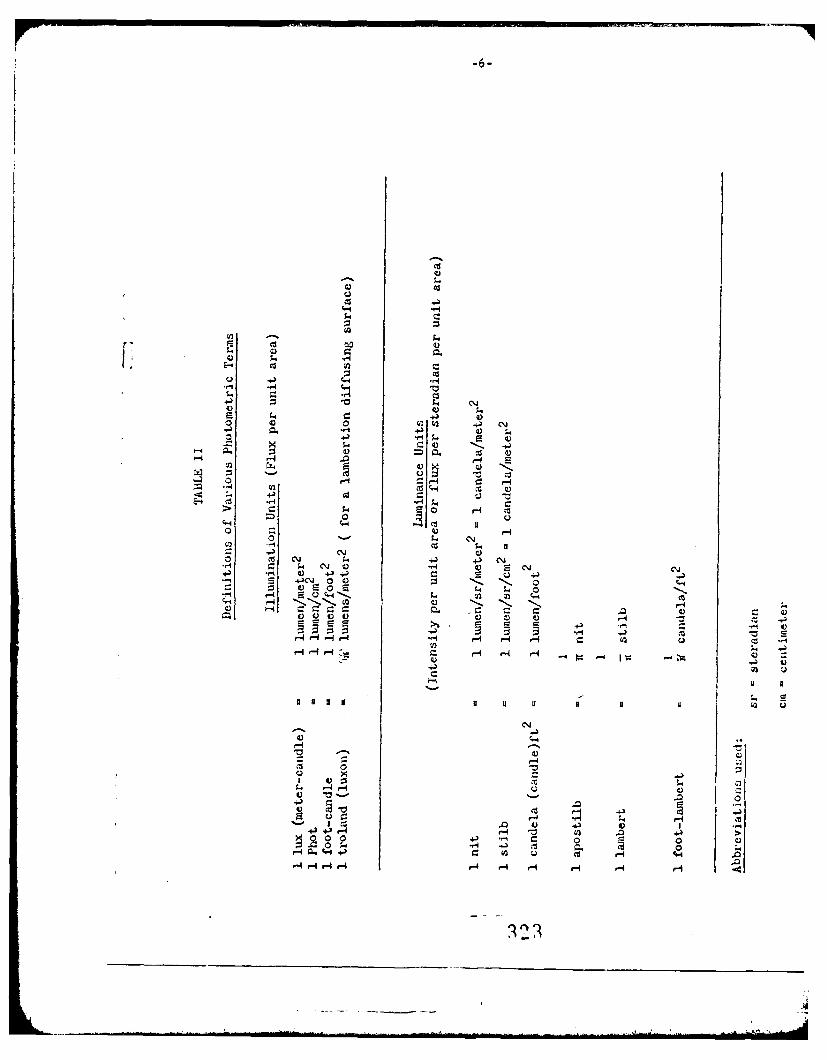
C-,
C-4)
0 Cd00
'*44a
od ... 4 Cd
E . ..I-4 ;.>5
cH 4-) 4\
4) C: +)
0~ 0 C
CU r4
CUN
oIt - )-45
5.Q 4-4 cu5- i
.41 CUr I 4) r- .3 +CU >%0~ *0 41 0~. 0)
A) a. +')c-4 P4 r4 f4 r4) 4 r4) HH

cQ ~ ~ -0% CZ %ci 0% V% *.
0%
03- .0 oN *~ 0% 0
o 0
40 Go Lo .. .
In4 Nr 4 '4 P
O - '04 3- N N N
c % __ Y 0 0 H0- 0
f'j >4 co l 0 N 0\en r-4 -T 0 C4 \

-8-
or, diensionally, on- joule has the dimensions (:. 2 T- 2 ) ad since one watt
equals one joule/secomd the dimensions of the watt are (:.ST- 3 ).
There are three basic systems of phxysical units on which radiometric
units can be based. '-he two metric systems are the KKS and the CaS, and
thd 4 tatdddp A taA Dr~jh Tho VWIX DoG5'fft 13 thf :WS% bdrfatnl.Y Ubed
system, although the QZS is sometimes used. The definition of some radio-
z.etric quantities and conversion factors are given in Table IV. Lcluded
also are the thermal =.its of energy, the calorie and the B.T.U., which
Vare occasionally used as radiometric units.
Calculations using the units of either system, within that system, is
straightforward once the unit of power (flux) is krnown. For example, a
radiation source emitting one watt of power, P, will, in one second, radiate
a total of one joule, U, of energy. A source with a luminous flux (power) of
one lumen will, in one sedond, radiate a luminous energy of one talbot.
For a source that emits radiation isotropically the radiant or photometric
power (flux) density will follow the inverse square law, that is, at a
distance R (centimeters, or meters) from a source of output P (or F) the
power density amjittance will be:
W" P/4R R2 watts/cm2 or L- F/4rR2 lumens/=n2
The intensity of the same source will be:
j.WR2 - P/41 or i-M 2 F /I41
It can be seen from the description of the two unit systems that the
radiometric system is fundamental, that is, they are based on the cor-on
units of physics and chemistry. On the other hand, the photometric units
are tailored for relative descriptions of visual sensation.
-325-
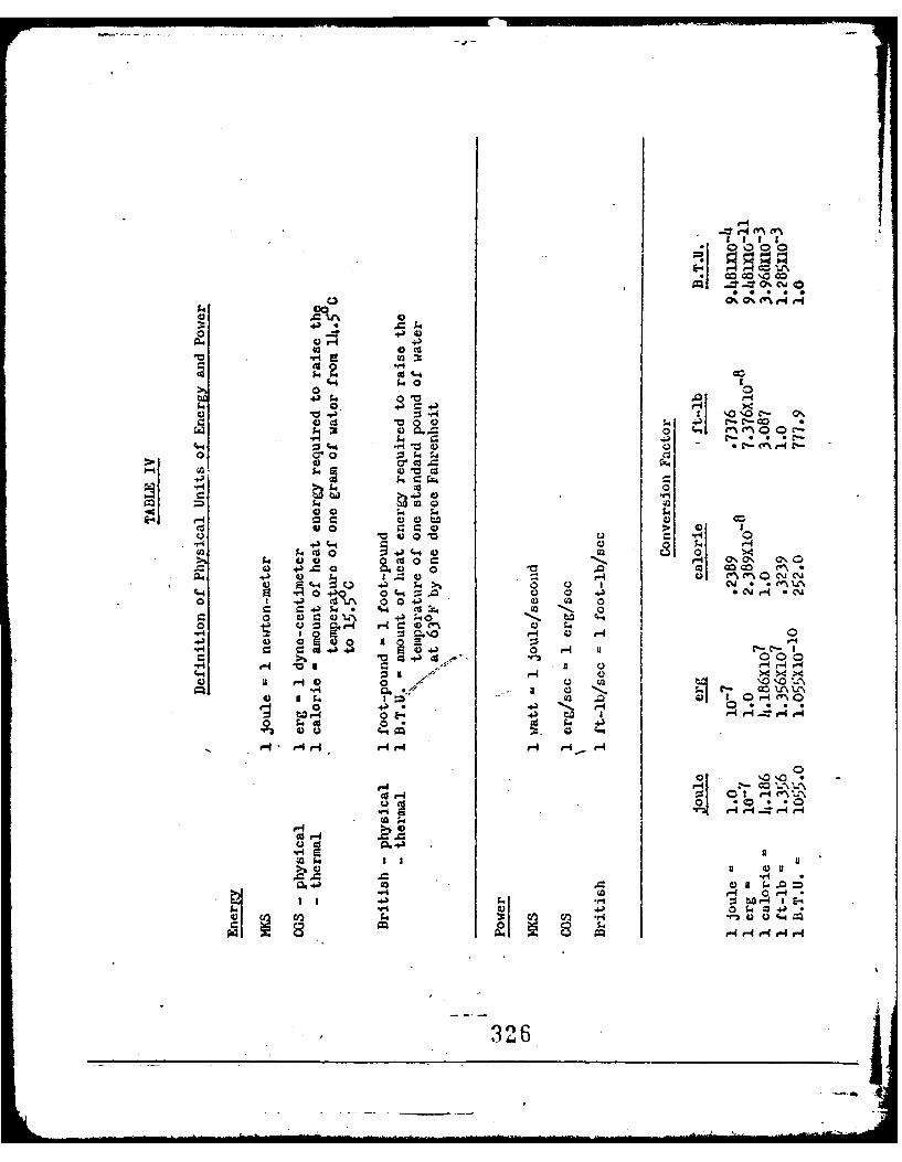
6Z ,4,
S" 0
M 0 0
S4 a CoL co 0
*)) 0
ul 00
0 a0V424
0. 00c04 ~ ~~~ ~ -. 1-4 S f
43 4-fl $I %Ob 00%a +31 0 O G C"4 :5~ .- -0
4 0 J 41.- 0 1. 0 *6C. -P;404C-
0 Q Q ;
V 10 s4~z 00) 0 -
0 00 f..4 £4C-j4 Hs V12 000
f" 00 >44 >. )9a. m00 0 0 11
40 a) 1. H 7
£4 to I00 4..= f-4 0% 4
0 H C) 41~0CJ\4)f 1.4b 0 0 C-4S-I f'O+) 014. 0 .J.Cia4*
0 04 0C. £4.~ I0 00.~ a-I ) H
1.4r- "Ia-
0~~- £4 *0 5
H aIHa-4H H..4
3 '3

-10 -
In order that the luminous output of a light source be specified, its
radiation distribution must be knon in radiometric units, watts per micron.
Thus a radiometric description of radiant processes can be converted to a
luminous description by use of equation one. Photometric descriptions,
on the other hand, cannot generally be converted to radiometric ones
since a given luminous unit is a number and not J-ndicative of the physical
power involved or its distribution in wavelength. In other words, a
large variety of radiation source of varied spectral radiant power can
[I be combined in equation one, to give the same value of luminous flux.
Unless the Px of the source is stated separately one cannot obtain a
radiometric description of events. In the case where the luminous output
of a source is given, as well as the spectral power distribution, physical
data can be interpreted directly in ter.s of the :F\ information. Table V
compares several light sources, including single wavelength lasers, .uder
the requirement that each source have the same luminous output. It can
readily be seen that, especially in the case of the lasers, the specifi-
cation of the luminous output does not characterize the power output.
Since most lasers operate at one wavelength, the k is found from the
luminosity curve and multiplied by 680 times the laser power output to
determine the luminous flux in lumens. The inverse process works equally
well, since only one wavelength is involved and the integral of equation
one is replaced by the simple equation
Fa - 680 , ? limens
In the assessment of ophthomological hazards with respect to laser
radiation, photometric units are not orly inadequate, since some infrared

TABLE V
Total Power Output of Several Light Sources of Different Spectral Distribution
Required to Produce a Luminous Output of 15CO Lumens
Source Soectral Power Total RadiometricDistribution Power 0utput
Incandescent Lamp Approx. 9-10 wattsBlackbody
Distribution*
Fluorescent La=. Phosphor Fluorescence 4-5 wattsDistribution*
Helium-Neon Laser Single Wavelength 88 wattsX -6328
Argon Laser Single W.4avelength 4 watts) -5145
GaAs Laser Single UndefinedX-8900 (very high)
Argon Laser % 3002
* See reference I
328
* F7~.
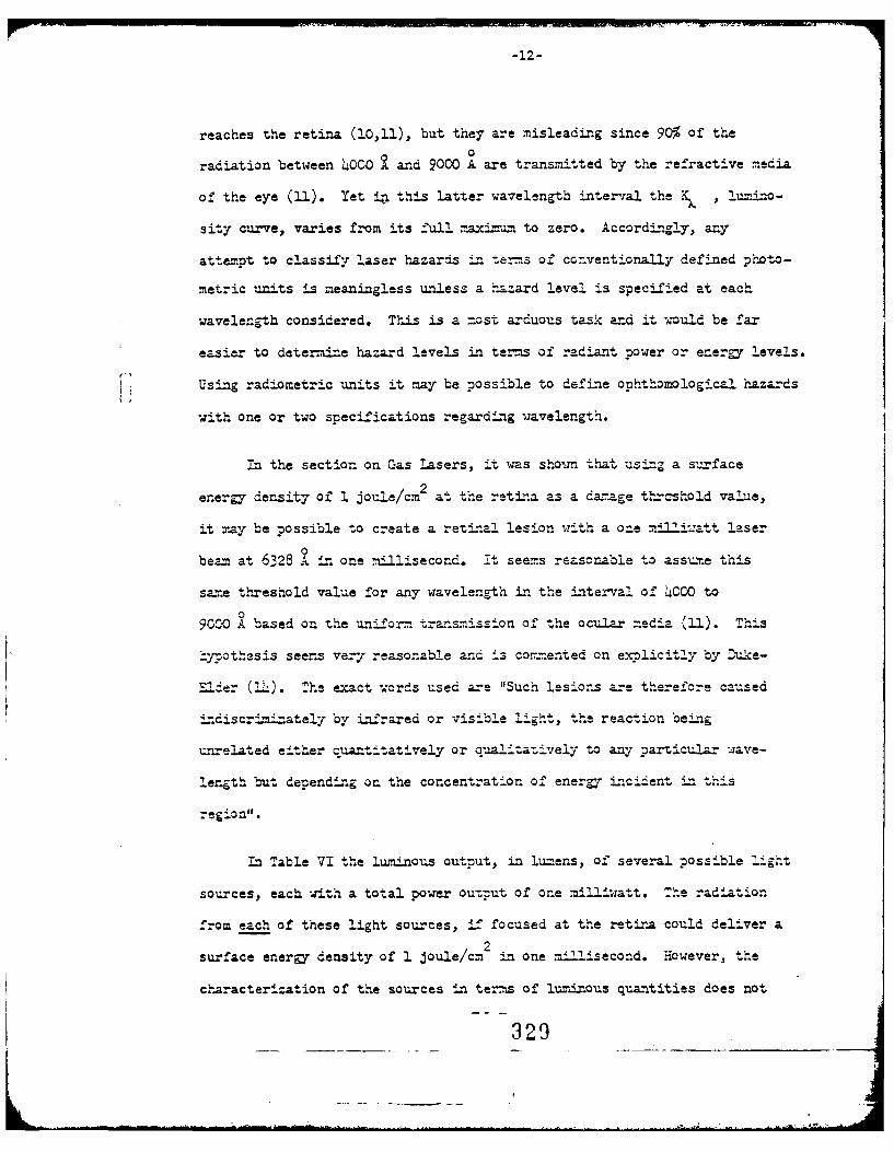
-12-
reaches the retina (10,11), but they are misleading since 90% of the0
radiation between 4OCO a and 9000 A are transmitted by the refractive media
of the eye (11). Yet in this latter wavelength interval the K , lu=ino -
sity curve, varies from its full maximuzn to zero. Accordingly, any
attempt to classify laser hazards L terms of ccnventionally defined photo-
metric units is meaningless unless a 'zard level is specified at each
wavelength considered. This is a most arduous task and it would be far
easier to determine hazard levels in terms of radiant power or energy levels.
Using radiometric units it may be possible to define ophthomalogic' hazards
with one or two specifications regarding wavelength.
In the section on Gas Lasers, it was show:n that using a surface
energy density of 1 joule/cm2 at the retina as a danage thrashold value,
it may be possible to create a retinal lesion ,.,ith a one mii.att laser
beam at 6328 9" in one millisecond. 7t seems reasonable to assume this
same threshold value for any wavelength in the interval of 4000 to
090CO A based on the uniform transmission of the ocular media (11). This
hypothesis seems vey reasonable and is commxented on explicitly by Duke-
Elder (Th). The exact crds used are "Such lesions are therefore caused
4discr!-iinately by infrared or visible ligl t, the reaction being
unrelated either quantitatively or qualitatively to any particular ave-
length but depending on the concentration of energy incident in this
egion".
in Table VI the l"minous output, in lu.ens, of several possible light
sources, each with a total power output of one milliw;att. The radiation
from each of these light sources, if focused at the retina could deliver a
surface energy density of 1 joule/cm 2 in one millisecond. However, the
characterization of the sources in terms of luminous quantities does not
329
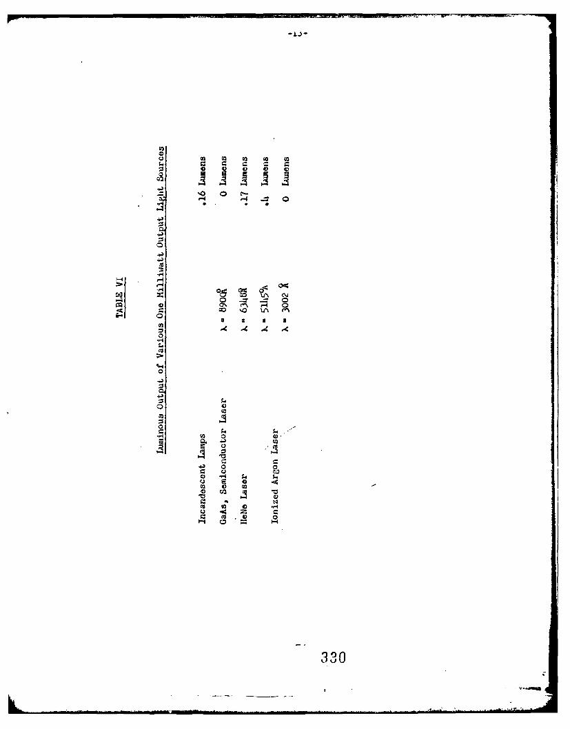
o
coC o o C
0
'-ri
0.0
I I

-14-
indicate the similarity of danger presented by each.
:n biophysics where pri-me consideration is given to physical phenomer,
such as damage thresholds, photochemical interactions and thermal inter-
actions, the energy and power of radiation must be unambiguous or at least
easily understood.
In studies primarily involved with physiological problems either unit
system can be used. Due to the vast aount of work that has been dane
pin the past, (for example reference 5), on the nhysiology of vision,
where illumination conditions are described photometrically, it seens
convenient to maintaLn the status quo. It .;ould 'ze advantageous if such
studies could indicate the P of the light source involved, since the
nterralation between physiological and physical phenomena is of interest
(5,6,7,3). This does imply additional work, but the methods of dete.-mining
P are well .. own (9). In addition, particularly in visual effects and
hazards, it is interesting to compare reversible and irreversible hazards;
the crssover moint from flash blindness to retinal damge is of interest
Li laser research. :However, the work done on flash blindness uses the..
photometric system aL .ost exclusiiely (12).
Retinal damage, thresholds are cha-racterized generally in ter ms
of radiometric units. Kinsey et. al. (15) uses photometric measurements
of the visual output of an electric weld4--ia arc to indicate the amount
of ultraviolet radiation present. This method is, at best, dubious and
any attermt to relate these measurements to a physical threshold value for
f'lash keratoconjuctivitis is equa'ly suspect. The only apparent "orthmhile
information that can be gleaned from this work of Kinsey is that the thresh-
old for onset of a photophthalic reaction in humans is about one half that
331
-15-
of rabbits and one fourth that of dogs.
Indeed, improper use of radiometric units occurs in the literature also
(1O,lh). The use of the unit erg-sec/cm2 by Duke-Elder (l) and repeated in
project Agile (10) when referring to previous work can cause confusion. Li one
instance the unit is used as a unit of power and in a second case it is used as
an energy unit.
Not only can the photoetric units be confusing but the misuse of the sim-
pler radiometric.ones must be avoided.
7To su-narize, the radiometric unit system should be used in describing
biophysical phenomena under exposure to optical radiation; the reasons for
this are as follows:
1. The units are frundamental and give a complete, unambiguous
description of the radiation.
2. Correlation between incident radiation and physical observables
such as temperature changes, are done easily w nen the radiation characteristics
are g-iven in radiometric units.
3. Radiometric measurements are not limited in wavelength as are photo-
metric units. Radiation beyond the visible region can only be described in
radiometric units.
4. A cormarison of physical effects initiated by various sources of
radiation (such as broad band Xenon flashtube radiation and narrow band laser
radiation) can be done more readily only in radiometric units. Other factors
such as the relative attenuation by ocular media, absorption, scattering and
attenuation must be considered.
5. Cormarisons or relations between physiological and physical effects
cannot be done by photometric descriptions. Radiometry must be used since
physical effects can be described quantitatively only in radiometric units.
332-

Go*ta2:oogical----a of car '-- describ-ed -'C:t u4.
sys-eams but -.he radiometric is t o beo treferr.-a sLrce a fe n-'-e~rs m.ay b.e
used to classi--y hazard levels for a vari-e-:y of 2.scr :.:venthI
ihotometric units are used, incividuZIL hazard 2'::smust be szecified f'cr
each wavalent..
The iroblem o1' whether the tine z'cr re~sbevisual :L- aIn : 4 r
a f'-ncti4on o1' the va-velenc-th ha-s not be-n consiaered . T' it is - de endezt of'
wavelangth, then radiometric units .iZadequately describ;,e both reversible
and 4-reversible tnz-a4-'ent. If reversible Lrmairneant is a 1'unction of
veegtthen a curve relating reversible mairnment to wavelength must
be considered i n conjun,-ction til the rad-io=Etr!c uis

.0680
1.2
4 - : -,
0.016.c '~-6.8 -
Scotopic .-= (v')
00
[ oz I-
.001 -t0
400 10683000 4000 5000 6000 7000 8000
Wavelength, angstroms
334
335

P .. E..;CES
1. MS Lighting Handbook, 3rd Edition; M1uin ating ngineering ociCty
New York, N.Y., (1959)
2. Photometry, 2=d Edition; J.U.T. Walsh, Constable & Co., L.I.D., London
3. Levi, L., "A Short Cou-rse in Photometr-y", Electronics Products; 16 (1965)
4. J. Otical Society of Limer., 52 (4); 490, 1962
5. Enoch, J.M., "Optical Properties of the Retinal Receptors", J. Optical
Soc. Amer., 53 (71), 1963
6. Boynton, R.1M., 'Quantun and Energy-Bared Visual Sensitiv--ty or. a Si-.age
Plot", J. Optical Soc. .f .Ar., 53 (641), 1963
7. of, E., Zigler, M.J., "Dark Adaption Level and Size of Testfield",
J. Optical Soc. of Amer., 4o, (211), 1950
8. Southa-1_, J.P.C., Introduction to Physiological Optics, Oxford Univer-
sity Press, London, 1937
9. Cb4istensen, ?.L., Lmes, 1., 1'Absolute Calibration of a Light Detector",
JOA,5L, (22h), 1961
10. Christner, C.R., et. al., "State-of-the-4.rt Study on Visual Izairment
by High-intensity F1as h of Visible, - 9 :Prared, or Ultraviolet Light",
Report No. -AT-171-9, Battelle Memorial Institute, Jan. 1965
11. Light Coagulation by Gerd Meyer-Sch-aickerath, C.V. Mosby Co.,
St. Louis, o., (1960)
12. Sperling, H.G., "Flash Blindness as a .-?uLnction of -Zavelength Specifi0city",
Fed. Proc. Vol. 24, No. 1, Part i, 0. S-73, 1965
336

13. Moon, P., Spencer, D.Z., "On the Stiles-Crawford Effect",
JOSA, _4,(319), 1944
14. Duke-Elder, S., Taxt5ook of onhthalmolgiv, Vol. VI, C.V. Mosby Co.,
St. Louis, Mo. (1954)
15. Kinsey, V.E., et.al., "Masuring Eye Flash from Arc Welding",
J.A.M.A., 123(7), (403), 1943
F
'3,
au3x f '.4.O .
Laser Eye Protection
Recent investigations have indicated that direct viewing of both
pulsed and C-W lasers can result in either reversible or irreversible
disturbances in vision. Protection of personnel from these hazards has
been the subject of a number of papers (1,2,3,4,5). These studies have
been oriented toward protection in the research laboratory and industry,
as well as in the field.
The extensive investigations by Ham et al. (6,7,8,9) on threshold
studies for the production of lesions in rabbit fundi, where thresholds
were determined by ophthalmoloscopic examination after irradiation by
carbon are, xenon lamp, or pulsed ruby laser, has been widely used as a
basis for estimation of the safe laser dose to the human eye. These
studies indicate that a mild, irreversible retinal lesion was ophthalmos-
copically visible five minutes after irradiation by a ruby laser pulse of
200 psec duration at a calculated energy density of 0.7 Joules/cm2 at
the retina. This radiation dose is approximately the same as that required
from conventional white light sources (xenon lamps and carbon arcs) for
exposure times of 200 sec. In addition, at this exposure time, the
threshold energy density showed no dependence on retinal'image diameter
for spots ranging from 1.05 =t o 0.54 mm diameter (8). A simple thermal
model of the f-ndus indicated chat, for exposures of 200 psec duration,
very little energy is lost due to radial heat conduction away from the
irradiated site during the pulse(S).
Straub (1,2) has considered several methods of providing protection
for the human eye from laser radiation, both in the laboratory and in
the field. He has suggested that warning can be given before a laser is
...338 .. .......

fired in the laboratory, thus giving personnel sufficient time to close
their eyes. It is probable, however, chat as the power and energy of lasers
increase the human eyelid will not provide sufficient opacity to protect
the eye from direct laser radiation or even scattered laser radiation.
Precautionary measures such as covering the eyes with the forearm, may
provide additional protection. Further study in this area is warranted.
In the field, where friendly and enemy lasers may be continually
operating, warning cannot be given to personnel. This situation necessi-
[ tates devising a method for continuous eye protection. Methods considered
by Straub include the automatic, high speed shutter triggered by the arriv-
ing laser pulse, and colored absorption filter or dielectric reflection
filters. He disqualifies Kerr cell shutters because of their small angle
of acceptance and low optical density in the fully closed state. Photo-
tropic filters acting as optical shutters from stimulus in the far red
with an appropriately short response time (10-8 sec) and sufficient opacity
were not available at that time. Such devices do not appear to be avail-
able in the literature at present.
In his well documented studies, Straub considers dielectric refleczors
or colored absorption filters as better possibilities for eye prctection in
the field. Dielectric reflectors are rejected, however, due to the angular
dependence of effective reflectance. Straub assumes that the hazardous
radiation associated with ruby lasers lies in a narrow band in the far red.
Thus it is possible that colored absorption filters can be constructed so
as to pass near red and visible radiation (to allow visual observation of
monitoring equipment, etc.) and yet protect the wearer against direct
viewing of laser light. Such filters may however not be completely sati-
sfactory, at present, for a number of reasons:
339

;"3xI
1. The source of ocular hazard to personnel does not necessarily lie
at the initial laser wavelength. Intense light from backscactered
plumes, as well as coherently scattered harmonic frequencies in
che visible range, is a distinct hazard to the wearer of such
glasses.
2. Under field conditions, laser frequencies being employed by the
enemy may not be known. Protective glasses with fixed abosrp-
tion bands are of little value against narrow band incident
laser light of unknown frequency.
Using Ham's result chat an energy density of 0.2 cal/cm 2 delivered to
a rabbit retina in 175 .sec. produces a mild, irreversible lesion, observable
three to five minutes after exposure, Straub concludes that 1.6 x 10⁻⁶ joules
at the iris, focused to a diffraction limited spot at the retina (2 x 10⁻⁶
cz2 when the dark adapted iris is the limiting aperture) is the least
energy required to produce irreversible retinal damage. Whether diffr-ac-
tion limited spoc size is justifiable merits further consideration. A
statistical approach to the problem of irreversible threshold damage may
be necessary. One problem associated with a consideration of a diffraction
limited spot size is that the extrapolation of the data of Ham et al. to one
diffraction limited may not be justifiable. Radial heat conduction away
from the irradiated site may occur and must be taken into account for a
diffraction limited spot size. It is possible that the assumption of a
diffraction limited spot may not represent the situation where-the total
energy required to produce irreversible damage to the retina is a minimum.
An irreversible lesion of a diffraction limited spot size may not be the
most disabling.
110
According to Ham et al. (10), heat dissipation by conduction at the
image borders will have an appreciable effect on the temperature at the
center of a 20 micron diameter spot at the retina, in approximately
180 isec. Straub's extrapolation to a 164 diffraction limited spot dia-
meter may therefore, be acceptable for shorter laser pulse:durations.
However, experimental verification of this model for a diffraction limit spot
is desirable.
V If it is assumed that the diameter of a perfectly collimated laser
beam is larger than the pupillary aperture of the eyes, then the diffrac-
tion limit of the retinal spot will be determined by the pupil diameter D,.
Since the diffraction limited spot diameter is then given by
DP
where H - Diameter of retinal image spot
F - Focal length of the eye
- Wavelength of incident radiation
Dp - Pupil diameter
It is apparent that, for a given focal length (given eye accomodation),
the diameter of the image spot at the retina will be a minimum when D. is
a maximum (i.e. when the eye is completely dark-adapted). It should be
noted that a perfectly collimated incident beam will be focused at the
retina only when the eye is accomodated for viewing a distant object.
For any other accomodations, the retinal image from a collimated beam will
be larger than the diffraction limit.
.341

The minimum energy required to produce irreversible retinal damage
occurs for a diffraction limited focal spot and maximum pupil diameter.
Straub has then chosen the case necessary to produce an irreversible
lesion for 175 tsec pulsed lasers operating in the far red, where the
ocular transmission to the recina is almost 100% (11). If one considers
longer pulse lengths, and/or a spot size larger than the diffraction
limit, the least energy at the pupil required to produce threshold ir-
reversible damage to the retina occurs for the smaller spot sizes (8).
Swopeand Koester (4) have published calculations of the attenuation
required to protect the human eye against pulsed laser radiation. These
calculations are based on published data (5,6) on threshold dosage for an
observable retinal lesion in rabbits, and data concerning human (invitro)
ocular transmission characteristics. The threshold energy theoretically
required to produce a retinal lesion in the huma- eye is determined for
both ruby and Nd-glass lasers with a given beam divergence. One of the
assumptions on which the studies by Swope and Koester (as well as chat
of many others) are based, is that human retinal damage will occur at
the same power densities as that required to damage a rabbit retina.
They conclude that a pulsed ruby laser with a beam divergence of 1/20
will produce irreversible retinal damage to the rabbit (and hence human)
when the entire beam enters the eye with an energy of 10-4 joules. This
result is a factor of 100 greater than that obtained by Straub for dif-
faction limited retinal images produced by a collimated beam.
Suitable eye attenuators are discussed (Swope and Koester). They
conclude that a combination of BG-18 and BG-38 glass, each 2 t thick,
provides the best eye protection with minimal hazard due to crazing of
the glass. A nomograph is then devised whereby a "Safe Laser Cutput"
342

is determined for personnel wearing BG-18 and BG-38 combination safety
glasses. The nomograph is based on the assumptions chat: 1). a suitable
safety factor can be established so chat the threshold energy for retinal
damage to the rabbit can be correlated to the human eye; 2) all light from
the laser enters the eye; 3) the area of the illuminated retinal spot
depends only on 9 , where 9 is the beam divergence as it enters the eye,
when the eye is focused at infinity.
Adequate data is required as to the correlation between the human anc
L rabbit eye. The importance of physiologic and anatomic differences between
the rabbit and human eye must be considered. It is, therefore, difficult
to assign a definitive value to threshold lesion data taken from studies
on various rabbits.
The assumption that the illuminated recinal soot dia=eter depends only
on the beam divergence angle when all light enters the eye, is true only
under certain conditions. In addition, if the eye is a distance L from
a laser aperture of diameter d and if the focal length of the eye is F,
then:d
(a) When L - (F + 9 )> 0 the minimum spot lies in the image
plane of the eye (retina)d
(b) Vhen L - (F + 9 ) = 0 the beam diverges within the eyed
(c) When L - (F + 9 )<0 the =in=um soot lies in the focal plane
It is important to realize that retinal spot diameter depends on eye
focus. The assumption that the eye is focused at infinity only gives a
minimum spot diameter at the retina in case (c) (and the diffraction
94.3

limited case). If the eye is focused ac infinity in case (a), the minimum
spot will lie behind the retina. If, however, the eye is focused at the
laser aperture in case (a), the minimum spot will lie on the retina. There-
fore, in case (a), a "safe laser output energy" that is calculated for the
eye focused at infinity will no longer be safe if the eye is focused upon
the laser aperture.
In summary, parameters, such as laser aperture, eye-to-laser distance,
eye focal length, as well as beam diver;ence, must be considered in a
theoretical model for damage to the human retina. Damage to ocher than
the retina must also be considered, particularly as other wavelengths
become available.
Although many of the factors associated with protective glasses,
particularly BG-18 and BG-38, have been extensively studied by Straub
(1,2) and by Swope and Koester (4), further investigation is necessary,
particularly as power and energy density levels, and ocher wavelengths
become available.
REF ZF C ES
1. Szraub, H.W., "Protection of the Human Eye from Laser Radiation",
Army Yaterials Co~and, Report TR-1153 (Harry Diamond Laboratories) 1963.
2. Scraub, H.W., "Use of Protective Goggles in Areas of Laser Radiation"
Fed. Proc. 24 (1) Part 3, Suppl. 14, S-78, 1965.
3. Fine, S., e al. "Ilechanisms and Control of Laser Hazards and Manage-
ment of Accidents", Proc. 2nd Conf. on Laser Technology, Illinois
Institute of Technology, Chicago, Illinois, April 1965.
4. Swope, C.H., and Koester, C.J., "Eye Protection Against Lasers" Appl.
Optics 4, 532, 1965.
5. Solon, L.R.; Aronson, R., Gould, G., Science 137, 1506, 1961.
6. Ham, W.T., Jr., ec al., Amer. J. Ophchal. 43, 711, 1957.
7. Ham, W.T., Jr., et al., Amer. J. Ophchal. 46, 700, 1958.
8. Ham, W.T., Jr., ec al., Acca Ophthal.,,Suppl 76, 60, 1963.
9. Ham, W.T., Jr., et al., Acta Ophchal., (Submitted for pub!i'ac.on).
10. Ham, W.T., Jr., et al., "Ocular Effects of Laser Radia:.on,.Part "
Defense Atomic Support Agency Report DASA-1574.
.ll. Geeraets, W.J., and Ridgeway, D., "Rer-na. Daoage from hnzensi y
Light", Acta Ophthal., Suppl. 75, :09, 1961.
345

Laser Dosimetry - Biological Systems
Few studies have been reported on laser dosimetry. Fine ec al. (1)
have discussed the difficulties involved with laser dosimetry from both a
physical and biological standpoint. Schlickman and Kingston, and Schlickman
and Diffley have reported the development of a pulsed laser dosimeter for
ocular hazards measurements (2,3).
The Schlickman - Kingston dosimeter utilizes a modified gamma-radiation
dosimeter as a high-impedance voltmeter. The instrument is calibrated with a
steady state light source and an optical spike filter so as to read full scale
when an energy density believed to be capable of causing retinal damage to the
human eye enters the instrument at a specified wavelength.
The instrument is used in the following manner. A laser target is
selected and the diameter of the area to be irradiated on the target is
determined by firsc irradiating a piece of carbon paper or exposed Polaroid
film. The distance from the target to the anticipated position of the eye
of an observer is measured. The aperture of the dosimeter is adjusted from
a chart (which appears in the article) to yield a full scale deflection for
a hazardous radiation dose. Schlickman and Kingston point out that the target
area is then illuminated by an incadescent source and the scattered light
focused to a sharp image (by means of.a viewing system attached to the dosi-
meter which they describe) on a diffusing plate that lies at the entrance to
a phototube within the dosimeter. The laser is then fired at the *target and
a reading taken on the dosimeter. A reading of 1/10 scale is assumed to
indicate unsafe viewing of the laser impact by the unprotected eye.
346

The authors explain that the instruments dynamic range encompasses all
0known, non-Q-switched lasers operating in the 6,000 to 10,000 A range. Pro-
visions have not been made for work with Q-switched lasers. This is due to
design difficulties and the lack of quantitative biological data regarding
such lasers. The dosimeter will accurately respond to laser pulses of at
least 23 Lsec duration composed of triangular spikes at lease 400 sec wide
at the base with approximately a 1 sec delay between the end of one spike
and the beginning of the next.
To set the level of "safe" radiation exposure for the human eye,
Schlickman and Kingston have incorporated the threshold data of Ham and
Geeraets (4). Schlickman and Kingston have used a safety factor of 10, in
correlating this data with the hazardous radiation dose to the human eye.
However, due to the lack of quantitative, in vivo data for correlation of the
physical and physiological characteristics of various strains of rabbit and
human eyes this safety factor requires further consideration.
The general effectiveness of single pulse laser dosimetry is
doubtful under normal laboratory conditions. Basic to the work of Schlickman
and Kingston is the assumption that, if a dosimeter reading in the field of
scattered laser light indicates a safe level for a given laser burst, then a
second laser burst at a similar target will also produce scattered light at
a safe level. The dosimeter cannot measure the level of either direct or
scattered laser radiation reaching the eyes of laboratory personnel during
a specific laser pulse. Safe radiation levels can only be inferred from
dosimeter measurements that are made prior to the exposure of personnel to
347

scattered (or direct) laser radiation. There are many instances where the
target surface to be irradiated is not the same from shot to shot (eg. bio-
logical targets). Changes in target surface will change the patterns of
scattered light intensity and thus reduce confidence in predicting scattered
light intensity at a specific location in the laboratory for subsequent laser
pulses. With many pulsed lasers, output power density iL not *asily reproduced
from pulse to pulse and should be considered as a factor in evaluating a single
pulse laser dosimeter.
The Schlickman-Kingston dosimeter is equipped with an adjustable iris
that reduces the total radiative energy entering the dosimeter for scattering
conditions that will produce a spot on the retina that is larger than the
diffraction lim-.t of the eye. Further data and a theoretical model of the eye
would be desirable to explain the curves shown in the article. A more complete
theoretical treatment of the focusing of specularly scattered light by the human
eye would be of interest.
In view of radial heat conduction within the pigment epithelium, the
retinal spot diameter as well as area is an important parameter for certain
laser pulse durations (5). Using xenon sources, Ham et al. (6) have shown that
for pulses from approximately 100 gsec to I msec duration, radial heat conduc-
tion is relatively unimportant even for diffraction limited spots on the retina.
Hence, for pulsed lasers operating in the 175 sec - 1 msec range, one can
consider the threshold energy required to permanently damage the retina as
being a function of only the retinal spot area. This being the case, it
becomes important to know the area of the human retina that is actually ir-
radiated by scattered &ht 1r3oua target surface.
8

A factor of importance with respect co the Schlickman-Kingscon dosimeter
is that i: is only calibrated over a narrow band near a desired laser wave-
length. There is no reason to assume that all the hazardous scattered radi-
ation will be confined to that frequency band. Brilliant, broad band plasmas
have been observed on laser radiation of both physical and biological targets
(7,8). These plasmas are generally small and may be focused to a small image
on the retina of an observer. The S-1 response of the Schlickman-Kingscon
dosimeter is relatively insensitive in che blue-green spectral regions, the
[ response at 5000 X being 20% of maximum. The biological effects of viewing
laser generated plasmas has not been investigated and should be considered
as a po.e,,cial hazard. Scattered light may also contain frequency doubled
components. An S-1 dosimeter response will not yield reliable data for
frequency doubled ruby or neodymium laser radiation.
The problem of monitoring laser radiation levels to determine safe
conditions for personnel involves measuring several parameters. Power, power
density, energy, wavelength, and anatomical site involved in laser inter-
action are some of the parameters that must be measured before a specific
biological reaction to laser impact can be predicted. There is no single
parameter that can be assigned to laser output that will also yield sufficient
biological information to ascertain possible hazards. "Radiation dose" is a
phrase that must be broad enough to include all parameters necessary to com-
pletely describe a specific biological reaction to a laser radiation field.
From the standpoint of instrumentation, it will be difficult to develop
a simple monitoring dosimeter that can measure a sufficient number of physical
and biological parameters to completely characterize the laser interaction.
349

Dosimecry for radiation from discretely pulsed lasers is difficult due to
variations in scattering, power output, beam heterogeneity, location of person-
nel and other biological and physical parameters from shot to shot. Extrapo-
lation of data obtained from one laser pulse to the next is difficult. Fine
at al. (1) have pointed out that if adequate instrumentation can be developed
two types of dosimetry are desirable. First, instantaneous detection of
unsafe conditions may be used for protection. Second, cummulative exposure
measurements may be important for medico-legal records.
350.. ....

w_ __~
R F ERENC ES
1. Fine, S., et al. "Mechanisms and Control of Laser Hazards and Management
of Accidents", Proc. 2nd Conf. on Laser Technology, Illinois Institute of
Technology, Chicago, Illinois, April 1965.
2. Schlickman, J.J., and Kingston, R.H., "The Dark Side of the Laser",
Electronics 38:8, 93, April 1965.
3. Schlickman, J.J., and Diffley, R.M., "Laser Dosimetry: The Design Engineer's
Viewpoint", 1st Annual Biomedical Laser Conference, Boston, June 1965.
" 4. Geeraets, W.J., et al., "Laser Versus Light Coagulator: A Funduscopic and
Histologic Study of Chorioretinal Injury as a Function of Exposure Time'.,
Fed. Proc 24, Suppl. 14:1 part 3, S-48, 1965.
5. Ham, W.T., Jr., at al., Acta Ophthal. (Submitted for publication).
6. Ham, W.T., Jr., at al., Acta Ophthal., Suppl. 76, 60, 1963.
7. Fine, S., et al., "Measurements and Hazards on Interaction of Laser Radi-
ation and Biological Systems", NEREM Record, 1964.
S. Fine, S., et al., "Interaction of Laser Radiation with Biologic Systems,
I. Studies on Interaction with Tissues", Fed. Proc. Suppl. 14, 24,
Part 3, S-35, 1965.
.......................... |
i
Polarization of Lasers - Effects on Energy Measurements
In most energy measurements of laser radiation, the output intensity
is often sampled by a low refaee~anse bPam splister, Tia proaodutt Ls
usually followed when it is desired to monitor the laser energy during
each pulse or when the full laser energy would destroy che monitor.
Generally the beam spliter has been placed at a 450 angle to the optical
axis, reflecting a small fraction of the lighc to an energy measuring
device while most of the beam is :r:nsmitced to the target. The angle of
05 ° has been chosen, since it oer=its pcement of the energy measurement
device at 900 to the beam axis. This procedure is ao-on in bio-physical
laser research, where various beam spli:ters are ased (1,2).
There is a very importzant. factor that is not usually considered in
estimating the reflectance of :he beam splitter. Depending on the angle
at which the beam splitter is set, the polarization properties of the
laser beam can be responsible for errors in excess of 100% of the calcu-
la:ed reflectance. In this section the source of these errors and a
method for minimizing them will be shown. For the sake of brevity only
single surface reflections will be considered.
Most optical phenomena c~n be described in terms of electromagnetic
theory. In the wave model of light, a light beam is considered as a
transverse wave with perpendicular electric (S) and magnetic (H) field
vectors. The simplest type of electromagnetic wave is one that can be
expressed in terms of a sine or cosine function. For example, using the
cartesian coordinate system, an electromagnetic wave traveling in the
352

-2-
positive x direction could have its electric field vector in the y
direction. in which case:
E - £y cos (Wt-kx)
H Hz cos (Wt-kx)
where *** > .
Ex E, 0
H - H = 0y x
and
where
= frequency, and X - wavelength
A wave which =aintains this orientation is said to be plane polarized or
linearly polarized. lndeed, any light wave in which the electric (and
thus the ma;nezic) field vector nainta.Lns a constant orientation is said
to be plane polarized. Most opical radiation is unpolarized, that is
the individual wave trains have their electric field vectors oriented in
a random fashinn, giving the overall effect of having every possible linear
polarizations at once.
Linear polarization can be selected from unpolarized iight by
passing the unpolarized beam :hrough a polarizer. Unpolarized light can
be theoretically considered as consisting of two mutually perpendicular
polarizations. This is because, once an orientation is chosen, all the
o.har vectors can be decomposed in the ordinary fashion into two
=utually perpendicular- components. Unpolarized light may be represented
by two orthogonal vectors depiccing the electric vector orientations as
shown in Figure 1.
A polarizer selects one polarization. If two polarizers are set to
accept perpendicular polarizations, then their combined action on a beam
is to pass nothing at all, as is shown in Figure 11.
%any practical polarizing devices are made from crystals which
naturally have the physical proparcias of birefringence or double refraction.
Many other materials exhibit b refringenca when placed under mechanical
stress for example polaroid plazzic, or under elactrical stress, for
example the Kerr effect, or under magnetic stress, for example the Voigt
effect.
The ruby used in ruby lasers is naturally bire-frigent znd the out-
put of ruby lasers has been shown to have a varied polarization (3,8)
M ost gas lasers operate with a linearily polarized output due co the
design of the discharge cube (-he Brewster angle windows will be cov=ented
on further in this section). Polarized outpucs have been observed in
gas lasers withouc Brewster angle windows however. This fact may be due
to a =echanical scress birefrinrance in the glass itself or induced
=agnetic birefringence (4,5). The output of Neodymium in glass may have
various Iolarizacion properties. However, studies do not appear to have
been carried out in any detail in this area. A review of the contradictory
observations on the polarization of light emitted from GaAs laser diodes
has been carried out by Nannichi (9). His study also showed that the
polarizations of the GaAs laser radiation shifted its orientation as a
!unct.;on of current. No cause of the polarization was found (9).
ft .

The najor point to emp-asize ca ce-n~ng laser polarization is the
fact that chose lasers which have been investi;ated are polarized to
various extents. The polarization in ruby not only changes from shot to
shot, but even varies during a single shot (2). Thare is almost no
-nfortatlon on the Neodymium laser, but there is no a oriori reason :o
expect uniformly oriented polarization -:hi- .he possiblities of random
polarizarion may exist (4,5). :ha followi analysis thus directzy
applies to ruby lasers and =ay :eil be significant for the case of
Neodynium in glass.
The basic optical law of raf-c'on, Snell's law scates that if a
light beam is in . cn at .he in-efaca -. :o media at an anle 9 .
relative to the normZ-l to the Sz-face :-.en :ha bean w:.-_ be refracted in
the second medi, a~n .ng of 6' frc:. tche am-is, "n :hat the ratio
o: che sines of the t=;o angles i the inverse ratio of the indices of
refraction of the t;o media.
S£:7LL'S T.Tsin' n
nac:ever, not a e z 4 zs trans .-t: ad into zhe sec cd ra on,
soma is reflectad at the n.erfazce at an angle equal to the angle of
incadence, Fi.,ure TIT.
Fh Fresnel laws ot rafleczn ;,va the reflection and -m 5he sslon
cotaiclants for the various beans depending on their polarization. Any
bean scriking the surface can be either .nearly polarized, with its
electric vector perpendicular to the plane of incidence or parallel to
355

the plane of incidence (see Figure Iii), or unpolarized and :epresentable
as a linear combination of the two linear polarizations (elliptical and
circular polarization must also be considered).
The reflectances are a function of the an.le of incidance and the
angle of refraction and may be written as (7):
an2 (9-r p -2
tan (e + )
S.: 2 .
sin 2 (9 + e)
where the subscripts:
P = parallel polarization
s - ?erpendicular polarization
and where the anga of refraction, 9 , must be calculated from Snell's law.
Ordinarily, the reflection coefficient of a surf-ce is calculated for
unpolarized light in which case the aver-age of the t.o polarized reflectances
is used (5). The average reflection coefficient is written as
r = I(r
Figure IV is a graph showing the three reflection coefficients for
air t O glass, (high dispersion crown at 6560 %), n = 1, n' = 1.517 as a
function of angle of incidence. It should be noted that at 56.580 the
reflectance of the parallel component goes to zero. This is the Brewster
angle. At this angle the parallel polarization is transmitted with no
356

-0-
reflaction loss. In ,as lasers the use of windows se at :he Brewster
ang'e causes the parallel polarization to be favored in the anplification
process.
The reflection coefficient for either polariza::on depends on :he
angle of incidence and the ratio of :he inices of refraction between the
two =edia. If an air-glass (or cuartz) interface is assumed, the variation
of reflectance for G = 00 as c " ion of refractive ine:: can be specified
in ter=s of the refractiwe dcx of the gKass (air is assumed to have
an index of one). This e s S ., Fin -iUre V. This curve is
calculated from therrac:ia in.ces of crown and flint glass and
quartz for vcrious .:aveen:-:s (5). Thus the rafractive index of a given
bea splitter, hence the rafIec:.cn coefficienz, is a function of :he
material used and the wavelength of the laser 'ean.
Figure V! shows the error rha: can axisr due to bea= polarization if
:he average reflection coefficient is the value soecified. The error is
olocted as a function of anle of incidenca. Az 450, the most co..only
used beazspIi:er ange, the error due co z~rpendicular polarizacion is
about ' a'. -: a: due to the --:cl olrizaction has a value of -70.
:t is reasonaoIe to concluce hat if tha average reflectance coeffizienz
for a ea= solicter iz used, tnen zhe energy of a aer beam wich vary ing
poarizazon cannot be =easurad accurazeCy cy the bea=spli:er technique,
as ordinarily used.
357
As an enample, consider a single surface beam splizter (n' = 1.517)
set at an angle of 450 to the beam axis (8 - 450). The average reflection
coefficient is 5.3%. The reflection coefficient for parallel polarization
is 1% while that for perpendicular polarization is 9.3%. A one thousand
joule, parallel polarized laser beam would be diagnosed as having an energy
of about 200 joules if the average reflaction coefficient were used.
Si:ilarly a reflected energy of 5 joules could be interpreted as 95 joules,
55 joules or 500 joules according to whethar the average, perpendicular or
parallel reflectance coefficient :ara used.
It is, thus, =osz important that the polarization properties of the
laser beam be taken into account when reflection dependent beam sampling
methods are used. A beam splitter set a: 450 can produce large errors in
energy measurement. Since the polarization nzoerties of lasers are not
widely explored, they must be consiared as coable and of random
orientation in the design of beam splitters.
A si=ple manner to overcoae the ambi~uity prasented by various beam
polarization prczrzies is to use a low angle of incidence beam splicter.
As can be seen from Fi, uze VI by setting the beam spliter at an angle of
100 , zhe error introduced by bear polarization is less than 5%. This
angular setting is reco-ended and should be followed unless some other
tachnique of eli.inating this source of error is found.
Due to the time factor involved these results must be viewed as
highly preliminary. Further considerat-)n must be given to the problem.
The increased complexity due to reflections from boch :he first and second
surfaces must be considered in detail. Experimental verification of the
conclusions determined analytically must be carried out.
358 1

Polariz.d t 'o Page
Un po Ia r izcd
unoolcrized side view
cn4 viev:
T~~ r I ___rPolarizrr____
No.

lncid ;ne bow -1 l ccdtz
.1
7 h C. ra a-3 St h s r.
3160

14,
1L
0
Angl ofIciec
?ercent reflection of light from a single surface, n =1.517
.a function of angle of incidence where rsis for perpendicularzation, r~ is for parallel polarization, rayi o
;.~--;zed.
36 1

'a
0
UID
0
0 -
'a -4
)
Uo -
C U
U, -0
'4
0
'4UU
ifl
'4
U
U
~C~J
ID
362

CC
C
cv%
0
0
363

-3-
aferances
1. Minton, J.?., et a!, "The Effect of ieodymium Laser Radiation on
Two Experimental lignant Tu-or Systems," Sur2. Gynecol. &Obstetrics, 120:481, 1965.
2. FON, J. L. , pIq J P.., n& 'Scrin, -1. N. , "The E oc of La a o.adiation on Intracranal Scruczures," Isc annual BiomedicalLaser Conference, of the Laser '.:edical Research Found. , Boston,
ass., june 1965.
3. Brunton, J. , "?olarization of tha Light Cutout from a Ruby Cpotcal
Maser," Aocl. Otics, 3:124-, i9-.
4. Doyle, W.I.., and ! 'ite, .I.B., ".--equenzy Soplizr-ing and Mode CO=.e!-itionin a Dual-?olarizaeion ?z-2:a Gas Laser," ADol. physics Lett. , _
193, 1964.
5. Handbook of Che.nis rv ad --'Zcs, 45ch Ed. ,(The chemical .Rubber Co.,Cleveland, Ohio, !1-S5).
6. Jzvan, A., Ballik, BodE.., and - ;.., "Frequency Characteristicsof a Continuous o ,,ave He-'a Coaical M azer," JOSA 52:961, 1962.
7. Jenkins . , and !hice, H.. E., ",U..amen-:als cf Ontics. (New York:
M'cGraw-wil1, 1957).
8. Ta=ai, T., and Achiwa, ,o.'. , "?oarizaz:on of a Ruby Laser Bea=," Jaman.J. A'ol. .=hvs., 3:60, -924.
9. Na.nichi, "Cn c.'e ?ooar-za.on of ,. rom :-ahe GaAs Diode Laser,"
Jaoan. J Aovl. ?hvs., 3:360, 196.
364

Discussion of Some Laser Output Detectors
Instruments available for measuring the output of lasers fall into
four primary catagories:
I. Those which convert the output to heat and measure the temperature
change of an absorber.
2. Those which sample the output beam directly, as in photodiodes.
3. Those which convert the output to mechanical energy.
4. Those which convert the output to chemical energy.
This section is concerned with the first two categories since these
are the most usual methods employed in commercial calorimeters. Some
general remarks, however, apply to all classes of laser r&diation detectors.
Whenever radiation is incident on matter, there is a reflection which
is dependent both on the parameters of. the radiation, including wavelength
and polarization, and on the characteristics of the reflecting medium,
such as surface texture and the reflecting properties as a function of
wavelength. In general, the polarization of a laser beam is not well
defined (1). Consequently it is difficult fo predict the properties of
the reflected radiation. Hence the transmitted radiation cannot be accurately
characterized. Furthermore, t4ere is no evidence that the parameters of
a laser beam, such as polarization and beam homogeneity, are constant from
pulse to pulse or during any given pulse.
In devices, which transform the output to heat (thermal energy), the
sensing device is usually an easily calibrated heat, sensor. This sensor
is in contact with a medium Uhich is heated by the incident radiation it
absorbs. The temperature rise is related to the absorbed- energy by a
365

response curve which is accurate only for specific values of absorption,
reflection, temperature distribution within the receiver, and in some
cases ambient or reference temperature. These and some other parameters
will be d.scussed below.
In most of the devices which transform the incident energy to heat
there are two components; the energy absorber, and a temperature sensor
(2,3,5,6). The energy absorber is generally a device employing the
Mendenhall Wedge Effect. Briefly the Mendenhall Wedge Effect is the
production of many reflections in a wedge or cone shaped enclosure of
specified absorptivity (Fig. 1). The absorptivity of a medium is the
fraction of incident radiation absorbed by that medium. The incident
energy is strongly attenuated within a cavity employing this effect. If
the end is not sharply pointed, fewer reflections take place, and conse-
quently less energy is absorbed (Fig. 2).
Usually a small angle cone is employed as the cavity. Daly (2) has
calculated greater than 95% absorption due to 15 reflections of the
radiation in a 12 degree cone, with light incident on the cone aperture
parallel to the axis, and with the cone having a specular reflectivity
of the order of 80%. His data does not include the cases of either non-
parallel light entering the cone, nor the cone being a diffuse, rather
than polished, reflector.
Zisenman et al (3) have reported greater than 95% absorption in the
infrared by using blackened cones. Their experiment was done by using the
two extreme cases of parallel light entering the cone, and a point source
of light situated on the axis of the cone. Their cone was assumed to be
a perfect specular reflector, and they therefore do not consider the case
of a diffuse reflector. Gouff' (4 ad others have analyzed the effective14 F
absorptive cavity of a conical receiver which has a perfectly diffusing
reflecting wall and in which the incident energy enters as a collimated
beam parallel to the cone axis. Although this may be an important theor-
etical case, it is usually not employed in practice, since the coatings
generally employed to increase absorptivity, such as carbon, tend to cause
specular rather than diffuse reflections. Therefore this paper will not
consider that case. In a consideration of the case of a point source
situated on the axis of the cone, the calculations carried out by
zisenman ec al (3) are useful to investigators who focus the laser beam
into the cone or who use diverging beams. However, the case of a beam
focused so that the focal point lies beyond the point of the cone is not
discussed.
In devices described by Ackerman (5) and by Dimeff and Neal (6) the
problem of manufacture and maintenance of sharply pointed cones is circum-
vented by the use of stacks of razor blades, which also employ the
Mendenhall Wedge Effect. However, a problem arises which is due to the
dependance of the reflection coefficient on the polarization of the
incident radiation.
All conductors have a complex index of refraction (21). The imaginary
component of this number acts as an absorption term, but, since it j an
imaginary number, it tends to change the phase of the light which it affects.
Since it was seen in the section on polarization of lasebeams that the
components of light with different polarizations will in ganeral have
different reflection coefficients, it follows that these differing components
will also have their phases changed by differing amounts. This implies
that if light is assumed to be plane polarized when incident on a conducting
367

reflector, it will in general become elliptically polarized after reflection
(22). Any further reflection will then change the polarization again, etc.
Now a receiver with circular cross section, such as a cone, will
present a normal to the surface in all directions. Hence it will average
the polarization of the beam within the cone at each reflection, making
V the coefficient of reflection independent of polarization. The coefficient
will be the same for each reflection.
Raceivers which do not present circular cross sections will not
perform this averaging. Hence each reflection with a given wedge will
have a different coefficient which depends on the state of polarization
of the beam which is altered by each of the previous reflections. It
is then difficult to predict the total attenuation of the beam within
each wedge formed by receivers such as a razor blade stack.
All studies of the absorption by the walls of a receiver assume that
:hose walls are smooth. In many commercial instruments, however, the
cones are produced by welding, riveting, butting, or otherwise mechanically
joining a sheet of metal to form the cone. The site of the joint will be
a perturbation odfche reflectivity of the cone and should be taken into
account in calibrating the instrument. In addition, the surface of most
metals is altered in time by oxidation or contamination, which will affect
the reflectivity. Stainless steel razor blades appear to have advantages
over cones in terms of design simplicity and surface resistance to
chemical and physical damage.
368
Other devices, such as the Westinghouse radiometer MN-1, measure the
change in resistivity of a metal wire as it is heated. The incident energy
is thus determined. The method is acceptable, provided the temperature of
the wire at all times exceeds the Debye temperature of the metal used in the
wire. For copper the Debye temperature is 420C, or 1080 F (7). Above this
temperature the resistivity of metals increases linearly with temperature
(8). The wire can then be considered as many small segments connected in
series, each with ics own resistance dependent on the temperature of that
segment. The suit of these resistances comprise the total resistance of the
wire. Under these conditions, the heat distribution in the wire is not
important; the tocal heat in the wire will determine the resistance. This
principle insures that the rapidity of data output is limited solely by
the circuitry involved. The errors due to heat loss occurring wnile the
heat is evenly distributed are minimized. :iese instruments however, are
not exempt from problems of reflection. The Westinghouse brochure on the
RN-I, indicates that a correction must be made for reflected radiation
which depends on both wavelength and polarization.
The majority of systems which employ devices which change the energy form
from radiant to thermal use thermocouples or thermopiles as a sensing
element. These thermocouples employ the thermoelectric effect (9), which
is in reality three separate effects. The three effects are the Seebeck
effect, the Peltier effect, and the Thomson effect.
In 1823, T.J. Seebeck discovered that if a circuit is formed consisting
of two dLssimilar metalic conductors, and if one of the junctions of the
circuit is at one temperature while the other junction is at a higher
369

temperature, a current will flow in the circuit. The current continues.
to flow as long as the two junctions are at differing temperatures. The
electromotive force producing this current is called the Seebeck Thermal
SM.
In 1834, M. Peltier discovered that if a current flows across the
junction of two metals it gives rise to an absorption or liberation of
heat (9). If the current flows across the junction in one directr.n heat
is absorbed, while if the current flows in the other direction heat is
liberated. If the current flows in the same direction as the current
produced by the Seebeck effect at the hot junction in a thermoelectric
circuit of two metals, heat is absorbed, while at the cold junction heat
is liberated. The heat liberated or absorbed is proportional to the
quantity of electricity which crosses the junction. The amount of heat
liberated or absorbed when one coulomb of electricity crosses the junction
is called the Peltier effect at the temperature of the junction. Expressing
the heat in joules, it can be shown that the magnitude of the ?eltler
effect is given by the product of the absolute temperature of the junction
and the rate of energy change of the thermal B T of :he junction at that
temperature of the junction when the only current across it is that due to
the thermal a: (9).
The Thomson effect was discovered by W.Thomson in 1847 from observacxons
of thermocouples. He noted that 1.f the only reversible thermal effects in
the circuit were the Peltier effects at the junc Dns, the ."N around the
circuit whose cold junction is kept at constant temperature should be
* It is assumed that each junction of the thermocouple has small mass
with respect to its temperature source. -- -
370

proportional to the difference between the temperatures of the hot and
cold junctions. This was contrary to experience. Thomson therefore;
looked for reversible heac effects when an electric current flows through
a conductor in which there is a temperature gradient. He studied a
copper wire whose temperature varied from point to point. When a current
flowed along the wire such that at some point the currenc and heat flows
were in the same direction, i.e. the current flowed from hot places to
cold, heat was liberated. When the current and heat flows were in opposite
directions, heat was absorbed. This effect produced by a current tlowing
along a conductor in which there is a temperature gradient is called the
Thomson effect. This effect differs from the Peltier effect in chat it
occurs in a homogeneous conductor rather than at the junction of two
dissimilar conductors. This effect has less influence on the temperature
of the conductor than the Peltier effect has on the junction. * The Thomson
effect can be detected experimentally only by the use of large currents
and sensitive devices for measuring differences in temperature.
Thus the Mh observed by Seebeck, which is the basis of thermo-
electric thermometry, is the algebraic sum of the Peltier EIV at the
junctions and the two Thomson E--F's in the two dissimilar wires. There
is therefore not a simple relationship between the temperature and the
L observed. The relationship is certainly never strictly linear. The
reason the £M versus temperature curves are not linear, is that the
Thomson W in one metal of the thermocouple circuit is not in general equal
to the Thomson EM in the other metal for all temperatures. However, for
* In all cases the Joulian heat loss has been neglected.
371

all thermocouples there is a range where the curve may be approximated by
a straight line. The LFD versus temperature curves of thermocouples are
tabulated (10). These tables are strictly empirical. The dependence of
ZMF on temperature can not yet be predicted with certainty for any given
thermocouple.
The effect of high peak power levels on the reflective property of
V! metals and on the energy measurement system requires further consideration.
Whether the various devices remain linear under high peak power impacts
requires further study. Ddly (2) presumes that the conical collecting
apparatus remains linear to the point of visible damage to the cone.
Measurement of low energy levels can be attempted by integration of the output
from photosensitive devices.
Several methods employing photomult-aplier tubes as detectors of laser
energy output have been reported (2,11, 12). Before individual methods
are discussed, those characteristics of photomultiplier tubes which will
affect all experiments will be noted.
Photomultiplier tubes consist of an envelope w.th a transparent
section called a window) within which is a photocathode and a high gain,
wide band current amplifier. The photocathode consists of a photosensitive
material deposited on a carrier.
Photocathodes are produced in two ways. Those which emit electrons on
the opposite side from which light is incident are called semi-transparent.
This type is used in end-window tubes. Those which emit electrons on the
same side as the light impinges are called opaque. These generally are
372

used in side window tubes. The semi-transparent photocathodes are produced
by coating the inside of the window with the photosensitive material while
in the opaque photocathodes the photoemitter is coated on an opaqueL
metalic base.
Although the opaque photocathodes are simpler to make and are more
sensitive, semi-transparent photocathodes are more generally employed due
to their many design features (12).
When light is incident oni a photomultiplier tube it must be transmitted
through the window and then be absorbed by the photocathode. Both processes
will give rise to a reflection which will in general be a function of the
wavelength of the light and possibly the polarization(l).
From the familiar Einstein Photoelectric equation:
T hV-
where T is the kinetic energy of the emitted electron,
H is Planck's constant (6.624 x 10⁻³⁴ j. sec.),
V is the frequency of the incident radiation,
0 is the work function of the photocathode,
it is evident that below a frequency V. such that
hVo - 0 ' 0
there will be no emission of photoelectrons unless low probability events
such as multiple photon absorption or electron tunneling take place. V0
corresponds to a wavelength X. through the equation:
373

VA= C
where C is the velocity of light. Thus there is a cutoff of the photo-
emission above a wavelengtho unless a low probabilicy event occurs.
Finally, the efficiency of imteraction of the incident radiadon with
the photocathode will vary as a function of wavelength (13). The combination
of these three effects yeild a spectral response curve for each photomulti-
plier tube. The response curves of various types of phocomultiplier tubes
can be found in phototube manuals (13,14,15), and are usually supplied to
the purchaser of a phocomultiplier tube.
In many cases of interest, such as pulsed and Q-spoiled lasers, the
radiant energy output is of sufficiently high density to destroy the photo-
tube. In these cases, filters (11), diffusers (2,12) and beam splitters
(16) have been employed to reduce the energy density before the beam is
sampled.
Filters yield unreliable data since they can be calibrated only for
low energy incidence and do not take into consideration either the spatial
inhomogeneicy of the beam, or the existence of hot and cold spots in the
detector (12) which may arise due to an inhomogeneity in either the photo-
multiplier tube window or the phococathode.
Diffusers are employed tu reduce the energy density incident on the
phototube in the following way. The laser is fired at the diffuser which
scatters the radiation in all directions. A uniformly diffusing surface
is defined as "one for which the lum"ious intensity per unit area in any
3,7;
BIOLOGICAL EFFECTS OF LASER RADIATION. VOLU#ME 1. REVIEW OF THE -ETC(U)OCT 75 S FINE, E KLEIN DA-9-193-ND-2436
UNCLASSIFIED NL
56 fllflfflflfflfllflfI lflfl.ll fIlfl lllEIIIIEIIIIIIE.EIIIIIEEIIIIEEEEIIIIEEEIIIEEEIIIEEEIIEIIIEEEllEEEEEllEE

direction varies as the coaine of th angle between that direction and the
normal to the surface, so that it appears equally bright whatever be the
direction from which it is viewed" (19). ! surface whiih exhibits this
property is sometimes called a Lambert surface.
Daly (2) describes a procedure using as a diffuser, a "MgO block with
reflectivity 1 ." He states that if the phototube has aperture A, and is
a distance D away from the diffuser, the fraction L of the original emitted
power intercepted by the phototube aperture is given by
ApL - D2
This formula is applicable providing/0 is known as a function of both the
position of the phototube and the wavelength of the light incident on the
diffuser. Hoever, decermination oftO may be difficult, particularly
at high peak power density levels. Extrapolation from data obtained at low
power levels is not necessarily justified. In addition, no correction has
been made for the specular reflections which have been shown to arise
whenever light is incident on a rough surface A relation between surface
roughness and specular reflectance at normal incidence has been found (17)
which is dependent on wavelength.
Bernal (18) has shown that single MgO crystals become strongly absorbing
when exposed to the focused beam of a normal pulsed laser. The strong
absorption coincides with the vaporization of the crystal by the beam.
375
Leite and Porto (12) use a BaSO4 coating on glass which they measured
to have a 98.5% reflectivity and to be a nearly perfect Lambert reflector.
They do not indicate the wavelength at which the reflectivity of 98.5% was
obtained, nor whether the value is wavelength independant. Most reflectors
are wavelength dependent (20).
In devices called beam splitters, the usual technique is to insert a
transparent medium at an angle to the direction of the beam, thereby
partially reflecting and partially transmitting the beam. The amount
reflected can be varied by changing the angle of the reflector with respect
to the beam. Since this device introduces another reflection into the
system, its effect is identical to those discussed elsewhere in this paper (l).
376
Summary of Thermocouples
Thermocouples employ the Seebeck effect which, if jouliam heat loss
is neglected, can be considered the sum of the Peltier and Thomson effects.
The Peltier effect is a reversible effect. The process involved is
the production of heat flow into or out of a junction between two different
metallic conductors when a current passes through the junction.
The Thomson effect is also reversible. It is a heat flow into or out
of a homogeneous conductor in which there is a temperature gradient. If
the current is in the same direction as the heat flow in the wire, heat is
liberated.
Thermocouples are nonlinear devices because the Thomson EIV in each
of the components of the device varied differently with temperature
gradient.
- ----

F;' 1 Mendenll~O wedge effect.
The above diagrom.o 04 a £PcuIty reflectingutOOshW 3 elctoS
It the Cone relItCtS 3Q/. of the light 0? each Point, I'Ci the retlecltd ray
has (.9)1 or about Z5%, of the en~~ tTe~cdn ra.-
F19, Z. Truncated cone.
Flat end inefee With wedqe tff tct,,, reby reduciflQ the number 01
r~eflect ions.
378
References
1. See section on polarization of laser beams
2. Daly, R., "Measuring Laser Performance," Microwaves, January 1964.
3. Zisenman, W. et al, "Black Radiation Detector," JOSA. 53:6, June 1963.
4. Gouffi, A., Rev. Optique, 24(1), 1945
5. Ackerman, J.A , "Laser Energy Measuring Device," Applied Optics, 3(24),1964.
6. Dimeff, J., and Neel, C.P., NASA Tech. Brief, 63-100-04, April 1964.
7. Dekker, A.J., Solid State Phvsics,(New Jersey:Prentice Hall, 1962) p.. 44
8. Ibid, p. 225.
9. Dike, P.H., Thermoelectric Thermometry,(Phila, Pa.: Leeds & Northcup Co.,
), p. 1.
10. Handbook of Chemistry and Physics, College Edition, 45th Edition,(Cleveland, Ohio: Chemical Rubber Pub. Co., 1964-65), p. E47.
11. Glick, A.L., "A Method for Calibration of Laser Energy Output," Proc.IRE, 50:1835, Aug. 1962
12. Leite, R.C.C., and Porto, S.P.S., "A Simple Method. for Calibration ofRuby Laser Output," Proc. IEEE, 51:606, 1963.
13. "Photomultipliers for Scintillation Counting," distributed by PhilipsElectron Tube Division.
14. "DuMont Multiplier Phototubes," Dumont Laboratories, Div. of FairchildCamera & Inst. Corp., Clifton, N.J., March 1963, p.2.
15. "R.C.A. Phototubes and Photocells," technical manual PT-60, Radio Corp.of America, Electronic components and devices, Lancaster , Pa., 1963.
16. Commercial devices such as the optical calorimeter model 600 by OpticsTechnology Inc.
17. Bennet, H.E., and Portens, J.0., "Relation Between Surface Roughnessand Specular Reflectance at Normal Incidence," JOSA, 51:123,1961.
18. Bernal, E.G., "Absorption of Laser R diation by Transparent CrystallineSolids," abstract presented at the 1965 Spring Meeting, OpticalSociety of Amer., Dallas, Texas, Mar. 31-Apr.2, 1965,(paper TAl6).
379
19. Walsh, J.W.T., Photometry (London: Constable & Co., LTD, 1953) p. 137.
20. Ibid. Appendix VII.
21. Ditchburn, R.W., light (New York: Interscience Publ., Inc. 1963) p. 551.
22. Ibid. p. 555.
380
Hazards Due to Light Scattering
In this section of the report the hazards to be expected due to scattered
light will be indicated. The problem (of scattered light) wili be considered in
three separate sections. First, the scattering properties of the atmosphere will
be considered under different atmospheric conditions. Second, some typical examples
to diffuse reflectance (backscatter from targets) that may be encountered in the
laboratory wl1.*, be cited. Finally, the "hazard regions" associated with conditions
considered in sections One and Two will be outlined. Since laser communications
and experimentation wiii be carried out at a variety of power levels, pulse widths
and beam geometries, it is not possible to specify a "hazards region" without also
specifying the characteristics of the associated laser. Consequently in Section
Three the power density of scattered radiation will be expressed relative to the
power output of the laser. The resulting ratio, I sea, will then be a function1o
only of the position of th, observer and the properties of scattering media under
consideration. In this way safe distances associated with any specific laser out-
put characteristics and scattering condition can be determined utilizing the hazard
power levels presented in other sections of this report. Some examples in which all
the variables have been fixed (re laser characteristics and scattering properties
of the media or target) will be given to illustrate how the information presented
in Section Three is utilized.
INTRODUCTION
At the usual low power levels the degree to which a differential layer of a
medium diminishes a beam of radiation is determined by the extinction coefficient
of the medium. Extinction may be due to scattering or absorption or both, so that
in general under the above condition
I (z+dz) . a' - -- I (a 4b
I (z) dz ext sca abs
where aabs, asca' and aext are the absorption, scattering and extinction coef-
ficients respectively, and I is the radiation intensity. The scattering coef-
ficient (turbidity) is a measure of the radiation which passes undiverted from
one layer of the medium to the next. The absorption coefficient indicates the
-381-

2
radiation absorbed in each layer.
Electromagnetic radiation is scattered whenever it passes through an imhomo-
geneous medium. The two mechanisms by which light is scattered are refraction and
diffraction. Refraction occurs when radiation is diverted crossing a boundary
between two dissimilar optical media. Diffraction is an interference phenomena
which is associated solely with the geometry of the scatterers (la).
The extent and spatial distribution of the scattered radiation depends on
the characteristics of the radiation and the inherent properties of the inhomo-
genieties and their number density. If the beam diameter is small compared with
other dimensions of the systems the intensity at any particular cross-section,
a distance z into the medium, along the direction of propagation will be
"Cext'I. e0
where I is the incident power density. If the beam width is increased, intensity0
will no longer be constant at any given cross-section.
When the inhomogenieties are particles the total radiation scattered to a
given point will be the sum of the radiation scattered from all particles plizs
the undisturbed part of the incident beam at that point. In the limiting case
in which particle separation is large enough so the inter-particle radiation is
negligible (single scattering) the scattering function associated with all particles
will be identical and the radiation scattered to any point may be evaluated by a
simple volume integral (1), such as that in Equation 1 and Figure 1.
I W fv 10 f(R, e,O) dVe
fv1 f(R, 0 o' 0 09 Re$ ee, Oe) dVe
where V is the total volume containing scatters, dVe is a small element of the
total volume which scatters the light incident upon it according to f(R, 9, 0)and the variables (R,e,0), (R ,0 0 ) and (R,9 ,0 ) are the position of a
0 o0 e1 e escattering element with respect to an observer, the position of the observer with
respect to the origin, and the position of the scattering element with respect
to the origin.
If interparticle radiation is significant (multiple scattering) the scatter-
ing function associated with an individual particle is dependent on the position
-382-
*1. -
3
of that particle and the distribution of particles in the system (2). Problems
of this nature can in general be numerically evaluated only with the aid of severe
approximation
Due to the complex structure associated with biological media, their turbidi-
ties are very high. In the liver, for example, the scattering coefficient is an
order of magnitude greater than the absorption coefficient (3). Consequently,
when lasers are incorporated in clinical treatment, it should be recognized that
radiation scattered to points outside the path of the beam may be of considerable
intensity.
Atmospheric Transmiss ion
Before considering atmospheric scattering it is necessary to consider atmos-
pheric absorption since radiation absorption by the atmosphere limits the useful
frequency range for communications purposes. The absorption coefficient, like
the scattering coefficient will depend upon the number density of various atmos-
pheric constituents and the wavelength of the radiation for normal atmospheric
haze. The frequency ranges in which absorption is high under these conditions
are in the ultraviolet and infrared but not in the visible, except for a few
narrow bands.
The absorption coefficient for atmospheric components as a function of
wavelength has been determined by Thompson et al. (4) for the range between
1850 X and 4000 X3(ultraviolet). His results are reproduced in Figures 2-9
and the pressure at which the tests were carried out in Table 1. The results
were obtained with a double beam spectrophotometer and a test cell with a 10 cm
path length. The normalization procedure for tests run with a particular gas
at different pressures was not indicated (see Table I, in particular data con-
cerning NH3 ).
The absorption coefficient of gases is highly dependent upon pressure. Con-
sequently the data of Figures 2-9 do not indicate cho relative absorption coef-
ficient of the atmospheric fractions as they are found in nature since the gases
are not present in equal amounts and certainly not at the concentration indicated
in the report. However, the high absorption characteristics of all constituents
below 2000 1 would limit transmission to longer wavelengths in the lower atmos-
phere. At high altitudes (20 Ku) the absorption of ozone in the middle ultraviolet
(2000 X to 3200 9) limits the useful range to above 3200 1 (2).
-383-
4
Atmospheric absorption in the infrared is determined to a great extent by
the behavior of two atmospheric components R2 0 and CO2 (6). A detailed theo-
retical evaluation of the absorption spectra of these molecules in the infrared
range averaged over frequency intervals of 2.5 cm-I for three temperatures oetween
200 and 3000K, at seven pressures from .01 to 1 atm. has been presented in two
Air Force reports (7,8). The analyses were based upon the quasi random model for
molecular band absorptance (9). Due to its complexity, a discussion of the model
will not be given here. A comparison of the spectra analytically determined in
the report by Wyatt, Stull and Pluss with experimental results of Burch et al. (10)
was given in subsequent articles (11,12). Graphs from-these articles have been
reproduced in Figures 10 and 11, along with the atmospheric conditions to which
they correspond. The high correlation between the two sets of data tends to
indicate the validity of the model used for analytical evaluation over the ranges
tested. Since considerable detail concerning atmospheric absorption at infrared
wavelengths has been given both analytically and experimentally by Wyatt, Stull
and Pluss, and Burch et al., respectively, only a brief summary of the character-
istics will be given here.
Unlike the ultraviolet region, the infrared contains many windows (spectral
bands of transmittance). Consequently it is difficult to define a transmission
cutoff point at long wavelengths. Like the ultraviolet there is a strong dependence
of absorption upon altitude. This is apparent when one considers the atmospheric
absorption spectra for beams of solar radiation reaching different altitudes. Two
such spectra are shown in Figure 12 (by Goody (13) ) for solar radiation reaching
the ground level and that reaching 11 Km. The specific atmospheric conditions
under which the data was taken is not indicated in the article, since the spectra
presented were intended by the author to illustrate relative absorption at dif-
ferent altitudes under "typical" conditions. The continuum in Figure 10 was
interpreted by the author as the result of the scattering continuum which was not
distinguishable from directly transmitted light.
In view of the data considered, it was concluded that vertical atmospheric
transmission of laser radiation for communications purposes is limited in general
to wavelengths between 0.3 and 1.54. For horizontal transmissions at altitudes
where ozone content is negligible, (less than 15 Km (14) ) the near ultraviolet
region (.2 to 3g) may also be utilized. Since the useful range will depend upon
atmospheric conditions, transmission bands will vary with geographical location
and season. However, transmission through the infrared windows may be
-384-

5
important... Consequently the transmission range presented here should be inter-
preted as corresponding to an average situation.
The scattering of light by the atmosphere has received considerable attention
in past literature with emphasis placed primarily on extinction (15,16,17) rather
than angular distribution. Although the extent and manner in which light will be
scattered by individual atmospheric particles can be predicted theoretically (18,
191 the problem of aggregate scattering, except under special conditions, can only
approximately be represented analytically. In fact, when the light scattering
properties of the atmosphere have been studied (20,21,22,23) it has been found that
the angular distribution of scattered light did not correspond to any simple dis-
tribution (in both size and type) of particles. The noted characteristics appeared
to be the additive effects of Lambertian scatterers (dust) large dielectric spheres
(water droplets) and Rayleigh scatterers (gas molecules). The extent and distribution
of scattered light is dependent on the concentration of these particles as well as
on their size distribution in the case of dust and water droplets. Assuming single,
incoherent scattering, the scattering coefficient per unit volume, a will be
the coefficient per particle times the number of particles per unit volume, summed
over all particles in the voIume or
a -c =f IN(i)a(i) di
Similarly, assuming incoherent (random phase) scattering the angular distri-
bution of scattered light is
s(e) =fN(i)S(i,e) di
where S(i,e) is the distribution of scattered light associated with the ith
particle. An evaluation similar to that described above was completed by
Deirmendjian (24). The evaluation was based on an empirical expression proposed
by Khrgian and Mazin (25,26) which described the size distribution of particles
found under various atmospheric conditions, with the characteristics of particles
of a specific size taken from Mie theory (27). Some of the results of this
evaluation are given in Figures 14-17.
Dust particles scatter light primarily to the second and third quadrants
(backscatter) water droplets to the first and fourth quadrants (forward scatter)
and molecules equally in all quadrants (28). Consequently the scattering of
-385-
LA-

6
light near urban centers will be primarily backscatter while maritime atmospheres
will exhibit forward scattering.
The power density of radiation at the retina of an observer is determined by
the amount of light scattered from specific points in the beam due to the imaging
properties of the eye. Consequently the scattering properties of a small volume of
scatterers (Figure 18) within the beam of radiation, must be considered.
The angular distribution of light scattered by such a volume irradiated by a
Xenon flashlamp beam of small divergence has been studied by Reeger and Siedetopf
(29,). The light emerging from this volume was measured with a directional photo-
meter. The field of view of the photometer determined the dimensions of the observed
scattering volume. The data associated with this study was taken in normal atmos--c z
pheric haze (of low turbulence, turbidity, and dust content). Since I=IPe sca f(e)
is the intensity per steradian it can be concluded that the intensity per square
centimeter is
-a z
e)s ca f(0 l watts/cm2
where I = the intensity of radiation at the source0
R = the radial distance of the observer to the scattering volume in meters
z = the distance from the source to the scattering volume
e = angle measured with reference to the forward direction
a = the turbidity of the atmospheres ca
If R >0.01 the rays will be almost parallel. As a worst case, assuming a dark
adapted eye the focused spot size at the retina will be 16p. (30) and
-ac z1 e sa f() 102
0 0 watts/cm2
retina R
where R is in meters.
Since a in general ranges from 0.1 to 0.05 Km" (31), for z5I Kms ca
I f(e)Iretina =0 102 watts/cm2
R 2
Figure 20 is a plot of = 10 3 with f(e) based on the data ofretina o
Reeges and Siedentopf. This Figure represents the integrated scattering pattern
of the light at all wavelengths transmitted by the flashlamp. Since very little
-386-

7
change is observed in extinction coefficient as one traverses the visible range,
this pattern and those at specific visible wavelengths would in general be similar.
However, at specific wavelengths, resonant conditions dependent on the size and
number density of atmospheric particles may alter the scattering pattern.
At wavelengths where the cornea has a significant absorption coefficient
(UV and 13) most of the radiation incident on the eye will be absorbed near the
surface. Consequently the total light scattered to a point must be considered
rather than the light scattered from a specific part of the beam. If analysis
is limited to distances from the beam, R, from which a R<<l
-a R5sa I
R2 R2
The scattered light intensity absorbed by the cornea at the observation
point, R , e (see Figure 19) due to the entire beam will then be
SazI e f(9) G(9)
I - R2 dz ()
where f(g) is the atmospheric scattering function (as described on page 6 of
this report) and G(e) is the angular dependence of absorption of the cornea.
Utilizing a stepwise approximation for G(O) and f(S)
I (I- a sca2z) f(O)n G n()n
n, n X2 + (z-z )2o
N 2a /csec en+'.
---- n csec2 en)
Xwhere 0 - arctan -_ (corresponding to zo).if% z
0
Due to insufficient data concerning both f(S) and G(S) at ultraviolet
and infrared wavelengths computer evaluation of I(e) was accomplished assuming
-387-

8
(1) G(e) - 1
(2) f(9) - the same in the near IR, visible, and near UV
The results of the evaluation are shown in Figure 21. It is emphasized that
Figure 21 is meant only as an indication of the distribution of scattered light.
A more detailed evaluation would be warranted only if more information concerning
f(e) and GXe) were available.
In fog, clouds, and other optically dense media turbidities are of the order
of 15 Km- 1 (32). It can be concluded that for turbidities this high a collimated
beam of light is diminished to roughly 0.2 of its original intensity in 100 M.
The distribution of the scattered energy cannot readily be determined analytically
due to multiple and directed scattering. Search of the literature to date has
provided little information concerning distribution of the light scattered from a
beam in a highly turbid media. Consequently the following model has been postulated
in order to determine the distribution of scattered light one may expect under
highly turbid conditions.
If it is assumed that the intensity of the beam a distance, z, into the
turbid media is
-a zW - W e sea watts
0
The power lost'in the length Az will then be-azdW sacazdW = zAZ =W c sca e AZ
Under the assumption of radial propagation of scattered light from the center line
of the beam and an exponential decay in the radial direction, a
I~Wa "es ca (z~ -2-2
W. asca eas 10-2 watts/cm2
where a is in meters (see Figure 19).
-388-

9
Assuming a 16$L diameter spot on the retina (33) the power density at the retina is
-C (z+a)
1I o W asca c X 104 watts/cm2retina 27ta
(Assuming an ocular gain of 106)
For 15 Km~
s ca
Iretina = 0exp (-1.5Xl (z+a) ) X 2.4 X 10"1 watts/cm2
a
The scattering properties of snow have been studied experimentally by Hogg
(34). A helium neon laser, 0.63g , was transmitted through a cassegrainian tele-
scope over a 2.6 Km path length. The intensity of the beam was measured at various
angles while the path length was kept constant. Data for various turbidities is
shown in Figure 22. It is noted at this point that, in general, the scattered radi-
ation may not be represented by
I f( o)0 2
0
where e and R are the coordinates of the observer with respect to the source.0 0
Consequently, experimental data such as reported by Hogg (35), concerned with
the angular dependance of scattered radiation intensity in turbid media, give
information only about the data points and may not be extrapolated to determine
light intensity at other points.
The turbidity of the atmosphere decreases as wavelength increases in the
visible and infrared regions (36). This is due to the fact that the scattering
cross-section of water droplets, large compared to wavelength decreases with
increasing wavelength. However, the decrease is generally less than an order
of magnitude as one traverses the entire visible spectrum and into the infrared
-4region. (This is in contrast to the X dependence found in the microwave region).
Consequently, one may conclude that the atmospheric scattering patterns, for all
wavelengths within the visible range, will be similar.
-389-

10
In addition to particulate scattering, spatial variations in atmospheric
conditions will also result in the scattering of radiation. Edwards and Steen
(37) have shown that a constant level optical signal transmitted through a
turbulent atmosphere will also be randomly modulated. The signal variation was
designated by a modulation index given by
M2 D (t)
where D(t) - deviation from the average
(t) = average signal
The data was taken in normal atmospheric haze with relatively low turbulence
(although no exact figures concerning turbulence were given). Observed modulation
indices were reported to vary according to
M, - constant/DP
where D = viewing aperture diameter, and a - constant nearly equal to unity.
This is probably due to the fact that spatial variations tend to average out
as the beam cross-section is increased.
Modulation indices as high as M2 - 0.4 were observed with signals of a2
3 cm cross-section. The data showed random seasonal dependence although the
specific effects of variable ambient atmospheric conditions were not indicated.
The significant effect of turbulence in a medium of low ambient turbidity suggests
that turbulence in optically dense media will play a significant role in atmos-
pheric scattering. Since the distribution will be random and time varying,statistical
analysis will be necessary in order to determine probable scattered radiation
intens ities.
Scattering of Light by Targets
Probably a more eminent hazard is diffuse reflection and scatter from targets.
It has been shown (38) that the diffuse reflectivity at angles within 150 from the
normal, of a surface with the following properties
-390-
11
(1) M roughness,d,(RM deviation from flatness)is small compared to wavelength.
(2) Distribution of heights of the surface is gaussian about the mean.
is that of Equation 2:
5 4Rd (2)2
where M - RM6 slope contained in the surface profile, R is the total normal0
reflectance, S - observation arc length, and R - radial distance to the center
of the surface. For a surface described by -q - 0.1, m - 0.01, and Rd - 103 c 2
at a radial distance of lm.
The optical characteristics of the target are time varying at high power
densities. This is partly related to volatilization and creation of a plasma
cloud or plume aver the surface. The diffuse reflectance described above will
consequently be an initial value. Estimated electron densities of plumes range
from 1015 electrons/cm 3 for dielectrics, to 1023 electrons/cm3 for metal surfaces.
The amount of energy reflected by the plasma is dependent upon its complex refract-
ive index which is given by (39)
Ne Ni " v W+ .m(vZ4+wY-) v =
where N - electron number density
• Wpermitivity of free space
m - electron mass
e - electron charge
v - plasma collision frequency
w - the frequency of the optical radiation
Initially if the plasma electron density is greater than 1020/cm3 , the
index of refraction is a pure imaginary number (I n, where n is a real number)
and the plasma is highly; reflecting. As the plasma disperses, its electron
density decreases until the refractive index becomes real and the plasma reflects
littleof the incident energy. When the plasma has dispersed to a point where the
-391-

12
20 2electron density reaches a critical value, which is of the order of 10 /cm2 ,
it will again be highly reflecting. Assuming that the leading edge of a plume
emerges from an irradiated surface is a spherical segment, the radiation will be
reflected according to
2 1I (R) 1 - cos 2 sin" I -1
0 r
(see Figure 23) for angles equal to or less than 2 sinl ar
where r - radius of curvature of the plume
a - radius of the incident beam
R - radial distance of the observer0
Thus for a plume with a radius of curvature three times the radius of the beam
at a distance, R0 , lm from the event, the scattered intensity is roughly 10 - 4
the incident radiation.
At a distance of only lm. from the source of scattered radiation one may
not assume ocular focusing to a diffraction limited spot. The image diameter is
given in this case by (40)
y= arctan (X/D)F+D
where X = object diameter
D = distance to the object
F = refractive power of the eye (=60m " ) (41)
Assuming that the scattered radiation emerges from an area of 1 cm diameter, the-2
imrage spot diameter at the retina is y = 1.67 X 10 cm. The corresponding image-4 2area is then 1.9 X 0 4 cm . Assuming that the pupil is dilated so that its area
is I cm the gain due to focusing of the eye in this case is roughly 5 X 10
The power density (watt/cm 2) at the retina will then be 5 X 10" the output power
(watts) of the laser at I meter from target.
-392-

13
When the plume is totally reflecting, the radiation it scatters from a
focused beam will be related to the incident power density in roughly the same
way as in the unfocused case. The divergence of the scattered radiation may be
either greater than or less than in the unfocused case depending on the focal
length of the lense used, but the scattered radiation intensity will in general
be of the same order of magnitude.
Safety Regions
The sparial dependence of scattered radiation (at visible wavelengths) under
the condition of normal atmospheric haze presented above has been expressed in
terms of a fixed scattering element and a variable observation point. In order
to establish positions of safety the spacial distribution of scattered light must
be expressed in relation to fixed observation points. The difference between these
two approaches is illustrated in Figures 18 and 19
On page 6 of this report it was shown thatat.visible wavelengths, the light
power density at the retina is given by
I re I f(e) 102 (3)
R2
Equation 3 may be expressed alternately by
1 0 2' 2Iretina -2 sin xl1a
e is the angle of the observation (see Figure 19). The radiation intensity of the
retina will be tolerable when Iretina< 'hazard or when:
'hazard 'Lf sin 2 i ; 102
I > 2o a
-393-

14
where T1 d is the hazard level at the retina.
The function of f(e) sin e X 10 is plotted in Figure 24 where f(e) is
taken from the data on atmospheric haze of Reeger and Siedentopf (42). The function
has a maximum of 1.6 X 10⁻² at approximately 12 which corresponds to the direction
of observation at which the scattered light intensity is maximum. Assuming this
value as a worst case, the minimum radial distance from the beam, under normal
atmospheric conditions, at which an observer will be safe is
a = 1.26 X 10 " 1 a10
haz ard
Thus for a 10 megawatt/cm2 laser pulse, of duration 1 millisecond, and a corresp-
onding hazard level at the retina of 1 Kw/cm 2 the safety distance is roughly 12
meters.
The distribution of scattered light in highly turbid media (asca 15 Km " )
was given on page (in the section on atmospheric transmission) by
retna = 2.4 X10- 1W0 e -1.5 X 10
-2 (z+a)
a
under the assumptions cited. The radial distance from the center line of the
beam at which an observer will be safe is then found from the solution of a trans-
cendental equation.
The radial distance, a, required for safety decreases exponentially with z,
the direction along the center line of the beam. Maximum hazard occurs consequently
at z - o where
W 0 +1.5 X 10⁻²ao ae lKl a 4 .2
hazard
W 3This equation is plotted in Figure 25 for o ranging from 1.0 to 103. The
Thazard
-394-

The safe radial distance, a, will then be given by
-a zscaa-ac 0
where a -the safe radial distance at z-o, as read from Figure 25. Thus0
for a 1 megawatt pulse of duration 1 millisecond and the corresponding
WF 2 ahazard level at the retina of 1 IV/ the ratio - - 10 and the safe
Thazard
distance under highly turbid atmospheric conditions ( sca - 15 Km) will
have a maximum value of 76m at z-o and decay exponential as indicated.
Until now beam divergence has not been discussed in this section. It is
apparent that since radial safety distances, associated wiih scattering by
normal atmospheric haze are small, at large values of z beam divergence
will be the controlling factor. For small z, however, i.e. positions near
the source, divergence does not result in appreciable beam spread, and the
safety distance associated with scattering predominates. (A discussion
of the effects of beam divergence has been presented in another section of
this report).
Under highly turbid conditions light is scattered to a much greater
extent than in normal haze. The effect may be thought of as beam broad-
ening due to scattering. This broadening effect will be much greater than
the broadening effect of inherent beam divergence. Consequently under
highly turbid conditions beam divergence is not a significant factor.
At intermediate values of turbidity the beam divergence and scatter-
ing may not be considered separately since their effects are comparable. A
more detailed investigation is required before safety distances may be
established for conditions of intermediate turbidity.
As was illustrated above, the power density at the retina that one
may expect due to scattered radiation in the laboratory could be of the
same order of magnitude as the power output of the laser. Consequently a
lOOJ laser could result in 12 J/cm2 at the retina of an observer two meters
from the target. This level is well above the damage threshold. if we
relax the assumption that the plume is 1007. reflecting and assume only
-395-
16
107. reflectance (lower electron density in the plasma) the scattered
radiation remains above the damage threshold. Moreover since the lesion
cross-section (10-4 cm 2) constitutes such a small percent of the total
retina area, the observer may not be aware that damage has been done until
several lesions have been produced. It is concluded that a laboratory in
which laser research is carried out should be considered a critical area.
An adequate warning system should be provided which will indicate when the
laser is "active" and protective precaution taken during the firing.
The hazards associated with scattered ultraviolet and infrared radi-
ation differ considerably from those connected with visible radiation.
The total radiation from all points within the field of view must be co,--
sidered addatively. Before this summation is carried out each component
incident on the cornea must be weighted with the abo.rption coefficient
characteristics of the angle of incidence of that component. It is con-
cluded that due to the complexity of the problem, a more thorough investi-
gation is required before a criteria for safety.°can be established con-
cerning scattered ultraviolet and infrared radiation.
-396-

Distance from scattering elementto observer (R,9,O)
(Rea ee.0e) (ROS01 0 0)
Zob servat ionpoint
origin
FIGURE 1 Geometry for Evaluation of Single Scattering
-397-4

10
E
4-
0
c103
ol
10
0
010 I
-10
164
-398
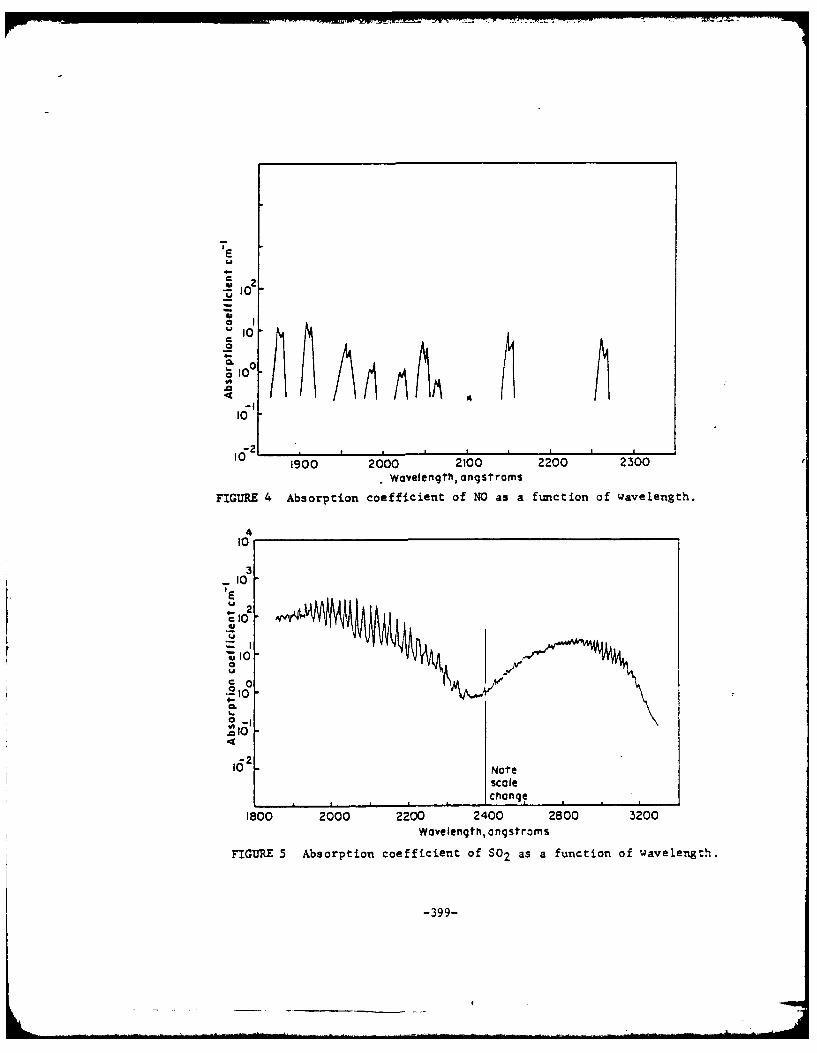
0.CL
oO.10
1900 2000 2100 2200 2300. Wavelength, angstroms
FIGURE 4 Absorption coefficient of NO as a function of wavelength.
410
'I
10
-2
0
N o t
108 100 2000 21 200 200 2300Wavelength, nqsroms
FIGRE 4 Absorption coefficient of 802 as a fuction of wavelenh.0
-Z 10
4161
162 Note
1-0 200 20 2400 2800 3'200
Wavelength, angstroms
FIGURE 5 Absorption coefficient of S02 as a function of wavelength.
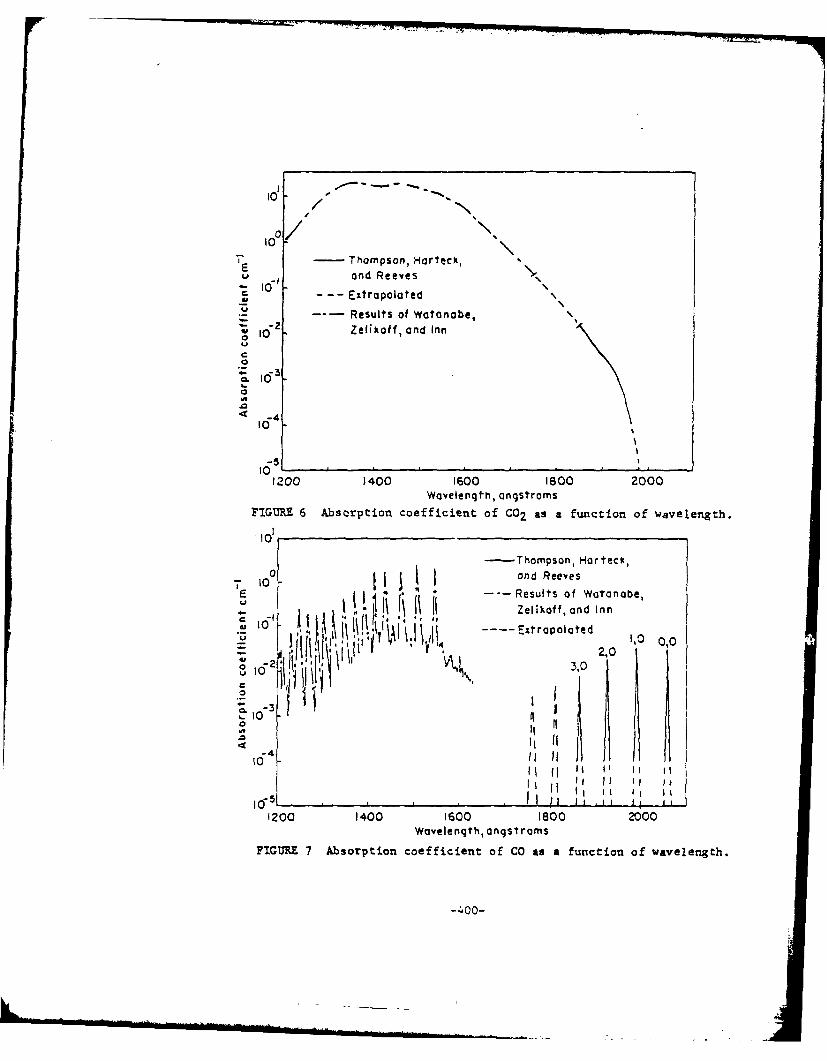
1 / -
10
7 - Thompson, Harteck, "U ond Reeves€ I0" Extrapolated
• --- Results of Watanobe,i 1 Zelikoff, and Inn
C3
i0-1.
10 0adRee
10,
1200 1400 1600 1800 2000
" 10-
-10 II
Wdvelenth, ngsroms
FIGIM 7 Absorption coefficient of CO2 as a function of wavelength.
10 *,Zeflkoff, and Inn
I T , :'\d~ I ' ,~ ;jP 1 ,0
o VL.
- 4 II I.~l0II
lI II lI
11 II II I '
-"0) Ii ?l Ig ,...I1200 1400 1600 1800 2000
Wavelength, angst rams
FIGURE 7 Absorption coefficient of CO as a function of wavelength.
-400-
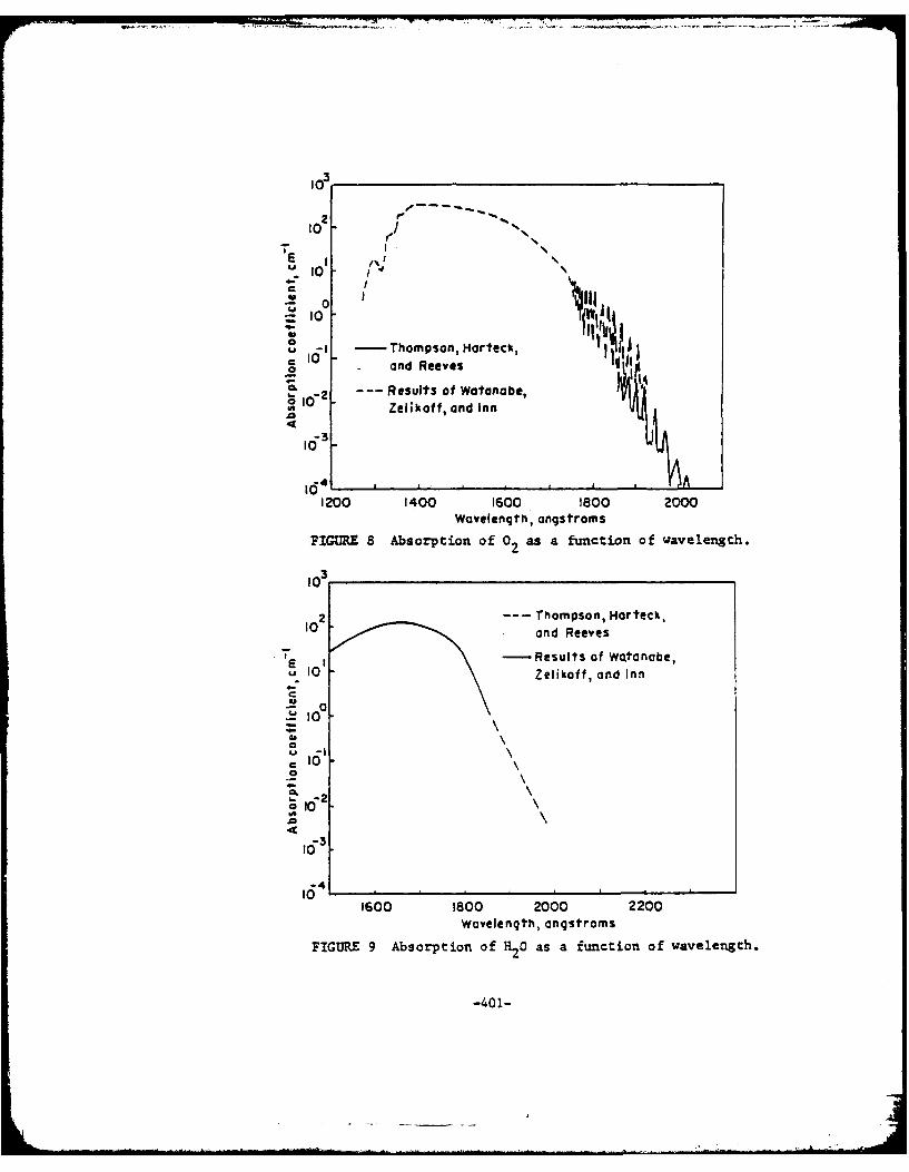
1 3
102
10%
0
161_I -Thompson, Horteck,-- and Reeves t
C" -- Results of Watanabe,le
16-2-z Zelikof f, and Inn , iU* 4 I , I I ,
1200 140O0 1600 1800 2000
Wavelength" , angstroms
FG=R 8 Absorption of 0 2 as a function of wavelengh.
1o3
12--- Thompson, Hrrceck,
o~an Reeveseve
- -Results of Watanabe,E 10 Zelikoff, and Inn
4
16-3
104
1600 1800 2000 2200
Wovelength, angstroms
FIGURE 9 Absorption of H20 as a function of wavelength.
-401-
l0s

0
oj U
- 0
Cu
oin
00
0 4J
0~ -
loE0 E
in 0
C.
0 -
w 0z,. 9*
E
0 0 0 00ON 0oI- t I T I
-4020

100
-Nq
90
80 /
70 1%J4.0
60 r* I,UV
0 I
5000 4 58
I I I30 1 _ .. oo,
Wave number, cm 1
FIGURE 11 Comparison of the theoretical calculations of the transmittanceof the 1.87-u band with the experimental measurements of Buch et al.The theoretical values have been averaged over::a 20 cm- l interval.
04. I
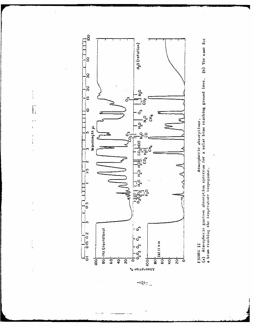
. . .. ..
700
CC
ot
I. CJ
ot
-- 0
CDD 00 0-2D w- 0 li
04

TABLE I
GAS PRESSURES USE FOR ABSORPTION
MEASUREMENTS
(Thompson, Harteck, and Reeves)
Gas Pressure,
C02 700
C0 660
02 720
H2'0 20
N20 326, 2050 - 4000 A.6, 1850 - 2050 A
NH3 760, 2250 - 4000 A100, 2250 - 4000 A
5, 1850 - 2150 A
0.1, 1850 - 2150 A
NO 6*
S02 5, 2000 - 4ooo A0.259, 1850 - 2200 A
CH4 750
* NO was measured at low pressures to avoid
interferences from dimerization.
-405-

0 - Haze C 0' 10
Haze M nir)
102 Haze model M
Cloud I XO.?70F Ax)n(r , r6je.l.r p (9)r O - JIO t
10' 10- .• - + 0 .4 0 5 -
10 10
.1 -- I0
I0- I 10-3 I I I
0.01 0.1 1.0 JOy 0 40 80 120 160r e(deqees)
FIGURE 14 Three size distribution FTGURE 15 Intensityfunctions forfunctions used in the integration haze particles at X0.70 and withof the Mie functions. Total con- real index 1.33 but a differentcentration 100 cm-3. distribution than Fig. . Com-
puted values at 9: 0(2.5)20(10)130(2.5)1800.
10' 0 1 1 1 1 I
Haze moe; 0 Cloud modelI00o - XO.45 .u A-I I00 " rc 41-4 07.r__ ' P(e) X5.3 . A--0.88
47 ". 47"*
lto 0.49
0.474
10- 2 .5 -, - - - 10-2
0 40 80 120 160 0 40 80 120 160
9(degrees) 9(degrees')
FIGURE 16 Integrated and noral- FIGURE 17 Intensity function forized intensity functions PI/ 4f water cloud illuminated by 5 .3 u(solid line) and P2/4v (dashed radiation. Computed values atline)for haze particles with com- 6: 0(2.5)30(10)140(2.5)1800.plex index and infrared illumina-tion. Computed values at e:0(1)5, 10(10)150(5)170(2)1800.
-406-
Variable obserationpoint (R,e)
Source Beam direction
zSmall volume of
scatterers in beam
FIGI=~ 18 Scattering from small volume of icatterers, fixed icatteringAlement and Variable 6bservation Ooint.
\Fixed observation point
eRadialdirection
Sourcee
Variable scatteringelement
FIOIIRE19 Scattering fromi a small volume of scatterers, fixed observationpoint, and variable scattering element.
-407-
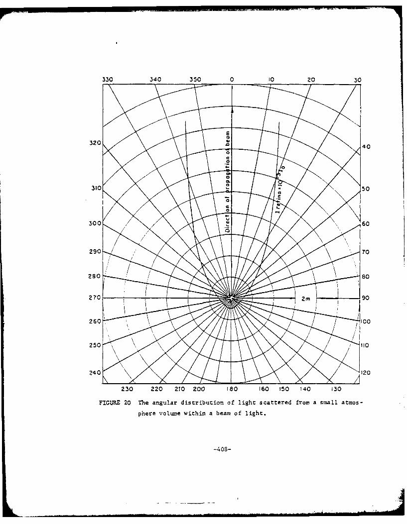
330 340 350 0 10 20 30
250 5
230 2260 00 10 10 15 4 3
phe0 2m~n 9ih0a emoflgt
24-408-
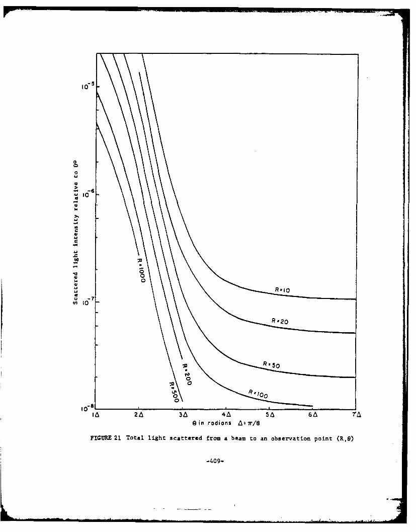
0
0000
0
R 82
-I -60
'J IC60
-409

0
-20
-20 -to 0 10 20 30 40 50Sec of orc, e
Sourcedire ction
FIGURE 22 Beam~ broadening- due to snow: 0.63 ;path length, 2-6 .Data of 2/10 and 2/11/64. 'Excess attenuations in curves 2.-Sare 3-5, 5-0, 1.3-6, 15-6 and 179 dblkrn respectively, correspond-~.ng to increasingly heavy snowfalls. The attenuatiors aremeasured at dead reckoning, that is, with the receiving apetureon the maxin-um of the beam. pattern. (qa0)
(yD.C. 11og8)
-410-

Scattered lightdefinition
Leading edgeof plume
Incident beam -
Observationpoint
FIGURE 23 The scattering of a cylindrical beam by a totally reflectingplu.
-411-
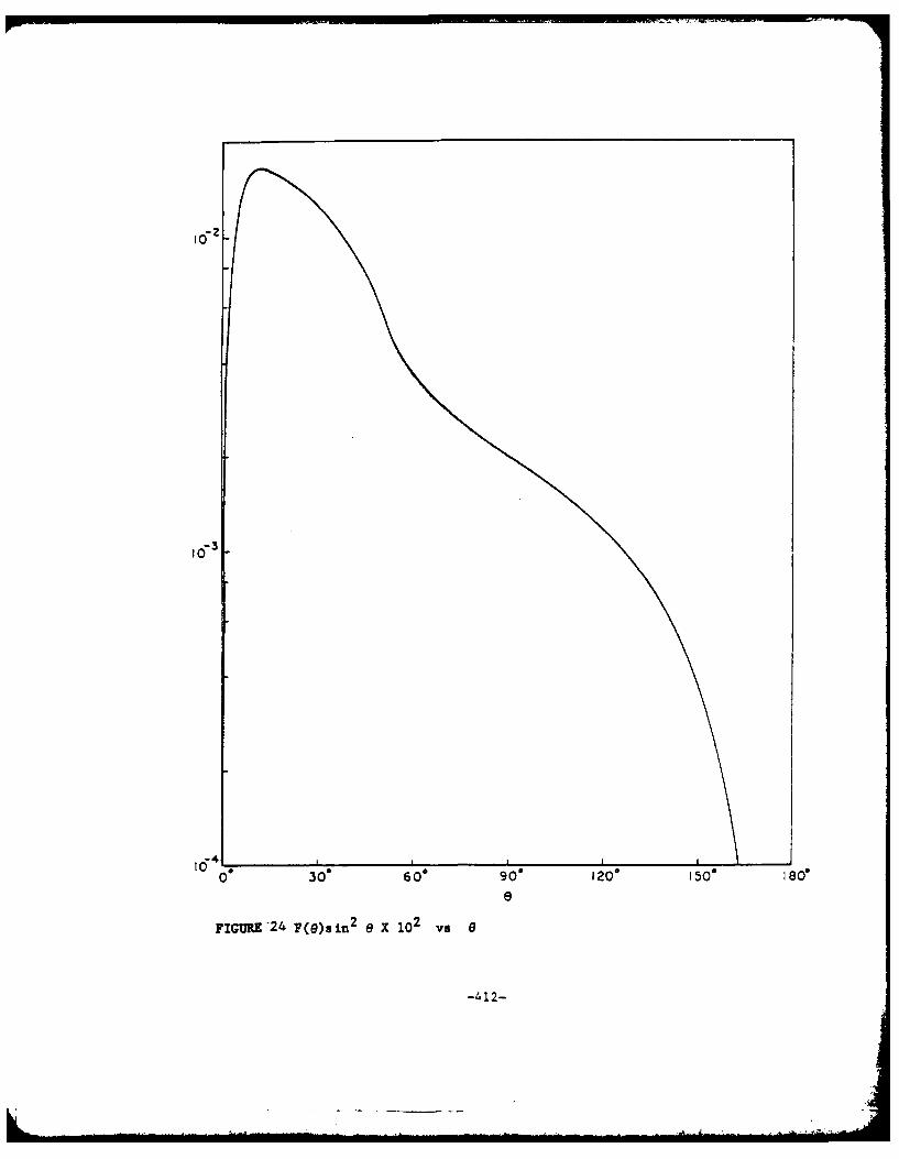
030' 600 90 0 120. 150- 18e
e
FIGURE'2 4 F(e)sin 2 e x 102 vs e
-412-
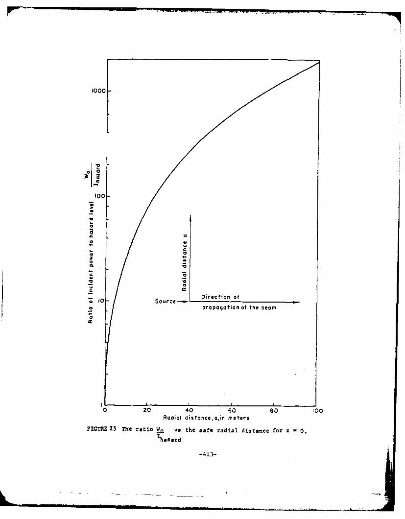
1000-
100-
0
CL
C0
10V
C M
S10- Source-. Direction ofo propagation of the bearn
0 20 40 60 80 100Radial distance, a,in meters
FIGURE 25 The ratio Wa vs the safe radial distance for z -0.
,hfaz ard
-413-

REFERENCES
1. Twersky, V., JOSA 52, 145-171 (1962)
1(a) Van der Hulst, Light Scattering by Small Particles, John Wiley-London (1957)
2. Van der Hulst, Light Scattering by Small Particles, John Wiley-London (1957)
3. Unpublished data
4. Thompson, B.A., et al., J. Geophys. Res. 68, 6431 (1963)
5. Green, A.E.S., Aoplied Optics 3, 203 (1964)
6. Wyatt, P.J., et al., Applied Optics 3, 229 (1964)
7. "The Infrared Absorption of Carbon Dioxide" - Report SSD-TDR-62-127, Vol. III,
Space Systems Division, Air Force Systems Com ,and, Los Angeles, Calif. (1963)
8. "The Infrared Absorption of Water" - Report SSD-TDR-62-127, Vol. II, Space
Systems Division, Air Force Systems Command, Los Angeles, Calif. (1963)
9. Wyatt, P.J., et al., JOSA 52, 1209 (1964)
10. Burch, D.E., et al., "Infrared Absorption of Carbon Dioxide, Water V1-or, and
Minor Atmospheric Constituents" - AF 191604, 2633, Ohio State Uhiversity Inc.,
(December 1960)
11. Wyatt, P.J., et al., Applied Optics 3, 229 (1964)
12. Stull, V.R., et al., Aoplied Optics, 243 (1964)
13. Goody, R.M., Atmospheric Radiation, Oxford Clarendon Press (1964)
14. Green, A.E.S., Aoolied Optics 3, 203 (1964)
15. Gibson, G., Laughridge, F.I., Nichols, J.R., Drause, N.A., JOSA 52, 38 (1962)
16. Khostrick, et al., JOSA 52, 416 (1962)
17. Peck, E.A., Khanna, B.N., JOSA 52, 1010 (1962)
18. Van der Hulst, Light Scattering by Small Particles, John Wiley-London (1957)
19. Gumprecht, R.O., et al., JOSA 42, 226 (1952)
20. Hulbert, C.O., JOSA 31, 467 (1941)
21. Reege, E., Siedentopf, H., Optik 1, 15 (1946)
22. Bulrich, K., Moller, F., Optik 2, 301, (1947)
23. Deirmendjian, D., Applied Optics 3, 187 (1964)
24. Deirmendjian, D., Applied Optics 3, 187 (1964)
25. Khrgian, A., Mazin, I.P., Tr. Tsentr. Aerolog. Observ. 7, 56 (1952)
26. Khrgian, A., Mazin, I.P., Tr. Tsentr. Aeroloq. Observ. 7, 36 (1956)
27. Mie, G., Ann Physik 25, 377 (1908)
28. Van der Hulst, Light Scattering by Small Particles, John Wiley-London (1957)
29. Reege, E., Siedentopf, H., Optik 1, 15 (1946)
30. See the section of this report titled "Laser Eye Protection"
-414-
31. Knestrick, G.L., et al., JOSA 52, 1010 (1962)
32. Eldridge, R.G., Johnson, J.C., JOSA 52, 7, 787
33. See section of this report titled "Laser Eye Protection"
34. Hogg, D.C., Nature 203, 396 (1964)
35. Hogg, D.C., Nature 203, 396 (1964)
36. Knestrick, et al., JOSA 52, 1010 (1962)
37. Edwards, B.N., Stean, R.R., Aplied Optics 4, 331 (1965)
38. Bennet, H.E., Portens, J.O., JOSA 51, 123 (1961)
39. Morin, T.J., Peacock, G.R., "Microwave and Optical Plasma Diagnostics"
AFCRL-64-903.
40. Southhall, P., Introduction to Physiological Ootics, Oxford University Press -
London (1937)
41. Southhall, P., Introduction to Physiological Otics,Oxford University Press
London (1937)
42. Reege, E., Siedentopf, H., Optik 1, 15 (1946)
-415-

Nonlinear Effects - ?ossible 1ih 1htensitv UV Source
Franken and Ward (1) have postLla:ed a schematic representation of
otical polarization as an illustration of the mechanism for second ha-monic
generation. The optical polarization as a function of applied electric field
is given by
XE (l+aI Ea --1 2
where , is the normal linear optical ?olariZation (of order uniZ;) is the
applied elactric field and aj (i=!,2,3,n) are nonlinear coefficients. If
the appliad field is Z = E si. wa the cuadradic term in the above expansion,0
- 2 . 2 _2p('w), will be p.2Zi) = xZ . '-- = xc. /2) (1-cos 2wt).-0
Since -a second harmonic power is proportional o p 2(2w)(la) it
follows that
p2 '24 .
0
or :. s. . hrm..on-c power da-ity is ?ropor:iona :o :he square of the
.. tan er.siy. Since a. is a very snai, apreci--ble second
h.--....c generation will occur only at high incident nower der-sieas.
.heories o second har.onic generation (2,3) indicate that only structures
lac::i.4 i.-ersion syetries will result in second harmonic generation.
Hawaver, effects of molecular interaction on local s.etry has been
obzerved (4) to result in frequency doublir from molecules which would
not othez,ise support the mechanism.
Second harmonic generation in liquids has been reported by Terhune
et al (5). A 1 megawatt peak power, 80 nsec. Q-switched ruby laser outpuz
416
-_ -
LI

2
was focused in H2 0, C Cl4 and CH3CN which had been filtered through 5.g
millipore filters. It was reported that about 10"13 the incident energy
emerged as scattered radiation near twice the laser frequency. From the
diagram of the arrangement of equipment used in this experiment it is con-
cluded that the second harmonic signal measured was the power radiated to
90 - 20 from the direction of propagation of the incident beam. No infor-
mation was given concerning second harmonic generation at other angles, and
it was not indicated how the measured value and the ratio of second harmonic
energy to incident energy were related. Since the total second harmonic
generation appears to be low, only minimal hazard is anticipated from
second harmonic generation in liquids unless more efficient conversion
efficiencies are found.
-% Conversion efficiencies in crystals can be much greater than those
in liquids, provided the crystals lack an inversion symmetry and possess
equal refractive indicies at fundamental and second harmonic frequencies
(6,7). The latter condition is met by proper orientation of crystals as
described by Giordmaine (8) and Maker et-al (9).
P.D. Maker et al (10) have observed that by focusing incident radi-
ation a gain of 3X0 3 in second harmonic intensity may be realized over the
unfocused value. It is estimated that this gain was composed of a factor
of 100 due to increased power density and a factor of 30 due to mixing
(beating) of divergent rays. Consequently one may expect increased con-
version efficiency under focused conditions, greater than that expected
from the increased energy density. Upon application of these principles,
conversion efficiencies as high as 20% have been achieved by focusing a
Q-switched ruby laser within a properly oriented block of KH2PO4(KDP) (11).
417

3
Efficient second harmonic generation in other crystals such as Li Nb 03 (12)
and KD2P04 (13) has also been observed.
Johnson (14) has produced ultraviolet radiation (2650 2) from infrared
(1.064) by quadruplying the frequency of a Q-switched 10 megawatt Neodymium-
in-glaSs laser using two KDP crystals in sucession. The conversion effiai-
encies were 27. from the IR (1.06L) to visible (5300 1-) and 1% from the visible
to UV (2650 1) resulting in a combined efficiency of 0.02%. Johnson points
out that the experiment was performed with the incident laser beam unfocused
to prevent damage to the KDP crystal. He estimated that had the beam been
focused, conversion efficiency at the first crystal would have been approxi-
mately 20% and the resulting increase in incident radiation at the second
crystal would have increased its efficiency also. The ultraviolet power
level at 2650 X, would consequently have been raised from the observed level,
2.4 Kw, to approximately 100 KU (15).
Efficient second harmonic generation in optically active amino acids
has been reported by Rieckhoff and Peticolas (16). The ultraviolet signals
were established as a second harmonic rather than some other luminescence
by accurate determination of spectral content. A very narrow line at the
second harmonic frequency was interpreted as sufficient evidence of second
harmonic generation rather than florescence. A 10 megawatt Q-switched ruby
laser (6943 X) was focused 7cm behind the sample to be tested with a 15 cm
focal length lens resulting in roughly 40 megawatts/cm2 at the sample. The
samples were in the form of a layer of powder Imm thick, enclosed between
a microscope glass slide and a cover glass. The authors indicated that the
conversion efficiency of the amino acids tested were as much as three and
four times that of KDP. This statement is highly questionable since the
conversion efficiency of KDP can be as high as 20%. The authors indicated
418 4

4
that particular acids may be capable of supporting the mechanism when in
the form commonly found in cells . No experimental work of this nature was
reported however.
Another nonlinear mechanism, Raman scattering, results in secondary
emission at frequencies other than harmonics of the incident radiation. Fol-
lowing interaction of a quantum of light energy, hv $with an atom of molecule
of energy E , the atom or molecule is excited to state E2 . If it returns to
its original state with emission of a photon the energy of that photon is
L hvand Rayleigh scattering is produced. If it returns to a state of other
than the original, E then emitted photon will have energy hv - E2-E When
E1 Eo, hVl< hv and the energy and frequency of the emitted photon are less
than those of the incident photon. If E Eo, hv> hv and the emitted
photon is of higher frequency and energy than the incident photon. The
former case is referred to as Raman Stokes radiation and the latter, Raman
anti-S tokes Radiation.
Most Raman scattering results from spontaneous emission in which case
the scattered radiation will be incoherent and diffuse. However, if the
Raman radiation is present in sufficient intensity or if the Raman and
exciting radiation are incident on the medium simultaneously, stimulated
Raman scattering will occur. A detailed review of both experimental and
theoretical work concerned with Raman scattering has been compiled by
Stoicheff (17) and detailed theory of stimulated Raman scatter is discussed
by Garmire et al (18) and Javan (19).
Some of the interesting properties of stimulated Stokes-Raman scatter-
ing have been reviewed by Eckhardt et al (20). They report an apparent
threshold for stimulated Raman radiation production. The resulting radi-
ation is coherent, collimated in the direction of the incident beam and is
419
5
contained in a series of narrow spectral lines. The intensity of the
stimulated radiation was reported to be as high as 107. the incident radi-
ation. Stimulated anti-Stokes-Raman radiation has been reported by Terhune
(21) and Stoicheff (22). The observed stimulated anti-Stokes radiation
was of intensity comparable to that of the incident beam and was confined
to cones 10 min (0.1570) wide within a few degrees of the forward direction.
Several orders of anti-Stokes radiation were observed, all of narrow line
width. As with stimulated Stokes-Raman scattering there was an apparent
threshold for stimulated anti-Stokes radiation.
Hazards may arise from nonlinear effects in both experimental targets
and biological material. Rieckhoff and Peticolas (23) have noted that
ultraviolet radiation generated due to nonlinear effects at specific sites
in biological substances may be absorbed by surrounding constituents.
Consequently it is concluded that media which are normally optically inactive
at the frequency of incident radiation may be effected when nonlinear effects
are significant.
Production of ultraviolet radiation due to nonlinear and Raman effects
in experimental targets could possibly present a hazard to exposed surfaces
of observers. This is due to the fact that the absorption spectra for bio-
logical media a abs (X), including the superficial layer of the eye, tends
to increase in the near ultraviolet.
By dissection of a rabbit eye and reassembling the various components
in quartz envelopes in a manner such as to duplicate their original geometry,
Kinsey (24) attempted to measure the absorption characteristics of ocular
420

6
constituents in the ultraviolet region. He concluded from his measurements
that almost all of the light (in the UV) incident on a whole rabbit eye
which is not reflected will be absorbed before reaching the retina. No
information was reported concerning the fraction of incident light that
was reflected. Absorption measurements made with this technique may not
necessarily yeild information concerning the fraction of incident radi-
ation absorbed in the different parts of the reconstructed globe. Kinsey
observed that abosrption in the aqueous and vitreous humor, and in the
[ cornea, peaks at 3000 1 while the corneal epithelium absorbs most strongly
at 2900
Veroeff and Bell (25) have noted that due to high ultraviolet absorp-
tion of the anterior part of the eye adverse effects will depend on the
total energy density at the surface from all angles rather than the amount
of incident from specific angles as in the case in the visible region.
Consequently, in contrast to visible radiation, the effects of a distributed
source of radiation are much the same as those of a point source.
The threshold for damage to the corneal epithelium was found by
2Cogan and Kinsey (26) to be approximately 0.01 joule/cm at peak sensiti-
vity (2800 r) while Verhoeff and Bell (27) found that the threshold for
nor=onochroatic ultraviolet light was roughly ten times that required at
peak sensitivity. Unfortunately no reference was made to time dependence
in either study (or spectral distribution in the study of Verhoeff and Bell).
Although damage at theselevels is to a great extent reversible, one may
consider 0.01 joules/cm2 a hazard level since at least temporary visual
impairment will be associated with the resulting disturbances.
Ocular shielding which will allow safe observation of experiments
becomes extremely difficult when inelastic (frequency shifted) scattering
42
7
is significant, This is due to the fact that resulting radiation will be
contained at several wavelengths in addition to that of the incident radi-
ation. The optical shield would consequently be required to attenuate light
only at a number of discrete frequencies.
422

REFERENCES
la. Corson, D., Lorrain, P., Introduction to Electromaznetic Fields and
Waves, W.H. Freeman and Co., San Francisco (1962)
1. Franken, P.A., Ward, J.F., Review of Modern Physics 35, 23 (1963).
2. Yin-Yan, L., Acta Physica Sinca,20, 164 (1964).
3. Kielich, S., Bulletin De L'Acadeie Polonaise des Sciences XII 53 (1964).
4. Terhune, R.W., Maker, P.D., Savage, C.M., Phys. Rev. Letters 14, 681 (1965).
5. Kielich, S., Bulletin De L'Academie Polonaise des Sciences XII 53 (1964).
6. Terhune, R.W., International Science and Tech., p. 38 (August 1964).
7. Maker, P.D., Tarhune, R.W., Nisenoff, M., Savage, C.M., Phys. Rev. Letters
_8, 21 (1962).
8. Giordaine, J.A., Phys. Rev. Letters 8, 19 (1962).
9. Maker, P.D., Terhune, R.W., Nisenoff, M., Savage, C.M., Phys. Rev. Letters
8, 21 (1962).
10. Maker, P.D., Terhune, R.W., Nisenoff, M., Savage, C.M., Phys. Rev. Letters
8, 21 (1962).
11. Giordmaine, J.A., Scientific American, 210, 38 (1964).
12. Boyd, G.C., Milles, R.C., Nasark, Bond, W.L., Savage, Applied Phys. Letters
, 234 (1964).
13. Van der Ziel, J.P., Bloembegen, N., Phys. Rev., 135 (1964).
14. johnson, F.M., Nature, 204, 985 (1964).
15. Johnson, F.M., Nature, 204, 985 (1964).
16. Riechoff, K.E., Peticolas, W.L., Science, 147, 610 (1964)
17. Stoieheff, B.P., Stimulated Raman Scattering, Division of Pure Physics,
National Research Council, Ottowa.
18. Barmire, E., Pandarese, F., Townes, C.H., Phys. Rev. Letters 11, 160 (1963).
19. Javan, A., Stimulated Raman Scattering, Division of Pure Physics, National
Research Council, Ottowa, p. 284.
20. Eckhardt, G., et al, Phys. Rev. Letters 9, 455 (1962).
423
,,
U,:
21. Terhune, R.W., Bull. Amer. Phys. Soc. 8, 359 (1963).
22. Stoicheff, B.P., Stimulated Raman Scattering, Division of Pure Physics,
National Research Council, Ottowa.
23. Riechoff, K.E., Peticolas, W.L., Science 147, 610 (1964).
24. Kisay, V.E., Arch. Induatrial Hoalth U., 305 (1955).
25. Verhoeff, F.H., Bell, L., Proc. Amer, Acad. Arts and Sciences, Boston (1916).
26. Kinsey, L.E., Cogan, D.G., Drinken, P., J.A.M.A. 123, (October 16, 1943)
27. Boyd, G.D., Milles, R.C., Nasark, Bond, Savage, Apoled Phys. Letters 5,
B234 (1964).
1.2
/4

Gas Lasers
The first sustained, continuous laser output was produced using a gas
mixture, the helium-neon system, which operated in the infrared with an
output power on the order of one milliwatt (1). In 1962, a review paper
by Bennett (2) listed some fifty-three gas laser wavelengths in pure gases
and mixtures. Laser transitions were identified in neon, cesium, oxygen,
helium, argon, krypton, and xenon. Today, over 500 transitions have been
observed in various gases and vapors and it seems likely that this number
will increase significantly over the next three years.
The high coherence properties, directionality, stability, and range
of wavelengths of gas and vapor lasers suggest many applications. In
addition to the improved optical instruments that can be fashioned by and
with them, gas lasers have many potential uses in communications, range
-finding, plasma diagnostics and optics. In fact, the gas laser comes
closest to exhibiting all the properties of an ideal laser. Although peak
powers do not approach those attained with the ruby or neodymium systems,
the gas laser is widely used in research and teaching.
The present state of the art of gas lasers is summed up very well in
a recent paper by Bennett (3). At present more than 500 lines are known to
exhibit laser action either continuously or under pulsed exitation. Laser
wavelengths range from the ultraviolet (neon 2678.8A) to the infrared
(HCN 337 microns). Power outputs ma7 be: continuous, from a fraction of a
milliwatt:to almost 20 watts (ionized argon); pulsed, 20-40 nanoseconds,
up to 100 watts (nitrogen); quasi-C, several watts for several milli-
second pulses in argon. Lasing action has been observed in the following
gases and vapors, over the wavelength ranges indicated (3).
425

-2-
1. Argon: 86 lines 2753 A-26.95u(strong lines from 3511-5145A)
2. Bromine: 4 lines in region of 8446A
3. Carbon: 9 lines 4647 A-5.5956u
4i. Cesium: 2 lines 3.204u and 7.1821u
5. Chlorine: U1 lines 4781 A-2.20603u
6. Helium: 2 lines 1.9543u and 2.0603u
7. Iodine: 8 lines 5407.4 A-3.431u
8. Krypton: 59 lines 3050 A-7.0565u
9. Mercury: 25 lines 4797 A-l.813u
10. Neon: 155 lines 2678.6A-132.8ur(line at 3.39u has gainh dS/meter)
11. Nitrogen: 9 lines 3478.7A-l.4547u
12. Oxygen: 13 lines 2984.6 A-8446.37A
13. Sulfur: 2 lines 1.0455u and 1.0636u
14. Xenon: 64 lines 2983.8k - 18.5u(line at 3.507u has highestgain 60 dB/meter)
15. CO three visible bands 5590.6 A-6613.Su(Carbon Monoxide)
16. CO2 two bands 9u and lOu(Carbon dioxide)
17. D20 16 unidentified transi- from 33.9u-107.7u
(Deuterium Oxide) tions
18. N2 4 bands 8683.5A-1.2347u
19. Ammonia 7 wavelengths 21.47u-31.95u(NH2, NH3, NH)
20. H20 (water vapor (32 unidentified transitions)
21. N20 (Nitrous Oxide) one band region of l0.8u
22. HCN (Hydrogen Cyanide) region of 337 microns
426
L "III. .. I ...li....I~i .. .. .

-3-
Hazards
The dispersion or 'beam spread of the laser beam from gas lasers is
axtremely small, charac-teristically in the range of ten milliradians or less
(see Fig. 1). Thus mamy gas lasers can be hazardous to the eye. Retinal
damage thresholds for v-Lsible gas laser radiation have not been firmly
established at any wave.ength. However, it seems reasonable to assume
approximately 1. Joule/€= 2 as a hazard threshold at a diffraction limited
retinal spot of the ordier of lOOu2 , based on the comprehensive studies of
Ham and Geerats (8,9),. In this approximation a 1llliwatt laser will produce
a radiant power density" at the retina of 1.0 watts/cm2 which will deliver
a total energy density Of 1 joule/cer 2 in 10- 3 seconds, or the apparent
threshold for damage. This also shows that the low power lasers of 100
microwatt power output :ay be hazardous in that a retinal lesion could
possibly be caused by a. 10 milli-second exposure, a time short compared to
the blink reaction time which is of the order of 100 mill±-seconds (10).
The exact thresholds for optical retinal damage with gas lasers and other
lasers are far from established. Recent studies i~ndicate that retinal
irreversible injury thresholds for pulsed irradiation may be as low as
0.14 joules/m 2 , non-Q-switched raby at 69h3 A (11), and as low as
0.07 joules/=m2 , Q-switched. Further 3tudies -n this area is needed
since the number of applications of low power gas lasers is increasing.
Both irreversible and reversible eye damage thresholds for infrared and
ultraviolet lasers also have not been established, although liminal dosage
for the onset of conjunctivitis due to non-laser ultraviolet wavelengths
has been studied as is pointed out in the section on Flash Lamps. The baz-
ards here may be to regions of the eye other than the retina and con-
junctiva depending on the absorption characteristics of the various
427
-Li_ -- . . .r4.

regions.
There is some specific information available concerning skin erythema
and its production by ultraviolet radiation of different wavelengths (12,13).
This in itself is a distinct hazard, usually reversible, but can be painful.
The possible carcinogenic effects of such radiation consequently cannot be
overlooked as a long term hazard. The threshold of ultraviolet erythema
has been established at various wavelengths (12,13), and at the most sens-
itive wavelength (Table-Z ), 2967 A, is 430 microwatts/cm2 for 2 minutes
[I exposure. Extrapolating under the assumption of reciprocity (cuzmlative
exposure-energy-density), this threshold may be stated as 5.16 X 10²
jou.les/cm2 . Thus even milliwatt continuous outputs of any of the lasers
listed as producing wavelengths shorther than 3000 A can present serious
erythemal, and possible carcinogenic, hazards. For example, at the 3002 A.
line in Argon II, a one milliwatt laser output, focused to a 0.1 mm spot
would deliver 10 watts/m 2 to the region. Using an erythemal effectiveness
of 50% (Table -I[), a one-tenth second exposure should produce a perceptible
erythemal response on normal untanned skin.
Perhaps the next most important hazard of gas lasers is the fact that
users are not properly warned concerning the potential hazard of viewing
even a ten times attenuated one milliwatt He-Ne beam. The relatively low
power output of small gas lasers does not suggest the potential hazard than
can exist..
Another hazard associated with gas lasers is the reflected beam from the
discharge tube windows. Most gas lasers have discharge tubes terminated at
the ends by Brewster angle windows. The windows are set at a specified angle,
the Brewster angle, to the discharge tube axis (about 3V), in order that the
-5-
laser amplifies only a silected polarization (See section on Polarization).
In open structures, where the Brewster angle windows are not shielded, a
certain fraction of the beam is reflected at both windows out of the optical
cavity at an angle of approximately 70 to the beam axis. The presence of
these reflected be.ams, nearly orthogonal to the laser axis, of lower power
than the main laser beam can increase the potential hazards in the laboratory.
However, proper shielding can eliminate this hazard.
Other hazards to be considered with respect to personnel are: back-
scatter radiation from targets, possible carcinogenic effects of ultraviolet
radiation from both direct short wavelength laser irradiation and radiation
emitted from unshielded quartz discharge tubes. This latter radiation hazard,
unevaluated as yet, may be particularly significant in the high repetition
rate pulsed gas lasers and ionized gas lasers where high density plasmas are
present. However, plasmas are also present in most other gas laser dis-
cnarge tubes. The He-Ne gas laser in particular ordinarily uses a quartz
or vycor tube discharge tube and in most commercial units is, at best, only
partially shielded. Since quartz and vycor transmit ultraviolet with wave-
lengths as short as 2000 A, ultraviolet radiation is present, but is, as yet,
unevaluated with respect to intensity and spectral distribution. The spectral
distribution of ultraviolet radiation is important in skin erythema considera-
tions and may have similar importance in ophthalmological considerations.
Table I shows the relative effectiveness for the production of skin er7thema
with ultraviolet radiation (12).
Ultraviolet gas lasers should be shielded since prolonged exposure to
such radiation from continuously operating discharge tubes may be hazardous.
Gas Laser Potential for Next Three Years
The estimates made by Bennett (2) in 1962 when compared to his recent
-29
• . . ... .. .. . . . " . . . . . .. . . i l
review of the state of the art of gas lasers (3) show a remarkable change
in the field. His estimates of power to be achieved have been exceeded by
almost an order of magnitude. The additional laser transitions found in the
last three years is a full order of magnitude increase from 1962. In short,
the whole area of gas lasers has expanded very rapidly. Two significant
contributions to the expansio" are the non-steady state inversions attained
in fast pulsed discharges, and the increased powers attainable in ionized
gas lasers (4,5,6).
Most gas lasers are four level systems. That is, a population inversionF
is maintained between two levels, #2 and #1. The upper level, #2, undergoes
stimulated emission to the lowr state #1. State 5 is depopulated by
spontaneous emission, not to the ground state, but to an intermediate, long
lived, metastable state #3. Before the atom can again contribute to laser
action it must return to the ground state #4, where it can be repumped to
state #2. The fact that the intermediate state #3 is metastable hinders the
decay of this state. Processes must come into play which can cause an atom
in the metastable state to return to state #. in these ci4cumstances the
population inversion density versus pumping rae can saturate or even decrease.
This so called =etastable "bottle-neck" has prevented -ost lasers from achiev-
ing higher power outputs or more efficient operating corditions. The ionized
gas laser seems to be the first step in overcoming this metastable drawback.
Lithough efficiency is low, 0.010%, no distinct saturation lixit on pumping
rate versus mower output has yet been observed. it seems readily apparent
that ion lasers will increase their power outputs by one or two orders of
magnitude in the next three years, achieving power leve&.s of 100 watts to
I kilowatt continuous. However, efficiency may poss-ib1y be increased by a
430

-7-
factor of 10 or 100 -- ich would still be a low efficiency, relative to semi-
conductor lasers.
In addition to .he ion lasers, a new class of lasers has been proposed
(3,7) called collisio= lasers, wherein the metastable problem can be overcome
by playing various atomic collision processes against one another. According
to Bennett (3), power outputs at a few wa~ts/M2 per unit Lasing volume at
50% efficiency is not an unreasonable figure to consider as possible. Thi
could possibly permit power levels of the order of 10 to 100 watts contin-
uous to be achieved with this system. Gould (7) also discusses potential
designs of gas lasers with efficiencies of 20% or better. He also suggests
the possibility of multijoule pulsed gas lasers operating in the infrared.
It is indeed very likely that high power outout gas lasers operating in
the infrared will become available in the next few years. This potential
is further enhanced by the recent operation of a 16 watt, C02 N laser at 40%
efficiency in the region of 10 microns by Patel (14).
I-

TABLE I
The relative spectral erythemic of the untanned human skin, of averagepigmentation, to equal amounts of radiant energy. (From Coblentz)
Wavelength (Angstroms) Resvonse
2400 0.56
2500 0.57
2600 0.42
2700 0.14
2804 0.06
2900 0.31
2950 0.98
2967 1.00
3000 0.83
3100 0.11
3200 0.005
3300 0.000
432

- 9-
1. Javan A., et. al., Phys. Rev. Letters, 6 (3): 206, !961
2. Bennett, W.R., Jr., Applied Optics - Optical MAser Suppl., p.2h, 1962
3. Bennett, W.R., Jr., Applied Optics, Chemical Laser Suppl., P.3, 1965
4. Gordon, Ea.l., et. al., Appl. Physics Lett., 4 (10): 17d, 196L
5. Bennett, W.I., Jr., et. al., Acol. Phys. Lett., (10)1 180, 1964
6. Bridges, W.3., Appl. Phys. Lett., _4 (2): 128, 1964
7. Gould, Anl. Optics - Chemical Laser Supply, p. 59, 1965
8. H1am, W.T., Geeraets, W.J., et. al., "Electronically Pulsed Light
Sources for the Production of Retinal Burrs", Amer. J. of Med.
Electronics, Vol. 2 #4, 30b, 1963
9. Geeraets, W.J., .am, W.T., et. al., "Laser versus Light Coagulator:
A funduscopic and histologic study of chorioretinal injuy as a
function of exposure time", ?ed. Proc., Vol. 2L #1, part ii7, S-48, 1965
10. Christner, C.R., eat al, "State-of-the-Art Study on Visual impairment
by High-Intensity Flash of Visible, Infrared, or Ultraviolet Light",
Report No. BAT-171-9, Batelle Memorial institute, Jan, 1965
11. jones, A.E., IcCartney, A.J., "Pulsed ruby Laser Effects on Primate
Ocular Structures", Biomedical laser Conference, Boston, June, 1965
12. Luckiesh, M., Taylor, A..H., "Ezrthemal and Tanning Zffectiveness of
Ultraviolet Energy", 0. S. Review, p. 27h, June 1939
13. Coblentz, W.W., Stair, R., Hogue, J.M., "The Spectral Z-ythemic Raaction
of the Untanned Human Skin to Ultraviolet Radiation", U.S. 3ur. of
Standards Journal of Research, Vol. 8, p. 541, 1932
14. The Laser Letter, Vol. 2, No. 11, p. 2, June 30, 1965
433

andmirror
Angular divergen~ce of laser beam
P.4#22
(a) Parallel end windows
Main laser beam: = 1 Stray beams s#2
(b) Anti-parallel end windows
Stray beams in gas lasers due to reflectionst from Brewster angle windows
434t(

Flash Lawns - Azocca 4 z- s
The use of flash lamps for punpin, lasers is widespread and historically
4s :he oldest ce thoa of laser excu ation. The considerations which follow
discuss the present state of the art in flash lamps or optical pumping
devices and .ndicates the hazards ssoc*a:ad wizh their use. The carm
flash lainp is rather generic and to bt more specific, the optical pumping
=ethods to be considered here will be:
I. Conventional gas filled tubes, both linear and helical
2. .4n.ular discharge tubes
3. Inductively coupled, aeta pinch discharges
4. Exploding wires.
7. Convencinnal Flash la-ns.
These lac=s, in both linear (including Pi -ecmetry) and helical
shapes are available comercially as very small la=ps, one inch or less
arc length. race at a few joules to cubes with six-fooc arc len6:hs,
capable of loadings in excess of 2C0,0O0 joules. The major manufacturers
cc such tubes are listed 4n Table I.
Characteristically the Lar ,r zubes, six-inch arc length or londer,
,enerate very intense bursts of optical radiation for ti=e durac.ons of
the order of a few milliseconds. The actual maxi=um tube loading per inch
arc length is a function of che discharge time and the tube bore size.
Figure is a graph depicting the blow up ratings of various diameter,
1-.ear tubes and a derating guide for various operating conditions.
435

- Inc.
- - - - . ..
-oa,36g
-3-
The li ghc output from flash lamps has a spectral distribution
governed by parameters such as:
I. .ype of fill (i.e. Xe, A, 02' He, etc.)
2. Fill pressure
3. Discharge characteristics (current density, discharge time, etc.)
An t=portant consideration -n :he use of zhese lanps is the spectral
conversion efficiency. This is a function of the above parameters and
assem~cialy tells how much of the input aleacricz. energy 's converted
co optical radiation at various opcical waveland-hs. Generally the
s~actral iis ribucion of the radi.:.t energy is characterized by lines
sunerimnozad on a cninuc-s batc crounc. There appears to be no coherent
iaaription which, -n simple for., anables one to preare=mine all the
;iof a van flash lamp. Considerable evidence is given by
--shak b)owever and some ganeral oboervations are given. The total
-:nversion efficiency for the conversion of electrical energy to radiated
optical energy in the 12.000 A - 2 000 A inarval tends to a single limit
independent of gas fill under special conditions. This conversion efficiency
increases so=ewhaL with the ato=c weight (the reason most Lubes are
iled with Xenon) of the gas but is nearly the same for gas pressures on
.he order of 100 = Hg, for tube radii of 6-12 = and for the electric
field across the tube about 100 volts per c= (i). As a general rule of
thumb, the overall conversion efficiency in this case is about 50 - 60%
of the inpuz energy (29). The balance of the energy is lost in heating the
tube, vaporizing parts of the tube and electrodes and heating in the external
leads (1,3).
~431

The spectral distribution of the emicted radiation covers the wave-
-0 0lng-th interval from below 12,000 A to more than 2,000 A and is determined
lar;ely by the axium current dansity which flows during the discharge
(1...4,5,6,7,9). At relatively low current censities, below 1,000 amps/
c-, the spectral distribution is przctically uniform across the entire0 0
spectrum from 3000 A to 12,000 A, waizh some prominence in the infrared.
~2As the current density is increased to the region of 2000 amps/cm the
Linfrared begins to saturate. As the energy and current density in the
discharge is increased the spectru= shifts towards the blue as shown in
V'i:ure I. The shift in spectral energy distribution continues further
:c :srds tha violet with saturation of outDut in the visible region occurring
in :he vicinity of 6000 - 7000 a=:ares/cm- '7). Higher current densities
hav been achieved using preinizing techniques (double pulsing) (4,5,6).
At current zensities of 20,000 =.s,'cm2 the spectral distribution is such
0that the ultraviolet (4500 A and shorter) makes up nearly 20% of the output
while the visible (4000 - 7000) is about3O%of the output and the infrared0
(7000 - 12,000 A) about 30. (4,5,6). A problem that occurs repeacedly in
t.ta literature is :"a: no one researcher shows the full spectral distri-
_--on, o- :!-a sh lo studied. The above figures must be regarded
with some caucion tinae they represent an approximate interpretation of
the results of zeveral investigators.
The largest linear tubes that are commercially available in the United
Sates today have a loading capability of 20Q100 joules. Helical tubes are
alzo manufactured with input ratings up to 240,0CO joules (see Table I).
Kt.."ver the maximum tube ratings are given in terms of joules/inch of
438

-5-
vzrious diameter tubes (Fig. 1) with no imiation on length. It is thus
. ite possible to theoretically =anufacture tubes of any energy rating
-zsired. The practical limit in langth appears dictatcd by the application.
In the laser field this appears, za present, to be limited to tubes with
a _ae ledfth of 1.8 r-zers (abour 6 frceC). In 1962 the Russlans reported
c-aratin of .- r:naa: tubes with an arc length of I meter at loadings in
e:-cass of 100,000 joules (1). Their continued work in the area of higher
anargy flash lamps does not seem to be in the open literature.
The most popular type of linear flash lamp is that represented by
-G&o; FX-47, that is, a 13 m bore, 1C c= arc lengrkt , 10,000 joules
I&, "anon filled tube. Typia. pulse operation with this type flash
_Lais a 1oua 10,00 joule input for pulse times between 1 and 3 milliseconds.
7:: thiz :oading, the total radiation energy will be approximately 5,000
ulas. Similarly a 20Q,000 Joule tube will radiate about 100,000 joules
in optical anergy, bazad on a 50 conversion efficiency.
2. Anru:.ar Flash Lamos
Fi-ah lamps based on a discharge in an annular section between ring
aeectrodes have been used in flash photolysis (8) and laser pumping (7).
AnMular lamps have been compared to helical lamps by Keubler and Nelson (9)
and to linear lamps by Church et al (7). A schematic drawing of an annular
lamp is shown in Figure II1. Kuebler reports efficiencies an order of
magnitude below that of helical lamps when the outer wall of the annular
=amp is opaque. Church, on the other hand, with a heavy coating of
r4f-lecive magnesium oxide covering the outer wall of the lamp reports
r,.by laser u=ping efficiencias af 0.3%, which is comparable to the
a::aienzias obtained with linc..r and helical flash lamps.
439 _

3. Theta Pinch Lazms
The recent application of the theta pinch discharge to laser pumping
(10,11,12,13,14,15) has introduced a new concept of optical pumping.
Primarily the theta pinch is an inductively coupled, electrodeless dis-
charge of similar geometry to the annular tube,except that the current
flows circumferencially, wherein relatively high energy electrons excite
resonant atomic transitions which emic the radiations. In this procedure
[the conversion efficiency of pulse energy to radiated energy is very high
and thus the temperature of the light source remains low. Since initial
V gas pressure is low, a few = of ig, and the atoms and ions do. not gain
appreciable kinetic energy , tha shock affects for short discharge times
is not a limiting factor on operation as it is in the case of the much
higher pressure Xenon flash !imps. Thus much faster light flashes can
be obtained.
Order of magnitude conversion efficiencies lie in the range 50% - 90%
of the induced energy. However, due to leakage inductance effects no more
than 50 of the stored electrical energy can be delivered to the tube.
The overa.1 efficiency of the theta pinch tube thus lies in the range
20% - 407 of the initially stored energy.
The output spectral distribution is reasonably flat across the-entire
opcical region, in Argon, at initial stored energies up to 800 joules (14).
There is little information concerning spectral distribution at other
loadings and in other gases, but the suggestion has been advanced that the
spectral distribution may be easily selected by proper choice of gas fill
(13,14,15), and that the ultraviolet predominates at higher loadings.
440
-7-
Theta pinch discharges as short as 2 sec have been used in laser
pumping experiments with loadings up to 800 joules (10,11,12,14). These
lamps were unshielded except for the current induction coil which was
wound around the periphery of the tube. Theta pinch discharges have also
been operated with 30 sec rise times at 1,500,000 joule loading by Kolb
and others (15). These large devices were extensively shielded to prevent
the emitted radiation from entaring the ambient atmosphere. This precaution
was necessary due t9 the very high peak power radiation which, when
absorbed by the ai.- in the viciniy of the tube, created an intense shock-
wave sufficient to destroy the tube. Kolb indicates that at loadings
in excess of 1000 joules the spectral distribution should be enhanced
significantly in zhe ultraviolet, determined mostly by the radiation from
the fast electrons.
4. Exclodina wires.
Various exploding wire light sources have been used in experimental
laser pumping studies at discharge times as short as 8Eseconds and
loadings up to 16,000 joules per wire (16,27). The spectral conversion
efficiency appears to be similar to that of Xenon flash lamps. The work
of Church et al (16) seems to indicate chat the exploding wire light
source is not as well suited for laser pumping as the more conventional
flash lamps.
1ash Lam .Mazar~is.
The most obvious hazards from the intense, transient radiation from
flash lamps and similar devices are irreversible and reversible damage
to the eye (17). A worst case analysis, under certain assumptions, can
441
.. .. " " ....... . . . .... ..... . .* . ... "A

indicate at least the minimum safe viewing distance at which irreversible
retinal damage and reversible external radiation effects may be minimal.
Other flash lamp hazards include ultraviolet erythema and possible carcino-
genic effects, X-ray production, electrical shock hazards, flash lamp
explosion and the possible production of poisonous gasses and vapors.
Optical Radiation Hazards.
Linear flash lamps, and to a good approximation, helical lamps are
essentially line radiation sources. Their radiant power distribution
does not follow the ordinary distribution for a point light source. For
a line source the radiation patte -.- is essentially toroidal (17) as shown
in Figure TV. At a distance RI from the lamp (R greater than the largest
lamp dimension) the radiation energy varies from a minimum at a = 900.
Thus at R, the surface enery tinezsity, for a la-p energy output of 0
joules is:
1,o0 2 R-2o
with the Lambertian angular dependence at any 9,
,0 = , cos 9
ha optical radiation hazards 'om intense transient light sources
can be best evaluated on a worst case basis. The worst case conditions
to be assumed here are:
442
-9-R
1. The lamp is unshielded.
2. A potential observer is located perpendicular to the tube axisat the mid-point of the tube, that is e = 00
3. The observer is copletely dark adapted with a pupil area of1 cm2 (for retinal dainage)
4. For light focused by the eye the retinal spot size is 100 sq.MrQn2o'.
5. The threshold for retinal damage is I joule/cm 2 and that thisvalue holds for flash times from a few microseconds to afew milliseconds *
6. The observer has normal, white i.nnanned skin (skin erythemahazard)
7. The threshold of LTV skin erythemal reaction is assumed as 0.05joules/cm2
8. The threshold for thie onset of photoinduced ultraviolet conjunc-tivitis is assumed as 0.07 joules/cm2
Under the above approximations the threshold hazard level for retinal
damage, conjunctivitis and s:in ey-thema can be given numerically in
relation to Zlash la-np input energies. Is an additional approximation
the radiation from the flash la---s will be considered to be related to
the flash lamp loading in the following manner.
1. Radiation taking ?art in retinal damage is approximately 507.of Lnput.
2. UV radiation taking part in conjunctiva and skin interaction isapproxi-ately 20. of inuz.
* From the work of .'&a, Geeraets, et al., sun-.arized by Christneret al (17).
"- This threshold has 'een -2.--mlnec at -he maxi um spectral sensi-.ivity for a "ave--;-h Z967 .1 (19,20). This value will
be assumed to hold for all ', radiation in the worst caseapproximation.
S This value is assumed from the work of Kinsey (21) who showedhumans to be , i:e as sensitive as rabbits and from thethreshold value discussed in Duke-Elder (22) for rabbitsat 2360 X - this value will be used, although its validityis questionable.
443

-10-
Threshold hazed distances can be plotted now as a function of flash
lamp inputs for thee three hazards. To repeat, the surface threshold
energy densities a-:
1. Retina. bu-in: 10-6 joulesicm 2 at pupil. ( joula/cm 2 a r nal.spot s:.Te of 100 sq. microns, I cm2 pupil area).
2. Conjunctiv tis: :7'x 10- 2 joules/cm2 at corneal surface
3. Skin eryth-ama: 5 x 10-Z joules/cm2 at skin surface.
The retinal da.-age thrashold versus distance is shown in Figure V.
For example, the =mt co=only used flash lamp (10,000 joule loading)
would have a -hres-.d for damage distance of about 220 meters i flashed
with no shielding.
The skin and e.=terior eye thresholds :or photo reaction :hresholds
are shown in Figure VI. For the 10,000 joule lamp, mentioned before,
perceptible skin erythema or conjunctiva effects could occur at distances
of about one half mater.
Thresholds for infrared damage to the cornea, conjunctiva, iris and
lens should be ccnsidered apart from the integrated intensity ot ultra-
violet effects. There does not however appear to be any literature
detailing :he effects at the longer wavelengths (7000 - 8000 O and longer)
with respects to other established hazard thresholds.
It must be pointed out that in almost all operating laser systems,
the optical pumping laps are enclosed in a shielded cavity. The cavity
is designed specifically to collect as much pumping radiation as possible
and deliver ti to the laser rod. Cavities can be made light tight, but
~444

in practice some pumping radiation usually leaks outside the cavity.
The worst case analysis above serves not only as a guide to viewing
exposed or unenclosed flash lamps, but also as a reinforce ment to the
suggestion of "don't look" when operating optically pumped lasers.
Other ?lash Lamp Hazards
The possibility of ionizing X-rays being produced under normal
flash lamp operation is very low. The electrons in the discharge can
never attain X-ray energies for the relativly long pulse times ordinarily
used and the relatively low voltages (3,000 - 5,000 volts) it seems natural
to expect the presence of X-rays. It appears as if this is not the case
however. Electron energy distributions i plas=as are discussed in
general by Loeb (23) and in the case of theta pinches by Kolb (15). On
a classical, kinetic cheory basis one would expect the highest electron
energies to appear in the lower pressure discharge, namely the theta
pinch. The data of Loeb confirms this. A personal communication with
4r. H.W. Klauer of Kemlite Laboratories Inc., indicates that in Xenon
flash lamps there is no danger of X-rays below 15,000 volts operating
potentials. However, above 15,000 - 20,000 volts he claims there may
be a danger of "soft gamma radiation." Kolb's calculations indicate
maximum electron temperatures (energies) of less than 100 electron volts
in the theta pinch and the work of Feldman seems to be similar (14). Our
own calculations based on a strictly kinetic theory model show maximum
electron energies to be of similar order of magnitude in Xenon discharges
at 20,000 and 40,000 volts. Experimental x-ray measurements by Vitkovitsky
It al (24) on exploding wires at driving voltages up to 320,000 volts
445

- , -]
showed no high energy X-ray production. 'axi=um X-rays observed in the
0
exploding wire studies were of the order of 5,000 electron volts (% - 2.5 A).
X-rays at this wavelength are attenuated rapidly by quartz and glass and
have a relatively short path length in air. Thus, althoagh no study
of possible X-ray production in fl sh !Z=ps has been undertaken, the
evidence clearly indicates chat the X-ray hazard from fash lamps them-
selves is minimal. The major X-ray hazard source associated with flash
tubes appears to be in the high vacuum rectifier tubes used in the
capacitor charging power supplies, for in high vacuum tubes the electrons
Vcan reach an energy, in electron volts, equal to the peak voltage across
the tube, as is the case in ordinary X-ray tubes.
Other potential hazards in flash lamps include:
1. Flashla=p failure produces a sharp acoustical report and flyingquartz fragments which can be very hazardous to unprotectedpersonnel in the in=ediete area.
2. ?oisonous gases can accu=ulate in the laboratory if not properlyventilated. The generation or 03 by ultraviolet irradiarionis well known and the decomposition of CO2 to CO under flashphotolysis has been shown (25).
3. The possibility of neutron production in theta pinch discharges:ue to low atomic weight impurities such as hydrogen, deuteriumand helium nas not been fully explored.
4. Flash lmps operate at several kilovolts or more so that properinsularion and good grounding procedures should be followed.
?otential Flash Tube Developments
Over the next three years no major advance in the state of the art
of flash lamps is to be expected. Present research in :.e doping of Xenon
flash lamp with various ele=ents will probably be continued in an attempt
to modify the output spectral characteristics of the flash (7).
446
1
Q] .QaJZ OUJA
- -13-
There is, as stated previously, no great technical problem in producing
higher energy flash lamps, the arc length and bore size are just increased.
However the trend in laser design seems to be in the utilization of
oscillator-amplifier arrays in which, at present, as many as ten flash
lamps modules have been used to crace haigh energy laser pulses (26).
This would seem to indicate that much larger linear lamps will not
probably be produced.
Helical flash lamps are expected to be produced with capabilities
up to 50OQO joules in the next three years according to Mr. H.W. Klauer
of Kemlite Laboratories, Inc.
The application of high energy theta pinch discharges to laser pumping
does not appear very extensive. Due to the enhanced ultraviolet output
at higher energies and the low overall efficiencies, this source does
not seem as potentially useful for efficient laser work (per~onal communi-
cation with Dr. Buser, Army Signal Corps Laboratories, Fort Yonmouth,
New Jersey).
/
447...
1arak,2.S.,"Sron-Crret ulse~('ak Discharges in Gas,
Used in Pulsed Lighz Sourcaz," Soviet Th-,sics tjspekhi,
:478, 1.962.
Goncz, j.ii., and lawe!!, 3., !'Xanon Flashtube Spectra FX-47 and
_7.-47A," EG&CG Technical Xazorandu= N'o. B-4302 1963.
2.Beack, A. , Erickson, R. , Bra_s, F. , "Dasi,-n and Operation of Xenon
L :~~Tash Tubes" J. 3~.?~s 4:211.3, 1963.
Zett, J.L., Schawlow, A.L., "Enhanced Ultraviolet: Output from Double-
"hz1ad --lash Lzans," A7zlia'd ?hvs. 7etc. 2:11, 1963.
5.Gar.:-, L.:.7. , at "Pasi: Enhzzced UV Radiation f~cm Double-
Sa FlaSh. 7rs" ~.-~s 4:11, .6.
?. ark-, S.W., "Dou~ble Pulse Tec-nique for 172-47 A," EG&G Raport, June
30, 1964.
7. Church-, C.H., ec 2 "High 77.er-y Cca::4i" 2. Lser P,=as," Laser Flash
LL-- Conference, :jafz~ ea:z:'i Tns4ituve, Fabiuary 1964.
2.Claesson, S. , Lindquist, L. , 1Lrkiv fr Kemi 2.2: , 13jZ.
K.Iueb2.er, N.A., Nelson, L S. , "2adiznt aer.ries and :rradiances of
Capacitor Discharge La=Ds," i O.S.A., 51:14.1, 1961
10. Brandewie, R.A. , Hitt, J.S.,2 and Faldr-an, J. M. , "Plasma Pinch
E~ecitation of A Ruby Laser," J. Ionl. Phvs. 34:3415, 1963.
1_'. Haswell, 7-.:. , qitt, J.S. , Feldnan, J.M. , "A High Repetition Rate
LzT 2i Systen," Proc. IEEE, 52:93, 1964.
.2. Fitt, .S. , Tadnan, J. M. , "Parrallel Theta-Pinch ?u~pig of aLaser
Csci2.tor~.~ifir,"Proc. IEEE, 52:616, 1904.
448

-15-
13. Aisenberg, et al, J. Anol. Phys, 25:3625, 1964.
14. Feldman, J.M., "Theta Pinch Lamp," Laser Flash Lamp Conference,
Stanford R.Isearch Institute, 1964.
15. Kolb, A.C., "N.L Research in Theta Pinch and Double Pulsed Lamps,"
Laser Flash Lamp Conference, Stanford Research Institute, 1964.
16. Church, C.H., et al, "Optical Pumping of Lasers Using Exploding
Wires," Westinghouse Research Laboratories Scientific Paper
62-112-259PL.
17. Christner, C.R., et al, "State-of-the-Art Study on Visual Impairment
by High-intensity Flash of Visible, Infrared, or Ultraviolet Light,"
Report No. BAT-171-9, Battelle 1 -orial Institute, Jan. 1965.
18. Uhl, R.J., ILI T., Servo=achanism La'. MXe-orandum No. 7668-T11-3, 1958.
19. Luckiesh, M., Tzylor, ..A., "Erythemal and Tanning Effectiveness of
Ultraviolet Snergy," G.E. Raview, ?. 274, June 1939.
20. Coblentz, W.W., Stair, R., Hogue, J.M.., "The Spectral Erythemic
Reaction of U Ed.a. Skin to Ultraviolet Rzdiacion," U.S.
Buravu of Stand. J. of acs., 8:541, 1932.
21. Kinsey, V.5., Cogan, D.C., and Drinker, P., "Measuring Eye Flash
from Arc Weldia," j., 123:7, 403, 1943.
22. Duke-Elder, S., Textbook of Cohthalmo1biv, Vol Vt, C.V. Mosby Co.,
St. Louis, Mo., 1954.
23. Loeb, L.B., Basic Processes of Gaseous Electronic, Univ. Calif Press,
1960.
24. Vitkovitsky, I.M., et.al,"Exploding Wires as A Source of X-rays,"
Exolodina Wires, Vol 2,(Q1.Y.:Plenum ?ress, 1962) ed.by W.G. Chase
and H.K Moore.
449

-16-
25. Bortner, M.P.., "Actinometric Determination of the Dissociation of
Carbon Dioxide by a Single Flash," JOSA, 50:172, 1959.
26. Dearman, J.R., et al, "Consideration in the Design of High Energy
Neodymium Lasers For A Surgical Device," First Annual Biomedical
Laser Conference, Boston, June 1963.
2. Clark, G.L., Chase, N.C., and Moore, H.I., "High Intensity Pump for
Optical Lasers," Technical Documentary Report No. ASD-TDR-63-651,
WPAAFB, Aug. 1963.
5
i 450

cy.i
ci
II.
o ,
419
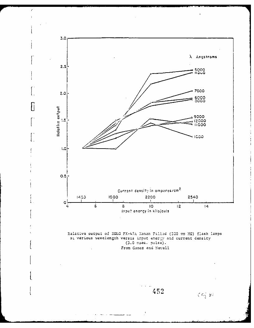
3.0
XAngs~roms
2.5- 00
5000
V 20L 0000
; 479000~200
0.5
Curr.,nt densit,1' in ornpares/cm2
I1403 IS00 2200 2540
4 3 8 10 12 14Input enerny in hilojouls
Rel.ative cutruz of EZG&C FX-47- X anozn 7F-ll-d ('ZOO1 = 'G) flash lampsaL Various wavelengzh versus ir-put ener~'; and cur-rent density
(3.0 =.sec. pulse).
Fro= Goncz an'd e:l
4 5

C
IjJ
C
U
L.
I..oC,
C- - - 0C,e
- IC,- C
-
I., -
3
3
-'3- C~J. -
C,
~iv 4-
o- 3~ *C,
453

;FF
co.--a tolras a-
3: 4 a

a
-4
-4
a
w
-4'-4
S0
as,o
00I
U00
C
0L. -.U 0
%. '~ 45 -as- 0 4-r U 0
0. -4) -
0 0
k ~) a.*~a ~ E00
- U,4) 0 -U - -4
JI~ 0~', .~'
4) 0~0 ~' '-4-
o 'a~V
4) U
U
-4
0
VS..
ino
o 0 --
sJa4aw U! ~UOJ.S!Q
45~
* -- ... ~aZ2$-t -
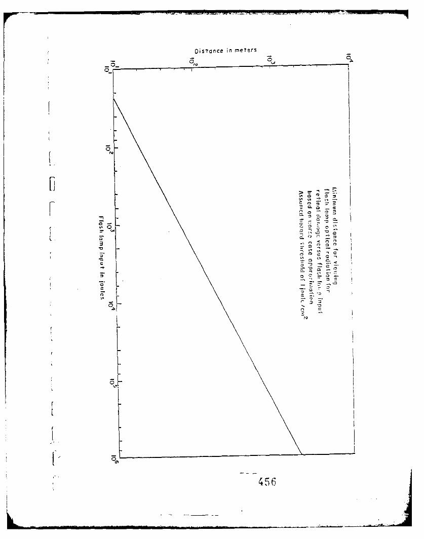
Distance in meer
3N>
V,~
L7
456 ~
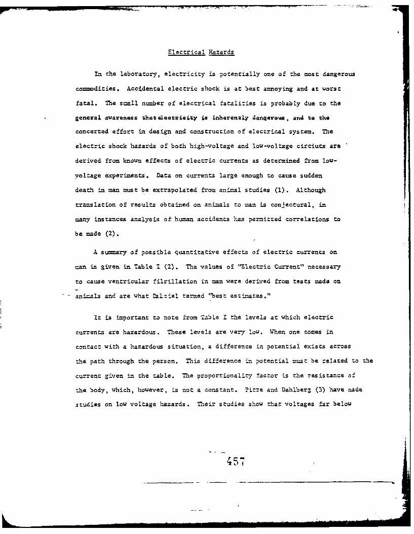
Electrical Hazards
In the laboratory, electricity is potentially one of the most dangerous
commodities. Accidental electric shock is at best annoying and at worst
fatal. The snall number of electrical fatalities is probably due to the
general awareness that eleetetyis i inherenty deangerems , and to the
concerted effort in design and construction of electrical system. The
electric shock hazards of both high-voltage and low-voltage circiuts are
derived from known effects of electric currents as determined from low-
voltage experiments. Data on currents large enough to cause sudden
death in man must be extrapolated from anfmal studies (1). Although
translation of results obtained on animals to man is conjectural, in
many instances analysis of human accidents has permitted correlations to
be made (2).
A summary of possible quantitative effects of electric currents on
man is given in Table I (2). The values of "Electric Current" necessary
to cause ventricular filrillation in man were derived from tests made on
ani=als and are what ralziel termed '"best estimates."
It is important to note from Table I the levels at which electric
currents are hazardous. These levels are very low. When one comes in
contact with a hazardous situation, a difference in potential exists across
the path through the person. This difference in potential must be related to the
current given in the table. The proportionality factor is the resistance of
the body, which, however, is not a constant. Pitre and Dahlberg (3) have made
studies on low voltage hazards. Their studies show that voltages far below
457

2
TAME~ i
Qlz~titative Effects of" Electric Cc-rent on Man
aJ-iaeres (tiousazandths of an a--=ere)
AltermatL'zig 0uz-entMNS 7a~ ., OC
Direct Cw'r~t 60 _CycIe- C'rclesmre~in =Cnn Ma!n Woren M!en WO7r-&-n
No sensation on h~d o .6 0.14~ 0.3 7 5
th-eshold S2 3.5 11 0.7 22 8Shock - no'. vainfull and
=c? I= control not lost 9 6 1.8 1.2 17?P.inf U, shock% -- nainf U, buit
=zculzz control not lost 62 9 6 55 37?aifAl shock - let-go
'--.eshold 76 51 1.6.0 10.5 75 5 0Pa~nfa amd sev~ere s.hock-
~ contractions.br3etbiag dIffiAcult 90 60 2 3 15 9.14 63
?assibole 7Tentricu1 p " fibril-ll-ion f.-o= s:-hort shlocks:
Shock duration 0.03 sec. 1300 1300 1000 10oo 1100 2-1 00Shock dnrtion 3.0 sec. 500 500 2ICO 100 500 5O
ve.ntricu~ar fib -Ut. oncertai..-n death XM2.tiply zb'e 2naat~ ove b7 2 3/L.
to be letha1 , h~o~ ~cs~to~ rs-asceptible n-hase of hezxt c-c2l
possi:0le ventric&2r- fibr-il-
s*hocks:DO shortshocks azd srgdischarges 27.0 ~t-eod
?amrfrcncy shortz~hck ad mfrcency7
oscillatory7 discharg-s 13.5~
458

that produced by the common 110 volt supply have caused fatal electrical
accidents. Fatal shock from low voltages usually occur under conditions
which are conducive to lowering skin resistance at the points of contact.
Working conditions which are particularly hazardous on low voltage
exposures include:
1. wet, humid or damp locations
2. High temperatures, since they are likely to cause perspiration
3. locations where the individual is exposed to grounded surfices.
Yany of the hazards associated with electrical circuits can be
allevia.ted by the use of proper precautions. Some basic cor..iaratious
are:
1. the prover selection of equipment for the type of exposurefor which it is to be used.
2. Installation of the equipment in accordance with establishedsafety standards.
3. Inspections at frequent intervals by competent personnel todevelop a proper maintenance program.
4. Maintenance to keep the equipment and circuits in safe operatingconditions.
The electrical shock hazard can be greatly reduced by any of the
following protective methods, or by a combination of them:
1. Effective grounding of non-current carrying parts.
2. Insulation of exposed metallic surfaces
3. Electrically isolating the operators.
4. Using voltage low enough to minimize the shock hazard.
459
Grounding of non-current-carrying parts of equipment is carried out
to protect personnel against electric shock from metal enclosures or
[frames of motors, transformers, switchboards, portable tools and lamps,
etc., in case shch parts accidentally come in contact with live parts of
r the circuit. Where protective grounding is found to be ispractical,
insulation of the non-current-carzying exposed metal parts may be
-- employed. Such insularion should be made to withstand the abuse expected
under working conditions and should be made resistant to oil, grease, and
moisture.
F W~here neither grounding nor insulation of non-current-carrying parts
' are employed, protection in hazardous exposures can be accomplished by
insulating the operator by the use of rubber mats or shields, or by
wooden platforms or barriers. The purpose is to prevent any return path
for the current through the body of the operator.
Those charged with the resposibility of the safety of the individuals
must carefully survey the electrical exposure, employ proper methods of
control and educate the workers regarding the hazards involved. Safety
personnal should be guided in their responsibilities by regualtions set down
by the National Electric Code (4).
TThitaker (5), and Inship and Watson (6) historically follow the
development of the gounding requirements of the National Electric Code dlbng
with the reasons for each. They also emphasize that the principle method
for grounding portable equipment is through the line~cord which includes
a grounding conductor with a 3-pole American Standard attachment
0L 460I _______________________________

plug cap which has % pair of parallel blades and a round or half-round
grounding blade. There should also be a receptable which establishes
a ground connection to the same electrode as used for the system ground
at the service entra.ce. Alternatively the same cap and receptable may be
used with a cable thzt has an armor or m.etal enclosure of the circuit
conductors with the enclosure being used as the grounding-conductor and at-
tached to the groundi:ng blade. A third method is also mentioned briefly.
This consists of a separate grounding conductor, as a bare or insulated flexible
wire or strap not in the supply cord, used only when part of approved equipment
or by special permission of the enforcing authority. Consequently, in laser
systems the first or second method should be used on-.all equipment to provide
adequate grounding.
Grounding does not always insure the greatest measure of protection
to personnel and, under certain conditions actually may increase the
hazard. The presence of well-grounded metal cabinets as opposed to isolated
metal contributes to the establishment of conditions necessary to cause
shock to persons who touch live current-carrying parts. This may or may
not have a particular application to a particular laser system. An individ-
ual system may require an iiolaced system but in general the grounded
system would be preferable because in this case it affords better protection
for the operators.
Grounding is not intended as a substitute for proper spacings, adequate
dielectric strength tests, or the use of proper insulating materials, but
is a supplozental safety measurc to all these features.
* 461
I ._ ]

r
r
LHcddesheimer (7) discussed plant and building grounds as well as
Fequipment grounds. He lists the grounded Y system as the safest for both
equipment and personnel. As for the establishment of grounds, all steel
frames of buildings must be adequately grounded. -As a minimum a ground
bus of not less than 'To 4.0 Am-erican 1.7ire gauge copper should be run around
the periphery of the building, connected to each outside building column,
and connected to earth at not greater than at 200 foot intervals.
Each water main enterig the building, if Suitable for use as a
grot-ud, should be connected to the ground bus at -:o or more points. In
large buildings the ground bus should be extended to connect to an adequate
number of internal building columns.
The connection to earth and the associated problems of keeping the
"to earth" resistance low are carefully covered by Heddesheimer (7).
Following proper grounding of the laser system, attention can be directed
to another electrical hazard. This is the co".leze and safe discharge
of any and all capacitor bants. The most dangerous situation of all
exist when the laswer system has been operating satisfactorily.. Then the
aser has fired and the capacitor bank has dumped most of its charge, the
capacitor bank is still -ansafe electrically. Referring to Table I, i4-t
can be seen that it is possible for as little as 27 watt-seconds (joules)
to be lethal. It is quite probable that after nor-al firing of the laser
much more than this a=ount of energy remains stored in the capacitor
bank. Since insulation resistance of typical capacitors is extremely high,
capacitors may retain stored charges for days (15). As a safety feature
some method should be provided to completely and safely discharge the bank
6: 462

manually as well as automatically to the point where no energy remains
stored, Calculations are carried out in the foll9wing part of this report to
indicate the necessity of this provision.
Dalziel (8) discusses the hazards of impulse currents. He attempts
to define shock intensity in terms of energy and then projects a hypothesis
that for short shocks, energy is the fundamental criteria, with current
magnitude,. duration and total charge being related quantities of secondary
importance.
These impulse studies were carried out both with oscillatory and
non-oscillatory discharges. It was found that the tolerance to surge
discharges was greater than for oscillatory discharges. This was based
on studies to determine the currents which are Just perceptible on the
hands, where the currents for pure direct current and 60-cycle alternating
current are in the ratio of 4.8 to 1 (10,11).
A comparison of impulse type shocks as opposed to the non-impulse
type is not readily apparent from the literature.
As an example of the hazard due to an impulse shock, the stored energy
(E M .2 ) is plotted as a parameter on a graph of capacity of the bank2
versus the potential across the bank. The curves are straight lines when
plotted on log-log scales. According to the data given by Dalziel (2,8),
any condition to the right or above the 27 joule line (the solid line) may be
considered extremely dangerous. It can be seen that as the capacitor banks
increase in size with a small potential across the bank a dangerous situation
can exist. For example for a 100,000 microfarad bank 25 volts could be
461?

fatal. Smaller banks can present significantly hazardous conditions.
A ruby laser that will deliver 100 joules and is 1. efficient will require
10,000 joules of pumping energy. If the bank is charged to 3,000 volts then
2,2-004f will be required. From the graph one can see that corresponding
to 2200 uf, 160 volts is extremely dangerous. If this laser system,
following firing, does not discharge to significantly less than this value
it must be considered dangerous. This will probably not be the case; 160
volts or more are left on the capacitor bank, and it will remain lethal and
[must be discharged previous to contact.
VThe high-voltage energy storage capacitors chosen for laser useshould be those designed specifically for discharge applications whibh
Icustomarily involve varying cycles of intermittent duty. These capacitors
are usually designed with a higher dielectric stress than is employed forrconventional d.c. capacitors (15). In addition, provision is made to
handle the high discharge current. The design also inclues minimization
of self-inductance to assure short discharge time.
Since most high energy storage capacitors, except the aroclors (12)
(which are also known as Askarels and are chlorinated diphenyls) are
inflammable, fire safety facilities near large capacitor banks should be
provided. When energy storage capacitors fail, resulting large currents
may cause heating followed by combustion. Consequently safety circuitry
designed to remove or monitor faulty capacitors are available.(12) and
should be employed. Since most fuses would add too much inductance to
the system, special fuses must be used in conjumction with protective
I

circuitry. Two such fuses are the silver sand current limiting type and
the exploding wire fuse. When particular safety circuitry is chosen it
should be kept in mind that major bus faults or failures in other parts
of the circuitry may cause undersized fuse operation. This can be
prevented by strategic placement of current limiting resistors. As a
substitute for fuses small lengths of transmission lines with destruction
levels below that contained in the unfaulted capacitor may be placed
between each capacitor and a common bus. A separate transmission line
should be run from each capacitor. Upon failure of a given capacitor, all
the energy in the remaining capacitors will discharge through the trans-
mission line associated with the faulty capacitors and destroy that
transmission line. This will remove the faulted capacitor from the
circuit, and permit operation with reduced capacity. However, it is
uncertain whether this is advantageous .-d disadvantageous from a safety
point of view.
An alternate method of fault protection is a method which monitors
the internal pressure of individual capacitors and relays this information
to a control panel. The impending fault may then be removed either
manually or automatically.
The life expectancy or number of charge-discharge cycles of rapid
discharge capacitors depends strongly on the operating conditions (12,
13, 14). An increase in voltage to as little as 1207. of rated voltage
may drop the life expectancy to 57. of its original value. The discharge
time or ringing frequency also strongly effects capacitor lifetime. When
the ringing frequency also strongly effects capacitor lifetime. When
the ringing frequency is increased from 105 to 10 6 cps the life expectancy

decreases by an order of magnitude. The life expectancy is also affected
by the amount of time the capacitor has been holding a charge, the degree
of voltage reversal and the ambient temperatures. In light of such extreme
conditions it is very advisable to stay well within the equipment ratings.
The capacitor life expectancy, for high energy storage capacitors
quoted by manufacturers varies from 103 to l05 operations. This figure
must be considered in light of the extreme variation in life expectancy
under operational conditions (12,13,14).
[7 Another item that is particularly prone to present safety problemsr are the high voltage current carrying cables. These cables should be
placed so that accidental contact with them cannot occur.
In addition special consideration should be given to the choice of
original cables. The cable chosen should be specified as corona free in
addition to the adequate dielectric strength for the laser with which it
is to be used. This will facilitate future field testing. Although
actual observation of the cable and continuity tests are very important,
the single most important test is the "hi-pot" or dielectric strength test.
These tests can be made with readily available testing apparatus or by
the cable manufacturers testing facility. The dielectric tests will give
an indication of the present strength of the insulation and the corona
test will test for the presence of the corona. If present, this corona
causes ozone which is highly corrosive to some dielectrics and may cause
ultimate failure of the cable. To be on the safe side, a cable that shows
L the presence of corona should be replaced. (*)
No discussion of electrical shock would be complete without mention
of rescueand resiscitation for victims .of serious electric shock accidents.
4
First aid should be restricted to minimal, essential manipulation of Che
patient as required for arrest of hemorrhage, coverage of the affected
region with sterile gauze, and immobilization of the affected region,
particularly in the event of fracture following electric shock. In the
case of respiratory arrest, mouth to mouth artificial respiration should
be iummediately begun, and continued until medical attention is obtained.
The equipment, techniques and knowledge necessary to apply controlled
counter-shock in the short period available if ventricular fibrillation
occurs and to attempt pacemaking is not available in the field. Considera-
tion can be given to maintenance of cardiac output by external message.
The literature in this field is in universal agreement that the application
of artificial respiration, if the victim is not breathing or if he appears
not 'to be breathing, should be continued without interruption until he
revives or until he is pronounced dead by a physician. All accidents
should receive immediate medical attention. A general physician, on
call at all times, should be available for accidents. His telephone
number should be posted in the laboratory.
4/W

I 99__
. -- - - -~~~~ .* ' . . ...... __ __ __
47

REFEREN~CES
1. Ferris, L.P., B.G. King, P.W. Spence, H.B. Williams: "Effect cf
Electric Shock on the Heart", Electrical Engineering 55:498, 1936.
2. Dalziel, C.F.: "Effects of Electric Shock on Man", AiEe transactions
75, July 1956.
3. Pitre, M.J.., S. Dahlherg: "Low Voltage Hazards", Safety Engineering
90:22, 1945.
4. National Electrical Code, National Electrical Contractors Association,
New York, N.Y.
5. Whitaker, H.B.: "Safety Aspects of Grounding Portable Electrical Equip-
ment", AIE! transactions 71:897, 1952.
6. tnskip, L.S., H.H. Watson, "Grounding of Portable Electrical Equipment"
AIE! transactions 74:286, 1955.
7. Heddesheimen, H.E.: "A Compedium of Grounding Techniques for Personnel
and Equipment Protections", AIEE transactions 77:1225, 1958.
8. Dalziel, C.F.: "A Study of the Hazards of Impulse Currents", A!
transactions 72:1032, 1953.
9. Dalziel, C.F., T.H. Mansfield" 'Effect of Frequency on Perception
Currents", AIEE transactions 69:1162, 1950.
10. Dalziel, C.F., E. Ogden, C. Abbott: "Effect of Frequency on Let Go
Currents", AM!! transactions 62:745, 1943.
11. Dalziel, C.F.: "Effect of Wave Form on Let Go Currents", AIZE trans-
actions 62:739, 1943.
12. 'Energy Storage and Laser Capacitors", Cornell-Dublier Electronics
Bulle.tin, QMas 210.30.
13. "Energy Discharge Capacitors", Sangamo Electric Co=Xanv, Bulletin
MeC-208 .
469

14. "Energy Storage Capacitors", General Electric Company.
15. "High-Voltage Energy Storage Capacitors for Laser, Photoflash, and
Allied Applications", Sprague Ensineering Bulletin, No. 2146.
(*) Telephone conversation with Mr. Paul Cardello, Boston Insulated Wire
and Cable Co., 65 3ay screoac, Dorchase.r, Maaeahusetta.
L
L
L
4 170

AD-AIBA 234 NORTHEASTERN UNIV BOSTON MASS DEPT OP BIOPHYSICS AN-ETC F/B 6/1B
OCT 78 S FINE, E KLEIN DA-49l193-MO-2I36
UNCLASSIFIED BMLGCLEFCSO ASRRDAIN OUEI.RVE PTE ECU

A D D E N D U M
ATMOSPHERIC SCATTERING OF LASER RADIATION
AND ASSOCIATED HAZARDS
General method of aaoroach
Atmospheric Scattering: Introduction
A laser transmitter employed for radar or communications
normally emits a highly-collimated beam (typically 10- 5 - 10- 4
radians) with a beam diameter ranging from I millimeter to
possibly I meter. Peak powers range from 10+ 9 to 10- 3 watts.
If the laser is to be useful, it must o-perate in a relatively
transparent atmosphere. Under optimum atmospheric conditions,
the attenuation of the beam as it passes through the atmosphere
is exceedingly small.
Various effects lead to beam attenuation. Even in a
clear, dry atmosphere, the beam is deflected and attenuated by
turbulent motion of the atmosphere. As with most of the atmos-
pheric scattering processes, the principal scattering direction
is forward. A second process effective even in clear, dry
atmospheres is molecular scattering. This process is strongly
wavelength-dependent and is also a function of the atmospheric
-471-

composition. This process is of particular interest in the case
of the CO2 laser where the C02 in the atmosphere, especially
urban atmospheres, leads to a substantial attenuation coefficient.
When the atmosphere is contaminated with aerosols and
solid matter, particulate scattering becomes important. With
the densities of particles found even in a moderate fog, each
scattering event is independent.* Consequently, one can des-1,cribe the scattering by a suitable average over the scattering
particles. Obtaining such averages is an extremely complicated
operation except in certain limiting cases. The more interesting
"middle values" (those which are not the limiting cases) have
only been recently examined with the aid of high-speed computers.
From the results of these studies cited below, one can conclude
that the most intense scattered fields are located within a small
cone ( 50) in the forward direction.
Scattering in a Clean, Dry Atmosphere
The laser beam propagating in a clean atmosphere will be
subject to turbulent and molecular scattering and possibly as-
sociated molecular absorption. This latter effect must be small
if the laser is to be very useful. The most complete measurements
of atmospheric transmission have been made using the sun as a
* This quite common assumption is exact for normal, relatively incoherentlight. With lasers, where coherence lengths are many meters, coherentscattering might prove important. This is further discussed below.
-472-

General method of approach (continued)
source. Figure 1 shows a typical total atmospheric absorption
spectrum for vertical transmission to 11 km and 0 km above sea
level (1). For horizontal transmission at low altitudes, one must
add or subtract appropriate components (e.g., increase the H2 0
absorption but remove 03 absorption.) Figure 2 shows a typical
IR horizontal transmittance curve for a S. 5 km path (2). From
Figures 1 and 2, one can infer that laser transmission, both
vertically and horizontally, will be within the two principal win-
dows--the quasi-visible (0.3 to 2 microns) and the middle infra-
red ( 8 to 13 microns)
Atmospheric turbulence will affect the entire band in a
very similar manner. The theory of laser beam propagation in the
turbulent atmopshere has been thoroughly discussed recently by
Davis (3). His results, a development based on the work of
Tatarski (4) suggest that the principal laser hazard associated
with turbulence is beam steering rather than scattering. For
plane waves the turbulent scattering typically (4) leads to in-
tensity fluctuations (scintillation) in the order of:
(log L-) 2 =1.23 C2 7 R/6
1° m
where I/I o is the ratio of the maximum intensity to the average
intensity, C is the structure constant characterizing
m-47 3-

General method of approach (continued)
8 -6 -1/3the turbulence (10- to 10- m ), R is the range in meters and
, the wavelength. Typical curves predicted and measured by
Tatarski confirmed in many recent measurements (e.g., Goldstein
et. al, (5)) are shown in Figure 3. The eye's aperture is too small
to do much averaging so the intensity variations will be as shown
in the curve. The immediate conclusion is that beams which are
well below damage threshold without turbulent scattering will not
normally become dangerous because of scintillation effects over
ranges to a few kilometers. Figure 3 shows that for longer ranges,
scintillation effects can lead to substantial hazard.
If the beam intensity is above hazard limits, atmospheric
beam steering, which causes the entire beam to meander about
within a narrow cone, could direct the beam into an unprepared
observer's eye. This hazard is confined to very narrow angles
(typically several to tens of microradians). For example, Goodwin
of Hughes Research Laboratories reports deviations of the order
of 50 microradians over a 29 km horizontal path. ThIs would
correspond to the center of the beam arriving 14.5 meters from its
designated target. For comparison, typical recollimated b'am
divergences for a gas laser are of the order of tens of micro-
radians. Thus, over the 29 km path, the beam occasionally
would meander a full beam width from its mean position. The
-474-

General method of approach (continued)
be reasonably constant so the absorption coefficient will be
linearly proportional to the partial pressure of CO 2 .) The next
question to ask is: What happened to the lost energy? If it
were strongly absorbed, one would expect it to appear as thermal
energy. On the other hand, with a CO2 partial pressure of
0.152 torr, collisional de-excitatlon is highly unlikely in an
excited lifetime (7) ( J=w0.3 milliseconds). Second absorptions
are also extremely unlikely. Therefore, it is not unreasonable to
assume that the energy is reradiated. It is important to note-10
that the mean time between collisions is -, 10 seconds. Thus,
the polarization and directionality associated with the absorption
of a photon are lost before the photon is reradiated. Each excited
atom simply contributes, on the average, a spherical wavelet.
Adding these along the entire path emphasizes scattering along
the beam axis simply from geometric considerations. However,
thetdependence prevents the development of any sub-
stantial scattered field. Accordingly, resonant molecular
scattering should not present much of a hazard.
Scattering in Atmosoheric Aerosols and Haze
It is obvious from visual observation that fog and snow
scatter light in all directions. The details of the process, however,
are far from obvious.
-475-

General method of aporoach (continued)
expectation values for r.m.s. angular deviations vary approxi-
0.6mately as R0 " so one may extrapolate to other ranges.
Molecular scattering becomes important only when there
is a gas species present with an appropriate absorption line.
(Nonlinear interactions such as Raman and Brillouin scattering
t. are important only at extremely high intensities more appropriate
to laboratory experimentation. They are not considered in this
portion of the report.) The most obvious atmospheric gas which
will result in this type of interaction is CO 2 where the radiation
is from a CO 2 laser. The usual laser transition is between two
vibrational levels (0001 to 1000) that lie, respectively, 0.303 and
0.17 ev above the ground state. At normal temperatures a signi-
ficant fraction of the atmospheric C02 will be found in the 1000
level where it w!4. absorb the laser radiation. Even in uncon-
taminated air, CO2 represents 200 ppm of the atmosphere. One
can estimate the attenuation coefficient for the CO2 laser radiation
in a clean atmosphere by comparing line strengths and attenuation
coefficients with the 4 .7 g C02 line. Using the values of
Burch et.al. (6) and ignoring the obvious problem of the rotational
structure of the absorption line, one obtains a value of = 0.05 km
Thus, in a 20 km horizontal path, the intensity would drop to
l/e of its initial value. (The molecular extinction coefficient will
-476-

General method of approach (continued)
Two classes of studies have been carried out which enable
one to make reasonably quantitative estimates of the scattering.
First, there has been the development of the Mie theory for
scattering by a spherical homogeneous particle. The first
development in detail was by Stratton and Houghton (8). The
result of this work is a rather difficult-to-evaluate integral
(or series) which gives the scattered intensity as a function of
angles 6 and t for a sphere of normalized radius x = 2W'r
and complex index of refraction n. Until recently only the
limiting cases of n-l<(, 1 (Rayleigh-Gans scattering) and /Lt << ).,
(Rayleigh scattering) had received detailed analysis. The advent
of high speed computers has allowed evaluation of almost any
combination of 27r r/',. and n. The work of Plass (9) is probably
the most extensive to date.
The second development was the measurement of the
actual particle diameters of atmospheric contaminants. With
solids, this is not too difficult, but with liquids, especially all-
important water, it has proved incredibly difficult. After much
effort and a great deal of ingenuity, some early data were obtained
such as that of Houghton (10) shown in Figure 4a. The early
work tended to underestimate the density of the smaller particles.
Recent work has shown that the distributions are very strongly
-477-

General method of approach (continued)
dependent on both the seeding particles (e.g., sea salt, traces of
HNO 3 or H2SO 4 , dust) and on atmospheric conditions. Generally,
though, distribution of water droplets in fog has one or several
maxima between 1 and 50 microns. (See Figure 4b for typical
cloud measurements.)
In nominally clear air, there is reason to suggest that
unstable distributions of minute water particles (less than I micron)
f are being generated at turbulpnt domain interfaces (11).
Particulate hazs are found to consist, in our civilization,
primarily of industrial and automotive waste products. Particle
sizes are often rather uniform and frequently of the order of 0.1
to 1 micron. Their optical characteristics are quite varied--black
or opaque for carbon, high index of refraction, but transparent for
many oxides, etc. Van der Hulst (12) gives a detailed discussion
of their scattering properties (data to 1957). The work of Plass (9)
gives more recent calculations.
With details of the scattering process and the scattering
ensemble at hand, one would like to develop graphs of the nor-
malized scattered intensity versus angle and range to determine
the appropriate danger zones. This process is quite possible
for any given set of boundary conditions. First, some general
comments are in order.
-478-
General method of approach (continued)
The scattering cross-section, T, of a particle is the
ratio of the total scattered power to the incident irradlance. For
small particles ( ((I) the scattering cross-section is much
less than the geometric cross-section, 7-g. As the radius in-
creases, the scattering cross-section increases rather rapidly
( as 4 ). However, its exact dependence is a function of the
complex index of refraction n = n, - jn 2 . From Babinet's principle,
one can state that the ratio Qs = s approaches 2 for a trans-
parent (or white) particle and I for a black particle. For an
opaque particle of albedo : 0, Qs approaches a value between
I and 2. Figure 5a shows Qs for the important case of n1 = 1.33
(water). For water illuminated at 0.5 microns, n2 = 0. On the
other hand, water at 10 microns has an imaginary component of
its index of refraction of 0.07. Since n2 > 0 implies absorption
as well as scattering, it is appropriate to define Ta as the ratio
of the absorbed power to the incident irradiance and Qa
The total attenuation (or extinction) cross-section is then
Tatt = *qs and Qatt = .tt Figures 5b and 5c show
Qa and Qatt for several values of n2 . It is interesting to note
that Qa is at the highest value for just those conditions that
describe C02 laser radiation propagating through a typical atmos-
pheric water aerosol: n2 = 0.1 with x ranging from 0.6 to 38.
-479-

General methods of aporoach (continued)
On the other hand, Q is much more significant for visible
radiation since x ranges from 13 to 755 for visible light.
Two other pieces of data are necessary to estimate the
danger zone. First, Figure 6 gives the scattering coefficient,
.a.(e) as a function of angle for three values of x for n, = 1.33
and n2 = 0 and 0.1. It will be noted that: small x leads to
almost equal forward and backward scattering, but as soon as
the particle circumference is much larger than the wavelength,
the scattering is almost entirely in the forward direction. From
the graph for x = 40 (e.g., 4.5.L, particles illuminated by ruby
laser light) it can be seen that the intensity has fallen by two
orders of magnitude in 10 degrees. Accordingly, one can state
that the principal danger zone for droplet scattering lies within
a narrow cone about the direction of propagation.
To obtain an order of magnitude measure of the hazards
involved, consider what would happen with a ruby laser emi:::ng
a beam of 1,000 watts/cr 2 at 0.694 microns. Figure 7 shows
how.;.(0) depends on x. To obtain the intensity in watts/
steradian for an illuminating intensity of I watt/cm2 , one
2 2multiplies . (0) by , /4' with N in centimeters. For our
2example of I kw/cm and?-.= 0.694 microns, a single 20 mic-on
particle (x = 182) yields 34.8 watts/steradian in the forward
-480-
General methods of approach (continued)
direction; while at 10.6 microns (x = 12) it yields only 0.112
watts/steradiam.
For purposes of estimating the hazard, let the particle
radii be 20 microns and its density equivalent to the water vapor
in the air at 40% relative humidity at 120 C. That is -.L. x 10- 2 gm3
of water/gm of air. Converting this to droplets and assuming the
beam is 1 cm 2 in cross-section, one obtains 3 x 10³ particles per
meter of path. To obtain an intensity estimate, one must integrate
the scattered light over the whole path with its usual I/R 2
dependence. This intensity illuminates the whole eye. To
obtain the intensity at the retina, one must find the contribution
from the volume that contributes to the most intense diffraction
limited spot (roughly 169.diameter). The calculation is simple
as long as one is far enough away from the beam so that the eye
subtends a very small solid angle from the particle (with, say,
S° off-axis limit, this restriction will always be met.)
The whole eye is subjected to an intensity 1e = 0.9d
watts/cm2 where d is the lateral separation between the observer
and the beam in meters. Visible light will be focused and thus
intensified. If one assumes that light from the nearest just
resolvable volume produces the brightest spot, one has
soA= I ( -6 area of p upil =200 Ie . (.--..) is theIspot =0area of spot 9
-481-

General methods of aporoach (continued)
percentage of the incident intensity that is focused into the
diffraction limited spot and the area ratio gives the optical gain
of the eye.)
A plot of le versus d is given as Figure 8. This plot,
of course, corresponds to the given set of conditions which were
rather artificial. However, several very interesting factors should
be discussed. First, the density of particles under discussion
corresponds to a moderate fog with an attenuation factor of
20 nepers/km. For comparison, clean air at sea level has an
4, of 0.02/km and moderate haze would be about 3/kn. Fogs
run as high as 200/km.
Second, the result is independent of the beam cross-
section (within the limits of subtending no more than 5- at 'he
eye). This follows from the fact that doubling the cross-sec-ion
halves the power density but doubles the number of scatterers.
Third, the result is linearly dependent on the radiated
power. Finally, the result of increasing the wavelength 's :o
decrease the scattered intensity (in accordance with Figure 1.
Note also that it is Qs that is small for the 10k radiation. Qa
and thus Qatt is large so the 20A particles will attenuate 1ip
radiation much more strongly than they will scatter it. For
this example of a 20)kmonodispersion, the difference is a
-482-

General methods of approach (continued)
factor of 190 between the ruby laser and the CO 2 laser. Finally,
it should be noted that the assumed power - 1 kw - is not un-
realistic for high power CW transmission in the present or near
future. However, for pulsed operation peak powers in excess of
+710 watts are routinely generated in radar applications. To
obtain a better measure of hazard in that case, a value of 10 joules
transmitted per pulse (or per eye integration time) is a reasonable
boundary. Assuming a pulse of 10-6 seconds gives a power of
10+ 7 watts. Thus, Ie = 9 kw/cm2 and I 1.8e spot %=-
d d2
megawatts/cm . Integrating for the pulse duration gives energy
densities of Ue = 009 joules/cm2 and Uso .18 )oules/cm2 ,d sPot d
respectively, (where d is in meters).
It should be reiterated that the numbers given in the
above example are very rough estimates based on a rather
artificial example. These numbers indicate that a substantial
hazard exists for an observer standing within several meters of
a powerful laser radar beam at visible wavelengths in a moderate
fog. If the weather was described as a "light fog" (ew,= 2 km-,
the danger zone would be reduced to well within a meter or
two of the beam. It is more difficult to draw so sharp a boundary
for the CW case. If one assumes the eye would blink in a time
of the order of 10- 2 seconds, the critical distance for the 1 kw
-483-

General methods of aporoach (continued)
laser at the same visible wavelength is about the same as for
the 10 joule pulse. On the other hand, to achieve a particular
safety limit--say an intensity of less than 10 watts/cm2 on the
retina spot--one should not come within about 10 meters of
the beam, as an initial very rough estimate. Such a conclusion
has obvious bearing on the appropriate ground clearance for
high power laser transmitters. Further study of this problem
is required.
The Problem of Snow
Scattering from snow flakes is a subject almost more
appropriate to "semi-diffuse reflectance" than atmospheric
scattering. A typical snowflake would intercept a substantial
portion of a 1 cm 2 beam. A priori, one would guess that randomly
tumbling snow flakes would attenuate a laser beam's energy
very rapidly. One might also expect to find large scintillations
in the intensity observed at any angle. If the beam were rapidly
attenuated, the random angle scattering near the transmitter
would pose the principal threat.
Data on beam attenuation and average beam width for a
laser beam propagating in snowy weather have been published
by Hogg (13). His data are reproduced in Figure 9. It is
important to note that the curves have been normalized to 0 db
-484-
A

General methods of approach (continued)
at the center of the beam. The attenuation at the center is given
on the individual curves. Hogg's data did not include metero-
logical data so that it is difficult to say more than "heavy snow
gives high attenuation."
Figure 10, derived from Hogg's data, shows that beam
broadening increases much more slowly than beam attenuation.
Two conclusions may be drawn from Figure 10: First, forward
scattering is not dominant in snow. Second, there is no new
hazard for near 0 observers as there is for the case of light
fog or rain.
On the question of large scintillation at large angles,
no data are available. However, since snow does not absorb
visible light, and the beam attenuation is high, it follows that
scattering is the principal effect. For 10J.iradiation, the
albedo may be substantially less than 1. From the shape of
a snow flake, it would seem probable that at each instant of
time the beam would be strongly deflected in some particular
direction or directions with little or no fields at other angles.
Since such behavior would constitute a substantial and rather
unpredictable hazard, an experimental investigation of large
angle, visible and infrared scattering in snow should be under-
taken.
-485-

I I I I
00
4-04-0g
0 0
0
94 az
00-0 0
CL
-0
z 0
0
000
00
in 10 . c1'4CN
C~Co
00M10
0w
0.
C 04
0 0
o 0
0 000 00 00 0 0 0 0 0 000 W v 0 CO0 CY to
% uoi4djosqV'
-486-
-A&. 4-
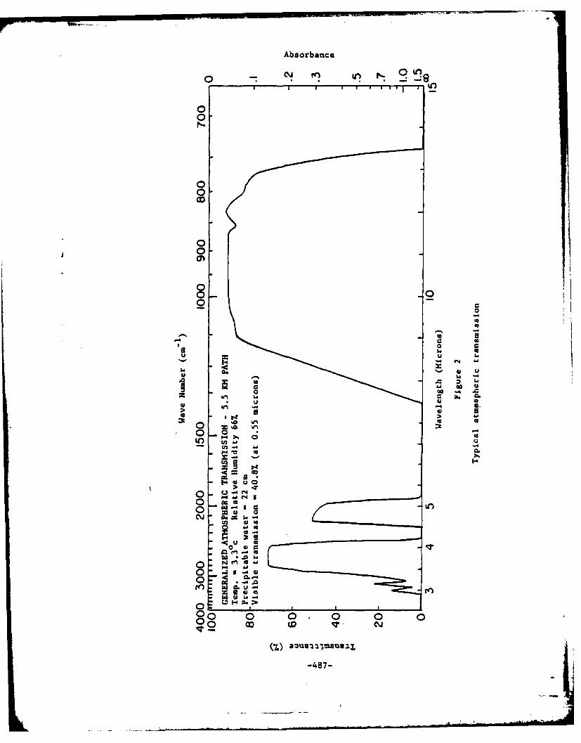
Absorbanice
0
0-0
00-
8
oog
00
-04
0 C4
0 4o .4 4
U3U
40 4ao 0-4..
W I1 6 u . ~4
00 CD ILO
41 ui -487-

r2
r0
" 0
LIl
oo - J
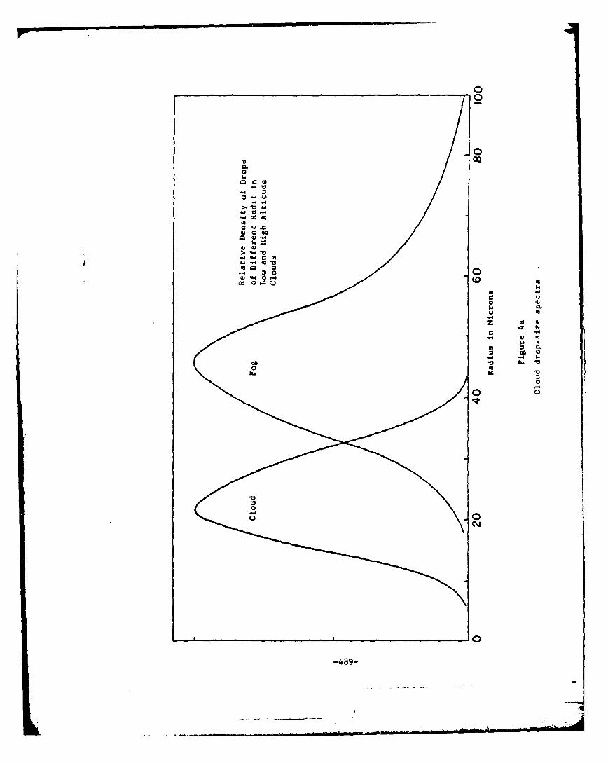
0
0
0.0
0.
w C
00
0
CY
100
.4489-

S .I , I I | I' I I I I I 5 I I II I l I l I I I
0i
0
a)
Q 4j
ac w
V3 fn
go
@10 0 00 0 0-
0 0
00
-490-
-=
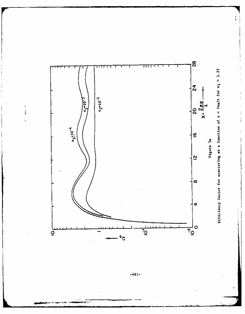
o 0 -0
0
U1h 1441 02
S 0
4CD1
0Vto
-4911

.101
2.1
102 n -10 "4
10
0 4 8 12 16 20 24 28Figure 5b-
Efficiency factor for absorption as a function of
x -2wa/A forn 1.3
-492-

0n
scale anSef
-493

n 2 1 -
0 I80
Figure ba
Scattered intensity as a function of scattering angle
for n, 1.33 and x - a/X 1.
-494-
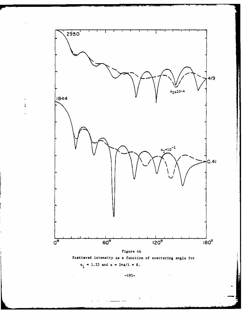
2950
1844
0060 0 1200 1800
Figure 66
Scattered intensity as a function of scattering angle for
- 1.33 and x -2ira/X - 8.
-495-
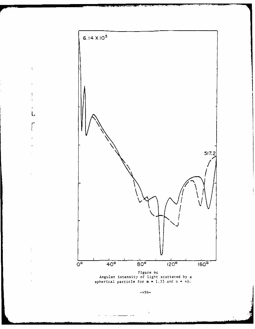
6.14 X 10~
517Z2
00"
400 800 1200 1600
Figure bcAngular intensity of light scattered by a
spherical particle for m 1.33 and a 40.
-496-

1010
9-10
J 10
0
0
-4
-10.
I-
0 100 200 300 400 X270 50 0 600Figure 7X
Normalized Scattered Intensity in the Forward Direction vs x -7r
-497-

For~
2.0
S
1.5
.0
C.4
1.0 -
l1
OW
0 1- 2 34distance from beam in meters
Figure8
-498-

00 -,f N ---
S\ 1465
1 0 5
3
o % .
-20 "Estimated beam Shapewith no atmosphericeffects
I I I I --20 -10 0 10 20 30 40 50
S Sec of arc, eSource
direction
FIGURE 9 Beam broadening due to snow: X -0.63 ; path length, 2.6 km.Data of 2/10 and 2/11/64. Excens attenuacions in curves 1-5are 3-5, 5"0, 13-6, 15-6 and 17-9 db/km respectively, correspond-ing to increasingly heavy snowfalls. The attenuations aremeasured at dead reckoning, that is, with the receivin4 apertureon the maximum of the beam pattern. (.9,0)
(by D.C. Hogg)
-499-

0
curve that would result if-Ia N~eam energy were constant
a ~Lurve from Hogg's data
V ~-20 "'
.0
00
-40
2 5 10 20 30 40Beam width (1/2 power points) at 2.6 km in seconds of arc
Figure 10.
-500-

r
- BIBLIOGRAPHY
r1. Goody, R. M., Atmospheric Radiation, Oxford, Clarendon Press, 1964.
2. Taylor, I. H., and Yates, H. W., JOSA 47, 223 (1957).
3. Davis, J. I., Appl. Opt. 5, #1, 139 (1966).
4. Tatarski, V. I., Wave Propagation in a Turbulent Medium, MtGraw-Hill , 1961.
S. Goldstein, et. al., Proceedings, IEEE, 53, No. 9, 1172 (1965).
6. Burch, D. E., et. al., Appl. Opt. 1 #6 (1962)Also, Curcio, I. A. and Estes Buttrey, D. V., Appl. Opt. 5, #2, 231 (1966)
7. Flynn, G. W., et. al., App. Phys. Let. 8, No. 3, 63 (1966)
8. Stratton, J. A., and Houghton, H. G., Phys. Rev. 38, 159 (1931) andLStratton, J. A., Electromagnetic Theory, McGraw-Hill, 1941
9. Plass, Gilbert N., Appl. Opt., 5, #2, 279 (1966) andAppl. Opt. _5, #1, 149 (1966)
10. Houghton, H. G., J. ofAer. Sci., 6, 103-107
11. Carlson, H. R., Appl. Opt., 4, #9, 1089 (1965)
12. Van der Hulst, H. C., Light Scattering by Small Particles, John Wiley, 1957.
13. Hogg, D. C., Nature, 203, 396, (1964)
i5
L -50 1-
-'- -". . ihi

PUBLICATIONS
A. Laser-Related
1. Fine, S., Klein, E., Scott, R.E., Aaronson, C. and Donoghue, J."Biological Effects of Laser Radiation," Second Boston LaserConference, August 1, 1963.
2. Fine, S., Maiman, T.H., Klein, W. and Scott, R.E. "BiologicalEffects of High Peak Power Radiation," Life Sciences, 3:309-322,March, 1964.
3. Fine, S. and Klein, E. "Effects of Pulsed Laser Irradiation of theForehead in Mice," Life Sciences, 3:199-207, March, 1964.
4. Fine, S., Klein, E. and Scott, R.E. "Studies on Interaction of LaserRadiation dith Biological Systems, " IEEE Spectrum, March, 1964.
5. Fine, S., Klein, E., Derr, V.E. and Nowak, W.B. "Hazards andBiological Effects of Laser Radiation," Proceedings of the MartinInterdivisional Solid State Symposium, March 1964.
6. Edlow, J., Farber, S., Fine, S. and Klein, E. "Prenatal and NeonatalEffects of Laser Radiation," Biological Abstracts of BostonLaser Conference, 1964.
7. Klein, E., Fine, S., Cohen, E., Ambrus, J., Neter, E., Lyman, R. andScott, R.E. "Effects of Laser Radiation on Biological Systems,"American College of Physicians (Atlantic City, N.J.), April 10, 1964.
8. Klein, E.and Fine, S. "Effects of Laser Radiation on Animal Tissues,"presented at Conference on Lasers, New York Academy of Sciences,May 4, 1964.
9. Klein, E., Fine, S. and Laor, Y. "Modification of Effects of LaserRadiation by Licht Absorbing Chemicals," Biological Abstractsof Boston Laser Conference, August, 1964.
10. Klein, E., Fine, S., Scott, R.E. and Farber, S. "Observations of LaserIrradiation of Experimental Tumors," Proceedings, American Associationfor Cancer Research, 1964.
11. Derr, V., Klein, E. and Fine, S. "Electron Spin Resonance Tests ofLaser Irradiated Biological Systems," Applied Optics, 3:786, 1964.
12. Fine, S., Klein, E., Aaronson, C., Hardway, G., King, W. and Scott, R.E."Closed Circuit Television in Laser Investigations," Journal ofInvest. Derm., 4:289-91, 1964.
13. Klein, E., Fine, S., Laor, Y., Litwin, M., Donoghue, J. and Englander, L."Laser Irradiation of the Skin," Journal of Invest. Derm., 43:565, 1964.
-1-

14. Fine, S., Klein, E., Ambrus, J., Cohen, E., Derr, V. and Ambrus, C."Interaction of Relatively Coherent Laser Radiation and BiologicalSystems," Federation Proceedings, 23, 1964.
15. Fine, S., Klein, E., Nowak, W.B., Hansen, W.P., Hergenrother, K.,Scott, R.E. and Donoghue, J. "Measurements and Hazards onInteraction of Laser Radiation and Biological Systems," NEREMRecord, 1958, 1964.
16. Nowak, W.B., Fine, S., Klein, E., Hergenrother, D., Hansen, W.P."On the Use of Thermocouples for Temperature Measurement DuringLaser Irradiation," Life Sciences, 3:1495-1581, 1964.
17. Derr, V., Klein, E. and Fine, S. "Free Radical Occurrence in SomeLaser Irradiated Biological Materials," Federation Proceedinas,24 (1), Supple. 14, Part III, January-February, 1965.
18. Fine, S., Klein, E., Nowak, W.B., Scott, R.E., Simpson, L., Crissey, J.and Donoghue, J. "Interaction of Laser Radiation with BiologicalSystems. I. Studies on Interaction with Tissues," FederationProceedings, 24 (1), Suppl. 14, Part III, January-February, 1965.
19. Klein, E., Fine, S., Laor, Y., Simpson, L., Ambrus, J., Richter, W.,Smith, G.K. and Aaronson, C. "Interaction of Laser Radiationwith Biological Systems. II. Experimental Tumors," FederationProceedings, 24 (1), Suppl. 14, Part III, January-February, 1965.
20. Klein, E., Fine, S., Ambrus, J., Cohen, E., Ambrus, C., Neter, I.,Bardos, T. and Lyman, R. "Interaction of Laser Radiation withBiological Systems. III. Studies on in Vitro Preparations,"Federation Proceedings, 24 (1), Suppl. 14TTt III, January-February, 1965.
21. Edlow, J., Fine, S. and Vawter, G.F. Federation Proceedings, 24:556,April, 1965.
22. Litwin, M., Fine, S., McCombs, H.L. and Klein, E. "Effects of LaserRadiation on the Surgically Exposed Canine Liver," FederationProceedings, 24 (1), Suppl. 14, Part II1, 566, March-April, 1965.
23. Fine, S., Klein, E., Fine, B.S., Litwin, M., Nowak, W.B., Hansen,W.P., Caron, J. and Forman, J. "Mechanisms and Control ofLaser Hazards and Management of Accidents," Laser TechnologyConference, April, 1965.
24. Laor, Y., Simpson, L.C., Klein, E. and Fine, S. "The Pathology of LaserIrradiation on the Skin and Body Wall of the Mouse," The AmericanJournal of Pathology, Vol. 47, No. 4, October, 1965, pp. 643-63.
25. Hansen, W.P., Fine, S., Peacock, G.R. and Klein, E. "Focusing ofLaser Light by Target Surfaces and Effects on Initial TemperatureConditions," NEREM Record, Vol. 7, 156-157, 1965.
-ii-
I
26. Stratton, K., Pathak, M.A. and Fine, S. "ESR Studies of MelaninContaining Tissues After Laser Irradiation," NEREM Record,p. 150, 1965.
27. Klein, E., Laor, Y., Fine, S., Simpson, L.C., Edlow, J., Litwin, M."Threshold Studies and Reversible Depigmentation in RodentSkin," NEREM Record, Vol. 7, pp. 108-109, 1965.
28. Edlow, J., Fine, S., Vawter, G.F., Jockin, H. and Klein, E. "LaserIrradiation Effect on Rat Embryo and Fetus in Utero," LifeSciences, 4:615-23, 1965.
29. Klein, E. and Fine, S. "Biological Aspects of Laser Radiation."Abstract presented at the 149th National Meeting of the AmericanChemical Society, Detroit, Michigan, 1965.
30. Fine, S. and Klein, E. "Biological Effects of Laser Irradiation,"in Advances in Biological and Medical Physics, Academic Press,10:149-225, 1965.
31. Fine, S., Klein, E., Litwin, M., Peacock, G., Hamar, M. and Hansen, W.P."Biological Effects of High Power Continuous N2-C02 Laser Radiationat 10.6 Microns," Federation Proceedings, Vol. 25, No. 2, Part I,March-April, 1966.
32. Laor, Y., Hust, F., Fine, S. and Klein, E. "Studies on BiologicalEffects of Laser Radiation," Symposium on Biomedical Engineering,Marquette University, Milwaukee, Wisconsin, Vol. 1, pp. 316-318,June, 1966.
33. Lobene, R. and Fine, S. "Interaction of Laser Radiation with OralHard Tissues," Journal of Prosthetic Dentistry, 16:3, 589-597,May-June, 1966.
34. Fine, S., Klein, E., Litwin, M. "Laser Radiation and Therapy ofMalignant Melanomas," New Views of Skin Diseases, Boston:Little, Brown & Co., 1966.
35. Fine, S., Klein, E., Fine, B.S., Hansen, W.P., Peacock, G.R. and Litwin, M."Implementation of Procedures and Techniques for Safe Operation ofLasers," Proceedings of the First Conference on Laser Safety,November, 1966.
36. Klein, E., Fine, S., Laor, Y., Hust, F. and MacKeen, D. "InjuriousEffects of Laser Radiation on Mammals," published in Proceedingsof the First Conference on Laser Safety, November, 1966.
37. Fine, S. and Klein, E. "Ultraviolet Lasers," presented at the FirstConference of the Biologic Effects of Ultraviolet Radiation,published in The Biologic Effects of Ultraviolet Radiation,"F. Urbach, editor, Pergamon Press, 1969.
-iii-

38. Fine, S., Hansen, W.P., Peacock, G.R., Klein, E., Hust, F. and Laor, Y."Bio-physical Studies with the CO2 Laser," NEREM Record,8:166-167, November, 1966.
39. Fine, B.S., Fine, S. and Zimmerman, L.E. "C02 Lasers Irradiation on theRabbit Eye, Clinical and Histopathologic Observations," NEREMRecord, 8:160-161, November 1966.
40. Fine, S., Klein, E. "Biological Effects of Laser Radiation," inMcGraw-Hill Yearbook of Science and Technology, 1966.
41. Klein, E., Fine, S., Laor, Y. and Hust, F. "Biological Effects ofLaser Radiation," Proceedings American Society for Cancer Research,1966.
42. Fine, S., Klein, E., Hansen, W.P. and Litwin, M. "Biological Effects ofLaser Radiation," Digest of Technical Papers, International QuantumElectronics Conference, 1966.
43. Fine, S., Klein, E., Haynie, W.H., Litwin, M., Laor, Y. and Hust, F.S."Biological Effects and Hazards of Laser Radiation," presented atAmerican College of Physicians Conference, 1966.
44. Fine, S., Klein, E., et al. "Management Study on Biological Effects ofLaser Radiation Program" for U.S. Air Force, Parts I, II, and III,1000 pages, 1966.
45. Klein, E., Fine, S., Laor, Y., Hust, F., Litwin, M. and Knubbe, K."Interaction of Laser Radiation with Experimental Melanoma,"Proc. Am. Assoc. Cancer Res., 7:36, 1966.
46. Litwin, M.S., Fine, S., Klein, E., Fine, B.S. and Raemer, H. "Hazardsof Laser Radiation Mechanisms, Control and Management," AmericanIndustrial Hygiene Assoc. Journal, 28:68-75, January-February, 1967.
47. Fine, S., Klein, E., Parr, W.H., Fine, B., Fisher, R.S., Peacock, G.R.,MacKeen, D., Hansen, W.P. and Feigen, L. "Hazard Studies withLaser Radiation," Conference on Laser Technology, April, 1967.
48. Hardy, L.B., Hardy, F.S., Fine, S. and Sokal, J. "Effect of Ruby Laseron Mouse Fibroblast Culture," Federation Proceedings, 26:688,April, 1967.
49. Fine, B.S., Fine, S., Peacock, G.R., Geeraets, W. and Klein, E."Preliminary Observations on Ocular Effects of High-Power,Continuous CO2 Laser Irradiation," American Journal of Oph:halmoiogy,Vol. 64, No. 2, pp. 209-222, August, 1967.
50. Peacock, G.R., Hansen, W.P. and Fine, S. "Increasing the Power Output fromInexpensive CO2 Lasers," American Journal of Physics, Vol. 35, No. 8,776-777, August, 1967.
-iv-

51. Fine, S., Feigen, L., MacKeen, 0. and Klein, E. "Hazards and ProtectiveDevices Associated with 10.6 u Radiation," presented at Conferenceon Engineering in Medicine and Biology, 1967. Published in Proceedings,November, 1967.
52. Hansen, W.P., Feigen, L. and Fine, S. "A 'Worst Case' Analysis ofContinuous Wave He-Ne Laser Hazards to the Eye," Applied Optics,Vol. 6, No. 11, pp. 1973-1975, November, 1967.
53. Laor, Y., Simpson, L., Klein, E., Fine, S. and Hust, F. "Effects ofLaser Radiation on the Skin and Underlying Tissue of Mice duringControlled Hair Growth Cycle," Journal of Invest. Derm.,48:297-298, 1967.
54. Fine, S., Edlow, J., MacKeen, D., Feigen, L., Ostrea, E. and Klein, E."Focal Hepatic Injury and Repair Produced by Laser Radiation:Pathologic and Biophysical Studies," American Journal of Patholcgy,Vol. 52, No. 1, pp. 155-176, January, 1968.
55. Hansen, W.P. and Fine, S. "Melanin Granule Models for Laser InducedRetinal Injury," Applied Optics, Vol. 7, No. 1, pp. 155-159,January, 1968.
56. Aaron, A., Fine, S. and Schetzen, M. "Safety improvement for UnattendedLasers," Laser Focus, February, 1968.
57. Lobene, R.R., Raj Bhussry, B. and Fine, S. "The Interaction of CarbonDioxide Laser Radiation with Enamel and Dentin," Journal of DentalResearch, Vol. 47, No. 2, pp. 311-317, March-April, 1968.
58. Cohen, E. and Fine, S. "In Vitro Effects of Laser Irradiation on HumanGanna Globulin," Federation Proceedings, 27:1, 473, March-April, 1968.
59. Parker, G.S., Bavley, H.A. and Fine, S. "Report on Massachusetts LaserSurvey," Laser Focus, 11:30-32, May, 1968.
60. Parker, G.S., Bavley, H., Fine, S., Powell, C. and Keene, B. "LaserSurvey in Massachusetts," Health Physics Society Annual Meeting,Denver, Colorado, June 16-20, 1968. (Abstract.)
61. Fine, B.S., Fine, S., Feigen, L. and MacKeen, 0. "Corneal injuryThreshold to Carbon Dioxide Laser Irradiation," 'ol. 66, No. 1,pp. 1-15, July, 1968, American Journal of Ophthalmolocy.
62. Cohen, E., Klein, E. and Fine, S. "Effects of Laser Irradiation on SomeSerologic Properties of Human Gamma Globulin," accepted forpublication.
63. MacKeen, D., Fine, S. and Klein, E. "Toxic and Explosive Hazards Associatedwith Lasers," Laser Focus, pp. 47-49, October, 1968.
64. Fine, B.S., Berkow, J.W., Fine, S. "Corneal Calcification," Science,Vol. 162, pp. 129-130, October 4, 1968.
-V-
65. Hansen, W.P. and Fine, S. "Application of Thermal Models to RetinalThreshold Injury," presented at Laser Industry Association meeting,October 24-26, 1968, published in Proceedings of the Laser Industry.Association Convention, 1968.
66. Bock, F., Laor, Y., Fine, S. and Klein, E. "Exploration of PotentialCarcinogenic Effects of Pulsed Laser Radiation," presented at LaserIndustry Association meeting, October 24-26, 1968, published inProceedings of the Laser Industry Association Convention, 1968.
67. Fine, S., MacKeen, D., Feigen, L. and Fine, B. "Anterior ChamberMeasurements on CO2 Laser Corneal Irradiation," Proceedings of theAnnual Conference on Engineering in Medicine and Bioloy, Voi. 10,p. 6, November 4, 1968.
68. Feigen, L., MacKeen, D. and Fine, S. "A Method for Detecting andMeasuring Frequency of Surface Vibrations Using a Helium-NeonLaser," Review of Scientific Instruments, Vol. 40, pp. 381-382,February 2, 1969.
69. Geeraets, W., Fine, B.S. and Fine, S. "Ophthalmic Studies on CO2Laser Irradiation," Acta Oohthalmologica, ((oberhavn), Vol. 47,pp. 80-92, 1969.
70. Laor, Y., Simpson, C.L. Klein, E. and Fine, S. "Pathology of InternalViscera Following Laser Radiation," American Journal of MedicalSciences, Vol. 257, pp. 242-252, April, 1969.
71. Litwin, M.S., Fine, S., Klein, E. and Fine, B.S. "Burn Injury AfterCarbon Dioxide Laser Irradiation," Arch. Surg., Vol. 98, pp. 219-222,February, 1969.
72. Fine, S. and Klein, E. "Lasers in Biology and Medicine," Laser Focus,pp. 28-36, July, 1969.
73. Fine, S., Bushor, W. and Cos, M. editors. Proceedings of the LaserIndustry Association Meeting, October 24-26, 1968.
74. Fine, S. and Klein, E. "Lasers in Biology and Medicine" published inDevelopment in Laser Technology, Society of Photo-Optical InstrumentationEngineers.
75. MacKeen, D., Fine, S., Feigen, L. and Fine, B.S. "Anterior ChamberMeasurements on CO2 Laser Radiation," Investigative Oohthalmology,Vol. 9, No. 5, pp. 366-371, May, 1970.
76. MacKeen, D., Edlow, J., Fine, S., Kopito, L. and Klein, E. "Calciumand Magnesium in Focal Hepatic Lesions," Federation Proceedings,Vol. 29, No. 2, March-April, 1970.
77. Klein, E., Laor, Y. and Fine, S. "Interaction of Laser Radiation with theSkin," Abstract-Laser Journal, Vol. 2, No. 1, January-February, 1970.
-vi-
78. Fine, S., MacKeen, D., Berkow, J. and Fine, B.S. "Biological Studieswith Laser Protective Materials," American Journal of Ophthalmology,Vol. 71, No. 4, April, 1971.
79. Campbell, J. and Fine, S. "Heat Sensation Thresholds for C02 LaserRadiation," Radiation Research, 43 (1), 1970.
80. MacKeen, D., Fine, S., Aaron, A. and Fine, B.S. "Cataract Productionin Rabbits with an Ultraviolet Laser," Laser Focus, April, 1971.
81. Fine, S. and Hansen, W.P. "Optical Second Harmonic Generation inBiological Systems," Applied Optics, October, 1971.
82. MacKeen, D.L., and Fine, S. "Effect of Suprathreshold C02 Laserirradiation of the Weanling Rabbit Eye on Lenticular AscorbicAcid and Reduced Glutathione," Federation Proceedings, Vol. 32,No. 3, Pt. 1, 1973.
83. MacKeen, D., Fine, S. and Fine, B.S. "Production of Cataracts inRabbits with an Ultraviolet Laser" - Ophthalmic Research, 5:317-324,1973.
84. MacKeen, 0., Cohen, J., and Fine, S. "Simultaneous Corneal Surface andAnterior Chamber Temperature Measurements on C02 Laser Irradiation,"Federation Proceedings, Vol. 33, No. 3, Pt. 1, March, 1974.(Abstract'.)
85. MacKeen, D.L., Szabo, G., and Fine, S. "The Effects of UV LaserRadiation at 325 nm on the Skin," The Yale Journal of Medicine,1973 (Abstract).
B. Non-Laser Related
in addition, credit was given to non-laser related studies, in which theprincipal investigator and his associate were involved. A number of thesewere listed in the annual progress reports; several are listed below.
86. Fine, S., Klein, E., R.E., Hainish, H. and Aaronson, C. "Bio-Engineeringin the Biological Sciences," IEEE Student Journal, January, 196a,1:33-39.
87. Litwin, S.B., Cohen, J., Fine, S. and Aaron, A. "Rupture and TensileStrength Measurements of Fresh and Teated Canine Aortic Tissue,"Proceedings of the Annual Conference on Engineering in Medicine andB ology, Vol. 10, p. 44', November'4, 1968.
88. Aaron, A., Litwin, S.B., Fine, S. and Sillin, L. "Pressure and FlowRelations in Canine Aortic-Pulmonary Shunts," presented at SecondInternational Conference on Medical Physics, Boston, Massachusetts,August, 1969, published in Abstracts of Conference.
89. Aaron, A., Litwin, S.B., Fine, S. and Rosenthal, A. "Determination ofCardiac Output by Dye Dilution," in Proceedings of the 23rd AnnualConference on Engineering in Medicine and Biology, Vol. 12, 1970.
-vii-

90. Cohen, J., Litwin, S.B., Aaron, A. and Fine, S. "The Rupture Force andTensile Strength of Canine Aortic Tissue," J. Surg. Research,December, 1972.
91. Litwin, S.B., Cohen, J. and Fine, S. "Effects of Sterilization andPreservation on the Rupture Force and Tensile Strength of CanineAortic Tissue," J. Surg. Research, 1973.
-viii-

C. Laser-Related Presentations as Invited Lecturer, Pertinent toContract to Which Credit Was Given
1. Conference on Biological Effects of Laser Radiation, Washington, D.C. -
Sponsored by U.S. Army Medical Research and Development Command, 1964
2. Conference on Lasers, New York Academy of Sciences, 1964
3. Gordon Research Conference on Biological Effects of Laser Radiation,1965
4. Conference on the Biological Effects of Lasers, National Institutes of
Health, Bethesda, Maryland, October 4-5, 1965
5. Gordon Research Conference on Biological Effects of Laser Radiation, 1966
6. Bell Telephone Laboratories - invited lecturer, 1966
7. The Martin Company - symposium on Biological Effects of LaserRadiation, 1966
8. Boston Medical Physics Society - Lecturer on Biophysical Studies withLaser Radiation, 1966
9. Seminar on Biological Effects of Laser Radiation, University of Texas,Austin, 1966
10. Conference on Development of Lasers in the Biological Sciences,Veterans Administration, Department of Medicine and Surgery,Washington, D.C., August 5, 1966
11. Presentation before the Physicians of the Association of AmericanRailroads, Montreal, June 4, 1967
12. Gordon Research Conference on Biological Effects of Laser Radiation, 1967(Session Chairman)
13. American College of Obstetrics - District I Meeting - Invited Participant -Biological Studies on Laser Radiation, October 1967
14. Invited Lecturer on Lasers - P.R. Mallory & Co. Laboratory for PhysicalSciences - Biological Effects of Laser Radiation, February, 1968
15. Brookhaven National Laboratory, Upton, New York "Biophysical Effects ofLaser Radiation", May, 1968
16. New England Chapter Health Physics Society, "Biological Effects and Hazardsof Laser Radiation", May, 1968
17. Case Western Reserve University, Cleveland, Ohio, Summer course on LaserTechnology and Applications, presented lecture "Lasers in Biologyand Medicine," July, 1968
18. G-APURSI Symposium (International Antenna and Propagation Symposium)"Electromagnetic Waves (Lasers) for Biological and MedicalApplications", September, 1968
-ix-

19. Rutgers University, New Brunswick, New Jersey, Participant in "Evaluationof Laser Hazards Course", October, 1968
20. S. Fine - "Biological Studies Relating to Laser Irradiation, Particularywith Respect to the Eye", Howe Laboratories, Massachusetts Eye andEar Infirmary, Harvard Medical School, December, 1968
21. S. Fine - "Control of Laser Hazards and Management of Accidents", NationalCenter for Radiological Health, U.S. Department of Health, Educationand Welfare, Rockville, Maryland, February, 1969
22. S. Fine - "The Application of Lasers to Biology and Medicine," Conferenceon Trends and Directions in Biological Sciences of the ThirteenColleges Curriculum Program Biology Teachers, Clark College, Atlanta,Georgia, March, 1969
23. S. Fine, Participation in Skin Laser Workshop, Second International LaserSafety Conference, Cincinnati, Ohio, March, 1969
24. S. Fine, Lasers--Characteristics, Use, Hazards and Biological Effects,Seminar Series, Environmental Health Engineering and Science, GraduateSchool of Engineering, Northeastern University, March, 1969
25. S. Fine, Lasers--Characteristics and Uses in Biology and Medicine, SurgicalSeminar Series, Boston University School of Medicine, March, 1969
26. S. Fine, Use of Lasers in Biology and Medicine, Laser Applications Course,Washington University, St. Louis, Missouri, May, 1969
27. E. Klein, and S. Fine, Tissue and Cell Effects of Laser Radiation--GordonResearch Conference on Lasers in Medicine and Biology, June, 1959
28. S. Fine, "Lasers in Biomedicine," I.E.E.E. Student Branch, NortheasternUniversity, July, 1969
29. S. Fine, "Biological Hazards and Effects of Laser Radiation," in courseon Fundamentals of Non-Ionizing Radiation Protection, NortheasternRadiological Health Laboratory, August, 1969
30. S. Fine, Lasers in industry, Associated Hazards and ?rotection, NationalSafety Congress, Chicago, Illinois, October 28, 1970
31. S. Fine, Non-invasive Testing in Medicine, >E.E.E. group on Engineeringin Biology and Medicine, Boston, Massachusetts, lovember, 197C
32. S. Fine, Uses and Hazards of Laser Radiation in :ndustry and in AtmosphericPollution Studies, 24th AMA Clinical ^onve,tion, Boston, Massachusetts,November 30, 1970
33. S. Fine, "Bioengineering", Massachusetts Epsilon Chapter, Tau Beta Pi(Northeastern University), September, 1969
_X"
34. S. Fine, "Laser Biology", in Laser Fundamentals and Applicationscourse, Polytecnnic Institute of Brooklyn Graduate Center,September, 1969
35. S. Fine and E. Klein, "Biological Effects of Laser Radiation," theTheobald Smith Society, New Jersey, October, 1969
36. S. Fine, "Lasers--Biological Effects and Medical Applications,"Society of Photo-optical Instrumentation Engineers Meeting,co-sponsored by the University of Rochester Institute ofOptics, Rochester, New York, November, 1969
37. S. Fine, Session Chairman, Laser and Ultraviolet Contributed Papers,Fourth Annual Midyear Topical Symposium, Health Physics SocietyMeeting, Louisville, Kentucky, January 28-30, 1970
38. S. Fine, Lasers in Industry, Associated Hazards and Protection, NationalSafety Congress, Chicago, Illinois, October 28, 1970
39. S. Fine, Non-invasive Testing in Medicine, I.E.E.E. group onngineering in Biology and Medicine, Boston, Massachusetts,November, 1970
40. S. Fine, Uses and Hazards of Laser Radiation in Industry and in AtmosphericPollution Studies, 24th AMA Clinical Convention, Boston, Massachusetts,November 30, 1970
41. S. Fine, "Lasers in Biology and Medicine," in course on Lasers andOptics for Applications, Massachusetts Institute of Technology,Cambridge, Massachusetts, July 30, 1971
42. S. Fine, Guest Lecturer in Graduate Course 2.77, "Biological Effectsand Medical Applications on Non-Ionizing Radiation," Fall, 1971,Massachusetts Institute of Technology
43. S. Fine, "Medical Applications, Research and Safety," Boston SectionI.E.E.E. 1972 Lecture Series, February, 1972
44. S. Fine, "Lasers in Biology and Medicine," a course on lasers andoptics for application, M.I.T., July, 1972
45. S. Fine, "Biological Effects and Medical Applications on Non-lonizingRadiation," guest lecturer for several sessions in graduate course2.77, M.I.T., 1972-1973
46. S. Fine, "Biological Effects and Medical Applications of Non-IonizingRadiation," guest lecturer in graduate course at M.I.T., 1973-9974
47. S. Fine, lectured at Raytheon Research Laboratory on Electrical Hazardsand Emergency Management of Accidents, 1974
48. S. Fine, "Biological Effects and Medical Applications of Non-IonizingRadiation", guest lecturer in summer session course, M.I.T.,July, 1975
-xi-
Most other conferences in which abstracts or papers were publishedare included in the preceding bibliography.
-xii-

0. Laser-Related Conference Organization and Planning; Pertinent to Contract
1. Boston Laser Conference, 1963
2. Boston Laser Conference, 1964
3. Institute of Electrical and Electronics Engineers - Member,NEREM Program Committee, 1965
4. Institute of Electrical and Electronics Engineers - Member,NEREM Program Committee, 1966
5. American Association for the Advancement of Science - Session Organizerand Chairman of Session on Biological Effects of Laser Radiation,1966
6. Laser Industry Association Convention, October 24-26, 1968
7. Course on "Fundamentals of Laser Radiation Protection" given topersonnel of U.S. Department of Health, Education and Welfare,1968
8. Member, program planning committee on seminar series in applicationsof physical chemical techniques in Biology and Medicine, EMB,IEEE, Boston Section, 1969
9. Laser Industry Association Meeting - Los Angeles, California, October20-22, 1969
10. Electro-Optical System Design Conference, September 22-24, 1970,New York Coliseum. Planning of sessions, session organizationand chairman.
11. Major participant in the organization, planning, and instruction ofpersonnel, and field work related to the first major survey onlasers and laser devices in the United States which was carriedout be the State of Massachusetts and Occupational Health andRadiological Health, H.E.W., 1968.
-xiii-
PERSONNEL RECEIVING CONTRACT SUPPORT AND GRADUATE DEGREES OBTAINED
Arnold Aaron Ph.D. in Engineering
Charles Aaronson M.S. in Engineering
John Campbell M.A. in Psychology
John Caron M.S. in Engineering
Joel Cohen M.S. in Biology
John Donoghue M.S. in Engineering
Larry Feigen M.S. in Physics
James Forman M.S. in Engineering
Peter Hansen Ph.D. in Engineering
Karl Hergenrother Ph.D. in Engineering
Donald MacKeen Ph.D. in Biology
Note: The above individuals were supported in full or in part
for contract related work while carrying out graduate
work. In some cases, the support was minimal.
OTHER NON-GRADUATE DEGREE PERSONNEL RECEIVING SUPPORT INCLUDE:
James Blout
Francis Daniels
Evelina Gorman
Mary Laananen
Audrey Marino
Angela Reed
Grace Woods

D?:?1BU710ON L:ST
USAnc (SuD-R2I1s)Fort Da::ick.-rederick, MD 21701
Defanse Technical infoazicn Cent~er (T)A:TN: DTI!C-DDACameron ScationAlexandria, VA 22314