Localize It: Rock, Cosmopolitanism, and the Nation in Trinidad

DOI: 10.2478/s11686-011-0078-y© W. Stefanski Institute of Parasitology, PASActa Parasitologica, 2011, 56(4), 000–000; ISSN 1230-2821
Ieredactylus rivuli gen. et sp. nov. (Monogenea, Gyrodactylidae) from Rivulus hartii
(Cyprinodontiformes, Rivulidae) in Trinidad
Bettina Schelkle1, Giuseppe Paladini2, Andrew P. Shinn2, Stanley King3, Mireille Johnson1, Cock van Oosterhout4, Ryan S. Mohammed5 and Joanne Cable1*
1School of Biosciences, Cardiff University, Cardiff, CF10 3AX, UK; 2Institute of Aquaculture, University of Stirling, Stirling, FK9 4LA, UK; 3Department of Biology, Dalhousie University, Halifax, Nova Scotia, B3H 4J1, Canada;
4 School of Environmental Sciences, University of East Anglia, Norwich NR4 7TJ, UK;5Strategic Environmental Services, 5 Henry Pierre Terrace, St. Augustine, Trinidad and Tobago
AbstractA new genus and species of Gyrodactylidae, Ieredactylus rivuli gen. et sp. nov. (Platyhelminthes, Monogenea), is described from
the skin of Hart’s Rivulus (Rivulus hartii Boulenger), a cyprinodontiform fish collected from streams of the Caroni and
Oropouche drainages and the Pitch Lake in Trinidad (prevalence all localities: 16.7–94.6%; mean parasite intensity 1–9 para-
sites/fish; range 1–34) with the type originating from a tributary of the Aripo River. This viviparous monogenean is distinctive
from other genera of Gyrodactylidae by its split ventral bar membrane, the shape of its male copulatory organ, the presence of
two conical accessory pieces associated with the hamulus root and two differently shaped marginal hook sickles. Its unique
rDNA sequence shows the closest ITS2 similarity (70%) to Gyrodactyloides andriaschewii Bychowsky et Poljansky, 1953.
The presence of I. rivuli gen. et sp. nov. in the Pitch Lake indicates an adaptation to extreme environmental conditions such as
high temperatures and hydrocarbons and adverse pH. Guppies may potentially serve as temporary hosts. The parasite displays
distinct behaviours, including a characteristic ‘swimming-like’ movement. The ecology and phylogeny of I. rivuli gen. et sp.
nov. is discussed in relation to the diversity of other gyrodactylids in Trinidad.
KeywordsGuppy, Gyrodactylidae, Hart’s Rivulus, Ieredactylus rivuli gen. et sp. nov., Monogenea, Poecilia reticulata
Introduction
In Trinidad and Tobago, only Gyrodactylus bullatarudis Turn-
bull, 1956 and Gyrodactylus turnbulli Harris, 1986 from the
guppy Poecilia reticulata Peters, and Gyrodactylus pictaeCable, van Oosterhout, Barson et Harris, 2005 from the
swamp guppy Micropoecilia (= Poecilia) picta (Regan) have
been recorded in genus Gyrodactylidae (Platyhelminthes,
Monogenea). Currently, due to the lack of parasite surveys
nothing is known of the gyrodactylid fauna from non-poeciliid
fish in Trinidad and Tobago (see Harris et al. 2004).
One of the most abundant predators of guppies in Trinidad
is Hart’s Rivulus (Rivulus hartii Boulenger). Its diet is princi-
pally represented by invertebrates, but it will also feed on
small guppies in areas of shared habitat (Endler 1978), poten-
tially allowing parasite transmission between host species.
Host switching is reportedly the major driver of speciation
amongst the Gyrodactylidae (see Ziętara and Lumme 2002)
leading to conjecture that interacting host species, including
predator-prey (e.g. R. hartii-P. reticulata), might share related
parasite species. Individuals of R. hartii (locally known as the
Jumping Guabine) can travel short distances across land al-
lowing them access to a range of waterways (Froese and Pauly
2011). This expands the contact network between this species
and other indigenous fish and therefore raises the potential for
host-switching (Bakke et al. 2007).
In the current study, R. hartii specimens collected in
streams and Trinidad’s Pitch Lake, co-inhabited by guppy
(P. reticulata) populations, were examined for parasites.
A monogenean parasite was recovered, the opisthaptoral ar-
mature of which differs from the three known Trinidadian
species of Gyrodactylus. Further, unique behavioural, mor-
*Corresponding author: [email protected]

phological and molecular differences identified this parasite
as belonging to a new gyrodactylid genus.
Materials and methods
Origin of samples
Specimens of Rivulus hartii (standard length 12.5–49.1 mm:
weight 0.028–1.366 g) were sampled in 2004 (n = 9) and
2006 (n = 5) from Pitch Lake, southern Trinidad (Grid. Ref.:
UTM 20P - 650341.45 E, 1131668.93 N), in 2006 (n = 37)
and 2008 (n = 6) in the Naranjo tributary of the Upper Aripo
River in northern Trinidad (UTM 20P - 692498.44 E,
118257.53 N), and in 2010 (n = 41) from the Upper Gua-
napo River (UTM 20P - 689796.25 E, 1183101.4 N). In ad-
dition, 118 specimens of Poecilia reticulata (standard length
4.9–29 mm; weight 0.003–0.489 g) collected from Pitch
Lake over the period 2004 and 2006 were screened for gy-
rodactylids.
Fish were euthanised with an overdose of anaesthetic
MS222 (Sigma, Poole, UK) and stored individually in 90%
ethanol until examined under a dissection microscope using
optic fibre illumination. Attached gyrodactylids were carefully
removed from the host using entomological pins and trans-
ferred into 0.5 ml Eppendorf tubes containing 90% ethanol;
no other ectoparasites were detected.
Light microscopy
From the monogenean material collected, a total of 22 speci-
mens was prepared as either whole mounts or partially di-
gested for morphological analysis, following the method
described by Paladini et al. (2009). Parasites were mounted
on a slide in ammonium picrate glycerine and the edges of the
coverslip sealed with Pertex (Histolab, Gothenburg, Sweden).
Seven specimens, including the two specimens collected from
P. reticulata, had their opisthaptors excised which were sub-
sequently prepared for morphological identification using the
digestion protocol cited above, whereas the corresponding
body was fixed in 90% ethanol for subsequent molecular
analyses.
The morphology of parasites was studied using an Olym-
pus BH2 compound microscope at ×40 and ×100 oil immer-
sion, a JVC KY-F30B 3CCD Zeiss AxioCam MRc digital
camera with an ×0.75 lens and KS300 ver. 3.0 image analysis
software (Carl Zeiss Vision GmbH 1997). A total of 30 point-
to-point measurements were made on the opisthaptor based
on the methods for 25 measurements described by Shinn et al.(2004) with an additional five measurements (HAPL: hamu-
lus accessory piece length measured from the root to the tip of
the accessory piece; HAPW: hamulus accessory piece width
measured as the width at the root; DBL × W: dorsal bar length
× width; DBAPL: dorsal bar attachment point length) to pro-
vide a comprehensive account of the new gyrodactylid genus.
The ventral bar membrane was measured in the median point
across the whole length of both membrane parts. Whole body
measurements (12 in total) included the total body length ×
width (TBL × W), opisthaptor length × width (HL × W), an-
terior pharynx length × width (APL × W), posterior pharynx
length × width (PPL × W), pharynx process length (PProL),
male copulatory organ length × width (MCOL × W) and the
length of the male copulatory organ principal spine (MCOPS).
All measurements are given in micrometres (μm).
Scanning electron microscopy (SEM)
Five alcohol preserved specimens were prepared for SEM
analysis of the opisthaptoral hard parts. Specimens were
placed in a drop of distilled water on a 12 mm diameter round
glass coverslip (Chance Propper Ltd., Warley, UK), which in
turn was attached to a glass slide using a drop of water. Tissue
surrounding the opisthaptoral hooks was removed by adding
2.5 μl of 10× digestion buffer consisting of 75 mM Tris-HCl
pH 8.0, 10 mM EDTA, 5% sodium dodecyl sulphate (SDS)
and proteinase K to a final concentration of 100 μg/ml (Har-
ris et al. 1999) and incubating at 55°C for 10 min. The speci-
mens were then rehydrated with a drop of distilled water and
examined under a dissection microscope. If necessary, a fur-
ther 2.5 μl of 10× digestion buffer was added, and the sample
was re-incubated for an additional 10 min. The dried digested
specimen remained superficially adhered to the coverslip and,
while viewed under a stereomicroscope, was washed 3–4
times with distilled water to produce tissue-free hook prepa-
rations. The specimen was then air-dried and the coverslip was
attached to a 0.5” aluminium pin stub (Agar Scientific Ltd.,
Essex, UK) using double-sided carbon tape and sputter-coated
with gold prior to examination with a LEO 1450VP scanning
electron microscope at 20 kV.
Molecular identification
DNA from seven gyrodactylids (five from R. hartii, two from
P. reticulata) was individually extracted in 15 µl TE buffer in-
cluding 3 µg of proteinase K and 0.45% Tween 20 by incu-
bating the mixture at 65°C overnight and neutralising it at
95°C for 10 min. rDNA was amplified using a forward P3b
and a reverse P4 primers (Cable et al. 2005) which anneal to
the 18S and 28S, respectively. Amplifications were carried out
in a Perkin Elmer thermocycler (9700) using an initial denat-
uration of 95°C, followed by 35 cycles of 94°C for 30 s, 50°Cfor 1 min, 72°C for 2 min and a final extension of 72°C for 10
min. PCR products were purified using Exonuclease I and
SAP (Shrimp Alkaline Phosphatase) (BioLabs) and both
strands were sequenced using BigDye (ver. 3.1; Applied
Biosystems) on an ABI3100 sequencer. Strands were manu-
ally aligned and corrected using the program BioEdit (Hall
1999).
The consensus sequences from the seven individuals were
aligned with EMBLALIGN: Align_000605 using CLUSTAL

X (Jeanmougin et al. 1998) following the criteria detailed by
Matějusová et al. (2003), generating a single haplotype. The
family Gyrodactylidae currently includes 31 genera (Bakke etal. 2007, Vianna et al. 2007, Přikrylová et al. 2009) but of
these, sequences are only available for 7 genera. Thus the hap-
lotype of the unknown specimen was aligned with the fol-
lowing sequences from GenBank: Acanthoplacatus sp.
(AF465784); Diplogyrodactylus martini Přikrylová, Matěju-
sová, Musilová, Gelnar et Harris, 2009 (AM943008); Fundu-lotrema foxi (Rawson 1973) Kritsky et Thatcher, 1977
(GQ918278); Fundulotrema porterensis King et Cone, 2009
(FJ845514); Fundulotrema prolongis (Hargis 1955) Kritsky
et Thatcher, 1977 (GQ918279); Fundulotrema stableri (Hath-
away et Herlevich, 1973) Kritsky et Thatcher, 1977
(AY099505); Gyrdicotylus gallieni Vercammen-Grandjean,
1960 (AJ001843); Gyrodactyloides bychowskii Albova, 1948
(AJ249348); Gyrodactylus anguillae Ergens, 1960 (AB063294);
Gyrodactylus arcuatus Bychowsky, 1933 (AJ001839); Gyro-dactylus branchicus Malmberg, 1964 (AF156669); Gyro-dactylus bullatarudis (AJ011410); Gyrodactylus derjavinoidesMalmberg, Collins, Cunningham et Jalali, 2007 (AJ132259);
Gyrodactylus macronychus Malmberg, 1957 (AJ407893); Gy-rodactylus poeciliae Harris et Cable, 2000 (AJ001844); Gy-rodactylus pseudonemachili Ergens et Bychowsky, 1967
(AJ567674); Gyrodactylus salaris Malmberg, 1957 (Z72477);
Gyrodactylus stephanus Müller, 1937 (FJ845515); Gyro-dactylus truttae Gläser, 1974 (AJ132260); Gyrodactylus turn-bulli (AJ001846); and Macrogyrodactylus polypteri Malm-
berg, 1957 (AJ567672). For the ITS1 analysis, three Dactylo-gyrus species were included: Dactylogyrus vastator Bychow-
sky, 1933 (AJ564159), Dactylogyrus vranoviensis Ergens,
1956 (AJ564163) and Dactylogyrus zandti Bychowsky, 1933
(AJ564165; ITS2 sequences not available for this genus). The
aligned sequences were imported into MEGA version 4.0
(Tamura et al. 2007) and phylogenetic analysis was carried
out using the Neighbor-Joining algorithm (NJ) and Maximum
Likelihood (ML) with bootstrap support n = 2000. Bayesian
analysis was also implemented using MrBayes 3.1 (Ronquist
and Huelsenbeck 2003) with 5 × 10⁶ Markov chain Monte
Carlo (MCMC) generations.
Results
Monogenea van Beneden, 1858
Gyrodactylidae Cobbold, 1864
Ieredactylus gen. nov. (Figs 1 and 2; Table I)
Body fusiform. Prohaptor with a single pair of cephalic lobes
each bearing posterior glands and a spike sensillum. No eye
spots. Spherical pharynx consisting of two bulbs (anterior
and posterior), the anterior bearing 8 processes opens ven-
trally. Oesophagus short, branching into two simple, blind
ended intestinal crura which extend beyond the ootype. Male
copulatory organ (MCO) ventrally positioned, bulbous with
a central principal spine which originates from the base of
the bulb as two spines that merge at the top and face a single
row of 8–10 closely arranged triangular spines. Two excre-
tory bladders present, in-line with the MCO. Vesicula semi-
nalis positioned posterior to the MCO. Female reproductive
system consists largely of tubular uterus and usually contains
an F1 embryo. F2 embryo observed in 3 and no embryo in 4
specimens (whole mount and partially digested prepara-
tions). No vagina. Viviparous. Opisthaptor trapezoidal-
shaped, demarcated from trunk, bears a single pair of hamuli
with conical accessory pieces associated with the roots. Ven-
tral bar with small processes bearing, posteriorly, a bi-lobed,
spatulate membrane. Sixteen marginal hooks directed to-
wards the posterior edge of the opisthaptor. Marginal hook
sickles of two different morphologies. The numbering sys-
tem of the marginal hook position follows that described by
Malmberg (1970). Pair 3 and 4 of the marginal hook sickles
are noticeably more angular, whereas the remaining sickles
are smoothly rounded. All marginal hook sickles bear a
prominent, circular process (‘button’) on their heel which re-
mains after proteolytic digestion of the soft tissue, and is
likely a point of muscle attachment. Marginal hook shafts
long and flexible compared to species of Gyrodactylus. Pres-
ence of two muscular pads adjacent to the hamuli, similar to
the ‘muscular adhesive disks’ observed by Přikrylová et al.(2009) in Diplogyrodactylus martini.
Ieredactylus rivuli gen. et sp. nov. (Figs 1 and 2; Table I)
Measurements for whole specimens and body parts are pre-
sented in Table I. Further, there are 8 pharynx processes and
the male copulatory organ (MCO) is positioned off centre to
the mid-line of the parasite and posterior to posterior pha-
ryngeal bulb; armed with a principal spine and a single row
of 8–10 smaller spines. Excretory bladders present. Dorsal
bars have elongated attachment points on hamuli. The ven-
tral bar is butterfly-shaped, the bar membrane bifurcated
with each wing attached at the mid-point of the ventral bar
proper, extending tangentially and increasing in width with
round ends. Two marginal hook types both with broad bases,
approximately divided into equal toe and heel regions. The
sickle shaft is marked by a deep groove on each side of the
hook along which the filament loop operates. Sickle shaft
proper for marginal hook pairs 1, 2, 5–8 is proportionately
broad, gently angled at three points, forward sloping, termi-
nating at a point well beyond the limit of the toe; sickle tip
sharply deflected. Inner blade face of the sickle gently
curved. Square heel with prominent circular muscle attach-
ment point on the upper edge. Marginal hook pairs 3 and 4
show a sickle shaft broader than those of pairs 1, 2, 5–8;
sharply angled at three points, terminating at a point well be-
yond the limit of the toe; sickle tip not as sharply deflected
as that of pairs 1, 2, 5–8. Inner blade face of the sickle is also
similarly angular.

Fig. 1. Light and scanning electron micrographs of Ieredactylus rivuli gen. et sp. nov. from Rivulus hartii Boulenger from Trinidad. A – opis-thaptoral central hook complex showing the hamuli, the accessory pieces on the hamulus root, the dorsal and the ventral bar (ventral view);B – hamuli (dorsal view); C – marginal hook representing pairs 1, 2, 5–8; D – marginal hook representing pairs 3 and 4; E, F – marginal hooksickle representing pairs 1, 2, 5–8. Note the prominent muscle attachment point at the level of the heel (arrow); G, H – marginal hook sicklerepresenting pairs 3 and 4; I, J – ventral bar, showing the split membrane; K, L – dorsal and ventral views of the male copulatory organ (MCO)showing the principal spine originating from the base of the bulb and facing a single row of 8–10 closely arranged triangular spines; M – triangular hamulus accessory pieces associated with the roots; N – detail of a flattened oval structure (arrows) positioned posterior to theuterus, presumably part of the reproductive system. Scale bars: A–D, I, J, N = 10 µm; E-H = 2 µm; K–M = 5 µm
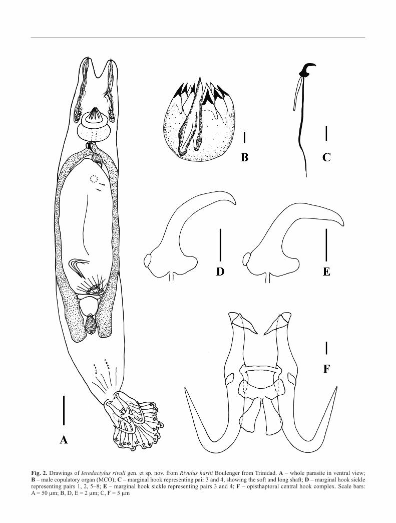
Fig. 2. Drawings of Ieredactylus rivuli gen. et sp. nov. from Rivulus hartii Boulenger from Trinidad. A – whole parasite in ventral view; B – male copulatory organ (MCO); C – marginal hook representing pair 3 and 4, showing the soft and long shaft; D – marginal hook sicklerepresenting pairs 1, 2, 5–8; E – marginal hook sickle representing pairs 3 and 4; F – opisthaptoral central hook complex. Scale bars: A = 50 µm; B, D, E = 2 µm; C, F = 5 µm
Taxonomic summary
Type and only species: Ieredactylus rivuli gen. et sp. nov.
Type host: Hart’s Rivulus Rivulus hartii Boulenger, 1890
(Cyprinodontiformes, Rivulidae).
Site on the host: Skin and fins.
Type locality: Mid-Naranjo tributary of the Aripo River
(Grid. Ref.: UTM 20P - 692498.44 E, 118257.53 N),
Trinidad.
Other reported localities: Guanapo River (689796.25 E,
1183101.4 N) and Pitch Lake (650341.45 E, 1131668.93 N),
Trinidad.
Type material: Holotype (acc. no. NHMUK 2011.9.19.1)
and 15 paratypes (acc. nos. NHMUK 2011.9.19.2-16) are de-
posited in the parasitic worm collection at The Natural His-
tory Museum, London. Additionally, 6 paratypes (acc. nos.
AHC 35253-8) are deposited in the gyrodactylid collection
held at the South Australian Museum.
DNA reference sequence: The 980 bp amplified fragment
consisting of partial 18S (17 bp) ITS1 (331 bp), 5.8S (156 bp),
ITS2 (421 bp) and partial 28S (55 bp) is deposited in Gen-
Bank under acc. no. HQ738514.
Etymology: The genus is named after the Arawak (an in-
digenous tribe of the West Indies) name for Trinidad, i.e.
Table I. Morphological measurements (mean ± 1 standard deviation followed by the range in parentheses;in µm) of Ieredactylus rivuli gen. et sp. nov. from Rivulus hartii collected from the Pitch Lake and theNaranjo tributary, Trinidad, in 2004, 2006 and 2008. Measurements were taken from 17 specimens
Measurement Ieredactylus rivuli gen. et sp. nov. from Trinidad(n = 27)
Total body length 675.5 ± 78 (470–805)Total body width 118.5 ± 22 (70–160)Opisthaptor length × width 94.8 ± 9.7 (70–111) × 80.7 ± 12.7 (58–102)Anterior pharynx length × width 8.7 ± 2.2 (5.5–13.3) × 38.4 ± 7.2 (22.8–50.1)Posterior pharynx length × width 17.3 ± 3.2 (10.8–21.5) × 48.8 ± 9.9 (27.3–65.6)Pharynx processes length 21.4 ± 2.7 (13.1–23.8)MCO length × width 12.1 ± 1.2 (10.3–14.3) × 12.1 ± 1.5 (9.9–14)MCO principal spine 14.1 ± 1 (11.7–15.3)
Hamulus (H)H aperture 16.6 ± 1.4 (14.8–19.4)H proximal shaft width 9.3 ± 0.5 (8.1–10.0)H point length 29.1 ± 0.7 (28.0-30.0)H distal shaft width 4.6 ± 0.4 (3.8–5.1)H shaft length 34.8 ± 1.1 (33.4–37.3)H inner curve length 2.2 ± 0.4 (1.3–2.7)H aperture angle (°) 30.0 ± 1.8 (26.8–32.6)H point curve angle (°) 5.7 ± 1.2 (3.3–7.5)Inner H aperture angle (°) 34.6 ± 2.1 (31.3–39.1)H root length 22.3 ± 1 (21.1–24.5)H total length 55.4 ± 2 (52.7–59.6)H accessory piece length × width 11.5 ± 1.2 (9.1–13.6) × 5.3 ± 0.4 (4.8–6.3)
Dorsal bar (DB)DB total length 12.0 ± 1.2 (9.4–13.4)DB width 2.0 ± 0.2 (1.5–2.3)DB attachment point length 10.2 ± 0.6 (9.5–11.5)
Ventral bar (VB)VB total width 17.6 ± 0.8 (16.4–19.2)VB total length 19.9 ± 0.8 (18.2–21.3)VB process-to-mid length 3.1 ± 0.3 (2.6–3.5)VB median length 6.0 ± 0.5 (5.1–7.1)VB process length 0.9 ± 0.3 (0.5–1.8)VB membrane length 12.5 ± 0.6 (11.4–13.5)
Marginal hook (MH) Pairs 1, 2, 5–8 Pairs 3 & 4MH total length 46.7 ± 2.6 (39.9–49.8) 41.6 ± 3.3 (35.8–46.1)MH shaft length 41.6 ± 2 (38.0–45.3) 36.6 ± 3.3 (30.7–42.0)MH sickle length 5.4 ± 0.3 (4.9–5.9) 4.6 ± 0.2 (4.3–5.0)MH sickle proximal width 3.5 ± 0.1 (3.3–3.8) 3.6 ± 0.2 (3.3–3.9)MH toe length 2.0 ± 0.1 (1.8–2.3) 2.1 ± 0.1 (1.9–2.3)MH sickle distal width 4.1 ± 0.2 (3.7–4.5) 3.8 ± 0.3 (3.2–4.3)MH aperture 6.3 ± 0.2 (5.9–6.7) 5.6 ± 0.3 (5.0–6.2)MH instep / arch height 0.4 ± 0.1 (0.3–0.4) 0.3 ± 0 (0.25–0.4)

“Iëre”. The species is named after the fish host on which this
parasite was first encountered.
General: The species profile including taxonomic details is
provided on www.monodb.org (Shinn et al. 2011).
Remarks
Within the family Gyrodactylidae, Diplogyrodactylus martini,Macrogyrodactylus simentiensis Přikrylová et Gelnar, 2008,
Gyrodactylus chologastris Mizelle, Whittaker et McDougal,
1969 and Gyrodactylus heterodactylus Rogers et Wellborn,
1965 have been reported as having two types of marginal hook
sickles (Rogers and Wellborn 1965; Mizelle et al. 1969;
Přikrylová and Gelnar 2008; Přikrylová et al. 2009), whilst Gy-rodactylus milleri Harris et Cable, 2000 possesses three dif-
ferent marginal hook sickle morphologies (Rubio-Godoy et al.
2010). Gyreteroncus spp. were reported to have marginal hooks
of different sizes and shapes, and their MCO to have many
small spinelets (Euzet and Birgy(i?) 1988), but this genus was
never formally described. Indeed, after re-examination of
Euzet’s drawings, Přikrylová et al. (2009) claim that the species
reported by Euzet and Birgy(i?) (1988) as Gyreteroncus sp.
from Polypterus senegalus senegalus Cuvier is identical to
D. martini, having a tubular and unarmed MCO, and the same
marginal hook morphology. Although a dorsal bar is present in
Euzet’s drawings, Přikrylová et al. (2009) discussed the lack of
this feature in D. martini. Confusion of all these genera with
Ieredactylus gen. nov., however, is unlikely as apart from the
two marginal hook types, all the other diagnostic features are
different. The new genus also features a ‘muscular adhesive
disk-like’ zone next to the hamuli, around the ventral bar at-
tachment points, characteristic of those also seen, for example,
in Diplogyrodactylus, Gyrdicotylus Vercammen-Grandjean,
1960 and Macrogyrodactylus Malmberg, 1956 (see Přikrylová
Fig. 3. Maximum Likelihood (ML) tree showing the relationship of Ieredactylus rivuli gen. et sp. nov. to the other gyrodactylid genera forwhich there are GenBank rDNA Internal Transcribed Spacer 2 sequences (421 bp) available (see Material and methods for individual acces-sion numbers for all species listed)

et al. 2009). There was no evidence of species variation based
on haptor morphology of specimens collected from both Rivu-lus hartii and Poecilia reticulata (although only two parasites
were recovered from the guppy host).
Molecular characterisation of Ieredactylus rivuli gen. et sp.
nov. (Figs 3 and 4)
Overall, the ITS2 region of Ieredactylus rivuli gen. et sp. nov.
showed the greatest genetic similarity (70% similarity with
47% coverage) to Gyrodactyloides bychowskii. All three phy-
logenetic analyses (Neighbor Joining, Maximum Likelihood
and Bayesian) of the Gyrodactylidae genera based on ITS2
data, grouped I. rivuli gen. et sp. nov. with Gyrodactyloides by-chowskii, Gyrdicotylus gallieni, Macrogyrodactylus polypteri
and Diplogyrodactylus martini. ITS2 Bayesian analysis (data
not shown) and Maximum Likelihood (Fig. 3) tree topologies
were almost identical. The major difference between the three
analyses was the position of Acanthoplacatus sp.
ITS1 analyses were conducted to assess the relationship
of the new genus with Dactylogyrus (as no ITS2 sequences of
the latter are currently available). As predicted the ITS1 re-
sults were highly variable (e.g. Cable et al. 1999) and align-
ments are problematic; but in all cases Ieredactylus gen. nov.
clearly clustered within the Gyrodactylidae (Figs 3 and 4).
Ecological observations (Table II)
Ieredactylus rivuli gen. et sp. nov. occurred at a prevalence of
16.7–94.6%, with a mean parasite intensity of 1–9 para-
Fig. 4. Neighbor-Joining (NJ) tree showing the relationship of Ieredactylus rivuli gen. et sp. nov. to the other gyrodactylid genera for whichthere are GenBank rDNA Internal Transcribed Spacer 1 sequences (331 bp) available (see Material and methods for individual accession num-bers for all species listed)
sites/fish on Hart’s Rivulus in three different fish populations
(Table II). This parasite can also naturally infect P. reticulata;
however, this was only observed in 2004 on Pitch Lake gup-
pies at a prevalence of 4.7% and a mean intensity of 1 para-
site/fish. Although we have screened nearly 5000 guppies
from the rest of Trinidad (Cable J. and van Oosterhout C., un-
publ. data), this new genus has not been recovered from guppy
populations elsewhere in Trinidad.
When detached from its host, the new species was observed
to move unidirectionally, characteristically twisting the body
in a repeated figure of eight movement causing the parasite to
move erratically, up or down in the water column (similar to the
‘swimming behaviour’ of Gyrodactylus rysavyi Ergens, 1973
described by El-Naggar et al. 2004). Occasionally the parasite
was observed motionless with its opisthaptor upright in the
water film, similar to G. turnbulli (see Cable et al. 2002). In
comparison to the Gyrodactylus species found on guppies,
I. rivuli gen. et sp. nov. is a distinctly larger parasite.
Discussion
The new genus Ieredactylus gen. nov. consists of a distinct vi-
viparous gyrodactylid with a unique ITS rDNA sequence, and
featuring a split ventral bar membrane, a MCO which differs
from the Gyrodactylus-type in having the principal spine
longer than the whole bulb (see Table I) and the smaller spines
not clearly delineated from the bulb (Figs 1K-L and 2B), the
presence of two accessory pieces attached to the roots of the
hamuli, and two types of marginal hooks. Both marginal hook
sickles show the presence of a “button” on the heel (Figs 1E-
H and 2D-E), which was resistant to the proteolytic digestion
alongside other the hard parts of the opisthaptor. A partial split
in the ventral bar membrane has previously been observed in
G. poeciliae (see Harris and Cable 2000), however, not to the
degree of a complete split as is characteristic for the newly de-
scribed genus. This cross-shaped ventral bar membrane is a
feature that is occasionally seen on species belonging to the
genus Dactylogyrus (i.e. Dactylogyrus crucifer Wagener, 1857
or D. zandti). Molecular analyses, however, clearly position
Ieredactylus rivuli gen. et sp. nov. within the family Gyro-
dactylidae, most similar to the genera Gyrodactyloides, Macr-ogyrodactylus, Gyrdicotylus and Diplogyrodactylus (see Figs
3 and 4).
The arrangement of the two types of marginal hook sick-
les can be divided in pairs 1, 2, 5–8, which are smoothly
rounded (similar to Gyrodactylus spp.), and pairs 3 and 4
sharply angular. Differences in sickle shape within a full set of
marginal hooks have previously been recorded for Diplogy-rodactylus martini (see Přikrylová et al. 2009), Gyrodactyluschologastris (see Mizelle et al. 1969), G. heterodactylus (see
Rogers and Wellborn 1965), G. milleri (see Rubio-Godoy etal. 2010) and Gyreteroncus spp. (see Euzet and Birgi(-y?)
1988), although one unidentified species of the latter has been
suggested to be identical with D. martini (see Přikrylová et al.2009). The size difference between the two sickle types in
Diplogyrodactylus is larger compared with Ieredactylus gen.
nov. and the ratio of the number of large type to the smaller
type hooks is 10 to 6, whereas Ieredactylus gen. nov. has 12
large and 4 smaller marginal hooks.
The prominent “button” on the heel of the marginal hook
sickles is a consistent feature and is not lost during the diges-
tion process which appears to be the case for most species of
Gyrodactylus. A similar feature, however, is retained in a new
Gyrodactylus species, currently under description, which in-
fects the skin and gills of the south European toothcarp Apha-nius fasciatus (Valenciennes) inhabiting hypersaline pools
(Paladini G., Huyse T. and Shinn A.P., unpubl. data). This
“button” most likely serves as a muscle attachment point, as
might the conical accessory pieces associated with the roots of
the hamuli, which disappear after proteolytic digestion.
The bulb of the MCO of Ieredactylus gen. nov. is in shape
similar to Gyrodactylus spp., but the size and the arrangement
of the principal spine differ from this genus. In Ieredactylusgen. nov. the MCO principal spine originates from the base of
the bulb, where appears to be slightly bifurcated, and it ex-
tends to the top as a single spine, having a total size longer
than the rest of the bulb (Figs 1K and 2B; Table I). These fea-
tures had been observed in other gyrodactylids, i.e. Gyro-dactyloides andriaschewi Bychowsky et Poljansky, 1953 and
Gyrodactyloides petruschewskii Bychowsky, 1947 in which
the MCO has a similar principal spine and four smaller spines,
Table II. Prevalence, mean intensity and range of Ieredactylus rivuli gen. et sp. nov. from Rivulus hartii collected in 2004,2006, 2007 and 2008. Details of Guanapo samples collected in 2010 are not available (n/a). For the Pitch Lake, specimenscollected from Poecilia reticulata are presented for 2004 only: the new genus has not been recovered from guppies elsewherein Trinidad nor on the Pitch Lake guppies collected in 2006
Year No. of specimens Location Prevalence (%) Mean intensity Range
Rivulus hartii2004 9 Pitch Lake 33.3 (3/9) 2.3 2–32006 5 Pitch Lake 20 (1/5) 9 92006 37 Mid-Naranjo 94.6 (35/37) 8.2 1–342008 6 Mid-Naranjo 16.7 (1/6) 1 12010 41 Guanapo n/a n/a n/a
Poecilia reticulata2004 64 Pitch Lake 4.7 (3/64) 1 –

and Laminiscus gussevi (Bychowsky et Poljansky, 1953) Páls-
son et Beverley-Burton, 1983 possessing one similar principal
spine and ten smaller spines (see figures in Pálsson and Bev-
erley-Burton 1983).
Preliminary laboratory experiments indicate I. rivuli gen.
et sp. nov. does not transmit from Hart’s Rivulus to what is
believed to be its temporary host, the guppy (P. reticulata). A
remarkable feature of I. rivuli gen. et sp. nov. is that, unlike
other monogeneans examined (Schelkle B., McMullan M.,
Mohammed R.S., Coogan M.P., Gillingham E., van Ooster-
hout C. and Cable J., unpubl. data), it has adapted to the ex-
treme physio-chemical conditions in the Pitch Lake which is
inhabited by Hart’s Rivulus, but also a potential temporary
host of I. rivuli, the guppy. This asphalt lake is a natural up-
welling of oil characterised by high hydrocarbon content and
the habitat is thought to act as a refuge for teleosts from most
parasites (Schelkle B. et al., unpubl. data). Nevertheless, some
parasites, such as I. rivuli gen. et sp. nov., appear to have
evolved tolerance to these extreme conditions, enabling it to
exploit hosts and conquer a niche in this unique habitat. In ad-
dition to infections on Hart’s Rivulus, three guppies (2.5%)
from Pitch Lake were also infected with I. rivuli gen. et sp.
nov., even though these fish were devoid of all other gyro-
dactylids.
Ieredactylus rivuli gen. et sp. nov. seems to be a robust
monogenean adapted to a host known for terrestrial locomo-
tion and inhabiting hostile environments. Further studies are
necessary to assess its potential role as a pathogen which can
infect other hosts in different environmental conditions.
Acknowledgements. We would like to thank Mark McMullan andFelipe Dargent for assistance with fieldwork. This work was sup-ported by a European Community Framework Programme 6 MarieCurie Host Fellowship for Transfer of Knowledge (MTKD-CT-2005-030018) and National Environment Research Council Advanced Re-search fellowship (NER/J/S/2002/00706) to J.C.
References
Bakke T.A., Cable J., Harris P.D. 2007. The biology of gyrodactylidmonogeneans: the “Russian-doll killers”. Advances in Par-asitology, 64, 161–376. DOI: 10.1016/S0065-308X(06)64003-7.
Cable J., Harris P.D., Tinsley R.C., Lazarus C.M. 1999. Phylogeneticanalysis of Gyrodactylus spp. (Platyhelminthes: Monogenea)using ribosomal DNA sequences. Canadian Journal of Zool-ogy, 77, 1439–1449. DOI: 10.1139/cjz-77-9-1439.
Cable J., Scott E.C.G., Tinsley R.C., Harris P.D. 2002. Behavior fa-voring transmission in the viviparous monogenean Gyro-dactylus turnbulli. Journal of Parasitology, 88, 183–184.DOI: 10.1645/0022-3395.
Cable J., van Oosterhout C., Barson N., Harris P.D. 2005. Gyro-dactylus pictae n. sp. from the Trinidadian swamp guppy Poe-cilia picta Regan, with a discussion on species ofGyrodactylus von Nordmann, 1832 and their poeciliid hosts.Systematic Parasitology, 60, 159–164. DOI: 10.1007/s11230-004-6348-4.
El-Naggar M.M., El-Naggar A.A., Kearn G.C. 2004. Swimming inGyrodactylus rysavyi (Monogenea, Gyrodactylidae) from theNile catfish, Clarias gariepinus. Acta Parasitologica, 49,102–107.
Endler J.A. 1978. A predator’s view of animal color patterns. Evolu-tionary Biology, 11, 319–364.
Euzet L., Birgi(-y) E. 1988. Gyreteroncus n. gen. (Gyrodactylidae)parasite of Mormyridae, in West Africa. In: Proceedings ofthe 1st International Symposium on Monogenea, August 1988,Institute of Parasitology, Czech Academy of Science, CěskéBudejovice, Czech Republic, p. 9.
Froese R., Pauly D. (Eds.) 2011. FishBase. World Wide Web elec-tronic publication. Available at http://www.fishbase.org (lastaccess 02/2011).
Hall T.A. 1999. ditor and analysis program for Windows 95/98/NT.Nucleic Acids Symposium Series, 41, 95–98.
Harris P.D., Shinn A.P., Cable J., Bakke T.A. 2004. Nominal speciesof the genus Gyrodactylus von Nordmann, 1832 (Monogenea,Gyrodactylidae), with a list of principal host species. System-atic Parasitology, 59, 1–27. DOI: 10.1023/B:SYPA0000038447.52015.e4.
Harris P.D., Cable J. 2000. Gyrodactylus poeciliae n. sp. and G. mil-leri n. sp. (Monogenea: Gyrodactylidae) from Poecilia cau-cana (Steindachner) from Venezuela. Systematic Parasito-logy, 47, 79–85. DOI: 10.1023/A:1006413804061.
Harris P.D., Cable J., Tinsley R.C., Lazarus C.M. 1999. Combined ri-bosomal DNA and morphological analysis of individual gy-rodactylid monogeneans. Journal of Parasitology, 85,188–191.
Jeanmougin F., Thompson J.D., Gouy M., Higgins D.G., Gibson T.J.1998. Multiple sequence alignment with Clustal X. Trends inBiochemical Sciences, 23, 403–405.
Malmberg G. 1970. The excretory systems and marginal hooks as abasis for the systematics of Gyrodactylus (Trematoda, Mono-genea). Arkiv för Zoologi, 23, 1–235.
Matĕjusová M., Gelnar M., Verneau O., Cunningham C.O., Little-wood D.T.J. 2003. Molecular phylogenetic analysis of thegenus Gyrodactylus (Platyhelminthes: Monogenea) inferredfrom rDNA ITS region: subgenera versus species groups.Parasitology, 127, 603–611. DOI: 10.1017/S0031182003004098.
Mizelle J.D., Whittaker F.H., McDougal H.D. 1969. Studies onmonogenetic trematodes. XLIII. Notes on Gyrodactylus,emendation of the genus, and description of G. chologastrissp. n. from Amblyopsids. American Midland Naturalist, 82,298–302.
Paladini G., Gustinelli A., Fioravanti M.L., Hansen H., Shinn A.P.2009. The first report of Gyrodactylus salaris Malmberg,1957 (Platyhelminthes, Monogenea) on Italian cultured stocksof rainbow trout (Oncorhynchus mykiss). Veterinary Para-sitology, 165, 290–297. DOI: 10.1016/j.vetpar.2009.07.025.
Pálsson J., Beverley-Burton M. 1983. Laminiscus n. g. (Monogenea:Gyrodactylidae) from capelin, Mallotus villosus (Müller),(Pisces: Osmeridae) in the northwest Atlantic with redescrip-tions of L. gussevi n. comb., Gyrodactyloides petruschewskii,and G. andriaschewi. Canadian Journal of Zoology, 61, 298–306.
Přikrylová I., Gelnar M. 2008. The first record of Macrogyrodacty-lus species (Monogenea, Gyrodactylidae) on freshwater fishesin Senegal with the description of Macrogyrodactylus simen-tiensis sp. nov., a parasite of Polypterus senegalus Cuvier.Acta Parasitologica, 53, 1–8. DOI: 10.2478/s11686-008-0001-3.
Přikrylová I., Matějusová I., Musilová N., Gelnar M., Harris P.D.2009. A new gyrodactylid (Monogenea) genus on Gray BichirPolypterus senegalus (Polipteridae) from Senegal (West
Africa). Journal of Parasitology, 95, 555–560. DOI: 10.1645/GE-1652.1.
Rogers W.A., Wellborn T.L. 1965. Studies on Gyrodactylus (Trema-toda: Monogenea) with descriptions of five new species fromthe south-eastern U.S. Journal of Parasitology, 51, 977–982.
Ronquist F., Huelsenbeck J.P. 2003. MrBayes 3: Bayesian phyloge-netic inference under mixed models. Bioinformatics, 19,1572–1574. DOI: 10.1093/bioinformatics/btg180.
Rubio-Godoy M., Paladini G., García-Vásquez A., Shinn A.P. 2010.Gyrodactylus jarocho sp. nov. and Gyrodactylus xalapensissp. nov. (Platyhelminthes: Monogenea) from Mexican poe-ciliids (Teleostei: Cyprinodontiformes), with comments on theknown gyrodactylid fauna infecting poeciliid fish. Zootaxa,2509, 1–29.
Shinn A.P., Hansen H., Olstad K., Bachmann L., Bakke T.A. 2004.The use of morphometric characters to discriminate specieslaboratory-reared and wild populations of Gyrodactylussalaris and G. thymalli (Monogenea). Folia Parasitologica,51, 239–252.
(Accepted October 11, 2011)
Shinn A.P., Paladini G., Rubio-Godoy M., Domingues M.V., Whit-tington I.D., Bron J.E. 2011. Editors. 2011. MonoDb. A web-host for Monogenea. World Wide Web electronic publication.Available at http://www.monodb.org (last access 02/2011).
Tamura K., Dudley J., Nei M., Suhdhir K. 2007. MEGA4: Molecu-lar Evolutionary Genetics Analysis (MEGA) Software ver-sion 4.0. Molecular Biology and Evolution, 24, 1596–1599.
Vianna R.T., Boeger W.A., Dove A.D.M. 2007. Neotropical Mono-genoidea. 51. Scutalatus magniancoratus gen. et sp. n. (Gy-rodactylidae) from the South-American electric eel, Elec-trophorus electricus (Gymnotidae, Gymnotiformes), andredescription of Mormyrogyrodactylus gemini from theAfrican bulldog, Marcusenius macrolepidotus (Mormyridae,Osteoglossiformes). Acta Zoologica, 88, 89–94. DOI: 10.1111/j.1463-6395.2007.00255.x.
Ziętara M.S., Lumme J. 2002. Speciation by host switch and adaptiveradiation in a fish parasite genus Gyrodactylus (Monogenea:Gyrodactylidae). Evolution, 56, 2445–2458. DOI: 10.1111/j.0014-3820.2002.tb00170.x.
Copyright © 2022 FDOKUMEN