
IDEXX-PACS 4.3 Operator's Guide
101
-
Upload
khangminh22 -
Category
Documents
-
view
4 -
download
0
Transcript of IDEXX-PACS 4.3 Operator's Guide


Proprietary rights noticeInformation in this document is subject to change without notice. Companies, names, anddata used in examples are fictitious unlessotherwise noted. No part of this document may be reproduced or transmitted in any form or by anymeans, electronic, mechanical, orotherwise, for any purpose, without the expresswritten permission of IDEXXLaboratories. IDEXXmay have patents or pending patentapplications, trademarks, copyrights, or other intellectual or industrial property rights covering this document or subject matter in thisdocument. The furnishing of this document does not give a license to these property rights except as expressly provided in any written licenseagreement from IDEXXLaboratories or an affiliate.
© 2018 IDEXXLaboratories, Inc. All rights reserved. • 06-0002342-13
*CardioPet, ClearCapture Dx, Cornerstone, DVMAX, EquiView, IDEXXEliteVision, IDEXX-CR, IDEXX-DR, IDEXX I-Vision CR, IDEXX I-Vision DR,IDEXX I-Vision Mobile, IDEXX-PACS, ImageBank, Image Coach, ImageVue, Neo, Patient Clipboard, Pet Health Network, SmartService,VetConnect, and VetMedStat are trademarks or registered trademarks of IDEXXLaboratories, Inc. or its affiliates in the United States and/orother countries.
DICOM is the registered trademark of the National ElectricalManufacturers Association for its standards publications relating to digitalcommunication of medical information. AmazonWebServices is a trademark of Amazon.com, Inc. or its affiliates in the United States and/orother countries. Microsoft, Windows, WindowsServer, and Vista are trademarks of Microsoft Corporation, registered in the United States andother countries. Mac, OSX, iPad, andSafari are trademarks of Apple, Inc. registered in the United States and other countries. Android andGoogle Play are trademarks of Google Inc. Dolphin Browser is a trademark or registered trademark of MoboTap, Inc. ImproMedandAVImarkare trademarks or registered trademarks of Henry Schein Animal Health. InstallShield is a registered trademark of Flexera Software. MySQL is atrademark of Oracle Corporation and/or its affiliates. All other product and company names and logos are trademarks of their respectiveholders.
The IDEXXDiagnostic Imaging systemsare intended for veterinary use only; they are not intended for human diagnostic use.
06/11/2018

Contents
Getting Started 6Statement of Intended Use 6About Your IDEXX Diagnostic ImagingHardware 6
Operating system recommendations 7Minimum hardware requirements 7
Using the IDEXX-PACS HomeWindow 7Shortcuts and Touch-Screen Gestures 10
Capturing Images 13Starting a STAT Image Request 13Capturing Images, Starting from the IDEXX-PACS* Software 13Capturing Images, Starting from the Cornerstone* Software or Other Integrated Systems 14Starting the Capture Request UsingModality Worklist 14Selecting the Shots 15Capturing Images with an IDEXX ImageVue*DR40, IDEXX I-Vision DR*, or IDEXX-DR* 1417Digital Imaging System 17Capturing Images with the IDEXX I-Vision CR* or IDEXX-CR* 1417 (Model 140/140R) DigitalImaging System 18Pausing a Study 20Workingwith the Capture Window 21Checking and Approving Images 22Reprocessing Images 25Deleting Pending Image Requests 26
Using Advanced Image Capture Tools 27Using Special Shot Options 27Creating and Using a Favorite Shots List 28Using Image Coach* and Other Technique Assistance 29Applying Image Markers 31
Viewing and Enhancing Images after Capture 32Using the Image Viewer Window 32SavingModified Images 34Using Image Viewer Tools 34Annotation Tools 34Measurement Tools 35Crop Tool 36Actual Size Tool 36Magnify Tools 37Contrast and Brightness (Window/Level) Tool 37Invert Tool 38Rotate and Flip Tools 38Restore Original Tool 38Reprocess Tool 38
iii

Toggle Overlays and Annotations Tool 39Left-Right Marker Tool 39Editing and Applying Image Overlays 39When to Adjust Contrast and Brightness (Window/Level) 40Special Measurement and Planning Tools 40Comparing Images 45ViewingDental Radiographs 45ViewingMultimedia Files 47ViewingCardioPet* ECG Files 47
Managing Images 49Reassigning a Study 49Editing Image Details 49Saving an Image in a Different Format 50Cloning an Image 51Deleting an Image 51Ensuring that Images Are Correctly Matched to Patients 51Using the Radiology Log 52BackingUp and Archiving Images 54Using IDEXX WebPACS or the IDEXX ImageBank* Storage System 56
Importing and Distributing Images 58Using the SharingWindow 58Printing Images 58Emailing Images 59Sending Images Automatically after Image Capture (Auto-Routing/Auto-Exporting) 60UsingDICOM to Send and Receive Images 60
Before you begin 62Importing a DICOM file based on patient ID 62Importing a DICOM file into a currently open study 63If the imported images are not visible in the Cornerstone Patient Clipboard 63
ImportingNon-DICOM Files 66Importing Images by Scanning 67Bringing a Template-Modified Image Back into the IDEXX-PACS Software 67Creating a Patient CD 68Viewing a Patient CD 69
Managing Client and Patient Records 70Adding a New Patient and Client Record 70Updating a Patient or Client Record 71Deleting a Patient or Client Record 71
Setting Up DICOM* Services 73Overview of DICOM Services Configuration 73Configuring the Local DICOM Server 73ConfiguringRemote DICOM Servers or Modality Worklist Connections 75DICOM File Mapping 76Setting Up a Telemedicine Connection 77
Setting Software Options 79iv

Setting Practice Options 79Setting Staff Options 84Setting Image Options 86SettingClient and Patient Options 91
Using Telemedicine 94Submitting a Telemedicine Case 94Viewing the Status of a Telemedicine Case 95Updating or Deleting a Telemedicine Case 95ImportingCases Submitted Outside the IDEXX-PACS* Software 95Viewing a List of All Telemedicine Cases 96
Customer Support and Software Upgrades 97Contacting IDEXX 97Upgrading Software with SmartService* Solutions 97
Glossary 99
v

Getting StartedYour IDEXX-PACS* Imaging Software works with your IDEXX Diagnostic Imaging system todeliver diagnostic radiographs without film or chemical development.In this chapter, you'll find information about:
l Learning resourcesl Using the Home window and finding a patient recordl How images are organized in order of study, series, and imagel How to determine which IDEXX Diagnostic Imaging system you havel Accessing other programs, like VetConnect* PLUSl Logging off and switching usersl Keyboard shortcuts
Tip: To save thisOperator’s Guide to your computer, the network, or a CD, at the top of the PDFwindow, select File > Save as.
l Capturing Images, Starting from the IDEXX-PACS Software
l Finding Patient Images and Records
Statement of Intended Use
The IDEXX Diagnostic Imaging systems are intended for veterinary use only; they are notintended for human diagnostic use. For a detailed list of species and settings, contact IDEXXDiagnostic ImagingCustomer Support at 1-877-433-9948.
About Your IDEXX Diagnostic Imaging Hardware
Identifying Your Diagnostic Imaging System
There are two types of IDEXX Diagnostic Imaging systems—computed radiography (CR) anddigital radiography (DR).It's easy to determine which system you have:An IDEXX-CR* system uses a portable cassette containing a phosphor screen that replacesx-ray film. After you capture an image, you must insert the cassette into a scanner, which readsthe digital x-ray image without chemical developing. The images are stored and viewed usingthe IDEXX-PACS* software.An IDEXX-DR* system does not have a portable cassette or scanner. In a DR system thedetector panel is attached to the underside of the radiography table. When you take an x-ray,the high-resolution detector plate is exposed to the x-ray source, and the x-rays are convertedimmediately into electrical signals that are read by the DR system and displayed on the monitorin just a few seconds.
Finding Your IDEXX-PACS Version Number
To locate your IDEXX-PACS version number:
On the IDEXX-PACS Home window, clickHelp > About IDEXX Capture.
6

Getting Started
Cleaning and Maintaining Your System
For technical specifications and for information on equipment maintenance, service, transportation,and storage for all IDEXX CRand DR systems, see the IDEXXDiagnostic Imaging Systems Hardware
Guide available on the Help menu in IDEXX-PACS Software. The guide is also located in the folderwhere your IDEXX Diagnostic Imaging software is installed.
System Requirements
Operating system recommendationsOperating system recommendations and requirements are slightly different for computers used tocapture images with IDEXX Diagnostic Imaging systems and computers used for viewing images andusing other software features:
l When capturing images, use only computers that are supplied by IDEXX.l When viewing images and using other features (except image capture), use a computer runningthe Windows* 7 Professional SP1, Windows* 10 Professional or Enterprise, Windows* 2012Server, or Windows* 2016 Server operating system.
The Patient CD feature can be used with home or business editions of Windows operating systems.The Patient CD is not compatible with the Mac*OS X* operating system.
Minimum hardware requirementsThe computer used to run the IDEXX-PACS software must meet these minimum requirements:
RAM: Internet speeds:– 4.0 GB RAM (Windows 32-bit operating system) – Upload: 5 Mbps– 8.0 GB RAM (Windows 64-bit operating system) – Download: 10 MbpsIntel* dual-core processor or better Monitor resolution:20GB free space on hard drive – Minimum: 1024 x 768CD/DVD burner – Recommended: 1280 x 1024 or higher10/100/1000Mbps Ethernet port
Contact IDEXX Diagnostic ImagingCustomer Support if you need help upgradingRAM or freeing diskspace.To capture images with IDEXX Diagnostic Imaging systems, use only computers provided by IDEXX.
Using the IDEXX-PACS Home Window
Using the Home Window on a Capture Station
From the IDEXX-PACS Home window on your image-capture computer you can:
l Find a patient record. See "Finding Patient Images and Records" on page 8.
l Use the Tools button to:o Import DICOM® files and use DICOM query and retrieve.
o Back up or archive your images.o Erase cassettes (for IDEXX I-Vision CR systems).o View the radiology log.
l Use the System Settings button to set software options.
7

Getting Started
l Start an image capture request (if you did not start it from your practice management software).See "Capturing Images, Starting from the IDEXX-PACS* Software" on page 13 for more information.
Home window on capture station
To minimize or resize the Home window:
In the upper right corner of the Home window, clickResize to display minimize, resize, and closebuttons.
Using the Home Window on a Noncapture Computer
The IDEXX-PACS Home window displays image-capture tools only on computers designated asimage-capture stations.If a computer is not used for image capture, the Home window does not display Capture or STATbuttons or the Pending list. You can still use the Home window to select a patient record to view itsimages. You can also use the Patient button to add a new patient record and import images into it.Computers are designated as either image-capture or viewing stations when your system is installed.To change a computer’s designation, contact IDEXX Diagnostic ImagingCustomer Support.
Finding Patient Images and Records
To find a patient record and view images:
1. In the Home window, search for patients either of these ways:
l In the Search box, enter a patient or client name or ID and click .OR
l Click a time period (such as ThisWeek), and then select an optional filter, such as a species.
A patient card is displayed for each patient found.
8

Getting Started
2. To open the patient record, click in the thumbnail area.
An image from the current study opens in the Image Viewer.
If there are no thumbnails in the patient card, clicking in the thumbnail area still opens the ImageViewer, so that you can import or capture images, update the patient record, and perform othertasks.
Using the Patient Card
A patient card summarizes information about the patient and the study including:
l Patient name, ID, and demographic information
Note: If the patient ID is surrounded by parentheses, e.g., (1234), the number was generated bythe IDEXX-PACS software. If the patient ID is surrounded by square brackets, e.g., [1234], thenumber is from your practice information management system (PIMS). If you see [Add ID]following the patient name, IDs are being assigned by your PIMS, but this patient record doesn’thave an ID; click Add ID to assign one.
l Study name (the default name is the study date)l Thumbnail images; each stack represents a series containing the indicated number of images ofimages in the series
l Paper clip if the study is associated with a telemedicine case; click the paperclip for details
l Heart icon if the study includes an ECG filel Menu with options to share, print, or save imagesl Image capture button
A patient card. The study contains one series with two images.
About Image, Series, and Study
Captured images are organized into a hierarchy as follows:
l Image—A single radiograph; the image name is the name of the shot, such as “THX - Lat"l Series—A group of related images, usually of the same body part or region; the series name is theexam category, such as “Thorax-Routine"
9
Getting Started
l Study—A group of series that were captured at the same time; the study name is the date and timeyou began the capture process
To change image, series, or study names:
Use the study panel to edit image details in the Image Viewer.
The DICOM standard requires series and study information to be recorded for every image, even if youtake only a single radiograph. In that case, there will be a single image in the series and a single seriesin the study.Because the terms "series" and "study" are part of the DICOM standard, you may encounter themwhenusing other DICOM-compliant equipment, such as endoscopes or ultrasound machines.
Accessing VetConnect* PLUS, VetMedStat*, and IDEXX Pet Health Network* Pro
l VetConnect* PLUS—All your IDEXX diagnostic results side-by-sidel VetMedStat*—The website for IDEXX Telemedicine Consultantsl IDEXX Pet Health Network* Pro—Easy-to-use tools for pet owner communication and education
To access the web-based services:
1. In the Home window, click in the upper left corner of the window.2. Click the icon (not the name) of the website you want to visit.
If you don’t have a logon, click the link on the logon page to create an account or to find contactinformation.
Switching Users
If staff members have different levels of access to software features, the current user should log offbefore another staff member uses the system. For more information, see "SettingUp StaffClassifications" on page 86Note:An "access denied" message is displayed if a user does not have access to a specific window orfeature.
To log off before switching users:
1. Click your user name in the upper right corner of the window, and then select Log off.2. ClickOK. The logon box is displayed so another user can log on.
To exit the software:
1. Click your user name in the upper right corner of the window, and then select Exit. You areprompted to back up your system.
2. (Optional) Select backup options as needed.3. ClickOK.
Shortcuts and Touch-Screen Gestures
Windows* Keyboard Shortcuts
The following shortcuts work with most Windows*-based software.
10

Getting Started
Task ShortcutClose a menu Click outside the menu.
Select a group of noncontiguousitems
Hold down the CTRL key while clicking theindividual items.
Select a group of contiguousitems
Click the first item in the group, and then holddown the SHIFT key while clicking the last item inthe group.
Deselect itemsHold down the CTRL key while clicking a selecteditem.
Select an area of an imageHold down the mouse key while dragging thepointer across the area you want. This is called“click and drag.”
Sort table informationClick the column name to sort by that column.Click the column name again to reverse the sortorder.
Open folders Click the plus sign (+).
Close folders Click the minus sign (-).
Display drop-down lists Click the down-arrow next to the list name.
IDEXX-PACS* Imaging ShortcutsImaging shortcuts let you move or enhance an image in the Image Viewer.Notes:
l To use imaging shortcuts, you must first enable them as described below.l Shortcuts involving the right mouse button do not work with touch-screen monitors.
To activate imaging shortcuts:
1. In the Home window, click Settings .2. On the left side of the System Settings window, clickUser Settings.3. Select Enable Imaging Shortcuts.4. Click Save.
11

Getting Started
Imaging shortcuts
Task Shortcut
Rotate image 90°counterclockwise
Press the A key.
Rotate image 90° clockwise Press the S key.
Change the window (contrast)Hold down the left mouse button and move themouse left and right (touch screen: drag fingerleft/right).
Change the level (brightness)Hold down the left mouse button and move themouse up and down (touch screen: drag fingerup/down).
Return to original window/levelsettings
Double-click the left mouse button (touch screen:double tap).
ZoomHold down the right mouse button and move themouse.
Return to original zoom (imagefits in window)
Double-click the right mouse button.
Move a magnified area aroundthe image
Hold down the SHIFT key, and click the rightmouse button.
Touch-Screen Gestures
Use these gestures if you have a touch-screen monitor.
Task GestureSelect an item Tap once.Select a group of contiguousitems Touch and drag to select the range of items.
Start a program from thedesktop
Tap twice quickly.
Display a "right-click" menuHold one finger on the touch screen while youtap with a second finger.
Pan an image in the ImageViewer
Hold one finger on the image and drag left orright. (You can pan only when the image islarger than the window.)
Change the window/level(contrast/brightness)
Imaging shortcuts must be turned on. Hold twofingers on the image in the Image Viewer anddrag left or right.
Enter text or numberTap once in the data-entry box. An onscreenkeyboard or number pad appears. Tap theletters or numbers.
12

Capturing ImagesThere are four steps in the image capture process:
1. Start the capture request from the IDEXX-PACS software or from your practicemanagement system.
2. Select the radiographs you want to take.3. Capture the radiographs(s), following the appropriate instructions in this chapter for yourdigital imaging system.
4. Review the radiographs. You can enhance, retake, or reprocess images, as needed.
You'll find basic instructions in this chapter. For additional information, see:
l Chapter 3, "Using Advanced Image Capture Tools" on page 27 for tools you can use tocapture better radiographs.
l Chapter 4, "Viewing and Enhancing Images after Capture" on page 32 for tools you can useto enhance or annotate images after capture.
Starting a STAT Image Request
To start a STAT image request:
1. In the IDEXX-PACS Home window, click .2. At the bottom of the Add Shots window, select the species.3. Select a body region, and then select individual shots (or click the series name to select anentire series). The capture window opens with the shots listed on the right.
4. After you capture the images, click at the bottom of the patient history panel to assignthe study to the correct patient. For more information, see "Reassigning a Study " on page49.
Note: If IDEXX-PACS software is integrated with Cornerstone software, the STAT image will bereturned to Cornerstone as a “not requested” image. See your Cornerstone user manual forinformation on handling not-requested images.
See also:
Reassigning a Study to Another Client or Patient
Capturing Images, Starting from the IDEXX-PACS* Software
Note: If your IDEXX-PACS Software is integrated with Cornerstone Software or another practicemanagement system, start the image capture request from that system. See "Capturing Images,Starting from the Cornerstone* Software or Other Integrated Systems" on page 14. If yourIDEXX-PACS software is not integrated with your practice management system, followinstructions below.
13

Capturing Images
To start a capture request for an existing patient:
1. In the IDEXX-PACS Home window, find the patient record, and in the patient card click .2. Select the shots.
To start a capture request for a new patient:
1. In the IDEXX-PACS Home window, click , and then clickNew Patient.2. Enter the patient and client information, and then click Save and Continue.3. Select the shots.
To start a capture request from the Image Viewer:
1. Click in the lower right corner of the Image Viewer to capture images for the current patient.2. Select the shots.
Capturing Images, Starting from the Cornerstone* Software or Other Integrated Systems
Some practice management systems, such as Cornerstone* Practice Management Software andAVImark* software, can be closely integrated with your IDEXX Diagnostic Imaging software.If your systems are integrated:
l Start your radiograph requests from the practice management software. The requests will be sentautomatically to the IDEXX-PACS software.
l Captured images will be returned automatically to the patient record and added to the patientinvoice.
To start the capture request:
Refer to your practice management system user guide. The steps depend on your system.After you start the request, you must select the request in the IDEXX-PACS software. See the nextsection for details.
To fulfill a request from a practice management system:
1. In the IDEXX-PACS Home window, click the patient name in the Pending list. (If the list isn’t visible,
click to display it.)
2. If radiographs were not specified in the initial request, the Add Shots window opens; select theshots. See "Selecting the Shots" on page 15. If radiographs were specified in the request, the shotsare already selected, and you can now capture the images. See "Capturing Images" on page 13.
Tip: You can also choose patients from the Census list, which contains the names of all patientscurrently checked-in through your practice management system.
Starting the Capture Request Using Modality Worklist
The modality worklist feature enables your IDEXX Diagnostic Imaging software to communicate withany DICOM®-enabled practice information management system. Usingmodality worklist, you can:
l Start image-capture requests from the practice management system by adding them to a modalityworklist queue. The requests appear on the IDEXX-PACS Pending list.
l Track the status of the requests from your practice management software.
14

Capturing Images
IMPORTANT: To set up a modality worklist connection between IDEXX-PACS software and yourpractice management system, you must configure your practice management system as a remoteDICOM server. See "ConfiguringRemote DICOM Servers or Modality Worklist Connections" on page75.
To start the image request:
Refer to your practice management system user guide. The steps depend on your system.Important:Always include notes that specify the shots to be taken.
To fulfill the image request:
1. In the Home window of the IDEXX-PACS software, click Pending, select the request from thePending List, and then clickContinue.
2. Look for the image request notes in the Request Notes box on the Add Shots window, and thenselect the specified shots. See "Selecting the Shots" on page 15.
If the request is not on the Pending list:
1. Above the Pending list, click Advanced Search.2. Enter search criteria (patient name, patient ID, image source, status, and/or date), and then clickSearch.
3. View the status of the request in the Status column:l Scheduled—The request was created, but the capture process has not started.l Arrived—You will see this status only if your practice information management system uses it.l Ready—You will see this status only if your practice information management system uses it.l Started—You began the capture process but exited before capturing the images.l Discontinued—The request has been discontinued.l Completed—Radiographs have been captured.
4. To fulfill a request, select the request, and then clickCapture.
Or
To cancel a request, select the request, and then clickDiscontinue.
To monitor the modality worklist queue:
1. In the Home window, click (button color may be different).2. On the MWL row, clickQueue.
Selecting the Shots
The Add Shots window opens after you start an image-capture request.In most cases, you see a species illustration called the Visual Shot Tree. (You will see a text list ofspecies if the patient is not canine, feline, or equine.)
15

Capturing Images
Visual Shot Tree
To select shots from the Visual Shot Tree:
1. For STAT patients that are not canine, select the correct species in the lower left corner of the
window .
Note: These options are not available if the species is already defined in the patient record or if youare using an IDEXX-CR systemwithout ClearCapture Dx* software.
2. In the information panel at the top of the window, review any request notes, and update studyand other names as needed.
Note:After you clickDone in this window, the study title will no longer be editable.
3. Click an anatomical region to view available shots (or in a text list, click the small+ signs to expandthe categories).
4. To select an entire series of shots, click the series name; to select specific shots, click the shotnames.
5. Repeat steps 3–4 to add other shots. You can add shots in any order and can add the same shotmore than once.
6. ClickDone. The selected shots are added to the list on the Capture window.7. (Optional) To select special shots, such as timed intervals, select the shot on the Capture window,and then click the Add Options tab. For more information, see "Using Special Shot Options" onpage 27
8. To capture the images, follow the instructions that apply to your digital imaging system.
16

Capturing Images
Capturing Images with an IDEXX ImageVue* DR40, IDEXX I-Vision DR*, or IDEXX-DR* 1417Digital Imaging System
IMPORTANT:Do not run other software programs while capturing images. Running other programscould result in loss of data.
To capture images:
1. The first shot in the capture list is automatically selected; to take a different image, select the shot inthe list.
The currently selected shot is surrounded by a light border; an arrow points to the next image to becaptured.
IMPORTANT: For best image quality, the body part that you are imagingmust match the selectedshot. The shot determines image processing values and image orientation.
2. Use the technique chart provided by your installer to select suitable exposure time and kVpsettings.
3. Position the animal on the table.4. Prep the x-ray switch by pressing the foot pedal half-way down (not all the way).
You will see a message that the system is prepping.
Note: For best image quality, take the image within 10 seconds of the system prepped message. Ifmore than 10 seconds elapse, release the switch and re-prep the system.
Other signs that the system is prepped include:
l The light on the small black switch box turns from green to red.l The green light on the white interface box is illuminated.l The x-ray generator tube makes the sounds that it usually makes when preparing tofire.
5. Press the switch all the way to capture the image.
IMPORTANT:When taking an image, do not perform any other operation on your computer (e.g.,printing, entering data, or emailing).
You will know that the x-ray generator has fired when you see a message that the image is beingcaptured. The generator makes its standard exposure notification.
The processed image is displayed with a dashed-line crop border:
l To accept this crop area, click the image, and then clickCrop , or simply captureanother image.
l To set a new crop area, click and drag the dotted crop line as needed, and then click
Crop .
l To reject the crop area, click the image, and then clickDelete .
In the capture list, the captured image is surrounded by a light border, and an arrow points to thenext shot in the list.
Tip:To capture a different image, click the shot you want.
6. Take the next radiograph as soon as the light on the black switch box turns green and the softwaremakes a three-note beep to signal that image processing is complete.
17

Capturing Images
7. Repeat the steps above until you have taken all the images for this animal.
Tips:
l To add shots to the list, click Add Shots at the top of the capture list.l To pause the capture session, click Pause in the lower right corner of the window. Formore information, see "Pausing a Study" on page 20.
8. Before completing the capture session, check and approve each image. (To view each image,click the image name in the capture list). For more information, see "Checking and ApprovingImages" on page 22.
If necessary, you can retake, replace, or delete an image. For more information, see "Retaking,replacing, or deleting radiographs" on page 24.
9. When finished taking all images for this session, clickDone. The images are saved, and the Homewindow is redisplayed.
Capturing Images with the IDEXX I-Vision CR* or IDEXX-CR* 1417 (Model 140/140R) DigitalImaging System
IMPORTANT:Do not run other software programs while capturing images. Running other programscould result in loss of data.The image capture process differs slightly, depending on the driver software installed on yourscanner:
l If your systemwas installed after June 2012, you are probably using the newer scanner software.Follow the instructions for the newer scanner software below.
l If you have not updated your IDEXX-PACS software since before June 1, 2012, or if you are usingthe Windows* XP operating system, follow the instructions for the default scanner softwarebelow.
Note: If you are using the default scanner software but would like to upgrade, contact IDEXXDiagnostic ImagingCustomer Support at 1-877-433-9948. With the upgrade, you can erase cassetteswith a single click, and you no longer need to reselect the view on the blue scanner interface window aspart of the capture process. To upgrade, you must be using the Windows* 7 operating system andhave updated your IDEXX-PACS software since June 1, 2012.
To capture images with newer scanner software:
1. The first shot in the capture list is automatically selected; to take a different image, select theshot in the list.
The currently selected shot is surrounded by a light border, and an arrow points to the nextimage to be captured.
IMPORTANT: For best image quality, the body part that you are imagingmust match theselected shot. The shot determines image processing values and image orientation.
2. Use the technique chart provided by your installer to select suitable exposure time and kVpsettings.
3. Position the animal on the table, and take the radiograph(s).4. Insert the cassette into the scanner.
Note: If the scanner is unable to identify the cassette size, a window opens so that you canspecify the size.
18

Capturing Images
5. ClickCapture. A scanningmessage appears on the screen, and the light(s) on the front of thescanner is/are green and blinking.
IMPORTANT:While the plate is being read, do not perform any other operation on yourcomputer (e.g., printing, entering data, or emailing).
When the scan is complete, the scanner interface closes, and the image is displayed in themain window.
The image is preserved, and an arrow points to the next shot in the list.
Tip: To capture a different image, select the shot you want.
6. Scan another cassette when both lights are green and steady.7. Repeat the steps above until you have taken all the images for this patient.
Tips:
l To take additional shots, click Add Shots at the top of the capture list.l To pause the capture session, click Pause in the lower right corner of the window.For more information, see "Pausing a Study" on page 20.
8. Before completing the capture session, check and approve each image. (To view each image,click the image name in the capture list). For more information, see "Checking and ApprovingImages" on page 22.
9. If necessary, you can retake, replace, or delete an image. For more information, see "Retaking,replacing, or deleting radiographs" on page 24.
10. When finished taking all images for this session, clickDone. The images are saved, and theHome window is redisplayed.
To capture images with default scanner software:
1. The first shot in the capture list is automatically selected; to take a different image, select theshot in the list.
IMPORTANT: For best image quality, the body part that you are imagingmust match theselected shot. The shot determines image processing values and image orientation.
2. Use the technique chart that was provided by your installer to select suitable exposure timeand kVp settings.
3. Position the animal on the table and take the radiograph(s).4. ClickCapture. The Scanner Interface window opens.5. Click the button for the body part, and then select the projection from the drop-down list.
Note: The body part and projection selected on the Scanner Interface windowmust match theshot selected on the Image Capture window. ClickUpper Extremities for foreleg projections orLower Extremities for hind leg projections.
6. Insert the cassette into the scanner.
Note: If you are using the IDEXX I-Vision CR* System and the scanner is unable to identify thecassette size, a window opens so that you can specify the size.
19

Capturing Images
The Scanner Interface window
7. Click Scan. A scanningmessage appears on the screen and the light(s) on the front of thescanner is/are green and blinking.
IMPORTANT:While the plate is being read, do not perform any other operation on yourcomputer (e.g., printing, entering data, or emailing).
When the scan is complete, the scanner interface closes, and the image is displayed in theIDEXX-PACS window.
The first image is preserved, and an arrow points to the next shot in the list.
Tip: To capture a different image, select the shot you want.
8. Scan another cassette when both lights are green and steady.9. Repeat these steps until you have taken all the images for this patient.
Tip: To pause the capture session, click Pause in the lower right corner of the window. Formore information, see "Pausing a Study" on page 20.
10. Before completing the capture session, check and approve each image. (To view each image,click the image name in the capture list). For more information, see "Checking and ApprovingImages" on page 22.
11. When finished taking all images for this session, clickDone. The images are saved, and theHome window is redisplayed.
Pausing a Study
Tip:By default, a paused study closes after 4 hours, but you can change this value. To change thissetting, see "Setting the Duration of Paused Studies" on page 91.
20

Capturing Images
To pause and then resume a study:
1. Click in the lower right corner of the Image Capture window.
The Home window is redisplayed. The paused study is listed in the Pending list.
2. To resume a paused study, select the paused request from the Pending list in the Home window .
When the study reopens, the first of the remaining shots is highlighted for capture. The remainingshots will be saved in the same study as those already captured.
Working with the Capture Window
To view or edit study information:
1. At the top of the capture window, click the down-arrow next to the study title to view any imagerequest notes and to update the technician or veterinarian names if needed.
2. Click the up-arrow to close the panel.
To select a shot to capture:
Click the shot in the capture list on the right.The first shot is always selected by default, but you can select shots in any order. The selected shot issurrounded by a light border, and an arrow points to the next shot to be captured.If there are any shots not captured when you reach the bottom of the list, the first of the not-capturedshots is selected.
To add special shots, such as timed series:
Click the shot name in the capture list, and then click Add Options. Enter the required information.Additional shots are added to the capture list as needed.
To add shots:
Click at the top of the capture list.Note: For IDEXX-DR systems, the software adds a new "miscellaneous" shot after you've captured allshots in the current list. The miscellaneous shot applies the processing settings for the last shot you
took. If you want to take an image of a different body region, always click instead of usingthe miscellaneous shot. This way you can select the correct body region and shot, to ensure theproper settings for best quality.
To delete shots:
ClickDelete next to the shot in the capture list.
To review captured images:
Click the thumbnail next to the shot in the capture list to display the image.
To replace, retake, or delete a radiograph:
Icons for replacing, retaking, or deleting images appear to the right of the thumbnail after you capturean image.
21

Capturing Images
l ClickRetake to take an additional image of the same shot.l ClickReplace to take a new image of the same shot, replacing the previous one.l ClickDelete to remove the image.
Checking and Approving Images
Before you click Done to save captured images, review them in the Image Viewer for content andquality.Consider the following:
l Is the area of interest positioned correctly in the image?l Is the image oriented properly?l Is the image of diagnostic quality?
As you assess the image, you can also:
l View the image full screen.l Edit image details, such as study name.l Reprocess the image.
See the sections below for more information.To learn more about the Image Viewer window, see "Using the Image Viewer Window" on page 32.
Positioning and Cropping the Area of Interest
To position (pan) the image:
Click the image in the Image Viewer, and then hold down the left mouse button as you drag the imageto the desired location. (Touch screen: press and dragwith one finger.)
To crop the image:
1. Click the Crop tool , and then hold down the left mouse button as you drag the mouse to draw aborder around the area to keep. (Touch screen: press and dragwith one finger.)
2. To adjust the crop border, click and drag an edge of the rectangle, or to rotate the crop border,click and drag one of the corners.
For example, if the patient and the image detector/cassette were not aligned, collimation mightproduce an irregular white area. You can rotate the crop border until the white area is excluded,and then apply the crop.
3. When ready to crop, clickCrop near the corner of the cropped area. The area within theborder fills the window.
Tip: After you capture an image, a crop border is automatically applied around the image toremove most of the unexposed plate area. You can rotate or adjust the border as needed beforeapplying the crop.
22

Capturing Images
Changing Image Orientation
Orientation rules (also called hanging rules) are applied automatically to orient the image correctly foreach shot, but you can rotate or flip an image as needed.Note:After an image is rotated, a marker shows the number of degrees clockwise the image has beenrotated; an "X!" marker indicates an image has been flipped horizontally; and a “Y!” marker indicate animage has been flipped vertically. These markers are always displayed with the image and cannot behidden.
To rotate or flip an image:
Click the Rotate and Flip tool , and then select the option you want:
Rotate 90° clockwise
Rotate 90° counterclockwise
Flip top to bottom
Flip left to right
Assessing Image Quality
Use the tools below to examine an image before you end the capture session.For information about other image enhancement tools, see "Using Image Viewer Tools" on page 34.
Zoom
Displays the zoom tools. You can adjust magnification any of these ways:—Click the Fit to Window button to see the entire image within the window.—Click a predefined percentage.—Click the plus or minus buttons to adjust size by 10% up or down.
InvertReverses light and dark values.
Window/LevelDisplays contrast/brightness controls:—Drag the upper scale left or right to change the level (brightness).— Drag the lower scale left or right to change window (contrast).Numerical values are displayed on the right side of the scales.
Reprocess:Returns the radiograph to its original appearance, and then applies new edgeenhancement. Previous changes or enhancements are removed. For moreinformation, see "Reprocessing Images" on page 25.
Viewing the Image Full Screen
To view an image full screen:
Click the small arrow on the right edge of the image to expand the image to the full width of the screen.To restore the default view, click the arrow again.
23

Capturing Images
Editing Image Details
Image details include series title, modality, and technique settings.Note: The image details feature should be used only in a manner consistent with local laws governingthe maintenance of veterinary records used for diagnostic purposes.
To view and edit image details:
1. With the image open in the Image Viewer, click the study name above the image and hold down themouse button as you pull down the study panel. (Touch screen: press the name and pull down.)
2. Edit the following, as needed:l Series title and commentsNote: The study title cannot be edited; it can be changed only on the shot selection screenbefore image capture.
l Image titlel Capture date, source, technician name, veterinarian name, and modalityNote: The ability to change the capture date is provided so you can enter the correct date forscanned documents.
3. Edit the following technique details, as needed:l Distance—The distance from the x-ray source to the detector or screen; select the unit ofmeasure from the drop-down list.
l Thickness—The thickness of the body part in centimeters.l Sedation—Select from the drop-down list.l Exposure mAs—The amount of x-ray exposure in milliampere-seconds (mAs). This iscalculated automatically if you enter values in the Exposure mA (current in milliamperes) andExposure Time (duration in seconds) text boxes.
l Exposure kVp—The amount of voltage (peak kilovoltage) used for this image.l Use of Grid—Select this check box if a grid was used.
4. When finished, pull the bottom of the panel back up to close it.
Retaking, replacing, or deleting radiographs
You can retake, replace, or delete radiographs during the capture session.
To retake a radiograph (take an additional shot of the same exam type):
1. In the capture list, select the thumbnail of the image you want to retake.
2. Click the Retake icon to the right of the thumbnail.3. Capture the new image. A thumbnail for the additional image is added to the series.
To replace a radiograph (substitute a new shot for an existing one):
1. In the capture list, click the thumbnail of the image you want to replace.
2. Click the Replace icon to the right of the thumbnail.3. Capture the image. A thumbnail for the new image replaces the original thumbnail.
24

Capturing Images
To delete a radiograph:
1. In the capture list, click the thumbnail of the image you want to delete.
2. Click the Delete icon to the right of the thumbnail. The image is removed from the series.
Note:After you clickDone to finish the capture session, only an administrator can delete images.
Reprocessing Images
Reprocessing returns the image to its original state and then applies new edge enhancement values.Most of the time, you will not need to reprocess images.IMPORTANT:Reprocessing removes any changes to the image, such as rotation, window/leveladjustments, and annotations.You might want to reprocess an image if:
l It was captured using an exam type that did not match the body part.
For instance, if an image was taken for a skull, but the animal’s abdomen was x-rayed, the IDEXX-PACS software applied the level and strength values appropriate for skull images. These valueswould not be the best for the abdomen.
l The animal has a metal implant, and edges next to the implant appear dark or hazy.
Try reprocessingwith a level value of 3 and a strength value of 40. If the edge enhancement stillneeds fine-tuning, try reprocessing again and lowering one or both values.
See the following sections for instructions.
Reprocessing with Standard Image Processing
Standard image processing is used for radiographs captured with any IDEXX-DR system. When youreprocess, you change the level and strength values that are applied to the image.
To reprocess an image with standard image processing:
1. With the image open in the Image Viewer, clickReprocess .2. Type new values for Level (level refers to the size of the structure that is to be enhanced):
l Lower numbers (1–2) sharpen the edges on fine details such as bone texture.l Higher numbers (4–5) sharpen the edges of organs, bones, and other larger structures.
3. Type a new value for Strength (strength tells the system howmuch to sharpen):l A high number (greater than 50) sharpens strongly, but will showmore noise in other parts ofthe image.
4. Click Apply. The image is restored to its original appearance, but with the new edge enhancementsettings applied.
Reprocessing with ClearCapture Dx* Image Processing Software
ClearCapture Dx* Image Processing Technology offers advanced image processing to users ofIDEXX-DR* 1417 and IDEXX-CR* 1417 systems.ClearCapture Dx technology requires a dongle connected to your computer. The dongle resembles aUSB flash drive. It contains security software that lets the IDEXX Digital Imaging system access thespecial software required for the ClearCapture Dx technology. The dongle should always be attachedto a USBport on the computer connected to the IDEXX Digital Imaging system.
25

Capturing Images
Removing the dongle is not recommended. Without the dongle:
l An IDEXX-CR 1417 system usingClearCapture Dx technology will capture radiographs in anunprocessed state; you cannot reprocess them.
l An IDEXX-DR 1417 system usingClearCapture Dx technology will capture radiographs usingstandard image processing; you can reprocess them, but only with standard image processing.
When the IDEXX-PACS software detects that the dongle is missing, it displays an error message withfurther instructions.
To reprocess an image captured with ClearCapture Dx Image Processing Software:
1. ClickReprocess.2. From the Body Part drop-down list, select large-scale processing parameters for a particularbody part. Most of the time, you will not want to change this setting.
3. Use the Preset drop-downmenu to select one of the finer-level processing presets (soft, medium,enhanced, or strong) for the selected body part. The preset adjusts the amount of edge detail inthe radiograph.
4. Click Apply. The image is displayed with the new edge enhancement applied.
Deleting Pending Image Requests
You can delete all pending image requests sent fromCornerstone or AVImark software. This optiondoes not apply to Modality Work List requests.IMPORTANT: You will also need to delete the requests from the Cornerstone or AVImark Software.
To delete pending image requests:
1. In the IDEXX-PACS Home window, click the Clear List button at the top of the Pending tab. This willremove all patient names from the Pending tab.
2. Click Yes in the confirmation message to delete all requests from the tab.
26

Using Advanced Image Capture ToolsIDEXX Diagnostic Imaging software offers advanced tools that let you:
l Use either a visual shot tree or a text list to select shots.l Create your own lists of favorite shots.l Add, delete, and retake shots.l Use special shot options, such as a timed series of shots.l Get real-time technique assistance to help you capture the best possible radiographs.l Add markers to images to indicate image position or handle location (for portable plates).l Use a DICOM®modality worklist to fulfill image requests started at your practicemanagement system.
Using Special Shot Options
Special shots are available for certain body regions. For example, if you select an abdomen shotin the Add Shots window, you can choose a timed series of shots for an excretory urogram.
For a list of all available special shots:
In the IDEXX-PACS Home window, go toSettings >Exam Trees>Shot Options.
To select special shot options:
1. Start the image capture request.2. In the Add Shots window, select the shot for the appropriate body part.3. In the capture list, click the shot you want to add the options to.4. Click the Add Options tab above the capture list.
A list of options appropriate for that shot is displayed.
5. Select an option.
The special shots are added to the capture list.
6. ClickOK to close the options list and then capture the shots.
27

UsingAdvanced Image Capture Tools
Tips:
l An on-screen keypad is displayed if you click a box that requires numeric values,
such as a time period. Use the key tomove to the next value, the key to back
space, and the key to close the keypad.l When capturing a timed series, remember to pause the capture session and return toit when it’s time to capture the next set of images. A timed seriesmust be completedwithin 24 hours. For more information, see "Pausing a Study" on page 20.
l All images within a timed series are stored within the same image study.
Creating and Using a Favorite Shots List
A favorite shots list is a custom list of shots. The software includes default favorite shots lists ready touse.Favorite shot lists are displayed as tabs on the Add Shots window.
To create a favorite shots list:
1. Start the image capture process for any patient (you won’t need to capture the images).2. In the Add Shots window, select the shots you want to include in the favorite shots list, and thenclickDone.
3. Above the capture list, click Favorites .4. Enter a name for the favorite shots list and click Save.5. ClickDone to close the Capture windowwithout capturing images. A tab for the new favorite shotslist will be added to the Add Shots window.
To use a favorite shots list:
1. In the Add Shots window, select the tab for the list you want to use. The shots are added to thecapture list.
2. Add any additional shots or shot options as you normally would.3. Capture the radiographs.
To update a favorite shots list:
1. Start the image capture process for any patient (you won't need to capture the images).2. In the Add Shots window, select the favorite shots list you want to update.3. Add or delete shots as needed.
4. When finished, from the Tools menu, selectCreate New Favorite.5. Click Yes to overwrite the existing favorite shots list.
Tip: To save this list as a new list, clickNo, and then enter a new list name and click Save. 28

UsingAdvanced Image Capture Tools
Using Image Coach* and Other Technique Assistance
Image Coach* features available on the Image Capture window include:
l The real-time positioning aid—a photo showing an animal correctly positioned for the shotl Reference radiograph—an example radiograph showing the animal correctly positioned for theshot
l Exposure index—a scale indicating the level of x-ray exposure
In addition, the IDEXX-PACS* technique prompt displays recommended kVp, mAs, and exposurelength.
Using the Real-Time Positioning Aid
Before you take a radiograph, you can review a photograph of a correctly positioned animal, alongwith hints for achieving the position.
To display the positioning aid:
On the right side of the Image Capture window, click Position to display the photograph.Next to the photo is a text description, alongwith recommended beam center, measurement point,and other variables, as well as hints for obtaining the best quality radiograph.
Using Reference Radiographs
Before you take a radiograph, you can review a reference radiograph that shows how the radiographshould look when the patient is positioned properly for this view.
To display a reference radiograph before you capture:
On the right side of the Image Capture window, clickReference. An example radiograph is displayedfor the selected view.IMPORTANT: This radiograph is provided to assist in taking the radiograph correctly. It is not anexample of any particular pathology.
Using the Exposure Index
After you capture a radiograph, a scale on the Image Capture window displays the level of x-rayexposure. This index shows whether the right amount of x-rays reached the detector or screen.Remember:
l Trust the image. If the radiograph is of diagnostic quality, don't worry about an exposure index thatis a little high or low. The Normal area on the index is not an absolute goal.
l Use the exposure index to troubleshoot nondiagnostic radiographs. For example, if you have anall-white or all-black image, a high- or low-exposure index is valuable information to share withIDEXX Diagnostic ImagingCustomer Support.
l The exposure index is relative—it is calculated differently for each shot.
29

UsingAdvanced Image Capture Tools
For nondiagnostic radiographs with low exposure index (underexposed):
Low exposure can cause graininess, a mottled appearance, or large amounts of visual noise.Try the following:
1. Check that you measured the thickness of the anatomy correctly.2. Check that you used the proper technique for this anatomy thickness and view and that youselected the correct shot.
3. Increase your mAs by one generator setting.4. Retake the same radiograph.5. If the new radiograph is not diagnostic, call IDEXX Diagnostic ImagingCustomer Support at1-877-433-9948.
For nondiagnostic radiographs with high exposure index (overexposed):
Overexposed images generally appear too dark. Because digital images can be adjusted tomanydifferent appearances, an overexposed image may look light in some areas and dark in others.Try the following:
1. Check that you measured the thickness of the anatomy correctly.2. Check that you used the proper technique for this anatomy thickness and view.3. Decrease the kVp by 5–10, or decrease the mAs by two generator settings.4. Retake the same radiograph.5. If the new radiograph is not diagnostic, call IDEXX Diagnostic ImagingCustomer Support at1-877-433-9948.
If a particular exam type is always nondiagnostic and index is always high or low:
Contact IDEXX Diagnostic ImagingCustomer Support about adjusting your technique chart for thatview.
If radiographs are always nondiagnostic and index is always high or low:
Ask your generator service technician about getting the generator calibrated. After generatorrecalibration, contact IDEXX Diagnostic ImagingCustomer Support to have a new technique chartcreated.
Using the Technique Prompt
If you have enabled the technique prompt feature, you can view the recommended technique settings(kVp, mAs, and seconds) for each shot before you capture the image.To enable the feature, see "SettingUp the Technique Prompt" on page 88.
To view the suggested technique:
On the right side of the Image Capture window, select the weight of the patient or the thickness of thebody part to be radiographed. The kVp, mAs, and seconds values for that view appear in largenumbers above the weight/thickness selection area.Note:
l For a DR system, your technique chart is set up by weight. Contact IDEXX Diagnostic ImagingCustomer Support at 1-877-433-9948 if you want to use thickness instead.
l For a CR system, the chart is set up by thickness. The technique prompt is not available for IDEXX-CR 1417 systems that do not have ClearCapture Dx* software installed
30

UsingAdvanced Image Capture Tools
Applying Image Markers
Adding a Position Marker
You can add a marker to a radiograph to indicate whether the image is left, right, lateral, or medial.The marker becomes a permanent part of the image and will be flipped and rotated whenever you flipor rotate the image.
To add a position marker:
1. After you capture an image, click in the toolbar below the image.2. Select a marker type:
l L for either lateral or leftl M for mediall R for right
Note: The D and Gmarker types support markers in other languages.
3. An "X" appears in all four corners of the image; click the X in the corner where you want the markerto appear.
Note: You can change the marker by repeating these steps. But once you leave the Image Capturewindow, the marker is fixed. You cannot move the marker to a different location or remove it from theimage.
Setting Up the Plate Handle Locator
If you are using an IDEXX-DR* 1417 or EquiView*Digital Imaging System, you can add a plate handlelocator to your images to indicate plate orientation.
To add the plate handle locator:
1. In the Home window, click Settings .2. On the left side of the System Settings window, clickGlobal Settings.3. Select the Show Plate Handle Locator check box.4. Click Save to save and close the window, or click Apply to save without closing.
31

Viewing and Enhancing Images after CaptureThe Image Viewer is the work space where you can view, enhance, and annotate images, asdescribed in the sections below, and where you can share images in various ways.See these chapters for more information:
l "Managing Images" on page 49l "Importing and Distributing Images" on page 58l "ManagingClient and Patient Records" on page 70
Using the Image Viewer Window
When you capture a new image or open a previously captured image, the image is displayed inthe Image Viewer. In this window you can:
l View the image full screen.l View and edit image information.l View patient information.l Select and view images from the patient’s radiographic history.l Annotate and enhance images using viewing tools.
Image Viewer
To view an image full screen:
Click the arrow on the right edge of the image. Click again to redisplay the patient history.
To view and edit image information:
Click the study name above the image, and then pull the cursor down. (Touch screen: touchand pull down.) You can edit these image details. For more information, see "Editing ImageDetails" on page 49.
32

Viewing and Enhancing Images after Capture
Image Viewerwith studypanel pulled down
To view patient information:
Click the patient name above the patient history area on the right, and then pull the cursor down.(Touch screen: touch and pull down.)
l The patient ID is next to the patient name. IDs generated by IDEXX-PACS are in parentheses(1234). IDs generated by your practice information management system (PIMS) are in squarebrackets [1234].
l If you see [Add ID] in place of a patient ID, IDEXX-PACS could not find a PIMS patient ID for thispatient. Click Add ID to open the Patient/Client Management window and enter a patient ID.
You cannot edit other patient information here. To edit patient information, click Tools , and thenselect Patient/Client Management. For more information, see "Updating a Patient or Client Record"on page 71
Image Viewer with patient panel pulled down
To select and view images from the patient history:
1. On the right side of the Image Viewer, find the study date you want, and then click the down-arrowto open the study.
2. Click a thumbnail to view the image.
Tip: You can also compare images for this patient. For more information, see "Comparing Images"on page 45
To use viewing and enhancement tools:
1. Click a tool in the toolbar located below the image.2. For information about each tool, see Using Image Viewer Tools.
33

Viewing and Enhancing Images after Capture
Saving Modified Images
To save modified images in the database:
ClickHome , and then click Yes when prompted to save the image.Tip:Do not use the Save button to save images in the database. Use the Save button only when youwant to save images outside of the database, such as in a personal folder, on a CD, or on anothercomputer.
Using Image Viewer Tools
The toolbar at the bottom of the Image Viewer lets you view, enhance, and annotate your images.
IMPORTANT:Each image can have only one enhanced version. If you make changes to theenhanced version, the previous image is overwritten. However, the original image is never overwritten.You can always use the Restore Original tool to return to the version you saved at the end of thecapture process.
To use an Image Viewer tool:
1. Click the tool icon; if a palette of tools opens, select the one you want. Refer to the individual tooldescriptions below to learn more.
2. When finished, click the tool again to close it.
Annotation Tools
Click the Annotation tool to display a palette of line, shape, and text tools. Use the tools asdescribed below. Click the Annotation tool again to close it.
Click the Pointer tool, and then click and drag to draw an arrow.
Click the Line tool, and then click and drag to draw a line.
Click the Rectangle tool, and then click and drag to draw a rectangle.
Click the Freehand tool, and then click and drag to draw a freehand line.
Click the Ellipse tool, and then click and drag to draw an ellipse.
Click the Text tool, and then click the image to display a box where you can entertext. Tomove the annotation, click and drag the text to a new location.
34

Viewing and Enhancing Images after Capture
To change or delete an annotation:
1. Click the Annotation tool, and then click the annotation on the image. Editing options aredisplayed.
2. Update the annotation, or click to delete it.3. Click the Annotation tool again to close it.
To hide or show annotations:
Click the Toggle tool on the main Image Viewer toolbar.
Measurement Tools
Click theMeasurement tool to display a palette of tools for calibrating images and for measuringlines, structures, and angles, as described below.
For instructions on calibrating images, see "Calibrating Images" on page 40.
For instructions on using the Norberg angle tool for measuring hip dysplasia, see"Calculating the Norberg Angle" on page 41.
For instructions on using the TPLO (Tibial Plateau Leveling Osteotomy) tool for surgicalplanning, see "Calculating the TPLO Angle" on page 42.
For instructions on using the VHS (Vertebral Heart Score) tool for measuring cardiacenlargement, see "Calculating the Vertebral Heart Score" on page 42.
Click the Protractor tool, and then click the image and drag to draw a line to the cornerof the angle. Release the mouse button, and then click again to place the endpoint ofthe second line. The angle measurement between the two lines is displayed.
Click the Ruler tool, and then click and drag to draw a measurement line on the image.Note: The Ruler tool is accurate only if the system or the image has been calibrated.Otherwise, a window opens so you can begin the calibration process. For moreinformation, see "Calibrating Images" on page 40.
Click the Image Rulers tool to display or hide an image ruler along the top and left sideof the Image Viewer. A marker on each ruler shows the current position of the mousepointer or imaging tool within the image.Note: The Image Ruler is accurate only if the system or the image has been calibrated.Otherwise, a window opens so you can begin the calibration process. For moreinformation, see "Calibrating Images" on page 40.
35

Viewing and Enhancing Images after Capture
Crop Tool
Click the Crop tool and then click and drag a crop border on theimage so that it contains the part of the image you want to keep.Right-click inside the border and select Apply Crop. (Touchscreen: touch and hold with one finger and then tap with anotherfinger to display the menu.)
Tip: To adjust the border, click and drag an edge of therectangle. To rotate the crop area, click and drag one of thecorners. For example, if the patient and the imagedetector/cassette were not aligned and collimation produced anirregular white area, you could rotate the crop area to excludethe white area from the image.
Actual Size Tool
Click the Actual Size tool to display the image at approximately life size.Because different monitors can vary slightly, the correlation may not be exact.Note: You must calibrate the software before using the Actual Size tool. Formore information, see "Calibrating Images" on page 40.
36

Viewing and Enhancing Images after Capture
Magnify Tools
Click theMagnify tool to open a palette of tools for changing image size, as describedbelow.
Click the Fit to Window tool to enlarge the image to fill the Image Viewer.
Click theMinus tool to reduce image size by 10%.
Click a value to display the image at the specified percentage.
Click the Plus tool to increase image size by 10%
Click theMagnifier tool, and then click the image tomagnify the specific area.As you move the tool, the magnified area moves. When the tool stops, a scaleappears at the bottom of the magnified area. Move the slider on the scale toincrease or decrease magnification.
To change the window and level (contrast and brightness) within the magnifiedarea, select the LockWindow check box and then click and drag the mousepointer within the magnified area. When finished, clear the check box tocontinue using the Magnify tool, or click theMagnifier tool again to deactivatethe magnifier.
Contrast and Brightness (Window/Level) Tool
Click the Contrast and Brightness tool to display slider bars you can use toadjust these values.
Drag the Level slider bar to adjust image brightness.
Drag theWindow slider bar to adjust image contrast.
For more information about image contrast and brightness and when to adjust these values, see "Whento Adjust Contrast and Brightness (Window/Level)" on page 40.
37

Viewing and Enhancing Images after Capture
Invert Tool
Click the Invert tool to invert the colors in your image. White becomes black,and black becomes white.
Rotate and Flip Tools
Click the Rotate and Flip tool to display a palette of tools to change imageorientation.Markers are automatically added to the image to indicate that orientation haschanged. The markers are displayed with the image and cannot be hidden.If the images includes annotations, the annotations rotate or flip, as well
Click once to rotate the image 90° clockwise. A marker shows the number ofdegrees or rotation.
Click once to rotate the image 90° counterclockwise. A marker shows thenumber of degrees or rotation.
Click once to flip the image top to bottom. An "X!" marker is added to theflipped image.
Click once to flip the image left to right. A "Y!" marker is added to the flippedimage.
Restore Original Tool
Click the Restore Original tool to return the image to the way itlooked when you opened the Image Viewer. All modifications to theimage will be lost.
Reprocess Tool
For more information about reprocessing, see "Reprocessing Images" on page 25.
Click the Reprocess tool to return the image to its original state and then applynew edge enhancement values. Reprocessing removes any changes youmade to the image, such as contrast and brightness adjustments andannotations.
38

Viewing and Enhancing Images after Capture
Toggle Overlays and Annotations Tool
Click the Toggle Overlays and Annotations tool to show or hide the overlaysand annotations on the image.
Left-Right Marker Tool
Click the Left-Right Marker tool to place a left-right marker on the image. Formore information, see "Adding a Position Marker " on page 31.
Editing and Applying Image Overlays
An overlay displays information about the image, such as patient name, date of birth, and image andseries name, in the corners of the image. You can choose which information to include in the overlays.Tip: You can also choose to include the overlay information whenever you export an image. For moreinformation, see "SettingUp ImagingUnits, Compression, and Export Options" on page 87.
To view or hide the image overlay:
With an image open in the Image Viewer, click .
To select overlay content:
1. With an image open in the Image Viewer, go to Tools >Edit Overlay.2. In the overlay window, select the check boxes for the information you want to include.3. For each selected item:
l In theGrouping column, select the corner where the information should be displayed.l If multiple information items are in the same corner, use theOrder and Line Number columnsto specify the order of the information and the lines on which it is displayed.
4. ClickOK.
Image Overlays and DICOM
If you create a DICOM file of an image, the DICOM header holdsmuch of the same information that isdisplayed in the overlays. For this reason, the ability to apply overlays is not available whenever you:
l Email an image as a DICOM filel Send an image via DICOMl Save an image in the DICOM (DCM) format using the Save As featurel Send a case usingDICOMl Create a Patient CD†
By default, the IDEXX-PACS software sends the original version of the radiograph, as it looked whenyou clicked Done at the end of the capture process (or when you imported or received the file).Changesmade to the image later are not part of the DICOM image. To send the current version of the
39

Viewing and Enhancing Images after Capture
DICOM image rather than the original version, contact IDEXX Diagnostic ImagingCustomer Support at1-877-433-9948.†Although a DICOM image cannot have annotations or overlays, the JPEG version of the same imagecan have them if you choose. When you display a DICOM image using the viewer that is included on aPatient CD, information from the DICOM header is displayed the same way the overlays are displayedwithin the IDEXX-PACS software.
When to Adjust Contrast and Brightness (Window/Level)
The Window/Level tool opens two scales you can use to adjust image contrast and brightness.
During initial processing, the software uses parameters defined by exam type to create a default look.You may need to adjust the look because of differences in collimation and variations in exposure andsize.
l Window indicates howmuch contrast is in the image.
A small window (500–2,000) creates high contrast, but some bone or lung detail may be "washedout." A very large window (around 5,000 or 10,000), causes the image to be flat and very evenlygray.The most frequent use of window is to adjust the contrast down to provide a high-contrastimage.
l Level sets the center point where the image changes fromwhite to black, similar to the brightnessadjustment on a TV. Changing the level makes the overall image lighter or darker and allows you tosee detail in different areas. For most images, the level will average around 8,000.
The most frequent use of level is to adjust brightness to view different areas, for example, to viewthe lungs and the abdomen in the same image.
To use the Window/Level tool:
Click the Window/Level tool, and then:
l Drag the upper scale left or right to change the level (brightness).
l Drag the lower scale left or right to change window (contrast).
Numerical values are displayed on the right side of the scales.
To adjust window/level without using the tool:
Make sure imaging shortcuts are enabled, and then:
l Click and drag the cursor left or right to decrease or increase the window.l Click and drag the cursor up or down to decrease or increase the level.
Special Measurement and Planning Tools
Calibrating Images
The Calibration tool lets you view an image at actual size and measure structures within the image.IMPORTANT: To calibrate, you must know the exact size of a feature within an image to use as astandard of measurement.
40

Viewing and Enhancing Images after Capture
The Calibration tool is most useful in small animal radiography, where the distance between the x-raysource and the x-ray detector is usually fixed.
Before you begin:
In the specify the units of measure you want to use (English or metric). For more information, see"SettingDisplay Formats" on page 82.
To calibrate images:
1. Open an image that has a feature for which you know the exact measurement.
2. Click theMeasurement tool , and then click the Calibrate tool .3. Click and drag to draw a line on the image that corresponds to the feature for which you know theexact measurement.
The Calibration Settings window opens.
4. In the Length box, type the number of units (inches, centimeters, etc.) that matches the length ofthe feature in the image.
5. ClickOK. The window closes and the Calibrated value in the image overlay reads “Calibrated:true.”
6. Close the Image Viewer and then reopen it. The new calibration setting takes effect when youreopen the Image Viewer.
You can now use the Ruler tool to measure other structures on this image or on other images ofthe same modality.Note: This calibration setting is applied to all radiographs captured with this modality. If the source-to-plate distance changes, you must recalibrate by repeating the steps above.
Calculating the Norberg Angle
The Norberg angle tool is used to assess hip dysplasia and hip laxity. Follow the instructions below toplace the Norberg angle template on the image, and then adjust the template to determine theNorberg angle.
To calculate the Norberg angle:
1. Click theMeasurement tool , and then click the Norberg tool.2. Place the mouse pointer in the center of one femoral head and click.3. Place the mouse pointer in the center of the second femoral head and click.
The Norberg angle template is displayed, with the default Norberg angles for each hip (0°) shownnear the femoral head circles and the default combined Norberg angle value shown in the center.
4. Adjust the Norberg angle template by doing the following:
l Tomove the entire template, click the solid lines anywhere outside of the circles anddrag.
l To change the angle of one of the femoral-head-to-rim-of-acetabulum lines, click anddrag the small square handle. This handle is on the line near the circle.
l Tomove a circle, click the center point and drag.
41

Viewing and Enhancing Images after Capture
l To adjust the size of a circle around the center point, click the small round handle anddrag.
l To adjust the size of a circle relative to a fixed edge point, click the small round handleand drag it to the point on the circle that you want to be fixed in place. Then clickelsewhere on the circle to create a second handle, and drag this handle.
Note: If you add a third handle, the circle is redrawn so that it passes through all threehandle points. The circle is adjusted as you click and drag any of the handles.
l To adjust a circle for the best fit to an irregular area, such as a deformed femoralhead, click to add points along the edge of the femoral head. The software redrawsthe circle to be the closest possible fit to the points and moves the center pointappropriately. You can have up to 10 points around the circle.
Calculating the TPLO Angle
The TPLO tool lets you place the tibial plateau leveling osteotomy (TPLO) template on an image, andthen adjust the template to determine the TPLO angle.The TPLO template is a set of three lines which intersect at a common point:
l Line 1 is vertical when the template is first displayed. This is the line that should be aligned with thelength of the bone so that it passes through the center of the talus and through the centers of theintercondylar eminences of the tibia.
l Line 2 is always perpendicular to line 1. When you move or rotate line 1, line 2 moves and rotateswith it.
l Line 3 is the line that you move to calculate the TPLO angle, which is measured between lines 2and 3. As you rotate line 3, the TPLO angle measurement changes.
To determine the TPLO angle:
1. Click theMeasurement tool , and then click the TPLO tool.
2. Click on the image at a point as close as possible to where you want the center point of thetemplate to be.
This limits the number of line adjustments you need tomake tomove the template into place.
3. Adjust line 1 (the vertical line): Click on the blue or green handle and drag to rotate the line aroundthe center point, or click anywhere on the line and drag tomove the line.
Line 1 must align with the length of the bone and pass through the center of the talus and throughthe centers of the intercondylar eminences of the tibia.
4. Move line 3 to calculate the TPLO angle: Click on the red or green handle and drag to rotate theline around the center point, or click anywhere on the line and drag tomove the line.
Calculating the Vertebral Heart Score
The vertebral heart score (VHS) tool is used to determine whether the heart is enlarged.The VHS is determined by measuring the length and width of the heart and then comparing thesemeasurements to a section of the thoracic vertebrae. Essentially, you measure howmany vertebraelong the heart is and howmany vertebrae wide. The sum of the twomeasurements is the VHS.The IDEXX-PACS* software calculates the VHS using three measurements on a thoracic lateralradiograph:
42
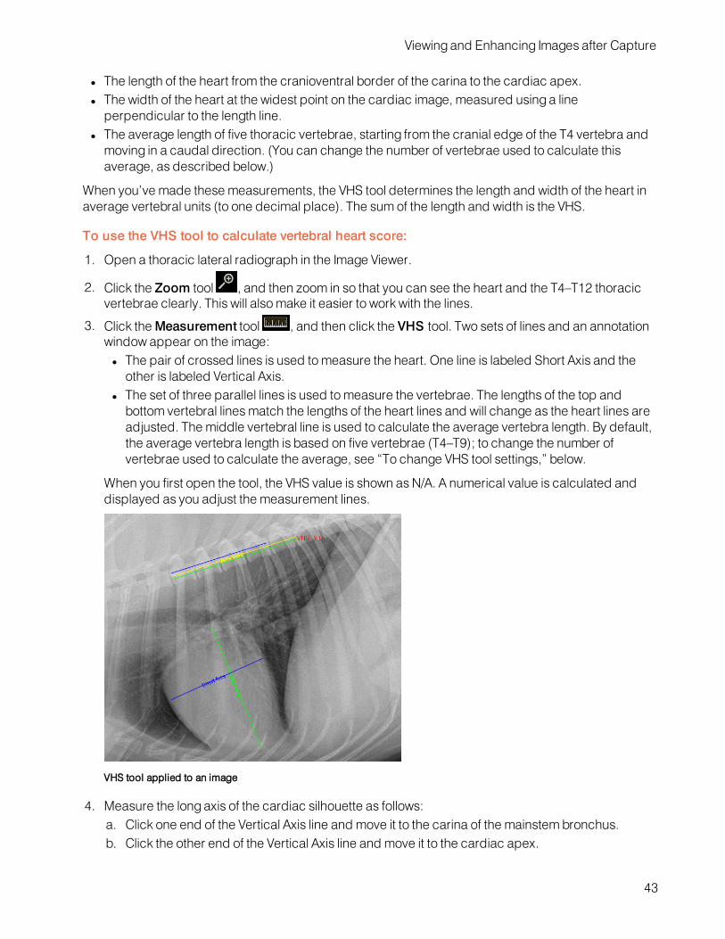
Viewing and Enhancing Images after Capture
l The length of the heart from the cranioventral border of the carina to the cardiac apex.l The width of the heart at the widest point on the cardiac image, measured using a lineperpendicular to the length line.
l The average length of five thoracic vertebrae, starting from the cranial edge of the T4 vertebra andmoving in a caudal direction. (You can change the number of vertebrae used to calculate thisaverage, as described below.)
When you’ve made these measurements, the VHS tool determines the length and width of the heart inaverage vertebral units (to one decimal place). The sum of the length and width is the VHS.
To use the VHS tool to calculate vertebral heart score:
1. Open a thoracic lateral radiograph in the Image Viewer.
2. Click the Zoom tool , and then zoom in so that you can see the heart and the T4–T12 thoracicvertebrae clearly. This will alsomake it easier to work with the lines.
3. Click theMeasurement tool , and then click the VHS tool. Two sets of lines and an annotationwindow appear on the image:l The pair of crossed lines is used tomeasure the heart. One line is labeled Short Axis and theother is labeled Vertical Axis.
l The set of three parallel lines is used tomeasure the vertebrae. The lengths of the top andbottom vertebral linesmatch the lengths of the heart lines and will change as the heart lines areadjusted. The middle vertebral line is used to calculate the average vertebra length. By default,the average vertebra length is based on five vertebrae (T4–T9); to change the number ofvertebrae used to calculate the average, see “To change VHS tool settings,” below.
When you first open the tool, the VHS value is shown as N/A. A numerical value is calculated anddisplayed as you adjust the measurement lines.
VHS tool applied to an image
4. Measure the long axis of the cardiac silhouette as follows:a. Click one end of the Vertical Axis line and move it to the carina of the mainstem bronchus.b. Click the other end of the Vertical Axis line and move it to the cardiac apex.
43

Viewing and Enhancing Images after Capture
5. Measure the short axis of the cardiac silhouette, perpendicular to the vertical axis, as follows:l Click one end of the Short Axis line and move it to one side of the widest axis of the heart.l Click the other end of the Short Axis line and move it to the other side of the widest axis, keepthe line perpendicular to the Vertical Axis.
6. Adjust the vertebral measurement line (middle line of the three parallel lines), as follows:a. Click one end of the middle line and move it to the cranial edge of the T4 vertebra.b. Click the other end of the middle line and move it to the cranial edge of the T9 vertebra (or to adifferent vertebra, if you have changed the tool settings). The VHS tool uses the length of thisline to calculate the average vertebral length.
7. Note the resulting VHS value displayed on screen and in the VHS annotation box.
Tips: To delete the VHS lines from the image, clickDelete in the VHS Annotation Settings window,and then click theMeasurement tool to close it.
To change VHS tool settings:
With the radiograph open in the Image Viewer, open the VHS tool again, and click on any of the VHSlines on the image. The VHS Annotation Settings window opens. Change settings as follows:
l To change the number of vertebrae used to calculate the average vertebra length, type a newnumber in the No. of Vertebrae text box, and then drag the middle measurement line to theappropriate vertebra.
l To override the calculated VHS value, type a new value in the VHS Annotation Settings window,and then click the image to close the window and apply the new value.
l To recalculate the value, delete the current value. Click the image to close the window. The VHSvalue is recalculated using the current line settings.
l To change the color, width, or style of any of the lines, select the appropriate tab in the VHSAnnotation Settings window, choose the desired values, and then click the image to close thewindow and apply the new values.
Using Surgical Templates
Surgical templates are not included in the IDEXX-PACS software, but if you have separate surgicaltemplate software installed, the Surgical Template tool is available in the Image Viewer.This tool allows you to launch the independent software so that you can add templates to a copy of anIDEXX-PACS software image for presurgical evaluation. You can drag and place templates oforthopedic implants onto an image and make surgical measurements.
To use surgical template software on an IDEXX-PACS software image:
1. Open the image in the Image Viewer and then click the Surgical Templates tool .
The surgical template software opens in a new window. The image you were viewing opens in thesurgical template window.
2. Use the surgical template software to apply templates to the image for presurgical planning. Referto the surgical template software user instructions for more detail.
3. After you have applied the templates, bring the template-modified images back into the IDEXX-PACS software. For more information, see "Bringing a Template-Modified Image Back into theIDEXX-PACS Software" on page 67.
44

Viewing and Enhancing Images after Capture
Comparing Images
Tips:
l You can compare up to four images.l You can compare images of different formats (e.g., DICOMwith JPG).l All images being compared must belong to the same patient.l If your IDEXX-PACS software is integrated with your Cornerstone software, always use the IDEXX-PACS software when comparing images for diagnostic purposes, even if the files are non-DICOMimages, such as JPGs.
To compare multiple images for the same patient:
1. Open the study, and then select the thumbnail of the first image you want to compare.
2. ClickCompare above the history panel on the right side of the window.3. Select the thumbnails for the other images. The images open alongside the first image.4. To close an image, select it in the main window and then click the X in the upper right corner; to
stop comparing, select the image you want to keep open, and click Single View .
Viewing Dental Radiographs
When you view dental radiographs, specialized dental viewingmodes are available on the ImageViewer window. These modes allow you to arrange images in a variety of dental layouts and tomoveback and forth among them.There are three modes:• Template layout mode• Review all imagesmode (all the images from the template layout are displayed in a grid)• Review selected imagesmode (selected images are viewed in isolation, at a larger size)Note:Dental image viewingmodes are available only if you have selected Intra-oral Radiography (IO)as the image modality. For more information, see "Labeling a Radiograph as Intra-Oral" on page 45.
Labeling a Radiograph as Intra-Oral
To view dental radiographs in a dental template, you must first label the radiographs as intra-oral.
To label a radiograph as intra-oral:
1. With a dental radiograph open in the Image Viewer, click the study name above the currentlydisplayed dental image and pull down to reveal the study panel.
2. In theModality box (in about the middle of the panel), click the down-arrow and select Intra-oralRadiography.
3. Drag the bottom of the panel up to close the panel.4. Repeat for each radiograph in the study.
5. ClickHome .6. When prompted to save each image, click Yes.
The next time you open the study, the radiographs appear in the dental template. You can select adifferent dental template if needed.
45
Viewing and Enhancing Images after Capture
Using Dental Template Mode
In dental template mode, the primary radiographs in the dental series are displayed in the templatepane and are arranged in a template layout so you can view them together. The rest of the images arein the overflow pane at the bottom of the window.The radiographs are placed in cells that are shown as black rectangles. You can move a radiographfrom one cell to another, either within the same pane or between panes. If you move a radiograph to anoccupied cell, the radiograph currently in the cell moves to the last overflow cell in the overflow pane.Note:Only images identified as Intra-Oral Radiography are displayed in dental templates. For moreinformation, see "Labeling a Radiograph as Intra-Oral" on page 45.
To display radiographs in a different template layout:
1. Click Templates in the Image Viewer toolbar. A menu displays layout choices.2. If using a mouse, move the mouse pointer over the choices in the list to see representations of thelayouts.
3. Select one of the template layouts. The radiographs are displayed in that layout.
To place a radiograph in a different cell:
1. Click the image and drag it to the cell.2. Release the mouse button when the cell turns white. The radiograph now occupies that cell.
Note: The radiograph that had occupied the cell is moved to the last overflow cell.
To remove a radiograph from the template layout:
Click the radiograph, drag it to the overflow pane, and release the mouse button.Note: If you release the mouse button when the radiograph is between cells in the overflow pane (nocell turns white), the radiograph you are moving is placed in the last overflow cell.
To save the current arrangement of radiographs for this series:
No action is necessary. The next time you open the series in the Image Viewer, the radiographs will bein the same cells they were in when you closed the series.
Review Selected Images Mode and Review All Images Mode
Dental image viewingmodes are available only if you have selected Intra-oral Radiography (IO) as theimage modality. For more information, see "Labeling a Radiograph as Intra-Oral" on page 45.
To view one or more radiographs in Review Selected Images mode:
1. Click a dental image or hold down the CTRL key and click more than one image.
2. ClickReview ,and then select Review Selected. The image or images are displayed in alarger size.
Tip: You can also double-click a single image to view it in a larger size.
3. To redisplay the image(s) in the dental template, clickBack to Series in the upper right corner ofthe viewing pane.
46

Viewing and Enhancing Images after Capture
To view radiographs in Review All Images mode:
1. ClickReview , and then select Review All. All images that appeared in the template nowappear in a grid format.
2. To redisplay the image(s) in the dental template, clickBack to Series in the upper right corner ofthe viewing pane.
Viewing Multimedia Files
You can viewmultiframe DICOM files and multimedia files, such as ultrasound images, CT scans, AVI(.avi) files, and MPEG (.mpg, .mpeg) files in the IDEXX-PACS Image Viewer.
To view multimedia files and capture images from them:
1. Import the file to a patient record. For more information, see "ImportingNon-DICOM Files" on page66
The file opens in the Image Viewer with the multimedia toolbar displayed along the bottom.
A thumbnail image is added to the current study. An arrow icon on the thumbnail indicates this is avideo file.
2. Use these tools to view the file:
3. Click the slide bar to quickly move to a specific spot in the video.
To use full-screen mode:
1. When the multimedia player is in full screen mode, press the SPACEBAR to pause or play thevideo.
2. PressESC to return to the Image Viewer in normal mode.
To capture images from the video:
To capture a single frame of the video file, click the Capture button in the video toolbar.Each captured frame is saved as a still image, with a thumbnail in the current study. You can annotatethe still images as needed.
Viewing CardioPet* ECG Files
You can view DICOM*ECG files in the Image Viewer, as long as they were recorded using an IDEXX
CardioPet ECGDevice. DICOM*ECG files are identified by a heart icon in the patient card and inthe history panel of the Image Viewer.
47

Viewing and Enhancing Images after Capture
To view DICOM ECG files:
1. Click the heart icon on the patient card or within the history panel of the Image Viewer. The ECGopens in a separate browser window.
2. Use the tools at the top of the window to adjust the feed and gain and to hide or display the grid.3. Use the scroll bar at the bottom of the window to scroll backward or forward through the ECG.
Note: The standard radiograph viewing tools, the Compare tool, and the Clone tool are not availablefor use with ECG files. However, you can use certain tools located below the history panel:Import/Export, Reassign, Save, Print, and Delete.
48

Managing ImagesReassigning a Study
You must assign a STAT study to a patient, following the steps below. You can also use thesesteps to reassign any existing study to a new or different patient.Note: You can reassign entire studies only, not individual images.IMPORTANT: If your IDEXX-PACS software is integrated with your Cornerstone software,always reassign diagnostic images in the IDEXX-PACS software, not in the Cornerstonesoftware. This way the updated information will be returned to Cornerstone, and theCornerstone record will be updated accordingly.
To reassign a study:
1. Find and open the patient study, and then clickReassign in the bottom right tool bar ofthe Image Viewer. The Reassign Images window opens.
Tip: If this is a STAT patient, you can also click the Reassign button at the top of theSTAT patient card in the Home window.
2. In the Reassign Images window, select the appropriate tab to assign the study either to anexisting patient or to a new patient (for an existing patient, you must then enter searchcriteria and select the patient from the search results).
3. If this is a new patient, enter the patient information; if this is an existing patient, update anyinformation if needed; when finished, clickReassign.
A confirmation message displays the patient and client information for the reassigned study.Note:A patient ID in square brackets is one generated by your practice informationmanagement system.
4. ClickOK to close the message.
If your IDEXX-PACS software is integrated with Cornerstone software, the reassigned study isremoved from the current Cornerstone Patient Clipboard and will be available in the PatientClipboard for the patient you assigned the study to.
Editing Image Details
Image details include series title, modality, and technique settings.Note: The image details feature should be used only in a manner consistent with local lawsgoverning the maintenance of veterinary records used for diagnostic purposes.
To view and edit image details:
1. With the image open in the Image Viewer, click the study name above the image and holddown the mouse button as you pull down the study panel. (Touch screen: press the nameand pull down.)
2. Edit the following, as needed:l Series title and comments
49

Managing Images
Note: The study title cannot be edited; it can be changed only on the shot selection screenbefore image capture.
l Image titlel Capture date, source, technician name, veterinarian name, and modalityNote: The ability to change the capture date is provided so you can enter the correct date forscanned documents.
3. Edit the following technique details, as needed:l Distance—The distance from the x-ray source to the detector or screen; select the unit ofmeasure from the drop-down list.
l Thickness—The thickness of the body part in centimeters.l Sedation—Select from the drop-down list.l Exposure mAs—The amount of x-ray exposure in milliampere-seconds (mAs). This iscalculated automatically if you enter values in the Exposure mA (current in milliamperes) andExposure Time (duration in seconds) text boxes.
l Exposure kVp—The amount of voltage (peak kilovoltage) used for this image.l Use of Grid—Select this check box if a grid was used.
4. When finished, pull the bottom of the panel back up to close it.
Saving an Image in a Different Format
After you click Done at the end of a capture session, all captured images are saved to the database.However, you can also save images to your local computer or network in various file formats, so thatthe images can be imported into other software programs or sent over the Internet.Available formats:
l Bitmapl JPEGl JPEG 2000l RAWl TIFFl DICOMl Video (.avi, .mpg, .mpeg)
To save images in a different file format:
1. In the Home window, find the patient record for the study date; then in the patient card click ,
and then click .
OR
If the image is already open in the Image Viewer, click in the lower right area of the window.
The Sharingwindow opens. Thumbnails from the current study are at the top left.
2. Use the Sharingwindow to select the images you want to save. For more information, see "Usingthe SharingWindow" on page 58.
3. When you have selected all the images, click .4. Choose a location and enter a file name.
50
Managing Images
5. Select the format from the Files of Type drop-down list.6. To include any annotations you applied in the Image Viewer, select Burn Annotations.7. To include the overlay information displayed in the Image Viewer, select Burn Overlays.8. Click Save.9. ClickClose to close the Sharingwindow.
Cloning an Image
When you clone an image, you make a copy of the current version of the image, including anyannotations or enhancements.
To clone an image:
1. With the image open in the Image Viewer, click in the lower right corner of the window. A copyof the image appears beside the original image. "(Copy)" is appended to the image name.
2. Modify or adjust the cloned image as needed.3. To save the cloned image, click the X in the upper right corner of the image, and then click Yeswhen prompted to save.
Deleting an Image
IMPORTANT:
l Only a system administrator can delete images. For more information, see "Creating a SystemAdministrator Account" on page 79.
l Only individual images can be deleted; you cannot delete series or studies. A series or study isautomatically deleted when all the images within it have been deleted.
To delete images:
1. Log on as the system administrator.2. In the Home window, find the patient record containing the image to be deleted.3. Click the thumbnail in the patient card to open the image in the Image Viewer.
4. In the lower right corner of the window, click .
You are prompted to confirm the deletion.
5. Click Yes to delete the image, or No to keep the image.
Ensuring that Images Are Correctly Matched to Patients
If you notice that some images are associated with the wrong patients or that there are duplicatepatient records in the IDEXX-PACS software, your system configuration may have changed sinceinstallation, and you may need to adjust your patient-matching settings.Follow the steps below to review and adjust your settings as needed.
Check settings on your image-capture computer
Your image-capture computer should use patient IDs from your practice information managementsystem, if that system is integrated with IDEXX-PACS. These IDs are known as PIMS IDs.Check these settings on the image-capture computer to ensure you are using the PIMS ID (if you havean integrated system):
51
Managing Images
l Check your Client and Patient settings. For integrated systems, choose the option to use patientIDs from your practice information management system.
l Under Global Settings select the practice information management system that you use.Note:Selecting the system name will also add a system identifier to each image (in the image DICOMtags).
l Check your local DICOM server configuration. In the Image Import Settings area, make sure thatPatient ID is selected in the Patient Match Level drop-down list and that the Use PIMS Patient IDoption is selected at the bottom of the window.
Check image import settings on your IDEXX-PACS server
On your IDEXX-PACS server, check your local DICOM server configuration. In the Image ImportSettings area, the Patient Match Level drop-down list should display one of the following:
l Patient ID—if your IDEXX-PACS software is used only with IDEXX digital imaging systems (forexample, you do not import images from an ultrasound, a dental, or a non-IDEXX radiographysystem).
l Patient Name, Sex, Species, DOB, Client Last Name—if your IDEXX-PACS software is usedwith any non-IDEXX systems (such as an ultrasound, a dental, or a non-IDEXX radiographysystem). In this case, also deselect the Use PIMS Patient ID check box at the bottom of thewindow.
Starting the image request
To avoid mismatched images, always begin the image request from your practice informationmanagement system.This is especially important if you use Cornerstone, AVImark, or ImproMed software. When you start animage request from one of these systems, IDEXX-PACS assigns a unique ID (separate from the PIMSor patient ID) to the requested study. That ID will be used to link the captured images to the imagerequest, ensuring that images are matched to the correct patient record, without relying on the patientID or other patient information.
Using the Radiology Log
Radiology log information is automatically recorded for each radiograph taken on your IDEXXDiagnostic Imaging System.You can record technique details during the capture process or when you are reviewing the imageslater.You can view the entire log, print it, email it, or export it as a PDF (Portable Document Format) file.
Viewing and Editing the Radiology Log
The radiology log captures information about each image. The information comes from the image'sstudy panel. For more information, see "Recording Technique Information in the Radiology Log " onpage 53.You can also add information to the log directly, as described below.
52

Managing Images
To view and edit the radiology log:
1. In the Home window, click Tools > Radiology Log....2. Enter any combination of search criteria:
l Date range (use the Calendar button )l Client or patient name or IDl Technician namel Modality
3. Click Search.4. Scroll down to see all the results, and scroll to the right to see all the information for each image.5. To sort the log, click the column header you want to sort by.6. To add an entry to the log, click Add Entry, and then enter information in the new row at the top ofthe table.
7. To edit information, click in the cell, and then enter or select a new value. You cannot edit patientand client information.
8. To delete an entry you added, click in the row to select it, and then clickDelete Entry.
Note: You cannot delete entries created automatically by the IDEXX-PACS* software.
9. To print or share the logwith others:
l Click Print Log, and then click Print in the Preview window.l Click Email Log, enter and address and a subject, and then click Send.l Click Export as PDF, enter or select a location to save the PDF, and then click Save.
10. When finished, click Save and thenClose.
Recording Technique Information in the Radiology Log
You can enter technique details for an individual image in the image's study panel. The information isthen stored in the radiology log.
To record information in the radiology log:
1. With the image open in the Image Viewer window, pull down the study panel. For more information,see "Editing Image Details" on page 49.
2. Enter or edit the following information to save it in the radiology log:l Distance—Distance between generator and body part.l Thickness—The thickness of the body part being imaged.l Sedation—Select from the drop-downmenu.l Exposure mAs—The amount of x-ray exposure in milliampere-seconds (mAs). This iscalculated automatically if you enter values in the Exposure mA and Exposure time boxes.
l Exposure mA—The amount of current in milliamperes (mA).l Exposure Time—The duration of the exposure in seconds as a decimal value, or use the drop-downmenu to select a fraction of a second.
l Exposure kVp—The amount of voltage (peak kilovoltage) used for this image.l Use of Grid—Select the check box if a grid was used.
53
Managing Images
Backing Up and Archiving Images
Be sure to back up your data frequently. IDEXX-PACS* Imaging Software offers several backupoptions.In addition, you may want to archive older images to save space on your computer. Archivingmovesimages from the local computer to another location for long-term storage.For more information about backing up and archiving, see the sections below.
Backing Up Your Data
IDEXX-PACS* software offers three backup options:
l Full—The IDEXX-PACS software stores the last 3 days of images and data. Data backups arestored in the cloud (in the event of a system failure, contact IDEXX Diagnostic ImagingCustomersupport to download data files to your server if needed).
Note:Backup folders are saved for 3 days only. After 3 days, the next backup will overwrite theoldest of the 3 folders. In addition, multiple backups in a single day overwrite any other backupsmade the same day. To keep images and data for a longer period, transfer them to a CD/DVD, anexternal drive, or other media.
l External—A third-party program of your choice manages the backup process. If this processcopies the local IDEXX-PACS backup folders, remind your backup vendor that these folderscontain only the last 3 days of images and data. To keep images and data for longer periods, workwith your backup vendor to establish an appropriate backup schedule and process.
l Data Only—A third-party program backs up the IDEXX-PACS images, while the IDEXX-PACSsoftware backs up the data files.
You can change your auto-backup option, if needed. For more information, see "SettingUp Auto-Backup Options" on page 90. In addition, you can always back up your systemmanually, as describedbelow.Follow the procedures below to perform the backup.
To back up while using the IDEXX-PACS software:
1. Do one of the following:
l In the Home window, click Tools >Backup System.OR
l Close the IDEXX-PACS software, and click Yes when asked if you want to back up your system.
2. Leave the default backup folder location (C:\Program Files\IDEXX\IDEXX-PACS\backup), or clickBrowse and select a different location.
Note: IDEXX strongly recommends that you back up your system to a network location.
3. ClickOK.4. If asked whether you want to overwrite existing files, click Yes to All.
IMPORTANT: If you use the Full or Data Only backup option, you must manage the backup folder forspace.
To manually back up your data:
1. Close the IDEXX-PACS software.2. Insert a writeable CD or DVD into your disc drive.
54

Managing Images
3. Double-click the Computer (orMy Computer) icon on your desktop, and then double-click theLocal Disk (C:) icon in the My Computer window.
4. Double-click the Program Files folder, and then the IDEXX folder, and then the IDEXX-PACSfolder, and then the Backup folder.
5. Hold down the CTRL key while you click to select all of the following: the Images folder and each ofthe three folders labeled with dates.
6. While the items are still selected, right-click any of the items and selectCopy.7. Double-click the Computer (orMy Computer) icon again.8. Find the drive location for your storage media (CD/DVD or other device), right-click the drive name,and select Paste to copy the files.
9. When the files have been copied, open the drive, and verify that all of the files are there:l Check dates of the backed-up files to ensure that they match the files in the C:\ProgramFiles\IDEXX\IDEXX-PACS folder.
l Check the total number of images in the backed-up Images folder to ensure that it matches theoriginal Images folder. The total is displayed on the left side of the window’s status bar.
To check backup status:
Backup status is visible in the Connections drop-down list in the Home window:
l Green: Backup < 3 days agol Yellow:Backup > 3 days agol Red:Backup > 2 weeks ago
Archiving Images
Unlike a backup, which saves a copy of your images and data, archiving removes image files fromyour computer and saves them to a CD or DVD, or to a network location, such as a server. Archivingcan save space on your computer and make image management easier.You can view archived images in the Image Viewer window. For more information, see "AccessingArchived Images" on page 56.IMPORTANT:Store archived images in a safe location! If archived files are lost or damaged, theimages cannot be recovered.Tip: You can set up the IDEXX-PACS software to alert you when it's time to archive images. For moreinformation, see "SettingUp Archive Alerts" on page 90.
To archive images:
1. In the Home window, click Tools >Archive Images.2. Enter any or all of the following search criteria:
l Patient and client names or IDsl A date range in which the images were capturedl Include Inactive, Include Deceased, or Show only inactive/deceased option(s)
3. Click Search. Each row in the results table represents a separate study.
4. By default, all of the studies are selected for archive; to select specific studies:l Hold down the CTRL key and click to deselect or select individual studies.l ClickDeselect All or Select All.
55

Managing Images
5. In the Archive To area:l SelectCD/DVD, and then enter the number of copies you want tomake.OR
l Select File Folder, and then enter the folder location, or clickBrowse and then find and selectthe location. Locations can include a folder on your computer, on another computer in yournetwork, or on an external hard drive.
6. In the Archive Label box, enter a name for the archive file.7. If using a CD/DVD, insert the disc; if using an external hard drive, connect the drive to yourcomputer.
Note:Be sure to label discs with the date. IDEXX recommends using the same format as theIDEXX-PACS software: "IDEXX-PACS YYYY-MM-DD (#)" where # is the CD number.
8. When ready, click Archive at the top of the window.
Note:Archived images are identified by an "Archived" banner across the patient card in the Homewindow.
Accessing Archived Images
When an image is archived, it is removed from the database and stored in your archive location (e.g.,archive hard drive, CD, network server, etc.). A thumbnail of the image remains in the patient record asa reference.
To view archived images:
1. In the Home window, search for the archived image, just as you would any image in the IDEXX-PACS software.
An "Archived" banner is displayed over the thumbnails on the patient card.
2. To view the image, click the image thumbnail:l If the image is readily accessible (e.g., on a networked computer or server), it opens in theImage Viewer.
l If the image is stored in a remote location (such as a CD), a message indicates the location.Insert the disc or browse to the location on the external hard drive or network, and clickOK.The image opens in the Image Viewer window.
3. To reactivate this image, click on the Image Viewer window to save the images in the database.You can now use this image like any other image in the IDEXX-PACS software.
Using IDEXX Web PACS or the IDEXX ImageBank* Storage System
IDEXX WebPACS and the IDEXX ImageBank* storage system store your digital images securely andaffordably in the cloud. You can view your digital radiographs wherever you have Internet access,making it easy to work from home or in the field, and making it simple to restore files whenevernecessary.You should continue to back up your client and patient data and software system files using yourstandard backup procedure. 56

Managing Images
Activating IDEXX Web PACS
Access to the IDEXX WebPACS software requires an IDEXX VetConnect* PLUS account, as well as anIDEXX WebPACS subscription.To learn more about subscribing to IDEXX WebPACS, contact your IDEXX representative.
How New Images Are Uploaded
When you subscribe to IDEXX WebPACS, your images are uploaded to the cloud shortly after youcapture them or import them into your IDEXX PACS system.The images are placed in a queue and then uploaded as your local bandwidth allows. Copies of theimages are also kept on your local system until storage limits are reached.Uploaded images include all DICOM images that you capture, import, retrieve, or place in your watchdirectory. You can upload other file types besides DICOM, as well. For more information, see "SettingUp IDEXX WebPACS Storage Options" on page 89.
To view the status of the image upload:
1. In the Home window, clickConnections.2. On the ImageBank row, clickQueue.
The queue displays a list of image uploads and their progress.
Uploading Legacy (Older) Images
To upload images captured before you subscribed to IDEXX WebPACS software (known as legacyimages), you will need an upload kit from IDEXX that contains a flash drive. Contact IDEXX DiagnosticImagingCustomer Support for pricing and upload options.Follow the instructions in the kit to copy the legacy images to the drive and to send the drive to IDEXXWebPACS at Amazon WebServices*.
Viewing Images Stored in the Cloud
To view images directly in IDEXX Web PACS:
1. In a web browser, go to vetconnectplus.com.
OR
In the IDEXX-PACS Home window, clickMenu and then click the VetConnect PLUS icon .
2. Log on to VetConnect PLUS, and then click Imaging in the VetConnect PLUS title bar.
Tip: If you are already logged on to VetConnect PLUS, the IDEXX WebPACS software will openautomatically.
To view cloud-based images in the IDEXX-PACS* software:
1. In the IDEXX-PACS Home window, search for the patient record and the study you want.2. Look for a small cloud icon in the upper right corner of the thumbnail on the patient card.
3. Click the thumbnail to view the images in the Image Viewer, just as you would any image storedlocally on your computer.
57

Importing and Distributing ImagesUsing the Sharing Window
Use the Sharingwindow to send images by DICOM or email, and to submit cases to IDEXXTelemedicine Consultants.Tip: You can send both images and ECG files from IDEXX-PACS software.
To open the sharing window:
In a patient card, click , and then click Share .OR
In the Image Viewer, click Share in the lower right corner of the window.
To use the sharing window:
1. At the top of the sharingwindow, on the left, click the arrows to find and select the studydate.
All studies and series for the selected date are listed below.
2. Click the Add Study or Add Series link tomove the images to the right pane.
OR
Click individual images tomove them to the right pane.
3. Repeat step 2 to select additional images, if needed.
Tip: To remove images from the right pane, clickRemove Study or Remove Series, orclick the individual image.
4. When all the images you want to share are in the right pane, click the button for the sharing
method, for example, Telemedicine or .5. Provide any additional information required by the sharingmethod.
Printing Images
You can print an image, a series, or a study.
To print an image:
1. In the Home window, find the patient record, and in the patient card click , and then
click Print .
OR
With the image open in the Image Viewer, click Print .
The Sharingwindow opens.
58

Importing and Distributing Images
2. Use the Sharingwindow to select the images you want to print. For more information, see "Usingthe SharingWindow" on page 58.
3. When you have selected all the images, click .
The images open in a Print Preview window.
4. (Optional) Use the tools at the top of the window to review the images before printing.
5. When ready, click Print , specify which images to print (all or a range), and then clickOK.
6. Click to close the Print Preview window, and then clickClose to close the Sharingwindow.
Emailing Images
You can email an image, a series, or a study from either the Home window or the Image Viewer.You can send the image in these formats:
l JPEG and TIFF formats, which can be viewed by most Internet browsers and many graphicsprograms.
l DICOM format, which can be viewed only by specialized medical or veterinary imaging softwarelike the IDEXX-PACS software.
Before you begin:
l Your system’s email default settingsmust be set up before you can email images. For moreinformation, see "SettingUp Email" on page 82.
l If you want to include annotations, overlays, or window/level adjustments in DICOM images, selectexport options as follows:
o Go toSettings > Imaging.o Select desired export options under Image Export on the right side of the window and thenclick Save:
To email images from the Home window or the Image Viewer:
1. In the Home window, find the patient record, and in the patient card click , and then click
Share .
OR
With the image open in the Image Viewer, click Share in the lower right corner of the window.
2. Use the Sharingwindow to select the images you want to email. For more information, see "Usingthe SharingWindow" on page 58.
3. When you have selected all the images, click .
An email message box opens.
59

Importing and Distributing Images
4. Enter the email address, a subject line, and a message.5. Select a file format for the images.
The JPEG, JPEG 2000, and TIFF formats can be viewed in most Internet browsers and manygraphics programs; the DICOM format can be viewed only by using specialized medical orveterinary imaging software.
6. Click Send.7. ClickClose to close the Sharingwindow.
Sending Images Automatically after Image Capture (Auto-Routing/Auto-Exporting)
Auto-routing sends images via DICOM® to a specified destination, such as a consultant, after imagecapture. You can specify fully automatic routing or semi-automatic routing.Auto-exporting sends copies of the images to a file folder on your computer or network.
Before you begin:
Enable the auto-routing and/or auto-exporting feature. For more information, see "SettingUp Auto-Routing and Auto-Exporting" on page 88.
To send images via fully automatic auto-routing or auto-exporting:
No intervention is needed. The captured images are sent to the predefined destination automatically.Use the DICOM queue to check progress, or to pause, restart, or cancel image sending. For moreinformation, "Using the Outbound DICOMQueue" on page 65.
To send images via semi-automatic auto-routing:
1. ClickDone at the end of the image capture session. The Send DICOMwindow opens.2. In the Images to Send box, select the images by holding down the CTRL key and clicking imagenames.
3. In the Remote Servers box, select the destination. (To send the images tomore than onedestination, hold down the CTRL key while clicking the destinations.)
4. Click Send.5. Use the DICOM queue to check progress, or to pause, restart, or cancel image sending. For moreinformation, see "Using the Outbound DICOMQueue" on page 65.
Using DICOM to Send and Receive Images
The DICOM® (digital imaging and communications in medicine) standard allows for the transfer,storage, and printing of digital images between various software programs or equipment, regardless ofthe manufacturer.The DICOM queue manages the sending and receiving of DICOM files. The DICOMQueue windowprovides two tabs:
l Outbound—Lists all DICOM files being sent out of your IDEXX-PACS systeml Inbound—Lists all DICOM files being received by your IDEXX-PACS system from external sources
Sending Images to Remote DICOM Servers
IMPORTANT:Before you can send your images to other DICOM-enabled servers, you must configureDICOM settings for the sending and receiving computers. For more information, "Overview of DICOMServices Configuration" on page 73.
60

Importing and Distributing Images
When you send a radiograph via DICOM, the original version of the radiograph is sent (the version as itlooked when you clicked Done Capture or when you imported or received the file). Changes such asannotations and window/level adjustments are not included. To send a version containing theseupdates, contact IDEXX Diagnostic ImagingCustomer Support at 1-877-433-9948.
To send images from the Home window:
1. In the Home window, find the patient record, click expand , and then click Share .
OR
In the Image Viewer, click Share .
Use the Sharingwindow to select the images you want to send. For more information, see "Usingthe SharingWindow" on page 58.
2. When you have selected all the images, click the DICOM button.
The Send DICOMwindow opens. The Images to Send list shows the images you have selected.
3. In the Remote Servers box, select the image destination. (To send the images tomore than onedestination, hold down the CTRL key while clicking on the destinations.)
4. Click Send.5. ClickClose to close the Sharingwindow.
Use the DICOM queue to check the progress of the images, or to pause, restart, or cancel imagesending. For more information, see "Using the Outbound DICOMQueue" on page 65.
Importing Images from Remote DICOM Servers Using Query and Retrieve
DICOMQuery and Retrieve lets you search for and import image studies from remote servers, so youcan view the images in the IDEXX-PACS software.
Before you begin:
The remote serversmust be defined in the IDEXX-PACS software. For more information, see"ConfiguringRemote DICOM Servers or Modality Worklist Connections" on page 75.
To import an image from a remote server:
1. In the Home window, click Tools > Query Retrieve.2. Type search criteria in the search text boxes, and select the remote server from the drop-down listin the Location box.
Tip:Servers appear in the list only if they have been defined in the IDEXX-PACS software.
3. Click Search.4. In the search results, select the study (or studies) you want to import and clickRetrieve. Amessage notifies you that the study has been requested.
5. ClickOK. The software begins importing the file(s).6. To check the status of the import request, in the Home window, clickConnections.7. On the DICOM row of the Connectionsmenu, clickQueue to open the DICOM queue window.8. Select the Inbound tab, find the request in the table, and note the status in the Status column. Formore information, see "Using the Inbound DICOMQueue" on page 64.
When the status says "Complete," the study has been imported.
61

Importing and Distributing Images
9. When the import process is complete, search for and open the images as you would any otherimages.
Importing Local DICOM Files Automatically
If you frequently import multiple DICOM images, you can import them automatically by placing theminto a DICOMwatch directory. The default location of the watch directory is C:\ProgramFiles\IDEXX\IDEXX-PACS\dicomdrop.The software checks the watch directory at specified intervals and automatically imports any images itfinds.Note: IDEXX recommends that you test the DICOMwatch directory with a single image to ensure thatall settings are properly configured.You can also import DICOM imagesmanually. For more information, see "Before you begin" on page62.
To use the DICOM watch directory:
1. Use the DICOM configuration window to specify the location of the watch directory (if you don'twant the default location) and to specify the interval at which the software checks the directory. Forinformation, see steps 12 and 13 in "Configuring the Local DICOM Server" on page 73.
2. When you have DICOM images to import, copy them to the DICOMwatch directory.
The images will be imported based on the specified schedule. If you are importing a large numberof images, the processmay take a couple of minutes.
Importing Local DICOM* Files Manually
You can import both DICOM radiographs and DICOM ECG files (identified by a heart icon ).You can import the files two ways:
l Based on patient IDl By importing files directly into the currently open patient record.
Both options are explained below.Tip: You can also import DICOM files automatically by adding them to a watch directory that importsfiles at specified intervals. For more information, see "Importing Local DICOM Files Automatically " onpage 62
Before you beginThe DICOM file must be located on your local computer or local network.
Importing a DICOM file based on patient IDWhen you import a DICOM file this way, the IDEXX-PACS software assigns the imported file to a patientrecord based on the patient ID in the imported file’s DICOM tags. If no patient record matches the ID,IDEXX-PACS creates a new patient record for the file.
To import the DICOM file:
1. In the Home window, click Tools > Import DICOM.2. Use the Look In drop-down list or the browsingwindow to locate the image.
Tip:Use these buttons to navigate to the image location:
Move up one folder62

Importing and Distributing Images
Return to the Home folder (Desktop)
View file names only
View file details (name, size, type, and date modified)
Note: If you do not see the files displayed in the browsingwindow, select All Files in the Files ofType drop-down list. This allows you to see DICOM files that have been saved without the standard.dcm or .dic file extensions.
3. Click the file name to select it, and then click Import.
Tip: To select multiple files, hold down the CTRL key while you click the file names. (Touch screen:you must import files individually.)
4. When the DICOM queue window opens, click the Inbound tab to view the file import status; youmay need to clickRefresh to see the most current status.
5. When the import is complete, the image is added to the patient record. If the Cornerstone Softwareis integrated with IDEXX-PACS software, the image will be available in the Patient Clipboard.
Importing a DICOM file into a currently open studyWhen you import a DICOM file this way, the file is added to the study that is currently open in the ImageViewer, regardless of information in the file’s DICOM tags.
To import the DICOM file directly into a study:
1. Find and open the patient study you want to add the DICOM file to.
2. In the Image Viewer window, click Tools > Import DICOM Direct.3. Find and select the file(s) to import and then click Import.
If the imported images are not visible in the Cornerstone Patient ClipboardThis can happen if the images are not imported to the correct patient ID. In this case, you mustreassign the imported images to the correct patient ID for the images to become visible in the PatientClipboard.
To reassign an imported image to a different patient ID:
1. In the Patient Clipboard, right-click the patient name that the imported image should be assignedto, and then select Imaging > IDEXX-PACS image viewer. The patient record opens in theIDEXX-PACS Image Viewer.
2. Note the patient ID located next to the patient name at the top of the Image Viewer. This is thepatient ID to which you should reassign the imported image.
3. ClickHome to display the IDEXX-PACS Home window.4. Open the imported images from the inbound DICOM queue, as follows:
a. In the Home window, click (button color may be different).b. On the DICOM row, clickQueue.c. Select the Inbound tab.d. ClickRefresh to see the most recent information.e. Double-click the row for the imported image to open the study in the IDEXX-PACS ImageViewer.
63

Importing and Distributing Images
5. In the Image Viewer, clickReassign on the bottom right side of the window.6. Use the Reassign window to reassign the images to the correct patient ID from step 2.
Viewing the Status of the DICOM Connection
To view the status of the DICOM connection:
1. In the Home window, click .2. On the DICOMQueue row, note the color of the circle. It tells you the status of the DICOMconnection:l Green—IDEXX DICOM services are running, and the queue does not contain any sent orreceived images with a status of Failed.
l Yellow—Check the queue for information about sent or received images that have failed. Youmay see this status when DICOM has either started or stopped.
l Gray—If DICOM services have already been configured but the connection button is gray, theIDEXX-PACS application services are not running. You must start the IDEXX-PACS ApplicationServer (see below).
l Red—The DICOM server is not running. If you are using the computer that is your IDEXXDiagnostic Imaging server, click Start on the DICOM row.
3. To open the DICOM queue, clickQueue on the DICOM row.
To start or stop the DICOM queue:
1. In the Home window, click .2. On the DICOMQueue row, click Start or Stop.
Starting the IDEXX-PACS Application Server
If DICOM services have been configured but the DICOM connection status is inactive (the DICOMconnection button is gray), the IDEXX-PACS Application Server is not running.
To start the IDEXX-PACS Application Server:
1. PressCtrl+Alt+Delete.2. Click Start Task Manager.3. Click the Services tab.4. Click the Name column heading to sort the services alphabetically.5. Scroll down until you find the service named IDEXXPACSApplicationServer.6. In the Status column, right-click and select Start Service.7. Click the X in the upper right corner of the window border to close the window.
Using the Inbound DICOM Queue
The inbound DICOM queue lists images that have been received by the system or are in the process ofbeing received.
To use the inbound DICOM queue:
1. In the Home window, click (button color may be different).2. On the DICOM row, clickQueue.
64

Importing and Distributing Images
3. Select the Inbound tab.4. ClickRefresh to see the most recent information.
Possible Status values for inbound DICOM files are:
l Requested—Images have been requested but not yet received.l Complete—Images were received successfully and can be viewed in the IDEXX-PACS software.l Receiving—Images are being received. You cannot remove images that have this status.l Pending—Requested images are waiting to be sent.l Failed—Images were not received successfully.
To remove a received or a failed image from the queue:
Select the image row, and then clickRemove on the right side of the window.
To remove all information from the inbound queue:
ClickClear Queue.
To close the DICOM Queue window:
Click the X in the upper right corner.
Using the Outbound DICOM Queue
The outbound DICOM queue lists images that are being sent to another location.
To view the outbound DICOM queue:
1. In the Home window, click (button color may be different).2. On the DICOM row, clickQueue.3. Select theOutbound tab.4. From the Destination drop-down list, select the destination you want to review.5. ClickRefresh to see the most recent information. The following information is displayed:
l Destination to which the image is being sentl Patient namel Owner namel Image namel Study datel Number of retriesl Status of the transmissionl Status details
6. Possible Status values are:l Pending—Images are waiting to be sent.l Sending—Images are in the process of being sent.l On Hold—Images are paused and will not be sent until you resend them.l Cancelled—Images will not be sent.l Failed—Images were not sent successfully, but you can resend them.l Permanently Failed—Image sending failed for a more serious reason and images cannot be
65
Importing and Distributing Images
resent.l Complete—Images have been sent successfully.
To change the status of images in the queue:
Select the image and then use the buttons on the right:
l Send—Sends the image.l Pause—Puts a sending or pending image on hold. The image will not be sent unless you restart it.l Remove—Removes a selected image from the list.
To remove Completed, Failed, and On Hold items from the outbound queue:
ClickClear Queue.
To close the DICOM Queue window:
Click the X in the upper right corner.
Importing Non-DICOM Files
You can import the following types of non-DICOM® files:
l .bmp—Microsoft Windows bitmapl .jpg or .jp2—Joint Photographic Experts Group (JPEG) file interchange format or JPEG 2000l .tif—Tagged image file format (TIFF)l .avi—Audio video interleaved formatl .mpg, .mpeg—Moving Picture Experts Group formatl .ecg—Electrocardiogram files
You can also import DICOM images. For more information, see ""Before you begin" on page 62.
Before you begin:
l You must import files into an existing patient record. If this is a new patient, create a patient recordfirst. For more information, see "To add a new patient without capturing radiographs" in "Adding aNew Patient and Client Record" on page 70. Then follow the steps below.
l The files to be imported must be stored on your network or local computer.
To import an image from the Image Viewer:
1. Find and open the patient study you want to add the filet to.
2. In the Image Viewer, click Tools > Import Non-DICOM.3. Use the Look In drop-down list, or the browsing area below it, to locate the image.
Tip:Use these buttons to navigate to the image location:
Move up one folder
Return to the Home folder (Desktop)
View file names only
View file details (name, size, type, and date)66

Importing and Distributing Images
4. To look for specific file types, click the down arrow in the Files of Type box, and select the imageformat.
5. Click the file name to select it, and then click Import. To select multiple images, hold down theCTRL key while you select the files. (Touch screen: you must import files individually.)
The image opens in the Image Viewer, and a thumbnail is added to the currently open series. Ifthere is no current series, a new study and series are created to contain the imported file(s).
6. Pull down the study panel at the top of the Image Viewer, and then edit the following as needed:a. Series titleb. Capture date
7. Pull the panel back up to close it.
8. ClickHome .9. When prompted to save the image(s), click Yes.
Importing Images by Scanning
Before you begin:
l You must define the scanner source in imaging settings. For more information, see "SettingUpImagingUnits, Compression, and Export Options" on page 87.
l Scanned imagesmust be added to a current patient record; therefore, create a new client andpatient record first, if needed. For more information, see "Adding a New Patient and Client Record"on page 70.
To scan images:
1. Find the patient record you want to add the images to, and open it in the Image Viewer.2. If your device scansmultiple pages at one time, place the pages into the scanner, and then in the
IDEXX-PACS Image Viewer, click Tools >Multi TWAIN. The images are acquired anddisplayed.
OR
If your device scans one image at a time (such as a flatbed scanner), place the image in the
scanner, and then in the IDEXX-PACS Image Viewer, click Tools > Single TWAIN. The imageis acquired and displayed. Repeat for each image you want to scan for this patient.
3. For each image, pull down the study panel at the top of the Image Viewer, and then edit thefollowing as needed:a. Series titleb. Capture date
4. Pull the panel back up to close it.
5. ClickHome .6. When prompted to save the image(s), click Yes.
Bringing a Template-Modified Image Back into the IDEXX-PACS Software
There are two ways to bring an image back into the IDEXX-PACS software after you have appliedsurgical templates to it. The method you use depends on whether DICOM services are enabled onyour system.
67

Importing and Distributing Images
If IDEXX DICOM Services are enabled on your system:
1. While using the template software, export the image as a DICOM file by using the Commit feature inthe template software. (See your surgical template software documentation for instructions.)
2. On the IDEXX-PACS Home window, open the study that contains the original image. The newtemplate-modified image is included in the same series as the original image.
If IDEXX DICOM Services are not enabled on your system:
1. While using the template software, save the image as a JPEG file. (See your surgical templatesoftware documentation for instructions.)
2. In the IDEXX-PACS software, open the patient study that contains the original image, and thenimport the JPEG file. For more information, see "ImportingNon-DICOM Files" on page 66.
The image appears in the Image Viewer window and is added to the same series as the originalimage.
Note:When a template-modified image is brought into the IDEXX-PACS software, the templatebecomes part of the image. If you want to save an image with a template and then make additionalmodifications, you must reopen the file using the surgical template software.
Creating a Patient CD
You can create a Patient CD that contains both JPEG and DICOM versions of the patient's images, aswell as patient information and image viewing software.Note: The Patient CD feature is not supported on virtual machines (i.e., emulated computer systems).By default, the images saved to the CD are the original versions (as they looked when you clicked Doneat the end of the capture process). Changes such as annotations and window/level adjustments arenot included.To create a version containing these updates, contact IDEXX Diagnostic ImagingCustomer Support 1-877-433-9948.
To create a Patient CD:
1. Insert a writeable CD into the disc drive (a CD that can be written only once); do not use a RE-writeable CD.
Note: You can use a DVD if you have a drive on your computer that will burn DVDs and if thecomputer used to view the images has a DVD drive.
2. In the Home window, find the patient record, click , and then click Share .
OR
With any of the patient's images open in the Image Viewer window, click Share .
Use the Sharingwindow to select the images you want to copy to the Patient CD. For moreinformation, see "Using the SharingWindow" on page 58.
3. When you have selected all the images, click . The Select Export Preference window opens.4. Enter the number of CD copies you want tomake, and then clickOK.
A message states that components are being compiled.
When the CD is finished, a message states that the Patient CD is complete.
68

Importing and Distributing Images
5. ClickOK. The finished CD is ejected automatically.6. ClickClose.7. To view the CD, see "Viewing a Patient CD" on page 69.
Note:After you create a Patient CD, you cannot add to or change the information on the CD.
Tip: You can choose to save the information to a file folder instead of a CD. See the next section.
To save Patient CD information to a file location:
You might choose to save the information to a file if you want to put the images in an email or on a USBflash drive; or you might want to compile the information for Patient CDs throughout the day and thencreate the CDs at a later time using the CD-creation capabilities in Windows* software.
1. Follow the previous Patient CD creation procedure through step 6.2. On the Select Export Preference window, select File Folder, clickBrowse, select a location, andthen click Save.
3. ClickOK. The files are copied to a folder at the designated location. The folder is named"PatientCD" followed by a number. When all files have been copied, a message states that thePatient CD is complete.
4. ClickOK.
Viewing a Patient CD
Note: The Patient CD has been tested on computers using the Windows* XP, Windows Vista*, andWindows* 7 operating systems. The Patient CD and the viewer software included on the CDmay notbe compatible with other operating systems, such asMac OS X.
To view a Patient CD:
1. Insert the Patient CD into the CD drive.2. Double-click theMy Computer or Computer icon on your computer desktop.3. Double-click the icon for your CD or DVD drive. A list of files on the CD is displayed.4. Double-click viewMyCD.bat. The Patient CD opens, displaying a viewer similar to the one inIDEXX-PACS software
5. Use the tools at the bottom of the window to view and enhance the images.
Note: You cannot save the changes you make to the images.
6. When finished, click the X in the upper right corner of the window to close the Patient CD viewer,and then remove the CD from the CD drive.
69

Managing Client and Patient RecordsIMPORTANT: If your IDEXX-PACS software is integrated with Cornerstone, ImproMed, orAVImark software, manage all client and patient records in those software systems, not inIDEXX-PACS. Any client or patient updates you make in the IDEXX-PACS software will not bereturned to your practice management system.
Adding a New Patient and Client Record
IMPORTANT: If your IDEXX-PACS software is integrated with your Cornerstone or AVImarksoftware, always add the client/patient record in the Cornerstone or AVImark software. Therecord will be added to IDEXX-PACS the next time you request an image for the patient. If youuse another practice management system, be sure to add the records in both systems.
To add a new patient:
IMPORTANT:All fields are required.
1. In the Home window, enter the patient name in the New Patient box.
Tip: If the New Patient box is not displayed, click at the top of the Home window.
2. If your system automatically assigns patient IDs, the next available ID appears in the PatientID box; otherwise, enter a patient ID (up to 10 characters).
3. Select the species, breed, and gender.4. Enter an age and a date of birth, and then enter the client first and last names.
70

ManagingClient and Patient Records
5. Enter the client information, including a client ID, if none is displayed automatically.6. Click Save and Continue.7. Select radiographs, and then capture the images for the new patient.
OR
8. To save this record without capturing images, clickDone on the Shot Selection Window, and thenclickDone in the Capture window. The Home window is redisplayed.
To add a new client to an existing patient:
1. Open any of the patient's images in the Image Viewer.
2. Click Tools > Patient/Client Management.3. In the Clients section, click Add. A new client row is added.4. Enter the new client information.5. (Optional) To remove the previous client, click in the client row, and then clickDelete. If you do notdelete the previous client, then both clients are considered owners. You can select which client isthe primary owner.
6. Click Save.
Updating a Patient or Client Record
IMPORTANT: If your IDEXX-PACS software is integrated with your Cornerstone or AVImark software,always update the client/patient record in the Cornerstone or AVImark software. The record will beupdated in IDEXX-PACS the next time you request an image for the patient. If you use another practicemanagement system, be sure to update the records in both systems.
To update a patient or client record:
1. Find the patient record.2. Click the thumbnail in the patient card to open the Image Viewer.
3. Click Tools >Patient/Client Management.4. If the patient does not already have an ID, enter an ID in the Patient ID box. It should be the same IDused by your practice management system for this patient.
Note: If the ID you enter is already in use, you will have the opportunity to merge this record with theexisting record or cancel and enter a different ID.
5. Update the remaining patient information; a first and last name for the primary client are required.6. To remove a client from this patient, click the client row in the Clients section, and then clickDelete.
7. To associate another client with this patient, click Add, and then search for and select the newclient record.
8. Click Save.
Deleting a Patient or Client Record
IMPORTANT: You cannot delete a patient record, but you can mark it as inactive, as described below.Because all patient recordsmust be associated with client (owner) records, you cannot delete a clientrecord unless you first reassign the associated patients to another client. Be sure to delete the clientrecord in your practice management system as well. For more information, see "Reassigning a Study "on page 49.
71

ManagingClient and Patient Records
To make a patient record inactive or to delete a client record:
1. In the Home window, find the patient record.2. Click the thumbnail in the patient card to open the Image Viewer.
3. Click Tools >Patient/Client Management.4. Tomake the patient record inactive, select Inactive in the upper right corner of the window.5. To delete a client record, select the client row in the table of search results, and then clickDelete.6. Click Save.
72

Setting Up DICOM* ServicesThe IDEXX DICOM*Services Configuration utility lets you configure your local server so that itcan communicate with external devices (e.g., image-capture hardware and other servers),resulting in the successful exchange of medical images and data files. Using the DICOMconformance statements for each device, you can set up your local server to receive data fromand transmit data to a wide array of devices.If you have any questions or require assistance as you configure your local DICOM server, callIDEXX Diagnostic ImagingCustomer Support at 1-877-433-9948.
Overview of DICOM Services Configuration
The DICOM configuration settings ensure successful communication between your local DICOMserver and any external device. There are three steps in DICOM configuration:
1. Local Server Configuration—Configure your local DICOM server.2. Remote Servers Configuration—Identify the external computers and devices that the localDICOM server will communicate with. This may include your practice informationmanagement server, and servers or devices outside your practice. Identify each device’sname, server address, and port number.
3. DICOM file mapping—Add the manufacturers of the devices your server will communicatewith, and then specify a specific device's default location for standard text boxes (e.g.,patient name, diagnosis, image type) as defined in the conformance statement for thatdevice. Then you can map that information to your local DICOM server, if necessary. Thisensures that the two devices can exchange information with each other.
After you configure your system to use DICOM, you can use DICOM query and retrieve, and youcan set up amodality worklist connection between the IDEXX-PACS* software and yourpractice management system. For more information, see ""Importing Images fromRemoteDICOM Servers UsingQuery and Retrieve" on page 61 and "ConfiguringRemote DICOMServers or Modality Worklist Connections" on page 75
Configuring the Local DICOM Server
IMPORTANT: If your Cornerstone server or your IDEXX-PACS server will send DICOM imagesto other systems or receive images from other systems, configure the Cornerstone or IDEXX-PACS server as a local server, as described below.
Define the local server configuration:
1. In the Home window, click Settings .2. On the left side of the Settings window, selectDICOM Services and then Local Server.3. Define Common Server Settings:a. Enter the server's application entity title in the AE Title box.Note: If you are setting up IDEXX DICOM services on more than one server, be sure togive the servers different names.
73

SettingUp DICOM*Services
a. Enter the server port number in the Port Number box.b. In the Transfer Queue Watch Interval box, specify (in seconds) how long the DICOM servershould wait between checking the DICOM queue for images to be sent.
4. Do not change anything in the C-Find Options area.
These advanced features should be changed only in consultation with IDEXX Diagnostic ImagingCustomer Support.
5. Define DICOM and network settings:a. In theMaximum Number of Clients box, specify the maximum number of external devicesthat can access the local DICOM server at one time.
b. In the DIMSE Timeout text box, specify the number of seconds that can elapse before theDICOMmessage service element (DIMSE) closes communication with an external device.After the local DICOM server opens a line of communication with an external device, it expectsto receive a request from that device within the DIMSE timeout period. If the request is notreceived within the timeout period, the line is closed and the association between the twodevices ends.
c. In the Socket Close Delay text box, enter the amount of time, in milliseconds, that can passbefore the socket between the local device and an external device closes after a line ofcommunication has ended.A socket is an endpoint for communication between two devices. When a socket is opened, aline of communication (or association) is created between the two devices. When that line ofcommunication is ended, the socket must be closed. This text box defines how long it takesbefore the socket is closed.
d. In theMaximum PDU Length text box, specify the maximumprotocol data unit (PDU). Thisvalue can be found in your local DICOM server’s conformance statement.For example, if this value is 60 seconds and the local DICOM server has received a request toaccept an image from a device, the server will open a line of communication to the sendingdevice. If no image is received within 60 seconds, the local server will close the line ofcommunication.
e. In the Request Timeout text box, specify the number of seconds that can elapse before thelocal DICOM server times out. This text box defines howmany seconds can pass after the localserver receives a request.
f. Select the Incoming DICOM Server Registration Required check box if you want the IDEXXsoftware to accept DICOM files only from servers that are on the remote servers list. If thischeck box is not selected, DICOM files will be accepted from any remote server.
6. Specify image import settings:a. In theWatch Directory text box, specify the folder fromwhich DICOM services shouldautomatically import images. The default folder name is dicomdrop.
b. In theWatch Interval (in seconds) box, specify how frequently DICOM Services should checkthe watch directory.
c. Under Image Import Settings, in the Patient Match Level drop-down list, select the informationused tomatch an image to a patient record:
l Patient ID—Select this option if you use IDEXX-PACS software only with IDEXX digital imagingsystems (for example, you do not import images from an ultrasound, dental, or ECG system).The patient ID will be used to link incomingDICOM images and telemedicine reports withpatients.
74

SettingUp DICOM*Services
l Patient Name, Sex, Species, DOB, Client Last Name—Select this option if you use IDEXX-PACS with non-IDEXX systems (such as an ultrasound, dental, or non-IDEXX radiographysystem). The patient information will be used to link incomingDICOM images and telemedicinereports with patients.
d. Select the Use PIMS Patient ID check box if you want to use the PIMS patient ID (as opposedto an ID assigned by IDEXX-PACS) to assign incomingDICOM and telemedicine files to existingpatient records.
7. Click Save to save and close the Settings window, or click Apply to save without closing.
Configuring Remote DICOM Servers or Modality Worklist Connections
Note: To set up a connection with IDEXX Telemedicine Consultants, see SettingUp a TelemedicineConnection. See "Setting Up a Telemedicine Connection" on page 77 for more information.To enable a modality worklist connection between your practice management system and the IDEXX-PACS software, follow the steps below to define your practice management system server as a remoteDICOM server.
To add or edit a remote or modality worklist server:
1. In the Home window, click Settings .2. On the left side of the System Settings window, selectDICOM Services and then RemoteServers.
3. Click Add New Server, or to edit a server, click in the server row.4. Add or edit the following information. The person or company in charge of the remote server canprovide values for items a through d.a. Server Name: A descriptive name of the server you want to send DICOM images to (or thename of your practice management system server if setting up a modality worklist connection).
b. Server Address: The server’s IP address.c. Port #: The server's port number.d. AE Title: The application entity title (i.e., the official DICOM name of the server).e. Support Query Retrieve: Select this box if you want to be able to search for images on theremote server and import them into your IDEXX-PACS server. For more information, see"Importing Images fromRemote DICOM Servers UsingQuery and Retrieve" on page 61.
Note: You may need to use the scroll bar at the bottom of the table to bring the Support QueryRetrieve text box into view.
5. If the remote server is your own practice management system server and you want to set up amodality worklist connection to that server:a. In theMWL SCPdrop-down list, select the practice management system server.b. If you want the IDEXX-PACS software to send statusmessages about the request back to thatserver, select the server name in theMPPS SCPdropdown list.
c. To send statusmessages for images captured without an image request, select the SendMPPS for non-requested check box.
Note: In some cases, a server is not able to receive these statusmessages. If so, the server willdisplay error messages related to the MPPSmessages. To stop receiving these errormessages, simply select the empty option in the MPPS SCP list.
6. Under PACS Server, select which server in the Remote Servers list is your IDEXX-PACS server.7. Leave the Send Fuji MWL option unchanged.
75

SettingUp DICOM*Services
This option allows AVImark systems to send image requests to the ImageVue DR50 or ImageVueCR20 systems via modality work list. Do not change this setting unless directed to by IDEXX.
8. Click Apply.9. To test the connection, select the server in the remote server table, and then click Test Server onthe right.l If the test is successful, a "Test Successful" message is displayed. ClickOK.l If the test fails, the message "Unable to echo server: ###.###.###.###" appears (# equalsthe IP address of the remote server). ClickOK to continue, or clickDetails to view data aboutthe failure.
Recheck the information you entered, check all cords and connections, and make sure thatthe server is powered on. If the connection still fails, contact a support representative for theserver software or contact IDEXX Diagnostic ImagingCustomer Support.
10. Click Save to save settings and close the window.11. To add or edit another server, repeat steps 3 and 4.
To delete a remote server:
1. In the Home window, click Settings .2. On the left side of the Settings window, selectDICOM Services and then Remote Servers.3. In the Remote Servers table, select the server, and then clickDelete.4. Click Save to save settings and close the window, or click Apply to save without closing.
DICOM File Mapping
IMPORTANT:Do not map DICOM files unless you are experienced in workingwith the DICOMstandard. For help with communication between the IDEXX-PACS server and other DICOM devices,contact IDEXX Diagnostic ImagingCustomer Support (1-877-433-9948). The IDEXX DICOMconformance statement is available at idexx.com/digital in the Quick Links box.
How fields are defined for a DICOM device
Each DICOM device has its own DICOM conformance statement. One component of this statementdefines the device’s fields.Most manufacturers use a DICOM-standard group number and element number (such as “0008,0070”) to define each common field (such as patient name, patient ID, study date, referring physicianname). But sometimes a manufacturer may assign a different group number and element number toone of these common fields, or may not assign a required value to the field, or may assign a value thatmust be changed for use with the IDEXX-PACS server.When that happens, the relevant fields from the sending device must be mapped to the fields of theIDEXX-PACS server.
How fields are mapped
TheMapping Rules window lets you select a manufacturer/modality and then create a mapping ruletomap specific fields from that manufacturer to the corresponding IDEXX-PACS fields.The window contains three tables:
l Rules table
A list of existingmapping rules.
Example rule name: Replace the WindowWidth value in EliteVision files. 76

SettingUp DICOM*Services
l Rule Triggers table
A list of tag values that, if present in a DICOM file, trigger application of the selected rule. In theexample below, the rule will be applied only if tag “0008,0070” has the value “EliteVision.”
Example rule trigger:
Tag Value0008, 0070 EliteVision
l Rule Mapping Actions table
The mapping actions that occur when the rule is triggered. There are two possible actions:Replace the current value of a tagwith a new value, or replace the current value of a tagwith thevalue from another tag. You can also specify whether mapping occurs always or only if the initialtag is blank. In the example below, the value in tag “0028, 1051” will always be replaced with thevalue “2000.”
Example mapping action:
Tag Action Value When0028,1051 Replace with value 2000 Always
To map the DICOM fields:
1. In the Home window, click Settings .2. On the left side of the Settings window, clickDICOM Services, and then selectDICOM FileMapping.
3. In the Rules table, click , and enter a name for the new rule. The name should clearly definethe function of the rule.
4. In the Rule Triggers table, click , and enter the tag IDs and tag values that must be present inthe DICOM file for the rule to be triggered.
5. In the Rule Mapping Actions table, click , and then:a. Enter the ID of the tag to be replaced.b. Select the type of replace action.c. Enter the replacement value.d. Select the check box if the rule should be applied only when the tag is blank.
6. Click Save to save settings and close the window, or click Apply to save without closing.
Setting Up a Telemedicine Connection
To set up an online connection to your telemedicine provider:
1. In the Home window, click Settings .2. On the left side of the System Settings window, click Telemedicine Provider.3. To add a provider, click Add.4. In the Name box, type a display name for this provider.5. Skip the Homepage text box—it has been provided to support future functionality.6. In the API URL text box, type the web address for the telemedicine provider. Be sure to start theaddress with http:// or https://, as instructed by your telemedicine provider.
77

SettingUp DICOM*Services
For IDEXX Telemedicine Consultants, type the following:https://api.vetmedstat.com/vmsintegrations/vmstms.asmx
7. In the Login and Password text boxes, type the login name and password you use to log in to thetelemedicine provider’s website.
8. In the Authentication drop-down list, choose the password authentication method that yourtelemedicine provider specifies.
9. Click the row to select it, and then click Apply.10. Click Test Tele-provider. A message tells you whether the connection is successful.11. Click Save to save and close the window, or click Apply to save without closing.
78

Setting Software OptionsIMPORTANT: If your IDEXX-PACS software is integrated with your practice managementsystem, set all non-imaging software options (e.g., client/patient, practice/staff) in thosesystems. Before modifying any exam tree (shot tree) settings, contact IDEXX Diagnostic ImagingCustomer Service.
The System Settings window lets you:
1. Add and delete staff members2. Add practice information.3. Set up DICOM® services.
4. Set up a telemedicine connection.5. Customize your software in many other ways.
To access system settings:
1. In the Home window, click Settings . The System Settings window opens.2. On the left side of the window, click the settings category you want.3. In the right pane, specify the setting values.4. Click Save to save and close the window, or click Apply to save without closing.
Setting Practice Options
IMPORTANT: If your IDEXX-PACS software is integrated with your practice managementsystem, set all non-imaging software options (e.g., client/patient, practice/staff) in thosesystems, not in IDEXX-PACS software. Use the IDEXX-PACS options only if your IDEXX-PACSsoftware is a standalone system.When you first start the IDEXX-PACS* imaging software, the Practice Configuration windowopens so that you can enter your practice information. Your installer can help you set up thepractice and staff options needed before you start using the software.You can change these settings if necessary. Refer to the sections below the system settingswindow.
Creating a System Administrator Account
When the IDEXX-PACS software is installed, there is one predefined user account namedadministrator (password: admin) to allow you to log in. The administrator account cannot bedeleted.Use this default administrator account to create another administrator account specific to yourpractice.The administrator is responsible for:
l Maintaining the IDEXX-PACS softwarel Setting staff passwords
79

Setting Software Options
l Designatingwhich parts of the software each staff member can accessl Deleting images if necessary
IMPORTANT: If your IDEXX-PACS software is integrated with your Cornerstone software, set theadministrator account in that system, not in the IDEXX-PACS software. Use the IDEXX-PACSadministrator account only if your IDEXX-PACS software is a standalone system.
To create an administrator account:
1. Log on asAdministrator, and type admin as the password (asterisks will appear in place of thecharacters); clickOK. If this is the first time you've started the IDEXX-PACS software, you areprompted to enter practice information:a. ClickOK.b. At the top of the Practice Configuration window, type the practice name and address. Thisinformation will be printed on reports and included when you send cases to specialists.
c. To add contact information, click Add Contact and enter your phone number, a description,and any notes.
d. Click Save.
2. In the Home window, click Settings .3. On the left side of the System Settings window, click Practice and then:a. Click Add to display an empty Staff Information form.b. Type an ID for the new system administrator.c. Enter the administrator's first and last names and other information, as needed.
4. Click Security.5. In the Password and Confirm Password text boxes, type a password for the administrator.6. Select the Administrator check box.7. At the bottom of the window, click Select All to give this account access to all parts of the software.
8. When finished, click Save.
You now have a system administrator account for the IDEXX-PACS software.
Updating Practice Information
IMPORTANT: If your IDEXX-PACS software is integrated with your practice management system, setall non-imaging software options (e.g., client/patient, practice/staff) in those systems, not in IDEXX-PACS software. Use the IDEXX-PACS options only if your IDEXX-PACS software is a standalonesystem.Practice information is printed on your telemedicine reports, included when you submit a case to aspecialist, and used when synchronizing remote and master computers.
To update practice information:
1. In the IDEXX-PACS Home window, click Settings .2. On the left side of the System Settings window, click Practice.3. At the top of the Practice Configuration window, type the practice name and address.4. To add contact information, click Add Contact. A line is added to the Contact Information table.5. Type your phone information, including a description and any notes.
80

Setting Software Options
6. To update existing information, click to select the text box and then edit the information.7. Click Save to save and close the window, or click Apply to save without closing.
Setting Single Species Mode
In single speciesmode, only one species and its related breeds are displayed as choices when yousearch for patients or set up patient information.
To set up single species mode:
1. In the Home window, click Settings .2. On the left side of the System Settings window, clickGlobal Settings.3. In the Set Single Species Mode drop-down list, select the species that you want.4. Click Save to save and close the window, or click Apply to save without closing.
Setting the Default Species
To specify default species for IDEXX-PACS:
1. In the Home window, click Settings .2. On the left side of the System Settings window, clickClient and Patient.3. In the Species box near the bottom of the window, select the default species for new patientrecords. You can always change the value when entering a record.
4. Click Save to save and close the window, or click Apply to save without closing.
Setting the System Type
The System Type options specify the kind of IDEXX Diagnostic Imaging system you are using.DO NOT change any of the system type options without consulting IDEXX Diagnostic ImagingCustomer Support. For service within the United States or Canada, call 1-877-433-9948.
Specifying Global Settings
Show Plate Handle Locator:Adds a marker to the image to indicate plate orientation (DR systemsonly).Enable Client Patient Security:Causes the software to display only images that you created or (if youare a veterinarian) images that were created with your name selected as the veterinarian.Enable Keeneland Functionality: Allows you to use a Keeneland shot tree.Set Single Species Mode:Causes the software to display only one species and its related breedswhen you search for patients or set up patient information. Select the species you want.Search Matching Options:Specifies requirements for entering search information. Select one of thefollowing:
None (no special search requirements).
[x]Characters (enter the minimum number of search characters required).
Exact Match (a full name or IDmust be entered exactly).
Study Duration Preferences:Specify how long a paused study will be kept open so you can resumetaking radiographs. Select either a duration in hours or a time of day.Practice Information Management System (PIMS): Select the practice information managementsystem that IDEXX-PACS is connected to. If IDEXX-PACS is a stand-alone system, selectNone.
81

Setting Software Options
Auto Logon:Select this option if you want users to be able to log on without entering a user name orpassword. When Auto Logon is enabled, all users have administrator privileges.
Setting Display Formats
You can specify the format to be used for dates, phone numbers, addresses, and units ofmeasurement.
To set format options:
1. In the Home window, click Settings .2. On the left side of the System Settings window, click Formats.3. Select your preferred date and phone number formats and your preferred units of measurement.4. Click Save to save and close the window, or click Apply to save without closing.
Setting Up Email
Tip: If your IDEXX-PACS software is integrated with your Cornerstone Software, email settings aretypically the same for both systems. However, you can change the IDEXX-PACS email settings, forexample, if you want a separate account used only for sending images.
To set up email:
1. In the Home window, click Settings .2. On the left side of the System Settings window, click Email.3. In the SMTP Server Name text box, type the name of your email server.4. In the SMTP User Name and SMTP Password text boxes, type the information that the softwareneeds to log into your email server.
5. In the SMTP Port text box, type the number of the port to be used for sending email.6. In the Default Email Attachment File Type drop-down list, select the default file format forsending images. You can select a different format when you email images.
7. In the Default Send Email Address text box, type the address to be listed as the sender.8. In the Reply Email Address text box, type the address to which the recipient should reply.9. Click Save to save and close the window, or click Apply to save without closing.
Setting Up Monitors and On-Screen Keyboard
To use multiple monitors:
IDEXX-PACS automatically detectsmultiple monitors and displays the Home window on monitor 1 andthe Image Viewer on monitor 2.If you have more than twomonitors, you can display a separate Image Viewer on each additionalmonitor.
To switch windows from one monitor to another:
Click and drag each window to the other monitor.
To enable the on-screen keyboard for a touch-screen monitor:
1. In the Home window, click Settings .2. On the left side of the System Settings window, clickMonitor Preferences.
82

Setting Software Options
3. Select Enable On-Screen Keyboard.4. Click Save to save and close the window, or click Apply to save without closing.
Setting Up Secure Search Display
To help protect confidentiality, you can have the search feature in the Home window return results onlyafter you have typed the full name exactly or only after you have typed a certain number of characters.
To set up secure search display options:
1. In the Home window, click Settings .2. On the left side of the System Settings window, clickGlobal Settings.3. Under Search Matching Options, select one of the following:
l None—Returns a list of results nomatter howmany characters you type.l The middle button (in front of a blank text box and the word “Characters”)—Specifies aminimum number of characters for searches. Type a number in the text box.†
l Exact Match—Returns results that exactly match what you type. For example, if you type Jo,the software returns patients or clients named Jo, but not Joseph or Joe.†
Note: If you specify a required number of characters or an exact match, you must use the patientname, the client first name, or the client last name in every search. If you search without thisinformation, no search results will be displayed.
4. Click Save to save and close the window, or click Apply to save without closing.
Setting Up Secure Image Display
In secure mode, the IDEXX-PACS software displays only images that you created or (if you are aveterinarian) images that were created for you as a veterinarian.
To set up secure mode:
1. In the Home window, click Settings .2. On the left side of the System Settings window, clickGlobal Settings.3. Select the Enable Client Patient Security check box.4. Click Save to save and close the window, or click Apply to save without closing.
Setting Up Auto-Logon
Auto-logon lets anyone at your practice use the IDEXX-PACS software without entering a user name orpassword. If IDEXX-PACS software is integrated with your practice management system, logon isautomatic; you do not need to set it up.IMPORTANT: Turning on auto-logon reduces the security of the IDEXX-PACS software. Anyone canopen the program if your computer is left unattended.
To turn on/off auto-logon:
1. In the Home window, click Settings .2. On the left side of the System Settings window, clickGlobal Settings.3. To turn on auto-logon, select the Auto Logon check box at the bottom of the window; to turn it off,clear the check box.
83

Setting Software Options
4. Click Save.5. Close and then restart the IDEXX-PACS software.
Setting Up the IDEXX I-Vision Mobile* Application
To set up the IDEXX I-Vision Mobile* Application on an iPad tablet:
1. Open the Safari browser.2. Type “http://” followed by the IP address or PC name for your server, and then type the followingexactly:
:8081/i-vision-mobile
For example: http://123.456.789.10:8081/i-vision-mobile
Note: The IDEXX representative who installed the IDEXX I-Vision Mobile Application should havegiven you the IP address or PC name.
3. Log in to the mobile application.
4. Tap and select Add to Desktop. Be sure to do this after logging in.5. Close the browser.6. Tap the IDEXX I-Vision Mobile Application icon on the tablet desktop to start the application.(From now on, always use the icon.)
To set up the IDEXX I-Vision Mobile Application on an Android™ tablet:
You’ll need both the default Android™ browser and the Dolphin Browser™ for Android.
1. Open the default Android browser.2. Download the Dolphin Browser for Android from the Google Play™ store.3. Type “http://” followed by the IP address for your server, then type the following exactly:
:8081/i-vision-mobile
For example: http://123.456.789.10:8081/i-vision-mobile
Note: The IDEXX representative who installed the IDEXX I-Vision Mobile Application should havegiven you the IP address.
4. Log on to the mobile application.
5. Tap in the upper right of the Android browser, and then tap+ (Add). Be sure to do this afterlogging on to the mobile application.
6. Tap Add to home screen.
7. Tap in the upper right of the Android browser again, and then tap .8. Tap Fullscreen. When you open the mobile application, the application will occupy the wholetablet screen.
9. Close the browser.
10. Tap the IDEXX I-Vision Mobile Application icon on the tablet desktop to start application.(From now on, always use the icon.)
Setting Staff Options
IMPORTANT: If your IDEXX-PACS software is integrated with your practice management system, setall non-imaging software options (e.g., client/patient, practice/staff) in those systems, not in IDEXX- 84

Setting Software Options
PACS software. Use the IDEXX-PACS options only if your IDEXX-PACS software is a standalonesystem.
Adding, Deleting, Updating Staff Information
If your IDEXX-PACS software is connected to your Cornerstone software, you should manage all staffinformation in Cornerstone; you do not need to follow the instructions below.However, if your IDEXX-PACS software is a standalone system, follow the steps below to:
l Add/delete/update user information.l Set user names and passwords. If you do not want to require user names and passwords, set upauto-logon. For more information, see "SettingUp Auto-Logon" on page 83.
l Specify administrators.
To manage user information:
1. In the Home window, click Settings .2. On the left side of the System Settings window, click Staff Settings, and then select Staff.3. Click Add, or select a staff name and click Edit.4. Enter the staff member title, name, and Staff ID as needed (the ID can consist of letters, numbers,or a combination and will become the staff person’s user name).
5. Select a classification from the drop-down list.6. Enter the staff member’s license information (if applicable).7. Enter any notes.8. To enter contact information:a. Click Add Phone to display a new row.b. Enter a description, phone number, extension, and any notes.
9. To assign a password, or designate an administrator:a. Click Security,b. Provide the requested information, and then click Save.
IMPORTANT: If your IDEXX-PACS software is integrated with your Cornerstone software,passwords are not required for IDEXX-PACS. If your IDEXX-PACS software is not integrated withCornerstone and you want to require users to log on, make sure that auto-logon is not selected onthe Global Settings page. For more information, see "SettingUp Auto-Logon" on page 83.
10. Tomark the staff member as Inactive, clear the check box at the bottom of the window. The defaultsetting is Active.
11. ClickClose.
To delete a staff member:
1. In the Home window, click Settings .2. On the left side of the System Settings window, click Staff Settings, and then click Staff.3. Select the staff member in the Current Staff table.4. ClickDelete.5. ClickClose.
85

Setting Software Options
Setting Up Staff Classifications
IMPORTANT: If your IDEXX-PACS software is integrated with your practice management system, setall non-imaging software options (e.g., client/patient, practice/staff) in those systems, not in the IDEXX-PACS software. Set these options in IDEXX-PACS only if your IDEXX-PACS software is a standalonesystem.Staff classifications organize staff members into groups, such as doctors or technicians.
To enter classifications:
1. In the Home window, click Settings .2. On the left side of the System Settings window, click Staff Settings, and then select StaffClassifications.
3. Click Add (or Edit if you are changing existing information).4. In the Description text box, type a name for a new classification or edit the current name.5. ClickOK.6. When you are finished, clickClose.
To set administrator privileges:
See "Adding, Deleting, Updating Staff Information" on page 85.
Changing Your Password
IMPORTANT:
l The Change Password option is not available if auto-logon is enabled.l If your IDEXX-PACS software is integrated with Cornerstone software, no password is needed forIDEXX-PACS.
l Passwords can also be changed on the Staff settings window.
To change your own password:
1. Click your user name in the upper right corner of the window, and then selectChange Password.2. Enter your old password and new password as required. As you type the new password, asterisksappear in place of the characters.
3. Click Save.
Setting Image Options
Setting Up the Plate Handle Locator
If you are using an IDEXX-DR* 1417 or EquiView*Digital Imaging System, you can add a plate handlelocator to your images to indicate plate orientation.
To add the plate handle locator:
1. In the Home window, click Settings .2. On the left side of the System Settings window, clickGlobal Settings.3. Select the Show Plate Handle Locator check box.4. Click Save to save and close the window, or click Apply to save without closing.
86

Setting Software Options
Setting Up Imaging Units, Compression, and Export Options
To set up imaging options:
1. In the Home window, click Settings .2. On the left side of the System Settings window, click Imaging.3. In the Image Scan Settings area, do the following:a. Select your scanner in the TWAIN Source text box.b. Adjust the resolution values in dots per inch (dpi) in the Horizontal Resolution and Verticalc. Resolution text boxes.d. Adjust the pixel type (black-and-white, grayscale, or color) in the Pixel Type text box.
4. In the Image Viewer area, select a unit of measure (centimeters, millimeters, or inches) for the Rulerannotation tool.
5. In the Image Capture Settings area:a. From the Distance Units drop-down list, select the default unit for the distance between thedetector plate and the x-ray source.
b. From the Exposure Units drop-down list, select the default unit for x-ray exposure time(AmpSeconds [As], milliAmpSeconds [mAs], or microAmpSeconds [µAs]).
6. Under DICOM Compression Settings, select the compression type used when sending orsavingDICOM images:l RAW images—DICOM images are not compressed.l JPEGLossless—Images are compressed with little or no loss of quality.
Note: In most cases, leave the default setting (RAW). Compressing an image may improve imagetransfer time, but it also reduces quality.
7. In the Image Export area:a. If images look considerably different after being sent or saved with DICOM, check ApplyWindow/Level to Exported Images in the Image Export area. This changes howwindow andlevel (contrast and brightness) values are saved.
b. If you want images that you print or send to contain overlay information from the Image Viewer,select Apply Overlays to Exported Images.
c. If you want the images that you print or send to contain annotations created from the ImageViewer, select Apply Annotations to Exported Images.
8. Click Save to save and close the window, or click Apply to save without closing.
Setting Up Image Rotation
Upon image capture, the IDEXX-PACS software automatically rotates each image based on the shot.However, if you use an IDEXX mobile equine system, you can customize the rotation settings for eachradiograph based on how you hold the detector plate.
To customize radiograph rotation:
1. In the Home window, select Settings .2. On the left side of the System Settings window, click Exam Trees.3. Click the+ signs to open the folders until you find the group containing the shot you want tocustomize. Shots are marked with page icons like this: .
87

Setting Software Options
4. Click the name of the shot. Information about that shot is displayed on the right.5. In theOrientation Setting drop-down list, select an orientation. The boxes below it displayinformation about your choice:l Angle—Displays the number of degrees the image will be rotated clockwise.l Flip—Displays the axis around which the image will be flipped (Vertical, Horizontal, or None).
Note:IDEXX strongly recommends that you do not adjust any of the other settings on the ExamTree Editor windowwithout consulting IDEXX Diagnostic ImagingCustomer Support and backingup your existing exam tree. If you want to add customized exam type collections or new individualshots, contact IDEXX Diagnostic ImagingCustomer Support. For service within the United States orCanada, call 1-877-433-9948..
6. When finished, click Save to save and close the window, or clickClose to exit without saving.
Setting Up the Technique Prompt
The technique prompt on the Image Capture window can display the recommended generatorsettings for each shot, as determined by the technique chart that IDEXX created for your system. Toset up the technique prompt, you must enter the technique chart values.
l For a DR system, your technique chart is set up by weight. Contact IDEXX Diagnostic ImagingCustomer Support at 1-877-433-9948 if you want to use thickness instead.
l For a CR system, the chart is set up by thickness. The technique prompt is not available for IDEXX-CR 1417 systems that do not have ClearCapture Dx* Image Processing Software installed.
To set up the technique prompt:
1. In the Home window, click Settings .2. On the left side of the System Settings window, click Exam Trees > Capture technique chart.The header row for a table of technique chart values appears.
IMPORTANT:Do not change any other settings related to the exam tree without consulting IDEXXDiagnostic ImagingCustomer Support.
3. In the Unit of Measurement drop-down list, select the unit you use for measuring patients, such askilograms (kg) or centimeters (cm).
4. Click Add. A blank row appears in the table.5. In theMeasurement Range text box, type a range of weights or thicknesses.6. In the kVp andmAs text boxes for each anatomical region, type the values from your techniquechart.
Note: The software extrapolates from these three general region settings to display techniquevalues for all shots.
7. To enter values for another anatomical thickness or weight, click Add and repeat steps 5 and 6.8. To delete a row, click in the row to highlight it, and then clickDelete.9. Click Save to save and close the System Settings window.10. Restart the IDEXX-PACS software to apply the new settings.
Setting Up Auto-Routing and Auto-Exporting
Auto-routing automatically sends images by DICOM® to a specified destination, such as a consultant.
Auto-exporting lets you send images to a file folder.For auto-routing, you have a choice of settings: 88

Setting Software Options
l Manual—Deactivates auto-routing. You can still send imagesmanually by means of DICOM. Forinformation about sending imagesmanually by DICOM, see "Sending Images to Remote DICOMServers" on page 60
l Semi-Automatic—Lets you select the images and the destinations before sending.l Automatic—At the end of the capture process, images are sent automatically to a designatedaddress.
The DICOM queue lets you check progress and pause, restart, or cancel image sending. For moreinformation, see"UsingDICOM to Send and Receive Images" on page 60.
To set up auto-routing and/or auto-exporting:
1. In the Home window, click Settings .2. On the left side of the System Settings window,clickDICOM Services, and then select Auto-Routing
3. Under Auto-Routing Mode in the upper left, selectManual, Semi-Automatic, or Automatic.4. In the Remote Servers box, select the image destination(s).5. To auto-export the images to a file folder, select the Auto-export to file folder check box, clickBrowse, and then select the folder location.
6. Click Save to save and close the window, or click Apply to save without closing
Setting Up IDEXX Web PACS Storage Options
If you subscribe to the IDEXX WebPACS software or the IDEXX ImageBank* storage system, follow thesteps below to:
l view storage limits.l specify the types of files you want to upload to the cloud.l adjust how long images are stored on your local computer after being uploaded to the cloud.
Note: Your images are uploaded to the cloud automatically. Therefore, do not click the Uploadbutton, which is for use by customer support only, and only follow the steps for uploading images viaflash drive if you have previously purchased a legacy image upload kit from IDEXX. Contact IDEXXDiagnostic ImagingCustomer Support if you have any questions about image upload.
To view cloud storage limits and to set file types:
1. In the Home window, go toSettings > IDEXX Web PACS Storage. The cloud storage limitsare displayed in the window.
Note: To change the Cloud Storage Level (the maximum number of studies you can upload permonth), contact your IDEXX representative.
2. To specify the types of files to upload to the cloud (besides DICOM), in the Upload File Types area,select options under DICOM plus.
3. To specify the storage duration on your local drive (that is, how long images should remain in yourlocal IDEXX-PACS software after being uploaded to the cloud), select an age in days or a diskspace limit in gigabytes.
Because your images are being stored in the cloud, copies of the images are kept on your localsystem only for a limited time.
4. Click Save to save and close the window, or click Apply to save without closing.
89

Setting Software Options
Setting Up Auto-Backup Options
These options control automatic backup functions, such as the time needed to shut down the serverbefore a backup.
To set auto-backup options:
1. In the Home window, click Settings .2. On the left side of the System Settings window, clickBackup.3. In the Server Shutdown Time box, enter the amount of time, in seconds, needed to shut down theserver. The IDEXX-PACS* software waits this specified amount of time before backing up.
4. In the Server Restart Time box, enter the amount of time, in seconds, needed to restart the server.5. In the Backup Mode drop-down list, select one of these options:
l Full—The software backs up a complete set of images and the entire database (the cstone.dbfile, the cstone.log file, custom .ini files, and installation configuration files created when theprogramwas installed). The software compares the images and data to the information in thebackup file and backs up only images or data files that have changed.
l External—You have a separate program in place tomanage the backup process.
IMPORTANT: This option turns off the automatic IDEXX-PACS backup functionality. It also
disables the Backup System command in the Tools menu.
l Data Only—The software backs up only your data files. Select this option if you use a separateprogram to back up your images. The Images folder will not be included in the backup process.
6. In the Backup Location box, enter the path to the desired backup location, or clickBrowse to findand select the location.
7. In the Auto Backup area, specify when to back up:l Auto Backup on Close—The system is backed up when you shut down the software.OR
l Auto Backup Ever—The system is backed up at timed intervals; enter a number of hours.
IMPORTANT: The backup timer counts only the time the software is running. The timer stops whenyou shut down the IDEXX-PACS software and resumes when you restart it.
8. Click Save.
Setting Up Archive Alerts
Archiving is not required, but if you choose to archive images, you can have the IDEXX-PACS softwarealert you whenever it's time to archive. For information on how to archive images, see "ArchivingImages" on page 55.
To set the archive alert:
1. In the Home window, click Settings .2. On the left side of the System Settings window, click Archive.3. Select the Enable Archive Alerts check box.4. In the Alert Frequency area, select whether to be alerted after a number of days or after a certainnumber of images, and then set the value.
5. In the Archive Location box, specify where to store the archived files.6. Click Save to save and close the window, or click Apply to save without closing.
90

Setting Software Options
When the value of days or images is reached, a message alerts you that it is time to archive yourimages. You will also be prompted to archive if your Images folder nears its storage capacity.
Setting the Duration of Paused Studies
By default a paused study closes after 4 hours, unless it is reopened before that time.You can change the length of time a paused study remains open, up to 24 hours. Or you can choose toclose paused studies at a specific time of day, for example, at the practice's closing time.
To set the duration for paused studies:
1. In the Home window, click Settings .2. On the left side of the System Settings window, clickGlobal Settings.3. Under Study Duration Preferences, select either Study Duration or Time of Day, and then usethe up/down arrows to set the value.
4. Click Save to save and close the window, or click Apply to save without closing.
Setting Up Imaging Shortcuts
For more information about shortcuts, see "IDEXX-PACS* Imaging Shortcuts" on page 11.
To turn shortcuts on or off:
1. In the Home window, click Settings .2. On the left side of the System Settings window, clickUser Settings.3. Check or clear the Enable Imaging Shortcuts check box.4. Click Save to save and close the window, or click Apply to save without closing.
Managing Favorite Shots Lists
Your favorite shots lists are displayed in tabs on the Add Shots window.
To change the order of the lists on the Add Shots window, or to delete a list:
1. In the Home window, click Settings .2. On the left side of the System Settings window, click Exam Trees, and then select Shot Favorites.3. To change the order of the lists on the Add Shots window, select a list, and then clickMove Up orMove Down.
4. To delete a favorite shots list, select it and then clickDelete.
Setting Client and Patient Options
IMPORTANT: If your IDEXX-PACS software is integrated with your practice management system, setall non-imaging software options (e.g., client/patient, practice/staff) in those systems, not in IDEXX-PACS software. Use the IDEXX-PACS options only if your IDEXX-PACS software is a standalonesystem.
Setting Up Client and Patient IDs
You have several options for how client and patient IDs are assigned and displayed in the IDEXX-PACSsoftware.
l Client IDs
91

Setting Software Options
You can choose to have IDEXX-PACS assign client IDs automatically, or you can choose to enterclient IDsmanually. Follow the instructions below.
l Patient IDs
If IDEXX-PACS is integrated with your practice information management system, you can use thepatient IDs from that system (recommended). Alternatively, you can have IDEXX-PACS generateits own patient IDs or you can enter patient IDsmanually. Follow the instructions below.
Tip: Patient IDs assigned by your practice information management system have square brackets[1234]. Patient IDs assigned by you or by the IDEXX-PACS software have parentheses (1234).
To specify how client and patient IDs are assigned:
1. In the Home window, click Settings .2. On the left side of the System Settings window, clickClient and Patient.3. Under Do you assign Client IDs in your practice? select whether to enter client IDs yourself orhave IDEXX-PACS assign them for you (if the latter, specify the number for the next client ID).
4. Under Patient ID to Display, select one of the following:a. PIMS—Choose this option if IDEXX-PACS is integrated with your practice informationmanagement system and you want to use that system’s patient IDs. IMPORTANT: You mustselect your practice information management system under .
OR
b. IDEXX-PACS—Choose this option if you don’twant to display patient IDs from your practiceinformation management system. You can then select whether to input your own patient IDsmanually or have IDEXX-PACS assign the IDs.
5. Click Save to save and close the window, or click Apply to save without closing.
Setting Up Patient Species and Breeds
The patient species that have been preloaded into the IDEXX-PACS software include canine, feline,equine, and several others. You can add other species and breeds as needed.
To add or edit a species or breed:
1. In the Home window, click .2. On the left side of the System Settings window, click Species and Breeds.3. In the Species table:
l To add a species, click Add Species, type an ID (of 1–3 characters), and type a name. Checkthe box in the Large column if this is a large animal. Use the drop-down list in theWeightcolumn to select a measure of weight.
l To update information, select the text box you want, and then edit the information.l To delete a species, click the row for that species, and then clickRemove Species.
When you select a row in the Species table, the related breeds appear in the Breeds for table.
Note: The name of this table changes to reflect the species. For example, if the species is feline,this table is Breeds for Feline.
92
Setting Software Options
4. In the Breeds for table:l To add a breed, click Add Breed, type an ID of nomore than three letters, and type a name forthe breed.
l To update information, select the text box you want, and then edit the information.l To delete a breed, click the row for that breed, and then clickRemove Breed.
5. Click Save to save and close the window, or click Apply to save without closing.
Setting Up Patient Sex Information
Patient sex information is preloaded in your software, but you can add or change the information.
To add or edit patient sex information:
1. In the Home window, click .2. On the left side of the System Settings window, click Sex.3. In the Sex Information table:a. To add a sex, click Add Sex, type a description, and, if necessary, select an option from theModified drop-down list.
b. To update information, select the text box, and then edit the information.c. To delete a sex, click the row, and then clickRemove Sex.
4. Click Save to save and close the window, or click Apply to save without closing.
93

Using TelemedicineYou can send images to IDEXX Telemedicine Consultants or to other telemedicine providersdirectly from the IDEXX-PACS* software. You can also track the status of the telemedicine caseand receive the consultant's report.First, set up the IDEXX-PACS software to communicate with your telemedicine provider. See"Setting Up a Telemedicine Connection" on page 77.After the setup is complete, you can start a telemedicine case from either the IDEXX-PACSHome window or the Image Viewer.Note: If you choose to submit the case directly through the telemedicine provider’s website (notthrough the IDEXX-PACS software), the case status will still be visible within the software, as longas the patient is in the IDEXX-PACS database. If the patient is new, or cannot be found in thedatabase, you can match telemedicine results to the patient by using the telemedicine queue toimport the missing case. For more information, see "ImportingCases Submitted Outside theIDEXX-PACS* Software" on page 95.
Submitting a Telemedicine Case
Before you can submit a case to a telemedicine provider, you must set up the telemedicineconnection. For more information, see "Setting Up a Telemedicine Connection" on page 77.
To start a telemedicine case:
1. In the Home window, find the patient record, and in the patient card click , and then click
Share .
OR
With the image open in the Image Viewer, click Share .
2. Use the Sharingwindow to select the radiographs to submit. For more information, see"Using the SharingWindow" on page 58.
Tip: You can also add DICOM ECG files to the case, if you have imported them into theIDEXX-PACS software or if the files have been uploaded from the CardioPet* software. ECG
files are identified by a heart icon .
3. Click . The telemedicine provider's website opens, and selected images areautomatically uploaded to the site.
4. Follow instructions on the website to fill out the required information, upload any supportingfiles, and submit the case.
5. Close the browser window.6. ClickClose to close the Sharingwindow.
94

Using Telemedicine
Viewing the Status of a Telemedicine Case
To view the case status:
1. In the Home window, find the patient study.
OR
With the patient record open in the Image Viewer, find the study on the right side of the window.
A paperclip means the study is associated with a telemedicine case:
2. Note the paperclip color:l Yellow—Submitted or in processl Green—Completedl Red—Error or missing informationl Gray—No images associated with case. The gray paper clip appears above the study in theImage Viewer window.
3. Click the paperclip to open the Case window to view case details and open case reports.
Updating or Deleting a Telemedicine Case
To update or delete a case:
1. In the Home window, find the patient study.
OR
With the patient record open in the Image Viewer, find the study on the right side of the window.
You'll know a study is associated with a telemedicine case by the paperclip icon:
2. Click the paperclip to open the Case window.3. Click View Case. The telemedicine provider's website opens.4. On the provider's website, update or delete the case, as needed, and then close the browser.
To access the IDEXX Telemedicine Consultants website without selecting a case:
1. In the Home window, click in the upper left corner of the window.2. Select VetMedStat. A web browser displays the website for IDEXX Telemedicine Consultants.
Importing Cases Submitted Outside the IDEXX-PACS* Software
If you submit a case to IDEXX Telemedicine Consultants outside of the IDEXX-PACS* software, theinformation is still communicated to the IDEXX-PACS software, which attempts tomatch thesubmission status and any resulting reports to the proper patient.But if the status or report does not show up automatically, follow the steps below tomanually assign thetelemedicine submission to the patient.
95

Using Telemedicine
To assign a telemedicine submission to a patient:
1. From the Home window, clickConnections.2. On the Telemedicine row, clickQueue. The Telemedicine Queue window opens and displays alltelemedicine submissions.
3. If the case is not in the list:a. Click Import Missing Case.b. Select VetMedStat, and enter the case ID.c. Click Import Missing Case. The case is added to the list of cases in the Telemedicine Queuewindow.
4. Click Assign. The Select Client and Patient window opens.5. Type the patient’s name or ID in the text box and click Search.6. Select the patient and click Select. A case containing this submission is created for this patient.7. ClickClose.
Viewing a List of All Telemedicine Cases
To view a list of all telemedicine cases:
1. In the Home window, clickConnections.2. On the Telemedicine row, clickQueue.
The Cases tab of the Telemedicine Queue window lists telemedicine submissions. The File Uploadtab lists files that have been uploaded to the telemedicine provider.
3. To see case details, click a case name and click View Case.
96

Customer Support and Software UpgradesContacting IDEXX
In the U.S. or Canada:
l IDEXX Diagnostic ImagingCustomer Support: 1-877-433-9948l General sales information: 1-888-622-1962
In Europe: 00800 1234 3399
Upgrading Software with SmartService* Solutions
If you have SmartService* Solutions, you will be alerted when a new software upgrade isavailable. You can upgrade immediately over your network without using software CDs.Upgrading usually takes from 15 to 60 minutes on each computer, not including the timeneeded to back up your database and images.
Before you begin:
Follow your usual image storage or backup process to be sure you have a backup copy of yourdatabase and images.
Important information if you have an IDEXX Diagnostic Imaging network:
l Upgrade the server software first. The server stores the database and image files. If youhave a backup drive, it is usually connected to the server. You must finish upgrading theserver, including step 5 in the following procedure, before you can use the software on anyother computer.
l If your server is not used for capturing radiographs, update the radiograph-capturecomputer next, so that you can get back to capturing images quickly.
l After the server and capture computers are upgraded, upgrade all other computers onyour network. The other computers will not run the software until they have been upgraded.
To upgrade:
1. Close all other programs, including IDEXX-PACS software, before you start the upgrade.
2. (IDEXX-PACS 4.1 or earlier) On the server, double-click the red-and-yellow SQL icon inthe lower right corner of your desktop, and then click Exit or Shutdown. You may need toclick a small arrow in the task bar to find the icon.
Note: If using IDEXX-PACS 4.2 or later you do not need to shut down the database first. To
find your software version clickHelp > About IDEXX Capture.
97

Customer Support and Software Upgrades
3. To start the upgrade, do one of the following:l In the upgrade message that tells you the upgrade is available, clickUpgrade Now.
OR
l If you've closed the upgrade message, in the Home window, clickHelp > Install Updates.
4. Follow the instructions in the installation windows, and restart the server when the installation iscomplete.
5. Start the IDEXX-PACS software (you may need to wait a minute or two for the software to open thefirst time), and then follow the onscreen instructions to update the database and complete theupgrade (this will take a fewminutes).
6. To upgrade workstations, start the IDEXX-PACS software on the workstation. The upgrade beginsimmediately. Do not upgrade more than three workstations at a time.
98

GlossaryAnnotation
Explanatory notes or shapes that a user can add to an image using the tools found in theAnnotation drop-down toolbar on the Image Viewer window.
Archiving
The process of moving images to another location, such as a CD or a portable hard disk.Generally, archiving is done tomake room on the hard disk by removing images that areunlikely to be used, such as older images. Unlike backing up, which makes copies ofimages, archiving removes the primary image files from the IDEXX-PACS* ImagingSoftware. Archived images can be brought back into the program if needed.
Authorization Code (Activation Key)
A code you must enter when you start the IDEXX-PACS software for the first time afterinstallation. Call IDEXX Diagnostic ImagingCustomer Support at 1-877-433-9948 to receiveyour authorization code and clinic ID. You will need to provide the registration code thatappears in the Register window at startup.
Backing Up
The process of making a copy of images and database information in another location, suchas a tape drive or portable hard disk. If the computer is damaged, or if the information on thecomputer is damaged, the images and information can be restored from the backup copy.Unlike archiving, backing up does not remove any images from the computer; it just createsextra copies for safety.
Bit
The basic building block of binary data, a bit is used to store information. The more bits perpixel per color type (RGBor grayscale), the greater the detail of the image.
Bucky Tray
A shallow tray under the x-ray table that holds the detector plate or cassette.
Cases
A collection of patient images, comments, and medical history you can send to a consultant.
Capture Station
A workstation or mobile computer where radiographs are captured.
Compression
The process of condensing images for the purpose of saving space and decreasingtransfer times when transmitting images via the Internet.
99

Glossary
DICOM®
An acronym for “Digital Imaging and Communications in Medicine,” a standard method fortransferring images and associated information between devices created by differentmanufacturers.
Image
A graphical “picture,” such as a radiograph.
Image Record
The information about an image, including a thumbnail view of the image, the image title, the type ofimage, the source of the image, the responsible staff member, comments about the image,compression information, bit depth, and status.
Network
A group of computers connected together to share resources and work more efficiently.
Overlays
Information about the image that can be displayed in the Image Viewer, such as patient name, dateof birth, series title, and modality. Much of the same information is also contained in a DICOM filecreated for the image.
PACS
An acronym for “picture archiving and communications system.”
Pixels
Small bits of data that make up an image.
Server
The computer that contains the IDEXX-PACS software database and copies of all the images.
SOAP
An acronym for “subjective data, objective data, assessment, and plan.” SOAP is the way thatprogress notes are organized in problem-oriented medical record keeping.
Thumbnail
A miniature view of an image in the patient’s image record and case.
TWAIN
A standard protocol and application programming interface that enables software applications tocommunicate with imaging devices (such as scanners and cameras). Applications that supportTWAIN have a standard method for acquiring images.
100

Glossary
Workstation
Computer connected to the IDEXX-PACS server. When images or related information is accessedon a workstation, the workstation contacts the IDEXX-PACS server to get the images andinformation.
101