Identification of the Genes that are Expressed in the Upper Layers of the Neocortex
9
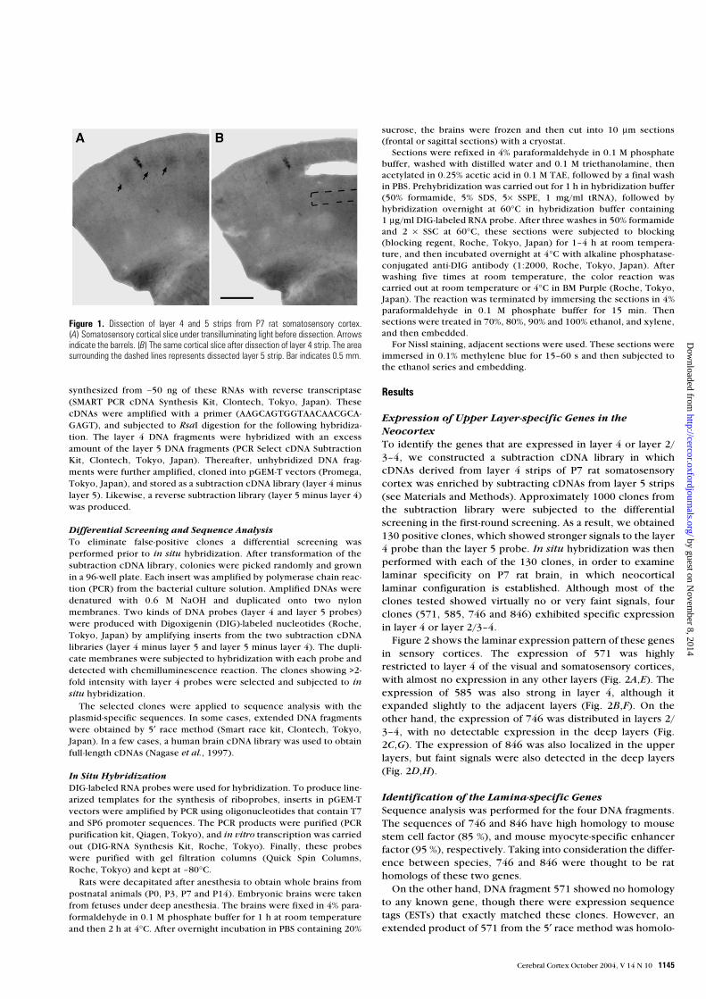
Cerebral Cortex V 14 N 10 © Oxford University Press 2004; all rights reserved Cerebral Cortex October 2004;14:1144–1152; doi:10.1093/cercor/bhh074 Identification of the Genes that are Expressed in the Upper Layers of the Neocortex Yun Zhong 1,3 , Makoto Takemoto 1 , Tsuyoshi Fukuda 1 , Yuki Hattori 1 , Fujio Murakami 1 , Daisuke Nakajima 2 , Manabu Nakayama 2 and Nobuhiko Yamamoto 1 1 Neuroscience Laboratories, Graduate School of Frontier Biosciences, Osaka University, Toyonaka, Osaka 560-8531, Japan and 2 Department of Human Gene Research, Kazusa DNA Research Institute, 2-6-7 Kazusa-kamatari, Chiba 292- 0818 Japan 3 Present address: The Rockefeller University, Box 57, 1230 York Avenue, New York, NY 10021, USA Laminar specificity is one of the most striking features of neocortical circuitry. To explore the molecular basis of this specificity, particu- larly in relation to thalamocortical connectivity, we searched for the genes expressed in the upper cortical layers by constructing a subtraction cDNA library that was enriched for genes expressed in layer 4 of perinatal rat somatosensory cortex. Differential screening, sequence analysis and in situ hybridization demonstrated that a new unc5 family member (unc5h4), deltex-like gene, stem cell factor (SCF) and myocyte-specific enhancer factor-2C (MEF-2C) were specifically expressed in layer 4 or layers 2/3–4 at postnatal day 7, by when laminar organization and fundamental cortical circuitries have been established. In terms of regional specificity, unc5h4 and SCF signals were stronger in sensory cortices, whereas MEF-2C and deltex-like gene were expressed rather uniformly in all neocortical regions. Analysis during development demonstrated that expression of these genes was pronounced between late embryonic and early postnatal developmental stages, except for MEF-2C expression, which continued in later stages. These results demonstrate that certain types of molecules including transcription factors, receptor and ligand molecules, are expressed specifically in the upper layers of the developing neocortex, suggesting a role in laminar specifica- tion of cortical cells and circuitry. Keywords: cerebral cortex, gene expression, layer specificity, thalamocortical projection, unc5h4 Introduction The neocortex is fundamentally composed of six cell layers, which are distinguishable by cellular morphology and the extrinsic and intrinsic connections they make (McConnell, 1989). An intriguing question is how cortical neurons differen- tiate into a particular laminar type, and are connected with a specific population of subcortical and cortical neurons. A plau- sible mechanism is that a set of transcriptional factors expressed in a given layer regulates laminar fate, and expres- sion of downstream molecules including ligand and receptor molecules, is required for cell type specification and axonal guidance of cortical afferents and efferents. Therefore, a key approach is to identify the molecules that are expressed with laminar specificity. Several molecules with lamina-specific expression patterns in the cortex have been identified. For instance, transcription factors of Otx1 and Id2 are primarily distributed in layer 5, and Tbr1, a T box gene, in layer 6 (Bulfone et al., 1995; Frantz et al., 1994; Hevner et al., 2001). Moreover, retinoid Z receptor (RZR-β) and chick ovalbumin upstream transcription factor 1 (COUP-TF1) are expressed rather specifically in layer 4 (Becker-Andre et al., 1994; Park et al., 1997; Liu et al., 2000). It has also been demonstrated that cadherin-6 and rCNL3 — homologous to G-protein-coupled receptors — are expressed in the upper layers (Suzuki et al., 1997; Chenn et al., 2001). Although molecular identification has progressed, a further understanding of gene expression in the developing cortex is necessary to pursue the mechanisms of laminar specification. In the present study, we attempted to identify lamina- specific genes, focusing on thalamocortical connectivity. The thalamocortical projection exhibits typical laminar specificity (Jones, 1981; Gilbert, 1983) and has also been well character- ized during development (Lund and Mustari, 1977; Ghosh and Shatz, 1992; Agmon et al., 1993; Kageyama and Robertson, 1993; Catalano et al., 1996; Molnár et al., 1998). To date, in vitro studies using organotypic co-cultures of the cortex with the thalamus have demonstrated that there is a target recogni- tion mechanism by which thalamocortical axons recognize layer 4, their target (Yamamoto et al., 1989, 1992; Molnár and Blakemore, 1991, 1999; Bolz et al., 1992). Our previous inves- tigations have further suggested that thalamocortical axon branching is induced by membrane-associated molecules in layer 4, whereas termination of axonal growth is regulated by growth-inhibitory molecules in layers 2/3 and 4 (Yamamoto et al., 1997, 2000a, 2000b). These findings suggest that the factors regulating thalamocortical axon targeting and differen- tiation of target layer cells are expressed specifically in the upper layers. Based on this hypothesis, we searched for the molecules expressed specifically in these layers, in particular, layer 4, by constructing a subtraction cDNA library. Four genes, including a new unc5 family member, were obtained. Their expression patterns were further investigated to gain an insight into their possible roles in thalamocortical connectivity and/or cortical cell identity. Materials and Methods Animals Sprague–Dawley rats were used for all experiments (Nihon Animals, Osaka, Japan). The day of vaginal plug detection was designated as E0, and the day of birth as postnatal day 0 (P0). Construction of a Subtraction cDNA Library The whole brain was removed from P7 rats. Coronal slices (250 µm thickness) were cut with a microtome in ice-cold Hanks’ solution. Layer 4 strips, ∼500 µm in length, were dissected from the somato- sensory cortex with a small scissors (Fig. 1). The cortical barrel struc- tures, which were visible under a trans-illuminating microscope, were used as a landmark for layer 4 (Fig. 1). Virtually the same size of layer 5 strip was dissected beneath layer 4 (Fig. 1). Three or four pieces for each layer were collected, from which total RNAs were extracted (RNeasy Mini Kit, Qiagen, Tokyo, Japan). Approximately 10–20 ng/µl of total RNA (30 µl) was obtained. Layer 4 and layer 5 cDNAs were by guest on November 8, 2014 http://cercor.oxfordjournals.org/ Downloaded from
-
Upload
independent -
Category
Documents
-
view
0 -
download
0
Transcript of Identification of the Genes that are Expressed in the Upper Layers of the Neocortex
Cerebral Cortex V 14 N 10 © Oxford University Press 2004; all rights reserved Cerebral Cortex October 2004;14:1144–1152; doi:10.1093/cercor/bhh074
Identification of the Genes that are Expressed in the Upper Layers of the Neocortex
Yun Zhong1,3, Makoto Takemoto1, Tsuyoshi Fukuda1, Yuki Hattori1, Fujio Murakami1, Daisuke Nakajima2, Manabu Nakayama2 and Nobuhiko Yamamoto1
1Neuroscience Laboratories, Graduate School of Frontier Biosciences, Osaka University, Toyonaka, Osaka 560-8531, Japan and 2Department of Human Gene Research, Kazusa DNA Research Institute, 2-6-7 Kazusa-kamatari, Chiba 292-0818 Japan3Present address: The Rockefeller University, Box 57, 1230 York Avenue, New York, NY 10021, USA
Laminar specificity is one of the most striking features of neocorticalcircuitry. To explore the molecular basis of this specificity, particu-larly in relation to thalamocortical connectivity, we searched for thegenes expressed in the upper cortical layers by constructing asubtraction cDNA library that was enriched for genes expressed inlayer 4 of perinatal rat somatosensory cortex. Differential screening,sequence analysis and in situ hybridization demonstrated that a newunc5 family member (unc5h4), deltex-like gene, stem cell factor(SCF) and myocyte-specific enhancer factor-2C (MEF-2C) werespecifically expressed in layer 4 or layers 2/3–4 at postnatal day 7,by when laminar organization and fundamental cortical circuitrieshave been established. In terms of regional specificity, unc5h4 andSCF signals were stronger in sensory cortices, whereas MEF-2C anddeltex-like gene were expressed rather uniformly in all neocorticalregions. Analysis during development demonstrated that expressionof these genes was pronounced between late embryonic and earlypostnatal developmental stages, except for MEF-2C expression,which continued in later stages. These results demonstrate thatcertain types of molecules including transcription factors, receptorand ligand molecules, are expressed specifically in the upper layersof the developing neocortex, suggesting a role in laminar specifica-tion of cortical cells and circuitry.
Keywords: cerebral cortex, gene expression, layer specificity, thalamocortical projection, unc5h4
IntroductionThe neocortex is fundamentally composed of six cell layers,which are distinguishable by cellular morphology and theextrinsic and intrinsic connections they make (McConnell,1989). An intriguing question is how cortical neurons differen-tiate into a particular laminar type, and are connected with aspecific population of subcortical and cortical neurons. A plau-sible mechanism is that a set of transcriptional factorsexpressed in a given layer regulates laminar fate, and expres-sion of downstream molecules including ligand and receptormolecules, is required for cell type specification and axonalguidance of cortical afferents and efferents. Therefore, a keyapproach is to identify the molecules that are expressed withlaminar specificity.
Several molecules with lamina-specific expression patternsin the cortex have been identified. For instance, transcriptionfactors of Otx1 and Id2 are primarily distributed in layer 5, andTbr1, a T box gene, in layer 6 (Bulfone et al., 1995; Frantz et al.,1994; Hevner et al., 2001). Moreover, retinoid Z receptor(RZR-β) and chick ovalbumin upstream transcription factor 1(COUP-TF1) are expressed rather specifically in layer 4(Becker-Andre et al., 1994; Park et al., 1997; Liu et al., 2000). It
has also been demonstrated that cadherin-6 and rCNL3 —homologous to G-protein-coupled receptors — are expressed inthe upper layers (Suzuki et al., 1997; Chenn et al., 2001).Although molecular identification has progressed, a furtherunderstanding of gene expression in the developing cortex isnecessary to pursue the mechanisms of laminar specification.
In the present study, we attempted to identify lamina-specific genes, focusing on thalamocortical connectivity. Thethalamocortical projection exhibits typical laminar specificity(Jones, 1981; Gilbert, 1983) and has also been well character-ized during development (Lund and Mustari, 1977; Ghosh andShatz, 1992; Agmon et al., 1993; Kageyama and Robertson,1993; Catalano et al., 1996; Molnár et al., 1998). To date, in
vitro studies using organotypic co-cultures of the cortex withthe thalamus have demonstrated that there is a target recogni-tion mechanism by which thalamocortical axons recognizelayer 4, their target (Yamamoto et al., 1989, 1992; Molnár andBlakemore, 1991, 1999; Bolz et al., 1992). Our previous inves-tigations have further suggested that thalamocortical axonbranching is induced by membrane-associated molecules inlayer 4, whereas termination of axonal growth is regulated bygrowth-inhibitory molecules in layers 2/3 and 4 (Yamamoto et
al., 1997, 2000a, 2000b). These findings suggest that thefactors regulating thalamocortical axon targeting and differen-tiation of target layer cells are expressed specifically in theupper layers. Based on this hypothesis, we searched for themolecules expressed specifically in these layers, in particular,layer 4, by constructing a subtraction cDNA library. Fourgenes, including a new unc5 family member, were obtained.Their expression patterns were further investigated to gain aninsight into their possible roles in thalamocortical connectivityand/or cortical cell identity.
Materials and Methods
AnimalsSprague–Dawley rats were used for all experiments (Nihon Animals,Osaka, Japan). The day of vaginal plug detection was designated as E0,and the day of birth as postnatal day 0 (P0).
Construction of a Subtraction cDNA LibraryThe whole brain was removed from P7 rats. Coronal slices (250 µmthickness) were cut with a microtome in ice-cold Hanks’ solution.Layer 4 strips, ∼500 µm in length, were dissected from the somato-sensory cortex with a small scissors (Fig. 1). The cortical barrel struc-tures, which were visible under a trans-illuminating microscope, wereused as a landmark for layer 4 (Fig. 1). Virtually the same size of layer5 strip was dissected beneath layer 4 (Fig. 1). Three or four pieces foreach layer were collected, from which total RNAs were extracted(RNeasy Mini Kit, Qiagen, Tokyo, Japan). Approximately 10–20 ng/µlof total RNA (30 µl) was obtained. Layer 4 and layer 5 cDNAs were
by guest on Novem
ber 8, 2014http://cercor.oxfordjournals.org/
Dow
nloaded from
Cerebral Cortex October 2004, V 14 N 10 1145
synthesized from ∼50 ng of these RNAs with reverse transcriptase(SMART PCR cDNA Synthesis Kit, Clontech, Tokyo, Japan). ThesecDNAs were amplified with a primer (AAGCAGTGGTAACAACGCA-GAGT), and subjected to RsaI digestion for the following hybridiza-tion. The layer 4 DNA fragments were hybridized with an excessamount of the layer 5 DNA fragments (PCR Select cDNA SubtractionKit, Clontech, Tokyo, Japan). Thereafter, unhybridized DNA frag-ments were further amplified, cloned into pGEM-T vectors (Promega,Tokyo, Japan), and stored as a subtraction cDNA library (layer 4 minuslayer 5). Likewise, a reverse subtraction library (layer 5 minus layer 4)was produced.
Differential Screening and Sequence AnalysisTo eliminate false-positive clones a differential screening wasperformed prior to in situ hybridization. After transformation of thesubtraction cDNA library, colonies were picked randomly and grownin a 96-well plate. Each insert was amplified by polymerase chain reac-tion (PCR) from the bacterial culture solution. Amplified DNAs weredenatured with 0.6 M NaOH and duplicated onto two nylonmembranes. Two kinds of DNA probes (layer 4 and layer 5 probes)were produced with Digoxigenin (DIG)-labeled nucleotides (Roche,Tokyo, Japan) by amplifying inserts from the two subtraction cDNAlibraries (layer 4 minus layer 5 and layer 5 minus layer 4). The dupli-cate membranes were subjected to hybridization with each probe anddetected with chemilluminescence reaction. The clones showing >2-fold intensity with layer 4 probes were selected and subjected to insitu hybridization.
The selected clones were applied to sequence analysis with theplasmid-specific sequences. In some cases, extended DNA fragmentswere obtained by 5′ race method (Smart race kit, Clontech, Tokyo,Japan). In a few cases, a human brain cDNA library was used to obtainfull-length cDNAs (Nagase et al., 1997).
In Situ HybridizationDIG-labeled RNA probes were used for hybridization. To produce line-arized templates for the synthesis of riboprobes, inserts in pGEM-Tvectors were amplified by PCR using oligonucleotides that contain T7and SP6 promoter sequences. The PCR products were purified (PCRpurification kit, Qiagen, Tokyo), and in vitro transcription was carriedout (DIG-RNA Synthesis Kit, Roche, Tokyo). Finally, these probeswere purified with gel filtration columns (Quick Spin Columns,Roche, Tokyo) and kept at –80°C.
Rats were decapitated after anesthesia to obtain whole brains frompostnatal animals (P0, P3, P7 and P14). Embryonic brains were takenfrom fetuses under deep anesthesia. The brains were fixed in 4% para-formaldehyde in 0.1 M phosphate buffer for 1 h at room temperatureand then 2 h at 4°C. After overnight incubation in PBS containing 20%
sucrose, the brains were frozen and then cut into 10 µm sections(frontal or sagittal sections) with a cryostat.
Sections were refixed in 4% paraformaldehyde in 0.1 M phosphatebuffer, washed with distilled water and 0.1 M triethanolamine, thenacetylated in 0.25% acetic acid in 0.1 M TAE, followed by a final washin PBS. Prehybridization was carried out for 1 h in hybridization buffer(50% formamide, 5% SDS, 5× SSPE, 1 mg/ml tRNA), followed byhybridization overnight at 60°C in hybridization buffer containing1 µg/ml DIG-labeled RNA probe. After three washes in 50% formamideand 2 × SSC at 60°C, these sections were subjected to blocking(blocking regent, Roche, Tokyo, Japan) for 1–4 h at room tempera-ture, and then incubated overnight at 4°C with alkaline phosphatase-conjugated anti-DIG antibody (1:2000, Roche, Tokyo, Japan). Afterwashing five times at room temperature, the color reaction wascarried out at room temperature or 4°C in BM Purple (Roche, Tokyo,Japan). The reaction was terminated by immersing the sections in 4%paraformaldehyde in 0.1 M phosphate buffer for 15 min. Thensections were treated in 70%, 80%, 90% and 100% ethanol, and xylene,and then embedded.
For Nissl staining, adjacent sections were used. These sections wereimmersed in 0.1% methylene blue for 15–60 s and then subjected tothe ethanol series and embedding.
Results
Expression of Upper Layer-specific Genes in the NeocortexTo identify the genes that are expressed in layer 4 or layer 2/3–4, we constructed a subtraction cDNA library in whichcDNAs derived from layer 4 strips of P7 rat somatosensorycortex was enriched by subtracting cDNAs from layer 5 strips(see Materials and Methods). Approximately 1000 clones fromthe subtraction library were subjected to the differentialscreening in the first-round screening. As a result, we obtained130 positive clones, which showed stronger signals to the layer4 probe than the layer 5 probe. In situ hybridization was thenperformed with each of the 130 clones, in order to examinelaminar specificity on P7 rat brain, in which neocorticallaminar configuration is established. Although most of theclones tested showed virtually no or very faint signals, fourclones (571, 585, 746 and 846) exhibited specific expressionin layer 4 or layer 2/3–4.
Figure 2 shows the laminar expression pattern of these genesin sensory cortices. The expression of 571 was highlyrestricted to layer 4 of the visual and somatosensory cortices,with almost no expression in any other layers (Fig. 2A,E). Theexpression of 585 was also strong in layer 4, although itexpanded slightly to the adjacent layers (Fig. 2B,F). On theother hand, the expression of 746 was distributed in layers 2/3–4, with no detectable expression in the deep layers (Fig.2C,G). The expression of 846 was also localized in the upperlayers, but faint signals were also detected in the deep layers(Fig. 2D,H).
Identification of the Lamina-specific GenesSequence analysis was performed for the four DNA fragments.The sequences of 746 and 846 have high homology to mousestem cell factor (85 %), and mouse myocyte-specific enhancerfactor (95 %), respectively. Taking into consideration the differ-ence between species, 746 and 846 were thought to be rathomologs of these two genes.
On the other hand, DNA fragment 571 showed no homologyto any known gene, though there were expression sequencetags (ESTs) that exactly matched these clones. However, anextended product of 571 from the 5′ race method was homolo-
Figure 1. Dissection of layer 4 and 5 strips from P7 rat somatosensory cortex.(A) Somatosensory cortical slice under transilluminating light before dissection. Arrowsindicate the barrels. (B) The same cortical slice after dissection of layer 4 strip. The areasurrounding the dashed lines represents dissected layer 5 strip. Bar indicates 0.5 mm.
by guest on Novem
ber 8, 2014http://cercor.oxfordjournals.org/
Dow
nloaded from

1146 Upper Layer-specific Genes in the Neocortex • Zhong et al.
gous to an unreported human cDNA, which was obtained froma human brain cDNA library (Nagase et al., 1997, 1999). Thetranscript contained an open reading frame (2847 bp) and 3′untranslated region (UTR) of >4000 bp. The deduced aminoacid sequence (948 aa) revealed its features to be a trans-membrane protein, including a signal peptide sequence, twoimmunoglobulin and thrombospondin domains (Fig. 3). Itscytoplasmic region consists of ZU5 and death domains, whichare common to unc5-like netrin receptors (Ackerman et al.,1997). We hereafter designate this novel member of the unc5family as unc5h4. The nucleotide sequence of human unc5h4/
KIAA1777 was deposited into DDBJ/GenBank/EMBL DNAdatabases (accession no. AB055056). KIAA1777 is an alias forthis new gene in the human brain cDNA database.
DNA fragment 585 was also extended by the 5′ race methodand found to highly match a coding region of KIAA0937,which was obtained from the sequence analysis of humancDNAs (Nagase et al., 1999). The deduced amino acidsequence (653 aa) contained two WWE domains, which arepredicted to mediate specific protein–protein interactions(Aravind, 2001), and had high homology (58 %) to humandeltex. Therefore, KIAA0937 is referred to as deltex-like gene.
Regional Specificity and Expression in Other Brain RegionsExpression patterns of the four obtained genes were furtherstudied in P7 rat brain. In particular, areal specificity was exam-ined in order to understand whether these genes are involvedin more generalized features of cortical laminar organization.
Unc5h4/KIA A1777 (571) exhibited a highly restrictedexpression in layer 4 across all neocortical regions. However,its expression was particularly strong in sensory corticesincluding somatosensory and visual areas (Fig. 4A,B). Anothercharacteristic of this gene was its prominent expression in theamygdala and hippocampus, especially in CA3 and dentategyrus. Moderate expression was also found in the hypo-thalamus. In the thalamus, weak expression was observed inthe ventral part of the lateral geniculate nucleus. In other brainregions, it was expressed in mitral cell layers of the olfactorybulb and Purkinje cells of the cerebellum.
The deltex-like gene (585) was also expressed in layer 4 of allneocortical areas. However, in the somatosensory area, thesignal was not only distributed in layer 4 but also in the upperpart of layer 2/3 (Fig. 4C,D). No apparent signal was found inareas other than the neocortex, except for weak expression inthe granule cell layer of the cerebellum.
Figure 2. Four clones which are expressed in layer 4 or in layer 2/3–4. In situ hybridization shows laminar expression patterns in the visual (A–D) and somatosensory (E–H) corticesof P7 rat. DIG-labeled probes were used. In each panel, in situ hybridization is shown to the left, and Nissl staining in adjacent sections is to the right. 571 (A, D) and 585 (B, E) geneswere more specifically expressed in layer 4, while 746 (C, G) and 846 (D, H) genes were expressed in layer 2/3–4. Bar represents 0.2 mm.
by guest on Novem
ber 8, 2014http://cercor.oxfordjournals.org/
Dow
nloaded from

Cerebral Cortex October 2004, V 14 N 10 1147
Figure 3. Amino acid sequence of human unc5h4/KIAA1777 and extended product of 571 by 5′ race method. TSP, thrombospondin; TM, transmembrane.
Figure 4. Layer 4-specific genes. Upper (A, B) and lower (C, D) panels show expression patterns of unc5h4 and deltex-like gene, respectively. Am, amygdala; Cb, cerebellum; DG,dentate gyrus; Hpt, hypothalamus; OB, olfactory bulb. Bar indicates 1 mm.
by guest on Novem
ber 8, 2014http://cercor.oxfordjournals.org/
Dow
nloaded from

1148 Upper Layer-specific Genes in the Neocortex • Zhong et al.
SCF (746) showed strong expression between layers 2/3 and4 across all neocortical regions (Fig. 5A,B). As observed forunc5h4/KIAA1777, SCF expression was strong in the somato-sensory and visual areas. Its strong expression in the limbicregion was another characteristic it shared with unc5h4/KIAA1777. The most striking aspect was the highly specificand strong expression in the thalamus. The expression waslocated in the dorsal lateral geniculate nucleus and the lateralposterior nucleus, which project to visual areas, and in theventral basal thalamic nucleus, which projects to somato-sensory areas. Strong signals were also found in the habenula,central lateral nucleus, central medial nucleus, parafascicularnucleus and intermediodorsal nucleus. In addition, this genewas expressed in the granule cells of the olfactory bulb andPurkinje cells of the cerebellum.
The expression of MEF-2C (846) was highly restricted to theneocortex (Fig. 5C,D). Its expression in layers 2/3–4 wasuniform across the whole neocortex, but no expression wasobserved in other brain regions.
Developmental Changes of Laminar Expression PatternsTo gain an insight into how each gene is associated withlaminar property, cellular differentiation and afferent invasion,their expression patterns were studied in the developing soma-tosensory cortex.
The expression of unc5h4/KIAA1777 (571) was observed inthe subventricular and intermediate zones at E18 (Fig. 6A). AtP0, the expression appeared just beneath the marginal zone,and then in slightly lower layers at P3. At P7, the message wasrestricted to layer 4 (Fig. 6A). Thus, the expression pattern ofthis gene during development was closely related to the
laminar locations of the cells destined to layer 4, but the signalwas rather weakened after P14.
The message of deltex-like gene (585) was expressed in boththe ventricular zone and the cortical plate (CP) at E18, but wasdistributed in the upper part of the CP from P0 to P3 (Fig. 6B),which is similar to unc5h4 expression pattern. At P7, theexpression was observed in layer 4 although it was slightlydiffuse. Moreover, the message was also observed in the upperpart of layer 2/3 in the somatosensory cortex. At P14, almostno expression was retained.
The message of SCF (746) was localized in the deepest partof the CP at E18 (Fig. 7A). At P0, the expression in layer 6 wasweaker, and even more so at P3, although the signal began toappear in the upper part of the CP. At P7, the expression wasstrong in layers 2/3 and 4, whereas it had virtually disappearedin layer 6. At P14, almost no expression was observed.
MEF-2C (846) was strongly expressed in the upper part ofthe CP from E18 to P3 (Fig. 7B). At P7, the expression wasprimarily observed in layers 2/3 and 4, and slightly in the upperpart of layer 5. Unlike the former three clones, the messagewas retained at P14 to a great extent.
DiscussionWe obtained four genes that were expressed in the upperlayers in P7 rat cortex when laminar configuration is estab-lished. Unc5h4/KIAA1777 and deltex-like gene/KIAA0937
were expressed specifically in layer 4, whereas SCF and MEF-
2C were expressed in layers 2/3–4. As for area specificity,unc5h4 and SCF signals were stronger in sensory cortices, butthe messages of MEF-2C and deltex-like gene were distributedrather broadly in the neocortex. All of the genes were strongly
Figure 5. Layer 2/3–4-specific genes. Upper (A, B) and lower (C, D) panels show SCF and MEF-2C expressions, respectively. Am, amygdala; Cb, cerebellum; Hb, habenularnucleus; MD, mediodorsal thalamus; OB, olfactory bulb; VB, ventrobasal complex of thalamus; vLG, ventral part of the lateral geniculate nucleus. Bar indicates 1 mm.
by guest on Novem
ber 8, 2014http://cercor.oxfordjournals.org/
Dow
nloaded from

Cerebral Cortex October 2004, V 14 N 10 1149
expressed in embryonic and early postnatal stages, except thatMEF-2C messages were still present in later stages. Thus, thepresent study using systematic screening of the subtractioncDNA library clearly demonstrates that certain types of mole-cules including extracellular proteins and transcription factors,which might be involved in cellular differentiation and neuralcircuit formation, are expressed in the upper layers of thedeveloping cortex.
Expression and Molecular Properties of Upper Layer-specific GenesUnc5h4, a new member of unc5 family, was expressed mostspecifically in layer 4 of P7 rat cortex. After the sequence ofhuman unc5h4/KIAA1777 had been registered in GenBank/DDBJ database, its mouse homolog was reported (Engelkamp,2002). However, the present study is the first to demonstrateits molecular characteristics and expression pattern in thebrain. The expression profile during development further
showed a migrating behavior of unc5h4 expression. Layer 4cells are born at E16–17 in the ventricular zone and migrate tothe subventricular and intermediate zones at E18. At P0, theymove to the most superficial part of the CP, and gradually settlein layer 4 by P6 (Berry and Rogers, 1965; Lund and Mustari,1977). Unc5h4 expression closely resembles this migrationpattern, suggesting that unc5h4 is expressed during develop-ment by the cells destined to form layer 4 of the cortex. Weakexpression in the frontal lobe is consistent with this view, asgranular cells, the major population in layer 4, are scarce in themotor cortex.
It has been shown that unc5 family members are involved inaxonal elongation and cell migration as netrin-1 receptors(Leonardo et al., 1997). In addition, these members have beenshown to suppress neuronal apoptosis, upon receiving a ligandsignal (Hofmann and Tschopp, 1995; Llambi et al., 2001).Although the molecules that interact with unc5h4 have not yetbeen identified, it is possible that the ligand may influence
Figure 6. Developmental changes of laminar expression of layer 4-specific genes. Unc5h4 (A) and deltex like gene (B) expressions in the parietal region (above the hippocampus)are shown. In (A), in situ signals are shown to the left of each stage, and Nissl staining is shown to the right. MZ, marginal zone; CP, cell-dense cortical plate; SP, subplate; SV,subventricular zone; VZ, ventricular zone. Bar represents 0.2 mm.
by guest on Novem
ber 8, 2014http://cercor.oxfordjournals.org/
Dow
nloaded from

1150 Upper Layer-specific Genes in the Neocortex • Zhong et al.
neuronal survival of layer 4 cells expressing unc5h4. Thesurvival effect might further influence neuronal connectivity.Another possibility is that unc5h4 acts as a ligand molecule.The extracellular domains containing immunoglobulin andthrombospondin domains might directly influence ingrowingthalamic axons. Identification of the molecules that interactwith unc5h4 would be necessary to elucidate its role to a greatextent.
We cloned and characterized another layer-4-specific mole-cule, deltex-like gene/KIAA0937, although its expression wasnot as localized as the unc5h4 message. Deltex has beenshown to mediate the Notch signaling pathway in the nucleusas well as in the cytoplasmic region (Matsuno et al., 1995; Kishiet al., 2001; Yamamoto et al., 2001). Therefore, deltex-likeprotein may be involved in the differentiation of layer 4 cells byregulating Notch activity transcriptionally and/or throughdirect binding.
As for MEF-2C, Leifer et al. (1993) have shown that MEF-2C
mRNA is expressed in the upper layers of the developing
cortex. Our results not only confirm their findings but alsodemonstrate that this lamina-specific expression is presentthroughout the neocortex, suggesting that MEF-2C may beinvolved in the upper cortical neuron identity. Although it hasbeen demonstrated that MEF-2C supports cell survival of post-mitotic neurons (Mao et al., 1999), the prolonged and ratheruniform expression in the neocortex indicates the possibilitythat MEF-2C may contribute to the maintenance and differen-tiation of upper cortical cells rather than simply to the differen-tiation of postmitotic neurons.
On the other hand, SCF expression during cortical develop-ment was more complicated, but seems to be related tothalamic axon invasion. Since the tips of thalamic axons start toinvade the CP at E18, extend into layer 5 at P0, and reachimmature layer 4 at P3 (Lund and Mustari, 1977; Kageyama andRobertson, 1993; Catalano et al., 1996; Molnár et al., 1998), acorrelation might exist between the expression of SCF andingrowth of thalamic axons in the neocortex. In accordancewith thalamic axon extension, SCF expression graduallybecame weaker in the deep layers and increased in the upperlayers. Indeed, immunohistochemistry with an antibodyagainst c-kit, the receptor of SCF, showed that c-kit wasexpressed in thalamic nuclei and axons (not shown), indi-cating the possibility that thalamic axons respond to SCFdistributed in the developing cortex. The weaker expression ofSCF in the frontal cortex might also be related to the lack ofsensory thalamic axons in this area.
Implications of the Presence of Upper Layer-specific GenesTo date, it has been shown that putative regulatory genes, RZR-
β (Becker-Andre et al., 1994; Park et al., 1997) and COUP-TF1
(Liu et al., 2000) are expressed in layer 4 of the neocortex,although the functions of these genes are not clear. Thalamo-cortical projections are disrupted in COUP-TF1 mutants, but itis likely that the phenotype is attributable to the lack ofsubplate neurons rather than its direct influences on thalamo-cortical axon targeting in layer 4 (Zhou et al., 1999). Thepresent results further demonstrate the presence of anotherputative regulatory gene, deltex-like gene, which is expressedprimarily in layer 4 and might act as a regulator of cellulardifferentiation (see above).
It is also important to reveal extracellular molecules such ascell surface or extracellular matrix molecules, in order tounderstand the molecular basis of cellular interactions andcircuit formation (Yamamoto, 2002). In this sense, unc5h4 isthe gene that encodes a cell surface protein and is expressedrather specifically in cortical cells destined for layer 4 duringdevelopment. Although how unc5h4 contributes to cellulardifferentiation and connectivity in the cortex is unknown, itsmolecular characteristics raise the possibility that it is involvedin the interactions between layer 4 cells, or between layer 4cells and thalamocortical fibers. Recent studies have shownthat ephrin-A5, an Eph ligand, affects thalamocortical axonbehavior by its expression in layer 4 (Mann et al., 2002), butthis is unlikely to be the case with the entire sensorythalamocortical projections (Donoghue and Rakic, 1999;Mackarehtschian et al., 1999; Vanderhaeghen et al., 2000;Yabuta et al., 2000). In contrast, localization of unc5h4/
KIAA1777 in layer 4 is found throughout the sensory cortices.Cadherin-6 and rCNL3 are known cell surface molecules that
are expressed in the upper layers of the developing cortex
Figure 7. Developmental changes of laminar expression of upper layer-specific genes.SCF (A) and MEF-2C (B) expressions in parietal cortex are shown. Abbreviations arethe same as those in Figure 6. Bar represents 0.2 mm.
by guest on Novem
ber 8, 2014http://cercor.oxfordjournals.org/
Dow
nloaded from

Cerebral Cortex October 2004, V 14 N 10 1151
(Suzuki et al., 1997; Chenn et al., 2001). Phosphacan, a prote-oglycan (Maeda and Noda, 1996), and SemaK1, a GPI-anchoredsemaphorin (Xu et al., 1998), are also expressed in the upperlayers of early postnatal stages, though their expressionpatterns during development are not well characterized. Itwould be worthwhile to examine whether these moleculesaffect thalamic axonal growth, as previous investigations haveindicated that thalamocortical axon termination is governed bymolecules distributed in layers 2/3–4. (Yamamoto et al.,2000a,b; Noctor et al., 2001; Yamamoto, 2002). In addition,the present findings of SCF expression pattern raises the possi-bility that a developmental change of laminar expression mayinfluence afferent fiber ingrowth (see above). A similar laminartransition has been reported in the expression of Sema3A, apotential molecule, which might be responsible for the forma-tion of thalamocortical projections (Skaliora et al., 1998).
Several transcriptional factors have been shown to beexpressed in a lamina-specific fashion: Otx1 (Frantz et al.,1994) and Id2 (Bulfone et al., 1995) in layer 5 and Tbr-1
(Bulfone et al., 1995; Hevner et al., 2001) in layer 6. As for layer4, COUP-TF1 and RZR-β are expressed in layer 4, as describedabove. The present results show that deltex-like gene and MEF-
2C are expressed in layer 4 and layer 2/3–4 as regulatoryfactors, respectively. Determining the relation between theseregulatory genes and their downstream molecules would alsobe useful in elucidating the molecular basis of cortical celldifferentiation and neural circuit formation.
NotesWe thank Dr R. Carney for critical reading of this manuscript. We alsothank Drs Y. Zhu and Y. Hatanaka for helpful comments on this manu-script, and Dr M. Mieda for helpful advice on construction of thesubtraction cDNA library. This work was supported by Grants-in-Aidfor Scientific Research Projects 12050227 and 13041038 from Japa-nese Ministry of Education, Culture, Science and Sports, by CoreResearch for Evolutional Science and Technology (CREST) from theJapan Science and Technology Corporation (JST), and by Human Fron-tier Science Project (RGP0107/2001).
Y. Zhong and M. Takemoto contributed equally to this study.Address correspondence to Nobuhiko Yamamoto, Neuroscience
Laboratories, Graduate School of Frontier Biosciences, Osaka Univer-sity, Toyonaka, Osaka 560-8531, Japan. Email: [email protected].
ReferencesAckerman SL, Kozak LP, Przyborski SA, Rund LA, Boyer BB, Knowles
BB (1997) The mouse rostral cerebellar malformation geneencodes an UNC-5-like protein. Nature 386:838–842.
Agmon A, Yang LT, O’Dowd DK, Jones EG (1993) Organized growthof thalamocortical axons from the deep tier of terminations intolayer IV of developing mouse barrel cortex. J Neurosci 13:5365–5382
Aravind L (2001) The WWE domain: a common interaction module inprotein ubiquitination and ADP ribosylation. Trends Biochem Sci26:273–275.
Becker-Andre M, Wiesenberg I, Schaeren-Wiemers N, Andre E,Missbach M, Saurat JH, Carlberg C (1994) Pineal gland hormonemelatonin binds and activates an orphan of the nuclear receptorsuperfamily. J Biol Chem 269:28531–28534.
Berry M, Rogers AW (1965) The migration of neuroblasts in the devel-oping cerebral cortex. J Anat 99:691–709.
Bolz J, Novak N, Staiger V (1992) Formation of specific afferentconnections in organotypic slice cultures from rat visual cortexwith lateral geniculate nucleus. J Neurosci 12:3054–3070.
Bulfone A, Smiga SM, Shimamura K, Peterson A, Puelles L, RubensteinJL (1995) T-brain-1: a homolog of Brachyury whose expression
defines molecularly distinct domains within the cerebral cortex.Neuron 15:63–78
Catalano S, Robertson RT, Killackey H (1996) Individual axonmorphology and thalamocortical topography in developing ratsomatosensory cortex. J Comp Neurol 366:36–53.
Chenn A, Levin ME, McConnell SK (2001) Temporally and spatiallyregulated expression of a candidate G-protein-coupled receptorduring cerebral cortical development. J Neurobiol 46:167–177.Erratum J Neurobiol 47:244.
Donoghue MJ, Rakic P (1999) Molecular evidence for the early specifi-cation of presumptive functional domains in the embryonicprimate cerebral cortex. J Neurosci 19:5967–79
Engelkamp D (2002) Cloning of three mouse Unc5 genes and theirexpression patterns at mid-gestation. Mech Dev 118:191–197.
Frantz GD, Weimann JM, Levin ME, McConnell SK (1994) Otx1 andOtx2 define layers and regions in developing cerebral cortex andcerebellum. J Neurosci 14:5725–5740.
Ghosh A, Shatz, CJ (1992) Pathfinding and target selection by devel-oping geniculocortical axons. J Neurosci 12:39–55.
Gilbert CD (1983) Microcircuitry of the visual cortex. Annu RevNeurosci 6:217–247.
Hevner RF, Shi L, Justice N, Hsueh Y, Sheng M, Smiga S, Bulfone A,Goffinet AM, Campagnoni AT, Rubenstein JL (2001) Tbr1 regulatesdifferentiation of the preplate and layer 6. Neuron 29:353–366.
Hofmann K, Tschopp J (1995) The death domain motif found in Fas(Apo-1) and TNF receptor is present in proteins involved in apop-tosis and axonal guidance. FEBS Lett 371:321–323.
Jones EG (1981) Anatomy of cerebral cortex: Columnar input–outputorganization. In: The organization of the cerebral cortex (SchmittFO, Worden FG, Adelman G, Dennis SG, eds), pp. 199–235.Cambridge, MA: The MIT press.
Kageyama, GH and Robertson, RT (1993) Development of geniculoco-rtical projections to visual cortex in rat. J Comp Neurol335:123–148.
Kishi N, Tang Z, Maeda Y, Hirai A, Mo R, Ito M, Suzuki S, Nakao K,Kinoshita T, Kadesch T, Hui C, Artavanis-Tsakonas S, Okano H,Matsuno K (2001) Murine homologs of deltex define a novel genefamily involved in vertebrate Notch signaling and neurogenesis. IntJ Dev Neurosci 19:21–35.
Leifer D, Krainc D, Yu YT, McDermott J, Breitbart RE, Heng J, NeveRL, Kosofsky B, Nadal-Ginard B, Lipton SA (1993) MEF2C, a MADS/MEF2-family transcription factor expressed in a laminar distribu-tion in cerebral cortex. Proc Natl Acad Sci U S A 90:1546–1550.
Leonardo ED, Hinck L, Masu M, Keino-Masu K, Ackerman SL, Tessier-Lavigne M (1997) Vertebrate homologues of C. elegans UNC-5 arecandidate netrin receptors. Nature 386:833–838.
Liu Q, Dwyer ND, O’Leary DD (2000) Differential expression ofCOUP-TFI, CHL1, and two novel genes in developing neocortexidentified by differential display PCR. J Neurosci 20:7682–7690.
Llambi F, Causeret F, Bloch-Gallego E, Mehlen P (2001) Netrin-1 actsas a survival factor via its receptors UNC5H and DCC. EMBO J20:2715–2722.
Lund RD, Mustari MJ (1977) Development of geniculocortical pathwayin rats. J Comp Neurol 173:289–306.
Mackarehtschian K, Lau CK, Caras I, McConnell SK (1999) Regionaldifferences in the developing cerebral cortex revealed by ephrin-A5 expression. Cereb Cortex 9:601–610.
Maeda N, Noda M (1996) 6B4 proteoglycan/phosphacan is a repulsivesubstratum but promotes morphological differentiation of corticalneurons. Development 122:647–658.
Mann F, Peuckert C, Dehner F, Zhou R, Bolz J (2002) Ephrins regulatethe formation of terminal axonal arbors during the development ofthalamocortical projections. Development 129:3945–3955.
Mao Z, Bonni A, Xia F, Nadal-Vicens M, Greenberg ME (1999) Neuronalactivity-dependent cell survival mediated by transcription factorMEF2. Science 286:785–790.
Matsuno K, Diederich RJ, Go MJ, Blaumueller CM, Artavanis-TsakonasS (1995) Deltex acts as a positive regulator of Notch signalingthrough interactions with the Notch ankyrin repeats. Development121:2633–2644.
by guest on Novem
ber 8, 2014http://cercor.oxfordjournals.org/
Dow
nloaded from

1152 Upper Layer-specific Genes in the Neocortex • Zhong et al.
McConnell SK (1989) The determination of neuronal fate in the cere-bral cortex. Trends Neurosci 12:342–349.
Molnár Z, Blakemore C (1991) Lack of regional specificity for connec-tions formed between thalamus and cortex in coculture. Nature351:475–477.
Molnár Z, Blakemore C (1999) Development of signals influencing thegrowth and termination of thalamocortical axons in organotypicculture. Exp Neurol 156:363–393.
Molnár Z, Adams R, Blakemore C (1998) Mechanisms underlying theearly establishment of thalamocortical connections in the rat. JNeurosci 18:5723–5745.
Nagase T, Ishikawa K, Nakajima D, Ohira M, Seki N, Miyajima N,Tanaka A, Kotani H, Nomura N, Ohara O (1997) Prediction of thecoding sequences of unidentified human genes. VII. The completesequences of 100 new cDNA clones from brain which can code forlarge proteins in vitro. DNA Res 4:141–150.
Nagase T, Ishikawa K, Suyama M, Kikuno R, Hirosawa M, Miyajima N,Tanaka A, Kotani H, Nomura N, Ohara O (1999) Prediction of thecoding sequences of unidentified human genes. XIII. Thecomplete sequence of 100 new cDNA clones from brain whichcode for large proteins in vitro. DNA Res 6:63–70.
Noctor SC, Palmer SL, McLaughlin DF, Juliano SL (2001) Disruption oflayers 3 and 4 during development results in altered thalamocor-tical projections in ferret somatosensory cortex. J Neurosci21:3184–3195.
Park HT, Kim YJ, Yoon S, Kim JB, Kim JJ (1997) Distributional charac-teristics of the mRNA for retinoid Z receptor beta (RZR beta), aputative nuclear melatonin receptor, in the rat brain and spinalcord. Brain Res 747:332–337.
Skaliora I, Singer W, Betz H, Püschel AW (1998) Differential patternsof semaphorin expression in the developing rat brain. Eur JNeurosci 10:1215–1229.
Suzuki SC, Inoue T, Kimura Y, Tanaka T, Takeichi M (1997) Neuronalcircuits are subdivided by differential expression of type-II classiccadherins in postnatal mouse brains. Mol Cell Neurosci 9:433–447.
Vanderhaeghen P, Lu Q, Prakash N, Frisén J, Walsh CA, Frostig RD,Flanagan JG (2000) A mapping label required for normal scale ofbody representation in the cortex. Nat Neurosci 3:358–365.
Xu X, Ng S, Wu ZL, Nguyen D, Homburger S, Seidel-Dugan C, Ebens A,Luo Y (1998) Human semaphorin K1 is glycosylphosphatidyli-nositol-linked and defines a new subfamily of viral-related sema-phorins. J Biol Chem 273:22428–22434.
Yabuta NH, Butler AK, Callaway EM (2000) Laminar specificity of localcircuits in barrel cortex of ephrin-a5 knockout mice. J Neurosci20(RC88):1–4.
Yamamoto N (2002) Cellular and molecular basis for the formation oflamina-specific thalamocortical projections. Neurosci Res42:167–173.
Yamamoto N, Kurotani T, Toyama K (1989) Neural connectionsbetween the lateral geniculate nucleus and visual cortex in vitro.Science 245:192–194.
Yamamoto N, Yamada K, Kurotani T, Toyama K (1992) Laminar specif-icity of extrinsic cortical connections studied in coculture prepara-tions. Neuron 9:217–228.
Yamamoto N, Higashi S, Toyama K (1997) Stop and branch behaviorsof geniculocortical axons: A time-lapse study in organotypic cocul-tures. J Neurosci 17:3653–3663.
Yamamoto N, Inui K, Matsuyama Y, Harada A, Hanamura K, MurakamiF, Ruthazer ES, Rutishauser U and Seki T (2000a) Inhibitory mech-anism by polysialic acid for lamina-specific branch formation ofthalamocortical axons. J Neurosci 20:9145–9151.
Yamamoto N, Matsuyama Y, Harada A, Inui K, Murakami F, HanamuraK (2000b) Characterization of factors regulating lamina-specificgrowth of thalamocortical axon. J Neurobiol 42:56–68.
Yamamoto N, Yamamoto S, Inagaki F, Kawaichi M, Fukamizu A, KishiN, Matsuno K, Nakamura K, Weinmaster G, Okano H, Nakafuku M(2001) Role of Deltex-1 as a transcriptional regulator downstreamof the Notch receptor. J Biol Chem. 276:45031–45040.
Zhou C, Qiu Y, Pereira FA, Crair MC, Tsai SY, Tsai MJ (1999) Thenuclear orphan receptor COUP-TFI is required for differentiationof subplate neurons and guidance of thalamocortical axons.Neuron 24:847–859. by guest on N
ovember 8, 2014
http://cercor.oxfordjournals.org/D
ownloaded from