Linear shear response of the upper skin layers
17
Biorheology 48 (2011) 229–245 229 DOI 10.3233/BIR-2011-0590 IOS Press Linear shear response of the upper skin layers Marion Geerligs a , Cees Oomens b,∗ , Paul Ackermans c , Frank Baaijens b and Gerrit Peters b a Philips Consumer Lifestyle, Drachten, The Netherlands b Eindhoven University of Technology, Eindhoven, The Netherlands c Philips Research, Eindhoven, The Netherlands Received 4 February 2011 Accepted in revised form 1 August 2011 Abstract. This study presents an in vitro experimental method to determine shear properties of the epidermis. Shear tests were performed with a parallel plate rheometer on samples of stratum corneum and the viable epidermis. The method was validated on very thin silicon sheets. Preliminary test were performed to determine the linear viscoelastic range, the effect of normal loading on the sample and the time to reach equilibrium after changes of temperature and relative humidity. The study shows that reproducible results can be obtained for the shear properties of epidermis in an in vitro set up. The dynamic shear modulus for stratum corneum ranges from about 4–12 kPa, decreasing with increasing relative humidity. The values are considerably lower than the shear modulus value based on tensile Young’s moduli in the literature, indicating a considerable anisotropic material behavior. Results for the epidermis were of the same order of magnitude, but were less consistent possibly due to a less well-defined tissue composition. Keywords: Stratum corneum, epidermis, mechanical properties, in vitro 1. Introduction For various clinical and cosmetic treatments it is essential to gain knowledge on the mechanical behav- ior of human skin. The human skin is composed of a non-uniform, layered structure and the mechanical behavior of these layers is very complex: i.e., anisotropic, inhomogeneous, nonlinear and viscoelastic. Therefore, the most appropriate approach is, to determine the mechanical properties of each individual skin layer in all loading directions in order to understand the full skin response. The present study focuses on the contribution of the outer skin layer, the epidermis, when in-plane forces are applied to the skin surface. Because of the anisotropic nature of the epidermis, the response in tension and shear is expected to be (very) different. Usually, tensile properties are addressed in research studies. However, the shear component plays a key role in applications such as the development of pressure ulcers, the removal of skin adhesives and skin-device contact such as with prosthetic limbs and shavers. All these applications will benefit from an improved knowledge of the mechanical response of the epidermis to shear. * Address for correspondence: Dr. Cees Oomens, Biomedical Engineering Faculty, Eindhoven University of Technology, P/O Box 513, 5600 MB Eindhoven, The Netherlands. Tel.: +31 40 247 2818; Fax: +31 40 244 7355; E-mail: c.w.j.oomens@ tue.nl. 0006-355X/11/$27.50 © 2011 – IOS Press and the authors. All rights reserved
Transcript of Linear shear response of the upper skin layers

Biorheology 48 (2011) 229–245 229DOI 10.3233/BIR-2011-0590IOS Press
Linear shear response of the upperskin layers
Marion Geerligs a, Cees Oomens b,∗, Paul Ackermans c, Frank Baaijens b and Gerrit Peters b
a Philips Consumer Lifestyle, Drachten, The Netherlandsb Eindhoven University of Technology, Eindhoven, The Netherlandsc Philips Research, Eindhoven, The Netherlands
Received 4 February 2011Accepted in revised form 1 August 2011
Abstract. This study presents an in vitro experimental method to determine shear properties of the epidermis. Shear tests wereperformed with a parallel plate rheometer on samples of stratum corneum and the viable epidermis. The method was validatedon very thin silicon sheets. Preliminary test were performed to determine the linear viscoelastic range, the effect of normalloading on the sample and the time to reach equilibrium after changes of temperature and relative humidity. The study showsthat reproducible results can be obtained for the shear properties of epidermis in an in vitro set up. The dynamic shear modulusfor stratum corneum ranges from about 4–12 kPa, decreasing with increasing relative humidity. The values are considerablylower than the shear modulus value based on tensile Young’s moduli in the literature, indicating a considerable anisotropicmaterial behavior. Results for the epidermis were of the same order of magnitude, but were less consistent possibly due to aless well-defined tissue composition.
Keywords: Stratum corneum, epidermis, mechanical properties, in vitro
1. Introduction
For various clinical and cosmetic treatments it is essential to gain knowledge on the mechanical behav-ior of human skin. The human skin is composed of a non-uniform, layered structure and the mechanicalbehavior of these layers is very complex: i.e., anisotropic, inhomogeneous, nonlinear and viscoelastic.Therefore, the most appropriate approach is, to determine the mechanical properties of each individualskin layer in all loading directions in order to understand the full skin response.
The present study focuses on the contribution of the outer skin layer, the epidermis, when in-planeforces are applied to the skin surface. Because of the anisotropic nature of the epidermis, the response intension and shear is expected to be (very) different. Usually, tensile properties are addressed in researchstudies. However, the shear component plays a key role in applications such as the development ofpressure ulcers, the removal of skin adhesives and skin-device contact such as with prosthetic limbs andshavers. All these applications will benefit from an improved knowledge of the mechanical response ofthe epidermis to shear.
*Address for correspondence: Dr. Cees Oomens, Biomedical Engineering Faculty, Eindhoven University of Technology,P/O Box 513, 5600 MB Eindhoven, The Netherlands. Tel.: +31 40 247 2818; Fax: +31 40 244 7355; E-mail: [email protected].
0006-355X/11/$27.50 © 2011 – IOS Press and the authors. All rights reserved
230 M. Geerligs et al. / Linear shear response of upper skin layers
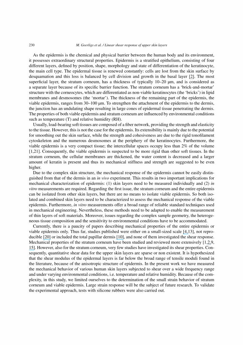
As the epidermis is the chemical and physical barrier between the human body and its environment,it possesses extraordinary structural properties. Epidermis is a stratified epithelium, consisting of fourdifferent layers, defined by position, shape, morphology and state of differentiation of the keratinocyte,the main cell type. The epidermal tissue is renewed constantly: cells are lost from the skin surface bydesquamation and this loss is balanced by cell division and growth in the basal layer [2]. The mostsuperficial layer, the stratum corneum, has a thickness of typically 10–20 µm, and is considered asa separate layer because of its specific barrier function. The stratum corneum has a ‘brick-and-mortar’structure with the corneocytes, which are differentiated as non-viable keratinocytes (the ‘bricks’) in lipidmembranes and desmosomes (the ‘mortar’). The thickness of the remaining part of the epidermis, theviable epidermis, ranges from 30–100 µm. To strengthen the attachment of the epidermis to the dermis,the junction has an undulating shape resulting in large cones of epidermal tissue penetrating the dermis.The properties of both viable epidermis and stratum corneum are influenced by environmental conditionssuch as temperature (T) and relative humidity (RH).
Usually, load-bearing soft tissues are composed of a fiber network, providing the strength and elasticityto the tissue. However, this is not the case for the epidermis. Its extensibility is mainly due to the potentialfor smoothing out the skin surface, while the strength and cohesiveness are due to the rigid tonofilamentcytoskeleton and the numerous desmosomes at the periphery of the keratinocytes. Furthermore, theviable epidermis is a very compact tissue; the intercellular spaces occupy less than 2% of the volume[1,21]. Consequently, the viable epidermis is suspected to be more rigid than other soft tissues. In thestratum corneum, the cellular membranes are thickened, the water content is decreased and a largeramount of keratin is present and thus its mechanical stiffness and strength are suggested to be evenhigher.
Due to the complex skin structure, the mechanical response of the epidermis cannot be easily distin-guished from that of the dermis in an in vivo experiment. This results in two important implications formechanical characterization of epidermis: (1) skin layers need to be measured individually and (2) invitro measurements are required. Regarding the first issue, the stratum corneum and the entire epidermiscan be isolated from other skin layers, but there are no means to isolate viable epidermis. So both iso-lated and combined skin layers need to be characterized to assess the mechanical response of the viableepidermis. Furthermore, in vitro measurements offer a broad range of reliable standard techniques usedin mechanical engineering. Nevertheless, these methods need to be adapted to enable the measurementof thin layers of soft materials. Moreover, issues regarding the complex sample geometry, the heteroge-neous tissue composition and the sensitivity to environmental conditions have to be accommodated.
Currently, there is a paucity of papers describing mechanical properties of the entire epidermis orviable epidermis only. Thus far, studies published were either on a small-sized scale [4,13], not repro-ducible [20] or included the total papillar dermis [10], and none of them investigated the shear response.Mechanical properties of the stratum corneum have been studied and reviewed more extensively [1,2,9,15]. However, also for the stratum corneum, very few studies have investigated its shear properties. Con-sequently, quantitative shear data for the upper skin layers are sparse or non existent. It is hypothesizedthat the shear modulus of the epidermal layers is far below the broad range of tensile moduli found inthe literature, because of the anisotropic structure of epidermis. In the present work we have measuredthe mechanical behavior of various human skin layers subjected to shear over a wide frequency rangeand under varying environmental conditions, i.e. temperature and relative humidity. Because of the com-plexity, in this study, we limited ourselves to the determination of the small strain behavior of stratumcorneum and viable epidermis. Large strain response will be the subject of future research. To validatethe experimental approach, tests with silicone rubbers were also carried out.

M. Geerligs et al. / Linear shear response of upper skin layers 231
2. Methods
2.1. Sample preparation
Because of a number of issues that arise with in vitro testing of these very thin skin samples, a seriesof preliminary studies have been done with various sample geometries. Table 1 gives an overview ofthe samples types that were used in the pilot studies and in the actual studies to determine the linearviscoelastic properties at different RH and temperature of epidermis and stratum corneum.
In order to validate the performance of the rheometer for very thin samples, a highly elastic sili-cone rubber (Köraform 42 A, Alpina Siliconee, Germany) with known properties was chosen (samplegroup R). The silicone rubber was poured under vacuum into various thicknesses: 5, 120 and 2000 µm.Circular samples were obtained by using an 8 mm diameter cork borer.
Skin was obtained from patients undergoing abdominoplastic surgery, who gave informed consentfor use of their skin for research purposes under a protocol approved by the ethics committee of theCatharina Hospital, Eindhoven, The Netherlands. Only abdominal skin of Caucasian women from anage group between 35 and 55 years old was used. Abdominal skin with stria, cellulite, damage dueto UV exposure or tremendously hairy skin was excluded from the study. Immediately after excision,the skin was brought into the laboratory and processed within 4 h. Skin slices were obtained using anelectric dermatome (D42, Humeca, The Netherlands) for which the accuracy of the prescribed thicknesswas improved for this purpose by the supplier.
Split-thickness skin of 200 and 400 µm in thickness (sample groups S200 and S400) was obtainedusing the dermatome. As can be seen in Fig. 1, the 200-µm split-thickness skin is composed of epidermisand papillar dermis. In the 400-µm split-thickness, reticular dermis is also present. For isolating thereticular dermis, the top layer of skin was dermatomed until the white opaque dermis was on top. Then,a 400-µm thick layer of reticular dermis was dermatomed.
In order to separate the epidermis (sample group E), the thickness was set to 100 µm. Subsequently,8-mm diameter circular tissue samples of the epidermis were obtained from the slices using the corkborer. The epidermis was estimated to vary from 50 to 150 µm on this body site [1,21]. Dependingon various factors such as skin surface roughness, tissue hydration, smoothness of the cutting, somepapillar dermis could remain attached (Fig. 1). The flaking off of stratum corneum in Fig. 1a and b is awell known phenomenon that is caused by the preparation technique to make histological coupes. Theactually used test samples all looked very well intact.
To obtain stratum corneum (Sample group SC), dermatomed skin slices of 300 µm were punched into8-mm diameter samples before immersion in a solution of 0.1% trypsin [16] (SV30037.01, Hyclone) in
Table 1
Overview of the different tissue samples and the experimental studies for which they were used
Samplegroup
Pilot studies Linear viscoelasticproperties at differentRH and temperaturesEffect of Stacking Linear viscoelastic Equilibration
thickness regimeSilicon rubber R n = 3Split-skin 200 µm S200 n = 3Split-skin 400 µm S400 n = 3Stratum corneum SC n = 3 n = 3 n = 3 n = 12Epidermis 100 µm E n = 3 n = 3 n = 9

232 M. Geerligs et al. / Linear shear response of upper skin layers
(a) (b)
(c) (d)
Fig. 1. Histological cross-sections of dermatomed skin: (a) 100 µm split-skin with stratum corneum (SC) and viable epidermis(VE), (b) 100 µm split-skin containing epidermis and some papillar dermis (PD), (c) 200 µm split-skin consisting of epidermisand papillar dermis, (d) 400 µm split-skin including reticular dermis (RD).
PBS at 37◦C for 2–3 h. Thereafter, samples were rinsed with PBS. The stratum corneum samples werestored in PBS at 4◦C for a maximum of 7 days, but dried when longer storage was needed. All othersamples were stored in a Hank’s HEPES Balanced Salt Solution (HHBSS) for a maximum of 72 h inan incubator prior to use. The viability of the samples was determined by a standard colometric MTT(Thiazolyl Blue Tetrazolium Bromide) assay. The tests proved that the tissue viability does not changeafter a storage period of 72 h (data not shown).
2.2. Experimental set-up
All experiments were performed on a rotational rheometer (ARES, Rheometric Scientific, NJ, USA)with parallel plate geometry in combination with a Peltier environmental control unit and a fluid bath.Plates were sand-blasted to prevent slippage. An eccentric configuration was used, where the sample wasplaced at the edge of the plate with a radius of 33 mm (Fig. 2), a method that was already successfullyused in the past, allowing for the measurement of soft tissues with a relatively low modulus [5,12,24].The shear stress τ and shear strain γ were then calculated from the measured torque M and the angle θusing:
τ =Mr
2π((r − r1)2/2 + r21/8)
, σ = θr
h, (1)
where r is the radius of the plate, r1 is the sample radius and h is the sample height. The advantages ofpositioning the sample at the edge of the plate are that the measured torque signal is increased and thedeformation is more homogeneous than in the conventional centered configuration.
Samples were gently placed in the correct position by using tweezers. In order to spread out thestratum corneum sample, a droplet of PBS was placed on top after which the stratum corneum sample

M. Geerligs et al. / Linear shear response of upper skin layers 233
Fig. 2. Eccentric configuration for rotational shear experiments. A sample with radius r1 isrotated at a radius r with a torque M .The groove following the perimeter facilitated the positioning of the samples.
Fig. 3. Measurement set-up. Pressurized air goes via the pressure switch (A), where after the air is split up into two tubes, passesflow regulators (B) and flow meters (C), before entering the humid and/or mixing chambers in the water bath (D). Then, theair goes via a temperature-controlled tube (E) into the measurement chamber of the rheometer (F), where a RH/T-sensor (G) isgiving feedback about the actual RH and temperature.
straightened. Subsequently, the droplet was removed by using a tissue. The other skin samples could beplaced using tweezers only. Visible droplets on the surface of all sample types were gently removed.Next, the upper plate was lowered until the sample was subjected to normal force.
Samples were measured in a controlled environment using a custom-built system, as shown in Fig. 3.Dry and fully hydrated air was mixed to obtain the desired RH by regulating the flow inlets. The mixingchamber as well as the chamber to obtain fully hydrated air, was placed in a water bath to control thetemperature. Finally, the air was transported via a temperature controlled tube (HT 20, Horst GmbH,Germany) into the measurement chamber, in which the temperature was controlled through the air inletas well as via the bottom plate by the Peltier environmental control unit. An RH/T-sensor (Hytemod-

234 M. Geerligs et al. / Linear shear response of upper skin layers
USB, Hygrosense Instruments GmbH, Germany) was located near the sample.All results are presented in the form of the dynamic modulus G∗ and the phase shift δ:
G∗ =√
(G′)2 + (G′ ′)2, (2)
δ =G′ ′
G′ , (3)
where G′ is the storage modulus and G′ ′ the loss modulus.
2.3. Experimental procedures
The ultimate goal of this study was to determine the linear viscoelastic behavior in terms of the lossand storage moduli of stratum corneum and viable epidermis and how these properties depend on tem-perature and relative humidity (RH). However, before these experiments were performed a number ofpilot studies were done to test the validity of the testing method and to find the appropriate measure-ment protocol. Because these issues play a role for any thin samples and are typical for other biologicalmaterials as well, they are also described in the present paper.
2.3.1. Tests with very thin rubberIt is recognized that the samples, particularly those involving the stratum corneum, are extremely thin
(less then 20 µm). Measuring such thin samples is at the limit of the specifications of the equipmentused. That is why experiments were conducted on silicone rubber samples with varying thickness ata constant diameter of 8 mm (group R). The shear modulus was determined for various frequenciesincreasing stepwise from 1 to 100 rad/s at 0.01 strain.
2.3.2. Stacking of samplesA possible way to resolve the problem of thin samples with a complex wrinkled sample geometry
is to stack a few of these samples. This approach was evaluated for 1, 3 and 5 layers of dried stratumcorneum, respectively (group SC). First, the dried samples were conditioned at room temperature for1 h. The natural wrinkling shape of the thin sample (see Fig. 1) may cause contact problems betweenthe sample and the parallel plates. Flattening the wrinkles may reduce these contact problems. That iswhy the normal force was varied between 10 and 100 mN, measuring the corresponding thickness andthe shear modulus at 10 rad/s and 0.01 strain at the same time. The measurements were performed underroom conditions of 50% RH and 20◦C. Of course there is a chance that slip occurs between samples, butthis is immediately visible in the rheological data. It is a highly nonlinear effect and results in remarkablejumps in the torque measurements immediately leading to dramatic changes in the phase shift δ. Becausethe samples are sticky this did not happen.
As the other skin layers were thicker and more pliable than stratum corneum, it was assumed that thespace between the plates was filled and that the skin surface roughness was negligible.
2.3.3. Linear viscoelastic strain regimeThe linear viscoelastic strain regime was determined using oscillatory shear experiments with constant
frequency and varying strain (strain sweep). The strain sweeps were performed at 10 rad/s for strainsvarying from 0.001 up to 0.1 under room conditions (50% RH, 20◦C) on all skin sample types: e.g., stra-tum corneum (group SC), epidermis (group E), epidermis and papillar dermis (group S200), epidermisand dermis and reticular dermis (group S400). A normal force of 10 mN was applied to these samples.

M. Geerligs et al. / Linear shear response of upper skin layers 235
It was assumed that 30 min adjustment to the temperature and RH conditions in the closed chamber ofthe measurement set-up prior to the start was sufficient.
2.3.4. Equilibration timesThe samples need time to come into equilibrium with the temperature and RH in the measurement
chamber. To determine the necessary equilibration time, oscillatory shear experiments with a strain of0.01 at 10 rad/s for 1 h at various RH at 22◦C were performed. These time sweep series were performedon stratum corneum and epidermis. Data points were collected every 30 s.
2.3.5. Determination of linear viscoelastic propertiesAll the above described tests were done to validate the experimental approach and to derive the proto-
col that was used in the final tests to determine the linear viscoelastic properties. As a result, frequencysweeps ranging from 0.1–100 rad/s at 0.01 strain were applied at 25%, 50%, 75% and 98% RH and at 22and 37◦C. The time needed to equilibrate with the environment increased with RH from 20 min at 25%RH, to 30 min at 50% and 75% RH, up to 45 min for 98% RH. The latter studies were performed onskin from 4 different subjects. For each test, 3 samples per subject were used (n = 12). Measurementswere started after at least 5 cycles of preconditioning.
2.4. Statistics
Stratum corneum and epidermis of four different subjects were used to investigate whether there is asignificant effect of relative humidity or temperature on the Gd (ω = 10 rad/s); see also Figs 9 and 10.Each subject delivered a set of skin samples. Samples were assigned at random to be treated at 20 or37◦C. Each sample was split into 4 subsamples and each subsample was assigned at random to one ofthe four relative humidity values. Due to the complexity of the measurements a large number of missingvalues were obtained, as shown in Tables 2 and 3 for the raw data. The design can be seen as a ‘split-plot’design with subjects acting as whole plots, their skin samples acting as sub plots and the four samplesections as sub-sub-plots. In order to be able to generalize the results to the population of subjects andsamples all plot effects have been taken as random. For each type of skin layer a linear mixed model canbe defined as:
Yijkl = F (ti, RHj) + Πk + Sl(ik) + εijkl, (4)
where Y = ln(Gd) at f = 10 rad/s and F (ti, RHj) denotes the fixed effect part due to temperature andhumidity, Πk the whole-plot effect (subjects), Sl(ik) the random sub-plot effect (samples) and εijkl therandom sub-sub-plot effect (residual error). All random effects are assumed to have a normal distributionwith zero expectation and a specific variance.
3. Results
For all tests, the linear viscoelastic behavior is presented in terms of the shear modulus, G∗ and thephase angle, δ. As δ appeared to remain nearly constant for all measured conditions, these data are notalways displayed.

236 M. Geerligs et al. / Linear shear response of upper skin layers
Table 2
log Gd (ω = 10 rad/s) for stratum corneum
Stratum corneum Temperature and relative humidity
Subject Sample 20◦C 37◦C
25 50 75 98 25 50 75 981 1 9.78 8.39
2 9.86 9.393 9.26 8.244 9.09 8.955 9.03 8.146 9.557 9.84 9.048 8.54
2 1 9.20 8.422 8.82 8.443 9.91 9.114 9.215 8.97 8.72 8.356 8.98 9.17 9.457 9.04 8.698 9.45 9.00 8.81
3 1 9.28 9.44 9.17 8.822 9.40 9.44 9.46 8.233 9.23 8.64 8.364 8.655 9.59 8.876 9.43 8.847 8.82 8.42 8.43
4 1 8.57 8.102 9.10 8.72 7.493 8.78 8.754 8.92 8.87 8.68 7.975 9.21 8.99 8.756 8.88 8.60 8.55 8.36
3.1. Validation of experimental approach
In order to prove that the experimental approach is appropriate for thin samples, frequency sweepswere applied for silicone rubbers of varying thickness. The results are shown in Fig. 4. It can be seenthat a change from 50 to 120 µm has an effect on the measured storage modulus G∗ and δ but thedifference between 120 and 2000 µm is very small. It was concluded that the experimental approach isappropriate for measuring thin, soft materials.
3.2. Stacking
In this test, stratum corneum samples were studied. Increasing the force from 10 to 30 mN resultsin large differences in the measured gap in the set-up, indicating that the wrinkling surface is unfolded

M. Geerligs et al. / Linear shear response of upper skin layers 237
Table 3
log Gds (ω = 10 rad/s) for epidermis
Epidermis Temperature and relative humidity
Subject Sample 20◦C 37◦C
25 50 75 98 25 50 75 982 1 9.52 9.05
2 9.68 8.743 9.81 8.974 9.36 9.215 9.26 8.356 9.37 9.06 9.457 9.22 9.348 9.37 9.099 9.23 9.2110 8.68 8.6811 8.96 8.57
3 1 8.852 8.02 8.23 8.34 8.053 8.04 8.43 8.47 8.504 9.38 9.41
4 1 8.11 8.72 8.102 10.03 9.08 9.153 9.534 8.86 9.535 8.98 8.516 9.48 8.31
(a) (b)
Fig. 4. Frequency sweeps performed on silicone rubber of various thickness: 50, 120 and 2000 µm.
(Fig. 5a). Increasing the force from 30 up to 100 mN causes relatively small deformations, indicatingthat unfolding is (nearly) completed and the sample volume is squeezed. Thus, a normal force of 30 mNapplied on one stratum corneum sample of 8 mm in diameter should provide sufficient contact betweenthe sample and the parallel plates. From Fig. 5b it is clear that at a normal force of 30 mN the measuredshear modulus does not depend on the number of layers in the stack, supporting the above conclusion.

238 M. Geerligs et al. / Linear shear response of upper skin layers
(a) (b)
Fig. 5. The effect of stacking dried stratum corneum samples: (a) total sample thickness vs. the measured gap at various normalforces: the dotted line represents the linear relationship between gap and number of stratum corneum (SC) samples stacked;(b) the shear modulus at varying axial forces vs. the number of SC layers at a frequency of 10 rad/s: the dashed line representsthe average of the measurements using an normal force of 30 mN (∇).
Fig. 6. The normalized G∗ of the average results of strain sweeps performed on various skin layers. For each skin layer,3 samples from each of the 3 specimens were tested.
3.3. Linear viscoelastic strain regime
As shown in Fig. 6, the linear viscoelastic strain regime is similar for stratum corneum, epidermis,dermis and split-thickness skin. For all those skin types, it was observed that the shear response isindependent of the applied shear strain up to a value of almost 0.01. As the correct equilibration time tothe right temperature and RH for epidermis and stratum corneum had not yet been well established forthis test, the measured value of G∗ might differ slightly from the actual G∗ when those skin layers areinvolved. Therefore, the data shown are normalized.
It should be noted that the value of G∗ for the reticular dermis is much less than for skin samplesincluding epidermis. Furthermore, the measured gap could deviate more than 50% from the set thicknessof the dermatome for samples containing epidermis and dermis (not shown).

M. Geerligs et al. / Linear shear response of upper skin layers 239
Fig. 7. Average values of 3 measurements for G∗ (ω = 10 rad/s, T = 20◦C) and the standard deviation over time (dotted lines)for the epidermis at various RH. The vertical grey band indicates the necessary equilibration time.
3.4. Equilibration
To reduce measurement time, the equilibration times were identified for epidermis. Since the thickerepidermis needs more time to adjust to a certain temperature and humidity, it is assumed that its equili-bration time will also be applicable for stratum corneum. The results of the time sweeps are depicted inFig. 7. At low RH, the mechanical response is stabilized within 20 min. Since hardly any difference isobserved between the settling times for 50% and 75% RH, both equilibration times were set at 30 min.At 98% RH, the moduli slightly decrease until about 40 min. Therefore, fully hydrated skin samples arepreferably conditioned for 45 min. A considerably increase in the standard deviation for the higher RHwas noted.
3.5. Determination of G′ and G′ ′
The dependency on RH and temperature were measured for both the epidermis and stratum corneum.For each RH/T combination, the tests were designed to measure 3 samples per subject. However, the testsequence could not be completed for subject 2 and 4 within 72 h. As only three measurements could beperformed on epidermis from subject 4, subject 4 was totally excluded from this part of the study.
For both stratum corneum and epidermis, the modulus was found to be slightly frequency dependent(see Fig. 8). However, there is hardly a difference in phase angle for the different RH. As similar re-sults were obtained for epidermis and stratum corneum, only results from the latter are shown in Fig. 8.Because of this small dependency on frequency, a comparison between the different environmental con-ditions was performed at only one frequency of 10 rad/s.
240 M. Geerligs et al. / Linear shear response of upper skin layers
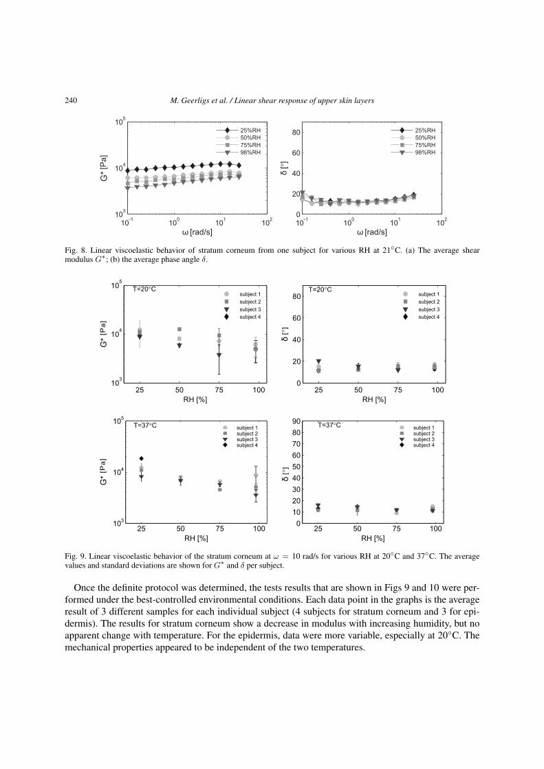
Fig. 8. Linear viscoelastic behavior of stratum corneum from one subject for various RH at 21◦C. (a) The average shearmodulus G∗; (b) the average phase angle δ.
Fig. 9. Linear viscoelastic behavior of the stratum corneum at ω = 10 rad/s for various RH at 20◦C and 37◦C. The averagevalues and standard deviations are shown for G∗ and δ per subject.
Once the definite protocol was determined, the tests results that are shown in Figs 9 and 10 were per-formed under the best-controlled environmental conditions. Each data point in the graphs is the averageresult of 3 different samples for each individual subject (4 subjects for stratum corneum and 3 for epi-dermis). The results for stratum corneum show a decrease in modulus with increasing humidity, but noapparent change with temperature. For the epidermis, data were more variable, especially at 20◦C. Themechanical properties appeared to be independent of the two temperatures.

M. Geerligs et al. / Linear shear response of upper skin layers 241
Fig. 10. Linear viscoelastic behavior of the epidermis at ω = 10 rad/s for various RH at 20◦C and 37◦C. The average valuesand standard deviations are shown for G∗ and δ per subject.
Table 4
Parameter estimates and standard errors
Parameter Skin type
Stratum corneum Epidermis
Estimate Standard error Estimate Standard errorβt 0.01330∗ 0.063 0.00905 0.0109βRH 0.01011∗ ∗ ∗ 0.012 −0.00400∗ 0.002Constant 9.1769 0.2232 8.9738 0.3451No. observations 69 47No. parameters 6 6Log-likelihood −35.3731 −37.5069
Notes: ∗p < 0.05; ∗ ∗p < 0.01 (not shown here); ∗ ∗ ∗p < 0.001.
When fitting model (1) for each type of layer and for various functions F (·, ·) including main effectsand interactions as well as polynomial models, it was found that for stratum corneum the (linear) effectof temperature and of relative humidity is significant (βt = 0.0133, p < 0.05; βRH = −0.01011,p < 0.001), and for epidermis only the linear effect of relative humidity (βRH = −0.0040, p < 0.05).This implies that the fixed part of model (1) can, for both types of skin, be written as Gd (ω = 10 rad/s,RH) = constant + β1t + βRHRH. Thus, these results suggest that RH did affect Gd for both stratumcorneum and epidermis. The significant effect of temperature is demonstrated for Gd of the stratumcorneum. Increasing the number of observations would help to define whether temperature also has astatistically significant effect on Gd for the epidermal tissue. Parameter estimates can be found in Table 4.Estimates have been obtained by using the REML option of the xtmixed procedure of STATA [22].

242 M. Geerligs et al. / Linear shear response of upper skin layers
4. Discussion
In the past, the mechanical behavior of epidermis has been described qualitatively due to the lackof experimental data. The skin curvature and the undulating dermal-epidermal junction cause inherentdifficulties during mechanical characterization of the epidermis in vivo. In addition, in vivo measurementmethods for shear, such as elastography, cannot be applied due to limitations in resolution. Therefore,this study presents an in vitro measurement method to determine shear properties of the epidermis.Preliminary testing was essential to validate the methods applied and to obtain the right experimentalconditions allowing for reliable final measurements.
In order to obtain meaningful results, it is very important that the structural integrity and viability ofthe skin are maintained. Although not fully described in the current paper, an extensive review and somepreliminary tests were performed on isolation methods for the epidermis and stratum corneum as wellas on preservation methods [6]. Since the cleavage plane is an indicator for the success rate of a method,the cleavage location was also considered. Although the variety of topics for in vitro skin research isenormous, it was concluded that isolation and storage protocols can be identical. Future in vitro researchshould make use of isolated epidermis, which is separated by the enzyme dispase [14,25] or cut usinga dermatome, as in the present study, because of its convenient geometry. When the epidermal samplesare subsequently stored for just a short period and tissue growth is not the goal, it is recommended to usea nutrient media such as HHBSS [3]. For long-term storage, the only option for intact viable epidermaltissue is cryopreservation. Regarding the stratum corneum, trypsin and drying remain by far the bestmethods to isolate and preserve this skin layer [16].
In order to measure shear properties of a soft biological material, measurement methods such as thosethat have been developed for muscle, brain and thrombus [5,12,24] could be used. However, pre-testingwas needed to prove that the experimental approach is also appropriate for samples within the order oftens of micrometers, while retaining a relative large diameter to avoid the effect of local properties. Asthere are inherent difficulties in determining the actual thickness of stratum corneum from histology, thesample thickness was defined by the measured gap between the plates. Although the thickness of thestratum corneum on the abdomen is reported to be 14 ± 4 µm [1], the measured gap at a normal force of30 mN varied from 15 to 60 µm due to local variations and skin surface roughness. Applying a highernormal force causes compression of the sample, which influences the measured shear modulus.
To resolve the problem of thin samples with a complex wrinkled sample geometry a technique ofstacking a few of samples was used. The measured properties from stacked samples rely on the assump-tion that there is perfect adhesion between stacked layers. However, it is not possible to be certain thatthe measured properties are not influenced by frictional interactions or incomplete adhesion betweenthese layers, despite efforts to minimize this and the apparent stickiness of the samples.
The skin surface roughness becomes less significant for the other samples involving thicker skin lay-ers. In addition, these layers are more pliable than stratum corneum. Whether stratum corneum, epider-mis only or epidermis and dermis together are measured, the shear response does not differ significantlyfor the strain sweeps. It is hypothesized that loading in shear causes cell deformation in the epidermiswithout affecting the desmosomes. In the dermis, the shear response will be determined by the groundsubstance, because the collagen and elastin fibers are mainly oriented transversally. It is likely that thissubstance has a lower shear resistance than the highly organized epidermis. By contrast, the tissue re-sponse to in-plane tensile loading will be determined by the mechanical integrity of the desmosomes,the elasticity of the dermal fibers and the direction of the Langer lines.

M. Geerligs et al. / Linear shear response of upper skin layers 243
Recently, the linear viscoelastic response of oscillatory shear strains of human whole skin and dermis-only was measured [7,18]. The increase of the moduli was more pronounced for the dermis-only athigher frequencies, so the authors concluded that the epidermis is only slightly frequency dependent. Atlower frequencies, G∗
dermis was of the order of kPa. In accord with this study, in our frequency sweeps weobserved that the epidermis is indeed slightly frequency dependent. Our strain sweeps also resulted in avalue for G∗
dermis of a few kPa.For the stratum corneum, the values of G∗ are similar to those of the epidermis, although some cor-
rections are needed to account for uncertainties in the sample thickness. The results for epidermis andstratum corneum suggest that the small strain shear properties of viable epidermis and stratum corneumare very similar. Currently, our shear moduli can only be compared with in-plane tensile properties ofstratum corneum from the literature. Accordingly, current values for the shear moduli are one order ofmagnitude lower than those in dry conditions and up to two orders of magnitude when fully hydrated,based on the lowest reported values for the tensile moduli [18,19,26]. This clearly supports the highlyanisotropic behavior of stratum corneum and epidermis.
A decrease in stiffness of the stratum corneum could be observed with increasing RH. In accordancewith our observations, delamination studies with stratum corneum, which also showed the pre-failuremechanical response, showed no temperature-dependence for this temperature range [27].
No clear relationship between the mechanical properties of the epidermis and RH could be established.Time sweeps showed that moduli stabilize to an equilibrium after a certain equilibration time. However,both time sweeps and frequency tests for epidermis showed larger variations per RH and per subjectcompared to stratum corneum. This might be related to the less well-defined tissue composition. Forexample, the direction of Langer lines or irregularities such as sweat pores and hair follicles, can have amore substantial role in the mechanical behavior in fully hydrated epidermis than for stratum corneum.Future experiments should clarify the variance in these results.
Longer equilibration times and larger variations were observed in fully hydrated epidermal samplesthan for less humid samples. Examination of fully hydrated stratum corneum has revealed swollen cor-neocytes and water pools in the extracellular spaces after storage in PBS [8]. Furthermore, water disruptsthe lipid lamellae to varying degrees and causes degradation of intercellular corneosomes [2,24]. It islikely that the desmosomes in the viable epidermis are also highly susceptible to damage. However, his-tological examination did not show any sign of degradation in the present samples. The prolonged timeof equilibration of the sample in the set-up at higher RH limited the number of experiments that couldbe performed with epidermis from one donor to within a time of 72 h.
The present study demonstrated that reproducible results can be obtained for the shear properties ofepidermis in an in vitro set up. Viable epidermis could not be measured as an isolated skin layer, but itsproperties can be derived from the other skin samples tested. The G∗ for stratum corneum ranges fromabout 4–12 kPa, decreasing with increasing RH. These values are considerably lower than the shearmodulus value based on tensile Young’s moduli (i.e., E = 3G∗) in the literature, assuming anisotropicmaterial behavior. Results for the epidermis were of the same order of magnitude, but were less consis-tent possibly due to a less well-defined tissue composition. Therefore, it would be interesting to combinemechanical testing with real-time imaging techniques to monitor changes in tissue deformation. It wasalready shown by histological examination after 2 days of loading that shear forces induce cell displace-ment in skin, and particularly in the epidermis [17].
Furthermore, electron microscope imaging techniques could support histological examination in as-sessing tissue damage due to preparation, storage or handling. In addition, it is important to correlate

244 M. Geerligs et al. / Linear shear response of upper skin layers
the shear response both with the tensile testing and with the effects of perpendicular loading, as accom-plished with indentation or compression testing.
Acknowledgement
We would like to thank the plastic surgery department of the Catharina hospital in Eindhoven forproviding the skin tissue.
References
[1] P. Agache and P. Humbert (eds), Measuring the Skin, Springer-Verlag, Berlin, 2004.[2] J.A. Bouwstra, A. de Graaff, G.S. Gooris, J. Nijsse, J.W. Wiechers and A.C. van Aelst, Water distribution and related
morphology in human stratum corneum at differenthydration levels, J. Invest. Derm. 123 (2003), 750–758.[3] D. Bravo, T.H. Rigley, N. Gibran, D.M. Strong and H. Newman-Gage, Effect of storage and preservation methods on
viability in transplantable human skin allografts, Burns 26 (2000), 367–378.[4] P. Chistolini, G. De Angelis, M. De Luca, G. Pellegrini and L. Ruspantini, Analysis of the mechanical properties of in
vitro reconstructed epidermis: preliminary results, Med. Biol. Eng. Comp. 37 (1999), 670–672.[5] E.A. van Dam, S.D. Dams, G.W. Peters, M.C. Rutten, G.W. Schurink, J. Buth and F.N. van de Vosse, Determination of
linear viscoelastic behavior of abdominal aortic aneurysm thrombus, Biorheology 43 (2006), 695–707.[6] M. Geerligs, Skin layer mechanics, PhD thesis, Eindhoven University of Technology, 2009, ISBN: 978–90-74445–92-4.[7] J.L. Gennisson, T. Baldeweck, M. Tanter, S. Catheline, M. Fink, L. Sandrin et al., Assessment of elastic parameters of
human skin using dynamic elastography, IEEE Trans. Ultrasonics Ferroelectrics Frequency Control 8 (2004), 980–989.[8] D.A. Van Hal, E. Jeremiasse, H.E. Junginger, F. Spies and J.A. Bouwstra, Structure of fully hydrated human stratum
corneum: a freeze fracture electron microscopy study, J. Invest. Derm. 106 (1996), 89–95.[9] F.M. Hendriks, Mechanical behaviour of human epidermal and dermal layers, PhD thesis, Eindhoven University of Tech-
nology, 2005.[10] F.M. Hendriks, C.W.J. Oomens, D.L. Bader, F.P.T. Baaijens and D. Brokken, The relative contributions of different skin
layers to the mechanical behavior of human skin in vivo using suction experiments, Med. Eng. Phys. 28 (2006), 259–266.[11] B. Holt, A. Tripathi and J. Morgan, Viscoelastic response of human skin to low magnitude physiologically relevant shear,
J. Biomech. 12 (2008), 2689–2695.[12] M. Hrapko, J.A. van Dommelen, G.W. Peters and J.S. Wismans, The mechanical behaviour of brain tissue: large strain
response and constitutive modeling, Biorheology 43 (2006), 623–636.[13] M.A. Kendall, Y.F. Chong and A. Cock, The mechanical properties of the skinepidermis in relation to targeted gene and
drug delivery, Biomaterials 28 (2007), 4968–4977.[14] Y. Kitano and N. Okada, Separation of the epidermal sheet by dispase, Br. J. Dermatol. 108 (1983) 555–560.[15] K.S. Koutroupi and J.C. Barbenel, Mechanical and failure behaviour of the stratum corneum, J. Biomech. 23 (1990),
281–287.[16] M. Nicollier, P. Agache, J.L. Kienzler, R, Laurant, R.Gibey, N.Cardot and J.C.Henry, Action of trypsin on human plantar
stratum corneum: an ultrastructural study, Arch. Dermatol. Res. 268 (1980), 53–64.[17] C. Pailler-Mattei and H. Zahouani, Study of adhesion forces and mechanical properties of human skin in vivo, J. Adhesion
Sci. Technol. 18 (2004), 1739–1758.[18] Y.S. Papir, K.H. Hsuand and R.H. Wildnauer, The mechanical properties of stratum corneum, I, The effect of water and
ambient temperature on the tensile properties of newborn rat stratum corneum, Biochim. Biophys. Acta 1 (1975), 170–180.[19] A.C. Park and C.B. Baddiel, Rheology of stratum corneum, 2, Physicochemical investigation of factors influencing water-
content of corneum, J. Soc. Cosmetic Chemists 23 (1972), 13–21.[20] A. Rochefort, P. Druot, M. Leduc, R.Vassalet and P. Agache, A new technique for the evaluation of cosmetics effect on
mechanical-properties of stratum corneum and epidermis in vitro, Int. J. Cosm. Sci. 8 (1986), 27–37.[21] F.H. Silver, L.M. Siperkoand and G.P. Seehra, Mechanobiology of force transduction in dermal tissue, Skin Res. Technol.
9 (2003), 3–23.[22] StataCorp, Stata statistical software, Release 11.2, StataCorp LP, College Station, TX, 2009.[23] K.S. Stenn, R. Link, G. Moellmann, J. Madri and E. Kuklinska, Dispase, a neutral protease from Bacillus polymyxa is a
powerful fibronectinase and type IV collagenase, Br. J. Dermatol. 93 (1989), 287–290.[24] M. van Turnhout, G. Peters, A. Stekelenburg and C. Oomens, Passive transverse mechanical properties as a function of
temperature of rat skeletal muscle in vitro, Biorheology 42 (2005), 193–207.

M. Geerligs et al. / Linear shear response of upper skin layers 245
[25] R.R. Warner, K.J. Stone and Y.L. Boissy, Hydration disrupts human stratum corneum ultrastructure, J. Invest. Derm. 120(2003), 275–284.
[26] R.H. Wildnauer, J.W. Bothwell and A.B. Douglass, Stratum corneum biomechanical properties. I. Influence of relativehumidity on normal and extracted human stratum corneum, J. Invest. Dermatol. 1 (1971), 72–78.
[27] K.S. Wu, J. Li, K.P. Ananthapadmanabhan and R.H. Dauskardt, Time-dependent intercellular delamination of humanstratum corneum, J. Mater. Sci. 42 (2007), 8986–8994.


![Menschenhaut [Human skin]](https://static.fdokumen.com/doc/165x107/6326d24f24adacd7250b1364/menschenhaut-human-skin.jpg)