Clear cell endometrial adenocarcinoma in a young woman: Report of a case detected by cytology
Upload
independentCategory
view
1download
0
Identification of pVHL as a Novel Substrate forAurora-A in Clear Cell Renal Cell Carcinoma(ccRCC)Benedicte Martin1, Franck Chesnel1, Jean-Guy Delcros2, Florence Jouan1, AnneCouturier1, Frederic Dugay1, Xavier Le Goff1, Jean-Jacques Patard3, Patricia Fergelot4,Cecile Vigneau1,5, Nathalie Rioux-Leclerq1,6, Yannick Arlot-Bonnemains1*
1 CNRS - UMR 6290 (IGDR) -Université Rennes 1- BIOSIT - Rennes, France, 2 UMR- INSERM U1052/CNRS 5286, Centre de Recherche enCancérologie de Lyon, Centre Léon Bérard, Lyon, France, 3 CHU Bordeaux, Service de Génétique Moléculaire, Bordeaux, France, 4 AP-HPHôpital Bicêtre, Service de Chirurgie Urologie, Le Kremlin Bicêtre, France, 5 CHU Pontchaillou, Département de Néphrologie, Rennes, France,6 CHU Pontchaillou, Département de Pathologie, Rennes, France
Abstract
Clear cell renal cell carcinoma (ccRCC) is the most common histological subtype of kidney cancer andis often characterized by mutations or deletions of the Von Hippel Lindau (VHL) tumour suppressorgene. Aurora gene family members are implicated in proper mitotic progression and spindlecheckpoint function and play a crucial role in cancer progression. In the present study, we assessedthe expression of Aurora-A in a cohort of 30 ccRCC with fully characterized VHL status (wt/wt or mut/del) and Fuhrman grade. Aurora-A transcript and protein levels were significantly increased in highFuhrman grade tumours and in VHLwt/wt tumours. These results suggest that Aurora-A and VHLinteract in the ccRCC. We demonstrated that the two proteins interact in vivo and identified the Ser72on the sequence of VHL as the unique site phosphorylated by Aurora-A.
Citation: Martin B, Chesnel F, Delcros J , Jouan F, Couturier A, et al. (2013) Identification of pVHL as a Novel Substrate for Aurora-A inClear Cell Renal Cell Carcinoma (ccRCC). PLoS ONE 8(6): e67071. doi:10.1371/journal.pone.0067071Editor: Claude Prigent, Institut de Génétique et Développement de Rennes, FranceReceived February 25, 2013; Accepted May 13, 2013; Published June 13, 2013Copyright: © 2013 Martin et al. This is an open-access article distributed under the terms of the Creative Commons AttributionLicense, which permits unrestricted use, distribution, and reproduction in any medium, provided the original author and source arecredited.Funding: This work was supported by the CNRS, the Ligue Nationale Contre le Cancer. The funders had no role in study design, datacollection and analysis, decision to publish, or preparation of the manuscript.Competing interests: The authors have declared that no competing interests exist.* E-mail [email protected]
Introduction
Clear cell renal cell carcinoma (ccRCC) is the mostcommon form of kidney cancer and the yearly numberof newly diagnosed cases is steadily increasing. Thistype of cancer develops in the renal proximal tube andis linked to biallelic inactivation of the von Hippel–Lindau (VHL) tumour suppressor gene. VHL syndromeis an inherited disorder caused by germline pointmutations in the VHL gene and is characterized by apredisposition to a variety of benign and malignanttumours [1]. Based on the patients’ genotype, VHLsyndrome can be divided in four subtypes (1, 2A, 2Band 2C) that are associated with different phenotypes[2] [3] [4]. Only the 1 and 2B subtypes predisposepatients to the development of ccRCC. Biallelicinactivation of VHL has also been reported in up to 80%of patients with sporadic ccRCC [5]; [6][7];. VHL
mutations are extremely heterogeneous anddistributed all along the gene (promoter, exons andintrons). Despite the observation that thereintroduction of wild type VHL in a VHL-null ccRCC cellline inhibits tumorigenesis [8], the molecular basis oftumour formation and progression upon VHLinactivation remains unclear. However, additionaloncogenic events are required for ccRCC formation asclearly evidenced by molecular and genetic approaches[9].
There are three isoforms of the VHL gene product(known as pVHL): the full-length pVHL30 protein (213amino acids) and two smaller isoforms of 160 (pVHL19,due to the use of an internal translational start site)and 172 amino acids (the result of alternative splicing)in length. pVHL is thought to be an E3 ubiquitin ligasewithin a tetramer complex with Cullin 2 and Elongins Band C that targets hypoxia inducible factor α (HIFα) for
PLOS ONE | www.plosone.org 1 June 2013 | Volume 8 | Issue 6 | e67071
-G

polyubiquitylation and proteasome degradation. Thisimplicates pVHL in the regulation of genes involved incell proliferation and survival and in angiogenesis. Inaddition, pVHL exerts other functions that arecompletely independent of its role in HIFα degradation,including regulation of microtubule stabilization, cellcycle progression [8,10] maintenance of appropriateMad2 protein levels and chromosomal stability [11].
Many protein kinases are key regulators of cell cycleprogression and are frequently deregulated in cancer.Among them, the mitotic Aurora-A serine/threonine(Ser/Thr) kinase has been identified as a putativeoncogenic protein [12]. Over-expression of Aurora-Aprotein in HeLa cells promotes the formation ofcolonies in soft agar and favours growth of tumourxenografts in mouse. However, it remains unclear howAurora-A over-expression can induce oncogenictransformation. Aurora-A expression is cell-cycleregulated as it is degraded via the ubiquitin-proteasome pathway upon mitotic exit [13]. It plays amajor role during duplication and separation ofcentrosomes and also contributes to spindle formationand stability. Aurora-A alterations are correlated withaggressive tumour behaviour such as differentiatedtumour grade, invasion and nodal metastasis [14] [15].Amplification of the Aurora-A gene and up-regulation ofAurora-A transcript/protein levels or kinase activityhave been observed in a variety of cancers [14] [16][17]. Aurora-A is often expressed in ccRCC and somestudies reported elevated expression levels of Aurora Aand Aurora B in advanced stage tumours associatedwith poor patients’ survival [18] [19]. In addition,abrogation of the mitotic spindle checkpoint has beeninvolved in the progression of ccRCC [19]. Altogetherthese results suggest that Aurora-A may be implicatedin an early event of ccRCC development [20] [21].
To test this hypothesis, we first assessed Aurora-Aand pVHL expression in ccRCC surgical samples withknown VHL status and then investigated their possibleinteraction. We show that the Aurora-A transcript andprotein levels in ccRCC were correlated with thetumour grade and VHL status. Moreover, wedemonstrate that Aurora-A interacts with pVHL andthat VHL is phosphorylated by Aurora-A.
Material and Methods
Tissue samplesTumour and matched normal tissue samples were
obtained from 30 patients with ccRCC who underwentpartial or total nephrectomy between 2002 and 2005.The Ethics Committee of the Medical School andHospital of Rennes University approved thisprospective study and all patients signed informedconsents. Upon collection, tissue samples wereimmediately frozen in liquid nitrogen and stored at-80°C at the Centre de Ressources Biologiques (CRB,Rennes, France) or formalin-fixed and paraffin-
embedded. The diagnosis and tumour staging weredetermined by histopathological analysis. To assessVHL status in the collected samples, denaturing high-performance liquid chromatography (DHPLC) wascarried out using a WAVE Nucleic Acid FragmentAnalysis system (Transgenomic, Glasgow, UK) andDNAsep columns [22]. Aberrant peaks were furtheranalysed by direct sequencing using standardprocedures. All mutations were confirmed by PCRanalysis and sequencing. Multiplex Ligation-dependentProbe Amplification (MLPA) was used to detect VHLdeletions as previously described [23]. The wild typestatus was hereafter defined as VHLwt/wt and thesamples deleted and mutated for VHL as VHLmut/del.
AntibodiesThe polyclonal antibodies against Aurora-A and Actin
were purchased from Abcam (Paris, France) and Sigma-Aldrich (L’Isle d’Abeau, France), respectively. The anti-pVHL monoclonal antibody was from BD Pharmingen(San Jose, CA). The horseradish peroxidase-conjugatedsecondary antibodies were from JacksonImmunoResearch Laboratories (Baltimore, MD).
Real time PCR analysisTotal RNA was extracted from tumour samples using
the Qiagen Qiamp Total RNA kit and then fivemicrograms of each sample were reverse-transcribedusing oligo-dT primers and the M-MLV reversetranscriptase. The resulting cDNAs were used astemplates for PCR assays. Primers were: 5’CTGCATTTCAGGACCTGTTAAGG-3’ (forward) and 5’-AACGCGCTGGGAAGAATTT-3’ (reverse) for humanAurora A; 5’-CTGACTTCAACAGCGACACC-3’ (forward)and 5’-TAGCCAAATTCGTTGTCATACC-3’ (reverse) forhuman GAPDH (control). Assays were performed intriplicate, using a RotorGene 3000 apparatus (CorbettResearch, Biolabo, Archamps, France) and the SYBRGreen I master mix (Roche Diagnostics, Mannheim,Germany). For each sample, the relative amount ofAurora A and GAPDH transcripts was calculated fromthe standard curves using the RotorGene software.
Cell cultureThe RCC4 cells come from ECACC [24] and 786-O and
HeLa from ATCC. HeLa and RCC4 cells were maintainedin DMEM medium and 786-O in RPMI medium,supplemented with 10% foetal bovine serum andantibiotics at 37°C in a humidified 5% CO2 atmosphere.Cells were transfected with the pCMV-VHL213 vectorkindly provided by Dr Buchberger (Heidelberg,Germany) using JetPRIMETM as recommended by themanufacturer (Polyplus, Illkirch, France). Cells werelysed in EB buffer (20 mM Tris-HCl pH 7.5, 100 mMNaCl, 5 mM MgCl2, 10% glycerol, 0.5 mM DTT, 0.2%NP40, 20 mM β-glycerophosphate, 1 mM NaF, 1 mMAEBSF) 24 hours after transfection.
pVHL Phosphorylation and Aurora-A in ccRCC
PLOS ONE | www.plosone.org 2 June 2013 | Volume 8 | Issue 6 | e67071

Immunoprecipitation assaysSeventy µl of Dynabeads coupled to Protein G
(Invitrogen, Saint-Aubin, France) were equilibrated with500 µl of 0.5M acetate buffer pH 5.5 and incubated at4°C with the anti-pVHL antibody or control IgG for 2hours, and then washed twice with 500 µl PBS. Beadswere then incubated at 4°C with 300 µg of HeLa orRCC4 protein extract or with an extract of 786-0 cellspreviously transfected for 24 hours with pCMV-VHL-213during 2 hours. Beads were washed once in 500 µl of0.5% NaCl and five times with 500 µl TBST (50 mM Tris-HCl, pH 7.5, 150mM NaCl, 0.05% Tween-20). Boundproteins were eluted in 10 µl of 2X Laemmli samplebuffer, separated on a 12.5% SDS polyacrylamide geland transferred to nitrocellulose membranes.Membranes were incubated with the anti-pVHL (1:100)or anti-Aurora-A antibody (1:200).
Protein purificationRecombinant Aurora-A-(His)6, pVHL213-(His)6 and pVHL
213S72A-(His)6 were produced in the Escherichia colistrain BL21(DE3) pLysS. Bacteria were lysed in IMAC 5buffer (20 mM Tris-HCl, pH 7.5; 500 mM NaCl; 10%glycerol, and 5 mM imidazole) containing 1 mg/mllysozyme and 1 mM PMSF for 1 h at 4 °C. Aftercentrifugation at 12,000g at 4 °C (JA-20 rotor, BeckmanInstruments) for 30 min, supernatants were filtered andproteins purified by Ni-NTA-agarose affinitychromatography following the manufacturer’sinstructions (Qiagen, Courtaboeuf, France), aspreviously described [25]. The concentration of theeluted His-tagged protein fractions was determinedaccording to the Bradford method and their purity wasassessed by electrophoresis on 12.5% SDS gels.
Protein Kinase AssayTwo µg of Aurora-A-(His) 6 were incubated in 15 µl of
kinase buffer (50 mM Tris-HCl, pH 7.5; 25 mM NaCl; 1mM dithiothreitol; 10 mM MgCl2) in the presence of 5µCi [γ -32P] ATP at 3,000 Ci/mmol and 2 µg pVHL213-(His)6 or pVHL 213S/A72-(His)6 at 30°C for 10 min.Reactions were stopped by addition of 5 µl ofdenaturing 5X Laemmli sample buffer and proteinswere separated on 12.5% SDS-PAGE gels. Incorporationof 32P was analysed by autoradiography.
Western blot analysisFrozen tissues were lysed in RIPA buffer (50 mM Tris-
HCl, pH 7.4, 1% NP-40, 0.5% sodium deoxycholate, 150mM sodium chloride, 1 mM EDTA, 1 mM sodiumfluoride, 1 mM AEBSF, 10 µg/ml aprotinin, 10 µg/mlleupeptin, 1 mM sodium orthovanadate) andcentrifuged at 9000g for 10 min. Bradford assays wereperformed to determine the protein concentrations inthe supernatants. Samples (50 µg of total proteins)were separated on 15% polyacrylamide gels andtransferred onto nitrocellulose membranes. Membranes
were washed with TBST, saturated with 5% low fat milkin TBST at room temperature for 2 hr and thenincubated with the primary antibodies in 2.5% low fatmilk in TBST at 4°C overnight. Antibody binding wasdetected with the appropriate horseradish peroxidaseconjugated secondary antibody (1:15,000 in TBST-2.5%BSA at RT for 1h) and the Western Blotchemiluminescence Super Signal kit (Pierce, Rockford,IL). Sample loading was controlled using an anti-Actinpolyclonal antibody (1:500).
ImmunohistochemistryFive-µm sections of formalin-fixed paraffin-embedded
tissue samples mounted on glass slides were dewaxedwith xylene and rehydrated through a gradient seriesof ethanol. They were then heated at 100°C in hot 0.01mM citrate buffer pH 8 for 40 min, left to cool down for20 minutes and washed in PBS/0.1% Tween for 5 min.Endogenous peroxidase was quenched in 3% hydrogenperoxide for 10 min. Slides were then washed in PBS/0.1% Tween for 5 min and incubated with the primaryantibodies against Aurora-A (1:1000) and pVHL (1:100).Antibody binding was revealed with HRP (horseradishperoxidase)-labelled polymer conjugated to secondaryantibodies (Envision™ + Dual Link System-HRP, DAKO)using diaminobenzidine as chromogen (Sigma-Aldrich).Antibody staining was assessed using a Leica™DMRXA microscope equipped with a CoolSnapsHQcamera (Photometrics™).
ImmunofluorescenceAfter blocking in PBS/5% BSA, Hela cells and RCC4
cells cultivated and fixed onto glass slides wereincubated at room temperature with the mouse anti-pVHL monoclonal antibody (1:200) and/or the rabbitanti-Aurora-A (1:200) antibody diluted in PBS/1% BSAfor 1 h. After two washings with PBS, slides wereincubated with Alexa Fluor® 546-conjugated anti-rabbitand Alexa Fluor® 488 donkey anti-mouse secondaryantibodies (1:15000 in PBS/1% BSA) at roomtemperature for one hour, then washed in PBS andmounted in Vectashield® containing 1µg/ml DAPI.Imaging was performed on a Leica DM 5500 confocalmicroscope using a X63 HCX PLAN-APO-ON 1.4 oilimmersion objective lens (MRic microscopy platform).Sequential images were captured at a single focalplane using appropriate laser settings for the detectionof DNA (TO-PRO®-3), Aurora-A and VHL. Imageprocessing was performed with the ImageJ 1.4software.
Statistical analysis. Analyses were performedusing the GraphPad software. The non parametricMann–Withney test was used to evaluate the statisticalrelationship between the expression of Aurora-Aprotein and the VHL status (wt/wt or VHL mut/del) aswell as with the Fuhrman grade of the tumour. For allanalyses, differences were considered to be significantwhen p < 0.05. A linear regression analysis was used to
pVHL Phosphorylation and Aurora-A in ccRCC
PLOS ONE | www.plosone.org 3 June 2013 | Volume 8 | Issue 6 | e67071

study the relation between the expression of Aurora-Aprotein and the expression of cytoplasmic VHL proteinleading to the determination of the Pearson coefficient.
Results
VHL status of tumour samplesFirst we determined the VHL status (wild type –
VHLwt/wt or presence of mutations/deletions – VHLmut/del) of the surgical ccRCC samples from a cohort of 30patients. Ten specimens (33% of the series) wereVHLwt/wt. In the remaining 20 tumours (67%) withVHLmut/del, deletion of the gene in one allele wasassociated with mutations in the other allele (VHLmut/del). Among the 20 samples which were characterisedas VHLmut /del, 10 samples were mutated in exon 1, 6samples in exon 2 and 4 samples in exon 3. They weremainly stop insertions, frame shift or missensemutations and 50% of them were in exon 1 and 13% inexon 3. Overall, 64% of patients had a Fuhrman grade3 to 4 tumour. VHLwt/wt ccRCCs mostly had a highFuhrman grade score (80%), while VHLmut/del tumourswere equally distributed among grades 2 to 4 (Table 1).
The expression level of Aurora-A transcripts inccRCCs is correlated with the Fuhrman gradeand the VHL status
We then measured Aurora-A expression in the 30surgical ccRCC samples (T) by quantitative real-timePCR (Figure 1A). The Aurora-A transcript levels weresignificantly higher in Fuhrman grade 3 and 4 than ingrade 1 and 2 tumours (p=0.0034) and in VHLwt/wtthan in VHL mut/del ccRCC samples (p=0.037) (Figure1B).
Aurora-A and VHL protein expression inmatching ccRCC and healthy kidney samples
We investigated VHL (pVHL30 and pVHL19) andAurora-A protein expression by western blot analysisusing protein extracts from ccRCC samples (T) andmatching neighbouring healthy tissues (N) from 2 VHLwt/wt samples and 9 VHL mut/del samples (Figure 2A).In all normal samples, the pVHL30 isoform waspredominant, whereas pVHL19 was weakly or notexpressed (Figure 2A: lanes 1, 3 and 5). In tumourtissues, the expression of both VHL isoforms was veryvariable and depended on the VHL status. VHLwt/wttumour samples expressed only pVHL19 (Figure 2A:lane 2). Conversely, in VHLmut/del tumours no pVHL oronly the pVHL30 isoform could be detected. Aurora-Awas detected at the predicted molecular weight of 42kDa in all normal tissue samples (Figure 2A: lanes 1, 3and 5) and in most tumour tissues, albeit at lower level(Figure 2A: lanes 2, 6). In few tumour samples, Aurora-A could not be detected (Figure 2A, lane 4).
We then analysed the localization of pVHL andAurora-A by IHC (Figure 2B). In normal renal tissues,
VHL and Aurora-A were homogeneously expressed inthe epithelial cells of proximal and distal tubules(Figure 2B: left panels). In tumours the tissuearchitecture was dramatically disorganized andimmunoreactivity towards Aurora-A and pVHL wasvariable (Figure 2B). All VHLwt/wt samples expressedpVHL, while four of the VHLmut/del tumours did not(data not shown), in agreement with the western blotanalysis. The intensity of pVHL staining was notcorrelated with the tumour grade (Figure 3A), whereasits localization differed with the tumour grade. In highgrade tumours, pVHL expression was predominantlycytoplasmic (grade 4 vs. grade 2: p=0.0035; grade 4vs. grade 3: p=0.002; Figure 3B), while in low gradetumours, pVHL was mostly membrane-associated(grade 2 vs. grade 4: p=0.0285; Figure 3C).
The intensity of Aurora-A staining was correlated withthe tumour grade (grade 4 vs. grade 2: p=0.0185)(Figure 3E), but not with the VHL status (p=0.0613)(Figure 3D), differently from what observed for thetranscript level. This discrepancy could be due to thelimited number of patients examined in our study(n=30). Moreover, Aurora-A staining was mainlyobserved in cells with pVHL cytoplasmic expression(Figure 3F, Pearson coefficient 0.658).
Aurora–A interacts with and phosphorylatespVHL
Aurora-A and pVHL co-expression in tumour cells andtheir implication in regulating the microtubule networkand cell cycle progression suggested that they mayinteract. To test this hypothesis, first we studied theirexpression and localization during the cell cycle inHeLa cells in which pVHL30 is endogenously expressed.In these cells, pVHL30 expression varied during the cellcycle, reaching a maximum in the G2 phase (Figure4A). As it was previously shown that Aurora-Aexpression and activity are cell cycle-regulated andthat Aurora-A is mainly associated with centrosomesfrom the end of S phase to the beginning of the G1phase [26], we assessed simultaneously thelocalization of Aurora-A and pVHL by doubleimmunofluorescence. In HeLa cells, as well as in RCC4cells, both proteins co-localized at the centrosome andat the mitotic spindle extremities (Figure 4B).
Then, we investigated whether Aurora-A and pVHLinteracted by immunoprecipitation assay using an anti-VHL antibody and total proteins extracts from HeLacells that had been transfected with the pCMV-hVHL213plasmid to over-express pVHL30. The interaction wasalso assayed by performing the immunoprecipitationon protein extracts of two different kidney cell lines(RCC4 and 786-0) (Figure 4C). As Aurora-A was co-immunoprecipitated with pVHL in all three cell lines(Figure 4C), we then asked whether pVHL was aphosphorylation substrate of Aurora-A. We thusincubated affinity-purified recombinant Aurora-A(His)6with recombinant pVHL-213(His)6 in the presence of
pVHL Phosphorylation and Aurora-A in ccRCC
PLOS ONE | www.plosone.org 4 June 2013 | Volume 8 | Issue 6 | e67071

[γ-32P] ATP (Figure 5). 32P was incorporated in VHL inthe presence of Aurora-A (Figure 5: lane 1) and massspectrometry analysis of 32P-VHL allowed identifyingSer72 as the most phosphorylated amino acid. Threedifferent productions and purifications of pVHL andAurora-A proteins were done and used in twoindependent kinase assays before identifying thephosphorylation site. Accordingly, the sequence which
includes Ser72 (…SREPS*QVIF…) matched the putativeAurora-A consensus phosphorylation site [(K/R) xx(S/T)](Figure 5). Mutation of Ser72 into alanine (VHL SA72)greatly diminished phosphorylation of pVHL by Aurora-A (Figure 5, lane 2).
Figure 1. Relative expression of the Aurora-A kinase in ccRCC and matching healthy tissuesamples. A-Expression of Aurora-A in tumours and neighbouring healthy tissues. Total RNAs extracted fromtumour tissues were reverse transcribed and Aurora-A expression was then analysed by PCR using specific Aurora-Aprimers that produced an amplicon of 150 bp. Expression values were normalized to GAPDH levels. B-Aurora-Atranscript levels are correlated with the tumour Fuhrman grade and VHL status. Quantitative RT-PCR data werenormalized to GAPDH and then their distribution according to the tumour grade and VHL status was analysed usingthe Mann-Whitney test. The Aurora-A transcription level of each tumour is symbolized by a dot and the median by ahorizontal line.doi: 10.1371/journal.pone.0067071.g001
pVHL Phosphorylation and Aurora-A in ccRCC
PLOS ONE | www.plosone.org 5 June 2013 | Volume 8 | Issue 6 | e67071

Figure 5. pVHL is phosphorylated on Ser72 byAurora-A in vitro. A- Purified recombinant 6xHis-tagged pVHL30 [VHL(His)6] (lane 1) or mutant 6xHis-tagged pVHL30 S72A [VHLSA72(His)6] ( (lane 2) wereincubated in the presence of [γ-32P] ATP andrecombinant Aurora-A(His) 6. Samples were separatedby SDS-PAGE and the gel was stained with CoomassieBlue (higher panel). pVHL phosphorylation status wasanalysed by autoradiography (lower panel). Theasterisk (*) indicate the pVHL protein.B-Localization and sequence of the putative Aurora-Aphosphorylation site on pVHL.doi: 10.1371/journal.pone.0067071.g005
Figure 2. Detection of Aurora-A and VHL in healthy and tumour tissues. A-Western blot analysis of proteinextracts from ccRCC and matching healthy tissue samples. Total proteins from tumour (T) and healthy (N) tissuesamples were extracted using the RIPA buffer and analysed by western blotting with the anti-Aurora-A polyclonalantibody (1:200) and the anti-pVHL monoclonal antibody (1:500). β-actin was used as loading control.B-Immunohistochemical analysis of Aurora-A and VHL expression in ccRCC and matching healthy tissue samples.Tissues samples were prepared for immunohistochemistry as described in Materials and Methods and analysedusing the polyclonal antibody against Aurora-A (1:100) and the monoclonal antibody against pVHL (1:200).Microphotographs were acquired with a Leica™ DMRXA microscope equipped with a CoolSnapsHQ camera(Photometrics™) (bar 10µm).doi: 10.1371/journal.pone.0067071.g002
pVHL Phosphorylation and Aurora-A in ccRCC
PLOS ONE | www.plosone.org 6 June 2013 | Volume 8 | Issue 6 | e67071

Figure 3. Statistical analysis of Aurora-A and pVHL levels and localization in ccRCCs according to theirFuhrman grade and VHL status. pVHL expression level (A) and localization (B and C) in ccRCCs were analysedrelative to the Fuhrman grade of the tumours. Aurora-A expression level was assessed relative to the VHL status (D)and Fuhrman grade (E) of the tumours. Aurora-A expression according to the VHL cytoplasmic localization was alsoevaluated (F). The non-parametric Mann-Whitney test was used to evaluate the statistical relationships between theexpression of Aurora-A and pVHL and the tumour parameters.doi: 10.1371/journal.pone.0067071.g003
pVHL Phosphorylation and Aurora-A in ccRCC
PLOS ONE | www.plosone.org 7 June 2013 | Volume 8 | Issue 6 | e67071

Figure 4. pVHL interacts with Aurora-A. A-HeLa cells were synchronized as described in Materials andMethods. Twenty µg of protein extracts were separated on a poly-acrylamide gel and VHL expression was assessedwith the monoclonal anti-pVHL antibody (1:200). Loading was controlled with the anti-Actin antibody (1:200).B-In HeLa and RCC4 cells, Aurora-A and VHL co-localize at the centrosome and at the mitotic spindle extremities(anti-Aurora-A antibody, 1:200 and anti-pVHL antibody, 1:200).C-HeLa, RCC4 and 786-0 cell protein extracts were immunoprecipitated with the anti-pVHL polyclonal antibody(lane 2) or with the IgG (lane 3, negative control). Aurora-A and pVHL were both detected in theimmunoprecipitates using a polyclonal antibody against Aurora-A (1:200) (upper panel) and a monoclonal antibodyagainst pVHL (1:200) (lower panel). Lane 1 was corresponding to the total cell extract.doi: 10.1371/journal.pone.0067071.g004
pVHL Phosphorylation and Aurora-A in ccRCC
PLOS ONE | www.plosone.org 8 June 2013 | Volume 8 | Issue 6 | e67071

Discussion
Inherited and sporadic forms of ccRCC account for85% of renal cancers and loss of the VHL tumoursuppressor gene is involved in 70% of sporadic ccRCC.In addition to genetic mutations, ccRCC is mainlyassociated with genetic instability that could be causedby abrogation of the cell cycle mitotic spindlecheckpoint and may involve the centrosomal kinaseAurora-A, which regulates centrosome stability andthus influences cell cycle progression [27].
In the present study, we determined the relationshipbetween Aurora-A and pVHL in ccRCC. Higher levels ofAurora-A transcripts were significantly associated withhigh Fuhrman grade ccRCC samples (grades 3 and 4)and with the VHLwt/wt status. Similarly, Aurora-Aprotein expression both at the cell membrane and inthe cytoplasm was significantly higher in grade 3-4tumours and tended to be more elevated in VHLwt/wttumours. These results suggest that low levels ofAurora A could be correlated with the presence of non-functional pVHL.
Previous studies in ductal breast cancer showed thatAurora-A over-expression was independent of thetumour histopathological type and was not correlatedwith tumour size and lymph node metastases [28].Conversely, other authors showed that Aurora-Aalterations were associated with poor prognosis ingastric cancers and high grade/late stage in breast andbladder cancers [29] [30]. An exhaustive analysis ofAurora-A expression in human RCC revealed nosignificant association with major pathologic factors[21]; however, in this study the relation with the VHLstatus was not assessed.
About 80% of the VHLwt/wt tumours included in ourstudy were grade 3-4, while only 50% of VHLmut/deltumours were classified as grade 3 and 4. It has beenreported that the outcome of patients with wild-typeVHL is significantly worse both in terms of progressionfree-survival (PFS) and RCC-specific survival (RCC-SS),whereas VHL alterations appear to be associated with amore favourable outcome [22]. pVHL expression in ourseries was very heterogeneous as previously reported[3] [31] and not correlated with the tumour grade.However, pVHL cytoplasmic localization was associatedwith grade 3-4 tumours and with higher Aurora-Aexpression, suggesting that Aurora-A oncogenic actionmight also involve interaction with pVHL to deregulate
its tumour suppressor function. Indeed, we furthershow that pVHL30 interacts with and is phosphorylatedby Aurora-A at Ser 72 in the β-domain of pVHL. Severalputative phosphorylation sites on pVHL have beenidentified by mass spectrometry. Three of these aminoacids are located in the acidic region of pVHL (Ser33,Ser38 and Ser48) and three others are in the β-domain(Ser68, Ser72 and Ser111). Ser111 of pVHL is aphysiological target of Checkpoint kinase 2 (CHK2) inresponse to DNA damage. Phosphorylation of Ser68 byglycogen synthase kinase 3 (GSK3) negativelyregulates pVHL-mediated microtubule stabilization[18,32]. GSK-3 regulation requires a primingphosphorylation on S72, which in vitro can beaccomplished by Casein kinase 1 (CK1). Our dataidentify Aurora A as the putative kinase that mightcarry out the priming phosphorylation on Ser 72 invivo. As pVHL was identified as a microtubule-associated protein which prevents microtubulesdepolymerisation, we therefore speculated that thephosphorylation of VHL by Aurora-A would modulatethe stability and/or dynamics of the microtubules.Dysregulation of such coordination may hence beresponsible of mitotic defects leading to aneuploidy,which contributes to tumorigenesis (Figure 6). Inconclusion, the present study describes for the firsttime the relationship between pVHL and the oncogenicAurora-A protein kinase. Even though the expressionlevels of both proteins might not be significantlycorrelated with tumour progression, we hypothesizethat the functional interaction between Aurora-A andpVHL may participate in cell cycle regulation and intumour initiation/progression through signallingpathway(s) that need to be further investigated.
Figure 6. Putative function of the interaction betweenVHL and Aurora-A in the ccRCC.doi: 10.1371/journal.pone.0067071.g006
Table 1. Fuhrman grade and VHL status of the 30clear cell renal cell carcinoma (ccRCC) specimensunder study.
Fuhrman grade 1 2 3 4VHLwt/wt % (nb)a 3 (1) 3 (1) 10 (3) 17 (5)VHLmut/del % (nb) 0 33 (10) 23 (7) 10 (3)a. (nb) : number of samples
pVHL Phosphorylation and Aurora-A in ccRCC
PLOS ONE | www.plosone.org 9 June 2013 | Volume 8 | Issue 6 | e67071
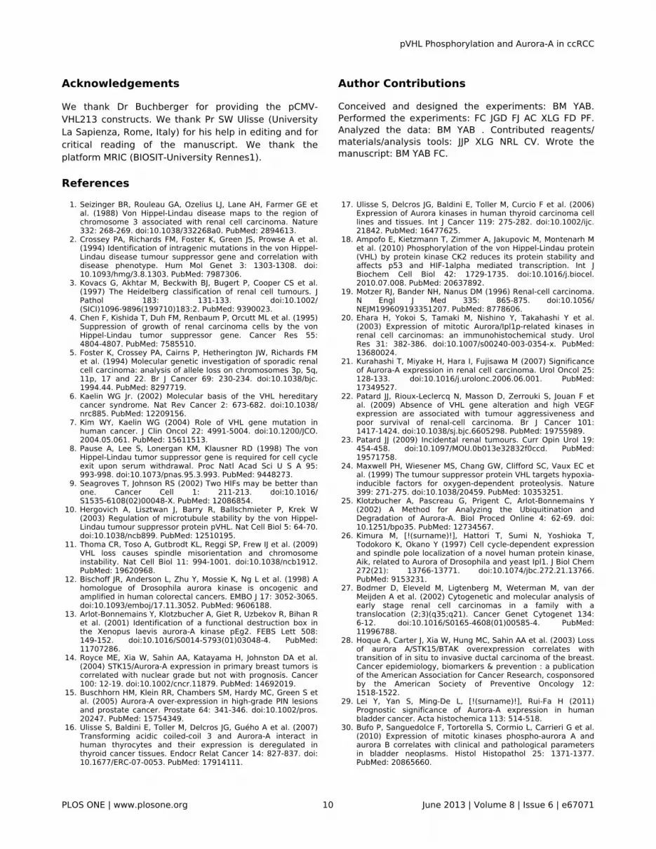
Acknowledgements
We thank Dr Buchberger for providing the pCMV-VHL213 constructs. We thank Pr SW Ulisse (UniversityLa Sapienza, Rome, Italy) for his help in editing and forcritical reading of the manuscript. We thank theplatform MRIC (BIOSIT-University Rennes1).
Author Contributions
Conceived and designed the experiments: BM YAB.Performed the experiments: FC JGD FJ AC XLG FD PF.Analyzed the data: BM YAB . Contributed reagents/materials/analysis tools: JJP XLG NRL CV. Wrote themanuscript: BM YAB FC.
References1. Seizinger BR, Rouleau GA, Ozelius LJ, Lane AH, Farmer GE et
al. (1988) Von Hippel-Lindau disease maps to the region ofchromosome 3 associated with renal cell carcinoma. Nature332: 268-269. doi:10.1038/332268a0. PubMed: 2894613.
2. Crossey PA, Richards FM, Foster K, Green JS, Prowse A et al.(1994) Identification of intragenic mutations in the von Hippel-Lindau disease tumour suppressor gene and correlation withdisease phenotype. Hum Mol Genet 3: 1303-1308. doi:10.1093/hmg/3.8.1303. PubMed: 7987306.
3. Kovacs G, Akhtar M, Beckwith BJ, Bugert P, Cooper CS et al.(1997) The Heidelberg classification of renal cell tumours. JPathol 183: 131-133. doi:10.1002/(SICI)1096-9896(199710)183:2. PubMed: 9390023.
4. Chen F, Kishida T, Duh FM, Renbaum P, Orcutt ML et al. (1995)Suppression of growth of renal carcinoma cells by the vonHippel-Lindau tumor suppressor gene. Cancer Res 55:4804-4807. PubMed: 7585510.
5. Foster K, Crossey PA, Cairns P, Hetherington JW, Richards FMet al. (1994) Molecular genetic investigation of sporadic renalcell carcinoma: analysis of allele loss on chromosomes 3p, 5q,11p, 17 and 22. Br J Cancer 69: 230-234. doi:10.1038/bjc.1994.44. PubMed: 8297719.
6. Kaelin WG Jr. (2002) Molecular basis of the VHL hereditarycancer syndrome. Nat Rev Cancer 2: 673-682. doi:10.1038/nrc885. PubMed: 12209156.
7. Kim WY, Kaelin WG (2004) Role of VHL gene mutation inhuman cancer. J Clin Oncol 22: 4991-5004. doi:10.1200/JCO.2004.05.061. PubMed: 15611513.
8. Pause A, Lee S, Lonergan KM, Klausner RD (1998) The vonHippel-Lindau tumor suppressor gene is required for cell cycleexit upon serum withdrawal. Proc Natl Acad Sci U S A 95:993-998. doi:10.1073/pnas.95.3.993. PubMed: 9448273.
9. Seagroves T, Johnson RS (2002) Two HIFs may be better thanone. Cancer Cell 1: 211-213. doi:10.1016/S1535-6108(02)00048-X. PubMed: 12086854.
10. Hergovich A, Lisztwan J, Barry R, Ballschmieter P, Krek W(2003) Regulation of microtubule stability by the von Hippel-Lindau tumour suppressor protein pVHL. Nat Cell Biol 5: 64-70.doi:10.1038/ncb899. PubMed: 12510195.
11. Thoma CR, Toso A, Gutbrodt KL, Reggi SP, Frew IJ et al. (2009)VHL loss causes spindle misorientation and chromosomeinstability. Nat Cell Biol 11: 994-1001. doi:10.1038/ncb1912.PubMed: 19620968.
12. Bischoff JR, Anderson L, Zhu Y, Mossie K, Ng L et al. (1998) Ahomologue of Drosophila aurora kinase is oncogenic andamplified in human colorectal cancers. EMBO J 17: 3052-3065.doi:10.1093/emboj/17.11.3052. PubMed: 9606188.
13. Arlot-Bonnemains Y, Klotzbucher A, Giet R, Uzbekov R, Bihan Ret al. (2001) Identification of a functional destruction box inthe Xenopus laevis aurora-A kinase pEg2. FEBS Lett 508:149-152. doi:10.1016/S0014-5793(01)03048-4. PubMed:11707286.
14. Royce ME, Xia W, Sahin AA, Katayama H, Johnston DA et al.(2004) STK15/Aurora-A expression in primary breast tumors iscorrelated with nuclear grade but not with prognosis. Cancer100: 12-19. doi:10.1002/cncr.11879. PubMed: 14692019.
15. Buschhorn HM, Klein RR, Chambers SM, Hardy MC, Green S etal. (2005) Aurora-A over-expression in high-grade PIN lesionsand prostate cancer. Prostate 64: 341-346. doi:10.1002/pros.20247. PubMed: 15754349.
16. Ulisse S, Baldini E, Toller M, Delcros JG, Guého A et al. (2007)Transforming acidic coiled-coil 3 and Aurora-A interact inhuman thyrocytes and their expression is deregulated inthyroid cancer tissues. Endocr Relat Cancer 14: 827-837. doi:10.1677/ERC-07-0053. PubMed: 17914111.
17. Ulisse S, Delcros JG, Baldini E, Toller M, Curcio F et al. (2006)Expression of Aurora kinases in human thyroid carcinoma celllines and tissues. Int J Cancer 119: 275-282. doi:10.1002/ijc.21842. PubMed: 16477625.
18. Ampofo E, Kietzmann T, Zimmer A, Jakupovic M, Montenarh Met al. (2010) Phosphorylation of the von Hippel-Lindau protein(VHL) by protein kinase CK2 reduces its protein stability andaffects p53 and HIF-1alpha mediated transcription. Int JBiochem Cell Biol 42: 1729-1735. doi:10.1016/j.biocel.2010.07.008. PubMed: 20637892.
19. Motzer RJ, Bander NH, Nanus DM (1996) Renal-cell carcinoma.N Engl J Med 335: 865-875. doi:10.1056/NEJM199609193351207. PubMed: 8778606.
20. Ehara H, Yokoi S, Tamaki M, Nishino Y, Takahashi Y et al.(2003) Expression of mitotic Aurora/Ipl1p-related kinases inrenal cell carcinomas: an immunohistochemical study. UrolRes 31: 382-386. doi:10.1007/s00240-003-0354-x. PubMed:13680024.
21. Kurahashi T, Miyake H, Hara I, Fujisawa M (2007) Significanceof Aurora-A expression in renal cell carcinoma. Urol Oncol 25:128-133. doi:10.1016/j.urolonc.2006.06.001. PubMed:17349527.
22. Patard JJ, Rioux-Leclercq N, Masson D, Zerrouki S, Jouan F etal. (2009) Absence of VHL gene alteration and high VEGFexpression are associated with tumour aggressiveness andpoor survival of renal-cell carcinoma. Br J Cancer 101:1417-1424. doi:10.1038/sj.bjc.6605298. PubMed: 19755989.
23. Patard JJ (2009) Incidental renal tumours. Curr Opin Urol 19:454-458. doi:10.1097/MOU.0b013e32832f0ccd. PubMed:19571758.
24. Maxwell PH, Wiesener MS, Chang GW, Clifford SC, Vaux EC etal. (1999) The tumour suppressor protein VHL targets hypoxia-inducible factors for oxygen-dependent proteolysis. Nature399: 271-275. doi:10.1038/20459. PubMed: 10353251.
25. Klotzbucher A, Pascreau G, Prigent C, Arlot-Bonnemains Y(2002) A Method for Analyzing the Ubiquitination andDegradation of Aurora-A. Biol Proced Online 4: 62-69. doi:10.1251/bpo35. PubMed: 12734567.
26. Kimura M, [!(surname)!], Hattori T, Sumi N, Yoshioka T,Todokoro K, Okano Y (1997) Cell cycle-dependent expressionand spindle pole localization of a novel human protein kinase,Aik, related to Aurora of Drosophila and yeast Ipl1. J Biol Chem272(21): 13766-13771. doi:10.1074/jbc.272.21.13766.PubMed: 9153231.
27. Bodmer D, Eleveld M, Ligtenberg M, Weterman M, van derMeijden A et al. (2002) Cytogenetic and molecular analysis ofearly stage renal cell carcinomas in a family with atranslocation (2;3)(q35;q21). Cancer Genet Cytogenet 134:6-12. doi:10.1016/S0165-4608(01)00585-4. PubMed:11996788.
28. Hoque A, Carter J, Xia W, Hung MC, Sahin AA et al. (2003) Lossof aurora A/STK15/BTAK overexpression correlates withtransition of in situ to invasive ductal carcinoma of the breast.Cancer epidemiology, biomarkers & prevention : a publicationof the American Association for Cancer Research, cosponsoredby the American Society of Preventive Oncology 12:1518-1522.
29. Lei Y, Yan S, Ming-De L, [!(surname)!], Rui-Fa H (2011)Prognostic significance of Aurora-A expression in humanbladder cancer. Acta histochemica 113: 514-518.
30. Bufo P, Sanguedolce F, Tortorella S, Cormio L, Carrieri G et al.(2010) Expression of mitotic kinases phospho-aurora A andaurora B correlates with clinical and pathological parametersin bladder neoplasms. Histol Histopathol 25: 1371-1377.PubMed: 20865660.
pVHL Phosphorylation and Aurora-A in ccRCC
PLOS ONE | www.plosone.org 10 June 2013 | Volume 8 | Issue 6 | e67071

31. Thoenes W, Storkel S, Rumpelt HJ (1986) Histopathology andclassification of renal cell tumors (adenomas, oncocytomasand carcinomas). The basic cytological and histopathologicalelements and their use for diagnostics. Pathol Res Practice181: 125-143. doi:10.1016/S0344-0338(86)80001-2.
32. Hergovich A, Lisztwan J, Thoma CR, Wirbelauer C, Barry RE etal. (2006) Priming-dependent phosphorylation and regulationof the tumor suppressor pVHL by glycogen synthase kinase 3.Mol Cell Biol 26: 5784-5796. doi:10.1128/MCB.00232-06.PubMed: 16847331.
pVHL Phosphorylation and Aurora-A in ccRCC
PLOS ONE | www.plosone.org 11 June 2013 | Volume 8 | Issue 6 | e67071
Copyright © 2022 FDOKUMEN