A Study of Agricultural Geography: Pomegranate (Punica granatum L.) Cultivation in Turkey
Upload
khangminh22Category
view
1download
0
agronomy
Article
Identification of Pomegranate as a New Host ofPassiflora Edulis Symptomless Virus (PeSV) andAnalysis of PeSV Diversity
Kadriye Caglayan 1,*, Mona Gazel 1, Vahid Roumi 2,* , Hamide Deniz Kocabag 1, Bahar Tunç 1,Jean Sebastien Reynard 3 , Ana Belén Ruiz-García 4, Antonio Olmos 4
and Thierry Candresse 5
1 Plant Protection Department, Faculty of Agriculture, Hatay Mustafa Kemal University, Hatay 31060, Turkey;[email protected] (M.G.); [email protected] (H.D.K.); [email protected] (B.T.)
2 Plant Protection Department, Faculty of Agriculture, University of Maragheh, Maragheh 55181, Iran3 Virology-Phytoplasmology Laboratory, Agroscope, 1260 Nyon, Switzerland;
[email protected] Instituto Valenciano de Investigaciones Agrarias (IVIA), 46113 Moncada, Spain;
[email protected] (A.B.R.-G.); [email protected] (A.O.)5 UMR BFP, University of Bordeaux, INRAE, 33882 Villenave d’Ornon CEDEX, France;
[email protected]* Correspondence: [email protected] (K.C.); [email protected] (V.R.)
Received: 20 October 2020; Accepted: 18 November 2020; Published: 20 November 2020 �����������������
Abstract: Pomegranate is an important crop in the Mediterranean Basin that can be affected by arange of pathogens. With the aim to better understand the impact of viral diseases on pomegranate,two leaf samples from Turkey showing virus-like symptoms such as chlorotic spots and oak-leafpatterns were subjected to high throughput sequencing (HTS). Data analysis indicated the presenceof passiflora edulis symptomless virus (PeSV: genus Roymovirus, Potyviridae family) in these twopomegranate samples, consistent with the observation by electron microscopy of flexuous filamentousviral particles 760 to 780 nm long. Further analysis of HTS reads revealed the presence of five PeSVvariants in one of the samples and another single variant in the other. PeSV occurrence was alsoidentified from publicly available SRA pomegranate RNA-Seq transcriptomic data from India andChina. The genome of these PeSV-pomegranate variants share 78.0–86.8% nucleotide identity withthat of the reference isolate from passionfruit (MH379332). The presence of PeSV in pomegranatewas confirmed by specific RT-PCR assays targeting either the coat protein (CP) or Nla-Pro genesin 37 cultivated and one ornamental pomegranate out of 133 samples collected from the EasternMediterranean region of Turkey. To our knowledge, this is the first application of HTS to assess virusoccurrence in pomegranate and the first recognition of pomegranate as a new host for PeSV.
Keywords: Punica granatum; potyviridae; HTS; electron microscopy; RT-PCR
1. Introduction
Pomegranate (Punica granatum L.) which belongs to the family Punicaceae, is originated from Iranand has been cultivated since ancient times throughout the Mediterranean parts of Asia, Africa andEurope as a medicinal and ornamental plant and as well as a fruit tree [1,2]. The main pomegranateproduction occurs in Asian countries such as Iran, India and China. However, Turkey is also one ofthe leading producers and exporter of this fruit, with a total pomegranate production that reached503 tons in 2017, corresponding to the 4th largest production in the World (https://trade.gov.tr/data/).
Agronomy 2020, 10, 1821; doi:10.3390/agronomy10111821 www.mdpi.com/journal/agronomy

Agronomy 2020, 10, 1821 2 of 11
Pomegranates have gained much attention in recent years because of their claimed antimicrobial,anticancer, antiviral and antioxidant properties [3,4].
Although there are many studies addressing a range of pathogens affecting pomegranates, such asfungi, bacteria and phytoplasmas [5], there are only scarce reports on the virus and virus-like diseasesaffecting this crop. To date, only cucumber mosaic virus (CMV), tomato ringspot virus (ToRSV)and hop stunt viroid (HSVd) have been reported in pomegranate trees [6–9]. Some recent studies inTurkey showed that “virus-like” symptoms such as vein clearing, oak-leaf discoloration patterns andleaf deformations are widespread in the local pomegranate cultivars grown in the Hatay province.While the symptomatic and suspicious plants were tested against some viruses by DAS-ELISA andRT-PCR, three samples out of twenty-three were found positive for grapevine leafroll-associated virus1 (GLRaV-1) [10]. This was the first report of a natural host for GLRaV-1 other than grapevine. As mostvegetatively propagated crops, pomegranates could host different viral pathogens transmitted toprogeny plants by vegetative propagation practices. When viruses multiply in their hosts, they canevolve through mutation or recombination with selection or genetic drift acting as filters on the geneticdiversity thus constituted, sometimes as a consequence to host-shift [11]. Furthermore, some virusescould be transmitted by insect vectors which could visit several different hosts, so that the viruspopulation dynamics and the host range of a given virus may shift over time [12]. The finding ofGLRaV-1 in pomegranates indicates that this plant might also be a host for other viruses or virus-likepathogens yet to be discovered.
The progress of science has been greatly boosted by the advent of revolutionary technologies suchas high throughput sequencing (HTS), which provides new ways and scales to formulate scientificquestions and advance knowledge. HTS has developed into a powerful tool and is changing theway we understand and address viruses, particularly in the areas of genome sequencing, evolution,ecology, biodiversity and virus discovery [13,14]. HTS has proved to be a fast and precise methodfor detection, identification and quantification of known or novel viruses. HTS technologies andbioinformatics have drastically changed the protocols of exhaustive search for viral infection in aplant sample (viral indexing in certification programs) and in the past few years have resulted in adramatic increase in the discovery rate of plant-infecting viruses and in the identification of the agentsresponsible for diseases of previously unknown etiology [15].
Potyviridae is a large family of plant viruses, some of them causing serious diseases in importantcrops. Viruses included in this family are classified into ten genera harboring 204 species and have amonopartite or bipartite single-stranded positive-sense RNA genome ranging from 8.2 up to 11.3 kb insize. All monopartite members of the family form flexuous filamentous particles with a 700–900 nmlength. Their genome consists of a positive single-stranded RNA (ssRNA) with a 3′ poly(A) tail.These viruses are transmitted mainly by arthropods [16]. Passiflora edulis symptomless virus (PeSV)has been recently proposed as a new member of the family Potiviridae, genus Roymovirus, identified inIsrael infecting passionfruit [17]. The nucleotide and deduced amino acid sequences of the polyproteinof roymoviruses share the greatest identities with other viruses in the family Potyviridae, but they arenot clearly closer to members of any particular genera in the family. Comparison of the deducedamino acid sequence of the large ORF revealed identities with other viruses of the family to be ratherlow, ranging from 13% for some bymoviruses to 23% for some potyviruses [18]. The type species ofthe Roymovirus genus is rose yellow mosaic virus (RoYMV) which was first isolated from severalrose cultivars in New York and Minnesota. PeSV is the second reported member of the genus andwas isolated from a symptomless passionfruit. Its 9928 nt genome encodes a polyprotein predictedto be processed into ten polypeptides. The genome of these two new viral species shows somepeculiarities, in particular, a putative additional NIa-Pro cleavage site at the protein 6K2-NIa-VPgjunction, the absence of aphid transmission motifs in both HC-Pro and CP, as well as the presence of aputative eriophyid mite transmission motif in the CP [17].

Agronomy 2020, 10, 1821 3 of 11
In the present study, we used HTS to investigate the possible presence of viruses in pomegranatesamples showing oak-leaf, mosaics and chlorotic leaf spots. The presence of PeSV was identified byHTS and further confirmed using RT-PCR and electron microscopy.
2. Materials and Methods
2.1. Plant Materials
Two pomegranate plants (Punica granatum cv. Hicaznar) (PS1 and PS4) showing strong chloroticspots and oak-leaf patterns on their leaves were used for HTS analysis in order to identify the etiologicalagent(s) involved. In addition, a total of 133 pomegranate samples (108 cultivated, 25 ornamentalpomegranates) were collected for field surveys during October–November 2019 from the provinces ofHatay, Adana, Mersin and Aydın of Turkey where pomegranate production is economically important.Among these plants, 82 exhibited typical symptoms of oak-leaf, chlorotic spots and mosaic on theirleaves, while others were symptomless.
2.2. Electron Microscopy
Virus particles were partially purified from symptomatic pomegranate leaves [19] and the enrichedfractions were negatively stained in 2% uranyl acetate and examined using a Tecnai Spirit BioTWIN(Thermo Fisher Scientific, Inc., Waltham, MA, USA) transmission electron microscope.
2.3. HTS Library Preparation and Sequencing
Total RNAs were extracted by a Silica capture method [20] and subsequently treatedwith Turbo DNase (Thermo Fisher Scientific, Inc.) and concentrated by ethanol precipitation.Nanodrop (Thermo Fisher Scientific, Inc.) and Agilent 2100 Bioanalyzer System (Agilent Technologies,Inc., Santa Clara, CA, USA) were used to assess the quality and quantity of the extracted RNAs.Ribosomal RNAs were removed using the Ribo-Zero™ Plant Leaf Kit (Illumina Inc., San Diego, CA,USA) and the library was prepared using the TruSeq Stranded Total RNA Library Prep Kit (Illumina Inc.)according to the manufacturer’s recommendations. The samples were sequenced on the IlluminaNextseq 500 platform with 2 × 150 nt paired-end sequencing (ADM Lifesequencing, Paterna, Spain).
2.4. Data Analysis
HTS data analysis was performed by CLC Genomics Workbench 10.1.1 (QIAGEN, Hilden,Germany) and Geneious Prime 2020 software (Biomatters Ltd., Auckland, New Zealand). The rawreads were subjected to quality control, trimming and host genome subtraction steps. The resultingreads were used to de novo assemble contigs using CLC Genomics Workbench. The similarity to viralsequences of contigs longer than 200 nucleotides was assessed by BLASTn and BLASTx with a cut-off
e-value of 10−4.For recovery of full-length genomes, contigs were extended by mapping the reads against the
recovered contigs, using stringent conditions (>98% of similarity) to avoid the recovery of a mixedsequence from different isolates present in a single sample.
Publicly available pomegranate transcriptomic RNA-Seq data deposited as sequence readsarchives (SRAs) in GenBank were also analyzed for the presence of viruses using CLC Workbench 10.1.1essentially as described above. In one case, a specific effort was made to assemble a complete genomeby manually scaffolding contigs followed by extension by rounds of reads mapping as described abovewhile for other SRAs, only the largest PeSV contig identified was considered for further analysis.
2.5. Virus Detection by RT-PCR
Based on the assembled contigs, two primer pairs amplifying partial sequences of theNIa-Pro (6564F: 5′-TTT GGT TTT CGT CAC TGG AT-3′/7030R: 5′-GTG ACT GGT GTG AAGTAG TT-3′) and of the coat protein (CP) regions (8985F: 5′-CTGGATGTGATTGAGTTACC-3’/9753R:
Agronomy 2020, 10, 1821 4 of 11
5′-CAAGAGGTTTTCATGCCAG-3′) were designed to confirm the HTS-assembled sequences andfulfillment of the survey. Reaction ingredients, conditions, and thermal cycling for RT-PCR wereas described previously [21]. The amplified fragments were cleaned up using PCR purification kit(Qiagen) and directly sequenced in both directions by Medsantek (Istanbul, Turkey).
2.6. Phylogenetic Analyses
Nucleotide and amino acid identities were determined from pairwise sequence alignment preparedwith the MAFFT v7.388 plugin [22] implemented in Geneious Prime. The phylogenetic analyseswere performed using a multiple alignment of complete genome nucleotide reference sequences ofmembers of the Potyviridae family and partial NIa-Pro and CP sequences prepared using MAFFTv7.388 and phylogenetic trees reconstructed using the Maximum Likelihood method implementedin MEGA X [23]. One hundred bootstrap replications [24] were applied to establish the solidity ofbranching relationships.
3. Results
3.1. Symptoms of Pomegranate Plants
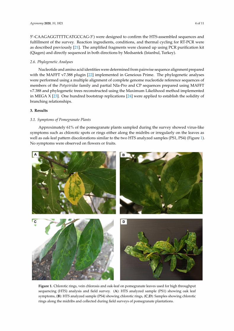
Approximately 61% of the pomegranate plants sampled during the survey showed virus-likesymptoms such as chlorotic spots or rings either along the midribs or irregularly on the leaves aswell as oak-leaf pattern discolorations similar to the two HTS analyzed samples (PS1, PS4) (Figure 1).No symptoms were observed on flowers or fruits.
Agronomy 2020, 10, x FOR PEER REVIEW 4 of 11
2.6. Phylogenetic Analyses
Nucleotide and amino acid identities were determined from pairwise sequence alignment prepared with the MAFFT v7.388 plugin [22] implemented in Geneious Prime. The phylogenetic analyses were performed using a multiple alignment of complete genome nucleotide reference sequences of members of the Potyviridae family and partial NIa-Pro and CP sequences prepared using MAFFT v7.388 and phylogenetic trees reconstructed using the Maximum Likelihood method implemented in MEGA X [23]. One hundred bootstrap replications [24] were applied to establish the solidity of branching relationships.
3. Results
3.1. Symptoms of Pomegranate Plants
Approximately 61% of the pomegranate plants sampled during the survey showed virus-like symptoms such as chlorotic spots or rings either along the midribs or irregularly on the leaves as well as oak-leaf pattern discolorations similar to the two HTS analyzed samples (PS1, PS4) (Figure 1). No symptoms were observed on flowers or fruits.
Figure 1. Chlorotic rings, vein chlorosis and oak-leaf on pomegranate leaves used for high throughput sequencing (HTS) analysis and field survey. (A): HTS analyzed sample (PS1) showing oak leaf symptoms, (B): HTS analyzed sample (PS4) showing chlorotic rings, (C,D): Samples showing chlorotic rings along the midribs and collected during field surveys of pomegranate plantations.
3.2. Transmission Electron Microscope (TEM) Analysis
Consistent with viruses from the family Potyviridae, electron microscopy revealed the presence of only flexuous filamentous viral particles, 760 to 780 nm long in purified virus preparations obtained from leaves of HTS analyzed samples PS1 and PS4 (Figure 2).
Figure 1. Chlorotic rings, vein chlorosis and oak-leaf on pomegranate leaves used for high throughputsequencing (HTS) analysis and field survey. (A): HTS analyzed sample (PS1) showing oak leafsymptoms, (B): HTS analyzed sample (PS4) showing chlorotic rings, (C,D): Samples showing chloroticrings along the midribs and collected during field surveys of pomegranate plantations.

Agronomy 2020, 10, 1821 5 of 11
3.2. Transmission Electron Microscope (TEM) Analysis
Consistent with viruses from the family Potyviridae, electron microscopy revealed the presence ofonly flexuous filamentous viral particles, 760 to 780 nm long in purified virus preparations obtainedfrom leaves of HTS analyzed samples PS1 and PS4 (Figure 2).
1
(a) (b)
Figure 2. Electron micrographs captured from HTS analyzed pomegranate samples, using TecnaiSpirit BioTWIN transmission electron microscope. (A):PS1, (B):PS4. The scale equals 100 nm for(A) and 200 nm for (B).
3.3. Virus Identification by High-Throughput Sequencing
Two HTS libraries were prepared, corresponding to samples PS1 and PS4. The raw numberof reads obtained from these libraries were respectively 13,611,066 and 20,918,456 reads whichresulted in the assembly of respectively 3690 and 6078 contigs longer than 200 nucleotides (Table S1).BLASTn analysis using the GenBank nr-nt database identified viral contigs with strong identity levelswith passiflora edulis symptomless virus (PeSV), a recently proposed new member of the familyPotiviridae, genus Roymovirus identified in Israel from passionfruit [17]. Complementary analysisusing BLASTx revealed also the presence of sequences related to the RdRp (MN532630; MN532627)of Plasmopara viticola lesion associated ourmia-like virus and RdRp (MN386961) of apple ourmia-likevirus 1. These homologies had not been detected using BLASTn.
Further analysis allowed the reconstruction of the near-full genome of five PeSV variants fromthe PS1 (PS1-1, PS1-2, PS1-3, PS1-4 and PS1-5) sample and of one variant from the PS4 sample(PS4-1). These variants shared 77.9–86.2% nucleotide identity with each other while having 78.0–86.8%nucleotide identity with PeSV-Rehovot from passion fruit (MH379332). All sequences have beendeposited in GenBank (see Table S2 for accession numbers).
A screening by BLAST of all publicly available GenBank SRA pomegranate RNA-Seqtranscriptomic data revealed the presence of PeSV in a total of seven SRAs from China and eight SRAsfrom India. Four datasets were selected on the basis of apparent differences in homology with the PeSVpassiflora isolate, downloaded and assembled using CLC GWB. Contigs were then screened by BLASTnagainst the passionfruit isolate of PeSV. In each case, one or more contigs with homology to PeSV wereidentified. In total, from the four datasets, eight contigs were thus obtained, covering at least the CPregion (one from SRX2735580 (Zhengzhou Fruit Research Institute, China), three from SRX2735578(Zhengzhou Fruit Research Institute, China), one from SRX2583192 (Anhui Academy of AgriculturalSciences, China) and three from SRX 2503675 (Indian Council of Agricultural Research, India). One ofthe contigs, from the SRX2583192 dataset, is a near-complete genome, missing only 29 nt at the 5′ endand 18 nt at the 3′ end. Out of the 81.055.700 reads of this particular dataset, the near-complete genome

Agronomy 2020, 10, 1821 6 of 11
contig assembles 5665 reads (0.01% of total reads). The near-complete genome and the seven partialones showed 77.8–78.6% and 76.7–88.1% nucleotide identity with PeSV-Rehovot and the PeSV HTScontigs, respectively.
3.4. Characterization of PeSV Genome Isolated from Pomegranate
The genome organization of the pomegranate isolate from the PS1 sample (PS1-5) which hadthe highest identity with PeSV-Rehovot isolate is presented in Figure 3. The complete sequence ofthe genome is 9938 nucleotides long, including the polyA tail. This genome encodes a polyproteinof 3174 amino acids which is later cleaved into ten functional proteins. The encoded polyproteinlacks known aphid transmission motifs in its HC-Pro and CP domains but possesses a C-2x-C motifputatively involved in eriophyid transmission in its HC-Pro [17].
Agronomy 2020, 10, x FOR PEER REVIEW 6 of 11
3.4. Characterization of PeSV Genome Isolated from Pomegranate
The genome organization of the pomegranate isolate from the PS1 sample (PS1-5) which had the highest identity with PeSV-Rehovot isolate is presented in Figure 3. The complete sequence of the genome is 9938 nucleotides long, including the polyA tail. This genome encodes a polyprotein of 3174 amino acids which is later cleaved into ten functional proteins. The encoded polyprotein lacks known aphid transmission motifs in its HC-Pro and CP domains but possesses a C-2x-C motif putatively involved in eriophyid transmission in its HC-Pro [17].
Figure 3. Genomic map of the passiflora edulis symptomless virus (PeSV)-PS1-5 variant.
The PeSV-PS1-5 genome shares an overall 86.8% nucleotide identity with that of PeSV-Rehovot. Among the various genes, the highest nucleotide identity is observed in the 6K1 gene (94.4%) while the lowest is observed in the HC-Pro gene (77.2%) (Table 1). The polyprotein of these two isolates shares 97.4% amino acid (aa) identity. The deduced amino acid sequences of their 6K1 and VPg proteins are identical, while the other proteins share 92.6–99.6% aa identity. The 5′-UTR and 3′-UTR of these isolates share 72.7 and 89.4% identity, respectively. The comparison of the various predicted polyprotein cleavage sites shows only a difference for the P1/HC-Pro cleavage site, which is EVYY433/G for PeSV-PS1-5 instead of EVYY433/S in PeSV-Rehovot.
Table 1. Length and pairwise identities between the various genes/proteins of PeSV-PG1-TR as compared to PeSV-Rehovot.
Genomic Region
Length (aa)
Predicted Cleavage Site PeSV-Rehovot
Predicted Cleavage Site PeSV-PS1-5
aa Identity
(%)
nt Identity
(%) 5′ UTR - - - - 72.7
P1 433 EVYY433/S EVYY433/G 92.6 81 HC-Pro 463 YQVG896/G YQVG896/G 94 77.2
P3 266 VEFQ1162/S VEFQ1162/S 97 84.6 6K1 54 VVFE1216/A VVFE1216/A 100 94.4 CI 640 VLFE1856/G VLFE1856/G 99.2 90.5
6K2 62 VDFE1918/G VDFE1918/G 98.4 92.5 VPg 186 VQFE2104/S VQFE2104/S 100 91
NIa-Pro 237 VTFQ2341/N VTFQ2341/N 99.6 90.4 NIb 504 VEFQ2845/M VEFQ2845/M 98.8 90 CP 328 - - 99.4 92.3
3 UTR - - - - 89.4
3.5. Phylogenetic Analysis
A phylogenentic analysis was performed using complete genome sequences of representative members of the various genera in the Potyviridae family: potato virus Y (PVY; Potyvirus), ryegrass mosaic virus (RyMV; Rymovirus), wheat streak mosaic virus (WSMV; Tritimovirus), barley yellow mosaic virus (BaYMV; Bymovirus), sweet potato mild mottle virus (SPMMV; Ipomovirus), blackberry virus Y (BVY; Brambyvirus), triticum mosaic virus (TriMV; Poacevirus), rose yellow mosaic virus (RoYMV: Roymovirus), bellflower veinal mottle virus (BVMoV; Bevemovirus), artichoke latent virus (ArLV; Macluravirus), celery latent virus (CeLV; Celavirus), areca palm necrotic ringspot virus (ANRSV; Arepavirus).
The phylogenetic analysis unambiguously showed that all PeSV pomegranate variants were grouped with the reference PeSV isolate from passionfruit. A robust bootstrap value supports the
Figure 3. Genomic map of the passiflora edulis symptomless virus (PeSV)-PS1-5 variant.
The PeSV-PS1-5 genome shares an overall 86.8% nucleotide identity with that of PeSV-Rehovot.Among the various genes, the highest nucleotide identity is observed in the 6K1 gene (94.4%) while thelowest is observed in the HC-Pro gene (77.2%) (Table 1). The polyprotein of these two isolates shares97.4% amino acid (aa) identity. The deduced amino acid sequences of their 6K1 and VPg proteins areidentical, while the other proteins share 92.6–99.6% aa identity. The 5′-UTR and 3′-UTR of these isolatesshare 72.7 and 89.4% identity, respectively. The comparison of the various predicted polyproteincleavage sites shows only a difference for the P1/HC-Pro cleavage site, which is EVYY433/G forPeSV-PS1-5 instead of EVYY433/S in PeSV-Rehovot.
Table 1. Length and pairwise identities between the various genes/proteins of PeSV-PG1-TR ascompared to PeSV-Rehovot.
Genomic Region Length (aa) Predicted CleavageSite PeSV-Rehovot
Predicted CleavageSite PeSV-PS1-5 aa Identity (%) nt Identity (%)
5′ UTR - - - - 72.7P1 433 EVYY433/S EVYY433/G 92.6 81
HC-Pro 463 YQVG896/G YQVG896/G 94 77.2P3 266 VEFQ1162/S VEFQ1162/S 97 84.6
6K1 54 VVFE1216/A VVFE1216/A 100 94.4CI 640 VLFE1856/G VLFE1856/G 99.2 90.5
6K2 62 VDFE1918/G VDFE1918/G 98.4 92.5VPg 186 VQFE2104/S VQFE2104/S 100 91
NIa-Pro 237 VTFQ2341/N VTFQ2341/N 99.6 90.4NIb 504 VEFQ2845/M VEFQ2845/M 98.8 90CP 328 - - 99.4 92.3
3 UTR - - - - 89.4
3.5. Phylogenetic Analysis
A phylogenentic analysis was performed using complete genome sequences of representativemembers of the various genera in the Potyviridae family: potato virus Y (PVY; Potyvirus), ryegrass mosaicvirus (RyMV; Rymovirus), wheat streak mosaic virus (WSMV; Tritimovirus), barley yellow mosaicvirus (BaYMV; Bymovirus), sweet potato mild mottle virus (SPMMV; Ipomovirus), blackberry virusY (BVY; Brambyvirus), triticum mosaic virus (TriMV; Poacevirus), rose yellow mosaic virus(RoYMV: Roymovirus), bellflower veinal mottle virus (BVMoV; Bevemovirus), artichoke latentvirus (ArLV; Macluravirus), celery latent virus (CeLV; Celavirus), areca palm necrotic ringspot virus(ANRSV; Arepavirus).
The phylogenetic analysis unambiguously showed that all PeSV pomegranate variants weregrouped with the reference PeSV isolate from passionfruit. A robust bootstrap value supports the

Agronomy 2020, 10, 1821 7 of 11
claim that PeSV belongs to the genus Roymovirus (Figure 4). A phylogenetic analysis using the viralpolyprotein sequences resulted in a similar topology (data not shown).
Agronomy 2020, 10, x FOR PEER REVIEW 7 of 11
claim that PeSV belongs to the genus Roymovirus (Figure 4). A phylogenetic analysis using the viral polyprotein sequences resulted in a similar topology (data not shown).
Figure 4. Phylogenetic analysis of PeSV isolates using complete genomes of representatives of Potyviridae by the Maximum Likelihood method and distances computed using the General Time Reversible model using gamma distribution with invariable sites (GTR+G+I). Bootstrap analysis (100 replicates) was used to evaluate branch validity and only bootstrap values higher than 70% are shown. The scale shows the number of substitutions per site.
3.6. PeSV Detection by RT-PCR
Two primer sets targeting the NIa-Pro and CP genes were designed based on the HTS-recovered PeSV genome sequences in order to evaluate the presence of PeSV in pomegranate by RT-PCR. The two primer pairs were validated using the HTS samples as positive controls. Among 133 pomegranate samples collected from the Eastern Mediterranean region of Turkey, the presence of PeSV was confirmed in 37 cultivated pomegranates and in one ornamental pomegranate. Among these PeSV-positive samples, all cultivated pomegranates exhibited leaf symptoms of variable severity but no symptoms were observed on the ornamental pomegranate, irrespective of its infection status. Besides the 37 symptomatic and PCR-positive cultivated pomegranates, another 45 showed virus-like symptoms but did not show positive PeSV RT-PCR amplification. PeSV was similarly not detected in any of the symptomless cultivated pomegranates assayed. The negative PCR results obtained with the symptomatic pomegranate samples might reflect the presence of other pathogenic agents or, alternatively, the too-narrow inclusiveness of the two detection primer pairs used and their failure to amplify some PeSV isolates.
From the tested samples, 24 CP (MT271616-MT271639) and 38 Nla-Pro (MT271640-MT271677) partial sequences were obtained and submitted to GenBank (Table S2). The partial CP nucleotide sequences shared 90.2–98.6% pairwise identity while they shared 90.3–93.7% nt identity with PeSV-
Figure 4. Phylogenetic analysis of PeSV isolates using complete genomes of representatives ofPotyviridae by the Maximum Likelihood method and distances computed using the General TimeReversible model using gamma distribution with invariable sites (GTR+G+I). Bootstrap analysis(100 replicates) was used to evaluate branch validity and only bootstrap values higher than 70% areshown. The scale shows the number of substitutions per site.
3.6. PeSV Detection by RT-PCR
Two primer sets targeting the NIa-Pro and CP genes were designed based on the HTS-recoveredPeSV genome sequences in order to evaluate the presence of PeSV in pomegranate by RT-PCR. The twoprimer pairs were validated using the HTS samples as positive controls. Among 133 pomegranatesamples collected from the Eastern Mediterranean region of Turkey, the presence of PeSV was confirmedin 37 cultivated pomegranates and in one ornamental pomegranate. Among these PeSV-positivesamples, all cultivated pomegranates exhibited leaf symptoms of variable severity but no symptomswere observed on the ornamental pomegranate, irrespective of its infection status. Besides the37 symptomatic and PCR-positive cultivated pomegranates, another 45 showed virus-like symptomsbut did not show positive PeSV RT-PCR amplification. PeSV was similarly not detected in anyof the symptomless cultivated pomegranates assayed. The negative PCR results obtained withthe symptomatic pomegranate samples might reflect the presence of other pathogenic agents or,alternatively, the too-narrow inclusiveness of the two detection primer pairs used and their failure toamplify some PeSV isolates.

Agronomy 2020, 10, 1821 8 of 11
From the tested samples, 24 CP (MT271616-MT271639) and 38 Nla-Pro (MT271640-MT271677)partial sequences were obtained and submitted to GenBank (Table S2). The partial CP nucleotidesequences shared 90.2–98.6% pairwise identity while they shared 90.3–93.7% nt identity withPeSV-Rehovot (Table S3). The pairwise nt identity for the partial NIa-Pro sequences ranged from 77.5%to 84.1% while they shared a 78.3–93.8% identity with PeSV-Rehovot (Table S4). Phylogenetic analysisusing nucleotide sequences of the partial CP (Figure 5a) and NIa-Pro (Figure 5b) regions showed alimited diversity of the amplified isolates for the CP region, with all detected variants closely related tothe PS1-5 variant while a higher diversity was observed for the NIa-Pro region, lending support to thehypothesis that the NIa-Pro primer pair might have higher inclusiveness than the CP one.
Agronomy 2020, 10, x FOR PEER REVIEW 8 of 11
Rehovot (Table S3). The pairwise nt identity for the partial NIa-Pro sequences ranged from 77.5% to 84.1% while they shared a 78.3–93.8% identity with PeSV-Rehovot (Table S4). Phylogenetic analysis using nucleotide sequences of the partial CP (Figure 5a) and NIa-Pro (Figure 5b) regions showed a limited diversity of the amplified isolates for the CP region, with all detected variants closely related to the PS1-5 variant while a higher diversity was observed for the NIa-Pro region, lending support to the hypothesis that the NIa-Pro primer pair might have higher inclusiveness than the CP one.
(a) (b)
Figure 5. Phylogenetic trees of PeSV isolates based on partial nucleotide sequences of CP (a) and NIa-pro (b). The trees were reconstructed using the Maximum Likelihood method (K2+G+I for CP and TN93+G+I for NIa-pro) and bootstrap values (100 replicates) greater than 70 are shown next to the branches.
4. Discussion
In this study, potyvirus-like particles were observed in symptomatic pomegranate plants using electron microscopy. Potyviridae family virions are flexuous filaments and usually are 680–900nm long and 11–20 nm wide, with helical symmetry and a pitch of about 3.4 nm [18]. Flexuous filamentous rods, ranging from 720 to 750 nm in length, were also observed for the rose yellow mosaic virus (RoYMV) [25] which is most closely related to passiflora edulis symptomless virus [17].
HTS analysis of two symptomatic pomegranate trees was performed. Based on the results, we report for the first time the presence of passiflora edulis symptomless virus (PeSV) in pomegranates. The complete genome sequences of six PeSV pomegranate variants were determined by in silico analyses and shown to share 78.0–86.8% nt identity with the PeSV-Rehovot genome obtained from passionfruit. Further analyses also allowed us to reconstruct the near-complete genome of PeSV variants from SRA data from China and India. All identified pomegranate variants illustrated a high level of variability for PeSV, as can be seen in the phylogenetic analyses.
Based on the recovered complete genomes of PeSV variants, two RT-PCR primer pairs were developed as detection primers for PeSV-pomegranate isolates. A survey in 133 pomegranate trees from several provinces of Turkey was carried out and 28.6% of tested samples were found infected by PeSV by using the Nla-Pro-specific primer pair. In parallel, 18.0% of samples tested positive using the CP-specific primer pair, indicating this second assay to be less sensitive or of lower inclusiveness.
Figure 5. Phylogenetic trees of PeSV isolates based on partial nucleotide sequences of CP (a) and NIa-pro(b). The trees were reconstructed using the Maximum Likelihood method (K2+G+I for CP andTN93+G+I for NIa-pro) and bootstrap values (100 replicates) greater than 70 are shown next tothe branches.
4. Discussion
In this study, potyvirus-like particles were observed in symptomatic pomegranate plants usingelectron microscopy. Potyviridae family virions are flexuous filaments and usually are 680–900nm longand 11–20 nm wide, with helical symmetry and a pitch of about 3.4 nm [18]. Flexuous filamentousrods, ranging from 720 to 750 nm in length, were also observed for the rose yellow mosaic virus(RoYMV) [25] which is most closely related to passiflora edulis symptomless virus [17].
HTS analysis of two symptomatic pomegranate trees was performed. Based on the results,we report for the first time the presence of passiflora edulis symptomless virus (PeSV) in pomegranates.The complete genome sequences of six PeSV pomegranate variants were determined by in silicoanalyses and shown to share 78.0–86.8% nt identity with the PeSV-Rehovot genome obtained frompassionfruit. Further analyses also allowed us to reconstruct the near-complete genome of PeSVvariants from SRA data from China and India. All identified pomegranate variants illustrated a highlevel of variability for PeSV, as can be seen in the phylogenetic analyses.

Agronomy 2020, 10, 1821 9 of 11
Based on the recovered complete genomes of PeSV variants, two RT-PCR primer pairs weredeveloped as detection primers for PeSV-pomegranate isolates. A survey in 133 pomegranate treesfrom several provinces of Turkey was carried out and 28.6% of tested samples were found infected byPeSV by using the Nla-Pro-specific primer pair. In parallel, 18.0% of samples tested positive usingthe CP-specific primer pair, indicating this second assay to be less sensitive or of lower inclusiveness.The hypothesis of lower inclusiveness of the CP primer pair is further reinforced by the reduceddiversity of the partial CP sequences recovered (Figure 5a), so that the NIa-Pro primers may be moresensitive than the CP primers for the screening of PeSV in pomegranate plantations.
While PeSV was not detected from symptomless field cultivated pomegranate samples cv. Hicaz,it was only detected from 45% of symptomatic samples (37/82). These results might reflect thetoo-narrow inclusiveness of the PCR assay used, including the NIa-Pro one, or alternatively a lack ofcorrelation between PeSV infection and symptoms development. This could in turn reflect an inabilityof PeVS to cause symptoms under some PeSV isolate/pomegranate genotype combinations (e.g., in theornamental plant) or the complete inability of PeSV to cause symptoms in this host. The observationthat PeSV was never detected in the tested symptomless cultivated field samples argues for, but doesnot prove, a contribution of PeSV to the symptoms.
In order to investigate the possibility that some other unknown viruses may be involved in thesymptoms observed, PeSV unrelated contigs without any BLASTn hits were subjected to BLASTxanalysis. This revealed putative viral contigs resembling Plasmopara viticola lesion associatedourmia-like virus and apple ourmia-like virus 1. Ourmiaviruses have tripartite genomes, with theRdRp, movement protein, and coat protein encoded each on separate genome segments. Up to date,three plant viruses are classified in the genus Ourmiavirus, ourmia melon virus, epirus cherry virus andcassava virus C [26]. Recently, the RdRp-encoding segment was also identified for three ourmia-likeviruses from apple trees with proposed names apple ourmia-like virus 1, 2 and 3. It was, however,not possible to decide whether the host of these viruses was apple or a fungus associated with theanalyzed apple samples [27]. Due to the very limited information on these new ourmia-like viruses,it is not possible to conclude on a possible contribution of the detected ourmia-like viruses to thesymptoms observed on pomegranate.
Potyviruses are known to be aphid transmitted [28] and this transmission is mediated bythe conserved HC-Pro motifs. PeSV-pomegranate isolates lack known aphid transmission motifsin its HC-Pro and CP but possess a C-2x-C motif putatively involved in eriophyid transmission.The presence of this motif could suggest that PeSV pomegranate and passion fruit isolates couldpossibly be transmitted by eriophyid mites, but this would have to be proven experimentally. Even withthe absence of aphid transmission domains in the PeSV proteins, the possibility of aphid transmissionshould not be overlooked due to the high population of aphids observed on pomegranate trees inearly spring and morphologically identified as Aphis punicae (Passerini) (Çaglayan, unpublished data).In fact, a new soybean virus, named soybean yellow shoot virus (SoyYSV) belonging to the Potyviridae,was recently shown to be transmitted by Myzus persicae and Aphis gossypii while lacking the HC-Prodomain required for aphid transmission found in other potyviruses [29].
When all the data obtained from field observations, HTS and PCR studies are consideredtogether, no clear correlation between virus and symptoms was detected although all PeSV positivesamples were symptomatic. In parallel, we show here that, at least under some circumstances(ornamental pomegranates), PeSV could cause asymptomatic infections. Further studies were clearlyneeded to clarify the potential contribution of PeSV to the virus-like symptoms observed in pomegranateand to better understand its transmission mechanism and epidemiology. Having detected its presencein India and China also raises the question of its presence in other pomegranate growing countries.
5. Conclusions
We describe here a new host for passiflora edulis symptomless virus (PeSV) which has beenrecently proposed as a new member of the family Potiviridae, genus Roymovirus, identified in Israel

Agronomy 2020, 10, 1821 10 of 11
infecting passionfruit. The complete genome is 9938 nucleotides long, including the polyA tail,shows the same organization and has the highest identity with the PeSV-passionfruit isolate. PeSVoccurrence was also identified from publicly available SRA pomegranate RNA-Seq transcriptomicdata from India and China. The presence of PeSV in pomegranate was further confirmed usingspecific RT-PCR and the virus showed to be widespread in the main commercial pomegranate growingregions of Turkey. There was, however, an only partial association between the presence of the virusand symptoms. The genetic diversity of PeSV pomegranate isolates was very high, with evidencefor complex mixed infections of divergent variants, which could be of importance for detection andcertification programs. As proposed in recently a published framework, investigating the epidemiologyand pathogenicity of new emerging viruses like PeSV represents a priority to evaluate the risk PeSVcould pose to pomegranate production in Turkey and in other pomegranate-producing countries.
Supplementary Materials: The following are available online at http://www.mdpi.com/2073-4395/10/11/1821/s1,Table S1: Numbers of raw reads, cleaned reads, assembled contigs, PeSV-related contigs and number of variantsidentified for each of the two analyzed pomegranate samples. Table S2: Characteristics of the isolates and variantsused in this study. Table S3: Pairwise nucleotide sequence identity in the CP gene between PeSV isolates. Table S4:Pairwise nucleotide sequence identity in the NIa-Pro gene between PeSV isolates.
Author Contributions: Conceptualization, K.C. and A.O.; methodology, K.C., V.R., A.O. and A.B.R.-G.; software,V.R. and A.O.; validation, M.G., H.D.K. and B.T.; formal analysis, V.R., A.O. and T.C.; investigation, K.C., V.R.and M.G.; resources, K.C. and A.O.; data curation, K.C. and J.S.R.; writing—original draft preparation, K.C.;writing—review and editing, K.C., V.R., A.O., A.B.R.-G. and T.C.; visualization, K.C. and M.G.; supervision, A.O.and T.C.; project administration, K.C. and V.R.; funding acquisition, K.C. and A.O. All authors have read andagreed to the published version of the manuscript.
Funding: This study was supported by funds from the European Union’s Horizon 2020 research and innovationprogramme under the Marie Skłodowska-Curie grant agreement No. 734736-VirFree project.
Acknowledgments: This study was supported by funds from the European Union’s Horizon 2020 research andinnovation programme under the Marie Skłodowska-Curie grant agreement No 734736- VirFree project.
Conflicts of Interest: The authors declare no conflict of interest.
References
1. Holland, D.A.; Hatib, K.; Bar-Ya’Akov, I. Pomegranate: Botany, Horticulture, Breeding. Hortic. Rev. 2009,35, 127–191. [CrossRef]
2. Lye, C. Pomegranate: Preliminary Assessment of the Potential for an Australian Industry; Publication No. 08/153;Rural Industries Research and Development Corporation of Australian Government: Kingston, Australia,2008; p. 17.
3. Caliskan, O.; Bayazit, S. Phytochemical and antioxidant attributes of autochthonous Turkish pomegranates.Sci. Hortic. 2012, 147, 81–88. [CrossRef]
4. Rosas-Burgos, E.C.; Burgos-Hernández, A.; Noguera-Artiaga, L.; Kacániová, M.; Hernández-García, F.;Cárdenas-López, J.L.; Carbonell-Barrachina, A.A. Antimicrobial activity of pomegranate peel extracts asaffected by cultivar. J. Sci. Food Agric. 2016, 97, 802–810. [CrossRef] [PubMed]
5. Gazel, M.; Caglayan, K.; Baspinar, H.; Mejia, J.F.; Paltrinieri, S.; Bertaccini, A.; Contaldo, N. Detectionand identification of phytoplasmas in pomegranate trees with yellows symptoms. J. Phytopathol. 2015,164, 136–140. [CrossRef]
6. Horvath, J. Isolation of cucumber mosaic virus from pomegranate (Punica granatum L.) in Yugoslavia.Acta Phytopathol. Acad. Sci. Hung. 1984, 4, 309–314.
7. Gomez, G.; Pallás, V. Detection of viroid-like RNAs in pomegranate (Punica granatum L.). Acta Hortic. 2001,550, 321–326. [CrossRef]
8. Astruc, N.; Marcos, J.F.; Macquaire, G.; Candresse, T.; Pallás, V. Studies on the diagnosis of hop stunt viroidin fruit trees: Identification of new hosts and application of a nucleic acid extraction procedure based onnon-organic solvents. Eur. J. Plant Pathol. 1996, 102, 837–846. [CrossRef]
9. Gazel, M.; Caglayan, K.; Serçe, Ç.U.; Durgac, C. Detection of hop stunt viroid in pomegranate(Punica granatum L.) trees in the east mediterranean region of turkey. Acta Hortic. 2009, 818, 273–276. [CrossRef]

Agronomy 2020, 10, 1821 11 of 11
10. Caglayan, K.; Elçi, E.; Gazel, M. Detection and partial characterization of grapevine leafroll-associated virus1 in pomegranate trees in Turkey. Eur. J. Plant Pathol. 2015, 145, 199–202. [CrossRef]
11. Ferris, M.T.; Joyce, P.; Burch, C.L. High frequency of mutations that expand the host range of an RNA virus.Genetics 2007, 176, 1013–1022. [CrossRef]
12. McLeish, M.J.; Fraile, A.; Garcia-Arenal, F. Ecological complexity in plant virus host range evolution.Int. Rev. Cytol. 2018, 101, 293–339. [CrossRef]
13. Massart, S.; Olmos, A.; Jijakli, H.; Candresse, T. Current impact and future directions of high throughputsequencing in plant virus diagnostics. Virus Res. 2014, 188, 90–96. [CrossRef] [PubMed]
14. Roossinck, M.J.; Martin, D.P.; Roumagnac, P. Plant virus metagenomics: Advances in virus discovery.Phytopathology 2015, 105, 716–727. [CrossRef] [PubMed]
15. Radford, A.D.; Chapman, D.; Dixon, L.; Chantrey, J.; Darby, A.C.; Hall, N. Application of next-generationsequencing technologies in virology. J. Gen. Virol. 2012, 93, 1853–1868. [CrossRef] [PubMed]
16. Rose, H.; Döring, I.; Vetten, H.-J.; Menzel, W.; Richert-Pöggeler, K.R.; Maiss, E. Complete genome sequenceand construction of an infectious full-length cDNA clone of celery latent virus—An unusual member of aputative new genus within the Potyviridae. J. Gen. Virol. 2019, 100, 308–320. [CrossRef] [PubMed]
17. Jover-Gil, S.; Beeri, A.; Fresnillo, P.; Samach, A.; Candela, H. Complete genome sequence of a novel virus,classifiable within the Potyviridae family, which infects passion fruit (Passiflora edulis). Arch. Virol. 2018,163, 3191–3194. [CrossRef]
18. Wylie, S.J.; Adams, M.; Chalam, C.; Kreuze, J.; López-Moya, J.J.; Ohshima, K.; Praveen, S.; Rabenstein, F.;Stenger, D.; Wang, A.; et al. ICTV Virus Taxonomy Profile: Potyviridae. J. Gen. Virol. 2017,98, 352–354. [CrossRef]
19. Gugerli, P.; Brugger, J.J.; Bovey, R. L’enroulement de la vigne: Mise en évidence de particules viraleset développement d’une méthode immuno-enzymatique pour le diagnostic rapide. Rev. suisse Vitic.Arboric. Hortic. 1984, 16, 299–304.
20. Poudel, B.; Wintermantel, W.M.; Cortez, A.A.; Ho, T.; Khadgi, A.; Tzanetakis, I.E. Epidemiology of Blackberryyellow vein associated virus. Plant Dis. 2013, 97, 1352–1357. [CrossRef]
21. Çaglayan, K.; Roumi, V.; Gazel, M.; Elçi, E.; Acioglu, M.; Pleško, I.M.; Reynard, J.-S.; Maclot, F.; Massart, S.Identification and Characterization of a Novel Robigovirus Species from Sweet Cherry in Turkey. Pathogens2019, 8, 57. [CrossRef]
22. Katoh, K.; Standley, D.M. MAFFT multiple sequence alignment software version 7: Improvements inperformance and usability. Mol. Biol. Evol. 2013, 30, 772–780. [CrossRef] [PubMed]
23. Kumar, S.; Stecher, G.; Li, M.; Knyaz, C.; Tamura, K. MEGA X: Molecular Evolutionary Genetics Analysisacross Computing Platforms. Mol. Biol. Evol. 2018, 35, 1547–1549. [CrossRef] [PubMed]
24. Felsenstein, J. Confidence limits on phylogenies: An approach using the bootstrap. Evolution 1985, 39, 783.[CrossRef] [PubMed]
25. Mollov, D.; Lockhart, B.; Zlesak, D. Complete nucleotide sequence of rose yellow mosaic virus, a novelmember of the family Potyviridae. Arch. Virol. 2013, 158, 1917–1923. [CrossRef] [PubMed]
26. Turina, M.; Hillman, B.I.; Izadpanah, K.; Rastgou, M.; Rosa, C. ICTV Report Consortium ICTV VirusTaxonomy Profile: Ourmiavirus. J. Gen. Virol. 2017, 98, 129–130. [CrossRef] [PubMed]
27. Wright, A.A.; Cross, A.R.; Harper, S.J. A bushel of viruses: Identification of seventeen novel putative virusesby RNA-seq in six apple trees. PLoS ONE 2020, 15, e0227669. [CrossRef]
28. Gibbs, A.J.; Ohshima, K.; Phillips, M.J.; Gibbs, M.J. The prehistory of potyviruses: Their initial radiation wasduring the dawn of agriculture. PLoS ONE 2008, 3, e2523. [CrossRef]
29. Figueira, A.D.R.; Geraldino-Duarte, P.S.; Nuñez, A.M.P.; Van Lent, J.; Galvino-Costa, S.B.F.; Farman, M.;Goodin, M. Characterization of Soybean yellow shoot virus, a New Member of the Family PotyviridaeInfecting Soybean Plants in Brazil. Plant Dis. 2019, 103, 1172–1180. [CrossRef]
Publisher’s Note: MDPI stays neutral with regard to jurisdictional claims in published maps and institutionalaffiliations.
© 2020 by the authors. Licensee MDPI, Basel, Switzerland. This article is an open accessarticle distributed under the terms and conditions of the Creative Commons Attribution(CC BY) license (http://creativecommons.org/licenses/by/4.0/).
Copyright © 2022 FDOKUMEN