02 host factors for disease resistance
28
Chapter 2 Host Factors for Disease Resistance Introduction J. M. Sharma Domesticated and wild birds are vulnerable to many microorgan- isms that contaminate the envirownent in which they live. The microorganisms include viruses, bacteria, fungi, and parasites. In confmed houses. such as the ODes used for intensive rearing of commercial poultry, the concentrations of microorganisms can reach very high levels. Often. these microorganisms are patho- genic and invasive and can cause severe clinical disease or death. The birds manage to survive the microbial challenge primarily because their immune systems provide protection against infec- tion and unrestricted replication of microorganisms. Without ef- fective immunity, the life span of birds would indeed be very short, and commercial poultry production would come to a halt. Because of the importance of immunity in health, the study of the mechanisms of immunity has received much attention within the last few decades, and many pioneering observations have been made that have lead to effective disease-control strategies. One of the most important contributions of immunity to human and an- imal health has been the development of vaccines that have dra- matically reduced the incidence of infectious diseases. Although the avian immune system has not been studied as extensively as that of mammals, important advances have been made. In the fIrst subchapter, a broad outline of the basic elements of the avian immune system is presented. Although great similarities exist be- tween immune mechanisms of the birds and mammals, there are also interesting differences. These differences are identified and briefly described. In the second subchapter, the role of genetics in regulating immune-mediated resistance to disease is covered. Genetic back- Avian Immune System J. M. Shanna Introduction The immune system plays a critical role in defending birds against environmental pathogens. The overall organization and mechanisms of immunity in birds are quite similar to those in mammals. Early studies on the bursa and the thymus of chickens provided some of the basic information that led to the identifica- ground of a host determines how the immune response to a given microorganism will evolve, and, ultimately, whether protective immunity will be generated. This is well demonstrated by the great variation that is often observed among individuals within a population in their response to a common disease agent. Some individuals may succumb to infection and die, whereas others may show no phenotypic signs of infection. This wide variation in response to the same agent is attributed to an intrinsic poly- morphism of genes that regulate the expression and interaction of various components of the immune system. Although genetic resistance to a disease is a multigenic trait, in commercial chicken populations, resistance or susceptibility to disease is often attributed to the genes that regulate the major his- tocompatibility complex (MHC). The MHC encodes a set of cell surface proteins that are necessary for antigen recognition by T cells and, consequently, the capability ofT cells to generate spe- cifIc immunity. The MHC proteins are genetically diverse and polymorphic. Association of specifIc MIlC haplotypes with dis- ease resistance bas been exploited by designing breeding pro- grams that select for resistance. Recently, the entire chicken genome has been sequenced, which has provided new opportuni- ties to identify and manipulate genes that control immunity and resistance to disease. The second subchapter provides informa- tion on how recent advances in molecular technology have facil- itated the study of genetic linkages to disease resistance. This new information is likely to have a significant impact on com- mercial poultry production. tion of the dichotomy of the immune system into B and T cell compartments. The recognition of this dichotomy initiated an era of extensive research on the mechanisms of immunity in mam- malian, avian, and amphibian species. This influx of research activity that began about five decades ago continues tmabated and is responsible for making immunology one of the fastest 47 Chapter 2 Host Factors for Disease Resistance Introduction J. M. Sharma Domesticated and wild birds are vulnerable to many microorgan- isms that contaminate the envirownent in which they live. The microorganisms include viruses, bacteria, fungi, and parasites. In confmed houses. such as the ODes used for intensive rearing of commercial poultry, the concentrations of microorganisms can reach very high levels. Often. these microorganisms are patho- genic and invasive and can cause severe clinical disease or death. The birds manage to survive the microbial challenge primarily because their immune systems provide protection against infec- tion and unrestricted replication of microorganisms. Without ef- fective immunity, the life span of birds would indeed be very short, and commercial poultry production would come to a halt. Because of the importance of immunity in health, the study of the mechanisms of immunity has received much attention within the last few decades, and many pioneering observations have been made that have lead to effective disease-control strategies. One of the most important contributions of immunity to human and an- imal health has been the development of vaccines that have dra- matically reduced the incidence of infectious diseases. Although the avian immune system has not been studied as extensively as that of mammals, important advances have been made. In the fIrst subchapter, a broad outline of the basic elements of the avian immune system is presented. Although great similarities exist be- tween immune mechanisms of the birds and mammals, there are also interesting differences. These differences are identified and briefly described. In the second subchapter, the role of genetics in regulating immune-mediated resistance to disease is covered. Genetic back- Avian Immune System J. M. Shanna Introduction The immune system plays a critical role in defending birds against environmental pathogens. The overall organization and mechanisms of immunity in birds are quite similar to those in mammals. Early studies on the bursa and the thymus of chickens provided some of the basic information that led to the identifica- ground of a host determines how the immune response to a given microorganism will evolve, and, ultimately, whether protective immunity will be generated. This is well demonstrated by the great variation that is often observed among individuals within a population in their response to a common disease agent. Some individuals may succumb to infection and die, whereas others may show no phenotypic signs of infection. This wide variation in response to the same agent is attributed to an intrinsic poly- morphism of genes that regulate the expression and interaction of various components of the immune system. Although genetic resistance to a disease is a multigenic trait, in commercial chicken populations, resistance or susceptibility to disease is often attributed to the genes that regulate the major his- tocompatibility complex (MHC). The MHC encodes a set of cell surface proteins that are necessary for antigen recognition by T cells and, consequently, the capability ofT cells to generate spe- cifIc immunity. The MHC proteins are genetically diverse and polymorphic. Association of specifIc MIlC haplotypes with dis- ease resistance bas been exploited by designing breeding pro- grams that select for resistance. Recently, the entire chicken genome has been sequenced, which has provided new opportuni- ties to identify and manipulate genes that control immunity and resistance to disease. The second subchapter provides informa- tion on how recent advances in molecular technology have facil- itated the study of genetic linkages to disease resistance. This new information is likely to have a significant impact on com- mercial poultry production. tion of the dichotomy of the immune system into B and T cell compartments. The recognition of this dichotomy initiated an era of extensive research on the mechanisms of immunity in mam- malian, avian, and amphibian species. This influx of research activity that began about five decades ago continues tmabated and is responsible for making immunology one of the fastest 47
-
Upload
independent -
Category
Documents
-
view
1 -
download
0
Transcript of 02 host factors for disease resistance

Chapter 2
Host Factors for Disease Resistance
IntroductionJ. M. Sharma
Domesticated and wild birds are vulnerable to many microorganisms that contaminate the envirownent in which they live. Themicroorganisms include viruses, bacteria, fungi, and parasites. Inconfmed houses. such as the ODes used for intensive rearing ofcommercial poultry, the concentrations of microorganisms canreach very high levels. Often. these microorganisms are pathogenic and invasive and can cause severe clinical disease or death.The birds manage to survive the microbial challenge primarilybecause their immune systems provide protection against infection and unrestricted replication of microorganisms. Without effective immunity, the life span of birds would indeed be veryshort, and commercial poultry production would come to a halt.Because ofthe importance ofimmunity in health, the study ofthemechanisms of immunity has received much attention within thelast few decades, and many pioneering observations have beenmade that have lead to effective disease-control strategies. One ofthe most important contributions of immunity to human and animal health has been the development ofvaccines that have dramatically reduced the incidence of infectious diseases. Althoughthe avian immune system has not been studied as extensively asthat of mammals, important advances have been made. In thefIrst subchapter, a broad outline ofthe basic elements ofthe avianimmune system is presented. Although great similarities exist between immune mechanisms of the birds and mammals, there arealso interesting differences. These differences are identified andbriefly described.
In the second subchapter, the role of genetics in regulatingimmune-mediated resistance to disease is covered. Genetic back-
Avian Immune SystemJ. M. Shanna
IntroductionThe immune system plays a critical role in defending birdsagainst environmental pathogens. The overall organization andmechanisms of immunity in birds are quite similar to those inmammals. Early studies on the bursa and the thymus ofchickensprovided some of the basic information that led to the identifica-
ground ofa host determines how the immune response to a givenmicroorganism will evolve, and, ultimately, whether protectiveimmunity will be generated. This is well demonstrated by thegreat variation that is often observed among individuals within apopulation in their response to a common disease agent. Someindividuals may succumb to infection and die, whereas othersmay show no phenotypic signs of infection. This wide variationin response to the same agent is attributed to an intrinsic polymorphism ofgenes that regulate the expression and interaction ofvarious components of the immune system.
Although genetic resistance to a disease is a multigenic trait,in commercial chicken populations, resistance or susceptibility todisease is often attributed to the genes that regulate the major histocompatibility complex (MHC). The MHC encodes a set ofcellsurface proteins that are necessary for antigen recognition by Tcells and, consequently, the capability ofT cells to generate specifIc immunity. The MHC proteins are genetically diverse andpolymorphic. Association of specifIc MIlC haplotypes with disease resistance bas been exploited by designing breeding programs that select for resistance. Recently, the entire chickengenome has been sequenced, which has provided new opportunities to identify and manipulate genes that control immunity andresistance to disease. The second subchapter provides information on how recent advances in molecular technology have facilitated the study of genetic linkages to disease resistance. Thisnew information is likely to have a significant impact on commercial poultry production.
tion of the dichotomy of the immune system into B and T cellcompartments. The recognition of this dichotomy initiated an eraof extensive research on the mechanisms of immunity in mammalian, avian, and amphibian species. This influx of researchactivity that began about five decades ago continues tmabatedand is responsible for making immunology one of the fastest
47
48 • Diseases of Poultry
growing branches of biology. Emerging concepts of immunemechanisms are constantly being revised by new information.
The immune system ofbirds, as ofmanunals, is complex and includes a ntimber ofcells ¥Id soluble factors that must work in concert to produce a protective immune response. A properly functioning immune system is of special importance to birds becausecommercial poultry flocks are raised under intensive rearing conditions. Under such conditions, the flocks are vulnerable to therapid spread of infectious agents and disease outbreaks. A varietyof vaccines must be used, often repeatedly, to protect the flocksagainst environmental exposure to virulent organisms. The protective efficacy ofa vaccine is dependent upon a vigorous immune response against the organism(s) present in the vaccine. If animalsare immunosuppressed and respond poorly to a vaccine, the flockhealth is placed in jeopardy. The understanding ofhow the immuneresponse is generated is of interest, as is the knowledge of how toprotect flocks from stresses that may induce immunosuppression.
TIlls subchapter is not a comprehensive review and is intendedto provide a broad overview of selected aspects of the avian immune system. For more detailed infonnation, the reader shouldconsult several books and reviews (17, 18,28,35,81,85,87, 101).
Anatomy of the Immune SystemThe immune cells reside in primary lymphoid organs (PLO) orsecondary lymphoid organs (SLO). The thymus and the bursa ofFabricius, respectively, are the PLO where T and B cell precursors differentiate and undergo maturation. The thymus is an elongated, multilobular structure located along the length of bothsides of the trachea with some lobes extending into the anteriorthoracic cavity (Fig. 2.lA). Thymic lobes are divided into lobules; each lobule has a peripheral cortical area in which lymphocytes are densely packed and a central medullary area in whichthe lymphocytes are less densely packed (Fig. 2.1 B). Bursa ofFabricius is a sac-like extension of the hindgut and is locatedabove the cloaca (Fig. 2.1 C). Bursa of Fabricius is organized intofollicles, each follicle is filled with lymphocytes. As in the thymus, the lymphocytes are arranged into a peripheral cortex and acentral medulla (Fig. 2.1 D).
Functional immune cells leave the PLO and populate SLO, theprincipal sites of antigen-induced immune response. SLO, characterized by aggregates of lymphocytes and antigen-presentingcells, are scattered through the body (Fig. 2.2). Examples ofSLOinclude spleen, bone marrow, gland of Harder (located ventraland posteriomedial to the eyeball), conjunctival-associated(CALT), bronchial-associated (BALT), and gut-associated lymphoid tissue (GALT). Bursa may also serve as SLO. Chickenslack the mammalian equivalent of lymph nodes but have lymphoid nodules along the course of lymphatics.
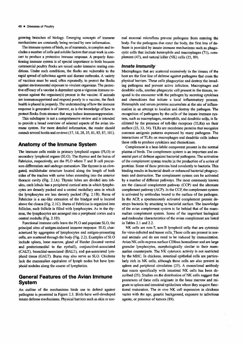
General Features of the Avian ImmuneSystemAn outline of the mechanisms birds use to defend againstpathogens is presented in Figure 2.3. Birds have well-developedinnate defense mechanisms. Physical barriers such as skin or nor-
mal mucosal microflora prevent pathogens from entering thebody. For the pathogens that enter the body, the first line of de·fense is provided by ilUlate immune mechanisms such as phagocytic cells that include heterophils and macrophages (71), complement (47), and natural killer (NK) cells (25, 89).
Innate ImmunityMacrophages that are scattered extensively in the tissues of thehost are the first line of defense against pathogens that cross thephysical barriers. These cells phagocytize and destroy the invading pathogens and prevent active infection. Macrophages anddendritic cells, another phagocytic cell present in the tissues, respond to the encounter with the pa:thogen by secreting cytokinesand chemokines that initiate a local inflammatory process.Hete~phils and serum proteins accumulate at the site of inflammation in an attempt to localize and destroy the pathogen. Therecognition of pathogens by the cells of the innate immune sys~
tem, such as macrophages, neutfophils, and dendritic cells, is facilitated by the presence of toll-like receptors (TLRs) on theirsurface (23, 33, 54). TLRs are membrane proteins that recognizecommon antigenic patterns expressed by many pathogens. Theinteractions ofTLRs on macrophages and dendritic cells inducethese cells to produce cytokines and chemokines.
Complement is a heat-labile component present in the normalplasma of birds. The complement system is an important and essential part ofdefense against bacterial pathogens. The activationof the complement system results in the production ofa series ofproteins. Some of these proteins bind covalently to bacteria. Thisbinding results in bacterial death or enhanced bacterial phagocytosis and destruction. The complement system can be activatedby a number of different pathways. The most commonly knownare the classical complement pathway (CCP) and the alternatecomplement pathway (ACP). In the CCp' the complement systemis activated by antibodies bound to the surface of the pathogen.In the ACP, a spontaneously activated complement protein destroys bacteria by attaching to bacterial surface. The knowledgeof the avian complement system is far behind that of the mammalian complement system. Some of the important biologicaland molecular characteristics of the avian complement are listedin Tables 2.1 and 2.2.
NK cells are non-T, non-B lymphoid cells that are cytotoxicfor virus-infected and tumor cells.. These cells are present in normal animals and do not need to be induced by immunization.Avian NK cells express surface CD8aa homodimer and are largegranular lymphocytes, morphologically similar to their mammalian counterparts. The NK cytotoxic activity is not restrictedby the MHC. In chickens, intestinal epithelial cells are particularly rich in NK cells, although these cells are also present inspleen and peripheral circulation (25). A monoclonal antibodythat reacts specifically with intestinal NK cells has been described (25). Studies on the distribution ofNK cells suggest thatprecursors of these cells originate in the bone marrow and migrate to spleen and intestinal epithelium where they acquire functional maturation. The in vivo NK cell expression in chickensvaries with the age, genetic background, exposure to infectiousagents, or presence of tumors (89).

2.1. A. Multiple lobes of the thymus lie on each side of the trachea. B. The bursa of Fabricius is the sac-like stnlcture extending from theend of the intestine. C. The multilobular histologic structure of the thymus is evident; each lobule is composed of the dark-staining cortexand the pale medulla. D. Bursal lymphoid follicles are separated by thin connective tissue septae. (From Fletcher, O. J., and H. J. Barnes.1998. Lymphoid organs and their anatomical distribution. In J. M. Shama (section ed.). Avian immunology. In P. P. Partoret, P. Griebel, H.Bazln, and A. Govaerts (ads.). Handbook of Veterinary Immunology. Academic Press. With permission.)
.,

2.2. A. The spleen is the oval organ in the centraJ-area of the photograph. B. Periarterlallymphoid sheaths are located in the white pulpof the spleen. C. A bursa-dependent lymphoid foillcle is located adjacent to a small artery and surrounded by thymus-dependent lymphoid
cells. D. Cecal tonsils, unopened ttop) and opened (bottom). E. Small nodules in the conjunctiva and the conjunctival-associated lymphoidtissue (CALl). F. The Harderian gland contains lymphoid cells in the connective tissue between the glands. G. Plasma cells are the predominant cell population in the Harderlan gland. H. Nodular deposits of lymphoid tissue are located in the mucosa of the trachea. (From Fletcher,
O. J., and H. J, Barnes, 1998. Lymphoid organs slid their anatomical distribution. In J. M. Sharma (section ad.), Avian immunology, In P. P.Partoret, P. Griebel, H. Bazin, and A. Govaerts (eds.). Handbook of Veterinary Immunology. Academic Press. With permission.)
CHAPTER 2 Host Factors for Disease Resistance • 49
Pathogens
(t.i~"""
1Physical barrier
(skin, normal mucosal flora, self cleaning)
1Innate immunity
(Non-adaptive immunity)NK cells, macrophages, phagocytes, complement
1
ICD'
ICytotoxic
cell
I
ICD'
IHelper
cell
ICytokines Lysis of cells
(provide help to express1f1gB cells. T cells foreign antigens
and macrophages)
ICytokines
(assist otherimmune cells)
IPhagocytosis
Pathogendestruction
IAntigen
presentation
IJg"I p~~~;~talion
Plasma cell
IAntibodies
Adaptive (or acquired) immunity
,-- H""'"m""'o~ca~I LI_,-~c~,~"~m~'~d~;'~"~d,--__,I I I
8 cell Macrophage T cellDendritic cell
Fig. 2.3. Physical and immunological mechanisms of defense against pathogens in birds.
NK cells and certain other effector cells may also induce target cell lysis if the target cells are coated with antibody. The an
. tibody molecules present on the surface of target cells interactwith Fc receptors present on NK cells, and this interaction triggers the cytotoxic attack against the target:'The destruction of anantibody-coated target cell is called antibody-dependent cellularcytotoxicity (ADCC). The ADCC activity contributes to host defense and has been detected in several avian species (90).
Pathogens that cannot be denied entry.bY physical barriern orcontrolled by innate immune defense mechanisms initiate a specific irrunune response (adaptive immunity). Adaptive immunityis highly specific to the agent that stimulates its developmentwhereas nonadaptive or innate immunity is nonspecific. Cellsmediating specific irrununity retain "memory" of their encounter
with the pathogen even after the pathogen has been cleared fromthe body and detectable immune response has subsided. Memorycells respond to the subsequent exposure to the same pathogen byinitiating a rapid and highly effective immune response. Boostervaccinations, used routinely in poultry, take advantage of thismemory response.
Adaptive irrununity is mediated by a variety of cells, the mostimportant ofwhich are T cells, B cells, and macrophages. T cells,the principal cells of cell-mediated immunity (CMI), recognizeforeign antigens after the antigens (such as microorganisms) havebeen processed by antigen-pr-esenting cells (APC). Macrophages,dendritic cells, and B cells are among the most important APC.The APe generally break down complex antigens and present toT cells small fragments of the antigen in conjunction with the

50 • Diseases of Poultry
Table 2.1 Molecular properties of avIan complement components.
Component
C3
FactorBClq
Species
Chicken
Quail
ChickenChicken
Approximatesorum
concentrationl~glmll
500
5O-10oa50-70
Refennce
(SO)
(51)
(36)
(46)(108)
From Koppenheffer, T.L. 1998. Complement. In J.M. Sharma (ed.). Avian immunology. In P. P. Partoret, P. Griebel, H. Barin, and A. Govaerts (eds.).Handbook ojVeterinary Immunology. Academic Press. With permission.-Estimated from data in Koch (46).
Table 2.2 Characteristics of avian complement.
• Antibody·independent ACP activity is demonstrable in vitro.• Microbial parasites activate the ACP in vivo.• The ACP is activated by avian antibodies.• Hemagglutinating levels of Ab produce maximum lysis.• CCP activity is difficult to demonstrate.• Both the ACP and CCP might utilize factor 8.• Cobra venom factor does not uniformly activate avian C.• The level of hemolytic C in chickens is MHC-linked.
From Koppenheffer, T.L. 1998. Complement. In J.M. Sharma (00.). Avianimmunology. In P. P. Partorct, P. Griebel, H. Barin, and A. Govaerts (eds.).Handbook ojVeterinary Immunology. Academic Press. With permission.
MHC molecules. T cells and the APC must share the same MHCbefore T cells can recognize and react to the antigen being presented.
The MHC molecules are glycoprotein receptors coded by thegenes within the MHC. The chicken MHC, also referred to as theB locus, is much smaller than the mammalian MHC and containsonly 19 genes in comparison with the human MHC that containsmore than 200. The organization of the chicken MHC is alsoquite different from that of the mammalian MHC (40). The Blocus consists of at least 3 loci: BF that encodes class I antigens,BL that encodes class II antigens, and BG that encodes class IVantigens. Class I and class II molecules are highly polymorphicand are critical for antigen presentation by the APe. The BF molecules (class I antigens) are present on a wide variety of nucleated cells including erythrocytes. The expression of the BL molecules (class II antigens) is much more restricted. Thesemolecules are expressed on macrophages, dendritic cells, monocytes, B cells, and activated T cells.
Whereas T cells require that the antigen be processed before itcan be recognized, the recognition ofan antigen by B cells is not
dependent on prior processing. B cells can recognize the antigenas it interacts with immunoglubulins that project from the cellsurface. B cells are responsible for humoral immunity and produce antibodies against the antigen.
Most microorganisms stimulate both CMI and humoral immunity although the type of immunity most critical for defense mayvary with the microorganism. Some of the important features ofthe eMI and humoral immunity are discussed in the followingsection.
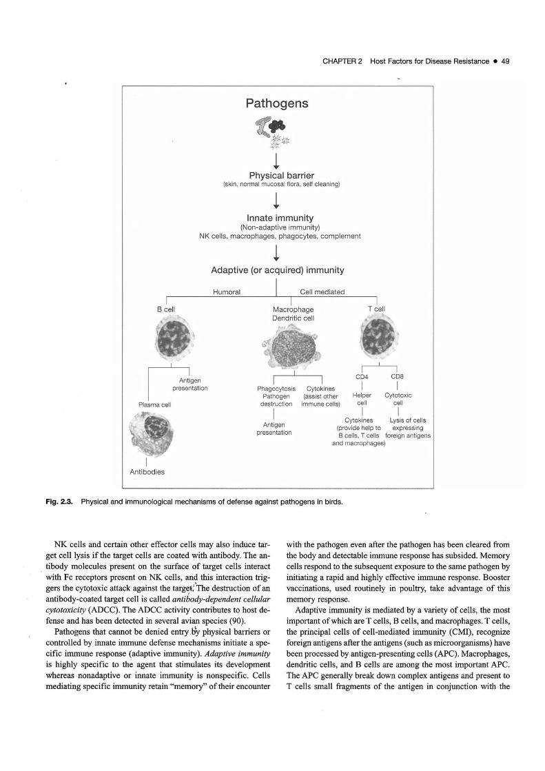
Adaptive ImmunityCell-mediated Immunity (CM!)T cells are the most important cells of CMf. Many subpopulations of T cells with diverse functions have been identified inchickens. These subpopulations express unique surface antigensthat can be detected with monoclonal antibodies. Table 2.3 showsa list of monoclonal antibodies that can recognize some of thesurface markers of chicken T cells. This table will undoubtedlyWldergo periodic revisions as new antibodies are developed.
As in mammals, avian T cells have two surface receptors thatbind antigens: T cell receptor (TCR)a[3 or TCRy8. Chickenshave a higher proportion of'V8 T cells than mice or humans andmay reach 30-50% of circulating lymphocytes (93). Both typesof TCRs (al3 and -y8) are closely associated with another molecule called CD3, which is present on all T cells. Only the TCRportion of the TCR·CD3 complex interacts with the antigen. TheCD3 molecule, which is composed of a complex set of proteins,transmits to the cell the signal of antigenITCR interaction. TheTCR molecules are diversified by rearrangement of single V, D,and J segments derived from multiple polymorphic copies ofgenes. The chicken TCRal3 locus is different from that of mammals and contains two V[3 families: Vfll and Vfl2 (9).
Swface molecules CD4 and CD8 differentiate two importantfunctional subsets of T cells. CD4 is expressed on the surface ofhelper T (TtV lymphocytes, whereas CDS is expressed on the S\D'-
CHAPTER 2 Host FactOfS for Disease Resistance • 51
.--2.3 MonodonaI antibodies for chicken T lymphocyte antigens.
......-."M__
Antigen antibodl•• mass (kD.) Homology (%) Distribution_.......
ChTl CT1, CT1a, T1oAe, 63 and 45 and 0 Thymocytes, some 15,8,30)
RR5-89, Mul83 dimers T lymphocytes
COO = 20,19,17,16 3&-40 AtITlymphocytes 14,11)
C04 CT4, 2-6, 2-35 64 23 Subpopulation of up (6,58)
Tlymphocytesand
thymocytos
COS 2-191,3-58 64 38 TandBlymphocylss R. Kosklnen andO. vano, U'lpl.tIIIshed-COw6 S3 110 SpienIc y8 and most up
T lymphocytes, some
Ihymocyt..
ChT6 INN-CH-16 SO Activated T lymphocytes (78)
ChT7 110 Activated T lymphocytes (52)
CD" eT8, EP72, 11-39, 34 37 Subpopulations of u, ~, "ill (6, 58, 64, 1(2)
3-298, AV12, AV13, and NK-like T lymphocytes
AV14, CVI·ChT-74.1 and thymocyt..
C08~ EP42 34 34 Subpopulation of u, ~T lymphocytes
ChTll A1. 120,90,28 IntesOOal and activated (27)
_TlymphocylssCD2S 2-4,2-102, AVT 40 SO upT~es (103, 109)
')18 TCR TCRl SO,4O 3O-'l3 .,.TIymphocytes (93)
atl1 TCR TCR2 SO,4O 26-35 Subpopulation of up (12, 14)
T lymphocytes
u~ TCR TCA3 48,40 26-35 Subpopulation of ap (7,10)T lymphocytes
From Jeurissen, S. H. M., O. Vainio, and M. J. H. Ratcliffe. 1998. Leukocyte markers in the chicken. In J. M. Sharma (section ed.). AVllIin immunOlogy. In P. P.Partoret, P. Griebel, H. Bazin, and A. Govaerts (cds.). Halldbook o/Veterillary lmmurwJogy. Academic ~u. With permission.
face ofcytoeoxicT tympbocytes (CTL). Great inteYspecies variationexists in the relative proportions ofcirculating CD4 and CD8 cells.
Helper T (THi CellsTHcells (CD4+ cells) recognize processed exogenous antigens inconjunction with MHC U and other costimuJatory molecules.When the TCR on the surface ofT cells comes in contact with thespecific antigenic fragment on the surface of the APe, T cells become activated and proliferate and initiate an inunune responsedirected against the antigen.
Studies in mammals have shown that antigen-induced activation stimulates THcells to differentiate into two types ofeffectorpopulations,: TNI andTH2. Differentiation ofTHcells into THI orT82 populations is determined by the nature of the stimulatingantigen and is mediated by soluble proteins called cytokines (seebelow). Intracellular antigens such as those accumulating withinmacrophages, dendritic, and other cells stimuJate the differentiation ofTHI cells. whereas extracellular antigens stimulate the differentiation ofTH2 cells. TH I effector cells promote proliferationofCD8+ crL. activate macrophages, enhance their microbicidalactivity, and facilitate B cells to produce antigen-specific antibodies with strong opsonizing properties. The principal function
ofTH2 effector cells is to help B cells to produce antigen-specificimmunoglobulins of various isotypes. Although defmitive dataare lacking, strong indications show that a similar dichotomy ofactivated THeffector cells into TH1and TH2 may also occur in thechicken (19).
Cytotoxic T Cells (CTL)Most crL express CD8 surface molecuJes. A small proportion ofmammalian CD4 T cells may also have cytotoxic activity, although the presence of avian CD4 T cells with cytotoxic abilityhas not been documented. CD8+ CTL recognize endogenousantigens in conjunction with MHC I (59). Internalized antigenssuch as viruses are degraded into small peptides by a large pr0
teolytic complex called a proteasome. Small antigen peptides,usually 7-13 amino acids long, are then transpOrted to the endoplasmic reticulum where the peptides become attached to MHCt. The peptide-MHC I complex is then transported to the cell surface for possible recognition by antigen-specific CTL
One of the most important functioos ofCTL is the eliminationofvirus-infected cells. Because most nucleated cells express surface MHC I, virus infection of almost any cell can lead to potential recognition and lysis by CTL. In vitro assays to quantitate

52 • Diseases of Pouttry
en.. in chickens have been difficult to establish because ofMHCrestriction of effe<::tor and target cells. Despite this difficulty,CfL activity has been shown to regulate pathogenesis of avianviral and neoplastic diseases (15. 35. 79).
Avian CytoldnesCytokines are small. biologically active proteins secreted by anwnber of cells, most notably immune cells. Cytokines bind tospecific recepton: on the surface of target cells and regulate immUDe response by signaling between cells. Receptor-bound cytokines and other membrane-associated molecules often act together to stimulate the effector function in a target cell. T cells, Bcells, macrophages, and dendritic cells all secrete eytokines.Cytokines produced by TH cells in panicular play a key role inmodulating an immune response. THI cells. which promote aeM! response, produce predominantly [fN--y. which activatesmacrophages and enhances destruction of cell-associatedpalbogens. Other major cytokines produced by THI cells includeIL-2 and tumor necrosis factor-a (TNF-a). IL-2 is critical for theproliferation ofa number ofimmune cells including TH'THI. andTH2 cells, CfL, NK cells. and B cells. The THI cell activity andcytokine secretion is stimulated by 1L-12 and lL-IS. both produced by macrophages, dendritic cells. and B cells.
Cytokines produced by T..2 cells. which promote B cell activation and antibody production, include IL-4. IL-5. and lL-tO.IL-l, a product of a number of cell types, most notablymacrophages, stimulates the TH2 cell activity.
Within the last few yean. a nwnber of avian eytokines havebeen isolated and characterized. Genes coding for avian cytokines and their receptors have been cloned and sequenced(Table 2.4). The biological activity ofavian cytokines is generallyquite similar to their mammalian counterparts, although aviancytokines show little cross-species biological reactivity.
Humoral ImmunityImnuUloglobuiins (Ig) or antibodies secreted by B cells constitutethe principal component of humoral immunity. Antibodies may bepresent in many body fluids but are most readily detected in theserwn or the plasma fraction of blood. Exposure of birds to microorganisms stimulates the production of specific antibodies,which, in tum, react with microorganisms and hasten lbeir destruc
tion. The three mechartisJM by which antibodies contribute to defense against pathogens are as follows: Neutralization, in which an·tibodies bind to and neutralize specific pathogens. particularlyviruses. Neutralized viroses are unable to attach to surface receptors
oftarget cells and are thus prevented from replicarion. Opsonizalionwttich includes bacterial pathogens that can replicate extracellu1arIy, and ... mo<e.-eadily mt=alized..ddestroyed by pbagocy1eSifthe pathogens are coated with antibodies. Complement activation.in which antibodies bound to the surface of pathogens, can activatecomplement and poduce new complement proteins. The complement proteins anach to receptors on phagoeytes, which facilitate thephagocytosis and destruction of pathogens.
Chickens have three main classes of immunog1obulins: IgM,180, and IgA (fable 2.S). Figure 2.4 shows the typical structureof an 19 molecule. All Ig molecules have two distinct types of
polypeptide chains. The smaller polypeptide chain, called the"light chain," is common to all classes of Ig, whereas the largerchain. called the "heavy chain," is structurally distinct for eachclass or subclass of 19. Covalent and noncovalent forces connectthe two chains. The structure of the heavy chain detennines thebiological function ofeach class of Ig. Genes encoding alllhreeclasses of avian Ig have been cloned and sequenced, which basfacilitated the generation of recombinant avian and chimeric antibodies in vitro and the expression of recombinant avian Ii inplants (16, 26, 60, 68, 77,106).
IgM is found on the surface of most B cells and is the first antibody produced following primary immunization. As the immune response progresses. the IgM-producing cells stop IgMproduction and start the production ofIgG or 19A. This phenomenon is called "class switch." The antigen-binding ability of theantibodies does not alter during or after the switch. The "classswitch" occurs because the antibody-producing B cell begins tosplice the variable (V) region genes (V genes) to the constant (C)region genes (C genes) of the heavy chain of a different class ofIg. Cytokines including 1L-4. TGF-p and IFN-oy stimulate the Bcell to undergo class switch (21).
A typical immune response ofa chicken begins with IgM production. After some time, 19M production switches over to 180production. IgG is also the principal antibody produced after secondary immunization and is the predominant Ig class in chickenblood. Because avian (and also amphibian, reptile. and piscine)IgG is larger than its mammalian counterpart, the chicken IgG isoften called IgY (104). Figure 2.4 compares the relative structureof manunalian and avian IgG. Molecular cloning data suggestthat IgY may be the ancestral precursor of mammalian IgG andIgE (104).
IgA is the most important Ig involved in mucosal immunity.Chicken secretory IgA (slgA) exists as a dimer in mucosal secretions, whereas circulating IgA is polymeric or monomeric. IgAcomplexes with a secretory component present on the surface ofmucosal epithelial cells to form sIgA (107). The acquisition ofthe secretory component protects 19A from proteolytic digestionin the gut. 19A is most concentrated on mucosal surfaces, although small quantities may be found in the circulation. Bile isalso a rich reservoir or IgA in birds. IgA protects mucosal surfaces against pathogens. particularly viruses. by neutralizing andpreventing their attachment to receptors on target cells.
As noted earlier. B cells use surface Ig to bind to antigens.Each B cell produces only one type ofheavy and light chain andis committed to one Idnd of antigenic determinant Thus, for anantigen to initiate antibody production and clonal expansion, theantigen must interact with a B cell that expresses the homologousIg receptor. Potentially thousands of antigens and millions ofantigenic shapes are in the environment How does the irwnunesystem maintain an inventory of B cells with such a wide varietyof antigenic specificities? This is accomplished by a nwnber ofgenetic mechanisms during the development and maturation ofBcells. In mammals. Ig gene rearrangement leads to extensive Igdiversity. In the chicken, because ofa relatively small numbers ofIg genes. the rearranged genes must undergo a process calledgene conversion to attain needed diversity (21). In gene conver-

chicken (72)chicken (97)chicken (29)
chicken (1, 37)chicken (1)chicken (1)chicken (37)chicken (1,37)chicken (37)
Table 2.4 Avian cytokine genes identified.
Cytokine gene cloned
IFN-aIFN-~
IFN-j'IL-1~
IL-2IL-3IL-4IL-5Il-GIL-7IL-9lL~10
lL-12tL-13tL-15IL-16lL-17lL-17A ,a,D.FlL-18Il-19IL-21IL-22IL-26IFN->..1 (lL-29),- A2 (IL-28A), - A3 (IL-28B)Granulocyte colony-stimulating factor (CSF3)GM-CSFStem cell factorChicken MGFTGF~1
TGF-132,TGF-133TGF134LymphotaetinMIP-1~
Chemokines (K60, K(03)ChemokineschXCl1 ,chCCLl5,chCCU6chCCli7,chCCli8,chCCU9chCCli1 O,ccCCU1 ,ccCCU2ccCCU3,chCCli4,chCCL17chCCL19,chCCL20,chCCL21chCXCU1,chCXCU20L-8)chCXCU3,chCXCL12,chCXCL1311 ,chCXCL13L2chCXCL13L3,chCLCXL14chCX3CL1Stromal cell derived factor-1ChTUALPS-itlduced TNF-alpha factor (liTAF)TRAILTNFAIITRAF5
VEGFCD30LCD40LBAFF
Avian species
chicken, turkey, duckchickenchicken, turkey, Japanese quail, guinea fowl, duckchickenchicken, turkey, Japanese quail, duck, goosechickenchickenchickenchickenchickenchickenchickenchickenchickenchickenchickenchickenchickenchicken, turkeychickenchickenchickenchickenchickenchickenchickenchickenchickenchickenchickenchickenchickenchickenchickenchickenchicken
Reference
(83,84,98)(92)(22,31,38,57,82)(105)(37,51,55,94,95,111)(2,37)
(2,37)
(37)(37,100)(37)
(37)(37,75)(3,20.37, 100)(2,37)(13,37,55)(37,63)
(63)(37)
(37,80)
(37)(37)(37)
(37)
(37)(2,37)(37,76)
(110)(53)(34)
(37)(87)(100)(70)(91)
(32)
(37)
(Data for this table provided by Mahesh Khatri, University of Minnesota, St. Paul. MN 55127.)
53

54 • Diseases of Poultry
Table 2.5 Properties of chicken immunoglobulin Isotypes.
IgM
IgG
IgA
Heavychain (kDs)
70kDa
87 kDa
65kDa
.Hehsln19 domains
5
4
4
Homology 1o
mammsllBn
About 30%
78% forTM"
3<l-05%
32-41%
Seromconcentration
1-2 mg/ml
5-10mg/ml
i!!3 mgl ml
Serum
Cell surface
SerumEgg Yolk
Se<Um
Bile
Mucosa (tears,saliva)
StructUI"8 end comments
900 kOs, consistent with heavily
glycosylated (~24)5 plus a J chain~2l2 monomer of membrane IgM,
no J chain
175 kOa, 't24 monomeric fonn
't2l2' high concentrations (10 mglm~
of IgG are found in egg yolkOowconcentrations in egg white)
170 kOs, (l24-monomeric formwithout J-chain
350 kOa, consistent with «(124>2 plusa J-chsin
600-700 kOs, consistent with ((124>.plus a J-chain
"'TM refers to the transmembrane and cytoplasmic domains of chicken sIgM.(From Demaries, S.L. and M.J.H. Ratcliffe. Cell surface and secreted immunoglubulins in B cell development. In: J.M. Sharma, ed., Avian Immunology. In:Partoret P.P:, P. Griebel, H. Bazin, and A. Govaerts, cds., Handbook ofVeterinary Immunology, Academic Press, 1998. With Pennission).
....--AvlM leGlor ltV)
Ag. 2.4. Typical structure of an 19 molecule and comparison be
tween avian and mammalian IgG molecule.
sicn, the rearranged light and heavy chain gene complexes acquire clusters of chromosomal pseudogenes. Large segments ofhighly homologous pseudogenes are present in the vicinity oflight and heavy chain genes in the chicken chromosome (73,74).
Maternal Transfer of ImmunityTransmission of immunity from the hen to the newly hatchedchick is critical for protecting the chick against infections duringearly life. In chickens, Ig are the principal mode oftransfer of immunity. There is little evidence that the mother's immune cellsare passed on to the embryo. Ig from hen's circulation are deposited in the superficial epithelial and glandular cells of theoviduct (45). From the oviduct, IgG is transferred into the maturing oocyst in the ovarian follicle and accumulates in the yolk sac.Ig produced locally in the oviduct likely constitutes an insignifi-
cant proportion of the transferred Ig. The developing chick acquires maternal IgG from the yolk sac. IgA and IgM are transferred via the amniotic fluid. The developing embryo swallows19A- and IgM-containing amniotic fluid.
Assays to Measure ImmunityNKCellsNK cell assays are based on in vitro cytotoxicity against susceptible target cells (24.49. 89). The most commonly used targetsare the cells ofthe line LSCC-RP9 (88). These cells were derivedfrom a retrovirus-induced tumor in a B2BIS male chicken (65).The target cells are labeled with SICr and incubated in vitro withvarying concentrations of cell suspension being tested for NKcell activity (effector cells). Two controls are important: a)adding "neutral" cells such as thymocytes to target cells at thesame effector:target ratios as used for the effector cells, and b)the use ofNK-resistant target cells. After 4 hours of incubation at37°C, the radioactivity released into the medium is quantitated.Specific cytotoxicity, a measure ofNK cell lysis, is calculated bythe following formula:
% cytotoxicity = counts per minute (cpm) in target cells mixedwith effector cells - cpm in target cells mixed with normal thymus cells I total cpm incorporated in target cells - cpm in tar·get cells mixed with normal thymus cells X 100.
MacrophagesMacrophages, a phenotypically diverse population of cells, arepresent in almost all tissues. Because most macrophages adhereto substrates, they can be readily isolated from short-term in vitrocultures of peripheral blood cells (PBL) or single cell suspensions of spleen (42). Peritoneal macrophages may also be induced in birds by intraperitoneal injections of inflammatory

stimulants such as Sephadex beads. Some of the assays used toassess macrophage functions include a) phagocytosis, b) cytokine production upon stimulation with mitogens (lipopolysaccharide), c) ability to lyse hlmor cells, and d) production of nitricoxide (NO) upon activation by T cell-produced cytokines, mostnotably IFN--y. Some of the functional characteristics of avianmacrophages have been described (41, 42, 62, 66, 71, 86).
TCe/lsMost TH cell assays are based on in vitro stimulation ofcells withmitogens or specific antigens (43, 44, 99). Stimulated cells proliferate and secrete cytokines. Mitogen-induced proliferation is acommon assay of T-cell competence. Concanavalin A (Con A)and phytohemagglutinin (PHA) are the mitogens of choice.These mitogens bind to cell surface glycoproteins on T cells andstimulate the cells to proliferate. In the typical assays, spleencells, PBL, or diluted whole blood are cultured in vitro inmedium containing Con A or PHA. After 40 hours of incubationat 37-41 oC, the cells are pulse labeled with radioactive thymidine. The incorporation of the label in cellular DNA is quantitated. Actively proliferating cultures incorporate higher levels ofradioactivity than non-proliferating cultures. If the proliferativeresponse of the test group of chickens is lower than the responsein age-matched healthy controls, the test group is viewed asbeing deficient in functional T cells. This general conclusionshould be viewed with caution because the mitogen-inducedproliferation is not antigen-specific, and response to mitogen isan in vitro function ofT cells. The in vivo relevance of this function to other in vitro or in vivo functions ofT cells is not known,and functional T cells may be prevented from proliferating bynon-T suppressor cells or suppressor products present in theculture (43, 69).
T cells recovered from immunized animals may proliferate invitro when co-cultured with the antigen used for immunization(44,99). This antigen·specific proliferation has been shown withseveral avian viruses, although the ideal assay conditions are notwell established, and the test is not widely used.
Mitogen- or antigen-induced stimulation ofT cells in vitro alsoresults in the secretion ofcytokines. Quantitation of cytokines inthe culture medium gives an indication of the functional capability ofT cells. A nitric oxide inducing factor (NOIF) test has beendescribed in which macrophage-stimulating cytokines such asIFN--y can be quantitated. Macrophage line cells are exposed tothe test supernatants, and NO concentration in the supernatants iscalculated (39).
CTL activity can be measured in vitro by co-culturing effectorcells with slCr-labeled target cells (59). The protocols are quitesimilar to those described previously for NK cell cytotoxicity assays. The cytotoxic activity of CTL is MHC I-restricted.Therefore, both effector and target cells must come from thesame or a genetically compatible bird. Because of this limitation,CTL assay is difficult to perfonn in outbred populations of birdsand remains a research tool.
Certain in vivo assays may also be used to assess T-cell functions. The delayed type hypersensitivity assay measures antigenspecific response. In this test, an animal immunized against an
CHAPTER 2 Host Factors for Disease Resistance • 55
antigen is intradennally injected with the same antigen. Swellingat the site of the injection comprises a positive response. Localswelling at the site of an intradermal injection of mitogens suchas PHA has also been attributed to a non-specific T cell response.
Antibody LevelsBirds exposed to pathogens develop circulating antibodies thatgenerally persist for several weeks after the antigen has beencleared. Detection of these antibodies is much more convenientthan detecting cellular immunity, and a number of serologicassays are available to quantitate antibodies. Some of thecommonly used serologic tests include agar gel precipitationtest, virus neutralization test, immunofluorescence test, hemagglutination inhibition test, and enzyme-linked immunosorbentassay (ELISA). Protocols for conducting these tests have beendescribed (98).
ELISA is by far the most common serologic assay used undercommercial settings. Automated technology allows rapid processing of large numbers of serum samples. Computerized datatransmission facilitates flock profiling and provides useful information on environmental exposure to pathogens and response tovaccination. ELISA kits that can be used to detect antibodiesagainst most of the common viral and bacterial pathogens ofpoultry are available commercially.
The transfer of IgG from the yolk sac to the embryo or thehatchling occurs by absorption into the recipient's circulation.Yolk sac is highly vascularized, and IgG is transferred by receptor-mediated endocytosis across the yolk sac epithelium (56).The transfer of IgG begins during the first week ofembryonationbut occurs most predominantly during the last three days beforehatching (48). The transfer from the yolk continues after hatch.Peak levels of maternal IgG in the circulation of the newlyhatched chick reach around 2-3 days of age. Maternally derivedantibodies decline linearly in the recipient and become undetectable after 2-5 weeks.
Although maternal antibodies are important for the well beingof the newly hatched chick, the antibodies may interfere with active immunization with live vaccines. Neonatal or in avo vaccination is often necessary in flocks being raised in heavily contaminated environment. Besides neutralizing the antigen presentin the vaccine, pre-existing antibodies may also interfere with thedevelopment of active immunity by providing negative feedbackto the immune system.
ReferencesI. Abdalla, S. A., H. Horiuchi, S. FUl1lsawa, and H. Matsuda. 2004.
Molecular cloning and characterization of chicken tumor necrosisfactor (TNF)-superfamily ligands, CDJOL and TNF-related apoptosis inducing ligand (TRAIL). J Vet Met! Sci 66:643-50.
2. AVery, S., L. Rothwell, W. D. Degen, V. E. Schijns, J. Young, J.Kaufinan, and P. Kaiser. 2004. Characterization of the first nonmammalian T2 cytokine gene cluster: the cluster contains functional single-eopy genes for IL-], IL-4, IL-13, and GM-CSF, a genefor IL-S that appears to be a pseudogene, and a gene encoding another cyIokinelike transcript, KK34. J Interferon Cytokine Res
24:600-10.

56 • Diseases of POUltry
3. Balu, S., and P. Kaiser. 2003. Avian interleukin-12beta (p40);cloning and characterization of the cDNA and gene. J InterferonCytokine Res 23:699-707.
4. Bernot, A., and C. Auffray. 1991. Primary structure and ontogenyof an avian CD3 transcript. Proc Natl Acad Sci USA 88:2550-4.
5. Boyd, R. 1., T. J. Wilson, A. G. Bean, H. A. Ward, and M. E.Gershwin. 1992. Phonotypic characterization of chicken thymicstromal elements. Dev. Immunol. 2;51--66.
6. Chan, M. M., C. L. Chen, L. L. Ager, and M. D. Cooper. 1988.Identification of the avian homologues of mammalian CD4 andCD8 antigens. J Immunoll40:2133-8.
7. Char, D., P. Sanchez, C. L. Chen, R. P. Bucy, and M. D. Cooper.1990. A third sublineage of avian T cells can be identified with a Tcell receptor-3-specific antibody. J ImmunoI145:3547-55.
8. Chen, C. H., T. C. Chanh, and M. D. Cooper. 1984. Chickenthymocyte-specific antigen identified by monoclonal antibodies:ontogeny, tissue distribution and biochemical characterization. EurJ ImmunoI14;385-91.
9. Chen, C~ H., T. W. Gobel, T. Kubota, and M. D. Cooper. 1994. T celldevelopment in the chicken. Poult Sci 73: 1012-8.
lO. Chen, C. H., J. T. Sowder, J. M. Lahti, J. Cihak, U. Losch, and M.D. Cooper. 1989. TCR3: a third T-cell receptor in the chicken. ProcNatlAcad Sci USA 86:2351-5.
11. Chen, C. 1., 1. L. Ager, G. 1. Gartland, and M. D. Cooper. 1986.Identification of a T3rr cell receptor complex in chickens. J ExpMed 164:375-80.
12. Chen, C. L., 1. Cihak, U. Losch, and M. D. Cooper. 1988. Differential expression of two T cell receptors, TcR1 and TcR2, onchicken lymphocytes. EurJ ImmunoI18:539-43.
13. Choi, K.. D., H. S. Lillehoj, K. D. Song, and J. Y. Han. 1999.Molecular and functional characterization of chicken 11-15. DevComp Immunol 23: 165-77.
14. Cihak, 1., H. W. Ziegier-Heitbrock, H. Trainer, I. Schranner, M.Merkenschlager, and U. Losch. 1988. Characterization and functional properties of a novel monoclonal antibody which idenlifies aT cell receptor in chickens. Eur J ImmWlo/18:533-7.
15. Collisson, E. w., 1. Pei, J. Dzielawa, and S. H. Seo. 2000. CylotoxicT lymphocytes are critical in the control of infectious bronchitisvirus in poultry. Dell Comp Immunol 24: 187-200.
16. Dahan, A., C. A. Reynaud, and J.C. Weill. 1983. Nucleotide sequenceofa chicken J1 heavy chain mRNA. NuclAcids Res. 11:5381-5389
17. Davison, T. E, K. B, and K. A. Schal (ed.). 2007. Avian Immunology. Elsevier Limited.
18. Davison, T. E, T. R. Morris, and L. N. Payne. 1996. Poultry Immunology. Poullry Science Symposium Series., vol. 24. Carfax Publishing Company. Abingdon, UK.
19. Degen, W. G., N. Daal, L. Rothwell, P. Kaiser, and V. E. Schijns.2005. Th1fTh2 polarization by viral and helminth infection inbirds. ~t Microbiol105:163-7.
20. Degen, W G., N. van Daal, H. I. van Zuilekom, J. Burnside, and V.E. Schijns. 2004. Identification and molecular cloning of functional chicken IL-12. J ImmunoI172:4371-80.
21. Demaries, S. L., and M. J. Ratcliffe. 1998. Cell surface and secretedimmunoglobulins in B cell development. In J. M. Sharma (ed.),Avian Immunology, vol. Academic Press. Handbook of VertebrateImmunology, Pastoret, P.P..P. Griebel, H. Bazin and A. Govaerts.
22. Digby, M. R., and J. W. Lowenthal. 1995. Cloning and expressionof the chicken interferon-gamma gene. J Inteiferon Cytokine Res15:939-45.
23. Fukui, A., N. Inoue, M. Matsumoto, M. Nomura, K. Yamada, Y.Matsuda, K. Toyoshima, and T. Seya. 2001. Molecular cloning and
functional characterization of chicken toll-like receptors. A singlechicken loll covers multiple molecular patterns. J Bioi Chem276:47143-9.
24. Gobel, T. W. 2000. Isolation and analysis of natural killer cells inchickens. Method Mol Bioi. 121:337-345.
25. Gobel, T. w., B. Kaspers, and M. Stangassinger. 2001. NK and Tcells constitute two major, functionally distinct intestinal epitheliallymphocyte subsets in lhe chicken. Inllmmunol13:757--62.
26. Greunke, K., E. Spillner, I. Sraren, H. Seismann, S. Kainz, U.Hahn, T. Grunwald, and R. Bredehorst. 2006. Bivalent monoclonalIgY antibody formats by conversion of recombinant antibody fragments. J BiotechnoI124:446--56.
27. Haury, M., Y. Kasahara, S. Schaal, R. P. Buey, and M. D. Cooper.1993. Intestinal T lymphocytes in the chicken express an integrinlike antigen. Eur J ImmunoI23:313-9.
28. Higgins, D. A., and G. W. Warr. 2000. The avian immune responseto infectious diseases. Special Issue. Developmental andComparative Immunology 24:85-101.
29. Hong, Y. H., H. S. Lillehoj, S. Hyen Lee, D. Woon Park, and E. P.Lillehoj. 2006. Molecular cloning and characterization of chickenlipopolysaccharide-induced TNF-alpha factor (LITAF). Dev CompImmuno130:919-29.
30. Houssaint, E., E. Diez, and F. V. Jotereau. 1985. Tissue dislributionand ontogenic appearance of a chicken T lymphocyte differentia·tion marker. Eur J ImmunolI5:305-8.
31. Huang, A., C. A. Scougall, 1. W. Lowenthal, A. R. Jilbert, and I.Kotlarski. 2001. Structural and functional homology between duckand chicken imerferon-gamma. Dev Comp ImmunoI25:55--68.
32. Hughes, S., and N. Bumstead. 2000. The gene encoding a chickenchemokine with homology to human SCYCI maps to chromosomeI. Anim Genet 31:142-3.
33. Iqbal, M., V. J. Philbin, and A. L. Smith. 2005. Expression patternsof chicken toll-like receptor mRNA in tissues, immune cell subsetsand cell lines. Vet /mmunollmmunopalholl04: 117-27.
34. Jak.owlew, S. B., P. 1. Dillard, M. B. Sporn, and A. B. Roberts. 1988.Nucleotide sequence of chicken transforming growth factor-beta 1(TGF-beta I). Nucleic Acids Res 16:8730.
35. Jeurissen, S. H., A. G. Boonstra-Blom, S. O. Al-Garib, 1. Hartog,
and G. Koch. 2000. Defense mechanisms against viral infection inpoullry: A Review. M?terinary Quart. 22:204--208.
36. Kai, c., K. Yoshikawa, K. Yamanouchi, and H. Okada. 1983.Isolation and identification of the third component of complementof Japanese quails. J Immunol. 130:2814-2820.
37. Kaiser, P., T. Y. Poh, 1. Rothwell, S. Avery, S. Balu, U. S. Pathania,S. Hughes, M. Goodchild, S. Morrell, M. Watson, N. Bumstead,1. Kaufman, and 1. R. Young. 2005. A genomic analysis ofchicken cytokines and chemokines. J Interferon Cytokine Res25:467-84.
38. Kaiser, P., D. Sonnemans, and L. M. Smith. 1998. Avian IFNgamma genes: sequence analysis suggests probable cross-speciesreactivily among galliforms. J Interferon Cytokine Res 18:711-9.
39. Karaca, K., I. 1. Kim, S. K. Reddy, and 1. M. Sharma. 1996. Nitricoxide inducing factor as a measure of antigen and mitogen-specificT cell responses in chick.ens. J lmmunol Methods 192:97~103.
40. Kaufman, J., 1. Jacob, I. Shaw, B. Walker, S. Milne, S. Beck, and J.Salomonsen. 1999. Gene organisation determines evolution offunction in the chicken MHC./mmunol Rev 167:101-17.
41. Khatri, M., 1. M. Palmquist, R. M. Cha, and J. M. Sharma. 2005.Infection and aClivation of bursal macrophages by virulent infectious bursal disease virus. Virus Res 113:44-50.

42. Khatri, M., and 1. M. Shanna. 2006. Infectious bursal disease virus
infection induces macrophage activation via p38 MAPK lind NF·
kappaB pathways. Virus Res 118:70-7.43. Kim, I. 1., and 1. M. Sharma. 2000. IBDV-induced bursal T lympho
cytes inhibit mitogenic response of normal splenocyles. VetImmunoIImmunopathoI74:47-57.
44. Kim, I. J., S. K. You, H. Kim, H. Y. Yeh, and 1. M. Sharma. 2000.
Characteristics of bursal T lymphocytes induced by infectious bur
sal disease virus. J ViroI74:8884-92.45. Kimijama, T., H. Y., H. Kitagawa, Y. Kan, and M. Sugimura. 1990.
Localization of immunoglogulins in the chicken oviduct. Japanese
J Vet &i 52:299-30546. Koch, C. 1986. The alternative complement pathway in chickens.
Purification of factor B and production of a nonspecific antibody
against it. Acta Path Microbiallmmunol Seand, Se. C. 94:253-259.47. Koppenheffer, T. L. 1998. Complement. In 1. Sharma (ed.), Avian
Immunology, vol. Academic Press. Handbook of Vertebrate
Immunology, Pastoret, P.P., P. GriebeL, H. Barin and A. Govaerts.48. Kowalczyk, K., 1. Doiss, 1. Halpern, and T. F. Roth. 1985.
Quantitation of maternal-fetal IgO transport in the chicken.ImmunoI54:755-762.
49. Kushima, K., M. Fujita, A. Shigeta, H. Horiuchi, H. Matsuda, and
S. Furusawa. 2003. Flow cytometric analysis of chicken NK activ
ity and its use on the effect of restraint stress. J Vet Med Sei65:995-1000.
50. Laursen, 1., and C. Koch. 1989. Purification of chicken C3 and a
structural and functional characterization. Scand J Immunol30:529-538.
51. Lawson, S., L. Rothwell, and P. Kaiser. 2000. Turkey and chicken
interleukin-2 cross-react in in vitro proliferation assays despite limited amino acid sequence identity. J Interferon Cytokine Res20:161-70.
52. Lee, T. H., and C. H. Tempelis. 1992. A possible IIO-kDa receptorfor interleukin-2 in the chicken. Dev Comp ImmunoI16:463-472.
53. Leutz, A., K. Damm, E. Stemeck, E. Kowenz, S. Ness, R. Frank, H.Gausepohl, Y. C. Pan, 1. Smart, M. Hayman, and et al. 1989.Molecular cloning of the chicken myelomonocytic growth factor
(cMGF) reveals relationship to interleukin 6 and granulocytecolony stimulating factor. Embo J 8: 175-81.
54. Leveque, G., V. Forgetta, S. Morroll, A. L. Smith, N. Bumstead, P.Barrow,1. C. Loredo-Osti, K. Morgan, and D. Malo. 2003. Allelicvariation in TLR4 is linked to susceptibility to Salmonella enterica
serovar Typhimurium infection in chickens. Infect Immun71:1116--24.
55. Lillehoj, H. S., W. Min, K. D. Choi, U. S. Babu, 1. Burnside, T.Miyamoto, B. M. Rosenthal, and E. P. Lillehoj. 2001. Molecular,
cellular, and functional characterization of chicken cylokines homologous to mammalian IL-15 and IL-2. Vet Immunof Immunopatlwl 82:229-44.
56. Linden, C. D., and T. F. Roth. 1978. IgO receptors on fetal chickenyolk sac. J Cell Sci 33:3174--328.
57. Loa, C. C., M. K. Hsieh, C. C. Wu, and T. L. Lin. 2001. Molecularidentification and characterization of turkey IFN-gamma gene.Comp Biochem Physiol B Biochem Mol Bioi 130:579-84.
58. Luhtala, M., 1. Salomonsen, Y. Hirota, T. Onodera, P. Toivanen, andO. Vainio. 1993. Analysis of chicken CD4 by monoclonal antibodies indicates evolutionary conservation between avian and mammalian species. Hybridoma 12:633-46.
59. Maccubin, D. L, and L. W. Schierman. 1986. MHC restricted cytotoxic response of chicken T cells: expression, augmentation and
clonal characterisation. J Immunof 136:12-16.
CHAPTER 2 Host Factors for Disease Resistance _ 57
60. Mansikka, A. 1992. Chicken IgA H chains. Implications concern
ing the evolution ofH chain genes. J Immunol149:855-861.61. Mavroidis, M., 1. D. Sunyer, and 1. D. Lambris. 1995. Isolation, pri
mary structure and evolution of the third component of chicken
complement and evidence for a new member of the x2-macroglobulin family. J Immunol 154:2164--2174.
62. Mellata, M., M. Dho-Moulin, C. M. Dozois, R. Curtiss, 3rd, B.Lehoux, and 1. M. Fairbrother. 2003. Role of avian pathogenic
Escherichia coli virulence factors in bacterial interaction with
chicken heterophils and macrophages. Infect Immun 71 :494--503.
63. Min, w., and H. S. Lillehoj. 2002. isolation and characterization ofchicken interIeukin-17 eDNA. J Interferon Cytokine Res22:1123-8.
64. Noteborn, M. H., G. F. de Boer, D. 1. van Roozelaar, C. Karreman,
O. Kranenburg, 1. G. Vas, S. H. Jeurissen, R. C. Hoeben, A.Zantema, O. Koch, and et al. 1991. Characterization of cloned
chicken anemia virus DNA thai contains all elements for the infectious replication cycle. J ViroI65:3131-9.
65. Okazaki, w., R. L. Witter, C. Romero, K. Nazerian, J. M. Sharma,
A. Fadly, and D. Ewert. 1980. Indication of lymphoid leukosis
transplantable tumours and the establishment of Iymphoblastoidcell lines. Avian Path 9:311-329
66. Palmquist, 1. M., M. Khatri, R. M. Cha, B. M. Goddeeris, B.Walcheck, and 1. M. Shanna. 2006. In vivo activation of chicken
macrophages by infectious bursal disease virus. Viral Immunol19:305-15.
67. Pan, H., and 1. Halper. 2003. Cloning, expression, and characteri
zation of chicken transforming growth factor beta 4. BiochemBiophys Res Commun 303:24--30.
68. Parvari, R., A. Avivi, F. Lentner, E. Ziv, S. Tel-Or, Y. Burstein, and1. Schechter. 1988. Chicken immunoglobulin gamma-heavy chains:
limited VH gene repertoire, combinatorial diversification by Dgene segments and evolution of the heavy chain locus. Embo J7:739-44.
69. Pertile, T. L., K.. Karaca. M. M. Walser, and 1. M. Shanna. 1996.
Suppressor macrophages mediate depressed Iymphoproliferation inchickens infected with avian reovirus. Vet ImmunolImmunopathol53:129-45.
70. Petrenko, 0., I. Ischenko, and P. 1. Enrietto. 1995. Isolation of a
cDNA encoding a novel chicken chemokine homologous tomammalian macrophage inflammatory protein-I beta. Gene 160:305-6.
71. Qureshi, M. A., C. L. Heggen, and I. Hussain. 2000. Avian
macrophage: effector functions in health and disease. Dev CompImmuno/24:103-19.
72. Read, L. R., 1. A. Cumberbatch, M. M. Buhr, A. 1. Bendall, and S.Sharif. 2005. Cloning and characterization of chicken stromal cellderived factor-I. Dev Comp ImmunoI29:143-52.
73. Reynaud, C. A., V Anquez, H. Grimal, and 1. C. Weill. 1987. A hyperconversion mechanism generates the chicken light chain preimmune repertoire. Cell 48:379-88.
74. Reynaud, C. A., A. Dahan, V Anquez, and J. C. Weill. 1989.Somatic hyperconversion diversifies the single Vb gene of the
chicken with a high incidence in the D region. Cell 59:171-83.75. Rothwell, L., 1. R. Young, R. 2oorob, C. A. Whittaker, P. Hesketh,
A. Archer, A. L. Smith, and P. Kaiser. 2004. Cloning and character
ization of chicken IL-10 and its role in the inunune response toEimeria maxima. J Immuno/173:2675-82.
76. Santos, M. D., M. Yasuike, I. Hirono, and T. Aoki. 2006. The granulocyte colony-stimulating factors (CSF3s) of fish and chicken.
Immunogenetics 58:422-32.

58 • Diseases of Poulby
77. Sapats, S. I., H. G. Heine, L. Trinidad, G. 1. Gould, A. J. Foord, S.
G. Doolan, S. Prowse, and 1. Ignjatovic. 2003. Generation ofchicken single chain antibody variable fragments (scFv) that differentiate and neutralize infectious bursal disease virus (IBDV). ArchVirolI48:497-515.
78. Scauenstein, K., G. Kronenn, K. Hala, G. Bock, and G. Wick.1988. Chicken-aetivated-T-Iymphocyte antigen (CATLA) recognized by monoclonal antibody INN-CH-16 represents the IL-2 receptor. Dev Comp Immuno/12:823-831.
79. Schal, K. A., and Z. Xing. 2000. Specific and nonspecific immuneresponses to Marek's disease virus. Dev Comp ImmunoI24:201-21.
80. Sclmeider, K., F. Puehler, D. Baeuerle, S. Elvers, P. Staeheli, B.Kaspers, and K. C. Weining. 2000. cDNA cloning of biologicallyactive chicken interleukin-18. J Interferon Cytokine Res20:879-83.
81. Schat, K. A., and Z. Xing. 2000. Specific and non-specific immuneresponses to Marek's disease., vol. 24. Developmental andComparative Immunology.
82. Schultz, 0., and F. V. Chisari. 1999. Recombinant duck interferongamma inhibits duck hepatitis B virus replication in primary hepatocytes. J Virol73:3162-8.
83. Schultz, U, B. Kaspers, C. Rinderle, M. J. Sekellick, P. I. Marcus,and P. Staeheli. 1995. Recombinant chicken interferon: a potent antiviral agent that lacks intrinsic macrophage activating factor activity. EurJ Immuno/25:847-51.
84. Sekellick, M. J., A. F. Ferrandino, D. A. Hopkins, and P. I. Marcus.1994. Chicken interferon gene: cloning, expression, and analysis. JInterferon Res 14:71-9.
85. Shanna, J. M. 1991. Avian Cellular Immunology. CRS Press.86. Sharma, J. M. 1983. Presence of adherent cytotoxic cells and non
adherent natural killer cells in progressive and regressive Marek'sdisease tumors. ~t lmmunollmmunopathol 5: 125-40.
87. Sharma, J. M. 1997. The structure and function of the avian immune system. Acta Vet Hung 45:229-38.
88. Sharma, J. M., and W Okazaki. 1981. Natural killer cell activity inchickens: target cell analysis and effect of antithymocyte serum oneffector cells. Infect lmmun 31: 1078-85.
89. Sharma, J. M., and K. A. SChal. 1991. Natural immune functionsvol. CRS Press. Avian Cellular Immunology, J.M. Shanna.
90. Sharma, J. M., and K. A. Schat. 1991. Natural immune functions,vol. CRC Press. Avian Cellular Immunology, Sharma, J.M.
91. Sick, C., K. Schneider, P. Staeheli, and K. C. Weining. 2000. Novelchicken CXC and CC chemokines. Cytokine 12:181--6.
92. Sick, C., U. Schultz, and P. Staeheli. 1996. A family of genes coding for two serologically distinct chicken interferons. J Bioi Chem271:7635-9.
93. Sowder, 1. T., C. 1. Chen, L. L. Ager, M. M. Chan, and M. D.Cooper. 1988. A large subpopulation of avian T cells express a homologue of the mammalian T gamma/delta receptor. J Exp Med
167:315-22.94. Sreekumar, E., A. Premraj, and T. 1. Rasool. 2005. Duck (Anas
platyrhynchos), Japanese quail (Coturnix coturnix japonica) andother avian interleukin-2 reveals significant conservation of geneorganization, promoter elements and functional residues. Inl JImmunogenet 32:355--65.
95. Sundick, R. S., and C. Gill-Dixon. 1997. A cloned chicken Iymphokine homologous to both mammalian IL-2 and IL-15. JImmunolI59:72o-5.
96. Suresh. M., K. Karaca, D. Foster, and J. M. Shanna. 1995. Molecular and functional characterization of turkey interferon. J Virol69:8159-63.
97. Takimoto, T., K. Takahashi, K. Sato, andY. Akiba. 2005. Molecularcloning and functional characterizations of chicken TLIA. DevComp Immuno129:895-905.
98. Thayer, S. G., and C. W Beard. 1998. Serologic Procedures, vol.Am. Assoc. Avian Pathologists. A Laboratory Manual for Isolationand Identification of Avian Pathogens, Swayne, D.E., J.R. Glisson,M.W Jackwood. J.E. Pearson and WM. Reed.
99. Timms, L. M., C. D. Bracewell, and D. J. Alexander. 1980. Cell mediated and humoral immune response in chickens infected withavian infectious bronchitis. Br Vel J 136:349--6.
100. Tirunagaru, V. G., L. Sofer, J. Cui, and J. Burnside. 2000. An ex·pressed sequence tag database ofT-cell-enriched activated chickensplenocytes: sequence analysis of 5251 clones. Genomics66:144-51.
101. Toivanen, A., and P. Toivanen (ed.). 1987. Avian Immunology, vol.I. CRS Press.
102. Tregaskes, C. A., F. K. Kong, E. Paramithiotis, C. L. Chen, M. 1.
Ratcliffe, T. F. Davison, and 1. R. Young. 1995. Identification andanalysis ofthe expression of CD8 alpha beta and CD8 alpha alphaisofonns in chickens reveals a major TCR-gamma delta CD8 alphabeta subset of intestinal intraepithelial lymphocytes. J Immunol154:4485-94.
103. Vainio, 0., B. Riwar, M. H. Brown, and O. Lassila. 1991. Characterization of the putative avian CD2 homologue. J lmmunol147:[593-9.
104. Warr, G. W, K. E. Magor, and D. A. Higgins. 1995. IgY: clues tothe origins of modem antibodies.lmmunol Today 16:392-8.
105. Weining, K. C., C. Sick, B. Kaspers, and P. Staeheli. 1998. Achicken homolog of mammalian interleukin-I beta: eDNA cloningand purification of active recombinant protein. Eur J Biochem258:994-1000.
106. Wieland, W. H., A. Lammers, A. Schots, and D. V Orzaez. 2006.Plant expression ofchicken secretory antibodies derived from combinatoriallibraries. J Biolechno/122:382-91.
107. Wieland, W. H., D. Orzaez, A. Lammers, H. K. Parmentier, M. WVerstegen, and A. Schots. 2004. A functional polymeric immunoglobulin receptor in chicken (Gallus gallus) indicates ancientrole of secretory IgA in mucosal immunity. BiochemJ380:669-76.
108. Yonemasu, K., and T. Sasaki. 1986. Purification, identification andcharacterization of chicken C 1Q, a subcomponent of the first component of complement. J Immunol Meth. 88:245-253.
109. Young, J. R., T. F. Davison, C. A. Tregaskes, M. C. Rennie, and O.Vainio. 1994. Monomeric homologue of manunalian CD28 is expressed on chicken T cells. J ImmunolI52:3848-51.
110. Zhou, 1. H., M. Ohtaki, and M. Sakurai. 1993. Sequence of a cDNAencoding chicken stem cell factor. Gene 127:269-70.
Ill. Zhou, J. Y., 1. G. Chen, J. Y. Wang, J. X. Wu, and H. Gong. 2005.cDNA cloning and functional analysis of goose interleukin-2.Cytokine 30:328-38.

Genetics of Disease ResistanceHans H. Cheng and Susan J. Lamont
IntroductionGenetic resistance is alluring from both the industrial and. academic viewpoints. With respect to poultry companies, losses dueto diseases induced by infectious pathogens continue to be a significant issue and can be the key factor in determining economicviability. This is because pathogens lead to loss or condemnationof birds; inhibit the inunune response, making birds susceptibleto other pathogens and diseases; divert critical resources from
growth and production; add expenses for vacdnation programs;and force changes in husbandry practices, all of which increasethe cost of production. Furthermore, certain pathogens maycause a disruption in trade between countries or produce a loss ofpublic confidence in food product safety. Consequently, geneticresistance can be a powerful approach in combination with othermanagement practices to eliminate or manage infectious diseasesof agronomic interest, especially as a long-term solution in lightof the emergence of new and more virulent pathogens and increasing restrictions on the use of antibiotics.
From the academic side, modern molecular genetics has pr0
vided an arsenal of new tools for idenrifying genes and allelesthat confer resistance to disease. Some of the complexity ofbiology, and in particular the immune response, may finally becomefully elucidated. It is reasonable to expect that genetics will identity genes, or at least genomic regions containing these genes(known as QTL or quantitative trait loci) that influence complextraits like disease resistance. It is also expected that informationwill be forthcoming on how these genes ftmetion and interact asweU as respond to changing environments to control disease.Ultimately, this information will be transferred to poultry companies to generate elite lines with superior disease resistance or better vaccinal response. On the other hand, it is clear that tbe fieldis in its fonnative years, and our ability to predict and model
complex traits is limited. And while advancements in biotechnology will continue, technology cannot speed up the maturationrate, generation intervals, the number of progeny produced perday or per bird, and other biologically-limited traits and resourcesthat are required for experimental studies. Consequently, it is anticipated with the rapidly changing landscape of biologicalknowledge that long-held assumptions will be shattered, requir.ing revised models and paradigms. Fortunately, the momentumfor continued progress in genontics remains high along with theseemingly unending string of te<:hnological advancements.
Besides Ulese interests, studies on genetic resistaoce and genetics in general are the forerunners ofchange that will undoubtedly occur in all areas of biology including veterinary medicineand diagnostics. With the advent of mole<:u1ar genetic maps, thegenome sequence, and genomics, "discovery-driven research"emerged as the preeminent method for dissecting and understanding complex traits like disease resistance. Consequently,while genetics has always been a field that used a holistic ap-
CHAPTER 2 Host Factors for Disease Resistance • 59
proach to examine the entire organism, with the ability to measure and record millions ofdata points at the DNA, RNA, and protein levels quickly and eeonomically, the power of the existingand upcoming technologies has and will continue to shift thefield toward large-scale unbiased screens using molecular andcomputational biology and their integration. This does not meanthat scientists and clinicians need to become facile in genomictechnologies. rather it indicates that knowledge and informationcan be more readily transferred to other fields.
In this section, we focus on recent advancements in genetic resistance to disease, namely, molecular and quantitative genetics;for reviews on classical genetics or specific genes for disease resistance, see 16,27,37,55,56,75,96. The targeted audience isanimal health professionals and others that may not be familiarwith molecular or quantitative genetics. We bope to convey the
high level of excitement (as well as limitations) in these areas,which has been brought about with the rectnt release of thechicken genome sequence and other related teehnologies. Toachieve these goals, sections are presented on (I) a review ofgenetic concepts necessary for a basic understanding, (2) molecular genetic approaches to identifY disease-resistance genes, (3)complementary functional genomic approaches that provide insights on the biochemical mechanisms and pathways of diseaseresistance, (4) brief sununaries of genetic resistance and experimenial studies for specific diseases, and (5) thoughts on how thisinformation may be applied in poultry breeding to improve hostresistance to infectious disease. 1be emphasis throughout thesesections is on basic concepts as given the short history and dynamics of the field, knowledge, and state-of-the art metbods willchange rapidly. Finally, some topics of general interest (e.g., thechicken genome assembly, use of molecular genetic tools for veterinary diagnostics) are briefly discussed as general interest ortools thai could be transferred to veterinary diagnostics.
Review of Quantitative and MolecularGeneticsClassical or Mendelian genetics trains us to think about biologyin mathematical (quantifiable) terms by using discrete phenotypes (measurable traits) thai are explained by one or a few ge·netic loci. Certainly, there are "simple" or qualitative traits as evidenced by the huge number of loci and alleles, often withinteresting and descriptive names that account for plwnage colorand pattems-e.g., dominant white, slow feathering, naked neck.However, for the majority of traits (phenotypes), there is naturalvariation with continuous (or quantitative) rather than discrete(or qualitative) phenotypic classes within a population. This phenotypic variation can arise from the segregation of a single genewith multiple alleles (forms of genetic variants), or numerousgenes, combined with modulating effects due to interactions with
60 • Diseases of Poultry
the environment. The critical difference is that for a quantitativetrait. the phenotypic variation among individuals of a specificgenotype, is relatively larger than the average phenotypic difference between genotypes.
Genetics strives to understand how the phenotypes of organisms are influenced by their genotypes (genetic makeup). Morespecifically with modern molecular genetics, we wish to knowthe relationship between genetic variation and phenotypic variation. In other words, the goal is to detennine how variation withina population can be accounted for at the genetic level. whichshould facilitate selection of the trait in a breeding program. Thisdoes not mean to say that a gene with no discemable allelic variation does not contribute to the trait but rather that there is novariation within this population for genetic improvement. At thesimplest level, given a genetic component, we wish to knowwhether different alleles for a given gene or locus give statistically significant differences in a particular trait. With the adventof molecular genetic maps, we can extend this same question tothe entire genome. In short, the goal ofgenetics is to predict whatkinds ofoffspring are produced by a specific cross. More specifically, the major goal of molecular and quantitative genetics is toidentify the genes and alleles that account for the observed phenotypic variation within a population. This result is achieved bymapping the traits genetically and by understanding the mechanistic basis of the genetic contributions to phenotype.
The basic genetics concepts, which are described in the following discussion, have been established for many years and utilized well as evidenced by the great progress made by poultrybreeding companies. What has changed is the emergence of molecular genetic maps and high-throughput screening of genotypes, which has had a dramatic and positive impact. It is nowpossible to identify the underlying genetic basis for complextraits such as disease resistance. It is anticipated that many genesand gene products discussed in the prior subchapter will contribute to genetic resistance to disease.
Phenotypic DistributionsEven within individuals with identical genotypes, there is a rangeof phenotypes. In other words, while an individual's genotype certainly influences its phenotypes, it does not mean that genotypeequals phenotype. This can be seen where field crops with thesame genetic makeup are very unifonn but upon closer inspection,subtle variations in height or other measurable traits are found.This range is due to the fact that traits are not the outcome of thecontributing genes only, but normal biological variation, responseto the environment, and other factors. For simple and qualitativetraits, the phenotypic distributions of unique genotypes may bevery tight and not overlap with others to give discrete classes. Thisis often not the situation when dealing with complex and quantitative traits where a continuum is fOlUld. This also implies that individuals with different genotypes may have the same phenotype.
Disease presents unique issues with analysis of quantitativetraits, as the definition often varies according to the pathogen.Disease is the unfavorable outcome of the interactions betweenthe host, pathogen, and environments whereby conditions exist tofavor pathogen growth and spread. Disease resistance could
mean absolute resistance to infection. For example, chickens thatare resistant to certain subgroups ofavian leukosis viruses (ALV)lack the corresponding cellular receptor for ALV attachment andentry. Alternatively, disease resistance could mean tolerancee.g., all birds are considered to harbor intestinal parasites-butthe resistant birds do not go on to show debilitating symptoms. Inother words, the resistant birds did not pass a certain threshold tobe classified as diseased. Regardless of the situation, it is necessary to define what disease means for that pathogen.
Disease can also be considered as simply a binary or two-statetrait, as a bird is classified as either resistant or susceptible. Itwould be advantageous for disease to be broken down into continuously variable, quantifiable, andlor measurable components.Using Marek's disease (MD) as an example, besides askingwhether a bird gets tumors or enlarged nerves (Le., diseased), onecould monitor the amount of virus after infection, the days untildeath. and a whole variety of other measurable MD-associatedtraits. Besides providing additional statistical power, it has thepotential advantage that disease resistance can be ascribed to oneor more components. It is in the ability to define and quantifydisease traits that veterinarian pathologists and immunologistsmay significantly help studies on genetic resistance.
HeritabilityA prerequisite ofgenetics and breeding (genetic improvement) isthat the trait of interest is heritable. This can be determined somewhat by observing whether related birds are more similar to eachother than unrelated ones. This assumes that the environmentalconditions are the same for all birds. A more direct test would beto take individuals at the two extreme ends of the phenotypic distributions, mate them to birds of similar phenotype, and then detennine whether the measurable differences between the twogroups ofoffspring are maintained, thus, inherited. For example,let's say in a population of broilers grown in the same environment under the same feed regiment that birds vary in the percentabdominal fat. To determine whether there is a genetic basis forthis variation in abdominal fat, one would mate sires and damswith high abdominal fat and sires and dams with low abdominalfat to produce two sets of chicks. Following the rearing of thesechicks in the same environment, if abdominal fat is heritable,then the phenotypic distribution of abdominal fat should differbetween the two groups with the chicks resembling their parentsin the percent abdominal fat.
This extent of similarity from parent to offspring, or heritability, can be quantified. Variation in a trait within a populationresults from variation in the genotypes (genetic effect) and variation due to mainly the environment. The genetic variation can befurther broken down into what are called the additive and dominance variance components. Suffice it to say that the additive genetic variance accounts for the average effect of each allele of agene, while the dominance variance component measures deviation from the predicted average of the two alleles. What is normally reported for the heritability of a trait is the amount of phenotypic variation that can be accounted for by the additivegenetic variation and is represented as I? Values range from 0(no heritability) to I, where the trait measures of the parents
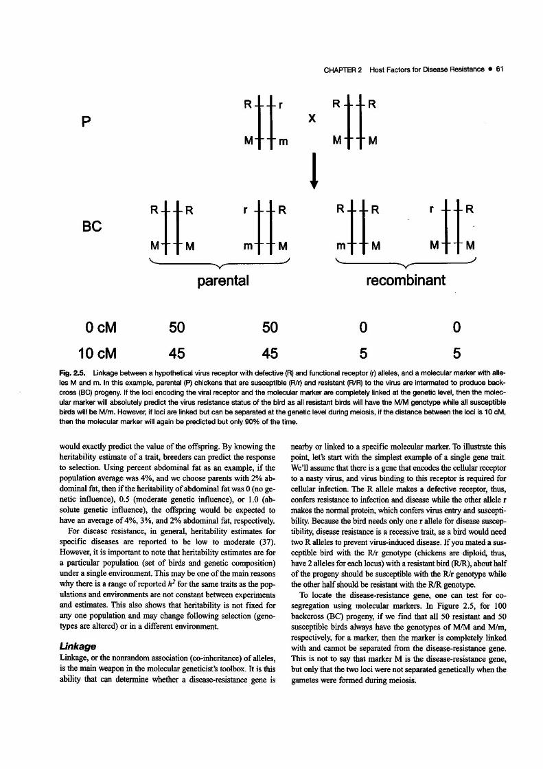
'"c_____----~.)- v-
p
Be
CHAPTER 2 Host Factors for Disease Resistance • 61
:tt x :tt:!
:tt: ~tt: :tt: ~tt:'"c____ _ ~/- ....."....
OcM
10cM
50
45
parental
50
45
recombinant
o5
o5
Fig. 2.5. Unkage between a hypothetical virus receptor with defective (R) and functional receptor (r) alleles, and a molecular marker with alle
les M and m. In this example, parental (p) chickens that are susceptible (RIr) and resistant (RIA) to the virus are intermated to produce backcross (Be) progeny. If the loci encoding the viral receptor and the molecular marker are completely linked at the genetic level, then the molec
ular marker will absolutely predict the virus resistance status of the bird as all resistant birds will have the MlM genotype while all susceptible
birds will be Mlm. However, if loci are linked but can be separated at the genetic level during meiosis, if the distance between the loci is 10 eM,then the molecular marker will again be predicted but only 90% of the time.
would exactly predict the value of the offspring. By knowing theheritability estimate of a trait, breeders can predict the responseto selection. Using percent abdominal fat as an example, if thepopulation average was 4%, and we choose parents with 2% abdominal fat, then ifthe heritability ofabdominal fat was 0 (no genetic influence), 0.5 (moderate genetic influence), or 1.0 (absolute genetic influence), the offspring would be expected tohave an average of4%, 3%, and 2% abdominal fat, respectively.
For disease resistance, in general, heritability estimates forspecific diseases are reported to be low to moderate (37).However, it is important to note that heritability estimates are fora particular population (set of birds and genetic composition)under a single environment. This may be one ofthe main reasonswhy there is a range of reported h1 for the same traits as the populations and environments are not constant between experimentsand estimates. This also shows that heritability is not fixed forany one population and may change following selection (genotypes are altered) or in a different environment.
UnkageLinkage, or the nonrandom association (co-inheritance) of alleles,is the main weapon in the molecular geneticist's toolbox. It is this
ability that can determine whether a disease-resistance gene is
nearby or linked to a specific molecular marker. To illustrate thispoint, let's start with the simplest example of a single gene trait.We'll assume that there is a gene that encodes the cellular receptorto a nasty virus, and virus binding to this receptor is required forcellular infection. The R allele makes a defective receptor, thus,confers resistance to infection and disease while the other allele rmakes the normal protein, which confers virus entry and susceptibility. Because the bird needs only one r allele for disease susceptibility, disease resistance is a recessive trait, as a bird would needtwo R alleles to prevent virus-induced disease. If you mated a susceptible bird with the Rir genotype (chickens are diploid, thus,have 2 alleles for each locus) with a resistant bird (RJR), about halfof the progeny should be susceptible with the Rlr genotype whilethe other half should be resistant with the RJR genotype.
To locate the disease-resistance gene, one can test for cosegregation using molecular markers. In Figure 2.5, for 100backcross (BC) progeny, if we find that aliSO resistant and 50susceptible birds always have the genotypes of MIM and Mlm,respectively, for a marker, then the marker is completely linkedwith and cannot be separated from the disease-resistance gene.This is not to say that marker M is the disease-resistance gene,but only that the two loci were not separated genetically when the
gametes were formed during meiosis.

62 • Diseases of Poultry
This also demonstrates that alleles M and R, and alleles m andr, are in linkage disequilibrium (LD). If the marker locus and disease resistance gene were unlinked, then the alleles should randomly segregate. But this is not the case as M occurs more frequently than is expected, and in this case, the marker allele ispredictive of the disease resistance gene allele.
But what ifthe two loci are not absolutely linked? Imagine thatof the 50 disease resistant birds, 45 had the MlM genotype and 5had the M/m genotype. Likewise, of the 50 susceptible birds, 45had the M/m genotype while 5 had the MIM genotype. Havingthe M allele is still a very good indicator ofdisease resistance (orvice versa with the m allele), but it is not 100% accurate. In thiscase, we can detennine the linkage of the two loci by quantifyingthe percentage of recombinant alleles (non-parental ones-e.g.,R and m or r and M). In this example, there are 10 recombinantprogeny out of 100, which means that the disease resistance geneand the marker are 10 centiMorgan (cM) away. The genetic tenncM reflects the number of recombination events between two locidetected in 100 progeny. In other words, the extent of linkage between these two loci is detennined by the amount of genetic recombination that occurs during meiosis, which can be determined by measuring how often particular alleles within eachlocus are inherited together.
To make the situation even more difficult and to put into perspective the challenge of finding genes for complex disease resistance, imagine now that each disease resistance gene accountsfor only a small percent (2-10010) of the total observed variationin the segregating population, as is typical for most diseases.Furthennore, not all Rlr birds are susceptible as the r allele maynot always be expressed at sufficient levels to allow virus entry.Or not all the birds get challenged with nasty virus as natural exposure is being used. While daunting, these challenges can beovercome with proper and controlled experiments, the use of molecular markers that encompass the entire chicken genome, manyprogeny to give more statistical power, accurate phenotypes, andbiometrical analyses.
Experimental Approaches forMolecular GeneticsThe development ofgenetic maps based on molecular markers defined the birth of genomics. No longer were scientists limited toassaying for genetic effects using markers at undefined locations.Now it was possible to systematically query the entire genome.This ability also changed the way experiments were primarilyconducted. Rather than using the reductionist approach of formulating a hypothesis about a specific gene and then testing it, genomics is a discovery-driven field where the end results allow definition of the next series of experiments. The genomics approachis growing in influence and is being reinforced and strengthenedwith whole genome sequences and high-throughput technologiesthat deliver quick, accurate, and low-cost data points.
Candidate GenesIn the preceding subchapter, a nwnber of genes and genes products have been identified that modulate the immune response.
Many of these immune function genes have or may be good candidates for conferring disease resistance. Consequently, priorknowledge to identify candidate genes for specific disease resistance is a valid method and often the best f'trst choice for testingfor a genetic effect. This approach requires a population that issegregating for disease or disease-associated traits, and an infonnative DNA polymorphism within or close to the gene of interest.
Although a gene and its gene product may be a critical component ofthe immune response and disease resistance, it is possiblethat there is no variation or genetic effect attributable to the genein a specific population. Consequently, the first requirement is toidentify polymorphisms (structural variations in the DNA) andalleles of the gene that are segregating within the population.Furthennore, evidence for a genetic effect does not prove causation by that gene but may be a result of linkage between thecausal gene and the analyzed gene. Finally, only that specificgene and genomic region is being screened for genetic effects,and no infonnation is generated about other genomic regions thatmay also influence resistance to the disease.
The MHC represents a unique situation and opportunity. Dueto its importance in many diseases, chicken lines have been developed that vary only in the MHC or B locus (1, 5, 40). Thus,these lines have the identical genetic background and carry different and unique MHC haplotypes. So, rather than using a genetic marker to the MHC and asking whether it is associated withdisease resistance, these "B congenic" strains can be used toquickly screen for MHC influences on disease resistance by challenging each line with the pathogen of interest and monitoringdisease and disease traits. Furthermore, due to the inbred natureof each line, there is less genetic variation that has to be accounted for, which translates into fewer chickens needing to bemeasured to detect genetic associations with traits.
Whole Genome ScansWith availability of the chicken molecular genetic map, it is possible to screen the entire chicken genome for disease-resistancegenes. The general approach is shown in Figure 2.6. In brief,what is called a resource population is identified or producedwhere birds segregate for disease resistance. Some or all of theanimals are measured for disease or disease-associated traits.Likewise, the same animals are genotyped for molecular markersspaced evenly throughout the entire chicken genome. Statisticalanalyses are perfonned to see whether certain genomic regions(genotypes) are associated with disease resistance (phenotypes)and, if yes, then how much does each region account for. The regions that contain one or more disease resistance gene are knownas a quantitative trait loci or QTL.
Two approaches can be used in genome-wide QTL scans. Thedifference between the two lies in how the resource populationsare produced, which also influences the density and, consequently, number ofgenetic markers required.
Linkage AnalysisIn this approach, for each marker, one tests whether the inheritance ofalleles influences disease resistance in a defined population. This type of linkage is known as identity by descent (IBD).

CHAPTER 2 Host Factors for Disease Resistance _ 63
resource population
statistical analyses
Molecular Markers. The beginning of genomics was markedwith the development of genetic maps composed of molecularmarkers. Unlike classical markers, molecular markers are typically based on DNA. Using an assay to type DNA variation, aile·les can be determined for each individual, which allows for thedevelopment of linkage maps and association studies.
The most commonly used molecular markers are called mi·crosatellites and SNPs. Both of these types of markers rely onspecific and unique sequences and can be anchored to a sequencetagged site (STS). Consequently, both genetic and physical distance can be detennined. for any pair of these molecular markers.
Genomic ToolsThe field ofgenetics and genomics is heavily influenced by technological advances. Key among these developments are mole·cular markers, which allow for the generation of high·densitygenetic maps and the chicken genome sequence. In short, com·prehensive genetic maps and the genome sequence allow for thegenetic dissection of simplex and complex traits.
Association MappingIn association or LD mapping, a pedigreed population is not re·quired, which makes it attractive for use in the corrunercial environment as existing populations can be tested. The power of thisapproach is that it relies on historical recombination events togreatly minimize the extent of LD. And rather than following theinheritance of specific alleles (identify by descent), associationmapping analyzes genetic marker allele frequency or identity bystate (IBS). Thus, resistance birds should be highly enriched. fora specific marker allele while the other allele(s) is found in thesusceptible birds. This method has both advantages and disadvantages. On the positive side, given the tight linkage required todetect a disease-resistance gene, any genetic marker with a sig·nificant association will be relatively close, which means that itis almost immediately amenable to further verification and implementation in a breeding program. On the other hand, since LDis small, one requires a much larger number of genetic markersso that each region is screened.
Association mapping has not been attempted in poultry mainlydue to the lack of genetic markers, cost of genotyping, and nothaving estimates on LD in commercial populations. This situation though has and will continue to change. The identification ofnearly 3 million single nucleotide polymorphisms (SNPs) (91)and high-throughput genotyping platforms makes this approachmuch more feasible.
ber of QTL that explained I to 5% of the total phenotypic variation for each trait were identified. And the QTL in total explainedaround 75% of the genetic variance of each trait. This indicatesthat it should be possible to achieve a great understanding ofdis·ease resistance in the chicken, given powerful enough popula·tions (and funds to perfonn these tasks). Thus said, even in thepowerful mouse study, each QTL was only resolved to a 2 cM in·terval, which still encompasses 25-50 genes, illustrating thegreat challenge in going from a QTL region down to a verification of a single gene's effect.
genotypes
//
phenotypes
Parents that differ markedly in disease resistance are selected andmated to produce progeny, which favors the use of inbred or divergently selected experimental lines due to their genetic simplicity and defined disease status. In initial QTL scans, the mating structures are often backcross (BC) or F2 populations. Due tothe limited nwnber of generations and recombination events thatcan erode the amount of LD, marker spacing can be relativelyhigh at 20-40 eM apart, which reduces the number and cost ofmarkers that need to be genotyped.
To validate and fine·map a QTL, additional generations are required to reduce LD and break apart distant marker-disease genelinkages through recombination, leaving only markers that aretightly linked to the disease resistance gene. A popular approach,known as advanced intercross lines (AILs), is to intennate progenyto get F3, F4, Fs,etc. (28). With each passing generation, the extentofLD grows smaller, which increases QTL resolving power.
Although simple in concept, the actual execution of the experiment entails a number offactors. Probably the biggest factor thatone can control is the number of progeny. The ability to detectQTL with smaller effects increases with more progeny. From apractical standpoint, this means at least 200 birds, if not 1000 ormore. Nonnally, the generation and measurements of resourcepopulations is the rate·limiting step.
A typical result is that a few to 15 QTL are revealed. These areclassified as suggestive or significant based on statistical analysesthat are corrected for the large number ofmultiple tests. The reason for including suggestive QTL at this point is that subsequentstudies are usually designed to validate potential QTL detected inthe fIrst-phase studies. Furthennore, it allows for comparisonsacross related studies conducted under varied conditions.
How robust is this experimental approach? No one reallyknows as the true architecture of anyone complex trait has notbeen completely deciphered. In the most advanced study to date(86), after examining 101 traits in 1904 F50 mice with 13,549 ge·netic markers, there was a surprising consistency in genetic ar·chitecture across all traits. For most traits, a relatively large num-
Fig. 2.6. General scheme for identifying genes or genomic regionsassociated with phenotypes. Using a resource population that is seg·regatlng for measurable traits, statistical analyses are used to associate genetic variation with the observed phenotypic variation.

64 • Diseases of Poultry
Microsatellites are sequence stretches that contain repeat unitsof I to 6 bases, e.g., CACACACACACACACA or (CA)s. Thenumber of repeat units often varies among individuals.Consequently, PCR primers that amplify this region will produceproducts that differ in size. The use of automatic DNA sequencers and fluorescently labeled PCR primers allows for therapid identification of each allele. On average, there is one microsatellite for every 75 kb in the chicken genome. In the chickengenome, I cM ofgenetic distance equates to -250 kb of physicaldistance, although this varies substantially across chromosomes.Therefore, there could be several informative and multi-allelemicrosatellites for every cM in the chicken genome.
SNPs, or single nucleotide polymorphisms, are sequence variants at a specific base in the DNA sequence. In theory, SNPsshould have 4 aUeles as there are 4 bases (A, C, G, and T).However, most SNPs have only 2 alleles and are often referred toas bi-allelic markers. SNPs are much more frequent than othermolecular markers with -5 found for every kb of chicken sequence. The presence of only 2 alleles greatly simplifies geneticanalyses, which makes platfonns based on this type of marker thecurrent method of choice for large~scale studies. On the otherhand, SNPs are not as informative as microsatellites as they arelimited to 2 alleles while microsatellites can have several.
Chicken GenomeAssembly. The chicken genome contains 38 autosomes and the Z and W sex chromosomes. In 2002, the NIHfunded the sequencing of the entire chicken genome, which wasreleased in 2004. To generate the draft genome sequence assembly, a framework was built using the genetic map as its foundation. Layered on this was a physical map derived from 180,000BAC (large insert) clones ordered through restriction enzymeanalyses. Cormections between the genetic and physical mapswere made through genetic markers located on individual BACclones. Finally, the whole-genome shotgun sequence was assembled into contigs and located onto the framework through common sequences; the BAC clones had their ends sequenced. As aresult, the initial assembly contained 1.05 Gb of assembled sequence of which 933 Mb were localized to specific chromosomes; the haploid content of the chicken genome is -1.2 Gb.Each base was sequenced -6.6 times on average, which is an indicator of genome coverage.
Like all other sequences including the human, there were gapsand assembly errors. In 2006, a second build that incorporatedmore genetic markers and sequence was generated. Additionalsequencing of targeted regions is still under way.
Useful websites on the chicken genome can be found at:
NCBI: http://www.ncbi.nlm.nih.gov/genome/guidelchickenlUCSC: http://genome.ucsc.edulcgi-binlhgGatewayEnsembl: http://www.ensembl.orglGallus....gallus/index.html
Functional GenomicsAs described previously, genetics relies on statistical associationof genotypic variation with phenotypic variation to identify genomic regions and detennine how much variation each region
contributes. Consequently, the ability to identify a disease resistance gene relies on statistical power and probabilities. Currently,this ability translates into QTL being localized to 5 or 20 cM intervals, at best. Even the best mapping studies in mice, with verylarge populations and dense marker placement, can only place aQTL of moderate effect to a 2 cM interval. This means that it willbe extremely difficult to resolve the location of a QTL down tothe single gene level. As the chicken genome contains 400 kb percM and 5 or more genes, on average for the larger chromosomes(43), even a 2 cM interval contains a lot of DNA sequence andcandidate genes.
To complement this genetic approach, there are a number oftools that query at the RNA, protein, or metabolite level. Thesefunctional genomic tools strive to identify components that varybetween two or more states, for example, gene transcripts that aredifferentially expressed between disease resistant and susceptiblechickens. The hope is that by identifying these molecules, genefunction and biological pathways can be ascribed to every gene.
Functional genomic assays identify gene transcripts, proteins,or other molecules, which combined with genome-wide QTLscans can reveal positional candidate genes. This integrative approach will be described in more detail for Marek's disease re~
sistance. Below are a few enabling technologies, which can alsobe applied in a number of ways beyond genetics.
DNA MicroarraysDuring the generation of whole genome sequences, it became obvious to scientists that there was a huge challenge to study all thegenes and gene products that would be identified. Furthennore, itwas clear that the reductionist approach of studying one gene orprotein at a time would not be sufficient to meet this requirement.
In one of the key technological developments, Pat Brown andcolleagues developed DNA microarrays (80). For many years,scientists had used hybridization, which detects complementarysequences for DNA and RNA analysis of specific sequencesusing labeled probes. In an elegant extension of this concept, allthe probe sequences instead of being individually labeled werearrayed onto a microscope slide and the total mRNA populationof the tested individual was labeled. As a consequence, the relative expression of thousands ofgenes in a single sample could besimultaneously measured in one step.
Within a relatively short period of time, this technology wasadapted for all species that had substantial RNA and DNA sequence infonnation or resources. With respect to chicken,Affymetrix presently makes the most comprehensive microarraywith coverage extending to all the -28,000 genes predicted in thefirst genome assembly. In addition, there are probes sets forgenes to 17 different avian viruses including Marek's diseasevirus, Newcastle disease virus, and avian influenza, which allowsthis tool to be used for veterinary diagnostics in the detection ofthese viruses.
DNA microarray experiments generate a list of genes that areexpressed more or less between two or more samples. For example, genes A, B, and C may be expressed more and genes X, Y, andZ less in the thymus of 5 day old resistant birds compared to agematched susceptible birds. It is important to note that because
gene expression (RNA) is being measured, unlike DNA, it canvary depending on the bird, tissue, time point, and other influences. Because of this natural biological variation, even for agiven set ofconditions, it is critical that several replicates are conducted. Unfortunately, at the moment, this is a relatively expensive procedure as each sample can cost $500 or more to process,which when combined with more variables (e.g., several tissues,more time points) inflates the total experiment cost quickly.
The list of genes can be further analyzed to provide a higherorder understanding. For example, if all the genes belonging to aspecific biological or functional class are elevated in the resistantbird compared to the susceptible bird, then most likely this pathway is involved in disease resistance. Tools to better analyze andextend the biological meaning of array results is an area of intense study.
With the success of this highly parallel process, it was quicklyadopted to many other molecules or situations. There are nowprotein arrays, tissue arrays, metabolite arrays, with the list goingon and on. Undoubtedly, many of these techniques will becomeviable and implemented in poultry in the near future. For reviewson diagnostic applications in animals, see references 33, 81, 89.
ProteomicsModerate to large scale global screening at the protein level isalso available today. For identification of proteins in a complexmixture, mass spectrometry is the technique of choice (29).'JYpically, protein samples are fractioned to reduce the complexity; then peptides are analyzed by the mass spectrometer; and finally the data sets are analyzed to deduce the identity of proteins. Although simple in concept, analysis of proteins is muchmore challenging than either DNA or RNA because for eachgene, there are many proteins with varying post-translationalmodifications, and the concentration of proteins encompassesmany orders of magnitude. Consequently, it is fairly simple toidentify the abundant proteins but difficult for the ones in lowquantity. And like DNA microarrays, cost can be a serious issue,which partially explains the minimal usage of this techniquethus far in poultry research, though the situation is graduallychanging (20).
Besides identifying individual proteins, it is possible to screenfor protein-protein interactions. The classic method, known as thetwo-hybrid screen (34, 35), takes advantage of the ability to reconstitute the activity ofa transcription factor that is in two parts.By fusing a cDNA (prey) library to the activation domain (AD)of a transcriptional activator, it is possible to identify proteinsthat interact with bait (protein of interest) that is fused with theDNA-binding domain (BD). As the AD and BD do not need to bephysically connected to promote transcription, if two proteins interact, a reporter gene is expressed ifAD and BD are brought intoclose proximity of each other. Due to the nature of the assay, allinteractions must be independently confirmed to eliminate falsepositives.
Higher order interactions can be revealed with gentle cell lysisand immunoprecipitation of a protein complex using antibodiesdirected against one member. The identity of the other interact
ing proteins can be quickly revealed through mass spectrometry.
CHAPTER 2 Host Factors for Disease Resistance • 65
Genetic Resistance to SpecificDiseasesAs discussed previously, one generally needs to first demonstrateheritability ofresistance to disease to demonstrate a genetic component. Although differences for disease resistance betweenchicken genetic lines may suggest a genetic basis, they are insufficient to prove it. Combined with the need in molecular geneticsfor controlled disease challenges and marker associations, thenumber of studies conducted to date is limited, especially forgenome-wide scans. There is, however, evidence for genetic control of poultry diseases caused by a wide range of pathogens, in
cluding viruses, bacteria and parasites.
Avian LeukosisAs described in Chapter 15, avian leukosis viruses (ALV) are agroup of retroviruses that can induce tumors. ALVs are classifiedinto various subgroups based on virus-specific cellular receptorsand virus envelope glycoproteins. The subgroups that infectchicken are A-E and J with all but subgroup J being exogenousALVs.
Genetic resistance to ALVs subgroups A-E is well defined andbased on specific cellular receptors. Since a single functioningreceptor allele is all that is required for virus entry, susceptibilityis dominant and the genetics is simple with only one locus involved. Molecular studies taking advantage of this fact have revealed not only the encoding gene but the basis for differencesbetween resistant and susceptible lines. Interestingly, althoughthe ALV subgroups are related and thought to have arisen from acommon ancestor, all the cellular receptors show no obvious sequence or structural similarity.
Resistance to ALV subgroup A is determined by the tva locusfound on chromosome 28, and encodes a protein a member ofthelow-density lipoprotein receptor (LDLR) family of unknownfunction (10, 30). There have been two alleles identified that confer resistance. The tva allele contains a single nucleotide mutation that results in a protein with very low binding afTmity to theALV subgroup A envelop while the other resistance allele, tva"2,has a 4-nucleotide insertion near the beginning of the coding sequencing, which results in an altered protein.
The tve locus, which confers resistance to ALV subgroup C, is""1 cM from tva (31). This receptor shows homology to butyrophilins, a member of the immunoglobulin superfamily. The resistant allele contains a premature stop codon and, thus, wouldnot produce a complete and functioning receptor (32).
Resistance toALV subgroups B, D, and E (endogenous) are allcontrolled by the tvb locus (2), which is located on chromosome22 (82). There are several reported alleles for this receptor, whichis related to the tumor necrosis factor receptor (TNFR) family. Asingle nucleotide change that generates a premature stop codon,results in a non-functioning receptor and resistance to all threesubgroups. The tvITI or wild-type allele is susceptible to all threesubgroups while the tvbS3 allele confers resistance to subgroup Eas the result of a different single nucleotide mutation (53).Molecular tests that can determine the tvb genotype have beenreported (97).
66 • Diseases of Poultry
Although the receptor for ALV subgroup 1 has been identifiedand shown to be Na+lH+ exchanger I (NHEI) (22), no cellularresistance has been observed, which suggests that variants that donot allow for virus binding are lethal. Field reports showing differences between lines suggest a genetic basis for differences inresistance to the myeloid leukosis.
Marek's DiseaseChickens resistant to Marek's disease (MD) are those that fail todevelop characteristic symptoms upon exposure to MDV (seeChapter 15). Genetic differences in resistance to fowl paralysis,assumed to be MD, have been reported for 60 years (3).Resistance to MD is complex and controlled by multiple genes orQTLs (6).
The best understood mechanism for the involvement ofgeneticresistance to MD involves the major histocompatibility complex(MHC) or, as it is known in the chicken, the B complex. TheMHC contains three tightly linked regions known as B~F (classI), B-G (class II), and B-L (class IV), which control cell surfaceantigens. The B-G locus is expressed in erythrocytes, which enables convenient typing of blood groups. By measuring the allelic frequency of specific blood groups, it has been observedthat certain B alleles are associated with resistance or susceptibility. Chickens with the B21 allele have been found to be moreresistant than those with other B haplotypes (4, 8). (Haplotyperefers to a set of alleles that is frequently co-inherited.) Otherstudies have allowed for the relative ranking of the other B alleles: moderate resistance, B2,!J6, B14; susceptibility, BI, 8 3, B5,B13, B15, B19, B27 (67) The MHC also influences vaccinal immunity as some haplotypes develop better protection with vaccinesofone serotype than of a different serotype (7, 9).
In addition to the MHC, chromosome 16 contains another setof class I and class II MHC genes that is called the Rfp-Y locus(73). Work by Wakenell (90) using data obtained from commercial chickens challenged with MDV suggests an association withMD resistance. However, results from matings with experimentallines indicate that the Rfp-Y genes do not influence any MD associated trait in these genetic backgrounds (88). These conflicting results further demonstrate the complexity of genetic resistance to MD, and the probable influence of genetic background.
Besides the MHC, other genetic factors have a major influenceon resistance to MD. For example, lines 6 and 7 chickens sharethe same B haplotype, B2, yet are MD resistant and susceptible,respectively. Due to the availability of these characterized linesand their genetic simplicity, genome-wide QTL scans have beenconducted. In one study, unvaccinated F2 progeny were challenged with MDV and measured for MD as well as a variety ofMD-associated traits such as viral titer, munber of tumors, andlength of survival (87. 92). Using genetic markers spacedthroughout the chicken genome, 14 QTL (7 significant and 7suggestive) were discovered that explain one or more MDassociated traits. The QTL were of small to moderate effect asthey explained 2 to 10% of the variance, with additive gene substitution effects from 0.01 to 1.05 phenotypic standard deviations. Collectively, the QTL explained up to 75% of the geneticvariance. Interestingly, 10 of the 14 QTL displayed non~additive
gene action; 3 with overdominance (individuals that are heterozygous have a phenotype better adapted than either class ofhomozygous individual) and 7 were recessive, With multipletraits being measured, the QTL could be grouped. In the first set,3 of the QTL were associated almost exclusively with viremialevels while the remaining QTL could account for disease, survival, tumors, nerve enlargement, and other disease associatedtraits. This suggests that disease resistance occurs at least at twolevels: initial viral replication and cellular transformation, whichoccurs later. It also highlights the added value ofmeasuring several components as it may functionally separate a complex traitas well as provide clues on positional candidate genes.
In the second study, Bumstead (17) used a (6 x 7) x 7 backcross population and measured MDV replication by quantitativepolymerase chain reaction (qPCR). A single large effect QTL onchromosome 1 was identified, which accounted for approximately half of the viral replication differences between the twoparental lines. This result also supports the relationship betweenvirus titers and disease resistance.
As described previously, going from a QTL to identification ofthe underlying causative genes is a tremendous challenge, even inmodel organisms. Consequently, additional functional genomicapproaches have been incorporated to give further screens. Thefirst approach utilizes DNA microarrays to profile transcripts thatare differentially expressed between MD resistant or susceptiblelines. Gene profiling has been conducted to identify differentiallyexpressed genes between lines 6 and 7 after MDV challenge (61),among B (MHC) congenic lines of chicken following inoculationwith different MD vaccines, and in chicken embryo fibroblasts(CEF) infected with MDV (74). Analyses of these experimentshave identified a number of genes and pathways that are consistently associated with either MD resistance or MDV infection.More importantly, the results suggest that chickens with immunesystems that are more stimulated by MDV infection are more susceptible. Initially, this seems counterintuitive but upon further reflection, MDV is thought to only infect activated lymphocytesand, thus, chickens with immune systems that are more responsivemay present more targets for MDV to infect and later transform.
The second approach uses the two-hybrid assay to identifyhost proteins that interact with specific MDV proteins. Thus far,9 MDV-chicken protein-protein interactions have been con:ftrmed by an in vitro binding assay. which demonstrates that theinteractions are direct and specific, and do not require other intermediary factors (e.g., yeast proteins) to be involved (76).
In both approaches, a finite number ofcandidate genes are obtained, which can be further evaluated for a genetic influence. Forexample. having confirmed the MDV SORF2-chicken growthhormone (GH) interaction, the GH gene (GHI) was evaluated asa candidate gene for MD resistance. GH1 variation was significantly associated (P<O.Ol) with a number of MD-associatedtraits in MHC B2/B15 chicks from commercial White Leghornlines (62). Furthermore, DNA microarray results indicate thatGH is differentially expressed between MD resistant (line 6) andsusceptible (line 7) chicks following MDV challenge (61). Thus,the combined results ofa specific MDV-chicken protein interaction, differential expression of GH between MD resistant and

susceptible chickens, and association of GHl with MD diseaserelated traits and selected lines for MD resistance, all stronglysuggest that GHl is a MD resistance gene (62). Using the samestrategy, LY6E [lymphocyte complex 6, locus E, aka, stem cellantigen 2 (SCA2) and thymic shared antigen I (TSAI)] (63) andBLB, the gene for MHC class II 13 chain (76), meet the samecriteria.
SalmonellosisControl of salmonellosis presents special challenges to the poul·try industry. As reviewed in detail elsewhere in this book(Chapter 16), some species of Salmonella bacteria are highlypathogenic and contagious in chickens, while other species causelittle response in the host birds, which can then become asymptomatic carriers. Because they are maintained in the productionflocks, birds with subclinical salmonellosis can transmit zoonoticbacteria into the human food chain. Chicks infected withSalmonella immediately after hatch can be persistently colonizedto maturity, when the bacteria are shed vertically to infect tableor hatching eggs, or horizontally to infect other hens (36).
Heritability estimates of various parameters ofSalmonella response indicate that genetic selection to improve resistance tosalmonellosis is feasible. The heritability of chick mortality afterSalmonella challenge was estimated at 0.14 and 0.62 for sire anddam components, respectively (11). Estimated heritability ofresistance to cecal carrier state, measured by eruiclunent culture, inlaying hens was 0.20 (13). Heritabilities of number of bacteriapersisting in internal organs ranged from 0.02 to 0.29 (39).Estimated heritability ofantibody response to Salmonella rangedwidely, from 0.03 to 0.26 (11, 52). Measuring antibody responseis less expensive and laborious than measuring bacterial colo·nization and because vaccine antibody has a negative genetic correlation with cecal colonization, vaccine antibody is a useful bio·marker to improve resistance to colonization (50).
Many genes are associated with genetic control of response toSalmonella species. The individual effect of most genes on thedisease phenotypic variation is relatively small, often only 3 to5% of the total variation, which is consistent with complex control of the disease by many genes. hnportant factors, such aschicken genetic line, population structure, defmition of response(antibody, mortality, systemic or enteric colonization), andserovar of Salmonella differed among studies, resulting in somevariation in results. However, a generally consistent picture ofgenetic control has emerged.
The strategy of comparative genomics was very effectivelyused to identify genes controlling salmonellosis resistance inchickens because response to Salmonella was previously studiedin detail in the mouse, a model organism. Chicken homologs ofmajor loci controlling natural resistance ofmice to infection withS. typhimurium were examined as candidate genes. Together,NRAMPl (natural resistance·associated macrophage protein I,now called SLCI1AI for solute carrier family 11 member 1) andTNC (a locus closely linked to LPS, now known as TLR4, whichbinds lipopolysaccharide, a major component of Gram·negativebacteria membranes) accounted for 33% ofthe differential resistance in Salmonella-induced mortality in a backcross population
CHAPTER 2 Host Factors for Disease Resistance • 67
of inbred lines (44). Subsequently, the NRAMP1 association wasconfirmed for several parameters of host response to Salmonellain other chicken populations (12, 54, 64), as was the TLR4 asso-ciation (12, 57, 68).
The success of the comparative genomics approach provided astarting point to select additional candidate genes to test. Somegenes were positional candidates based upon genomic position,such as CD28 and VILl in the NRAMPl region (38). The CD28gene was associated with enteric Salmonella infection (68), andVILI with visceral infection (39).
Other candidate genes were selected based upon involvementin pathways that are hypothesized to be important in host re
sponse to Salmonella. The product of the MD2 gene interactswith the TLR4 receptor on the cell surface, and SNPs in MD2 areassociated with persistence of Salmonella colonization in thececum (68). The MHC, because of its crucial role in antigen processing and presentation, was investigated. In a series of 12 B·complex congenic lines, line differences occurred in morbidityand mortality after Salmonella challenge (26). The MHC class Iwas associated with resistance to Salmonella colonization in thespleen in an experimental cross (66). Genes in apoptotic pathways include CASPI and lAPI. A CAS?1 SNP was associatedwith Salmonella persistence in the spleen and cecwn in an experimental cross (65) and in the liver and cecum in commercialbroilers (54). The IAPl gene was associated with spleen (65) andcecum bacterial load (54). The genomic region containing genesencoding a newly identified family ofantimicrobial peptides, thegallinacins, was associated with Salmonella vaccine antibody response (41).
Although the design of most candidate gene experiments doesnot preclude that the causal gene could be a nearby gene ratherthan the specific one studied, having supporting Jines ofevidencesuch as confinnation in independent populations, with QTLscans, by gene expression data, or from comparative genomics,helps to add confidence in the detected gene·resistance associa·tions. Studies on gene expression in Salmonella challenged versus unchallenged, or resistant versus susceptible animals, are re·vealing differential expression in genes that may be active inpathways controlling resistance (23, 49, 98).
Genome-scan studies have identified QTL regions controllingSalmonella resistance. In analysis of a backcross population ofresistant and susceptible parental inbred lines (lines 6, and 151,respectively), genotyping of animals with extreme bacterialcounts in spleen showed significant linkage between markers onchromosome 5 and the resistance trait (70). Fine-mapping of thechromosomal location of the QTL fOWld a very strong effect, accOWlting for 50% of the parental variation difference, in the re·gion near the CKB and DNCHI genes. The specific gene associ·ated with resistance is not positively identified, however, and theSalmonella resistance QTL in this region is considered a novellocus named SALJ. The distance of almost 50 cM between SALIand the Salmonella resistance QTL linked to marker ADL0298(51) makes them unlikely to represent the same locus althoughboth influenced bacterial colonization in the spleen.
Another genome scan for Salmonella resistance QTL used F2
and Be populations formed from line N and 6" and assessment
68 • Diseases of Poultry
of cloacal and cecal carrier state (84). This study confirmed anassociation of the SAL} region with enteric carrier state.Additionally, new QTL regions were identified at significant orsuggestive levels on chromosomes I, 2, 5, and 16, with somehaving effects as large as 37.5% of the phenotypic variance (84).The QTL on chromosome 16 lies in the MHC, although the twoinbred lines are considered to have the same MHC haplotype.The QTL on chromosome 5 maps near TGFB3, which was shownin independent populations to be associated with spleen (54) andcecal colonization (69).
ColibacillosisEscherichia coli is an important pathogen of poultry, both as acause of colibacillosis in production flocks and as a potentialfood-safety pathogen (Chapter 18). Elucidation of the geneticcontrol of resistance to E. coli has primarily been approachedthrough analysis oflines that were divergently selected for circulating antibody level to E. coli vaccine in young birds. The principle underlying this approach is that if vaccine antibody is related to resistance and if resistance is under genetic control, thenthe genetic selection for antibody response will generate linesthat differ in resistance genetics. These lines are then a resourceto identify the specific genetic differences controlling resistance.
Divergent selection for E. coli vaccine antibody response successfully modulated mortality rate, and the immune response topathogenic E. coli and several other antigens (42). Genetic control of E. coli response was studied in an F2 population generatedfrom crossing the divergently selected lines. Molecular probeswere used for three candidate genes in the MHC region: B-1'; Bo and TAP2 (94). Each probe revealed genetic variation in thepopulation (assessed by RFLP bands), and each gene had multiple bands associated with antibody traits. Also, analyzing the effect of all probes simultaneously revealed more significant effects than analyzing each separately.
Genome scans of resource populations produced from thesame divergently selected lines revealed QTL for antibody re
sponse and mortality. The initial low-density scan with 25 markers found three markers associated with antibody response to E.coli, Newcastle disease virus, and/or SRBC and one with mortality (93). In a more extensive study, PI males derived from a crossof the divergently selected lines were mated with females of fourgenetic backgrounds (the Flo the two divergent lines, and a commercialline) to produce 1700 progeny that were immunized withE. coli and Salmonella enteritidis vaccine (95). Individuals withthe highest or lowest average antibody were selectively genotyped within each mating type and sire family. About 125 markers were heterozygous in each sire. Twelve markers were associated with QTL for E. coli antibody, and six of these were alsoassociated with S. enteritidis antibody, with two markers suggesting a Qn on chromosome 2. The experimental design, with siresmated to dams ofmultiple lines, also demonstrated that the effectof markers was modulated by the genetic backround.
CoccidiosisCoccidiosis is caused by several species of parasites of the genusEimeria, each of which has tropism for a different area of the
gastrointestinal tract (Chapter 7). The cost ofcontrol ofcoccidiosis by pharmaceutical means, continual emergence of drug-resistant strains, and profit loss due to reduced growth efficiencyall mandate the need for other approaches, such as genetic resistance, to control this disease. Population differences, and the ability to genetically select lines for divergent resistance/susceptibility, illustrate the feasibility of improvement in genetic resistanceto coccidiosis (15, 46, 77).
Blood group antigens comprise the major category ofgenes associated with coccidiosis resistance, with the wel1-characterizedB locus, or MHC, representing most of the reported associations.Several studies compared MHC (or B)-congenic Hnes, whichshare background genes but differ for the B locus region (21,58,79), or populations in which MHC haplotypes segregate withinline or differ among lines (14, 24, 72, 85). Collectively, thesestudies provide strong support for the MHC being a coccidiosisresistance locus. However, effects of specific MHC haplotypesgreatly vary, dependent upon factors such as Eimeria challengestrain and the specific trait used to characterize resistance (forexample, antibody, oocyst shedding, weight gain, severity of intestinal lesions). A study of birds that were recombinants between the B-F and B--G (class I and II, respectively) regions ofthe MHC suggested that the MHC class I region had the greaterrole in resistance (25). Although studies of MHC-congenic linesreveal a clear picture regarding the role of the specific MHC haplotypes studied. they do not shed light on what impact genomicvariation at other regions might have on resistance. Experimentsthat included variation in both the MHC haplotypes and the genetic background determined that both MHC and non-MHCgenes control resistance to coccidiosis (59, 60). Erythrocyte loci(identified serological1y) in addition to the B locus have been associated with resistance to coccidiosis, including Ea-A, Ea-E(46), Ea-C (47), and Ea-I (71).
An F2 cross ofcommercial broiler lines was used to conduct agenome scan for QTL associated with resistance to coccidiosiscaused by Eimeria maxima (99). Using 119 markers that gaveabout 80% genome coverage, a strong QTL for oocyst sheddingwas located on chromosome I.
Infectious Bursal DiseaseInfectious bursal disease virus (IBDV) causes acute infection anddepletion of B cells in the bursa and other organs, often resultingin severe and pennanent immunosuppression ofthe host (Chapter4). Mating of resistant and susceptible lines to form F 1> F2and BCpopulations demonstrated lBDV-induced mortality to be under thecontrol ofa fully or partially dominant autosomal resistance gene(18,19). Multiple studies have examined the MHC for associationwith IBDV resistance and detected no effect (18, 19.45). In onestudy, however, an MHC effect was detected on resistance parameters of specific antibody to IBDV and bursal histopathology (48).The differences among studies are likely to be a result of examination of different MHC haplotypes in varied genetic backgrounds. Collectively, studies on genetics oflBDV resistance suggest involvement of the MHC and at least one other, currentlyunidentified. autosomal gene. Global transcriptional profiling hasrevealed numerous genes that are differentially regulated between
lBDV resistant and susceptible lines, and suggests that resistancemay be mediated by more rapid inflammatory response and more
extensive p53-related apoptosis of target B cells, thus limiting
viral replication in resistant birds (78).
Future PerspectivesGenetic resistance to disease for anyone individual or line of
chickens is the overall outcome of a very complex set of signals
and responses with which the chicken and pathogen interact. The
ability for molecular genetics to unravel this biological complexity has been very impressive. Despite the rapid pace of advance
ments, there is a long way to go before scientists develop a de
tailed Wlderstanding of the molecular pathways, and poultrybreeders are able to transfer this information to improve their
commercial products.Poultry breeding companies have achieved enhanced disease
resistance in their flocks to many pathogens using traditional
methods. However, this is a laborious process that requires the
intentional exposure of individuals from elite lines to pathogens,and progresses rather slowly due to indirect selection on overall
livability. With the emergence of molecular genetics and the
field of genomics, there is a optimism that studies will be ableto identify genes and alleles that confer superior disease resist
ance. When applied to breeding, the information will provide for
the rapid and accurate improvement of commercial lines. In ap
plication, the poultry breeders do not necessarily rely on the
identification of the disease resistance gene itself, but may usemarkers in linkage disequilibrium to increase the frequency offavorable alleles. But as science has shown many times, increas
ing knowledge gives increasing power that can often be applied
in novel ways. So the ultimate goal should be to identify diseaseresistance genes and their pathways to reveal biological function
and pathways.
So what will hasten the advancement of knowledge? Withhigh-throughput platforms to determine genotypes, the rate
limiting step is in producing and measuring resource populations.
With respect to animal production, one solution would be to in
crease the partnerships between poultry breeding companies andmolecular geneticists, which allows for the leveraging of expert
ise and resources. This partnership also allows for scientists towork at the population level with all the diverse set of genes and
allele combinations. Beyond this, trait measurements will be critical. As DNA microarrays and proteomics have already shown,
breaking down a trait into specific components provides criticalinformation on biological processes that cannot be or are difficult
to obtain by traditional phenotypic measurements. Thus, veterinary medicine can make key contributions by refining trait meas
urements associated with disease and disease progression.
ReferencesI. Abplanalp, H. 1992. Inbred lines as genetic resources of chickens.
Poultry Science Reviews 4, 29--39.2. Adkins, H. B., 1. Brojatsch. and J. A. Young. 2000. Identification and
characterization of a shared TNFR-related receptor for subgroup B,
CHAPTER 2 Host Factors for Disease Resistance. 69
D, and E avian leukosis viruses reveal cysteine residues requiredspecifically for subgroup E viral entry. J Viro174, 3572-78.
3. Asmundson, V. S., and 1. Biely. 1932. Inheritance and resistance tofowl paralysis (neuro-Iymphomatosis gallinarum). 1. Differences insusceptibility. Canadian Journal ofResearch 6, 171-176.
4. Bacon, L. D. 1987. Influence of the major histocompatibility comple1\ on disease resistance and productivity. Poult Sci 66, 802-11.
5. Bacon, L. D., H. D. Hunt, and H. H. Cheng. 2000. A review of thedevelopment of chicken lines to resolve genes determining resistance to diseases. Poult Sci 79, 1082-93.
6. Bacon, L. D., H. D. Hunt, and H. H. Cheng. 2001. Genetic resistanceto Marek's disease. Curr Top Microbiollmmunol255. 121-41.
7. Bacon, L. D., and R. L. Witter. 1992. Influence oftwkey herpesvirusvaccination on the B-haplotype effect on Marek's disease resistancein 15.B-congenic chickens. Avian Dis 36, 378-85.
8. Bacon, L. D., and R. L. Witter. 1994. B haplotype influence on therelative efficacy of Marek's disease vaccines in commercial chickens. Poult Sci 73, 481-7.
9. Bacon, L. D., and R. L. Witter. 1994. Serotype specificity of Bhaplotype influence on the relalive efficacy of Marek's disease vaccines. Avian Dis 38, 65-71.
10. Bates, P., L. Rong, H. E. Varmus, 1. A. Young, and L. B. Crittenden.1998. Genetic mapping of the cloned subgroup A avian sarcoma andleukosis virus receptor gene 10 the TVA locus. J Virol 72, 2505-8.
II. Beaumont. C.• 1. Protais, 1. F. Guillot, P: Colin, K. Proux, N. Millet,and P. Pardon. 1999. Genetic resistance to mortality of day-oldchicks and carrier-state of hens after inoculation with Salomella enteritidis. Avian Pathology 28, 131-135.
12. Beaumont, C., 1. Protais, F. Pitel, G. Leveque, D. Malo, F. Lantier, F.Plisson-Petit, P. Colin, M. Protais, P. Le Roy, 1. M. Elsen, D. Milan,I. Lantier, A. Neau, G. Salval, and A. Vignal. 2003. Effect of twocandidate genes on the Salmonella carrier state in fowl. Poult Sci 82,721--6.
13. Berthelot, E, C. Beaumont, E Mompart, O. Girard-Santosuosso, P.Pardon, and M. Duchet-Suchaux. 1998. Estimated heritability oftheresistance to cecal carrier state ofSalmonella enteritidis in chickens.Poult Sci 77, 797-801.
14. Brake, D. A., C. H. Fedor, B. W. Werner, T. 1. Miller, R. L. Taylor, Jr.,and R. A. Clare. 1997. Characterization of immune response toEimeria tenella antigens in a natural immunity model with hostswhich differ serologically at the B locus of the major histocompatibility complex. Infect lmmun 65, 1204-10.
IS. Bumstead, J. M., N. Bumstead, L. Rothwell, and F. M. Tomley. 1995.Comparison of immune responses in inbred lines of chickens toEimeria maxima and Eimeria tenella. Parasitology III (Pt. 2),143-51.
16. Bumstead, N. 1998. Genetic resistance to avian viruses. Rev SciTe<:h 17, 249-55.
17. Bumstead, N. 1998. Genomic mapping of resistance to Marek's disease. Avian Pathology 27, S78-S81.
18. Bumstead, N., M. B. Huggins, and 1. K. Cook. 1989. Genetic differences in susceptibility 10 a mixture of avian infectious bronchitisvirus and Escherichia coli. Br Poult Sci 30, 39-48.
19. Bumstead, N., R. L. Reece, and 1. K. Cook. 1993. Genetic differences in susceptibility of chicken lines to infection with infectiousbursa1 disease virus. Poult Sci 72, 403-10.
20. Burgess, S. C. 2004. Proteomics in the chicken: tools for understanding inunune responses 10 avian diseases. Poult Sci 83, 552-73.
21. Caron, L. A., H. Abplanalp, and R. L. Taylor, Jr. 1997. Resistance,susceptibility. and immunity 10 Eimeria tenella in major histocompatibility (B) comple1\ congenic lines. Poult Sci 76, 677-82.
70 • DIseases of Poultry
22. Chai, N. and P. Bates. 2006. Na+lH+ exchanger type I is a receptorfor pathogenic subgroup J avian leukosis virus. Prrx Natl Acad SciUSA 103,5531--6.
23. Cheeseman, J. H., M. G. Kaiser, C. Ciraci, P. Kaiser, and S. J.Lamont. 2006. Breed effect on early cytokine mRNA expression inspleen and cecum of chickens wilh and without Salmonella enteri·tidis infection. Dev Camp Immunol31, 52--60.
24. Clare, R. A., R. G. Strout, R. L. Taylor, Jr., W. M. Collins, and W. E.Briles. 1985. Major histocompatibility (B) complex effects on acquired immunity to cecal coccidiosis. Immunogenetics 22, 593-9.
25. Clare, R. A., R. L. Taylor, Jr., W E. Briles, and R. G. Strout. 1989.Characterization of resistance and immunity to Eimeria tenellaamong major histocompatibility complex B--FIB-G recombinanthosts. Poult Sci 68, 639-45.
26. Cotter, P. F., R. L. Taylor, Jr. and H. Abplanalp. 1998. B-complex associated immunity to Salmonella enteritidis challenge in congenicchickens. Poult Sci 77, 1846-51.
27. Crawford, R. D. 1990. Poultry Breeding and Genetics, Elsevier,Amsterdam, New York.
28. Darvasi, A., and M. Soller. 1995. Advanced intercross lines, an experimental population for fine genetic mapping. Genetics 141,1199-207.
29. Domon, 8., and R. Aebersold. 2006. Mass spectrometry and proteinanalysis. Science 312, 212-7.
30. Elleder, D., D. C. Melder, K. Trejbalova, J. Svoboda, and M. J.Federspiel. 2004. Two different molecular defects in the Tva receptor gene explain the resistance of two tvar lines of chickens to infection by subgroup A avian sarcoma and leukosis viruses. J Viral 78,13489-500.
31. Elleder, D., J. Plachy, J. Hejnar, J. Geryk, and J. Svoboda. 2004.Close linkage ofgenes encoding receptors for subgroups A and C ofavian sarcomalleucosis virus on chicken chromosome 28. AnimGenet 35,176-81.
32. Elleder, D., V. Stepanets, D. C. Melder, F. Senigl, J. Geryk, P. Pajer,J. Plachy, J. Hejnar, J. Svoboda, and M. J. Federspiel. 2005. The receptor for the subgroup C avian sarcoma and leukosis viruses, Tvc,is related to mammalian butyrophilins, members of the immunoglobulin superfamily. J Viro/ 79, 10408-19.
33. Feilotter, H. E. 2004. Microarrays in veterinary diagnostics. AnimHealth Res Rev 5, 249-55.
34. Fields, S. 2005. High-throughput two-hybrid analysis. The promiseand the peril. Febs J 272, 5391-9.
35. Fields, S., and O. Song. 1989. A novel genetic system to detectprotein-protein interactions. Nature 340, 245--{j.
36. Gast, R. K., and P. S. Holt. 1998. Persistence of Salmonella enteritidis from one day of age until maturity in experimentally infectedlayer chickens. Poult &i 77, 1759--62.
37. Gavora, J. S. 1990. Disease Genetics. In Poultry Breeding andGenetics (Crawford, R. D., ed.), 805-846. Elsevier SciencePublishers, 8. v., New York.
38. Girard-Santosuosso, 0., N. Bumstead, 1. Lantier, 1. Protais, P. Colin,J. F. Gui1!ot, C. Beaumont, D. Malo, and F. Lantier. 1997. Partialconservation of the mammalian NRAMPI syntenic group onchicken chromosome 7. Mamm Genome 8, 614--6.
39. Girard-Santosuosso, 0., F. Lantier, 1. Lantier, N. Bumstead, J. M.Elsen, and C. Beaumont. 2002. Heritability of susceptibility to Salmonella enteritidis infection in fowls and test ofthe role ofthe chromosome carrying the NRAMP1 gene. Genet Sel Evo134, 211-9.
40. Hala, K. 1987. Inbred lines of avian species. In Avian Immunology:Basis and Practice (Toivanen, A. and P. Toivanen, eds.), Vol. 11,85-99. CRC Press, Bacon Raton.
41. Hasenstein, J. R., G. Zhang, and S. J. Lamont. 2006. Analyses offivegallinacin genes and the Salmonella enterica serovar Enteritidis response in poultry. Infect Immun 74, 3375-80.
42. Heller, E. D., G. Leitner, A. Friedman, Z. Uni, M. Gutman, and A.Cahaner. 1992. Immunological parameters in meat-type chickenlines divergently selected by antibody response to Escherichia colivaccination. Vet Immunol Immunopathol 34, 159-72.
43. Hillier, L. w., W. Miller, E. Birney, W. Warren, R. C. Hardison, C. P.Ponting, P: Bolt, D. W Burt, M. A. Groenen, M. E. Delany, J. 8.Dodgson, A. T. ChinwaUa. P. F. Cliften, S. W. Clifton, K. D.De1ehaunty, C. Fronick, R. S. Fulton, T. A. Graves, C. Kretnitzki, D.Layman, V. Magrini, J. D. McPherson, T. L. Miner, P. Minx, W. E.Nash, M. N. Nhan. J. O. Nelson, L. G. Oddy, C. S. Pchl, J. RandallMaher, S. M. Smith, J. W. Wallis, S. P. Yang, M. N. Romanov, C. M.Rondelli, B. Paton, J. Smith, D. Morrice, L. Daniels, H. G. Tempest,1. Robertson, J. S. Masabanda. D. K. GriffIn, A. Vignal, V. Filion, L.Jacobbson, S. Kerje, L. Andersson, R. P. Crooijmans, J. Aerts, J. J.van der Poel, H. Ellegren, R. 8. Caldwell, S. J. Hubbard, D. V.Grafham, A. M. Kierzek, S. R. McLaren, I. M. Overton, H. Arakawa,
K. J. Beanie, Y. Bezzubov, P. E. Boardman, J. K. Bonfield, M. D.Croning, R. M. Davies, M. D. Francis, S. 1. Humphray, C. E. Scott,R. G. Taylor, C. Tickle, W. R. Brown, J. Rogers, J. M. Buerstedde, S.A. Wilson, 1. Stubbs, I. Ovcharenko, L. Gordon, S. Lucas, M. M.Miller, H. Inoko, T. Shiina, J. Kaufinan, 1. Salomonsen, K. Skjoedt,G. K. Wong, J. Wang, B. tiu, J. Wang, J. Yu, H. Yang, M. Nefedov, M.Koriabine, P. J. Dejong, 1. Goodstadt, C. Webber, N. 1. Dickens, I.Letunic, M. Suyama, D. Torrents, C. von Mering, et al. 2004.Sequence and comparative analysis of the chicken genome provideunique perspectives on vertebrate evolution. Nature 432, 695-716.
44. Hu, J., N. Bumstead, P. Barrow, G. Sebastiani, 1. Olien, K. Morgan,and D. Malo. 1997. Resistance to salmonellosis in the chicken islinked to NRAMPI and TNC. Genome Res 7, 693-704.
45. Hudson, J. C., E. 1. Hoerr, S. H. Parker, and S. J. Ewald. 2002.Quantitative measures of disease in broiler breeder chicks of differ·ent major histocompatibility complex genotypes after challengewith infectious bursal disease virus. Avian Dis 46, 581-92.
46. Johnson, L. W, and S. A. Edgar. 1984. Ea-A and Ea-E cellular antigen genes in Leghorn lines resistant and susceptible to acute cecalcoccidiosis. Poult Sci 63, 1695-704.
47. Johnson, L. W, and S. A. Edgar. 1986. Ea-B and Ea-C cellular antigen genes in Leghorn lines resistant and susceptible to acute cecalcoccidiosis. Poult Sci 65, 241-52.
48. Juul-Madsen, H. R., O. 1. Nielsen, T. Krogh-Maibom, C. M.Rontved, T. S. Dalgaard, N. Bumstead, and P. H. Jorgensen. 2002.Major histocompatibility complex-linked immune response ofyoung chickens vaccinated with an attenuated live infectious bursaldisease virus vaccine followed by an infection. Poult Sci 81,649-56.
49. Kaiser, M. G., J. H. Cheeseman, P: Kaiser, and S. J. Lamont. 2006.Cytokine expression in chicken peripheral blood mononuclear cellsafter in vitro exposure to Salmonella enterica serovar Enteritidis.Poultry Sci 85, 1907-1911.
50. Kaiser, M. G., N. Lakshmanan, T. Wing, and S. J. Lamont. 2002.Salmonella enterica serovar enteritidis burden in broiler breederchicks genetically associated with vaccine antibody response. AvianDis 46, 25-31.
51. Kaiser, M. G., and S. J. Lamont. 2002. Microsatellites linked toSalmonella enterica Serovar Enteritidis burden in spleen and cecalcontent of young Fl broiler-cross chicks. Poult Sci 81, 657--63.
52. Kaiser, M. G., T. Wing, A. Cahaner, and S. J. Lamont. 1997.Aviagen, 12th International Symposium on Current Problems inAvian Genetics, Prague, Czech Republic.
53. Klucking, S., H. B. Adkins, and 1. A. Young. 2002. Resistance to infection by subgroups B, 0, and E avian sarcoma and leukosis virusesis explained by a premature stop codon within a resistance allele ofthe tvb receptor gene. J Viro176, 7918-21.
54. Kramer, 1., M. Malek, and S. 1. Lamont. 2003. Association of twelvecandidate gene polymorphisms and response 10 challenge withSalmonella enteritidis in poultry. Anim Genet 34, 339-48.
55. Lamont, S. 1. 1998. Impact of genetics on disease resistance. Poult&i 77, 1111-8.
56. Lamont, S. 1. 1998. The chicken major histocompatibility complexand disease. Rev Sci Tech 17, 128-42.
57. Leveque, G., V. Forgena, S. Morroll, A. 1. Smith, N. Bumstead, P.Barrow, 1. C. Loredo-Osti, K. Morgan, and D. Malo. 2003. Allelicvariation in TLR4 is linked to susceptibility to Salmonella entericaserovar Typhimurium infection in chickens. Infect Immun 71,1116-24.
58. Lillehoj, H. S., M. C. Jenkins, and 1. D. Bacon. 1990. Effects ofmajor histocompatibility genes and antigen delivery on induction ofprotective mucosal immunity to E. acervulina following immuniza·tion with a recombinant merozoite antigen. Immunology 71, 127-32.
59. Lillehoj, H. S., M. C. Jenkins, 1. D. Bacon, R. H. Fetterer, and W. E.Briles. 1988. Eimeria acervulina: evaluation of the cellular and antibody responses to the recombinant coccidial antigens in B-congenicchickens. Exp Parasitol67, 148-58.
60. Lillehoj, H. S., M. D. Ruff, 1. D. Bacon, S. 1. Lamont, and T. K.Jeffers. 1989. Genetic control of immunity to Eimeria tenella.Interaction of MHC genes and non·MHC linked genes influenceslevels of disease susceptibility in chickens. Vet Immunol Immu
nopathol20, 135-48.61. Liu, H. C., H. H. Cheng, V. Tirunagaru, 1. Safer, and 1. Burnside.
2001. A strategy to identify positional candidate genes conferringMarek's disease resistance by integrating DNA microarrays and genetic mapping. Anim Genet 32,351-9.
62. Liu, H. C., H. J. Kung, I. E. Fulton,R. W. Morgan, and H. H. Cheng.2001. Growth hormone interacts with the Marek's disease virusSORF2 protein and is associated with disease resistance in chicken.Proc Natl Acad Sci USA 98, 9203--8.
63. Liu, H. C., M. Niikura, 1. E. Fullon, and H. H. Cheng. 2003. Identi·fication of chicken lymphocyte antigen 6 complex, locus E (LY6E,alias SCAl) as a putative Marek's disease resistance gene via a virushost protein interaction screen. Cytogenet Genome Res 102, 304-8.
64. Liu, w., M. G. Kaiser, and $. J. Lamont. 2003. Natural resistanceassociated macrophage protein I gene polymorphisms and responseto vaccine against or challenge with Salmonella enteritidis in youngchicks. Poult Sci 82, 259-66.
65. Liu, w., and S. 1. Lamont. 2003. Candidate gene approach: potentional association of caspase-I, inhibitor of apoptosis protein-I, andprosaposin gene polymorphisms with response to Salmonella enteritidis challenge or vaccination in young chicks. Anim Bjotechnol14,61-76.
66. Liu, w., M. M. Miller, and S. 1. Lamont. 2002.Association ofMHCclass I and class II gene polymorphisms with vaccine or challengeresponse to Salmonella enteritidis in young chicks. Immunogenetics54,582--90.
67. Longenecker, B. M., and T. R. Mosmann. 1981. Structure and properties of the major histocompatibility complex of the chicken.Speculations on the advantages and evolution of polymorphism.Immuoogenetics 13, 1-23.
68. Malek, M., 1. R. Hasenslein, and S. J. Lamont. 2004. Analysis ofchicken TLR4, CD28, MIF, MD.2, and LITAF genes in aSalmonella enteritidis resource population. Poult Sci 83, 544-9.
CHAPTER 2 Host Factors for Disease Resistance • 71
69. Malek, M., and S. 1. Lamont. 2003. Association of !NOS, TRAIL,TGF·beta2, TGF-beta3, and 19L genes with response to Salmonellaenteritidis in poultry. Genet Sel Eval 35 Suppl I, S99-1 II.
70. Mariani, P., P. A. Barrow, H. H. Cheng, M. M. Groenen, R. Negrini,and N. Bumstead. 2001. Localization to chicken chromosome 5 ofanovel locus detennining salmonellosis resistance. Immunogenetics53,786-91.
71. Martin, A., W. B. Gross, E. A. Dunnington, R. W. Briles, W. E.Briles, P. B. Siegel. 1986. Resistance to natural and controlled exposures to Eimeria tenella: genetic variation and alloantigen systems.Poult Sci 65, 1847-52.
72. Medarova, Z., W. E. Briles, and R. L. Taylor, Jr. 2003. Resistance,susceptibility, and immunity to cecal coccidiosis: effects ofB complex and alloantigen system 1. Poult Sci 82, I I13-7.
73. Miller, M. M., R. M. Goto, R. 1. Taylor, Jr., R. loorob, C. Auffi"ay,R. W. Briles, W. E. Briles, and S. E. Bloom. 1996. Assignment ofRfp-Y to the chicken major histocompatibility complexlNOR microchromosome and evidence for high.frequency recombination associated with the nucleolar organizer region. Proc Natl Acad Sci
USA 93,3958--62.74. Morgan, R. w.. L. Sofer, A. S. Anderson, E. L. Bemberg, 1. Cui, and
J. Burnside. 2001. Induction of host gene expression following infection of chicken embryo fibroblasts with oncogenic Marek's dis-ease virus. J Viro175, 533-9.
75. Muir, W. M., and S. E. Aggrey. 2003. Poultry Genetics, Breeding,and Biotechnology, CABI Publishing, Wallingford, Oxon, UK,Cambridge, MA.
76. Niikura, M., H. C. Liu, 1. B. Dodgson, and H. H. Cheng. 2004. Acomprehensive screen for chicken proteins that interact with proteins unique to virulent strains of Marek's disease virus. Poult Sci83, 1117-23.
77. Pinard·van der Laan, M. H., 1. 1. Monvoisin, P. Pery, N. Hamel, andM. Thomas. 1998. Comparison of outbred lines of chickens for resistance to experimental infection with coccidiosis (Eimeria tenella).PaultSci77,185-91.
78. Ruby, T., C. Whittaker, D. R. Withers, M. K. Chelbi-Alix, V. Morin,A. Oudin, 1. R. Young, and R. Zoorob. 2006. Transcriptional profil.ing reveals a possible role for the timing of the inflammatory response in determining susceptibility to a viral infection. J Virol 80,9207-16.
79. Ruff, M. D., and L. D. Bacon. 1989. Eimeriaacervulina and Eimeriatenella in 15.B-congenic White Leghorns. Poult Sci 68, 380-5.
80. Schena, M., D. Sha10n, R. W. Davis, and P. O. Brown. 1995.Quanlitative monitoring of gene expression patterns with a complementary DNA microarray. Science 270, 467-70.
81. Sclunitl, B., and 1. Henderson. 2005. Diagnostic tools for animaldiseases. Rev Sci Tech 24, 243-50.
82. Smith, E. J., and H. H. Cheng. 1998. Mapping chicken genes usingpreferential amplification of specific alleles. Microb CampGenomics 3, 13-20.
83. Staudt, L. M., and S. Dave. 2005. The biology of human lymphoidmalignancies revealed by gene expression profiling. Ad\! Immunol87, 163-208.
84. Tilquin, P., P. A. Barrow,1. Marly, F. Pitel, F. Plisson.Petil, P. Velge,A. Vignal, P. V. Baret, N. Bumstead, and C. Beaumont. 2005. Agenome scan for quantitative trait loci affecting the Salmonella car·rier-state in the chicken. Genet Sel Eva/37, 539-61.
85. Uni, Z., D. Sklan, N. Haklay, N. Yonash, and D. Heller. 1995.Response of three class-IV major histocompatibility complex haplotypes to Eimeria acervulina in meat-type chickens. Dr Poult Sci 36,555-61.

72 • Diseases of Poultry
86. Yaldar, w., L. C. Solberg, D. Gauguier, S. Burnett, P. Klenerman, W.O. Cookson, M. S. Taylor, 1. N. Rawlins, R. Mott, and 1. Flint. 2006.Genome-wide genetic association of complex traits in heterogene<lus stock mice. Nat Genet 38, 879-87.
87. Vallejo, R. L., L. D. Bacon, H. C. Liu, R. L. Witter, M. A. Groenen,J. Hillel, and H. H. Cheng. 1998. Genetic mapping of quantitativetrait loci affecting susceptibility to Marek's disease virus induced tu
mors in F2 intercross chickens. Genetics 148, 349--60.88. Vallejo, R. L., G. T. Pharr, H. C. Liu, H. H. Cheng, R. L. Winer, and
L. D. Bacon. 1997. Non-association between Rfp-Y major histocompatibility complex-like genes and susceptibility to Marek's disease virus-induced tumours in 6(3) x 7(2) F2 intercross chickens.Anim Genet 28,331-7.
89. van de Rijn, M., and C. B. Gilks. 2004. Applications of microarraysto histopathology. Histopathology 44, 97-108.
90. Wakenell, P. S., M. M. Miller, R. M. Goto, W. J. Gauderman, and W.E. Briles. ]996. Association between the Rfp--Y haplotype and the incidence of Marek's disease in chickens. Immunogenetics 44, 242-5.
91. Wong, G. K., B. Liu, J. Wang, Y Zhang, X. Yang, Z. Zhang, Q.Meng, J. Zhou, D. Li, J. Zhang, P. Ni, S. Li, L. Ran, H. Li, 1. Zhang,R. Li, S. Li, H. Zheng, W. Lin, G. Li, X. Wang, W. Zhao, J. Li, C. Ye,M. Dai, J. Ruan, Y. Zhou, Y. Li, X. He, Y. Zhang, J. Wang, X. Huang,W. Tong, J. Chen, J. Ye, C. Chen, N. Wei, G. Li, L. Dong, F. Lan, Y.Sun, Z. Zhang, Z. Yang, Y. Yu, Y. Huang, D. He, Y. Xi, D. Wei, Q. Qi,W. Li, J. Shi, M. Wang, F. Xie, J. Wang, X. Zhang, P. Wang, Y. Zhao,N. Li, N. Yang, W. Dong, S. Hu, C. Zeng, W. Zheng, B. Hao, L. W.Hillier, S. P. Yang, W. C. Warren, R. K. Wilson, M. Brandstrom, H.Ellegren, R. P. Crooijmans, 1. J. van der Poel, H. Bovenhuis, M. A.Groenen, I. Ovcharenko, L. Gordon, L. Stubbs, S. Lucas, T. Glavina,A. Aerts, P. Kaiser, L. Rothwell, 1. R. Young, S. Rogers, B. A.Walker, A. van Hateren, J. Kaufman, N. Bumstead, S. J. Lamont, H.Zhou, P. M. Hocking, D. Morrice, D. J. de Koning, A. Law, N.Bartley, D. W. Burt, H. Hunt, H. H. Cheng, U. Gunnarsson, P.
Wahlberg, et al. 2004. A genetic variation map for chicken with 2.8million single-nucleotide polymorphisms. Nature 432, 717-22.
92. Yonash, N., L. D. Bacon, R. L. Winer, and H. H. Cheng. 1999. Highresolution mapping and identification of new quantitative trait loci(QTL) affecting susceptibility to Marek's disease. Anim Genet 30,126-35.
93. Yonash, N.• H. H. Cheng. J. Hillel. D. E. Heller, and A. COOaner.2001. DNA microsatellites linked to quantitative trait loci affectingantibody response and survival rate in meat-type chickens. Poult Sci
80,22-8.94. Yonash, N., M. G. Kaiser, E. D. Heller, A. Cabaner, and S. J. Lamont.
1999. Major histocompatibility complex (MHC) related cDNAprobes associated with antibody response in meat-type chickens.Anim Genet 30, 92-101.
95. Yunis, R., E. D. Heller, J. Hillel, and A. COOaner. 2002. Microsatellite markers associated with quantitative trait loci controllingantibody response to Escherichia coli and Salmonella enteritidis inyoung broilers. Anim Genet 33, 407-14.
96. Zekarias, B., A. A. Ter Huurne, W. J. Landman, J. M. Rebel, J. M.Pol, and E. Gruys. 2002. Immunological basis of differences in disease resistance in the chicken. Vet Res 33, 109-25.
97. Zhang, H. M., 1. D. Bacon. H. H. Cheng, and H. D. Hunt. 2005.Development and validation of a PCR-RFLP assay to evaluate TVBhaplotypes coding receptors for subgroup B and subgroup E avianleukosis viruses in White Leghorns. Avian Pathol34, 324-31.
98. Zhou, H., and S. J. Lamont. 2007. Global gene expression profileafter Salmonella enterica serovar Enteritidis challenge in two F8advanced intercross chicken lines. Cytogenet Genome Res 117:131-138.
99. Zhu, J. J., H. S. Lillehoj, P. C. Allen. C. P. Van Tassell, T. S.Sonstegard, H. H. Cheng, D. Pollock, M. Sadjadi, W. Min, and M. G.Emara. 2003. Mapping quantitative trait loci associated with resistance to coccidiosis and growth. Poult &i 82, 9-16.