The effect of methadone and buprenorphine on human placental aromatase
Upload
independentCategory
view
0download
0
Identification of Patients at Risk for Early Onset and/or SeverePreeclampsia With the Use of Uterine Artery Doppler Velocimetryand Placental Growth Factor
Jimmy Espinoza, MD1,2, Roberto Romero, MD1,3, Jyh Kae Nien, MD1, Ricardo Gomez,MD4, Juan Pedro Kusanovic, MD1, Luis F. Gonçalves, MD1,2, Luis Medina, MD4, Sam Edwin,PhD1, Sonia Hassan, MD1,2, Mario Carstens, MD4, and Rogelio Gonzalez, MD4
1 Perinatology Research Branch, NICHD/NIH/DHHS, Bethesda, Maryland, and Detroit, Michigan, USA
2 Department of Obstetrics and Gynecology, Wayne State University/Hutzel Hospital, Detroit, Michigan,USA
3 Center for Molecular Medicine and Genetics, Wayne State University, Detroit, Michigan, USA
4 CEDIP, Department of Obstetrics and Gynecology, Sotero del Rio Hospital, Puente Alto, Chile
AbstractOBJECTIVE: Preeclampsia has been proposed to be an anti-angiogenic state that may be detectedby the determination of the concentrations of the soluble vascular endothelial growth factorreceptor-1 (sVEGFR-1) and placental growth factor (PlGF) in maternal blood even before the clinicaldevelopment of the disease. The purpose of this study was to determine the role of the combined useof uterine artery Doppler velocimetry (UADV) and maternal plasma PlGF and sVEGFR-1concentrations in the second trimester for the identification of patients at risk for severe and/or earlyonset preeclampsia.
STUDY DESIGN: A prospective cohort study was designed to examine the relationship betweenabnormal UADV and plasma concentrations of PlGF and sVEGFR-1 in 3348 pregnant women.Plasma samples were obtained between 22 and 26 weeks of gestation at the time of ultrasoundexamination. Abnormal UADV was defined as the presence of bilateral uterine artery notches and/or a mean pulsatility index above the 95th percentile for the gestational age. Maternal plasma PlGFand sVEGFR-1 concentrations were determined with the use of sensitive and specific immunoassays.The primary outcome was the development of early onset preeclampsia (≤34 weeks of gestation)and/or severe preeclampsia. Secondary outcomes included preeclampsia, the delivery of a small forgestational age (SGA) neonate without preeclampsia, spontaneous preterm birth at ≤32 and ≤35weeks of gestation, and a composite of severe neonatal morbidity. Contingency tables, chi-squaretest, receiver operating characteristic curve, and multivariate logistic regression were used forstatistical analyses. A probability value of <.05 was considered significant.
RESULTS: (1) The prevalence of preeclampsia, severe preeclampsia, and early onset preeclampsiawere 3.4% (113/3296), 1.0% (33/3296), and 0.8% (25/3208), respectively. UADV was performedin 95.4% (3146/3296) and maternal plasma PlGF concentrations were determined in 93.5%(3081/3296) of the study population. (2) Abnormal UADV and a maternal plasma PlGF <280 pg/mL were independent risk factors for the occurrence of preeclampsia, severe preeclampsia, earlyonset preeclampsia, and SGA without preeclampsia. (3) Among patients with abnormal UADV,maternal plasma PlGF concentration contributed significantly in the identification of patients
Address correspondence to: Roberto Romero, MD Perinatology Research Branch, NICHD/NIH/DHHS Wayne State University/HutzelWomen's Hospital 3990 John R. – Box #4 Detroit, MI 48201 Phone: (313) 993-2700; Fax: (313) 993-2694 E-mail:[email protected].
NIH Public AccessAuthor ManuscriptAm J Obstet Gynecol. Author manuscript; available in PMC 2008 January 10.
Published in final edited form as:Am J Obstet Gynecol. 2007 April ; 196(4): 326.e1–326.13.
NIH
-PA Author Manuscript
NIH
-PA Author Manuscript
NIH
-PA Author Manuscript
destined to develop early onset preeclampsia (area under the curve, 0.80; P<.001) and severepreeclampsia (area under the curve, 0.77; P<.001). (4) In contrast, maternal plasma sVEGFR-1concentration was of limited use in the prediction of early onset and/or severe preeclampsia. (5) Thecombination of abnormal UADV and maternal plasma PlGF of <280 pg/mL was associated with anodds ratio (OR) of 43.8 (95% CI, 18.48-103.89) for the development of early onset preeclampsia, anOR of 37.4 (95% CI, 17.64-79.07) for the development of severe preeclampsia, an OR of 8.6 (95%CI, 5.35-13.74) for the development of preeclampsia, and an OR of 2.7 (95% CI, 1.73-4.26) for thedelivery of a SGA neonate in the absence of preeclampsia.
CONCLUSION: The combination of abnormal UADV and maternal plasma PlGF concentration of<280 pg/mL in the second trimester is associated with a high risk for preeclampsia and early onsetand/or severe preeclampsia in a low-risk population. Among those with abnormal UADV, a maternalplasma concentration of PlGF of <280 pg/mL identifies most patients who will experience early onsetand/or severe preeclampsia.
Keywordsgestational hypertension; placental growth factor (PlGF); preeclampsia small for gestational age;soluble vascular endothelial growth factor receptor-1 (sVEGFR-1); uterine artery Dopplervelocimetry; vascular endothelial growth factor (VEGF)
INTRODUCTIONPreeclampsia (PE) is a leading cause of pregnancy-related maternal death.1-3 The earlier thegestational age at diagnosis, the higher the risk of maternal death exists.1 For example, the riskof maternal death is 4 times higher if preeclampsia develops between 32 weeks of gestationthan after this gestational age. Thus, the identification of patients at risk for severe and/or earlyonset preeclampsia followed by prophylactic interventions may prevent or delay the clinicalpresentation of the disease and/or reduce its severity.
Abnormal uterine artery Doppler velocimetry (UADV)4-8 as well as abnormal maternalplasma concentration of proangiogenic and antiangiogenic factors are risk factors for thesubsequent development of preeclampsia.9-14 Recently, it has been reported that UADVbetween 22 and 25 weeks of gestation is the “best test” for the identification of patients destinedto develop preeclampsia, compared with biochemical indicators in the maternal plasma, suchas markers for (1) lipid peroxidation (F2-isoprostane), (2) total antioxidant capacity of plasma(ferric reducing ability of plasma and uric acid concentrations), (3) antioxidant enzymes inerythrocytes (catalase, superoxide dismutase, and glutathione peroxidase), (4) putative markersfor endothelial cell dysfunction (von Willebrand factor, plasminogen activator inhibitor types1 and 2, and thrombomodulin), and (5) pro- and antiangiogenic factors (placental growth factor[PlGF], vascular endothelial growth factor [VEGF], and soluble vascular endothelial growthfactor receptor-1 [sVEGFR-1]).15 The purpose of this study was to determine whether thematernal plasma concentration of the angiogenic factor PlGF and the antiangiogenic factorsVEGFR-1 in the mid trimester of pregnancy can improve the risk assessment determined byUADV for severe and/or early onset preeclampsia.
MATERIAL AND METHODSStudy design
A prospective cohort study was conducted between January 1998 and April 2004 to examinethe relationship between UADV and plasma concentrations of PlGF and sVEGFR-1 inpregnant women. Plasma samples were obtained at the time of ultrasound examination between22 and 26 weeks of gestation. Preeclampsia was diagnosed in the presence of gestational
Espinoza et al. Page 2
Am J Obstet Gynecol. Author manuscript; available in PMC 2008 January 10.
NIH
-PA Author Manuscript
NIH
-PA Author Manuscript
NIH
-PA Author Manuscript
hypertension (systolic blood pressure ≥140 mmHg or diastolic blood pressure ≥90 mmHg onat least two occasions, 6 hours to 1 week apart) and proteinuria (≥300 mg in a 24-hour urinecollection or 1 dipstick measurement ≥2+). Patients with preeclampsia were sub-classified aseither early-onset (≤34 weeks of gestation) or late-onset (>34 weeks of gestation) diseaseaccording to the gestational age at which preeclampsia was diagnosed. Severe preeclampsiawas defined as severe gestational hypertension (diastolic blood pressure ≥110 mmHg) and mildproteinuria, or mild gestational hypertension and severe proteinuria (a 24-hour urine samplethat contained ≥3.5 g protein or a urine specimen ≥3+ protein by dipstick measurement).Patients with an abnormal liver function test (aspartate aminotransferase >70 IU/L) andthrombocytopenia (platelet count <100,000/cm3) were also classified as having severepreeclampsia. Small for gestational age (SGA) neonate was defined as a birthweight of<10th percentile for the gestational age at birth, according to the national birthweightdistribution of a Hispanic population.16 Patients with chronic hypertension, multiplepregnancies, fetal anomalies, or chronic renal disease were excluded from the study. All womenprovided written informed consent before the collection of plasma samples. The collection anduse of samples was approved by the Human Investigation Committee of the Sotero del RioHospital, Santiago, Chile (an affiliate of the Pontificia Catholic University of Santiago), andthe Institutional Review Board of the National Institute of Child Health and HumanDevelopment of the National Institutes of Health.
Uterine artery Doppler velocimetryFive experienced sonographers performed Doppler ultrasound of the uterine arteries at the timeof blood sampling using real-time ultrasound equipment (ACUSON 128-XP AcusonCorporation, Mountain View, CA) with a 3.5-MHz or a 5-MHz curvilinear probe. The rightand left uterine arteries were identified in an oblique plane of the pelvis at the crossover withthe external iliac arteries, and the Doppler signals were sampled. When 3 similar consecutivewaveforms were obtained, the pulsatility index of the right and left uterine arteries weremeasured, and the mean pulsatility index of the 2 vessels was calculated. The presence of anearly diastolic notch in the uterine arteries was determined according to the criteria proposedby Bower et al.17 An abnormal UADV was defined as the presence of bilateral uterine arterynotches and/or a mean pulsatility index of >95th percentile for the gestational age.
Sample collection and human sVEGFR-1 immunoassayVenipuncture was performed, and the blood was collected into tubes that contained EDTA.The samples were centrifuged for 10 minutes at 4° C and stored at −70° C until assayed. Theconcentrations of sVEGFR-1 were measured with an enzyme-linked immunosorbent assay(R&D Systems, Minneapolis, MN). The details of the method have been described previously.18 The inter- and intraassay coefficients of variation for human sVEGFR-1 immunoassay inour laboratory were 4.8% and 6.9%, respectively. The sensitivity of the assay was 17.8 pg/mL.The maternal plasma concentrations of sVEGFR-1 were determined only among patients withabnormal UADV.
Human PlGF assaysA specific and sensitive enzyme-linked immunosorbent assay was used to determineconcentrations of PlGF in maternal plasma (R&D Systems). Briefly, human plasma sampleswere incubated in duplicate wells of the microtiter plates, which had been coated withmonoclonal antibodies to human PlGF. During this incubation, plasma PlGF (antigen) bindsto monoclonal antibodies of PlGF to form antigen-antibody complexes. After unboundsubstances were washed away, horseradish peroxidase-conjugated polyclonal antibodiesspecific for PlGF were added to each well of the microtiter plate. After the second incubation,the unbound antibody-enzyme reagent was removed by repeated washing, and a substrate
Espinoza et al. Page 3
Am J Obstet Gynecol. Author manuscript; available in PMC 2008 January 10.
NIH
-PA Author Manuscript
NIH
-PA Author Manuscript
NIH
-PA Author Manuscript

solution was added. Color developed in proportion to the amount of PlGF in each well. Theintensity of color was measured by a programmable spectrophotometer (Ceres 900 Micro plateWorkstation, Bio-Tek Instruments, Winooski, VT). Concentrations of the samples werederived by interpolation of the absorbance readings from a standard curve that was generatedwith known concentrations of PlGF. Calculated inter- and intraassay coefficients of variationfor PlGF immunoassays in our laboratory were 4.60% and 2.27%, respectively. The detectionlimit (sensitivity) of the assay was 10.7 pg/mL.
Study outcomesThe primary outcome was the diagnosis of early onset preeclampsia and/or severepreeclampsia. Secondary outcomes included preeclampsia, SGA without preeclampsia,spontaneous preterm delivery at ≤35 and ≤32 weeks of gestation, examination-to-diagnosisinterval among patients who developed preeclampsia, and a composite of severe neonatalmorbidity that included intraventricular hemorrhage, necrotizing enterocolitis and hyalinemembrane disease with sonographic/radiological confirmation. Additional secondaryoutcomes included abruptio placentae and eclampsia. We have also included non-objectivedefinitions of early onset preeclampsia (such as “preeclampsia requiring delivery at ≤34weeks,”19,20) to provide the basis for comparison with previous reports.
Statistical analysisComparisons between proportions were performed with chi-square or Fisher's exact test.Receiver operating characteristic curves were constructed to describe the relationship betweensensitivity and the false-positive rate (1-specificity) of plasma PlGF and sVEGFR-1 in theidentification of patients destined to develop early onset and/or severe preeclampsia.
Survival analysis was used to compare the examination-to-diagnosis interval in patients whodeveloped preeclampsia, according to the results of the UADV and maternal plasma PlGFconcentration. Logistic regression analysis was used to explore the relationship between theoccurrence of the outcomes and the following explanatory variables: maternal plasma PlGFconcentration, maternal age of >35 years, previous preeclampsia, nulliparity, first trimesterbody mass index of >30kg/m2, smoking status, and sample storage time. A power analysisindicated that this study had adequate power (>90%) in determining the role of the combinationof abnormal UADV and maternal plasma PlGF concentration in the prediction of the outcomesexcept for spontaneous preterm delivery at <32 weeks of gestation. The statistical packagesused were SPSS software (version 12.0; SPSS Inc, Chicago, IL), MedCalc software (version7.4.4.1; MedCalc Software, Mariakerke, Belgium), and PASS software (NCSS, Kaysville,UT). A probability value of <0.05 was considered significant.
RESULTSPrevalence of the outcomes
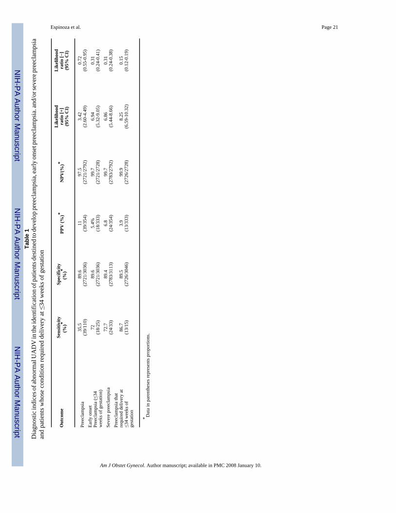
This study included 3348 patients (52 patients were lost to follow-up evaluations). Theprevalence of preeclampsia, severe preeclampsia, and early onset preeclampsia was 3.4%(113/3296), 1.0% (33/3296), and 0.8% (25/3208), respectively. UADV was performed in95.4% (3146/3296) and plasma PlGF concentrations were determined in 93.5% (3081/3296)of the study population. Early onset preeclampsia developed in 5 patients, severe preeclampsiadeveloped in 13 patients, and both early onset and severe preeclampsia developed in 20 patients(this group does not include the former 18 patients).
Espinoza et al. Page 4
Am J Obstet Gynecol. Author manuscript; available in PMC 2008 January 10.
NIH
-PA Author Manuscript
NIH
-PA Author Manuscript
NIH
-PA Author Manuscript

Diagnostic indices, predictive values, and likelihood ratios of UADVAn abnormal UADV was present in 11.3% (354/3146) of the study population. The diagnosticindices, predictive values and likelihood ratios for the primary outcomes are displayed in TableI. Logistic regression analysis indicated that an abnormal UADV, between 22 and 26 weeksof gestation, was an independent explanatory variable for the occurrence of preeclampsia, earlyonset preeclampsia, severe preeclampsia, and SGA without preeclampsia, after an adjustmentwas made for maternal age of >35 years, previous preeclampsia, nulliparity, smoking, firsttrimester body mass index of >30 kg/m2, maternal plasma PlGF, and sample storage time(Table 2).
Diagnostic indices, predictive values, and likelihood ratios of maternal plasma PlGFconcentration
Receiver operating characteristic curve analysis was performed to examine the diagnosticperformance of maternal plasma PlGF concentrations in the identification of the patientdestined to develop early onset and/or severe preeclampsia; a cut-off of 280 pg/mL wasselected. Logistic regression analysis indicated that a maternal plasma concentration of PlGF<280 pg/mL was an independent explanatory variable for the occurrence of preeclampsia, earlyonset preeclampsia, severe preeclampsia, and SGA without preeclampsia after an adjustmentwas made for the aforementioned covariates and abnormal UADV. The odds ratio and 95%CI of a low maternal plasma PlGF concentration (<280 pg/mL) in the identification of theoutcomes are displayed in Table 2, and the diagnostic indices of a low maternal plasmaconcentration for the primary outcomes are given in Table 3.
The combination of abnormal UADV and maternal plasma PlGF <280 pg/mL in theidentification of the outcomes
Table 4 gives the diagnostic indices, predictive values and likelihood ratios of the combinationof abnormal UADV and maternal plasma PlGF <280 pg/mL for the identification of the primaryoutcomes. This combined approach improved the positive predictive value of an abnormalUADV in the prediction of the primary study outcomes without a significant reduction in thesensitivity. Multivariate logistic regression analysis indicated that the combination of abnormalUADV and maternal plasma PlGF <280 pg/mL was an independent explanatory variable forthe outcomes after being controlled for the aforementioned covariates. This parametercombination was associated with an odds ratio of 43.8 (95% CI; 18.48-103.9) and 37.4 (95%CI, 17.6-79.1) to develop early onset preeclampsia and severe preeclampsia, respectively.Thus, abnormal UADV and low maternal plasma concentration of PlGF conferred a muchhigher risk for the development of early onset and/or severe preeclampsia than abnormalUADV alone (Tables 2 and 5).
Maternal plasma PlGF and sVEGFR-1 in the identification of patients destined to have earlyonset and/or severe preeclampsia according to the results of the UADV
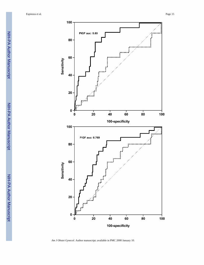
A sub-analysis indicated that, among patients with an abnormal UADV result, the maternalplasma PlGF concentration contributed significantly to the identification of patients destinedto have early onset preeclampsia (area under the curve, 0.80; P <.001) and severe preeclampsia(area under the curve, 0.77; P <.001; see Figure 1A and 1B). Indeed, 89% of the women (16/18)with abnormal UADV results who had early onset preeclampsia and 84% of the women (21/25)who had severe preeclampsia had a plasma PlGF concentration <280 pg/mL. In contrast,maternal plasma sVEGFR-1 concentration was of limited value in the prediction of early onset(area under the curve, 0.49; P = .9) and severe preeclampsia (area under the curve, 0.54; P = .5; Figure 1A and 1B).
Espinoza et al. Page 5
Am J Obstet Gynecol. Author manuscript; available in PMC 2008 January 10.
NIH
-PA Author Manuscript
NIH
-PA Author Manuscript
NIH
-PA Author Manuscript

Among patients with normal UADV, the maternal plasma PlGF concentration did notcontribute to the identification of patients destined to develop early onset preeclampsia (areaunder the curve, 0.56; P = .6) or severe preeclampsia (area under the curve, 0.62; P = .2).
Demographic and clinical characteristics of the study population, according to UADV andmaternal plasma PlGF concentration
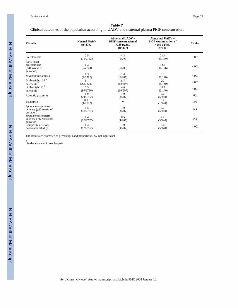
Table 6 and Table 7 display the demographic and clinical characteristics of the population aswell as the outcomes according to the results of the UADV and maternal plasma PlGFconcentration of <280 pg/mL, respectively. There were no differences in gestational ages atultrasound examination among the study groups. Patients with abnormal UADV and maternalplasma PlGF concentrations <280 pg/mL had a higher frequency of preeclampsia, early onsetpreeclampsia, severe preeclampsia, SGA without preeclampsia, placental abruption,eclampsia, and a composite of severe neonatal morbidity than both patients with normal UADVresults and those with abnormal UADV results and a maternal plasma concentration of PlGF≥280 pg/mL (chi square for trend; P < .001).
Sequential screening with UADV and maternal plasma PlGF concentration in theidentification of patients destined to develop early onset and/or severe PE
Figures 2 and 3 display the distribution of the study population according to the results ofsequential assessment with the use of UADV followed by maternal plasma PlGFdeterminations in the second trimester. The prevalence of early onset and/or severepreeclampsia among patients with abnormal UADV and PlGF concentrations <280 pg/mL was11 times higher than among those with abnormal UADV and PlGF concentrations ≥280 pg/mL (15.7% [22/140] vs. 1.4% [3/207]; P < .001) and 30 times higher than among patients withnormal UADV results, regardless of the maternal plasma PlGF concentration (15.7% [22/140]vs. 0.5% [13/2792]; P < .001; Figure 3).
Figure 4 displays the distribution of the study population as a result of the sequentialdetermination of maternal plasma concentration of PlGF followed by UADV. This flowdiagram was generated because many centers in the United States do not use UADV.
The survival analysis indicated that patients with abnormal UADV and low PlGF have a shorterexamination-to-diagnosis interval than those in the other 2 groups (log rank test, 37.9; P < .001; Figure 5).
COMMENTThe results of this study indicate that a combination of an abnormal UADV and a maternalplasma PlGF concentration of <280 pg/mL, between 22 and 26 weeks of gestation, identifiespatients at a very high risk for preeclampsia, early onset preeclampsia and severe preeclampsia.
These novel observations are consistent with previous reports indicating that a low maternalplasma concentration of PlGF in the first9,12 or second trimester of pregnancy9-11,21 andabnormal UADV results between 23 and 25 weeks of gestation5-8 are risk factors for thedevelopment of preeclampsia. Similarly, a low urine concentration of PlGF between 25 and28 weeks has been associated recently with a high risk for preeclampsia.13
The results presented herein differ from those reported recently,22 indicating a lack ofassociation between abnormal UADV and low PlGF. Differences in sample size, gestationalage at ultrasound, and study outcomes may account for these discrepancies. A recentlongitudinal study that included 81 patients at risk for preeclampsia reported that the maternalplasma PlGF concentration at 24 weeks of gestation contributed significantly to the predictionof the disease.23 However, this study did not include enough patients to determine the value
Espinoza et al. Page 6
Am J Obstet Gynecol. Author manuscript; available in PMC 2008 January 10.
NIH
-PA Author Manuscript
NIH
-PA Author Manuscript
NIH
-PA Author Manuscript
of maternal plasma PlGF for the prediction of early onset preeclampsia, and the authorscautioned that “large prospective cohort studies in unselected women are required to ascertainany clinical usefulness.”23
The regulation of vascular growth and remodeling, also known as angiogenesis, is consideredto be central to normal placental and fetal growth and development.24-26 In the humanplacenta, angiogenesis is biphasic, with peaks at mid gestation and at term as the result ofendothelial proliferation early in pregnancy and vascular remodeling in the second half ofpregnancy.27 This is consistent with the model of placental angiogenesis proposed byKingdom et al,28 whereby branching angiogenesis is predominant in the first trimester and isassociated with high placental production of VEGF. In contrast, nonbranching angiogenesis ispredominant in the third trimester and is associated with a high placental production of PlGF.28
Angiogenesis is regulated by at least three growth factor families, which include VEGFs,angiopoietins, and ephrins.29 Other nonspecific factors that have been proposed to regulateangiogenesis include fibroblast growth factors, transforming growth factors α and β, tumornecrosis factor α, Interleukin-8, hepatocyte growth factor, angiogenin, and members of theNotch family.26,30,31 Recent evidence indicates that angiogenesis requires the sequentialactivation of several receptors (which include Tie1, Tie2, and platelet-derived growth factorreceptor β) by ligands in endothelial and mural cells.32 However, VEGF signaling representsa critical rate-limiting step in physiological angiogenesis.32
VEGFs are a family of dimeric proteins whose members include VEGF-A, VEGF-B, VEGF-C, VEGF-D and PlGF.31 The function of VEGF is to promote survival, migration, anddifferentiation of endothelial cells as well as to mediate vascular permeability.30,31 VEGFexerts its biologic effect through VEGFR-2, whereas the precise function of VEGFR-1 is stilla subject of debate. Most investigators believe that VEGFR-1 might not be a receptor thattransmits a mitogenic signal, but rather a “decoy” receptor that prevents the binding of VEGFto VEGFR-2.31 The decoy function can be performed not only by the transmembrane, but alsoby the soluble isoform (sVEGFR-1).31 An additional mechanism by which sVEGFR-1 mayregulate the bioavailability of VEGF is the formation of heterodimers with the VEGF receptorsin the cell surface, which abolishes their signal transduction.33 Thus, sVEGFR-1 is consideredan antiangiogenic factor.
PlGF is another ligand for VEGFR-1 that enhances the angiogenic response of VEGF.34,35This has been proposed to be accomplished by (1) intermolecular cross-talk between VEGFR-1and VEGFR-2 (transphophorylation and activation of VEGFR-2 following activation ofVEGFR-1 by PlGF); (2) PlGF displacement of VEGF from sVEGFR-1, which makes moreVEGF available to bind VEGFR-2, and (3) PlGF homodimers that can destabilize inactiveheterodimers of VEGFR-2 and sVEGFR-1, which makes more VEGFR-2 available for theformation of functional homodimers.34,35
The results of the current study indicate that both an abnormal UADV and a maternal plasmaPlGF concentration of <280 pg/mL are independent factors for the prediction of the outcomes.The predictive value of an abnormal UADV between 22 and 26 weeks of gestation for theoccurrence of preeclampsia and early onset preeclampsia in the study population is consistentwith previous reports.5-8 However, this study further demonstrates that the combination ofabnormal UADV and a maternal plasma PlGF of <280pg/mL in the second trimester confersa much higher risk for preeclampsia and early onset or severe preeclampsia than abnormalUADV alone. Moreover, patients with abnormal UADV results and a plasma PlGFconcentration of <280 pg/mL, which represented only 4.5% of the population (140/3146
Espinoza et al. Page 7
Am J Obstet Gynecol. Author manuscript; available in PMC 2008 January 10.
NIH
-PA Author Manuscript
NIH
-PA Author Manuscript
NIH
-PA Author Manuscript
women), contained 60% of the patients (22/38 women) who developed early onset and/orsevere preeclampsia.
The sequential determination of UADV and maternal plasma concentration of PlGF, or viceversa, identifies patients at a very high risk for early onset and/or severe preeclampsia. Theflow diagrams in Figures 2 and 3 indicate that centers favoring the use of UADV first canobtain the same results if the determination of maternal plasma PlGF concentrations is offeredonly to patients with abnormal UADV (approximately 10% of the population), rather than thewhole study population. This is because a maternal plasma concentration of PlGF did notcontribute to the identification of patients at risk for early onset and/or severe preeclampsiaamong patients with normal UADV results. Among centers that may favor the determinationof maternal plasma concentration of PlGF followed by UADV, the identification of patientsat risk for early onset and/or severe preeclampsia can be accomplished if UADV is offeredonly to those with a plasma concentration of PlGF of <280 pg/mL. However, this will requireoffering UADV to about one-half of the population.
It is noteworthy that patients with abnormal UADV results and a plasma PlGF of <280 pg/mLalso had a higher proportion of spontaneous preterm delivery than patients with normal UADVresults and those with abnormal UADV results and a PlGF concentration of ≥280 pg/mL.However, the difference did not reach statistical significance. This observation is consistentwith a growing body of evidence showing that chronic uteroplacental ischemia may representthe mechanism of disease in a subset of patients with preterm delivery.36-38 Additional studies,with larger sample sizes, may be required to determine the risk of spontaneous preterm birthamong patients with abnormal UADV results and a low PlGF in the second trimester.
sVEGFR-1 has recently been implicated recently in the pathophysiologic condition ofpreeclampsia.11,18,39 Indeed, clinical and experimental evidence indicates that a highmaternal plasma concentration of sVEGFR-1 in patients with preeclampsia is associated witha reduction in the bioavailability of the free form of VEGF and PlGF,39 with the subsequentendothelial cell dysfunction. The observation that maternal plasma concentration of sVEGFR-1among patients with abnormal UADV results were of limited value in the prediction of earlyonset and/or severe preeclampsia is consistent with recent reports that the elevation of thisantiangiogenic factor occurs rather late in the course of the disease (approximately 5 weeksbefore the clinical presentation of preeclampsia).13,14 We did not determine the maternalplasma concentration of sVEGFR-1 among patients with normal UADV results, given thatsVEGFR-1 did not improve the diagnostic indices of an abnormal UADV (Figure 1).
An Abnormal UADV result between 22 and 26 weeks of gestation is considered to be asurrogate marker of chronic uteroplacental ischemia. Evidence in favor of this view includesthe following observations: (1) embolization of the uterine arterioles and spiral arteries withGelfoam particles in pregnant animals reduced the uterine blood flow and increased the uterineartery pulsatility index in a dose dependant manner40,41; (2) an abnormal UADV result isassociated with failure of physiologic transformation of the myometrial segment of the spiralarteries in placental bed biopsy specimens obtained from patients with preeclampsia andintrauterine growth restriction42-47; and (3) an abnormal UADV result is associated withplacental ischemic/hypoxic changes (excessive syncytial knots, proliferation of villouscytotrophoblast cells, intervillous fibrin deposition, thickening of the basement membranes,and mature small villi) in patients with preeclampsia48 and intrauterine growth restriction.49
In vitro studies had demonstrated that hypoxia may have opposite effects in the expression ofangiogenic factors in the trophoblast.50,51 Indeed, the incubation of isolated human termsyncytiotrophoblast under hypoxic conditions increased VEGF messenger RNA expression 8-fold but reduced the PlGF messenger RNA expression by 75%.51 Similarly, in primary
Espinoza et al. Page 8
Am J Obstet Gynecol. Author manuscript; available in PMC 2008 January 10.
NIH
-PA Author Manuscript
NIH
-PA Author Manuscript
NIH
-PA Author Manuscript
cytotrophoblast cultures, hypoxia increases the VEGF messenger RNA expression (but not therelease of free VEGF in the supernatant) and reduces the PlGF concentrations in thesupernatant.50 Thus, it is possible that chronic uteroplacental ischemia may account for thelow maternal plasma PlGF concentration observed among patients destined to develop earlyonset and/or severe preeclampsia and that the combination of an abnormal UADV result anda maternal plasma PlGF concentration of <280 pg/mL identifies patients at risk forpreeclampsia whose primary insult is a chronic reduction in the uteroplacental blood flow.
Abnormal UADV results 4,5,50,52-56 and biochemical markers10,15,57-59 in the secondtrimester had been reported to have low positive predictive values in the prediction ofpreeclampsia and early onset preeclampsia. Therefore, most patients with a positive test willnot experience the disease; any prophylactic intervention that targets the screen-positive groupwill expose a large proportion of patients that will not benefit from the intervention.Preeclampsia is 1 of the great obstetrical syndromes,60 and several mechanisms of disease hadbeen implicated in the pathophysiologic makeup of this complex disease, including (1)uteroplacental ischemia,61 (2) immune maladaptation,61 (3) very low-density lipoproteintoxicity,61 (4) genetic imprinting,61 (5) increased trophoblast apoptosis/necrosis,62-64 and(6) an exaggerated maternal inflammatory response to deported trophoblast.65-68 Theheterogeneity of the disease is exemplified by the observation that chronic uteroplacentalischemia may be more relevant in the pathogenesis of early onset preeclampsia than in termor post-term preeclampsia.69,70 Indeed, an abnormal UADV result in the second trimester isassociated with a higher risk for preeclampsia at ≤34 weeks of gestation than at >34 weeks ofgestation5-8. Moreover, early onset preeclampsia is more severe71 and has a higher proportionof growth-restricted fetuses,71 a higher risk of maternal death,1 and a higher frequency ofplacental pathologic condition72,73 than late onset preeclampsia.
Because of the syndromic nature of preeclampsia,74 it is unlikely that a single marker orcombination of markers will identify all patients destined to develop this pregnancycomplication, regardless of their mechanism of disease. We have provided the diagnosticindices of a combination of abnormal UADV result and low PlGF concentration for theprediction of the outcomes to provide the basis for comparison with previous reports. However,we propose that risk assessment of preeclampsia should not focus on the prediction of thisheterogeneous disorder, but on the identification of patients at high risk for the early and/or amore severe form of preeclampsia, in whom prophylactic interventions are more likely toreduce the morbidity and mortality rates associated with this obstetrical syndrome.
Collectively, the results presented herein indicate that a combination of abnormal UADV resultand a maternal plasma concentration of PlGF <280 pg/mL between 22 and 26 weeks ofgestation is associated with a high risk for preeclampsia, early onset preeclampsia, and severepreeclampsia. Moreover, a maternal plasma PlGF concentration of <280 pg/mL can identifymost patients at risk for early onset and/or severe preeclampsia among patients with abnormalUADV results. The identification of this subset of patients may allow for prophylacticinterventions, such as the modulation of the antiangiogenic state before the clinical presentationof early onset and/or severe preeclampsia, which potentially could reduce the perinatal andmaternal mortality rate that is associated with this syndrome.
The results of 2 recent randomized clinical trials of vitamin C and E supplementation for theprevention of preeclampsia in low-75 and high-risk76 patients yielded negative results. Thus,there is a need for alternative prophylactic interventions. VEGF and PlGF have been proposedas potential candidates to modulate the antiangiogenic state in severe preeclampsia.77Recently, it has been reported that the administration of recombinant VEGF121 to a rat modelof preeclampsia, which was induced by administration of adenovirus expressing the sVEGFR-1gene, attenuated the preeclamptic phenotype. Indeed, the authors reported that the
Espinoza et al. Page 9
Am J Obstet Gynecol. Author manuscript; available in PMC 2008 January 10.
NIH
-PA Author Manuscript
NIH
-PA Author Manuscript
NIH
-PA Author Manuscript

subcutaneous administration of 100, 200 or 400 μ/kg of VEGF121 reduced the systolic bloodpressure, limited the kidney damage (as indicated by a lower glomerular lesion index), andreduced proteinuria among the treated animals.78 The authors proposed that VEGF121 couldbe a potential therapeutic agent for preeclampsia in humans.78 Similarly, VEGF and PlGF hadbeen reported to stimulate angiogenesis in an animal model of myocardial and limb ischemia.79 However, PlGF appears to be a better candidate for clinical trials, because it did not inducethe side effects associated with VEGF (such as edema, hypotension, bleeding and the formationof fragile capillaries that were prone to regression).79 If clinical trials prove that proangiogenicfactors are effective in delaying the clinical presentation of preeclampsia and/or reducing itsseverity, we have estimated that 15 patients with abnormal UADV results (95% CI, 11-26) inthe second trimester would need to be treated in order to prevent 1 case of early onset and/orsevere preeclampsia. In contrast, only 7 patients with abnormal UADV results and low PlGFconcentrations (95% CI, 5-11) would need to be exposed to these interventions to prevent 1case of severe and/or early onset preeclampsia.
Acknowledgment
This research was supported by the Intramural Research Program of the National Institute of Child Health and HumanDevelopment, NIH, DHHS.
Reference1. MacKay AP, Berg CJ, Atrash HK. Pregnancy-related mortality from preeclampsia and eclampsia.
Obstet Gynecol 2001;97:533–38. [PubMed: 11275024]2. Redman CW, Sargent IL. Latest advances in understanding preeclampsia. Science 2005;308:1592–94.
[PubMed: 15947178]3. Roberts JM, Pearson G, Cutler J, Lindheimer M. Summary of the NHLBI Working Group on Research
on Hypertension During Pregnancy. Hypertension 2003;41:437–45. [PubMed: 12623940]4. Bower S, Bewley S, Campbell S. Improved prediction of preeclampsia by two-stage screening of
uterine arteries using the early diastolic notch and color Doppler imaging. Obstet Gynecol 1993;82:78–83. [PubMed: 8515930]
5. Harrington K, Cooper D, Lees C, Hecher K, Campbell S. Doppler ultrasound of the uterine arteries:the importance of bilateral notching in the prediction of preeclampsia, placental abruption or deliveryof a small-for-gestational-age baby. Ultrasound Obstet Gynecol 1996;7:182–88. [PubMed: 8705410]
6. Albaiges G, Missfelder-Lobos H, Lees C, Parra M, Nicolaides KH. One-stage screening for pregnancycomplications by color Doppler assessment of the uterine arteries at 23 weeks' gestation. ObstetGynecol 2000;96:559–64. [PubMed: 11004359]
7. Papageorghiou AT, Yu CK, Cicero S, Bower S, Nicolaides KH. Second-trimester uterine arteryDoppler screening in unselected populations: a review. J Matern Fetal Neonatal Med 2002;12:78–88.[PubMed: 12420836]
8. Papageorghiou AT, Yu CK, Bindra R, Pandis G, Nicolaides KH. Multicenter screening for pre-eclampsia and fetal growth restriction by transvaginal uterine artery Doppler at 23 weeks of gestation.Ultrasound Obstet Gynecol 2001;18:441–49. [PubMed: 11844162]
9. Tidwell SC, Ho HN, Chiu WH, Torry RJ, Torry DS. Low maternal serum levels of placenta growthfactor as an antecedent of clinical preeclampsia. Am J Obstet Gynecol 2001;184:1267–72. [PubMed:11349200]
10. Krauss T, Pauer HU, Augustin HG. Prospective analysis of placenta growth factor (PlGF)concentrations in the plasma of women with normal pregnancy and pregnancies complicated bypreeclampsia. Hypertens Pregnancy 2004;23:101–11. [PubMed: 15117604]
11. Levine RJ, Maynard SE, Qian C, Lim KH, England LJ, Yu KF, et al. Circulating angiogenic factorsand the risk of preeclampsia. N Engl J Med 2004;350:672–83. [PubMed: 14764923]
12. Stepan H, Faber R, Wessel N, Wallukat G, Schultheiss HP, Walther T. Relation between circulatingangiotensin II type 1 receptor agonistic autoantibodies and soluble fms-like tyrosine kinase 1 in thepathogenesis of preeclampsia. J Clin.Endocrinol.Metab 2006;91:2424–27. [PubMed: 16569734]
Espinoza et al. Page 10
Am J Obstet Gynecol. Author manuscript; available in PMC 2008 January 10.
NIH
-PA Author Manuscript
NIH
-PA Author Manuscript
NIH
-PA Author Manuscript
13. Levine RJ, Thadhani R, Qian C, Lam C, Lim KH, Yu KF, et al. Urinary placental growth factor andrisk of preeclampsia. JAMA 2005;293:77–85. [PubMed: 15632339]
14. Chaiworapongsa T, Romero R, Kim YM, Kim GJ, Kim MR, Espinoza J, et al. Plasma soluble vascularendothelial growth factor receptor-1 concentration is elevated prior to the clinical diagnosis of pre-eclampsia. J Matern Fetal Neonatal Med 2005;17:3–18. [PubMed: 15804781]
15. Parra M, Rodrigo R, Barja P, Bosco C, Fernandez V, Munoz H, et al. Screening test for preeclampsiathrough assessment of uteroplacental blood flow and biochemical markers of oxidative stress andendothelial dysfunction. Am J Obstet Gynecol 2005;193:1486–91. [PubMed: 16202744]
16. Gonzalez RP, Gomez RM, Castro RS, Nien JK, Merino PO, Etchegaray AB, et al. [A national birthweight distribution curve according to gestational age in chile from 1993 to 2000]. Rev Med Chil2004;132:1155–65. [PubMed: 15631202]
17. Bower S, Schuchter K, Campbell S. Doppler ultrasound screening as part of routine antenatalscanning: prediction of pre-eclampsia and intrauterine growth retardation. Br J Obstet Gynaecol1993;100:989–94. [PubMed: 8251470]
18. Chaiworapongsa T, Romero R, Espinoza J, Bujold E, Mee KY, Goncalves LF, et al. Evidencesupporting a role for blockade of the vascular endothelial growth factor system in the pathophysiologyof preeclampsia. Young Investigator Award. Am J Obstet Gynecol 2004;190:1541–47. [PubMed:15284729]
19. Haddad B, Deis S, Goffinet F, Paniel BJ, Cabrol D, Siba BM. Maternal and perinatal outcomes duringexpectant management of 239 severe preeclamptic women between 24 and 33 weeks' gestation. AmJ Obstet Gynecol 2004;190:1590–95. [PubMed: 15284743]
20. Hall DR, Grove D, Carstens E. Early pre-eclampsia: What proportion of women qualify for expectantmanagement and if not, why not? Eur J Obstet Gynecol Reprod Biol. 2006
21. Tjoa ML, van Vugt JM, Mulders MA, Schutgens RB, Oudejans CB, van Wijk IJ. Plasma placentagrowth factor levels in midtrimester pregnancies. Obstet Gynecol 2001;98:600–07. [PubMed:11576575]
22. Muller PR, James AH, Murtha PP, Yonish B, Jamison MG, Dekker G. Circulating angiogenic factorsand abnormal uterine artery Doppler velocimetry in the second trimester. Hypertens Pregnancy2006;25:183–92. [PubMed: 17065039]
23. Chappell LC, Seed PT, Briley A, Kelly FJ, Hunt BJ, Charnock-Jones DS, et al. A longitudinal studyof biochemical variables in women at risk of preeclampsia. Am J Obstet Gynecol 2002;187:127–36.[PubMed: 12114900]
24. Wulff C, Wilson H, Dickson SE, Wiegand SJ, Fraser HM. Hemochorial placentation in the primate:expression of vascular endothelial growth factor, angiopoietins, and their receptors throughoutpregnancy. Biol Reprod 2002;66:802–12. [PubMed: 11870089]
25. Carmeliet P. Mechanisms of angiogenesis and arteriogenesis. Nat Med 2000;6:389–95. [PubMed:10742145]
26. Yancopoulos GD, Davis S, Gale NW, Rudge JS, Wiegand SJ, Holash J. Vascular-specific growthfactors and blood vessel formation. Nature 2000;407:242–48. [PubMed: 11001067]
27. Mayhew TM. Fetoplacental angiogenesis during gestation is biphasic, longitudinal and occurs byproliferation and remodelling of vascular endothelial cells. Placenta 2002;23:742–50. [PubMed:12398814]
28. Kingdom J, Huppertz B, Seaward G, Kaufmann P. Development of the placental villous tree and itsconsequences for fetal growth. Eur J Obstet Gynecol Reprod Biol 2000;92:35–43. [PubMed:10986432]
29. Gale NW, Yancopoulos GD. Growth factors acting via endothelial cell-specific receptor tyrosinekinases: VEGFs, angiopoietins, and ephrins in vascular development. Genes Dev 1999;13:1055–66.[PubMed: 10323857]
30. Matsumoto T, Claesson-Welsh L. VEGF receptor signal transduction. Sci STKE 2001;2001:RE21.[PubMed: 11741095]
31. Ferrara N, Gerber HP, LeCouter J. The biology of VEGF and its receptors. Nat Med 2003;9:669–76.[PubMed: 12778165]
32. Ferrara N. Vascular endothelial growth factor: basic science and clinical progress. Endocr Rev2004;25:581–611. [PubMed: 15294883]
Espinoza et al. Page 11
Am J Obstet Gynecol. Author manuscript; available in PMC 2008 January 10.
NIH
-PA Author Manuscript
NIH
-PA Author Manuscript
NIH
-PA Author Manuscript
33. Barleon B, Reusch P, Totzke F, Herzog C, Keck C, Martiny-Baron G, et al. Soluble VEGFR-1 secretedby endothelial cells and monocytes is present in human serum and plasma from healthy donors.Angiogenesis 2001;4:143–54. [PubMed: 11806246]
34. Autiero M, Luttun A, Tjwa M, Carmeliet P. Placental growth factor and its receptor, vascularendothelial growth factor receptor-1: novel targets for stimulation of ischemic tissuerevascularization and inhibition of angiogenic and inflammatory disorders. J Thromb Haemost2003;1:1356–70. [PubMed: 12871269]
35. Autiero M, Waltenberger J, Communi D, Kranz A, Moons L, Lambrechts D, et al. Role of PlGF inthe intra- and intermolecular cross talk between the VEGF receptors Flt1 and Flk1. Nat Med2003;9:936–43. [PubMed: 12796773]
36. Kim YM, Bujold E, Chaiworapongsa T, Gomez R, Yoon BH, Thaler HT, et al. Failure of physiologictransformation of the spiral arteries in patients with preterm labor and intact membranes. Am J ObstetGynecol 2003;189:1063–69. [PubMed: 14586356]
37. Kim YM, Chaiworapongsa T, Gomez R, Bujold E, Yoon BH, Rotmensch S, et al. Failure ofphysiologic transformation of the spiral arteries in the placental bed in preterm premature rupture ofmembranes. Am J Obstet Gynecol 2002;187:1137–42. [PubMed: 12439491]
38. Arias F, Rodriquez L, Rayne SC, Kraus FT. Maternal placental vasculopathy and infection: twodistinct subgroups among patients with preterm labor and preterm ruptured membranes. Am J ObstetGynecol 1993;168:585–91. [PubMed: 8438933]
39. Maynard SE, Min JY, Merchan J, Lim KH, Li J, Mondal S, et al. Excess placental soluble fms-liketyrosine kinase 1 (sFlt1) may contribute to endothelial dysfunction, hypertension, and proteinuria inpreeclampsia. J Clin Invest 2003;111:649–58. [PubMed: 12618519]
40. Ochi H, Matsubara K, Kusanagi Y, Taniguchi H, Ito M. Significance of a diastolic notch in the uterineartery flow velocity waveform induced by uterine embolisation in the pregnant ewe. Br J ObstetGynaecol 1998;105:1118–21. [PubMed: 9800937]
41. Ochi H, Suginami H, Matsubara K, Taniguchi H, Yano J, Matsuura S. Micro-bead embolization ofuterine spiral arteries and changes in uterine arterial flow velocity waveforms in the pregnant ewe.Ultrasound Obstet Gynecol 1995;6:272–76. [PubMed: 8590190]
42. Aardema MW, Oosterhof H, Timmer A, van R, Aarnoudse JG. Uterine artery Doppler flow anduteroplacental vascular pathology in normal pregnancies and pregnancies complicated by pre-eclampsia and small for gestational age fetuses. Placenta 2001;22:405–11. [PubMed: 11373150]I
43. Lin S, Shimizu I, Suehara N, Nakayama M, Aono T. Uterine artery Doppler velocimetry in relationto trophoblast migration into the myometrium of the placental bed. Obstet Gynecol 1995;85:760–65.[PubMed: 7724109]
44. Madazli R, Somunkiran A, Calay Z, Ilvan S, Aksu MF. Histomorphology of the placenta and theplacental bed of growth restricted foetuses and correlation with the Doppler velocimetries of theuterine and umbilical arteries. Placenta 2003;24:510–16. [PubMed: 12744927]
45. Olofsson P, Laurini RN, Marsal K. A high uterine artery pulsatility index reflects a defectivedevelopment of placental bed spiral arteries in pregnancies complicated by hypertension and fetalgrowth retardation. Eur J Obstet Gynecol Reprod Biol 1993;49:161–68. [PubMed: 8405630]
46. Sagol S, Ozkinay E, Oztekin K, Ozdemir N. The comparison of uterine artery Doppler velocimetrywith the histopathology of the placental bed. Aust N Z J Obstet Gynaecol 1999;39:324–29. [PubMed:10554944]
47. Voigt HJ, Becker V. Doppler flow measurements and histomorphology of the placental bed inuteroplacental insufficiency. J Perinat Med 1992;20:139–47. [PubMed: 1501057]
48. Ferrazzi E, Bulfamante G, Mezzopane R, Barbera A, Ghidini A, Pardi G. Uterine Doppler velocimetryand placental hypoxic-ischemic lesion in pregnancies with fetal intrauterine growth restriction.Placenta 1999;20:389–94. [PubMed: 10419803]
49. Iwata M, Matsuzaki N, Shimizu I, Mitsuda N, Nakayama M, Suehara N. Prenatal detection of ischemicchanges in the placenta of the growth-retarded fetus by Doppler flow velocimetry of the maternaluterine artery. Obstet Gynecol 1993;82:494–99. [PubMed: 8377970]
50. Nagamatsu T, Fujii T, Kusumi M, Zou L, Yamashita T, Osuga Y, et al. Cytotrophoblasts up-regulatesoluble fms-like tyrosine kinase-1 expression under reduced oxygen: an implication for the placental
Espinoza et al. Page 12
Am J Obstet Gynecol. Author manuscript; available in PMC 2008 January 10.
NIH
-PA Author Manuscript
NIH
-PA Author Manuscript
NIH
-PA Author Manuscript
vascular development and the pathophysiology of preeclampsia. Endocrinology 2004;145:4838–45.[PubMed: 15284201]
51. Shore VH, Wang TH, Wang CL, Torry RJ, Caudle MR, Torry DS. Vascular endothelial growth factor,placenta growth factor and their receptors in isolated human trophoblast. Placenta 1997;18:657–65.[PubMed: 9364601]
52. Steel SA, Pearce JM, McParland P, Chamberlain GV. Early Doppler ultrasound screening inprediction of hypertensive disorders of pregnancy. Lancet 1990;335:1548–51. [PubMed: 1972486]
53. North RA, Ferrier C, Long D, Townend K, Kincaid-Smith P. Uterine artery Doppler flow velocitywaveforms in the second trimester for the prediction of preeclampsia and fetal growth retardation.Obstet Gynecol 1994;83:378–86. [PubMed: 8127529]
54. Irion O, Masse J, Forest JC, Moutquin JM. Prediction of pre-eclampsia, low birthweight for gestationand prematurity by uterine artery blood flow velocity waveforms analysis in low risk nulliparouswomen. Br J Obstet Gynaecol 1998;105:422–29. [PubMed: 9609270]
55. Harrington KF, Campbell S, Bewley S, Bower S. Doppler velocimetry studies of the uterine arteryin the early prediction of pre-eclampsia and intra-uterine growth retardation. Eur J Obstet GynecolReprod Biol 1991;42(Suppl):S14–S20. [PubMed: 1809604]
56. Aquilina J, Barnett A, Thompson O, Harrington K. Comprehensive analysis of uterine artery flowvelocity waveforms for the prediction of pre-eclampsia. Ultrasound Obstet Gynecol 2000;16:163–70. [PubMed: 11117088]
57. Aquilina J, Maplethorpe R, Ellis P, Harrington K. Correlation between second trimester maternalserum inhibin-A and human chorionic gonadotrophin for the prediction of pre-eclampsia. Placenta2000;21:487–92. [PubMed: 10940198]
58. Masse J, Forest JC, Moutquin JM, Marcoux S, Brideau NA, Belanger M. A prospective study ofseveral potential biologic markers for early prediction of the development of preeclampsia. Am JObstet Gynecol 1993;169:501–08. [PubMed: 8372852]
59. Stamilio DM, Sehdev HM, Morgan MA, Propert K, Macones GA. Can antenatal clinical andbiochemical markers predict the development of severe preeclampsia? Am J Obstet Gynecol2000;182:589–94. [PubMed: 10739512]
60. Romero R, Lockwood C, Oyarzun E, Hobbins JC. Toxemia: New Concepts in an Old Disease.Seminars in Perinatology 1988;12:302–23. [PubMed: 3065943]
61. Dekker GA, Sibai BM. Etiology and pathogenesis of preeclampsia: current concepts. Am J ObstetGynecol 1998;179:1359–75. [PubMed: 9822529]
62. Austgulen R, Isaksen CV, Chedwick L, Romundstad P, Vatten L, Craven C. Preeclampsia: associatedwith increased syncytial apoptosis when the infant is small-for-gestational-age. J Reprod Immunol2004;61:39–50. [PubMed: 14967220]
63. Crocker IP, Cooper S, Ong SC, Baker PN. Differences in apoptotic susceptibility of cytotrophoblastsand syncytiotrophoblasts in normal pregnancy to those complicated with preeclampsia andintrauterine growth restriction. Am J Pathol 2003;162:637–43. [PubMed: 12547721]
64. Leung DN, Smith SC, To KF, Sahota DS, Baker PN. Increased placental apoptosis in pregnanciescomplicated by preeclampsia. Am J Obstet Gynecol 2001;184:1249–50. [PubMed: 11349196]
65. Sargent IL, Germain SJ, Sacks GP, Kumar S, Redman CW. Trophoblast deportation and the maternalinflammatory response in pre-eclampsia. J Reprod Immunol 2003;59:153–60. [PubMed: 12896819]
66. Johansen M, Redman CW, Wilkins T, Sargent IL. Trophoblast deportation in human pregnancy--itsrelevance for pre-eclampsia. Placenta 1999;20:531–39. [PubMed: 10452906]
67. Knight M, Redman CW, Linton EA, Sargent IL. Shedding of syncytiotrophoblast microvilli into thematernal circulation in pre-eclamptic pregnancies. Br J Obstet Gynaecol 1998;105:632–40.[PubMed: 9647154]
68. Chua S, Wilkins T, Sargent I, Redman C. Trophoblast deportation in pre-eclamptic pregnancy. Br JObstet Gynaecol 1991;98:973–79. [PubMed: 1751443]
69. Rasmussen S, Irgens LM. Fetal growth and body proportion in preeclampsia. Obstet Gynecol2003;101:575–83. [PubMed: 12636965]
70. Aardema MW, Saro MC, Lander M, De Wolf BT, Oosterhof H, Aarnoudse JG. Second trimesterDoppler ultrasound screening of the uterine arteries differentiates between subsequent normal and
Espinoza et al. Page 13
Am J Obstet Gynecol. Author manuscript; available in PMC 2008 January 10.
NIH
-PA Author Manuscript
NIH
-PA Author Manuscript
NIH
-PA Author Manuscript
poor outcomes of hypertensive pregnancy: two different pathophysiological entities? Clin Sci (Lond)2004;106:377–82. [PubMed: 14636154]
71. Odegard RA, Vatten LJ, Nilsen ST, Salvesen KA, Austgulen R. Preeclampsia and fetal growth. ObstetGynecol 2000;96:950–55. [PubMed: 11084184]
72. Moldenhauer JS, Stanek J, Warshak C, Khoury J, Sibai B. The frequency and severity of placentalfindings in women with preeclampsia are gestational age dependent. Am J Obstet Gynecol2003;189:1173–77. [PubMed: 14586374]
73. Sebire NJ, Goldin RD, Regan L. Term preeclampsia is associated with minimal histopathologicalplacental features regardless of clinical severity. J Obstet Gynaecol 2005;25:117–18. [PubMed:15814385]
74. Ness RB, Roberts JM. Heterogeneous causes constituting the single syndrome of preeclampsia: ahypothesis and its implications. Am J Obstet Gynecol 1996;175:1365–70. [PubMed: 8942516]
75. Rumbold AR, Crowther CA, Haslam RR, Dekker GA, Robinson JS. Vitamins C and E and the risksof preeclampsia and perinatal complications. N Engl J Med 2006;354:1796–806. [PubMed:16641396]
76. Poston L, Briley AL, Seed PT, Kelly FJ, Shennan AH. Vitamin C and vitamin E in pregnant womenat risk for pre-eclampsia (VIP trial): randomised placebo-controlled trial. Lancet 2006;367:1145–54.[PubMed: 16616557]
77. Levine RJ, Karumanchi SA. Circulating Angiogenic Factors in Preeclampsia. Clin Obstet Gynecol2005;48:372–86. [PubMed: 15805796]
78. Zhang Y, Jing-Ying M, Almirez R, de Forest N, Schellenberger U, Pollit S, et al. Overexpression ofSoluble FMS-Like Tyrosine Kinase-1 (sFLT-1) induced Experimental Preeclampsia in Rats andAttenuation of Preeclamptic Phenotype by Recombinant Vascular Endothelial Growth Factor 121(VEGF121). J Soc Gynecol Investig 2006;2:287A.
79. Luttun A, Tjwa M, Carmeliet P. Placental growth factor (PlGF) and its receptor Flt-1 (VEGFR-1):novel therapeutic targets for angiogenic disorders. Ann N Y Acad Sci 2002;979:80–93. [PubMed:12543719]
Espinoza et al. Page 14
Am J Obstet Gynecol. Author manuscript; available in PMC 2008 January 10.
NIH
-PA Author Manuscript
NIH
-PA Author Manuscript
NIH
-PA Author Manuscript
Espinoza et al. Page 15
Am J Obstet Gynecol. Author manuscript; available in PMC 2008 January 10.
NIH
-PA Author Manuscript
NIH
-PA Author Manuscript
NIH
-PA Author Manuscript

Figure 1.Maternal plasma PlGF concentration (solid line) contributed significantly to the prediction ofpatients destined to develop A, early onset preeclampsia (PE; P < .001) and B, severepreeclampsia (P < .001). In contrast, maternal plasma sVEGFR-1 concentration (dotted line)was of limited use in the prediction of A, early onset preeclampsia (area under the curve [AUC],0.49; P = .9) and B, severe preeclampsia (area under the curve, 0.54; P = .5;).
Espinoza et al. Page 16
Am J Obstet Gynecol. Author manuscript; available in PMC 2008 January 10.
NIH
-PA Author Manuscript
NIH
-PA Author Manuscript
NIH
-PA Author Manuscript
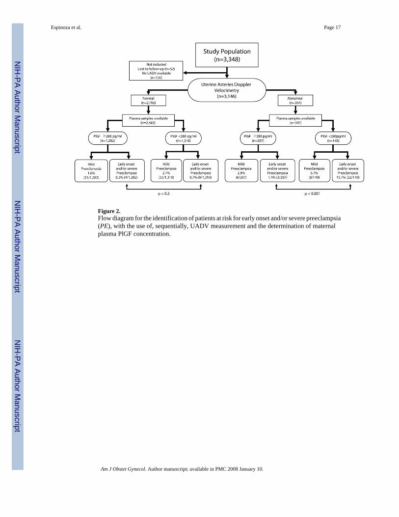
Figure 2.Flow diagram for the identification of patients at risk for early onset and/or severe preeclampsia(PE), with the use of, sequentially, UADV measurement and the determination of maternalplasma PlGF concentration.
Espinoza et al. Page 17
Am J Obstet Gynecol. Author manuscript; available in PMC 2008 January 10.
NIH
-PA Author Manuscript
NIH
-PA Author Manuscript
NIH
-PA Author Manuscript

Figure 3.Simplified flow diagram for the identification of patients at risk for early onset and/or severepreeclampsia (PE), with the use of UADV followed by maternal plasma PlGF determinations.The asterisk denotes that data for patients with normal UADV were combined, regardless ofthe plasma PlGF concentration.
Espinoza et al. Page 18
Am J Obstet Gynecol. Author manuscript; available in PMC 2008 January 10.
NIH
-PA Author Manuscript
NIH
-PA Author Manuscript
NIH
-PA Author Manuscript
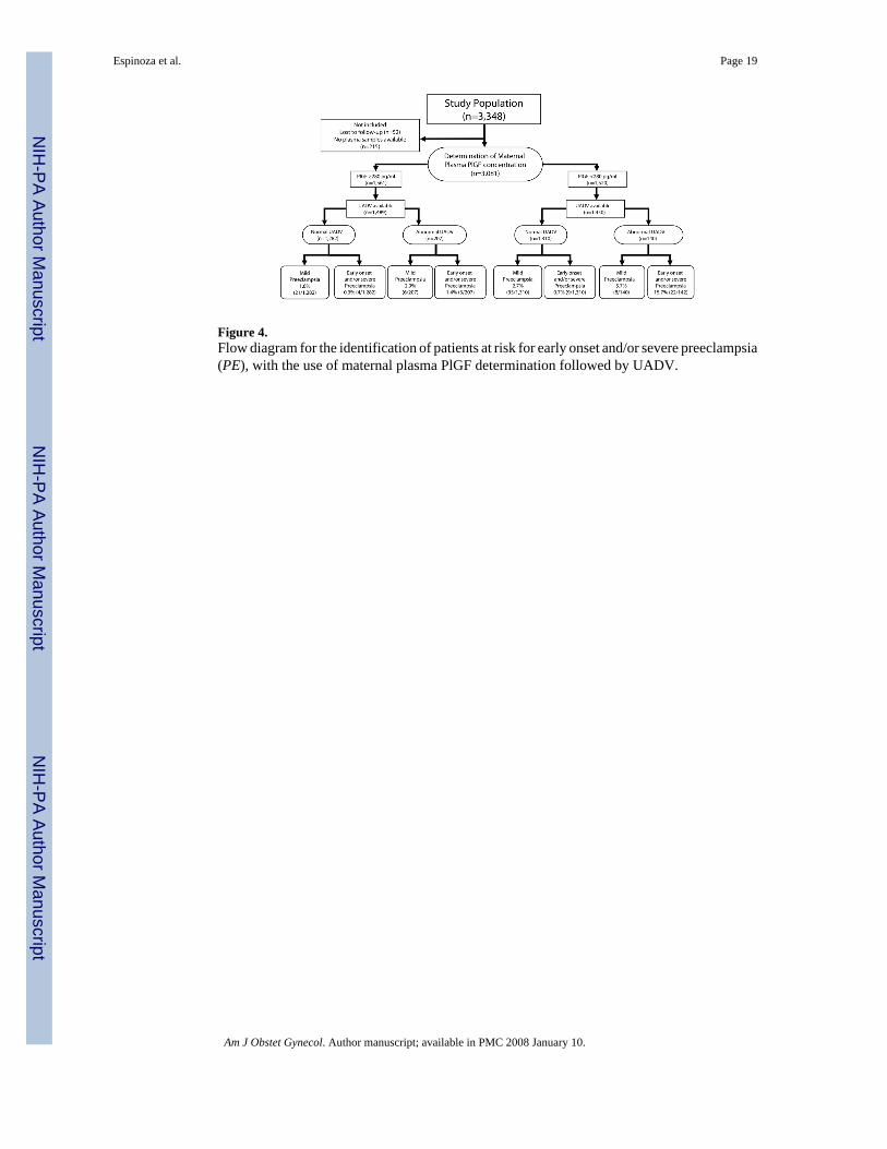
Figure 4.Flow diagram for the identification of patients at risk for early onset and/or severe preeclampsia(PE), with the use of maternal plasma PlGF determination followed by UADV.
Espinoza et al. Page 19
Am J Obstet Gynecol. Author manuscript; available in PMC 2008 January 10.
NIH
-PA Author Manuscript
NIH
-PA Author Manuscript
NIH
-PA Author Manuscript
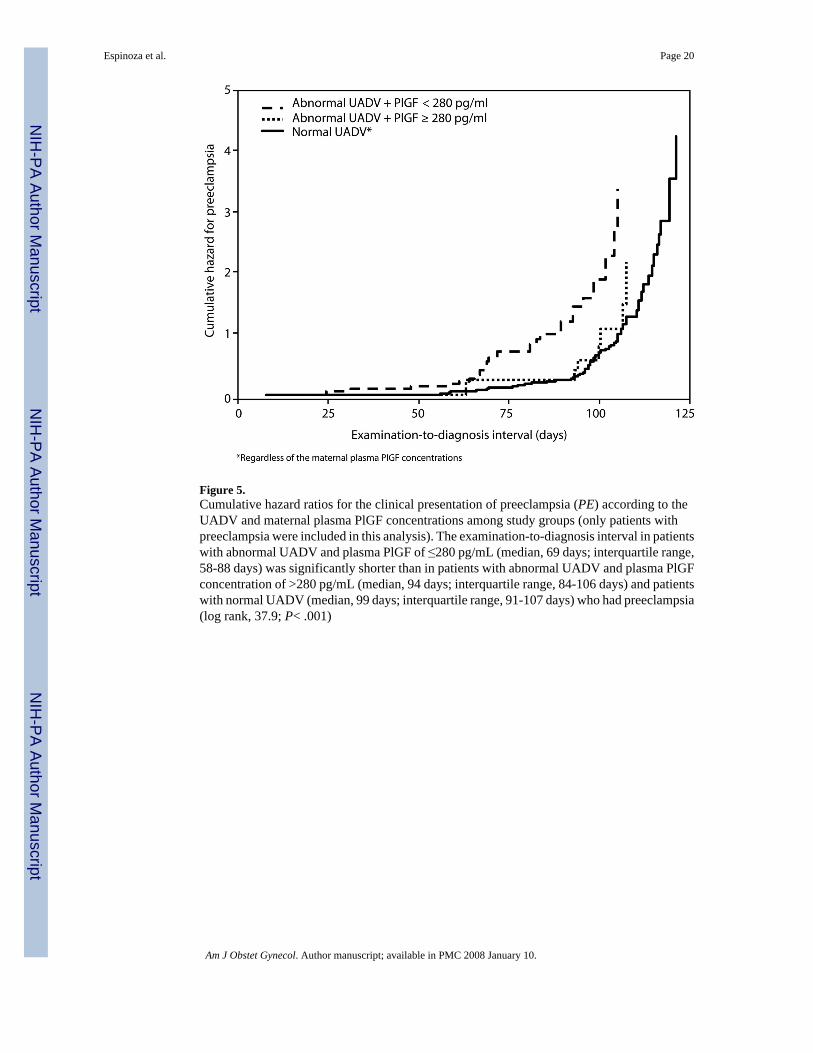
Figure 5.Cumulative hazard ratios for the clinical presentation of preeclampsia (PE) according to theUADV and maternal plasma PlGF concentrations among study groups (only patients withpreeclampsia were included in this analysis). The examination-to-diagnosis interval in patientswith abnormal UADV and plasma PlGF of ≤280 pg/mL (median, 69 days; interquartile range,58-88 days) was significantly shorter than in patients with abnormal UADV and plasma PlGFconcentration of >280 pg/mL (median, 94 days; interquartile range, 84-106 days) and patientswith normal UADV (median, 99 days; interquartile range, 91-107 days) who had preeclampsia(log rank, 37.9; P< .001)
Espinoza et al. Page 20
Am J Obstet Gynecol. Author manuscript; available in PMC 2008 January 10.
NIH
-PA Author Manuscript
NIH
-PA Author Manuscript
NIH
-PA Author Manuscript
NIH
-PA Author Manuscript
NIH
-PA Author Manuscript
NIH
-PA Author Manuscript
Espinoza et al. Page 21Ta
ble
1D
iagn
ostic
indi
ces o
f abn
orm
al U
AD
V in
the i
dent
ifica
tion
of p
atie
nts d
estin
ed to
dev
elop
pre
ecla
mps
ia, e
arly
ons
et p
reec
lam
psia
. and
/or s
ever
e pre
ecla
mps
iaan
d pa
tient
s who
se c
ondi
tion
requ
ired
deliv
ery
at ≤
34 w
eeks
of g
esta
tion
Out
com
eSe
nsiti
vity
(%)*
Spec
ifici
ty(%
)*PP
V (%
)*N
PV(%
)*L
ikel
ihoo
dra
tio [+
](9
5% C
I)
Lik
elih
ood
ratio
[−]
(95%
CI)
Pree
clam
psia
35.5
(39/
110)
89.6
(272
1/30
36)
11(3
9/35
4)97
.5(2
721/
2792
)3.
42(2
.60-
4.49
)0.
72(0
.55-
0.95
)Ea
rly o
nset
Pree
clam
psia
(≤34
wee
ks o
f ges
tatio
n)72
(18/
25)
89.6
(272
1/30
36)
5.4%
(18/
333)
99.7
(272
1/27
28)
6.94
(5.3
2-9.
05)
0.31
(0.2
4-0.
41)
Seve
re p
reec
lam
psia
72.7
(24/
33)
89.4
(278
3/31
13)
6.8
(24/
354)
99.7
(278
3/27
92)
6.86
(5.4
4-8.
66)
0.31
(0.2
4-0.
38)
Pree
clam
psia
that
requ
ired
deliv
ery
at≤3
4 w
eeks
of
gest
atio
n
86.7
(13/
15)
89.5
(272
6/30
46)
3.9
(13/
333)
99.9
(272
6/27
28)
8.25
(6.5
9-10
.32)
0.15
(0.1
2-0.
19)
* Dat
a in
par
enth
eses
repr
esen
ts p
ropo
rtion
s.
Am J Obstet Gynecol. Author manuscript; available in PMC 2008 January 10.

NIH
-PA Author Manuscript
NIH
-PA Author Manuscript
NIH
-PA Author Manuscript
Espinoza et al. Page 22
Table 2Logistic regression analysis of abnormal UADV or maternal plasma PlGF concentration of <280 pg/mL, for theprediction of the outcomes adjusted for maternal age, previous preeclampsia, nulliparity, smoking status, bodymass index, and sample storage time.
Outcome
Abnormal UADV PlGF concentration of <280 pg/mLOdds ratio 95% CI Odds ratio 95% C.I
Preeclampsia 4.3 2.82-6.66 2.6 1.67-3.94Preeclampsia≤34 weeks ofgestation 24.1 9.61-60.44 5.5 1.98-15.08Severe preeclampsia 21.1 9.47-47.14 6.5 2.59-16.34SGA withoutpreeclampsia 1.7 1.16-2.35 1.6 1.20-2.04
Am J Obstet Gynecol. Author manuscript; available in PMC 2008 January 10.

NIH
-PA Author Manuscript
NIH
-PA Author Manuscript
NIH
-PA Author Manuscript
Espinoza et al. Page 23Ta
ble
3D
iagn
ostic
ind
ices
of
a m
ater
nal
PlG
F co
ncen
tratio
n of
<28
0 pg
/mL
in t
he i
dent
ifica
tion
of p
atie
nts
dest
ined
to
deve
lop
pree
clam
psia
, ea
rly o
nset
pree
clam
psia
, and
/or s
ever
e pr
eecl
amps
ia a
nd p
atie
nts w
hose
con
dtio
n re
quire
d de
liver
y at
≤34
wee
ks o
f ges
tatio
n
Out
com
eSe
nsiti
vity
(%)*
Spec
ifici
ty(%
)*PP
V(%
)*N
PV(%
)*L
ikel
ihoo
dra
tio [+
](9
5% C
I)
Lik
elih
ood
ratio
[−]
(95%
CI)
Pree
clam
psia
69.1
(76/
110)
51.4
(153
6/29
88)
5(7
6/15
28)
97.8
(153
6/15
70)
1.42
(1.2
5-1.
62)
0.60
(0.5
3-0.
68)
Early
ons
etpr
eecl
amps
ia (≤
34w
eeks
of
gest
atio
n)80
(20/
25)
51.4
(152
7/29
71)
1.4
(20/
1464
)99
.7(1
527/
1532
)1.
65(1
.35-
2.01
)0.
39(0
.32-
0.48
)
Seve
repr
eecl
amps
ia81
.8(2
7/33
)51
.0 (1
555/
3048
)1.
8 (2
7/15
20)
99.6
(155
5/15
61)
1.67
(1.4
2-1.
97)
0.36
(0.3
0-0.
42)
Pree
clam
psia
that
requ
ired
deliv
ery
at≤3
4 w
eeks
of
gest
atio
n
86.7
(13/
15)
51.3
%(1
530/
2981
)0.
9(1
3/14
64)
99.9
(153
0/15
32)
1.78
(1.4
6-2.
18)
0.26
(0.2
1-0.
32)
* Dat
a in
par
enth
eses
repr
esen
ts p
ropo
rtion
s.
Am J Obstet Gynecol. Author manuscript; available in PMC 2008 January 10.

NIH
-PA Author Manuscript
NIH
-PA Author Manuscript
NIH
-PA Author Manuscript
Espinoza et al. Page 24Ta
ble
4D
iagn
ostic
indi
ces o
f a c
ombi
natio
n of
abn
orm
al U
AD
V a
nd m
ater
nal p
lasm
a Pl
GF
conc
entra
tion
of <
280
pg/m
L in
the
iden
tific
atio
n of
pat
ient
s des
tined
to d
evel
op p
reec
lam
psia
, ear
ly o
nset
pre
ecla
mps
ia, a
nd/o
r sev
ere
pree
clam
psia
and
pat
ient
s who
se c
ondi
tion
requ
ired
deliv
ery
at ≤
34 w
eeks
of g
esta
tion
Out
com
eSe
nsiti
vity
(%)*
Spec
ifici
ty(%
)*
Posi
tive
pred
ictiv
eva
lue
(%)*
Neg
ativ
epr
edic
tive
valu
e (%
)*
Lik
elih
ood
ratio
[+]
(95%
CI)
Lik
elih
ood
ratio
[−]
(95%
CI)
Pree
clam
psia
27.3
(30/
110)
96.4
(292
6/30
36)
21.4
(30/
140)
97.3
(292
6/30
06)
7.53
(5.2
7-10
.75)
0.75
(0.5
3-1.
08)
Early
ons
etpr
eecl
amps
ia (≤
34w
eeks
of g
esta
tion)
64(1
6/25
)96
.5(3
066/
3176
)12
.7(1
6/12
6)99
.7(3
066/
3075
)18
.48
(13.
07-2
6.13
)0.
37(0
.26-
0.53
)
Seve
re p
reec
lam
psia
63.6
(21/
33)
96.3
(313
6/32
55)
15.0
(21/
140)
99.6
(313
6/31
48)
17.4
1(1
2.74
-23.
79)
0.38
(0.2
8-0.
52)
Pree
clam
psia
that
requ
ired
deliv
ery
at≤3
4 w
eeks
of
gest
atio
n
73.3
(11/
15)
96.4
(307
1/31
86)
8.7
(11/
126)
99.9
(307
1/30
75)
20.3
2(1
4.26
-28.
95)
0.28
(0.1
9-0.
40)
* Dat
a in
par
enth
eses
repr
esen
ts p
ropo
rtion
s
Am J Obstet Gynecol. Author manuscript; available in PMC 2008 January 10.

NIH
-PA Author Manuscript
NIH
-PA Author Manuscript
NIH
-PA Author Manuscript
Espinoza et al. Page 25
Table 5Logistic regression analysis of a combination of abnormal UADV and maternal plasma PlGF concentration of<280 pg/mL for the prediction of the outcomes that are adjusted for maternal age, previous preeclampsia,nulliparity, smoking status, body mass index, and sample storage time.
Outcome
Abnormal UADV + PlGF concentration of <280pg/mL
Odds ratio 95% CI
Preeclampsia 8.6 5.35-13.74Preeclampsia ≤34weeks of gestation 43.8 18.48-103.89Severe preeclampsia 37.4 17.64-79.07SGA withoutpreeclampsia 2.7 1.73-4.26
Am J Obstet Gynecol. Author manuscript; available in PMC 2008 January 10.

NIH
-PA Author Manuscript
NIH
-PA Author Manuscript
NIH
-PA Author Manuscript
Espinoza et al. Page 26
Table 6Demographic and clinical characteristics of the study population according to UADV and maternal plasma PlGFconcentration.
Variable Normal UADV*(n=2792)
Abnormal UADV +PlGF concentration
of ≥280 pg/mL(n=207)
Abnormal UADV +PlGF concentration
of <280 pg/mL(n=140)
P value
Maternal age (y)† 27(14-46)
23(14-43)
24(16-42) <.001
Nulliparity (%)‡ 33.9(947/2792)
57(118/207)
54.3(76/140) <.001
Preeclampsia in aprevious pregnancy(%)‡
2(56/2792)
1.9(4/207)
3.6(5/140) NS
Body mass index(kg/m2)†
24.5(16.2-89)
23.1(15.9-37.1)
24.6(18.0-44.0) <.001
Smokers (%)‡ 7.4(207/2792)
8.7(18/207)
3.6%(5/140) NS
Gestational age atultrasound (wk)§ 24.1 ± 0.64 24.1 ± 0.60 24.1 ± 0.64 NS
Maternal plasmaPlGF (pg/mL)†
279.1(8.9-2572)
456.8(282.3-2040)
172.9(0-279.8) <.001
Maternal plasmasVEGFR-1(pg/mL)†
- 844(0-2550)
735(0-9450) NS
Mean uterine arterypulsatility index†
0.77(0.3-1.4)
1.10(0.6-2.7)
1.36(0.6-2.8) <.001
*Regardless of the maternal plasma PlGF concentrations.
†Data are given as median (range)
‡Data in parentheses represents proportions
§Data are given as mean ± SD
Am J Obstet Gynecol. Author manuscript; available in PMC 2008 January 10.
NIH
-PA Author Manuscript
NIH
-PA Author Manuscript
NIH
-PA Author Manuscript
Espinoza et al. Page 27
Table 7Clinical outcomes of the population according to UADV and maternal plasma PlGF concentration.
Variable Normal UADV(n=2792)
Abnormal UADV +PlGF concentration of
≥280 pg/mL(n=207)
Abnormal UADV +PlGF concentration of
<280 pg/mL(n=140)
P value
Preeclampsia 2.5(71/2792)
4.3(9/207)
21.4(30/140) <.001
Early onsetpreeclampsia(≥34 weeks ofgestation)
0.3(7/2728)
1(2/200)
12.7(16/126) <.001
Severe preeclampsia 0.3(9/2792)
1.4(3/207)
15(21/140) <.001
Birthweight <10th
percentile*8.1
(225/2786)8.7
(18/207)20
(28/140) <.001
Birthweight <5th
percentile*3.5
(97/2786)4.8
(10/207)10.7
(15/140) <.001
Abruptio placentae 0.9(24/2792)
1.9(4/207)
3.6(5/140) .001
Eclampsia 0.03(1/2792) 0 0.7
(1/140) .01Spontaneous pretermdelivery (≥35 weeks ofgestation)
1.5(41/2787)
1.9(4/207)
3.6(5/140) NS
Spontaneous pretermdelivery (≥32 weeks ofgestation)
0.4(10/2787)
0.5(1/207)
2.2(3/140) NS
Composite of severeneonatal morbidity
0.4(12/2792)
1.9(4/207)
3.6(5/140) <.001
The results are expressed as percentages and proportions. NS, not significant
*In the absence of preeclampsia
Am J Obstet Gynecol. Author manuscript; available in PMC 2008 January 10.
Copyright © 2022 FDOKUMEN