Identification of highly conserved regions in L-segment of Crimean–Congo hemorrhagic fever virus...
10
© 2015 Oany et al. This work is published by Dove Medical Press Limited, and licensed under Creative Commons Attribution – Non Commercial (unported, v3.0) License. The full terms of the License are available at http://creativecommons.org/licenses/by-nc/3.0/. Non-commercial uses of the work are permitted without any further permission from Dove Medical Press Limited, provided the work is properly attributed. Permissions beyond the scope of the License are administered by Dove Medical Press Limited. Information on how to request permission may be found at: http://www.dovepress.com/permissions.php Advances and Applications in Bioinformatics and Chemistry 2015:8 1–10 Advances and Applications in Bioinformatics and Chemistry Dovepress submit your manuscript | www.dovepress.com Dovepress 1 ORIGINAL RESEARCH open access to scientific and medical research Open Access Full Text Article http://dx.doi.org/10.2147/AABC.S75250 Identification of highly conserved regions in L-segment of Crimean–Congo hemorrhagic fever virus and immunoinformatic prediction about potential novel vaccine Arafat Rahman Oany 1 Shah Adil Ishtiyaq Ahmad 1 Mohammad Uzzal Hossain 1 Tahmina Pervin Jyoti 2 1 Department of Biotechnology and Genetic Engineering, Faculty of Life Science, Mawlana Bhashani Science and Technology University, Santosh, Tangail, Bangladesh; 2 Biotechnology and Genetic Engineering Discipline, Life Science School, Khulna University, Khulna, Bangladesh Correspondence: Arafat Rahman Oany Department of Biotechnology and Genetic Engineering, Faculty of Life Science, Mawlana Bhashani Science and Technology University, Santosh, Tangail 1902, Bangladesh Tel +880 15 5881 9130 Email [email protected] Abstract: Crimean–Congo hemorrhagic fever (CCHF) is a tick-borne zoonotic viral disease with a disease fatality rate between 15% and 70%. Despite the wide range of distribution, the virus (CCHFV) is basically endemic in Africa, Asia, eastern Europe, and the Middle East. Acute febrile illness associated with petechiae, disseminated intravascular coagulation, and multiple-organ failure are the main symptoms of the disease. With all these fatal effects, CCHFV is considered a huge threat as no successful therapeutic approach is currently available for the treatment of this disease. In the present study, we have used the immunoinformatics approach to design a potential epitope-based vaccine against the RNA-dependent RNA polymerase-L of CCHFV. Both the T-cell and B-cell epitopes were assessed, and the epitope “DCSSTPPDR” was found to be the most potential one, with 100% conservancy among all the strains of CCHFV. The epitope was also found to interact with both type I and II major histocompatibility complex molecules and is considered nonallergenic as well. In vivo study of our proposed peptide is advised for novel universal vaccine production, which might be an effective path to prevent CCHF disease. Keywords: single-stranded RNA, immunoinformatics, RNA-dependent RNA polymerase, epitope Introduction Crimean–Congo hemorrhagic fever virus (CCHFV) is the causative agent of the hemorrhagic fever, which was first described in Cremia in 1945. 1 The virus consists of a negative-sense single-stranded RNA, and its tripartite genome comprises the small (S), medium (M), and large (L) segments, which encode the viral nucleocapsid (N), glycoprotein precursor, and polymerase-L proteins, respectively. 2 Africa, Asia, eastern Europe, and the Middle East are the hotspots of this viral infection, with a high fatality rate between 15% and 70%. 3,4 In case of human infection, the progres- sion of this disease is very rapid and it causes acute febrile illness, associated with petechiae, ecchymosis, disseminated intravascular coagulation, and multiple-organ failure. 5 A wide range of ticks that belong to the Hyalomma genus are considered the main vectors of this viral disease and the disease is spread through the bite of these ticks to humans or other domestic animals. 6 Due to inadequate hospital care, the disease is also spread nosocomially and a case reported that, in South Africa, about 33% medical personnel are infected through needlestick injury. 7 Alternate routes of infection, such as aerosol and droplet respiratory route of infection, were also suspected for several cases in Russia. 8 With a broad geographic distribution,
Transcript of Identification of highly conserved regions in L-segment of Crimean–Congo hemorrhagic fever virus...
© 2015 Oany et al. This work is published by Dove Medical Press Limited, and licensed under Creative Commons Attribution – Non Commercial (unported, v3.0) License. The full terms of the License are available at http://creativecommons.org/licenses/by-nc/3.0/. Non-commercial uses of the work are permitted without any further
permission from Dove Medical Press Limited, provided the work is properly attributed. Permissions beyond the scope of the License are administered by Dove Medical Press Limited. Information on how to request permission may be found at: http://www.dovepress.com/permissions.php
Advances and Applications in Bioinformatics and Chemistry 2015:8 1–10
Advances and Applications in Bioinformatics and Chemistry Dovepress
submit your manuscript | www.dovepress.com
Dovepress 1
O r i g i n A l r e s e A r C h
open access to scientific and medical research
Open Access Full Text Article
http://dx.doi.org/10.2147/AABC.S75250
Identification of highly conserved regions in L-segment of Crimean–Congo hemorrhagic fever virus and immunoinformatic prediction about potential novel vaccine
Arafat rahman Oany1
shah Adil ishtiyaq Ahmad1
Mohammad Uzzal hossain1
Tahmina Pervin Jyoti2
1Department of Biotechnology and Genetic Engineering, Faculty of Life Science, Mawlana Bhashani Science and Technology University, Santosh, Tangail, Bangladesh; 2Biotechnology and Genetic Engineering Discipline, Life Science School, Khulna University, Khulna, Bangladesh
Correspondence: Arafat rahman Oany Department of Biotechnology and Genetic Engineering, Faculty of Life Science, Mawlana Bhashani Science and Technology University, Santosh, Tangail 1902, Bangladesh Tel +880 15 5881 9130 email [email protected]
Abstract: Crimean–Congo hemorrhagic fever (CCHF) is a tick-borne zoonotic viral disease with
a disease fatality rate between 15% and 70%. Despite the wide range of distribution, the virus
(CCHFV) is basically endemic in Africa, Asia, eastern Europe, and the Middle East. Acute febrile
illness associated with petechiae, disseminated intravascular coagulation, and multiple-organ
failure are the main symptoms of the disease. With all these fatal effects, CCHFV is considered
a huge threat as no successful therapeutic approach is currently available for the treatment of this
disease. In the present study, we have used the immunoinformatics approach to design a potential
epitope-based vaccine against the RNA-dependent RNA polymerase-L of CCHFV. Both the
T-cell and B-cell epitopes were assessed, and the epitope “DCSSTPPDR” was found to be the
most potential one, with 100% conservancy among all the strains of CCHFV. The epitope was
also found to interact with both type I and II major histocompatibility complex molecules and
is considered nonallergenic as well. In vivo study of our proposed peptide is advised for novel
universal vaccine production, which might be an effective path to prevent CCHF disease.
Keywords: single-stranded RNA, immunoinformatics, RNA-dependent RNA polymerase,
epitope
IntroductionCrimean–Congo hemorrhagic fever virus (CCHFV) is the causative agent of the
hemorrhagic fever, which was first described in Cremia in 1945.1 The virus consists
of a negative-sense single-stranded RNA, and its tripartite genome comprises the
small (S), medium (M), and large (L) segments, which encode the viral nucleocapsid
(N), glycoprotein precursor, and polymerase-L proteins, respectively.2 Africa, Asia,
eastern Europe, and the Middle East are the hotspots of this viral infection, with a
high fatality rate between 15% and 70%.3,4 In case of human infection, the progres-
sion of this disease is very rapid and it causes acute febrile illness, associated with
petechiae, ecchymosis, disseminated intravascular coagulation, and multiple-organ
failure.5 A wide range of ticks that belong to the Hyalomma genus are considered
the main vectors of this viral disease and the disease is spread through the bite of
these ticks to humans or other domestic animals.6 Due to inadequate hospital care,
the disease is also spread nosocomially and a case reported that, in South Africa,
about 33% medical personnel are infected through needlestick injury.7 Alternate
routes of infection, such as aerosol and droplet respiratory route of infection, were
also suspected for several cases in Russia.8 With a broad geographic distribution,

Advances and Applications in Bioinformatics and Chemistry 2015:8
Figure 1 Phylogenetic tree showing the evolutionary divergence among the different RNA-dependent RNA polymerase-L molecules of the CCHFV.Notes: Here, cladogram view is shown with appropriate distance among the different strains. The blue dotted view indicates the node of the tree.Abbreviation: CCHFV, Crimean–Congo hemorrhagic fever virus.
submit your manuscript | www.dovepress.com
Dovepress
Dovepress
2
Oany et al
this virus already covers .30 countries across Africa,
southeastern Europe, the Middle East, western Asia,
and more recently, some places of southwestern Europe,
particularly Spain.9–11 The wide distribution of this virus
indicates the ability of the tick hosts to adapt across dif-
ferent environmental and geographic regions.12 Despite
the outbreaks, there is no reliable vaccine or drug for the
treatment of CCHFV infection in animals or humans.13
As a single-stranded-RNA-containing genome and due
to the error-prone nature of its polymerase, the CCHFV
incorporates random mutations into the genome. The rate
of recombination in its RNA is also very high. As a con-
sequence, the development of a vaccine or antiviral drug
against CCHFV is very difficult.14
The design of epitope-based vaccines against some deadly
viruses has gained much popularity due to its increased
potency and safety.15,16 The application of bioinformatics
in immunology, which is also termed immunoinformatics,
is now widely accepted. Immunoinformatics can assist in
designing new vaccines through the identification of poten-
tial T-cell epitope, B-cell epitope, and human leukocyte
antigen (HLA) ligands.17–19 This novel approach has proven
its efficacy in the case of multiple sclerosis,20 malaria,21
human immunodeficiency virus,22 and tuberculosis,23 with
desired results. In our present study, we have proposed the
design of a potential conserved epitope candidate through
the immunoinformatics approaches in order to minimize
the fatal effects of the CCHFV, with the expectation of
finding a novel candidate for the vaccine via wet laboratory
validation.
Materials and methodssequence retrieval and conserved region identificationUniProtKB24 database was used for the retrieval of the
sequences of RNA-dependent RNA polymerase-L25 and
Advances and Applications in Bioinformatics and Chemistry 2015:8
Table 1 Antigenicity determination of the conserved peptide by Vaxijen server
Peptide number
Peptide Region Vaxijen score (threshold: 0.4)
1 LIQTLFPDKFEDFLDRTQLHPEFRDLTPDFSLTQKVHFKRNQIPSVEN VQISIDATLPESVEAVPVTERKMFPLPETPLSEVHSIERIMENFTRLM
3,563–3,658 0.6556
2 DYGERGIVEENHMKFSGEDQLETRQLLLVEVGFQTDIDGKIRT DHKKWKDILKLLELLGIKCSFIACADCSSTPPDRWWI
3,694–3,773 0.4268
3 EDRVRVLKNSVSFLFNKLSRNSPTEVTDIVVGAISTQKVRSY LKAGTATKTPVSTKDVLETWEK
3,775–3,838 0.3662
4 MKEHILNRPTGLTLPTSLEQAMRKGLVEGVVISKEGSESCINMLK ENLDRITDEFERTKFKHELTQNITTSEKML
3,841–3,915 0.1044
Figure 2 MSA of the conserved region of RNA-dependent RNA polymerase-L. Only the partial sequences containing the proposed epitope sequence are shown here.Notes: Clustalx color is used here. Different colors indicate different amino acid residues.Abbreviation: MSA, multiple-sequence alignment.
submit your manuscript | www.dovepress.com
Dovepress
Dovepress
3
Highly conserved antigenic epitope for CCHF vaccine designing
the envelope glycoprotein26 of the CCHFV in the FASTA
protein format.
BioEdit v7.2.3 sequence alignment editor27 was used
for the identification of the conserved region among the
sequences through multiple-sequence alignment (MSA) with
ClustalW.28 Finally, Jalview v2 tool29 was used to retrieve the
alignment and the CLC Sequence Viewer v7.0.2 (http://www.
clcbio.com) was used for analysis of the divergence among
the different strains of the CCHFV.
Antigenicity determination of the conserved peptidesVaxiJen v2. 0, a Web-based server,30 was used for the deter-
mination of the antigenicity of the conserved sequences.
Herein, we used the default parameters for the prediction,
with a threshold value of 0.4.
T-cell epitope predictionTwo online servers were used for the prediction of the
T-cell epitope. Initially, the NetCTL v1.2 server was used
for the identification of the potential T-cell epitope.31 We
used the default approach to predict the epitopes, including
major histocompatibility complex class I (MHC-I) bind-
ing, proteasomal C terminal cleavage, and transporter of
antigenic peptide (TAP) transport efficiency. The epitope
prediction was restricted to 12 MHC-I supertypes. MHC-I
binding and proteasomal cleavage were performed through
artificial neural networks and the weight matrix was used
Advances and Applications in Bioinformatics and Chemistry 2015:8
Table 2 Prediction of the T-cell epitope by netCTl server on the basis of combined score
Number Epitope Combined score (nM)
1 CSSTPPDRW 1.72352 DVLETWEKM 1.64913 WEKMKEHIL 1.50894 ERTKFKHEL 1.49555 VPVTERKMF 1.4734
Table 3 Prediction of the T-cell epitope by CTlPred server
Epitope Start position Score (ANN/SVM)
lnrPTglTl 246 1.00/–0.23718777FSLTQKVHF 30 0.99/–0.23718777ADCssTPPD 164 0.99/–0.23718777LSRNSPTEV 194 0.99/–0.23718777TSLEQAMRK 256 0.99/–0.23718777
Abbreviations: ANN/SVM, artificial neural networks/support vector machines; CTLP, Cytotoxic T Lymphocyte Prediction (CTLPred).
Table 4 MhC-i and MhC-ii interaction of the proposed sequence by ieDB-Ar
Epitope MHC interaction
MHC-I interaction analysisDCssTPPDr HLA-C*12:03, HLA-C*03:03, HLA-
C*14:02, HLA-B*58:01, HLA-C*12:03, HLA-C*03:03, HLA-C*15:02, HLA-B*57:01
MHC-II interaction analysisFIACADCSSTPPDRW HLA-DRB1*04:01, HLA-DRB1*03:01,
HLA-DRB1*08:02, HLA-DRB3*01:01, DQB1*03:01, HLA-DRB1*09:01, HLA-DRB5*01:01, HLA-DRB1*04:05, DQB1*04:02, HLA-DRB1*11:01, HLA-DRB1*07:01, DQB1*02:01, HLA-DRB1*13:02, DQB1*03:02, DQB1*05:01, HLA-DRB4*01:01, HLA-DRB1*01:01, HLA-DRB3*02:02, DPB1*04:01, DQB1*06:02, DPB1*14:01, HLA-DRB1*15:01, DPB1*02:01, HLA-DRB1*12:01, DPB1*04:02, DPB1*05:01, DPB1*01:01
Abbreviations: IEDB-AR, Immune Epitope Database and Analysis Resource; MHC, major histocompatibility complex.
submit your manuscript | www.dovepress.com
Dovepress
Dovepress
4
Oany et al
for estimating TAP transport efficiency. The threshold for
epitope identification was set at 0.5 to maintain sensitivity and
specificity of 0.89 and 0.94, respectively. Finally, CTLPred32
was implemented additionally for further confirmation about
the prediction with default parameters.
MhC-i and MhC-ii restriction analysisT Cell Epitope Prediction Tools from Immune Epitope
Database and Analysis Resource (IEDB-AR) was used for
the prediction of MHC-I33 and MHC-II34,35 binding of the
peptide. The Stabilized Matrix Method36 was used to cal-
culate the half-maximal inhibitory concentration (IC50
) of
peptide binding to MHC-I molecules from different predic-
tion methods, with a preselected 9.0-mer epitope. In case of
MHC-II binding analysis, the IEDB-recommended method
was used for the specific HLA-DQ, HLA-DP, and HLA-DR
loci. Here, we used specific peptides for the prediction of
MHC-II interaction on the basis of the antigenic conservancy
and MHC-I analysis.
B-cell epitope predictionB-cell epitope initiates immunoresponse through the inter-
action with B lymphocytes and causes the differentiation of
B lymphocytes into plasma and memory cells.37 IEDB-AR
hosts a number of Web-based tools for the prediction of
B-cell epitope. Multiple tools, including the Kolaskar and
Tongaonkar antigenicity scale,38 Emini surface accessibility
prediction39 and Bepipred linear epitope prediction analy-
sis, were used for the B-cell epitope prediction with high
accuracy.40
Homology modeling and protein variability determination of the conserved regionHomology model of the conserved region was obtained
by MODELLER v9,41 and the predicted model was
assessed by PROCHECK42 and QMEAN43 servers of the
SWISS-MODEL Workspace.44 For the disorder prediction
among the amino acid sequences, DISOPRED v345 was
used. The Protein variability server was used to calcu-
late protein variability index using Shannon variability
coefficient.46
Allergenicity and epitope conservancy analysisIn order to predict the allergenicity of the proposed epitopes
with high accuracy, a Web-based server AlgPred47 was used.
Herein, we used a hybrid prediction (SVMc + IgEepitope +
ARPs BLAST + MAST) approach to predict the allerge-
nicity with an accuracy of about 86% at a threshold value
of −0.4. The prediction procedure follows the guidelines
of the Food and Agriculture Organization/World Health
Organization, 2003. A Web-based tool from IEDB-AR48
was used in order to identify the specific conservancy of
the proposed epitopes.
Advances and Applications in Bioinformatics and Chemistry 2015:8
Table 5 Kolaskar and Tongaonkar antigenicity analysis
Number Start position End position Peptide Peptide length
1 16 23 rTQlhPeF 82 28 39 PDFSLTQKVHFK 123 43 67 IPSVENVQISIDVTLPESVEAVPVT 254 72 85 FPLPETPLSEVHSI 145 117 130 QSAVEHESPSISAF 146 154 163 TRQLLLVEVG 107 182 204 ILKLLELLGIKCSFIACADCSST 238 215 229 RVRVLKNSVSFLFNK 159 238 246 VTDIVVGAI 910 248 258 TQKVRSYLKAG 1111 262 273 KTPVSTKDVLET 1212 287 295 glTlPAsle 913 302 310 LVEGVVISK 914 314 319 esCinM 6
submit your manuscript | www.dovepress.com
Dovepress
Dovepress
5
Highly conserved antigenic epitope for CCHF vaccine designing
ResultsAnalysis of the retrieved sequences and their divergenceA total of 80 envelope glycoproteins and 34 RNA-dependent
RNA polymerase-L molecules from different variants of
the CCHFV were retrieved from the UniProt database. The
MSA of two different types of proteins was retrieved from
BioEdit tool through ClustalW with 1,000 bootstrap replicates
(Figures S1 and S2). Conserved regions were grouped for
the antigenic property analysis. CLC Sequence Viewer was
used to construct phylograms for both proteins from the MSA
obtained from BioEdit, in order to analyze the divergence
120Threshold
110
115
100
105
0.95
0.90
0.850 20 40 60 80 100 120 140 160 180
Position
Sco
re
200 220 240 260 280 300 320 340 360
Figure 3 Kolaskar and Tongaonkar antigenicity prediction of the conserved peptide, ranging from 3,563 to 3,915 MSA number.Notes: The region from 197 to 202 is the proposed epitope. The X- and Y-axes represent the sequence position and antigenic propensity score, respectively. The threshold value is 1.0. The regions above the threshold are antigenic, shown in yellow.Abbreviation: MSA, multiple-sequence alignment.
among the retrieved sequences. Phylogram of RNA-dependent
RNA polymerase-L is depicted in Figure 1 and the phylogram
for the envelope glycoprotein is provided in Figure S3.
Antigenic peptide identificationInitially, the conserved sequences (MSA number: 3,563–
3,915) of RNA-dependent RNA polymerase-L were separated
into four conserved peptides, according to their continuity in
the MSA. Then the VaxiJen v2.0 server was used to predict the
antigenicity of all the grouped conserved peptides (Table 1).
On the basis of the VaxiJen score, the top two conserved
peptides (MSA number: 3,563–3,658 and 3,694–3,773) were
Advances and Applications in Bioinformatics and Chemistry 2015:8submit your manuscript | www.dovepress.com
Dovepress
Dovepress
6
Oany et al
1.6Threshold
1.4
0.8
1.0
1.2
0.6
0.4
0.20 20
Position
Sco
re
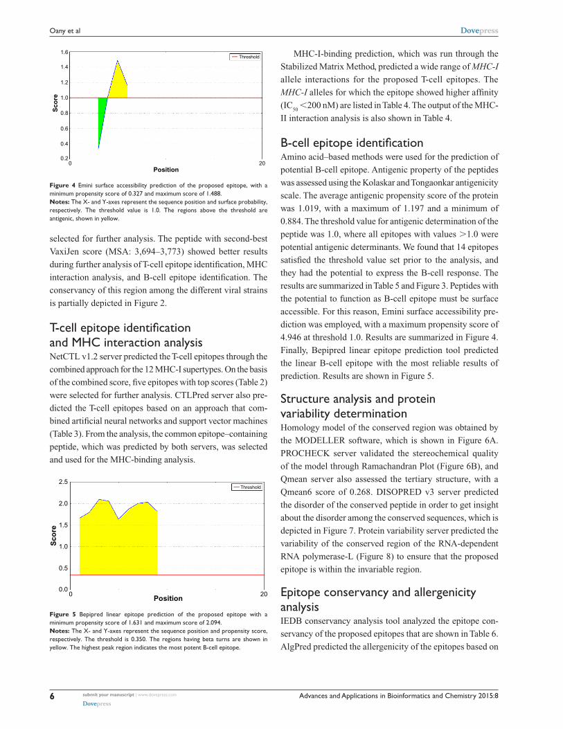
Figure 4 Emini surface accessibility prediction of the proposed epitope, with a minimum propensity score of 0.327 and maximum score of 1.488.Notes: The X- and Y-axes represent the sequence position and surface probability, respectively. The threshold value is 1.0. The regions above the threshold are antigenic, shown in yellow.
2.5Threshold
2.0
1.5
1.0
0.5
0.00 20
Position
Sco
re
Figure 5 Bepipred linear epitope prediction of the proposed epitope with a minimum propensity score of 1.631 and maximum score of 2.094.Notes: The X- and Y-axes represent the sequence position and propensity score, respectively. The threshold is 0.350. The regions having beta turns are shown in yellow. The highest peak region indicates the most potent B-cell epitope.
MHC-I-binding prediction, which was run through the
Stabilized Matrix Method, predicted a wide range of MHC-I
allele interactions for the proposed T-cell epitopes. The
MHC-I alleles for which the epitope showed higher affinity
(IC50
,200 nM) are listed in Table 4. The output of the MHC-
II interaction analysis is also shown in Table 4.
B-cell epitope identificationAmino acid–based methods were used for the prediction of
potential B-cell epitope. Antigenic property of the peptides
was assessed using the Kolaskar and Tongaonkar antigenicity
scale. The average antigenic propensity score of the protein
was 1.019, with a maximum of 1.197 and a minimum of
0.884. The threshold value for antigenic determination of the
peptide was 1.0, where all epitopes with values .1.0 were
potential antigenic determinants. We found that 14 epitopes
satisfied the threshold value set prior to the analysis, and
they had the potential to express the B-cell response. The
results are summarized in Table 5 and Figure 3. Peptides with
the potential to function as B-cell epitope must be surface
accessible. For this reason, Emini surface accessibility pre-
diction was employed, with a maximum propensity score of
4.946 at threshold 1.0. Results are summarized in Figure 4.
Finally, Bepipred linear epitope prediction tool predicted
the linear B-cell epitope with the most reliable results of
prediction. Results are shown in Figure 5.
structure analysis and protein variability determinationHomology model of the conserved region was obtained by
the MODELLER software, which is shown in Figure 6A.
PROCHECK server validated the stereochemical quality
of the model through Ramachandran Plot (Figure 6B), and
Qmean server also assessed the tertiary structure, with a
Qmean6 score of 0.268. DISOPRED v3 server predicted
the disorder of the conserved peptide in order to get insight
about the disorder among the conserved sequences, which is
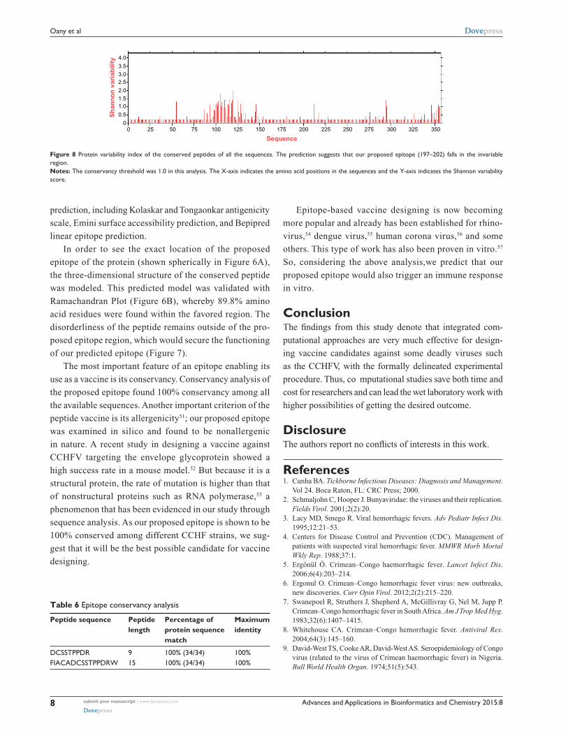
depicted in Figure 7. Protein variability server predicted the
variability of the conserved region of the RNA-dependent
RNA polymerase-L (Figure 8) to ensure that the proposed
epitope is within the invariable region.
Epitope conservancy and allergenicity analysisIEDB conservancy analysis tool analyzed the epitope con-
servancy of the proposed epitopes that are shown in Table 6.
AlgPred predicted the allergenicity of the epitopes based on
selected for further analysis. The peptide with second-best
VaxiJen score (MSA: 3,694–3,773) showed better results
during further analysis of T-cell epitope identification, MHC
interaction analysis, and B-cell epitope identification. The
conservancy of this region among the different viral strains
is partially depicted in Figure 2.
T-cell epitope identification and MhC interaction analysisNetCTL v1.2 server predicted the T-cell epitopes through the
combined approach for the 12 MHC-I supertypes. On the basis
of the combined score, five epitopes with top scores (Table 2)
were selected for further analysis. CTLPred server also pre-
dicted the T-cell epitopes based on an approach that com-
bined artificial neural networks and support vector machines
(Table 3). From the analysis, the common epitope–containing
peptide, which was predicted by both servers, was selected
and used for the MHC-binding analysis.

Advances and Applications in Bioinformatics and Chemistry 2015:8 submit your manuscript | www.dovepress.com
Dovepress
Dovepress
7
Highly conserved antigenic epitope for CCHF vaccine designing
180BA
135
90
0
−45
45
−90
−135
−180 −135 −90 −45 0 45 90Phi (degrees)
Psi
(d
egre
es)
135 180
Figure 6 Three-dimensional structure prediction and validation.Notes: (A) Three-dimensional model of the conserved region. Here, the epitope “DCSSTPPDR” is shown spherically. The outerside location of the epitope indicates its surface accessibility. (B) Ramachandran plot of the predicted model shows that most of the residues are in the allowed region of the plot, proving the validity of the model.
1
0.6
0.8
0.4
Co
nfi
den
ce s
core
0.2
00 50 100 150 200 250
Amino acid position300 350 400
Disordered stateProtein binding
Figure 7 Disorder prediction of the conserved antigenic amino acid sequences. Here, our proposed epitope lies outside (197–202) of the disordered region to secure its potentiality as an effective epitope.Notes: Amino acids in the input sequence are considered disordered when the blue line is above the gray dashed line, that is, when the confidence score is .0.5. The orange line shows the confidence score of the disordered protein-binding residue predictions.
amino acid composition. The prediction score of AlgPred
for the two epitopes in combination was 0.49752311 at
threshold of –0.4.
DiscussionWith a widely distributed endemically affected region
and a randomly mutated genome, CCHFV imposes a
great challenge to researchers in developing a success-
ful therapeutic approach against it. The ability of an
epitope-based vaccine to stimulate an effective specific
immune response with a minute structure and without any
unexpected side effects has made it a good choice for vac-
cine development.49 In this instance, we started with the
preferable target, namely, the envelope glycoprotein, but
failed to identify any unique conserved region (Figure S1;
multiple-sequence alignment of the envelope glycoprotein
of CCHFV.) to design a peptide vaccine against the enve-
lope glycoprotein. This was also revealed by phylogeny
analysis, which is shown in Figure S3; phylogenetic tree
showing the evolutionary divergence among the different
envelope glycoproteins of CCHFV. RNA-dependent RNA
polymerase, a product of the L-segment of the genome,
comprises a unique conserved region among all the avail-
able strains of CCHFV (Figure S2; multiple-sequence
alignment of the RNA-dependent RNA polymerase-L of
CCHFV). This was the pedestal to think about a novel
vaccine candidate. To ensure a firm immune response, we
looked for the activation of both T-cell and B-cell immu-
nity with a single epitope.50 Antigenicity of the conserved
peptides indicated their ability to provoke potential immune
response and they were used for further analysis involving
T-cell epitope prediction. Through the analysis of the out-
put of both NetCTL and CTLPred, it was found that the
epitope “DCSSTPPDR” would be the best candidate for the
activation of T-cell immunity with potential antigenicity.
Analysis of the MHC ligands for both type I and II revealed
that the core epitope “DCSSTPPDR” would interact with
the highest number of HLA molecules and that it would
support the MHC molecules to present the epitope on the
T-cell surface. The complete peptide for MHC-II restriction
was FIACADCSSTPPDRW. The peptide DCSSTPPDR was
also found to be the most potential candidate to raise B-cell
immune response by amino acid–based B-cell epitope
Advances and Applications in Bioinformatics and Chemistry 2015:8submit your manuscript | www.dovepress.com
Dovepress
Dovepress
8
Oany et al
prediction, including Kolaskar and Tongaonkar antigenicity
scale, Emini surface accessibility prediction, and Bepipred
linear epitope prediction.
In order to see the exact location of the proposed
epitope of the protein (shown spherically in Figure 6A),
the three-dimensional structure of the conserved peptide
was modeled. This predicted model was validated with
Ramachandran Plot (Figure 6B), whereby 89.8% amino
acid residues were found within the favored region. The
disorderliness of the peptide remains outside of the pro-
posed epitope region, which would secure the functioning
of our predicted epitope (Figure 7).
The most important feature of an epitope enabling its
use as a vaccine is its conservancy. Conservancy analysis of
the proposed epitope found 100% conservancy among all
the available sequences. Another important criterion of the
peptide vaccine is its allergenicity51; our proposed epitope
was examined in silico and found to be nonallergenic
in nature. A recent study in designing a vaccine against
CCHFV targeting the envelope glycoprotein showed a
high success rate in a mouse model.52 But because it is a
structural protein, the rate of mutation is higher than that
of nonstructural proteins such as RNA polymerase,53 a
phenomenon that has been evidenced in our study through
sequence analysis. As our proposed epitope is shown to be
100% conserved among different CCHF strains, we sug-
gest that it will be the best possible candidate for vaccine
designing.
Table 6 epitope conservancy analysis
Peptide sequence Peptide length
Percentage of protein sequence match
Maximum identity
DCssTPPDr 9 100% (34/34) 100%FIACADCSSTPPDRW 15 100% (34/34) 100%
4.0
3.5
3.02.5
1.52.0
1.0
0.5
00 25 50 75 100 125 150 175 200
Sequence
Sh
ann
on
var
iab
ility
225 250 275 300 325 350
Figure 8 Protein variability index of the conserved peptides of all the sequences. The prediction suggests that our proposed epitope (197–202) falls in the invariable region.Notes: The conservancy threshold was 1.0 in this analysis. The X-axis indicates the amino acid positions in the sequences and the Y-axis indicates the Shannon variability score.
Epitope-based vaccine designing is now becoming
more popular and already has been established for rhino-
virus,54 dengue virus,55 human corona virus,56 and some
others. This type of work has also been proven in vitro.57
So, considering the above analysis,we predict that our
proposed epitope would also trigger an immune response
in vitro.
ConclusionThe findings from this study denote that integrated com-
putational approaches are very much effective for design-
ing vaccine candidates against some deadly viruses such
as the CCHFV, with the formally delineated experimental
procedure. Thus, co mputational studies save both time and
cost for researchers and can lead the wet laboratory work with
higher possibilities of getting the desired outcome.
DisclosureThe authors report no conflicts of interests in this work.
References1. Cunha BA. Tickborne Infectious Diseases: Diagnosis and Management.
Vol 24. Boca Raton, FL: CRC Press; 2000.2. Schmaljohn C, Hooper J. Bunyaviridae: the viruses and their replication.
Fields Virol. 2001;2(2):20.3. Lacy MD, Smego R. Viral hemorrhagic fevers. Adv Pediatr Infect Dis.
1995;12:21–53.4. Centers for Disease Control and Prevention (CDC). Management of
patients with suspected viral hemorrhagic fever. MMWR Morb Mortal Wkly Rep. 1988;37:1.
5. Ergönül Ö. Crimean–Congo haemorrhagic fever. Lancet Infect Dis. 2006;6(4):203–214.
6. Ergonul O. Crimean–Congo hemorrhagic fever virus: new outbreaks, new discoveries. Curr Opin Virol. 2012;2(2):215–220.
7. Swanepoel R, Struthers J, Shepherd A, McGillivray G, Nel M, Jupp P. Crimean–Congo hemorrhagic fever in South Africa. Am J Trop Med Hyg. 1983;32(6):1407–1415.
8. Whitehouse CA. Crimean–Congo hemorrhagic fever. Antiviral Res. 2004;64(3):145–160.
9. David-West TS, Cooke AR, David-West AS. Seroepidemiology of Congo virus (related to the virus of Crimean haemorrhagic fever) in Nigeria. Bull World Health Organ. 1974;51(5):543.
Advances and Applications in Bioinformatics and Chemistry 2015:8 submit your manuscript | www.dovepress.com
Dovepress
Dovepress
9
Highly conserved antigenic epitope for CCHF vaccine designing
10. Gonzalez J-P, LeGuenno B, Guillaud M, Wilson ML. A fatal case of Crimean–Congo haemorrhagic fever in Mauritania: virological and serological evidence suggesting epidemic transmission. Trans R Soc Trop Med Hyg. 1990;84(4):573–576.
11. Wilson ML, LeGuenno B, Guillaud M, Desoutter D, Gonzalez J-P, Camicas J-L. Distribution of Crimean–Congo hemorrhagic fever viral antibody in Senegal: environmental and vectorial correlates. Am J Trop Med Hyg. 1990;43(5):557–566.
12. Estrada-Peña A, Ruiz-Fons F, Acevedo P, Gortazar C, la Fuente J. Factors driving the circulation and possible expansion of Crimean–Congo haemorrhagic fever virus in the western Palearctic. J Appl Microbiol. 2013;114(1):278–286.
13. Hoogstraal H. The epidemiology of tick-borne Crimean–Congo hemorrhagic fever in Asia, Europe, and Africa. J Med Entomol. 1979;15(4):307–417.
14. Kraus AA, Mirazimi A. Molecular biology and pathogenesis of Crimean–Congo hemorrhagic fever virus. Future Virol. 2010;5(4):469–479.
15. Holland J, Domingo E. Origin and evolution of viruses. Virus Genes. 1998;16(1):13–21.
16. Sette A, Newman M, Livingston B, et al. Optimizing vaccine design for cellular processing, MHC binding and TCR recognition. Tissue Antigens. 2002;59(6):443–451.
17. Sette A, Fikes J. Epitope-based vaccines: an update on epitope identi-fication, vaccine design and delivery. Curr Opin Immunol. 2003;15(4): 461–470.
18. Poland GA, Ovsyannikova IG, Jacobson RM. Application of pharma-cogenomics to vaccines. Pharmacogenomics. 2009;10(5):837–852.
19. Petrovsky N, Brusic V. Computational immunology: the coming of age. Immunol Cell Biol. 2002;80(3):248–254.
20. Bourdette DN, Edmonds E, Smith C, et al. A highly immunogenic trivalent T cell receptor peptide vaccine for multiple sclerosis. Mult Scler. 2005;11(5):552–561.
21. López JA, Weilenman C, Audran R, et al. A synthetic malaria vac-cine elicits a potent CD8+ and CD4+ T lymphocyte immune response in humans. Implications for vaccination strategies. Eur J Immunol. 2001;31(7):1989–1998.
22. Wilson CC, McKinney D, Anders M, et al. Development of a DNA vac-cine designed to induce cytotoxic T lymphocyte responses to multiple conserved epitopes in HIV-1. J Immunol. 2003;171(10):5611–5623.
23. Robinson HL, Amara RR. T cell vaccines for microbial infections. Nat Med. 2005;11:S25–S32.
24. Apweiler R, Bairoch A, Wu CH, et al. UniProt: the universal protein knowledgebase. Nucleic Acids Res. 2004;32(suppl 1):D115–D119.
25. Kinsella E, Martin SG, Grolla A, Czub M, Feldmann H, Flick R. Sequence determination of the Crimean–Congo hemorrhagic fever virus L segment. Virology. 2004;321(1):23–28.
26. Sanchez AJ, Vincent MJ, Nichol ST. Characterization of the glycopro-teins of Crimean–Congo hemorrhagic fever virus. J Virol. 2002;76(14): 7263–7275.
27. Hall TA. BioEdit: A User-Friendly Biological Sequence Alignment Editor and analysis Program for Windows 95/98/NT. Paper presented at: Nucleic Acids Symposium Series; 1999; London.
28. Thompson JD, Higgins DG, Gibson TJ. CLUSTAL W: improving the sensitivity of progressive multiple sequence alignment through sequence weighting, position-specific gap penalties and weight matrix choice. Nucleic Acids Res. 1994;22(22):4673–4680.
29. Waterhouse AM, Procter JB, Martin DM, Clamp M, Barton GJ. Jalview version 2 – a multiple sequence alignment editor and analysis workbench. Bioinformatics. 2009;25(9):1189–1191.
30. Doytchinova IA, Flower DR. VaxiJen: a server for prediction of protective antigens, tumour antigens and subunit vaccines. BMC Bioinformatics. 2007;8(1):4.
31. Larsen MV, Lundegaard C, Lamberth K, Buus S, Lund O, Nielsen M. Large-scale validation of methods for cytotoxic T-lymphocyte epitope prediction. BMC Bioinformatics. 2007;8(1):424.
32. Bhasin M, Raghava G. Prediction of CTL epitopes using QM, SVM and ANN techniques. Vaccine. 2004;22(23):3195–3204.
33. Buus S, Lauemøller SL, Worning P, et al. Sensitive quantitative pre-dictions of peptide-MHC binding by a ‘Query by Committee’ artificial neural network approach. Tissue Antigens. 2003;62(5):378–384.
34. Wang P, Sidney J, Kim Y, et al. Peptide binding predictions for HLA DR, DP and DQ molecules. BMC Bioinformatics. 2010;11(1):568.
35. Wang P, Sidney J, Dow C, Mothe B, Sette A, Peters B. A systematic assessment of MHC class II peptide binding predictions and evaluation of a consensus approach. PLoS Comput Biol. 2008;4(4):e1000048.
36. Peters B, Sette A. Generating quantitative models describing the sequence specificity of biological processes with the stabilized matrix method. BMC Bioinformatics. 2005;6(1):132.
37. Nair DT, Singh K, Siddiqui Z, Nayak BP, Rao KV, Salunke DM. Epitope recognition by diverse antibodies suggests conformational convergence in an antibody response. J Immunol. 2002;168(5):2371–2382.
38. Kolaskar A, Tongaonkar PC. A semi-empirical method for prediction of antigenic determinants on protein antigens. FEBS Lett. 1990;276(1): 172–174.
39. Emini EA, Hughes JV, Perlow D, Boger J. Induction of hepatitis A virus-neutralizing antibody by a virus-specific synthetic peptide. J Virol. 1985;55(3):836–839.
40. Larsen JE, Lund O, Nielsen M. Improved method for predicting linear B-cell epitopes. Immunome Res. 2006;2(1):2.
41. Šali A, Potterton L, Yuan F, van Vlijmen H, Karplus M. Evaluation of comparative protein modeling by MODELLER. Proteins. 1995;23(3): 318–326.
42. Laskowski RA, Rullmann JAC, MacArthur MW, Kaptein R, Thornton JM. AQUA and PROCHECK-NMR: programs for checking the quality of protein structures solved by NMR. J Biomol NMR. 1996;8(4): 477–486.
43. Benkert P, Biasini M, Schwede T. Toward the estimation of the abso-lute quality of individual protein structure models. Bioinformatics. 2011;27(3):343–350.
44. Arnold K, Bordoli L, Kopp J, Schwede T. The SWISS-MODEL workspace: a web-based environment for protein structure homology modelling. Bioinformatics. 2006;22(2):195–201.
45. Ward JJ, McGuff in LJ, Bryson K, Buxton BF, Jones DT. The DISOPRED server for the prediction of protein disorder. Bioinformatics. 2004;20(13):2138–2139.
46. Garcia-Boronat M, Diez-Rivero CM, Reinherz EL, Reche PA. PVS: a web server for protein sequence variability analysis tuned to facilitate conserved epitope discovery. Nucleic Acids Res. 2008;36(Suppl 2): W35–W41.
47. Saha S, Raghava G. AlgPred: prediction of allergenic proteins and mapping of IgE epitopes. Nucleic Acids Res. 2006;34(Suppl 2): W202–W209.
48. Bui H-H, Sidney J, Li W, Fusseder N, Sette A. Development of an epitope conservancy analysis tool to facilitate the design of epitope-based diagnostics and vaccines. BMC Bioinformatics. 2007; 8(1):361.
49. Shrestha B, Diamond MS. Role of CD8+ T cells in control of West Nile virus infection. J Virol. 2004;78(15):8312–8321.
50. Arnon R. A novel approach to vaccine design – epitope-based vaccines. FEBS J. 2006;273:33–34.
51. McKeever TM, Lewis SA, Smith C, Hubbard R. Vaccination and allergic disease: a birth cohort study. Am J Public Health. 2004; 94(6):985.
52. Buttigieg KR, Dowall SD, Findlay-Wilson S, et al. A novel vac-cine against Crimean–Congo haemorrhagic fever protects 100% of animals against lethal challenge in a mouse model. PLoS One. 2014;9(3):e91516.
53. Lu R, Yu X, Wang W, et al. Characterization of human coronavirus etiology in Chinese adults with acute upper respiratory tract infection by real-time RT-PCR assays. PLoS One. 2012;7(6):e38638.
54. Lapelosa M, Gallicchio E, Arnold GF, Arnold E, Levy RM. In silico vaccine design based on molecular simulations of rhinovirus chimeras presenting HIV-1 gp41 epitopes. J Mol Biol. 2009;385(2): 675–691.

Advances and Applications in Bioinformatics and Chemistry
Publish your work in this journal
Submit your manuscript here: http://www.dovepress.com/advances-and-applications-in-bioinformatics-and-chemistry-journal
Advances and Applications in Bioinformatics and Chemistry is an inter-national, peer-reviewed open-access journal that publishes articles in the following fields: Computational biomodeling; Bioinformatics; Compu-tational genomics; Molecular modeling; Protein structure modeling and structural genomics; Systems Biology; Computational Biochemistry;
Computational Biophysics; Chemoinformatics and Drug Design; In silico ADME/Tox prediction. The manuscript management system is completely online and includes a very quick and fair peer-review system, which is all easy to use. Visit http://www.dovepress.com/testimonials.php to read real quotes from published authors.
Advances and Applications in Bioinformatics and Chemistry 2015:8submit your manuscript | www.dovepress.com
Dovepress
Dovepress
Dovepress
10
Oany et al
55. Chakraborty S, Chakravorty R, Ahmed M, et al. A computational approach for identification of epitopes in dengue virus envelope protein: a step towards designing a universal dengue vaccine targeting endemic regions. In Silico Biol. 2010;10(5):235–246.
56. Oany AR, Emran AA, Jyoti TP. Design of an epitope-based peptide vac-cine against spike protein of human corona virus: an in silico approach. Drug Des Devel Ther. 2014;8:1139–1149.
57. Khan MK, Zaman S, Chakraborty S, et al. In silico predicted myco-bacterial epitope elicits in vitro T-cell responses. Mol Immunol. 2014;61(1):16–22.