Identification of cis-regulatory modules encoding temporal ...
16
HAL Id: hal-01619084 https://hal-amu.archives-ouvertes.fr/hal-01619084 Submitted on 19 Dec 2018 HAL is a multi-disciplinary open access archive for the deposit and dissemination of sci- entific research documents, whether they are pub- lished or not. The documents may come from teaching and research institutions in France or abroad, or from public or private research centers. L’archive ouverte pluridisciplinaire HAL, est destinée au dépôt et à la diffusion de documents scientifiques de niveau recherche, publiés ou non, émanant des établissements d’enseignement et de recherche français ou étrangers, des laboratoires publics ou privés. Distributed under a Creative Commons Attribution| 4.0 International License Identification of cis-regulatory modules encoding temporal dynamics during development Delphine Potier, Denis Seyres, Céline Guichard, Magali Iche-Torres, Stein Aerts, Carl Herrmann, Laurent Perrin To cite this version: Delphine Potier, Denis Seyres, Céline Guichard, Magali Iche-Torres, Stein Aerts, et al.. Identification of cis-regulatory modules encoding temporal dynamics during development. BMC Genomics, BioMed Central, 2014, 15 (1), pp.534. 10.1186/1471-2164-15-534. hal-01619084
-
Upload
khangminh22 -
Category
Documents
-
view
0 -
download
0
Transcript of Identification of cis-regulatory modules encoding temporal ...

HAL Id: hal-01619084https://hal-amu.archives-ouvertes.fr/hal-01619084
Submitted on 19 Dec 2018
HAL is a multi-disciplinary open accessarchive for the deposit and dissemination of sci-entific research documents, whether they are pub-lished or not. The documents may come fromteaching and research institutions in France orabroad, or from public or private research centers.
L’archive ouverte pluridisciplinaire HAL, estdestinée au dépôt et à la diffusion de documentsscientifiques de niveau recherche, publiés ou non,émanant des établissements d’enseignement et derecherche français ou étrangers, des laboratoirespublics ou privés.
Distributed under a Creative Commons Attribution| 4.0 International License
Identification of cis-regulatory modules encodingtemporal dynamics during development
Delphine Potier, Denis Seyres, Céline Guichard, Magali Iche-Torres, SteinAerts, Carl Herrmann, Laurent Perrin
To cite this version:Delphine Potier, Denis Seyres, Céline Guichard, Magali Iche-Torres, Stein Aerts, et al.. Identificationof cis-regulatory modules encoding temporal dynamics during development. BMC Genomics, BioMedCentral, 2014, 15 (1), pp.534. �10.1186/1471-2164-15-534�. �hal-01619084�

Potier et al. BMC Genomics 2014, 15:534http://www.biomedcentral.com/1471-2164/15/534
RESEARCH ARTICLE Open Access
Identification of cis-regulatory modules encodingtemporal dynamics during developmentDelphine Potier1,2,4, Denis Seyres1,2, Céline Guichard1,2, Magali Iche-Torres1,2, Stein Aerts4, Carl Herrmann1,2,5*
and Laurent Perrin1,2,3*
Abstract
Background: Developmental transcriptional regulatory networks are circuits of transcription factors (TFs) andcis-acting DNA elements (Cis Regulatory Modules, CRMs) that dynamically control expression of downstream genes.Comprehensive knowledge of these networks is an essential step towards our understanding of developmentalprocesses. However, this knowledge is mostly based on genome-wide mapping of transcription factor binding sites,and therefore requires prior knowledge regarding the TFs involved in the network.
Results: Focusing on how temporal control of gene expression is integrated within a developmental network,we applied an in silico approach to discover regulatory motifs and CRMs of co-expressed genes, with no priorknowledge about the involved TFs. Our aim was to identify regulatory motifs and potential trans-acting factorswhich regulate the temporal expression of co-expressed gene sets during a particular process of organogenesis,namely adult heart formation in Drosophila. Starting from whole genome tissue specific expression dynamics, weused an in silico method, cisTargetX, to predict TF binding motifs and CRMs. Potential Nuclear Receptor (NR)binding motifs were predicted to control the temporal expression profile of a gene set with increased expressionlevels during mid metamorphosis. The predicted CRMs and NR motifs were validated in vivo by reporter geneessays. In addition, we provide evidence that three NRs modulate CRM activity and behave as temporal regulatorsof target enhancers.
Conclusions: Our approach was successful in identifying CRMs and potential TFs acting on the temporal regulationof target genes. In addition, our results suggest a modular architecture of the regulatory machinery, in which thetemporal and spatial regulation can be uncoupled and encoded by distinct CRMs.
Keywords: Cis-regulatory modules, Temporal control, Motif discovery, Transcription, Drosophila metamorphosis,Cardiogenesis
BackgroundEmbryonic development is regulated by extensive tran-scriptional networks that drive cell-specific patterns ofgene expression [1,2]. At a molecular level, transcriptionalprograms are orchestrated by the recruitment of tran-scription factors (TFs) to enhancer elements or cis-regula-tory modules (CRMs). CRMs act as modular units thatintegrate inputs from multiple TFs giving rise to a specificspatio-temporal output of gene expression [3]. Develop-mental gene regulatory networks (GRNs) are circuits oftranscription factors and cis-acting DNA elements that
* Correspondence: [email protected]; [email protected], UMR1090 TAGC, Marseille F-13288, France2Aix-Marseille Université, UMR1090 TAGC, Marseille F-13288, FranceFull list of author information is available at the end of the article
© 2014 Potier et al.; licensee BioMed Central LCommons Attribution License (http://creativecreproduction in any medium, provided the orDedication waiver (http://creativecommons.orunless otherwise stated.
control expression of downstream regulatory and effec-tors genes. Understanding how the underlying cis-regulatory networks produce temporal and spatial geneexpression is an essential step towards deciphering meta-zoan development.Constructing such networks requires identification of
the regulatory genes involved and the characterization oftheir temporal and spatial expression patterns. Identifi-cation of downstream genes and associated CRMs, andmapping TF binding sites within them is a prerequisite.Two approaches are classically used to identify target genesand CRMs. The first is based on chromatin immuno-precipitation (ChIP) using an antibody against a particularTF, followed by next generation sequencing (ChIP-seq).This approach, combined with the computational analysis
td. This is an Open Access article distributed under the terms of the Creativeommons.org/licenses/by/4.0), which permits unrestricted use, distribution, andiginal work is properly credited. The Creative Commons Public Domaing/publicdomain/zero/1.0/) applies to the data made available in this article,

Potier et al. BMC Genomics 2014, 15:534 Page 2 of 15http://www.biomedcentral.com/1471-2164/15/534
of conserved binding sites, has been successful in identify-ing target genes and improving our knowledge of involvedGRNs in a number of studies. For example, in Drosophila,it has allowed exhaustive identification of CRMs and targetgenes of TFs involved in mesoderm specification and diver-sification [4-7]. The second approach (sometimes used incombination with ChIP-seq) is based on genetic per-turbation of identified TFs and large scale analysis ofexpression level changes. This allows identification ofdevelopmental programs and their downstream genes,such as those associated with myoblast diversification[8]. A similar strategy, in which the proneural transcrip-tion factor Atonal was over-expressed, lead to the iden-tification of 204 Atonal target genes [9]. However, bothof these approaches require pre-existing knowledge re-garding the TFs involved. In cases in which such know-ledge is lacking, when searching for TFs beyond thosealready identified, in silico approaches often fail to iden-tify relevant signals in metazoan genomes. Given thelarge size of the non-coding genome, approaches basedon motif-discovery often fail to distinguish signal frombackground noise. Reducing the search space by focus-ing on proximal regions may help uncover some signal,albeit at the expense of deliberately ignoring large, po-tentially functional regions [10,11].Others have investigated alternative strategies, based
on machine-learning approaches applied to training setsof validated enhancers. These studies have extracted se-quence features from a set of CRMs driving similar ex-pression, which were used to build a model that wasapplied genome-wide to predict further CRM candidates[12,13]. These are “TF-blind” approaches in that they donot require pre-existing knowledge of the relevant tran-scription factors, and the CRM search can be carried outwithout restriction to particular regions. However, theydepend crucially on the availability of a sufficiently largeset of homogeneous, experimentally validated CRMsshowing similar spatio-temporal activity, which is not al-ways available.In this study, we applied an alternative strategy, focus-
ing on how temporal control of gene expression is inte-grated within a gene regulatory network. Our aim was toidentify CRMs (and their associated potential transcrip-tion factor binding sites (TFBSs)), responsible for a stricttemporal expression profile of sets of co-expressed genesduring adult heart formation in Drosophila. Starting fromwhole genome tissue specific expression dynamics, a re-cently developed in silico method, cisTargetX [9,14] wasused to predict TF binding motifs and CRMs. It combinesgenome wide motif cluster predictions with gene setenrichment analysis. Because cisTargetX uses a large se-quence space (5 kb upstream from the transcription startsite and all introns) and a large motif collection (1981 pos-ition weight matrices) to predict potential regulatory
motifs, we reasoned that it could allow prediction of regu-latory motifs without prior definition of the potential TFs.Adult heart formation in Drosophila occurs by complete
remodeling of the larval organ during metamorphosis.This process is cell autonomously controlled by the ecdys-one Receptors (EcRs). All aspects of heart remodeling aredependent on the activity of these nuclear receptors(NRs), which, in particular, modulates expression and ac-tivity of Hox genes [15]. We previously investigated thesequence of events that occur at the transcriptional levelduring the remodeling process. A precise molecular por-trait of adult heart formation was drawn through wholegenome analysis of the temporal dynamics of heart-specific gene expression [16]. This led to the descriptionof clusters of genes expressed in the cardiac tube withstrict temporal expression patterns. This study highlightedthe involvement of a handful of conserved signaling path-ways, each being involved in specific aspects of cardiac re-modeling. It further supported the central role played byecdysone signaling. Indeed, as observed in other tissues,the cardiac specific transcriptome dynamics revealed thesequential activation of ecdysone responsive genes, whichare known downstream effectors of activated EcRs. Anumber of these ecdysone response genes are themselvesnuclear receptors that constitute a transcriptional cascadeand may drive the dynamics of cardiac remodeling. Start-ing from this biological system, our goal was to evaluatethe feasibility of predicting transcriptional control modal-ities at the temporal level, without prior knowledge of theCRMs and TFs involved.To predict potential transcription factor motifs involved
in the regulatory process, we used cisTargetX, a computa-tional approach described previously [9]. This tool uses acomprehensive library of 1981 position weight matrices,combined with phylogenetic conservation, to identify po-tential cis-regulatory modules common to a cluster of co-expressed genes. It produces high confidence target pre-dictions from statistical correlations between the input, aco-expressed gene set and the background, genome widetarget prioritization [9]. Recently, we successfully usedcisTargetX to predict a regulatory network at play duringcardiac aging [17]. Here we used cisTargetX to predictmotifs for TFs involved in the temporal control of geneexpression during heart metamorphosis, and to predict as-sociated CRMs.We focused on one particular set of co-expressed genes
whose expression is initiated late during remodeling, at42 hours after puparium formation. To our knowledge, nomaster regulators have been described that control thisspecific up-regulation of gene expression at this time point.Potential regulatory motifs and CRMs were predicted forthis gene set based on their evolutionary conservation, andover-representation in the surrounding non-coding se-quences of co-expressed genes with a high statistical

Potier et al. BMC Genomics 2014, 15:534 Page 3 of 15http://www.biomedcentral.com/1471-2164/15/534
significance. Next, we performed in vivo validations of pre-dicted CRMs, and demonstrated that the tested CRMs re-produce the expected temporal expression pattern. Inaddition, we demonstrated that this temporal expressionpattern is abolished when the motifs are mutated. Thesemotifs resemble the nuclear receptor family motifs. Wefurther demonstrate that several nuclear receptors, namelyHr39, Eip75B and Hr46, displaying dynamic expressionduring metamorphosis, are essential for the temporal pat-tern of the enhancer-reporter. Hence, our approach wassuccessful in identifying CRMs and potential TFs regulat-ing the temporal activation of the target genes. In addition,our results suggest a modular architecture of the regulatorymachinery, in which the temporal and spatial regulationsare distinct.
ResultsRegulatory motifs and target gene discovery withinspatio-temporally co-expressed gene sets usingconserved binding sites and gene rankingBy using a high sampling of time points from 21 to48 hours after puparium formation (APF), a precisepicture of the transcriptome landscape of adult heart for-mation could be described [16]. In this experiment, 1660genes showed significant levels of differential expressionthroughout the time-course. Self-organizing map (SOM)clustering of these dynamically expressed genes demon-strated temporal and progressive gene expression changes,across 13 distinct clusters, with diverse patterns of tem-poral changes, including continuous up-regulation, con-tinuous down-regulation, an early peak of expression, alate peak of expression, and more complex temporal pat-terns (Figure 1).To identify potential transcriptional regulatory motifs
and associated target genes from these gene sets, wechoose to use the cisTargetX method, recently published[9,14]. Statistical over-representation of motifs in the non-coding DNA around genes was calculated for all clustersof temporally co-expressed genes (Figure 1 and Additionalfile 1: Figure S1). Some analyzed gene sets were wellranked for motifs expected to be bound by known compo-nents of the cardiac GRN. This is the case for GATA-likeposition weight matrices (PWMs) recovered for clusters 2,3 and 6 and of the Mef2 related PWMs in clusters 6(M00232-V-mef2) and 13 (M00215-SRF). Pnr/GATA4and Mef2 have central roles in the cardiac GRN (see[19] for review) and the score observed here may sug-gest that they have a role in the regulation of corre-sponding gene sets. Several matrices for basic regionleucine zipper (bZIP) transcription factors were also sig-nificantly well ranked for several gene sets (Figure 1 andAdditional file 1: Figure S1), which may point to a po-tential role of a bZIP TF in adult cardiac GRN. As amatter of fact, two bZIP TFs (Vrille (Vri) and Pdp1) are
dynamically expressed during cardiac tube remodeling(respectively in cluster 4 and 11, see [16]). Of particularinterest were PWMs representing the DNA binding speci-ficity of NRs that were recovered in several clusters,namely clusters 5 (VRGKTYAWTGAMMYY-Ecdysone),6 (YGTCAWTGAC, closely related to ecdysone Receptors(EcRs) matrices) and 12 (M00526-V-GCNF_01, bindingspecificity of the vertebrate NR GCNF). Indeed, NRs areknown temporal regulators of Drosophila development[20] and the good ranking observed, suggested that theyplay a role in temporal regulation of corresponding genesets. Indeed, we previously demonstrated that EcR is cellautonomously required for heart remodeling and that itscardiac specific loss of function fully prevents adult heartformation [15]. Hence, the recovery of an EcR-type motifin cluster 6 which corresponds to genes induced early dur-ing the remodeling process may be viewed as a validationof our approach.The best enrichment score across all clusters was found
in cluster 12 for the NR motif M00526-V-GCNF_01. Inaddition, genes in cluster 12 are well ranked for a numberof other NR PWMs (Figure 1 and Additional file 1: TableS1), supporting a central role for NR motifs in the regula-tion of associated genes. To test this hypothesis and valid-ate our approach, we decided to investigate the potentialrole of corresponding motifs in vivo. Among the 10 bestranked genes from cluster 12, we selected genomic frag-ments that contain high-scoring clusters of the GNCF-liketype matrix motif in the vicinity of six genes (Figure 2)and manually extended the fragments on both sides,retaining flanking sequences with high phastCons [21]conservation scores across 12 Drosophila genomes toprevent potential CRM fragmentation (Additional file 1:Figure S2). Six putative CRMs of genes from cluster 12were predicted in this way, with sizes ranging from 646 bpto 1383 bp (Figure 2 and Additional file 1: Table S2).
Predicted CRMs reproduce the temporal expressionpattern of associated genesThe six predicted CRMs were tested using reporter geneassays in transgenic Drosophila pupae for in vivo valid-ation, using Lac-Z as a reporter gene. Remarkably, all sixtested constructs showed β −Galactosidase (βGal) expres-sion in a temporal pattern comparable to their predictedassociated genes. Indeed, similarly to genes in cluster 12which show increased expression at 48 h APF, all sixtested CRMs activate βGal expression between 24 h APFand 48 h APF in a variety of tissues (Figure 3). In addition,examination of their expression dynamics at later pupalstages shows further increase in expression after 48 h withmaximum βGal activity around 72 h APF (Figure 3).To facilitate reporter gene expression analysis during
metamorphosis, and to increase the spatial and temporalcharacterization of the reporter expression pattern, we

Cluster ES PWM IUPAC code (color: STAMP cluster)
1
- - -
2
4.38 M00348-V-GATA2_02 rsaGATAAsr
3
4.35 KHGATAASR-serpent khGATAAsr
3.78 bcd ytAAkCys
3.69 M00347-V-GATA1_06 mbaGATAAsr
4
4.37 M00114-V-TAXCREB_01 gGGGGkTGACGymga
5
4.49VRGKTYAWTGAMMYY-
EcdysonevrGkTyAwTGAmmyy
6
4.37 M00232-V-MEF2_03 bsksgwkCTawAAATAGmmycs
4.34 YGTCAWTGAC yGTCAwTGAC
4.33 Elemento-ATCTTATC GATAAGAT
4.22 MA0067 mGTCayKs
7
- - -
8
3.72 Elemento-CGATCGC CGATCGC
9
5.00 M00228-V-VBP_01 GTTaCrTmAk
4.77 MA0025 TTAyGTAAyvy
10
4.27 M00344-P-RAV1_02 ayCaCCTGrssc
11
3.95 Deaf1 yTCGks
3.90 MA0096 ACGTCAK
12
6.65 M00526-V-GCNF_01 stCaAGkTCAAGkTCAcc
5.30 PF0038 CAAGGTCA
5.02 TIFDMEM0000033 awCCrrttbs
4.56 Elemento-CAAGATCA CAAGATCA
4.56 M00398-N-CES2_01 rTTACGyAAy
4.43 Elemento-CAAGTTCA CAAGTTCA
4.19 PF0001 GCGCAkGCGC
13
4.17 M00215-V-SRF_C gCCwtatatGGcCak
Figure 1 In silico predictions of transcriptional regulatory motifs within gene sets defined from cardiac remodelling transcriptomedynamics. PWM matrices logos are provided in Additional file 1: Figure S1. The temporal expression pattern of the 13 co-expressed gene sets areshown (as defined in [16]; y axis: expression level, x axis: time). Cardiac expression dynamics was analyzed at 21, 24, 27, 30, 33, 36, 42 and 48 hoursAPF. The PWMs that rank corresponding gene sets above the automatic enrichment score (ES) threshold are displayed, together with theirenrichment score and IUPAC sequence (PWMs converted to IUPAC sequence with RSATools “convert matrix” using Drosophila melanogaster asbackground model [18]). Color code corresponds to stamp clustering of all retrieved PWM across all clusters. Blue: Nuclear receptor R typemotifs; green: bZIP motifs; purple: Mef2 like motifs; Pale purple: GATA-like motifs. Note that no PWMs were recovered above the automaticthreshold in cluster 1 and 7.
Potier et al. BMC Genomics 2014, 15:534 Page 4 of 15http://www.biomedcentral.com/1471-2164/15/534
analyzed a subset using a fluorescent reporter expressionassay (Figure 4). Although each CRM displays a specificspatial expression pattern with expression in diverse tis-sues such as various cuticle parts, the wings, legs anddifferent structures in the head, all displayed identicaltemporal profiles, with reporter activation starting at48 h APF and peaking at 72 h APF. This expression profilefits very well with the cardiac temporal expression dynam-ics we started from. Almost all reporter constructs droveexpression in all, or a subset of the forming adult wings(Figures 3 and 4). Hence, to confirm the temporal ex-pression profile of genes in cluster 12 and reporter
construct in this tissue, green fluorescent protein (GFP)expression was monitored in precisely staged pupalwings of CG15545-GFP flies by quantitative reversetranscription polymerase chain reaction (qRT-PCR) atdifferent time points. A marked increase in expressionof GFP was seen between 30 and 48 h APF and between48 and 72 h APF, as with all endogenous genes exam-ined (Additional file 1: Figure S3). Therefore, CRMs re-produce the temporal pattern of gene expression, and asubset of its spatial pattern. Surprisingly however noneof the tested regions drive expression of reporter genes(neither LacZ nor GFP) in the heart. This suggests that

CG identifier
CG15545 CG15545 FBgn0039806 24 1201CG17298 CG17298 FBgn0038879 26 694CG4998 CG4998 FBgn0036612 72 701CG11380 CG11380 FBgn0040359 109 1383
Ace CG17907 FBgn0000024 171 -CG10175 CG10175 FBgn0039084 325 646CG32645 CG32645 FBgn0052645 470 -CG3902 CG3902 FBgn0036824 494 1202
CG10076 FBgn0003475 647 -CG32499 CG32499 FBgn0052499 734 -
NameFBgn
identifierPosition in M00526-V-GCNF_01 ranking
tested CRMsize (bp)
spir
Cum
ulat
ive
num
ber
ofcl
uste
r12
gene
s
21h 24h 27h 30h 33h 36h 42h 48h-2
-1,5
-1
-0,5
0
0,5
1
1,5
2
2,5
3CG3902CG4998CG10175CG11380CG15545CG17298
time APF
Exp
ress
ion
leve
ls(s
tan
dard
ized
log
2)
A B
C
0 200 400 600 800 1000Genomic rank (first 1000 shown)
10
8
6
4
2
M00526-V-GCNF_01
Figure 2 Enrichment of the nuclear receptor – type GCNF motifs in cluster 12 gene set. A) ROC curve showing significant enrichment inputative NR binding sites (GCNF position weight matrix) among the 42 genes constituting cluster 12 (y axis) compared to a randomized set of1000 Drosophila genes (x axis) using cisTargetX. The blue curve shows the detection of cluster 12 genes, the red line a random distribution, andthe green curve shows a 2 sigma interval from random. B) Expression profile of the 10 best ranked genes. All genes display marked expressionincrease after 42 h after puparium formation (APF). C) The 10 genes are listed together with the size of the 6 tested CRMs (see Additional file 1:Table S2 and Additional file 1: Figure S2 for details).
Potier et al. BMC Genomics 2014, 15:534 Page 5 of 15http://www.biomedcentral.com/1471-2164/15/534
in this tissue, spatial and temporal cis-regulatory inputsmay be seperate, and that the CRMs with spatial infor-mation remain to be discovered (see Discussion).
NR motifs are required for accurate CRM activityWe next investigated whether the predicted nuclear re-ceptor binding sites are necessary for CRM activity bymutating the predicted binding motifs for NR in twoenhancers, CG17298 and CG4998 (Figure 5A). In eachcase, the CRMs display two putative NR motifs, and bothwere mutated simultaneously. Importantly, as shown inFigure 5B, these mutations strongly increased the GFP sig-nal indicating that the NR motifs represent functional ele-ments within the tested enhancers. Note, that the generalde-repression of reporter expression is observed withoutaffecting the spatial pattern. Detailed temporal expressionpattern was further analyzed for CG4998-CRM (wild type(WT) and mutated) by qRT-PCR (Figure 5C). Dynamicexpression of reporter RNA confirmed the temporal ex-pression pattern observed at the protein level in vivo, and
demonstrated a peak expression at 60 h APF. Followingmutation of the NR motifs, GFP expression was detectedat higher levels at all time points analyzed between 48 and96 h APF.In conclusion, our in silico predictions – which were
centered on temporal modalities of gene expression - suc-cessfully predicted the cis-regulatory inputs responsiblefor the temporal expression profile of associated genesand suggest an important role for Nuclear Receptors inthis process.
Nuclear receptors modulate CRM activity with a balancedactivation and repression on their target enhancersThe central role played by GNCF-like motifs in the ac-tivity of the tested CRMs suggests that one or severalNRs may regulate these CRMs and their associated tar-get genes. Indeed, the GCNF PWM that is significantlywell ranked among genes from cluster 12 is closely re-lated to several PWMs for Drosophila NRs (Additionalfile 1: Figure S4). Of note, two of these PWMs are also

Figure 3 Gene reporter assays reveal the temporal dynamics driven by tested CRM. Right: Each CRM predicted by cisTargetX isschematically represented (see Additional file 1: Figure S2 for a detailed description). Vertical black lines represent evolutionarily conserved GCNFclusters as predicted by cisTargetX. Horizontal boxes summarize regions tested by transgenic assays. Left: Reporter activity was examined atdifferent times after puparium formation (APF, indicated on top). A ventral and a dorsal view are shown in each case. Top: CG15545 was alsoanalyzed using a GFP reporter (Figure 4) which serves as a control for βGal staining. No βGal expression is visible, except on pharate adults (96 hAPF) in pericardiac cells, which indicate endogenous βGal activity in this tissue. 24 h APF: No βGal activity was observed, except in a few discretetissues in CG3902-LacZ and CG10175-LacZ flies (respectively in the developing eye and in discrete spots at the basis of the head). 48 h APF: In allLacZ transgenic lines, reporter construct induce βGal expression in a variety of tissues (weak staining was occasionally observed at 42 h, notshown). a: antennae, e: eye, h: head, l: legs, m: muscles, w: wings. 72 h APF: Maximum βGal activity was observed around this time point. 96 hAPF: X-Gal staining in pharate adults is due to stability of βGal, since no expression at this stage was observed on GFP-reporter constructs(see Figure 4).
Potier et al. BMC Genomics 2014, 15:534 Page 6 of 15http://www.biomedcentral.com/1471-2164/15/534
significantly enriched in the cluster 12 gene set, albeitto a lesser extent compared to M00526-V-GCNF_01(Additional file 1: Table S1). This moderate enrichmentmight be attributed to the quality of the respectivePWMs. Actually, while M00526-V-GCNF_01 is consti-tuted by a tandem repeat, fitting with the known hetero-or homo-dimerization of NRs at their target sequences[20], the Drosophila NR PWMs that are available are inmost cases constituted by a single motif, suggesting thatthese PWMs only partially reflect the DNA binding prop-erties of corresponding active NRs.
NRs are present across all eukaryotes and possess atleast one zinc finger-C4 domain involved in DNA bind-ing and a helical domain involved in hormone binding[20]. In Drosophila, 21 NRs have been identified of which7 are expressed during heart remodeling (Figure 6A).Since we started from a gene set dynamically expressed inthis tissue, we focused on these NRs even though none ofthe CRMs tested drove expression in this tissue. All ofthese 7 NRs share extensive similarities in their DNAbinding affinity with the vertebrate GCNF (Additionalfile 1: Figure S4) and all are expressed at moderate to
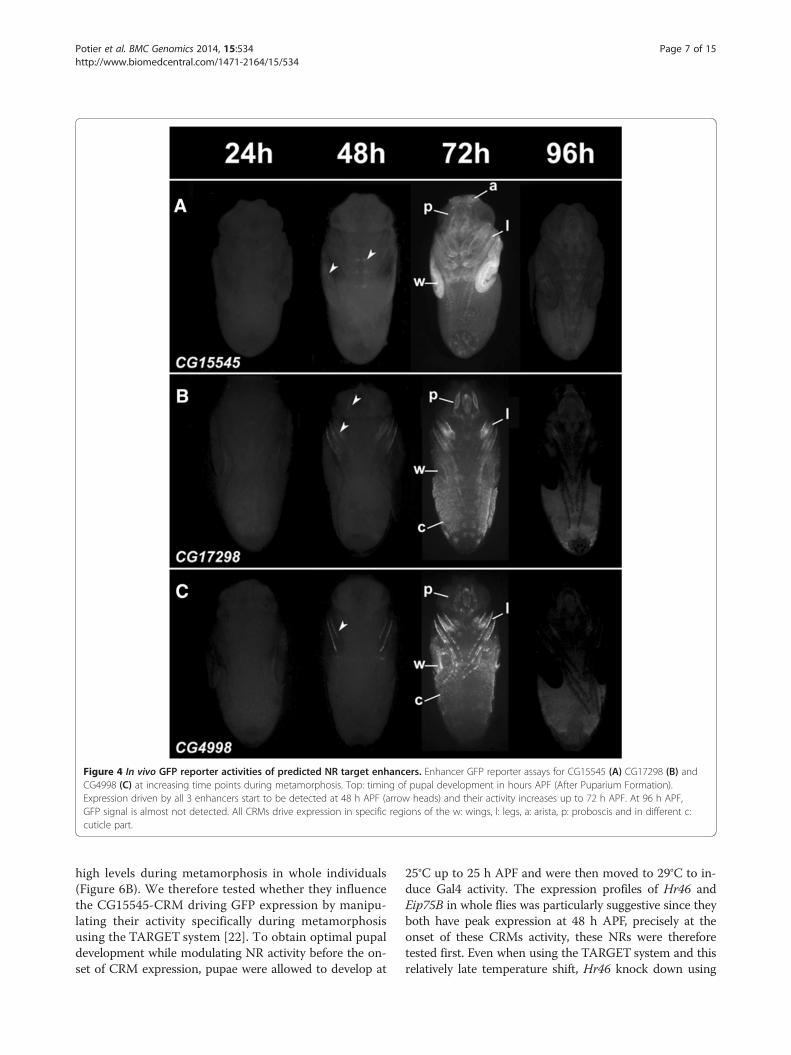
Figure 4 In vivo GFP reporter activities of predicted NR target enhancers. Enhancer GFP reporter assays for CG15545 (A) CG17298 (B) andCG4998 (C) at increasing time points during metamorphosis. Top: timing of pupal development in hours APF (After Puparium Formation).Expression driven by all 3 enhancers start to be detected at 48 h APF (arrow heads) and their activity increases up to 72 h APF. At 96 h APF,GFP signal is almost not detected. All CRMs drive expression in specific regions of the w: wings, l: legs, a: arista, p: proboscis and in different c:cuticle part.
Potier et al. BMC Genomics 2014, 15:534 Page 7 of 15http://www.biomedcentral.com/1471-2164/15/534
high levels during metamorphosis in whole individuals(Figure 6B). We therefore tested whether they influencethe CG15545-CRM driving GFP expression by manipu-lating their activity specifically during metamorphosisusing the TARGET system [22]. To obtain optimal pupaldevelopment while modulating NR activity before the on-set of CRM expression, pupae were allowed to develop at
25°C up to 25 h APF and were then moved to 29°C to in-duce Gal4 activity. The expression profiles of Hr46 andEip75B in whole flies was particularly suggestive since theyboth have peak expression at 48 h APF, precisely at theonset of these CRMs activity, these NRs were thereforetested first. Even when using the TARGET system and thisrelatively late temperature shift, Hr46 knock down using

Figure 5 NR motifs are functional within CRMsNR motifs are functional within CRMs. A) Schematic representation of mutations performedin NR motifs of CG4998 (top) and CG7298 (bottom) CRMs. The location of CRMs (black rectangles) with respect to corresponding genes isindicated on top, and putative NR motifs are represented as grey boxes. NR motifs sequences are indicated below, together with thecorresponding mutated sequences. Note that in CG4998-CRM both putative motifs are comprised of two inverted overlapping motifs. Motifs inlight grey are of lesser quality and were not analyzed in this study. B) GFP expression pattern at 72 h APF in WT (left) and mutated (right)individuals for CG4998 (top) and CG17298 (bottom) CRMs. A ventral and a dorsal view are shown in each case. NR motif mutations lead to amarked increase of GFP signal. C) Time course of GFP expression between 24 and 96 h APF driven by CG4998 and CG4998-mutated CRMsanalyzed by qRT-PCR. GFP expression was normalized to RP49 expression levels and expression ratio relative to GFP expression at 48 h innon-mutated CRM are represented (*p < 0.05; **p < 0.001, Student t-test). C’) The dynamics of GFP expression in the mutated CG4998 CRM issignificantly different from the one observed with wild type CRM (*p < 0.05 χ2 test).
Potier et al. BMC Genomics 2014, 15:534 Page 8 of 15http://www.biomedcentral.com/1471-2164/15/534
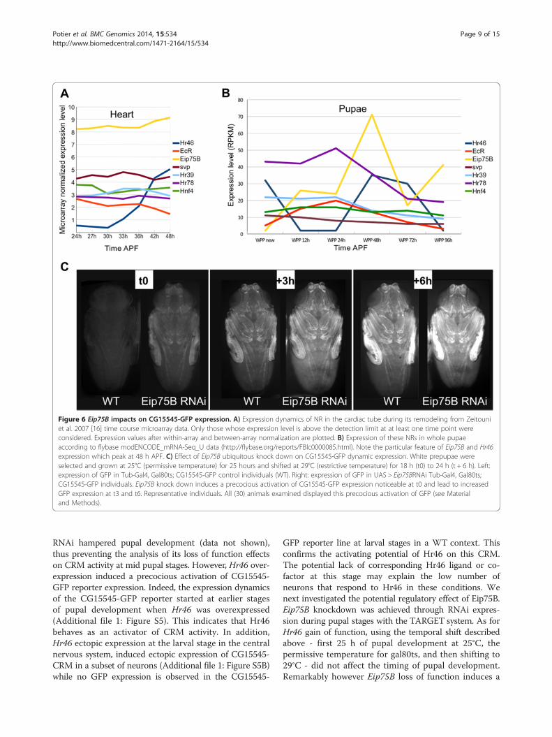
Figure 6 Eip75B impacts on CG15545-GFP expression. A) Expression dynamics of NR in the cardiac tube during its remodeling from Zeitouniet al. 2007 [16] time course microarray data. Only those whose expression level is above the detection limit at at least one time point wereconsidered. Expression values after within-array and between-array normalization are plotted. B) Expression of these NRs in whole pupaeaccording to flybase modENCODE_mRNA-Seq_U data (http://flybase.org/reports/FBlc0000085.html). Note the particular feature of Eip75B and Hr46expression which peak at 48 h APF. C) Effect of Eip75B ubiquitous knock down on CG15545-GFP dynamic expression. White prepupae wereselected and grown at 25°C (permissive temperature) for 25 hours and shifted at 29°C (restrictive temperature) for 18 h (t0) to 24 h (t + 6 h). Left:expression of GFP in Tub-Gal4, Gal80ts; CG15545-GFP control individuals (WT). Right: expression of GFP in UAS > Eip75BRNAi Tub-Gal4, Gal80ts;CG15545-GFP individuals. Eip75B knock down induces a precocious activation of CG15545-GFP expression noticeable at t0 and lead to increasedGFP expression at t3 and t6. Representative individuals. All (30) animals examined displayed this precocious activation of GFP (see Materialand Methods).
Potier et al. BMC Genomics 2014, 15:534 Page 9 of 15http://www.biomedcentral.com/1471-2164/15/534
RNAi hampered pupal development (data not shown),thus preventing the analysis of its loss of function effectson CRM activity at mid pupal stages. However, Hr46 over-expression induced a precocious activation of CG15545-GFP reporter expression. Indeed, the expression dynamicsof the CG15545-GFP reporter started at earlier stagesof pupal development when Hr46 was overexpressed(Additional file 1: Figure S5). This indicates that Hr46behaves as an activator of CRM activity. In addition,Hr46 ectopic expression at the larval stage in the centralnervous system, induced ectopic expression of CG15545-CRM in a subset of neurons (Additional file 1: Figure S5B)while no GFP expression is observed in the CG15545-
GFP reporter line at larval stages in a WT context. Thisconfirms the activating potential of Hr46 on this CRM.The potential lack of corresponding Hr46 ligand or co-factor at this stage may explain the low number ofneurons that respond to Hr46 in these conditions. Wenext investigated the potential regulatory effect of Eip75B.Eip75B knockdown was achieved through RNAi expres-sion during pupal stages with the TARGET system. As forHr46 gain of function, using the temporal shift describedabove - first 25 h of pupal development at 25°C, thepermissive temperature for gal80ts, and then shifting to29°C - did not affect the timing of pupal development.Remarkably however Eip75B loss of function induces a

Potier et al. BMC Genomics 2014, 15:534 Page 10 of 15http://www.biomedcentral.com/1471-2164/15/534
clear precocious activation of CG15545-GFP expression(Figure 6C), thus pointing to a repressive role of Eip75Bupon the activity of this CRM.Other expressed NRs were also tested for their activity
with respect to CG15545-GFP expression. However, EcRinactivation during metamorphosis blocks pupal develop-ment [15] and was therefore not tested. Hnf4 and Hr78RNAi mediated knockdown had no effect on CG15545-GFP expression dynamics (not shown). On the contrary,Hr39 knockdown accelerated CG15545-GFP expressiondynamics (Additional file 1: Figure S5C) very much likeEip75B, indicating that both genes behave as negative reg-ulators of the temporal pattern.In conclusion, both their expression and the genetic ma-
nipulations described here point to a central role of Hr39and Eip75B in the temporal repression of the CRMs iden-tified in this study. Given that putative NR binding motifsare functional within tested CRMs and are involved intheir repression, Hr39 and/or Eip75B may well be directlyinvolved in their temporal expression control. Eip75B en-codes three protein isoforms designated E75A, E75B, andE75C [23]. Interestingly it has been recently demonstratedthat E75A directly represses transcription of EcR targetgenes by opposing to EcR/Usp binding on enhancers [24].It is therefore conceivable that Eip75B directly repress theCRMs tested here.At metamorphosis, ecdysone, through EcR activation,
induces a cascade of transcriptional activation involvinga number of NRs that are intricately coordinated. Theobserved effect of Hr46 on CRM activity could thereforebe due to an indirect effect on Eip75B and Hr39 expres-sion. Alternatively, several reports have established thatEip75B gene products can form a complex with Hr46which switches Hr46 activating potential into a repres-sive one (see for example [25]). Since Hr46 and Eip75Bare induced at 48 hours APF both in the heart, and thewhole organism, their products may indeed dimerize toform a repressive complex that controls the expressionof the CRMs. In this case, the activating potential ofHr46 reported above may be due to a lack of Eip75Bgene expression in neurons, and to a titration of Eip75Bgene products by Hr46 overexpressed at metamorphosis.
DiscussionStarting from co-expressed gene sets and without priorknowledge of the TFs involved in their spatio-temporaltranscriptional regulation, our analysis shows that wewere able to identify the correct motif from a set of co-expressed genes and accurately predict target genes ofindividual TFs. The unbiased approach of cisTargetX,based on the enrichment of a comprehensive collectionof TF motifs, allows discovery of relevant motifs withoutrestricting the analysis to a particular class of transcriptionfactors. The robustness of the approach is confirmed by
the fact that (i) several motifs corresponding to nuclear re-ceptors are found to be enriched for the transient clusteranalyzed here, and (ii) applying this method to other clus-ters identifies further motifs corresponding to nuclear re-ceptors, corresponding well to the fact that these clustersmust have strict temporal control. A striking result is thefact that the motifs discovered here appear to form homo-typic clusters. While it is not possible to rule out the factthat an un-characterized TFBS might also be present, thedense arrangement of NR binding sites is reminiscent ofthe homotypic CRMs involved in early embryogenesiswhich respond to spatial morphogen gradients. It is there-fore tempting to hypothesize a mechanism by which theclusters of NRs respond to a temporal gradient of nuclearreceptors. Hence, this organization of binding sites mightrepresent a more general feature during development, butnot limited to early Drosophila embryogenesis [26].
Dissecting spatial and temporal cis-regulatory inputsSince regulatory motifs and CRMs were predicted andcompared for gene sets that have distinct temporal pat-terns but common spatial expression, our objectives werespecifically directed towards predicting regulatory inputsthat achieve temporal expression pattern. Indeed, whilewe started from genes co-expressed in the cardiac tube,none of the tested CRMs drove detectable expression incardiac myocytes, indicating that our predictions did notretrieve the heart specific spatial information. This failureto reproduce cardiac expression pattern indicates that forthe genes analyzed, the cis-regulatory modules that drivespatial expression in the cardiac tube might be distinctfrom, and located at a distance from the temporal CRMs.In an attempt to predict the regulatory sequences drivingspatial expression in the cardiac tube, we have used cisTar-getX with all 1660 dynamically expressed genes duringadult heart formation. The common property of this geneset is its spatial expression in the heart. Therefore, usingthis gene set, cisTargetX might allow predicting regulatorymotifs responsible for this spatial expression pattern.Interestingly, a number of PWMs known to be bound bybZIP transcription factors were significantly enriched inthis gene set (not shown), suggesting that one or severalbZIP transcription factor(s) may play a role in the expres-sion pattern of these genes. Of note, among the few CRMsknown to drive spatial expression in the cardiac tube, one,the cardiac specific Tin enhancer (TinC) was shown tohave functional bZIP motifs [27]. In addition, a recentstudy has underlined the important role of the bZIP TFE4BP4 in embryonic heart development in vertebrates[28], thus supporting a potential function of one or severalbZIP TF(s) in the cardiac GRN. Future work aimed atdeciphering the cardiac function of bZIP transcriptionfactors, and of their potential cognate enhancers shouldallow some light to be shed on the modalities of cardiac

Potier et al. BMC Genomics 2014, 15:534 Page 11 of 15http://www.biomedcentral.com/1471-2164/15/534
restricted spatial expression at metamorphosis and its re-latedness to temporal control of gene expression.The identified regulatory regions nevertheless display
characteristic spatial expression patterns, for instance inthe developing pupal wings and legs. This most probablyindicates that in these tissues, the spatial control of geneexpression is driven by TFs that bind in the vicinity of themotifs responsible for temporal control. Alternatively, onecould hypothesize that NRs themselves are responsible forthe spatial expression pattern of the CRMs. However afunction of NRs in the spatial control of genes and CRMexpression is not supported by our experimental data. In-deed, mutations of NR binding motifs only affect the tim-ing of expression, and have no effect on spatial control ofreporter gene expression. The same is true regarding thephenotypes induced by NR gain and loss of function atpupal stages. Furthermore all tested NRs are expressedin the heart (this was our selection criteria) but none ofthe analysed CRMs are expressed there, indicating thatthe spatial expression pattern of NRs cannot explain thespatial expression pattern of the CRMs.
Integrating temporal control within GRNsDevelopmental timing mechanisms are intricately linkedto pattern formation, and disruption of temporal pro-grams can cause organism wide changes in developmentthat can result in catastrophic birth defects [29]. At thelevel of GRNs, this implies that robust mechanisms en-sure precise coordination of both spatial and temporaltranscriptional control. Some of these mechanisms are“encoded” in the structure of the network themselves.For instance, network motifs such as feed-forward loopsconfer dynamics to the networks and ensure definingthe temporal order of specification events [30]. Thereare other mechanisms which also drive time-directed de-velopmental processes. One such mechanism concernsthe molecular oscillators that govern the vertebrate seg-mentation clock. In this case however, the nature of theclock pacemaker still remains elusive [31]. Another mech-anism -based on the temporal control of TF activity- wasrecently pointed to, in a well-documented example of ter-minal differentiation in Drosophila. Indeed, Kondo et al.[32] demonstrated that a small peptide (pri) triggersamino terminal truncation of the svb protein, switching itsactivity from a full-length repressor to a cleaved activator,providing temporal control to the GRN of epidermal dif-ferentiation. A study of heterochronic genes in C. elegans -which ensure that stage specific developmental programsoccur in the appropriate sequence- demonstrated thatmiRNAs play a central role in the temporal control ofgene expression during larval development [33]. Whilethe transcriptional regulation of these miRNA encodinggenes remain elusive, recent studies indicated that a cen-tral transcriptional regulator of heterochronic genes is the
nuclear receptor DAF-12 [34]. Moreover, a number ofstudies in different model organisms and humans identi-fied NRs as major regulators of developmental timing,usually as targets of hormonal cues. For instance, NRs areinvolved in humans to trigger the marked changes thatoccur during puberty and adolescence. In Drosophila,pulses of the steroid hormone ecdysone, which bind to,and activate EcR, control a number of developmentaltransitions during embryonic and post-embryonic de-velopment. In particular, three ecdysone pulses activategenetic regulatory hierarchies that coordinate the devel-opmental changes associated with Drosophila metamor-phosis [35]. Ecdysone pulses trigger the progression ofthe pupae into different stages through the transcriptionof a particular cascade of genes, most of which arethemselves nuclear receptors. We previously demon-strated that EcR function is required for all aspects ofcardiac tube remodeling, thus indicating that it is lo-cated at the top of adult heart formation GRN [15]. Wenow show that NR motifs are central in the temporalregulation of CRMs of genes activated late during theremodeling process. In addition, we provide evidencesuggesting that NRs might be direct trans-regulators ofthese CRMs, thus suggesting that they are part of thecardiac GRN during metamorphosis, acting downstreamof EcR function to control the expression of these lategenes. Other NR PWMs were recovered using cisTar-getX on different co-expressed gene sets, suggestingthat other NRs may play similar roles at earlier stages ofcardiac remodeling.Alternative modalities of temporal control of gene ex-
pression have been reported in Drosophila metamorphosis.In particular, it has recently been shown that Eip93F, oneof the primary targets of the ecdysone receptors that en-codes a non-nuclear receptor transcription factor, plays acentral role in the timely restricted expression of distalless(and most probably of many other genes) during adultmorphogenesis [36]. Eip93F is expressed at high levels dur-ing the early steps of heart remodeling [16] and it is there-fore possible that it participates in the temporal control ofcorresponding gene sets. The current lack of knowledgeabout its DNA binding specificity however prevents chal-lenging its involvement in the process using our approach.Another study implicated the importance of core pro-moters’ choice in the temporal control of gene expressionduring metamorphosis of the wing [37]. Although it wasnot in the focus of our study, it is possible that in the heartalso the type of core promoters’ usage may play an add-itional role in the temporal control of gene expression dur-ing adult heart formation.
ConclusionsUsing an in silico strategy, and without any prior know-ledge regarding the involved TFs, our approach identified

Potier et al. BMC Genomics 2014, 15:534 Page 12 of 15http://www.biomedcentral.com/1471-2164/15/534
CRMs and potential TFs acting on the temporal regu-lation of a co-expressed gene set. Indeed, based onevolutionary conservation and over-representation inthe surrounding non-coding sequences of co-expressedgenes, potential regulatory motifs and CRMs were pre-dicted and were both validated in vivo. We furtherdemonstrate that several nuclear receptors, displayingdynamic expression during the biological process ana-lyzed, are essential for the temporal pattern of theenhancer-reporter; the fact that some act as activatorswhile others are repressors suggests a subtle balance be-tween these opposite effects, as is the case for the spatialexpression during early embryogenesis. Therefore, ourstrategy was successful at identifying CRMs and TFs in-volved in the temporal dynamics of gene expression. Inaddition, our results suggest a modular architecture ofthe regulatory machinery, in which the temporal andspatial regulations can be uncoupled and encoded bydistinct CRMs.
MethodsDatasetThe gene expression dynamics during heart remodelingfrom 21 h to 48 h After Puparium Formation (8 timepoints) was described previously [16]. In this experiment,we identified 2394 genes that exhibited significant differ-ential expression between time-points in using modified t-statistic significance analysis of microarrays (SAM) [38]with estimated q-values (false discovery rates) of ≤ 0.05.Among them, 1660 genes showed significant levels of dif-ferential expression at least 1.8-fold in at least one condi-tion through our time-course analysis. Those 1660 geneswere clustered in 13 expression profiles using SOM clus-tering. All 13 clusters were independently submitted forcisTargetX analysis.
Motif over representation analysis in the 13 clusterscisTargetX was used with the following parameters: − As-sembly & Scoring: “dm2 (April 2004) with Cluster-Buster”; − motif collection: “Used in Aerts & al. PLoS Biol-ogy 2010 (1981 PWM)”; Z-score thresold: “Determinethreshold automatically”; receiver operating characteristic(ROC) threshold for area under the curve (AUC) calcula-tion: “0.03”; Genomic threshold for visualization: “1000”.The search space used is 5 kb upstream of all transcriptsTSS (Transcription Start Site) and their introns. If a neigh-boring gene (or a host gene exon in case of an intronicallyhosted gene) is less than 5 kb away, the search space is re-duced and truncated at this neighboring gene (or exon)boundary. In the case of cluster 12, in order to inspectfurther PWM, cisTargetX was run a second time with theprevious parameters except for the Z-score threshold,which was decreased to 2.5.
PWM clusteringIn order to visualize similar PWM (given the redun-dancy in the PWM library), we used STAMP [39] withdefault parameters and the “-chp” option to obtain clus-ters of similar PWM. A color code was used to visualizePWM belonging to related families.
Determination of drosophila TFs predicted to bind GCNFmotifAs GCNF is a motif representing the binding site of avertebrate NRs, we selected the 22 Drosophila TFs be-longing to the Nuclear hormone receptor family in theUniProt database (Hr39, EcR, tll, usp, Hr46, Hr96, Hr78,Hr4, Hr38, ftz-f1, svp, kni, Eip75B, eg, Eip78C, knrl,Hnf4, usp, Hr51, ERR, dsf, Hr83) with the following re-quest, family: “nuclear hormone receptor family” ANDorganism: “7227”. We collected PWMs representing NRsbinding sites present in the cisTargetX library (1981PWMs) or in the Fly Factor Survey database and useSTAMP with default options to build a similarity tree(Additional file 1: Figure S4).
CRM selection and delimitationTo delineate the putative CRMs, we selected DNA frag-ments around clusters of GNCF TFBS in the vicinity ofthe top ranked genes. Fragments were manually ex-tended on each side in order to retain conserved flank-ing sequences with high phastCons conservation scoresin the UCSC genome browser (“conservation” track inthe “Comparative Genomics” section). All tested frag-ments are presented as UCSC genome browser screen-shots in Additional file 1: Figure S3.
Molecular cloning and transgenesisAll fragments described in Figure 2 were amplified start-ing from W1118; cantonS genomic DNA and cloned bystandard molecular cloning techniques using the gatewaysystem in three different reporter vectors; a GFP reporter(pH-attB-Dest, [9]), a LacZ reporter vector produced byreplacing eGFP by LacZ in the “pH-attB-Dest” vectorfrom and a gal4 reporter (SMG4-Gal4 generous gift fromNicolas Gompel). The intermediate cloning vector used isthe pDONR221. Vectors were introduced by electro-poration in DH5α competent cells to be amplified. Weperformed mutations for two binding sites predictedfor GCNF motif in CG4998 (attgcttttggacttgaactgcgato tttttttttttttttttttttttt and gcgaggtccaagttcaagttcagt togggggggggggggggggggggggg) and in CG17298 (gatgatcttgatcttgag to tttttttttttttttttg and gagaaacatgaaattgat tocccccccccccccccccc). Long polymerase chain reaction(PCR) primers containing the mutations were usedand the mutated fragments were cloned in the SMG4-gal4 vector.

Potier et al. BMC Genomics 2014, 15:534 Page 13 of 15http://www.biomedcentral.com/1471-2164/15/534
To avoid position dependent expression variability, weused targeted PhiC31 transgenesis [40]. All constructswere inserted at P2(3 L)68A4 landing site using PhiC31integrase. Lines containing the SMG4-Gal vector werecrossed with a UAS_GFP line.
RNA extraction and qRT-PCRFor qRT-PCR on dissected pupal wings, dissected wingsfrom four CG15545-GFP individuals were used for eachbiological replicate and three biological replicates wereanalyzed. Staged pupae were dissected under stereo micro-scope and dissected wings immediately placed in Trizol®(Life Technologies) on ice and RNA extraction (see bellow)was performed extemporaneously.For the analysis of GFP expression on CG4998 and
CG4998* -CRMs, total RNAs was extracted from 5 indi-viduals per replicate and 3 biological replicates wereanalyzed. Staged pupae of corresponding genotypes werecollected, ground in Trizol® and RNA extraction (seebelow) was performed extemporaneously. A list of pri-mer pairs used for qRT-PCR analysis is provided bellow.Total RNA was extracted from samples in triplicate
using Trizol® according to standard procedures. The in-tegrity of RNA samples was assessed using Agilent 2100Bioanalyzer and RNA Nano CHIP kit (Agilent). TotalRNA concentration was measured using a NanoDropND-1000 Spectrophotometer (ThermoScientific) and itspurity was evaluated by absorbance ratios, 260/280 and260/230. cDNA was synthesized from 500 ng of DNase I(Promega)-treated total RNA in a 25 μl reaction volumeusing qScript cDNA SuperMix (Quanta Biosciences) ac-cording to supplier instructions. cDNA samples werediluted five-fold for real-time qRT-PCR reactions. Gene-specific transcription levels were determined in a 15 μlreaction volume in triplicate using SYBR Green (Invitrogen)and a BioRad CFX (Biorad) for pupal wings; and a Stra-tagene MX3000P real-time qPCR system (Agilent) forwhole pupae total RNA analysis following the manufac-turer’s instructions. Standard cDNA samples with 4-foldserial dilutions were used for PCR efficiency calculations.Amplifications were performed as follows: for pupal wings,8 min at 95°C, 40 cycles of 10s at 95°C/30s at 60°C andfor pupae total RNA, 5 min30 at 95°C, 40 cycles of 15 sat 95°C/1 min at 60°C. To verify the specificity of ampli-cons, a melting curve analysis was carried out from 60°Cto 95°C. Real-time qRT-PCR reactions of the standard, testcDNA samples and no template controls using the sameprimer set were analyzed together in the same 96-deepwell plate (Agilent or BioRad) in order to minimize run-to-run variations and use exactly the same thresholdsetting (user defined baseline subtracted curve fit) fordetermination of the threshold cycle values (Ct). Parallelsamples were processed using the same batch of reagents
to minimize overall sample-to-sample variations. Genespecific primers are listed in Additional file 1: Table S3.
Data analysesData analyses were manually performed to calculate thekey variables including PCR efficiency (E(%) = (10exp[−1/slope]-1)×100) and squared correlation coefficients (R2) ofprimer sets and expression ratios of target genes were nor-malized according to expression levels of RP49. In Figure 5,GFP expression values are displayed relative to GFP ex-pression in the CG4998-GFP reporter line at 48 h. InAdditional file 1: Figure S3, expression values are displayedrelative to expression at 30 hrs. In addition, a logarithmicscale of the fold change was used for better display. Todetermine significance of the results, statistic compari-sons were performed with a (one sample test or pairedtest) Student’s t-test. Statistical comparisons of dynamictemporal curve (Figure 5C’) were performed with a chi-Square test.
βGalactosidase staining24 h, 48 h, 72 h and 96 h APF individuals were extractedfrom their pupal cage. They were fixed using a PBS 1×,formaldehyde 3,7% and triton 0.01% solution and thenwashed with Phosphate-Buffered Tween (PBT) three timesand with Phosphate-Buffered Saline (PBS) once. The pupaewere then sonicated to perforate their membrane in orderto allow a X-Gal staining (10/15 min in X-gal buffer: PSB1×; KferriCN 4 mM; KferroCN 4 mM; MgCl2 2 mM;NP40 1%; X-gal 0,04 mg/ml).
GFP expression analysisFor GFP expression analysis, a batch of 10 flies per condi-tions and genotypes were analyzed in triplicate and repre-sentative individuals are shown. Images were acquiredusing a high-resolution video camera (CoolSNAP HQMonochrome, Roper Scientific, Inc), mounted on a ZeissStereo Lumar V12 binocular microscope with a Neolumar0.83 objective. Image acquisition was performed with theMetamorph/Metafluor software (universal Imaging, WestChester, PA). Individuals shown in Figures 4, 5 and 6 weredissected out their pupal case and mounted in Voltaleff oil3S prior imaging.
Drosophila strains and fly husbandryThe following stocks were obtained from the BloomingtonDrosophila Stock Centre. UAS-mcd8-GFP, UAS-dsRNA >Eip75B, UAS-dsRNA >Hr39, UAS-dsRNA >Hfn4, UAS-dsRNA >Hr78, P(tub-GAL80[ts]), P(tub-GAL4). UAS >Hr46; UAS-dsRNA >Hr46 and the elav > GeneSwithGal4were obtained from V Monnier (Paris).

Potier et al. BMC Genomics 2014, 15:534 Page 14 of 15http://www.biomedcentral.com/1471-2164/15/534
Timing of pupal development and control of Gal4inductionOnset of pupal development corresponds to white pupaethat were selected on the basis of spiracle eversion, ab-sence of reaction following forceps contact and absenceof tanning. Individuals were kept for further developmentin an air incubator at 25°C. Tub(Gal4); P(tub-GAL80[ts])and CG15545CRM>GFP transgenes were combined inthe same lines and crossed with appropriate UAS lines.Crosses with wild type flies served as controls. Develop-ment was allowed to proceed at 22°C until white pupalstage and individuals were then shifted to 25°C for 25 hoursand to restrictive temperature (29°C) for the indicatedperiod of time. Except for Hr46 and EcR whose knock-down hampered pupal development, none of the analysedNR affected the timing of pupal development when knock-down was accomplished following this time schedule.Therefore the effect seen after Eip75B and Hr39 RNAi me-diated loss of function cannot be attributed to a precociouspupal development.For GeneSwitch experiments, second instar larvae were
shifted to regular food medium containing either 0 μg/ml(control) or 100 μg/ml of RU486 (Mifepristone, Sigma)and GFP expression pattern was analyzed at third instarlarval stage.
Additional file
Additional file 1: Table S1. CisTargetX analysis of cluster 12 gene setwith 2.5 Z score cutoff. Table S2. Detailed genomic coordinates of testedCRMs. Table S3. List of primer pairs used for Q-RT PCR analysis. FigureS1. Logos of PWM outlined in Figure 1. Figure S2. Details of tested CRM.Figure S3. qRT-PCR in developing pupal wings of CG15545-GFPindividuals. Figure S4. Sequence based comparison of NR motifs.Figure S5. Hr46 and Hr39 regulate CG15547-GFP expression.
AbbreviationsNR: Nuclear receptor; GRN: Gene regulatory network; CRM: Cis regulatorymodule; PWM: Position weight matrice; TF: Transcription factor;TFBS: Transcription factor binding site; bZIP: Basic region leucine ZIPper;EcR: Ecdysone receptor; ChIP: Chromatin immuno-Precipitation; APF: Afterpuparium formation; SOM: Self-organizing map; ROC: Receiver operatingcharacteristic; AUC: Area under the curve; PCR: Polymerase chain reaction;qRT-PCR: Quantitative reverse transcription polymerase chain reaction;WT: Wild type; GFP: Green fluorescent protein; TSS: Transcription start site;SAM: Significance analysis of microarrays.
Competing interestsThe authors declare that they have no competing interest.
Authors’ contributionsPerformed experiments: DP, DS, MT, CG, LP. Analysed data: DP, DS, CH, LP. Wrotemanuscript: DP, SA, CH, LP. All authors read and approved the final manuscript.
AcknowledgmentsThis work was supported by ANR, partner of the ERASysBio + initiativesupported under the EU ERA-NET Plus scheme in FP7 (CH and LP), by an ANR“JCJC” grant to CH and LP and by funding from the AFM (LP). DP and DS weresupported by a doctoral fellowship from MESR. We thank the BloomingtonStock center, V. Monnier for fly stocks, K. Davie for careful reading of themanuscript and Annelies Clayes for her help on molecular cloning.
Author details1INSERM, UMR1090 TAGC, Marseille F-13288, France. 2Aix-Marseille Université,UMR1090 TAGC, Marseille F-13288, France. 3CNRS, Marseille, France.4Laboratory of Computational Biology, Center for Human Genetics, Universityof Leuven, Herestraat 49, P.O. Box 602 3000 Leuven, Belgium. 5IPMB,University of Heidelberg and German Cancer Research Center, div. ofTheoretical Bioinformatics, Im Neuenheimer Feld 280, Heidelberg 69120,Germany.
Received: 12 February 2014 Accepted: 13 June 2014Published: 27 June 2014
References1. Bonn S, Furlong EEM: cis-regulatory networks during development: a
view of drosophila. Curr Opin Genet Dev 2008, 18:513–520.2. Davidson EH, Levine MS: Properties of developmental gene regulatory
networks. Proc Natl Acad Sci U S A 2008, 105:20063–20066.3. Li L, Zhu Q, He X, Sinha S, Halfon MS: Large-scale analysis of
transcriptional cis-regulatory modules reveals both common featuresand distinct subclasses. Genome Biol 2007, 8:R101.
4. Sandmann T, Girardot C, Brehme M, Tongprasit W, Stolc V, Furlong EEM: Acore transcriptional network for early mesoderm development indrosophila melanogaster. Genes Dev 2007, 21:436–449.
5. Liu Y-H, Jakobsen JS, Valentin G, Amarantos I, Gilmour DT, Furlong EEM: Asystematic analysis of Tinman function reveals Eya and JAK-STATsignaling as essential regulators of muscle development. Dev Cell 2009,16:280–291.
6. Zinzen RP, Girardot C, Gagneur J, Braun M, Furlong EEM: Combinatorialbinding predicts spatio-temporal cis-regulatory activity. Nature 2009,462:65–70.
7. Junion G, Spivakov M, Girardot C, Braun M, Gustafson EH, Birney E, FurlongEEM: A transcription factor collective defines cardiac cell fate and reflectslineage history. Cell 2012, 148:473–486.
8. Estrada B, Choe SE, Gisselbrecht SS, Michaud S, Raj L, Busser BW, Halfon MS,Church GM, Michelson AM: An integrated strategy for analyzing theunique developmental programs of different myoblast subtypes. PLoSGenet 2006, 2:e16.
9. Aerts S, Quan X-J, Claeys A, Naval Sanchez M, Tate P, Yan J, Hassan BA:Robust target gene discovery through transcriptome perturbations andgenome-wide enhancer predictions in drosophila uncovers a regulatorybasis for sensory specification. PLoS Biol 2010, 8:e1000435.
10. Mayer H, Bilban M, Kurtev V, Gruber F, Wagner O, Binder BR, de Martin R:Deciphering regulatory patterns of inflammatory gene expression frominterleukin-1-stimulated human endothelial cells. Arterioscler Thromb VascBiol 2004, 24:1192–1198.
11. Tapia A, Vilos C, Marín JC, Croxatto HB, Devoto L: Bioinformatic detectionof E47, E2F1 and SREBP1 transcription factors as potential regulators ofgenes associated to acquisition of endometrial receptivity. Reprod BiolEndocrinol 2011, 9:14.
12. Kantorovitz MR, Kazemian M, Kinston S, Miranda-Saavedra D, Zhu Q,Robinson GE, Göttgens B, Halfon MS, Sinha S: Motif-blind, genome-widediscovery of cis-regulatory modules in drosophila and mouse. Dev Cell2009, 17:568–579.
13. Ahmad SM, Busser BW, Huang D, Cozart EJ, Michaud S, Zhu X, Jeffries N,Aboukhalil A, Bulyk ML, Ovcharenko I, Michelson AM: Machine learningclassification of cell-specific cardiac enhancers uncovers developmentalsubnetworks regulating progenitor cell division and cell fatespecification. Development 2014, 141:878–888.
14. Potier D, Atak ZK, Sanchez MN, Herrmann C, Aerts S: Using cisTargetX topredict transcriptional targets and networks in drosophila. Methods MolBiol Clift Nj 2012, 786:291–314. Methods in Molecular Biology.
15. Monier B, Astier M, Sémériva M, Perrin L: Steroid-dependent modificationof Hox function drives myocyte reprogramming in the drosophila heart.Development 2005, 132:5283–5293.
16. Zeitouni B, Sénatore S, Séverac D, Aknin C, Sémériva M, Perrin L: Signallingpathways involved in adult heart formation revealed by gene expressionprofiling in drosophila. PLoS Genet 2007, 3:1907–1921.
17. Monnier V, Iché-Torres M, Rera M, Contremoulins V, Guichard C, Lalevée N,Tricoire H, Perrin L: dJun and Vri/dNFIL3 are major regulators of cardiacaging in drosophila. PLoS Genet 2012, 8:e1003081.

Potier et al. BMC Genomics 2014, 15:534 Page 15 of 15http://www.biomedcentral.com/1471-2164/15/534
18. Thomas-Chollier M, Defrance M, Medina-Rivera A, Sand O, Herrmann C,Thieffry D, van Helden J: RSAT 2011: regulatory sequence analysis tools.Nucleic Acids Res 2011, 39(suppl):W86–W91.
19. Seyres D, Röder L, Perrin L: Genes and networks regulating cardiacdevelopment and function in flies: genetic and functional genomicapproaches. Brief Funct Genomics 2012, 11:366–374.
20. King-Jones K, Thummel CS: Nuclear receptors–a perspective fromdrosophila. Nat Rev Genet 2005, 6:311–323.
21. Siepel A, Bejerano G, Pedersen JS, Hinrichs AS, Hou M, Rosenbloom K,Clawson H, Spieth J, Hillier LW, Richards S, Weinstock GM, Wilson RK,Gibbs RA, Kent WJ, Miller W, Haussler D: Evolutionarily conserved elementsin vertebrate, insect, worm, and yeast genomes. Genome Res 2005,15:1034–1050.
22. McGuire SE, Le PT, Osborn AJ, Matsumoto K, Davis RL: Spatiotemporal rescueof memory dysfunction in drosophila. Science 2003, 302:1765–1768.
23. Segraves WA, Hogness DS: The E75 ecdysone-inducible gene responsiblefor the 75B early puff in drosophila encodes two new members of thesteroid receptor superfamily. Genes Dev 1990, 4:204–219.
24. Johnston DM, Sedkov Y, Petruk S, Riley KM, Fujioka M, Jaynes JB, Mazo A:Ecdysone- and NO-mediated gene regulation by competing EcR/Uspand E75A nuclear receptors during drosophila development. Mol Cell2011, 44:51–61.
25. Reinking J, Lam MMS, Pardee K, Sampson HM, Liu S, Yang P, Williams S,White W, Lajoie G, Edwards A, Krause HM: The drosophila nuclear receptore75 contains heme and is gas responsive. Cell 2005, 122:195–207.
26. Gotea V, Visel A, Westlund JM, Nobrega MA, Pennacchio LA, Ovcharenko I:Homotypic clusters of transcription factor binding sites are a keycomponent of human promoters and enhancers. Genome Res 2010,20:565–577.
27. Venkatesh TV, Park M, Ocorr K, Nemaceck J, Golden K, Wemple M, Bodmer R:Cardiac enhancer activity of the homeobox gene tinman depends on CREBconsensus binding sites in drosophila. Genesis 2000, 26:55–66.
28. Weng Y-J, Hsieh DJ-Y, Kuo W-W, Lai T-Y, Hsu H-H, Tsai C-H, Tsai F-J, Lin D-Y,Lin JA, Huang C-Y, Tung K-C: E4BP4 is a cardiac survival factor andessential for embryonic heart development. Mol Cell Biochem 2010,340:187–194.
29. Wilson GN: Heterochrony and human malformation. Am J Med Genet1988, 29:311–321.
30. Ciglar L, Furlong EEM: Conservation and divergence in developmentalnetworks: a view from drosophila myogenesis. Curr Opin Cell Biol 2009,21:754–760.
31. Ozbudak EM, Pourquié O: The vertebrate segmentation clock: the tip ofthe iceberg. Curr Opin Genet Dev 2008, 18:317–323.
32. Kondo T, Plaza S, Zanet J, Benrabah E, Valenti P, Hashimoto Y, Kobayashi S,Payre F, Kageyama Y: Small peptides switch the transcriptional activity ofshavenbaby during drosophila embryogenesis. Science 2010, 329:336–339.
33. Resnick TD, McCulloch KA, Rougvie AE: MiRNAs give worms the time oftheir lives: small RNAs and temporal control in caenorhabditis elegans.Dev Dyn 2010, 239:1477–1489.
34. Martinez NJ, Walhout AJM: The interplay between transcription factorsand microRNAs in genome-scale regulatory networks. Bioessays 2009,31:435–445.
35. Kozlova T, Thummel CS: Steroid regulation of postembryonicdevelopment and reproduction in drosophila. Trends Endocrinol Metab2000, 11:276–280.
36. Mou X, Duncan DM, Baehrecke EH, Duncan I: Control of target genespecificity during metamorphosis by the steroid response gene E93.Proc Natl Acad Sci U S A 2012, 109:2949–2954.
37. O’Keefe DD, Thomas SR, Bolin K, Griggs E, Edgar BA, Buttitta LA:Combinatorial control of temporal gene expression in the drosophilawing by enhancers and core promoters. BMC Genomics 2012, 13:498.
38. Tusher VG, Tibshirani R, Chu G: Significance analysis of microarraysapplied to the ionizing radiation response. Proc Natl Acad Sci U S A 2001,98:5116–5121.
39. Mahony S, Benos PV: STAMP: a web tool for exploring DNA-binding motifsimilarities. Nucleic Acids Res 2007, 35(Web Server issue):W253–W258.
40. Bischof J, Maeda RK, Hediger M, Karch F, Basler K: An optimizedtransgenesis system for drosophila using germ-line-specific phiC31integrases. Proc Natl Acad Sci U S A 2007, 104:3312–3317.
doi:10.1186/1471-2164-15-534Cite this article as: Potier et al.: Identification of cis-regulatory modulesencoding temporal dynamics during development. BMC Genomics2014 15:534.
Submit your next manuscript to BioMed Centraland take full advantage of:
• Convenient online submission
• Thorough peer review
• No space constraints or color figure charges
• Immediate publication on acceptance
• Inclusion in PubMed, CAS, Scopus and Google Scholar
• Research which is freely available for redistribution
Submit your manuscript at www.biomedcentral.com/submit