Identification of CD24 as a cancer stem cell marker in human nasopharyngeal carcinoma
14
Identification of CD24 as a Cancer Stem Cell Marker in Human Nasopharyngeal Carcinoma Chun-Hung Yang 1,2. , Hui-Ling Wang 3,4. , Yi-Sheng Lin 3,4. , K. P. Shravan Kumar 3,4 , Hung-Chi Lin 1,2 , Chih-Jung Chang 3,4,5 , Chia-Chen Lu 6 , Tsung-Teng Huang 3,4,7,8 , Jan Martel 7 , David M. Ojcius 7,9 , Yu-Sun Chang 10 , John D. Young 7,11,12 *, Hsin-Chih Lai 3,4,7,8 * 1 Graduate Institute of Biomedical Sciences, College of Medicine, Chang Gung University, Taoyuan, Taiwan, Republic of China, 2 Division of Applied Toxicology, Taiwan Agricultural Chemicals and Toxic Substances Research Institute, Council of Agriculture, Executive Yuan, Taichung, Taiwan, Republic of China, 3 Department of Medical Biotechnology and Laboratory Sciences, College of Medicine, Chang Gung University, Taoyuan, Taiwan, Republic of China, 4 Department of Laboratory Medicine, Linkou Chang Gung Memorial Hospital, Taoyuan, Taiwan, Republic of China, 5 Department of Microbiology and Immunology, College of Medicine, Chang Gung University, Taoyuan, Taiwan, Republic of China, 6 Department of Respiratory Therapy, Fu Jen Catholic University, Taipei, Taiwan, Republic of China, 7 Center for Molecular and Clinical Immunology, College of Medicine, Chang Gung University, Taoyuan, Taiwan, Republic of China, 8 Research Center of Bacterial Pathogenesis, College of Medicine, Chang Gung University, Taoyuan, Taiwan, Republic of China, 9 Department of Molecular Cell Biology and Health Sciences Research Institute, University of California Merced, Merced, California, United States of America, 10 Molecular Medicine Research Center, College of Medicine, Chang Gung University, Taoyuan, Taiwan, Republic of China, 11 Laboratory of Cellular Physiology and Immunology, Rockefeller University, New York, New York, United States of America, 12 Biochemical Engineering Research Center, Ming Chi University of Technology, Taipei, Taiwan, Republic of China Abstract Cancer stem cells (CSCs) represent a unique sub-population of tumor cells with the ability to initiate tumor growth and sustain self-renewal. Although CSC biomarkers have been described for various tumors, only a few markers have been identified for nasopharyngeal carcinoma (NPC). In this study, we show that CD24 + cells isolated from human NPC cell lines express stem cell genes (Sox2, Oct4, Nanog, Bmi-1, and Rex-1), and show activation of the Wnt/b-catenin signaling pathway. CD24 + cells possess typical CSC characteristics that include enhanced cell proliferation, increased colony and sphere formation, maintenance of cell differentiation potential in prolonged culture, and enhanced resistance to chemotherapeutic drugs. Notably, CD24 + cells produce tumors following inoculation of as few as 500 cells in immunodeficient NOD/SCID mice. CD24 + cells further show increased invasion ability in vitro, which correlates with enhanced expression of matrix metalloproteinase 2 and 9. In summary, our results suggest that CD24 represents a novel CSC biomarker in NPC. Citation: Yang C-H, Wang H-L, Lin Y-S, Kumar KPS, Lin H-C, et al. (2014) Identification of CD24 as a Cancer Stem Cell Marker in Human Nasopharyngeal Carcinoma. PLOS ONE 9(6): e99412. doi:10.1371/journal.pone.0099412 Editor: Masaharu Seno, Okayama University, Japan Received July 4, 2013; Accepted May 14, 2014; Published June 23, 2014 Copyright: ß 2014 Yang et al. This is an open-access article distributed under the terms of the Creative Commons Attribution License, which permits unrestricted use, distribution, and reproduction in any medium, provided the original author and source are credited. Funding: This study was supported by grants NSC101-2325-B-182-005 and NSC101-2320-B-030-011 from the National Science Council of Taiwan, and grant CMRPD1B0052 from Chang Gung Memorial Hospital (Linkou, Taiwan). The funders had no role in study design, data collection and analysis, decision to publish, or preparation of the manuscript. Competing Interests: The authors have declared that no competing interests exist. * Email: [email protected] (JDY); [email protected] (HCL) . These authors contributed equally to this work. Introduction NPC incidence rates vary throughout the world but are highest in Southeast Asia, especially in the Chinese province of Guangdong as well as in Hong Kong and Taiwan [1]. Intermediate NPC incidence is observed in countries of the Mediterranean basin along with Greenland and Alaska, while a low incidence is found in most Western countries [2]. In Taiwan, the incidence for females and males in 2007 was 2.93 and 8.41 cases per 100,000 individuals, respectively [3]. NPC usually shows relatively high sensitivity to radiation, and radiotherapy thus remains the major treatment for early-stage NPC [4,5]. Although the combination of radiotherapy and chemotherapy has improved treatment outcomes for patients with advanced-stage NPC, cancer recurrence still occurs frequently [6]. Identification and characterization of tumor-initiating cells, also called cancer stem cells (CSCs), has revealed cellular mechanisms that could account for the refractoriness to treatment and dormant behavior of many tumors [7]. The progenitor cell hypothesis of cancer development suggests that only a small sub-population of cells within a tumor (the CSCs) has stem-like properties and the ability to initiate new tumors [8]. CSCs may not only initiate primary tumors, but may also be responsible for recurrence following treatment, a phenomenon that may be due to the self- renewal capacity and inherent chemo- and radio-resistance of these cells [9]. CSCs have been identified and isolated from various tumors using specific cell surface markers, including CD44 for head and neck cancer [10], CD133 for prostate cancer [11], CD90 for liver tumor [12], CD24 for ovarian cancer [13], CD133 for brain tumor [14], CD44 + CD24 2/low for breast cancer [15], and CD133 for lung cancer [16]. Although specific biomarkers have been used to isolate CSCs, each type of tumor shows specific cell surface markers [17]. To isolate potential sub-populations of CSCs from tumors in which CSC biomarkers have not yet been identified, researchers have made use of the ability of CSCs to exclude the dyes, Hoechst PLOS ONE | www.plosone.org 1 June 2014 | Volume 9 | Issue 6 | e99412
-
Upload
independent -
Category
Documents
-
view
0 -
download
0
Transcript of Identification of CD24 as a cancer stem cell marker in human nasopharyngeal carcinoma

Identification of CD24 as a Cancer Stem Cell Marker inHuman Nasopharyngeal CarcinomaChun-Hung Yang1,2., Hui-Ling Wang3,4., Yi-Sheng Lin3,4., K. P. Shravan Kumar3,4, Hung-Chi Lin1,2,
Chih-Jung Chang3,4,5, Chia-Chen Lu6, Tsung-Teng Huang3,4,7,8, Jan Martel7, David M. Ojcius7,9,
Yu-Sun Chang10, John D. Young7,11,12*, Hsin-Chih Lai3,4,7,8*
1 Graduate Institute of Biomedical Sciences, College of Medicine, Chang Gung University, Taoyuan, Taiwan, Republic of China, 2 Division of Applied Toxicology, Taiwan
Agricultural Chemicals and Toxic Substances Research Institute, Council of Agriculture, Executive Yuan, Taichung, Taiwan, Republic of China, 3 Department of Medical
Biotechnology and Laboratory Sciences, College of Medicine, Chang Gung University, Taoyuan, Taiwan, Republic of China, 4 Department of Laboratory Medicine, Linkou
Chang Gung Memorial Hospital, Taoyuan, Taiwan, Republic of China, 5 Department of Microbiology and Immunology, College of Medicine, Chang Gung University,
Taoyuan, Taiwan, Republic of China, 6 Department of Respiratory Therapy, Fu Jen Catholic University, Taipei, Taiwan, Republic of China, 7 Center for Molecular and Clinical
Immunology, College of Medicine, Chang Gung University, Taoyuan, Taiwan, Republic of China, 8 Research Center of Bacterial Pathogenesis, College of Medicine, Chang
Gung University, Taoyuan, Taiwan, Republic of China, 9 Department of Molecular Cell Biology and Health Sciences Research Institute, University of California Merced,
Merced, California, United States of America, 10 Molecular Medicine Research Center, College of Medicine, Chang Gung University, Taoyuan, Taiwan, Republic of China,
11 Laboratory of Cellular Physiology and Immunology, Rockefeller University, New York, New York, United States of America, 12 Biochemical Engineering Research Center,
Ming Chi University of Technology, Taipei, Taiwan, Republic of China
Abstract
Cancer stem cells (CSCs) represent a unique sub-population of tumor cells with the ability to initiate tumor growth andsustain self-renewal. Although CSC biomarkers have been described for various tumors, only a few markers have beenidentified for nasopharyngeal carcinoma (NPC). In this study, we show that CD24+ cells isolated from human NPC cell linesexpress stem cell genes (Sox2, Oct4, Nanog, Bmi-1, and Rex-1), and show activation of the Wnt/b-catenin signaling pathway.CD24+ cells possess typical CSC characteristics that include enhanced cell proliferation, increased colony and sphereformation, maintenance of cell differentiation potential in prolonged culture, and enhanced resistance to chemotherapeuticdrugs. Notably, CD24+ cells produce tumors following inoculation of as few as 500 cells in immunodeficient NOD/SCID mice.CD24+ cells further show increased invasion ability in vitro, which correlates with enhanced expression of matrixmetalloproteinase 2 and 9. In summary, our results suggest that CD24 represents a novel CSC biomarker in NPC.
Citation: Yang C-H, Wang H-L, Lin Y-S, Kumar KPS, Lin H-C, et al. (2014) Identification of CD24 as a Cancer Stem Cell Marker in Human NasopharyngealCarcinoma. PLOS ONE 9(6): e99412. doi:10.1371/journal.pone.0099412
Editor: Masaharu Seno, Okayama University, Japan
Received July 4, 2013; Accepted May 14, 2014; Published June 23, 2014
Copyright: � 2014 Yang et al. This is an open-access article distributed under the terms of the Creative Commons Attribution License, which permitsunrestricted use, distribution, and reproduction in any medium, provided the original author and source are credited.
Funding: This study was supported by grants NSC101-2325-B-182-005 and NSC101-2320-B-030-011 from the National Science Council of Taiwan, and grantCMRPD1B0052 from Chang Gung Memorial Hospital (Linkou, Taiwan). The funders had no role in study design, data collection and analysis, decision to publish, orpreparation of the manuscript.
Competing Interests: The authors have declared that no competing interests exist.
* Email: [email protected] (JDY); [email protected] (HCL)
. These authors contributed equally to this work.
Introduction
NPC incidence rates vary throughout the world but are highest
in Southeast Asia, especially in the Chinese province of
Guangdong as well as in Hong Kong and Taiwan [1].
Intermediate NPC incidence is observed in countries of the
Mediterranean basin along with Greenland and Alaska, while a
low incidence is found in most Western countries [2]. In Taiwan,
the incidence for females and males in 2007 was 2.93 and 8.41
cases per 100,000 individuals, respectively [3]. NPC usually shows
relatively high sensitivity to radiation, and radiotherapy thus
remains the major treatment for early-stage NPC [4,5]. Although
the combination of radiotherapy and chemotherapy has improved
treatment outcomes for patients with advanced-stage NPC, cancer
recurrence still occurs frequently [6].
Identification and characterization of tumor-initiating cells, also
called cancer stem cells (CSCs), has revealed cellular mechanisms
that could account for the refractoriness to treatment and dormant
behavior of many tumors [7]. The progenitor cell hypothesis of
cancer development suggests that only a small sub-population of
cells within a tumor (the CSCs) has stem-like properties and the
ability to initiate new tumors [8]. CSCs may not only initiate
primary tumors, but may also be responsible for recurrence
following treatment, a phenomenon that may be due to the self-
renewal capacity and inherent chemo- and radio-resistance of
these cells [9]. CSCs have been identified and isolated from
various tumors using specific cell surface markers, including CD44
for head and neck cancer [10], CD133 for prostate cancer [11],
CD90 for liver tumor [12], CD24 for ovarian cancer [13], CD133
for brain tumor [14], CD44+CD242/low for breast cancer [15],
and CD133 for lung cancer [16].
Although specific biomarkers have been used to isolate CSCs,
each type of tumor shows specific cell surface markers [17]. To
isolate potential sub-populations of CSCs from tumors in which
CSC biomarkers have not yet been identified, researchers have
made use of the ability of CSCs to exclude the dyes, Hoechst
PLOS ONE | www.plosone.org 1 June 2014 | Volume 9 | Issue 6 | e99412
33342 or PKH26 [18–20]. Using this method, a sub-population of
cells displaying CSC characteristics has been identified in NPC
[21,22]. However, isolation of NPC CSCs using cell surface
makers has not been possible so far, with the exception of CD44,
which was identified in a previous report [23]. In the present
study, we describe the isolation of a sub-population of CD24+ cells
from two NPC cell lines, and show that the isolated cells express
stem cell gene markers. The isolated CD24+ cells exhibit typical
CSC characteristics, and initiate tumor growth in non-obese
diabetic/severe combined immunodeficiency (NOD/SCID) mice
at a low cell number. Higher invasion ability and increased
expression of metalloproteinase 2 and 9 (MMP2 and MMP9) were
also observed. Our observations suggest that CD24+ cells represent
a CSC biomarker in human NPC. These findings may lead to the
development of novel therapeutic strategies to target NPC CSCs
in human patients.
Results
Isolation of a sub-population of CD24+ cells from NPC celllines
To characterize cells with specific cell surface markers, we
analyzed several NPC cell lines using flow cytometry and
monoclonal antibodies that recognize cell surface biomarkers.
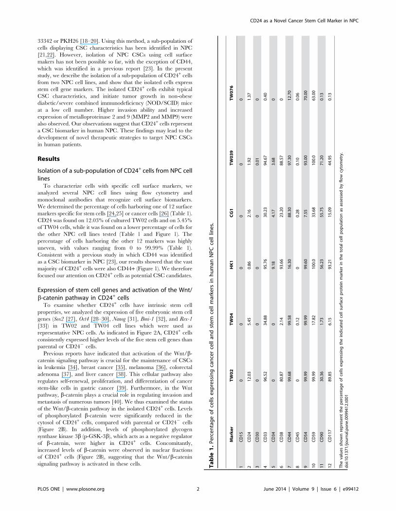
We determined the percentage of cells harboring one of 12 surface
markers specific for stem cells [24,25] or cancer cells [26] (Table 1).
CD24 was found on 12.03% of cultured TW02 cells and on 5.45%
of TW04 cells, while it was found on a lower percentage of cells for
the other NPC cell lines tested (Table 1 and Figure 1). The
percentage of cells harboring the other 12 markers was highly
uneven, with values ranging from 0 to 99.99% (Table 1).
Consistent with a previous study in which CD44 was identified
as a CSC biomarker in NPC [23], our results showed that the vast
majority of CD24+ cells were also CD44+ (Figure 1). We therefore
focused our attention on CD24+ cells as potential CSC candidates.
Expression of stem cell genes and activation of the Wnt/b-catenin pathway in CD24+ cells
To examine whether CD24+ cells have intrinsic stem cell
properties, we analyzed the expression of five embryonic stem cell
genes (Sox2 [27], Oct4 [28–30], Nanog [31], Bmi-1 [32], and Rex-1
[33]) in TW02 and TW04 cell lines which were used as
representative NPC cells. As indicated in Figure 2A, CD24+ cells
consistently expressed higher levels of the five stem cell genes than
parental or CD242 cells.
Previous reports have indicated that activation of the Wnt/b-
catenin signaling pathway is crucial for the maintenance of CSCs
in leukemia [34], breast cancer [35], melanoma [36], colorectal
adenoma [37], and liver cancer [38]. This cellular pathway also
regulates self-renewal, proliferation, and differentiation of cancer
stem-like cells in gastric cancer [39]. Furthermore, in the Wnt
pathway, b-catenin plays a crucial role in regulating invasion and
metastasis of numerous tumors [40]. We thus examined the status
of the Wnt/b-catenin pathway in the isolated CD24+ cells. Levels
of phosphorylated b-catenin were significantly reduced in the
cytosol of CD24+ cells, compared with parental or CD242 cells
(Figure 2B). In addition, levels of phosphorylated glycogen
synthase kinase 3b (p-GSK-3b), which acts as a negative regulator
of b-catenin, were higher in CD24+ cells. Concomitantly,
increased levels of b-catenin were observed in nuclear fractions
of CD24+ cells (Figure 2B), suggesting that the Wnt/b-catenin
signaling pathway is activated in these cells. Ta
ble
1.
Pe
rce
nta
ge
of
cells
exp
ress
ing
can
cer
cell
and
ste
mce
llm
arke
rsin
hu
man
NP
Cce
lllin
es.
Ma
rke
rT
W0
2T
W0
4H
K1
CG
1T
W0
39
TW
07
6
1C
D1
50
00
00
0
2C
D2
41
2.0
35
.45
0.8
62
.16
1.9
21
.37
3C
D3
00
00
00
.01
0
4C
D3
39
6.5
23
4.8
89
5.7
63
0.2
39
4.6
70
.40
5C
D3
40
09
.18
4.1
73
.68
0
6C
D3
88
0.8
72
.14
93
.66
23
.20
88
.57
0
7C
D4
49
9.6
89
9.5
81
6.3
08
8.3
09
7.3
01
2.7
0
8C
D4
50
0.1
20
0.2
80
.10
0.0
6
9C
D5
49
9.9
99
9.9
99
9.6
07
.55
93
.00
70
.00
10
CD
59
99
.99
17
.82
10
0.0
33
.68
10
0.0
63
.00
11
CD
90
30
.36
1.7
35
6.2
35
5.7
57
1.2
00
.13
12
CD
11
78
9.8
56
.15
93
.21
15
.09
44
.95
0.1
3
Th
eva
lue
ssh
ow
nre
pre
sen
tth
ep
erc
en
tag
eo
fce
llse
xpre
ssin
gth
ein
dic
ate
dce
llsu
rfac
ep
rote
inm
arke
rin
the
tota
lce
llp
op
ula
tio
nas
asse
sse
db
yfl
ow
cyto
me
try.
do
i:10
.13
71
/jo
urn
al.p
on
e.0
09
94
12
.t0
01
CD24 as a Novel Cancer Stem Cell Marker in NPC
PLOS ONE | www.plosone.org 2 June 2014 | Volume 9 | Issue 6 | e99412

Enhanced proliferation and clone/sphere formation inCD24+ cells
We measured the proliferation rate of CD24+ cells cultured in
complete DMEM. Compared with parental and CD242 cells,
CD24+ cells showed enhanced proliferation, starting on the 5th day
of culture for TW02 cells and the 7th day for TW04 cells, with
doubling times of about 42 h, 42 h, and 30 h for parental TW02,
CD242, and CD24+ cells, respectively, and of 40 h, 40 h and
28 h for parental TW04, CD242, and CD24+ cells (Figure 3A).
The clone formation efficiency (CFE) of CD24+ cells was also
determined. After 10 days of culture, when most clones were likely
to contain over 50 cells, the calculated CFE values of TW02 cells
were 15.1%, 80.8% and 12.2% for parental, CD24+, and CD242
cells, respectively. For TW04 cells, CFE values of 20.2%, 90.3%
and 13.0% were obtained for parental, CD24+, and CD242 cells,
respectively (Figure 3B). The formation of spheroid cell aggregates
(or simply spheres), which is indicative of self-renewal ability, was
further analyzed by culturing the cells in non-adherent conditions
consisting of serum-free DMEM containing only epithelial growth
factor (EGF) and basic fibroblast growth factor (bFGF). After 30
days of culture, CD24+ cells showed the highest number of
spheres, reaching diameters between 100 and 200 mm, whereas
parental cells and CD242 cells formed significantly fewer spheres,
with diameters below 100 mm (Figure 3C). These results indicate
that CD24+ cells show enhanced proliferation and ability to form
clones/spheres.
We also examined the effects of the Wnt inhibitor ICG-001 on
the sphere formation ability of CD24+ cells. After treatment with
the Wnt inhibitor for 7 days (10 mM), sphere size was considerably
reduced by an average of 20% and 45% in TW02 and TW04
cells, respectively (Figure 3D), suggesting that the Wnt pathway
may contribute to the CSC phenotype of CD24+ cells.
Long-term differentiation potential of CD24+ cellsA long-term culture of CSCs may increase its cell mass but also
undergo asymmetric division to generate a low tumorigenic cell
population with heterogeneous phenotypes [9]. To monitor the
cell differentiation potential of CD24+ cells, we analyzed 10-day
old cultures of CD24+ and CD242 cells by flow cytometry. After
culturing TW02 cells for 10 days, 11.56% of CD24+ cells
remained CD24+, whereas 0.14% of the cells that were initially
CD242 expressed CD24 after culture (Figure 4A). Similarly,
following culture of TW04 cells, 5.64% of CD24+ cells remained
CD24+, while 0.03% of cells that were CD242 initially were
CD24+ after culture (Figure 4B). These results suggest that CD24+
cells show cell differentiation potential after prolonged culture,
although the differentiation process may be only partial under
these conditions.
CD24+ cells show enhanced resistance tochemotherapeutic drugs
Higher resistance to chemotherapeutic drugs represents a
critical characteristic of CSCs [9]. Previous studies have reported
that CSCs can exclude the DNA-binding dye, Hoechst 33342, due
to enhanced expression of the ABC transporter, ABCG2 [41,42].
To determine whether CD24+ cells exclude Hoechst 33342 better
than parental or CD242 cells, we performed the dye exclusion
assay after treating TW02 cells with the ABC transporter inhibitor,
verapamil [43]. As shown in Figure 5A, a larger population of
Hoechst 33342-negative cells was observed for CD24+ cells
(12.9%) compared with CD242 cells (7.99%). Notably, verapamil
treatment reversed the dye exclusion phenotype observed in
CD24+ cells, while this treatment had no significant effect on
CD242 cells (Figure 5A). Similar results were obtained for the
TW04 cell line (verapamil produced no significant effect in this
case since almost no TW04 CD242 cells were Hoechst 33342-
negative). These observations suggest that CD24+ cells possess an
enhanced ability to exclude dye.
To examine whether CD24+ cells show higher resistance to
chemotherapeutic drugs, we treated the cells with either cisplatin
or docetaxel, and assessed cell viability using the MTT assay.
Cisplatin and docetaxel produced higher cytotoxic effects against
parental or CD242 cells compared with CD24+ cells (Figure 5B,
p,0.01). For the TW02 cell line, the half maximal inhibitory
concentration (IC50) of cisplatin against parental, CD24+, and
Figure 1. Flow cytometry analysis of CD24 and CD44 cell surface markers in NPC cell lines. Flow cytometry analysis of CD24+ and CD44+
sub-populations of TW02 and TW04 cells. 16106 cancer cells were collected and stained with anti-human CD24-fluorescein isothiocyanate (FITC) and/or anti-human CD44-phycoerythrin (PE) antibodies. Isotype-matched human antibodies served as control.doi:10.1371/journal.pone.0099412.g001
CD24 as a Novel Cancer Stem Cell Marker in NPC
PLOS ONE | www.plosone.org 3 June 2014 | Volume 9 | Issue 6 | e99412

CD242 cells was 1.84, 3.32, and 1.66 mg/ml, respectively, while
the IC50 values of docetaxel against parental, CD24+, and CD242
cells was 4.23, 7.45, and 3.53 mg/ml, respectively (Figure 5C). For
the TW04 cell line, cisplatin produced IC50 values of 0.70, 2.45,
and 0.46 mg/ml against parental, CD24+, and CD242 cells,
respectively, whereas the IC50 of docetaxel was 1.50, 3.93, and
1.41 mg/ml against parental, CD24+, and CD242 cells, respec-
tively (Figure 5D).
We also observed that the protein levels of the ABC transporter
ABCG2 were 1.42 and 2.12 fold higher in CD24+ cells than in
parental and CD242 cells, respectively (Figure 6A). Similarly, in
TW04 cells, the protein levels of ABCG2 in CD24+ cells were 1.85
and 3.56 fold higher than in parental and CD242 cells,
respectively (Figure 6A). Consistent with these results, the mRNA
expression level of ABCG2 was higher in CD24+ cells than in
parental or CD242 cells (Figure 6B). These results indicate that
CD24+ cells show enhanced chemoresistance, and that this
phenotype may be attributed at least in part to enhance expression
of ABCG2 transporter.
A small number of CD24+ cells is sufficient to producetumors in NOD/SCID mice
We tested the tumorigenic potential of CD24+ cells following
injection in the axillary fossa of immunodeficient NOD/SCID
Figure 2. Expression levels of stem cell genes and activation of the Wnt/b-catenin pathway in CD24+ cells. (A) mRNA expression levelsof Sox2, Oct4, Nanog, Bmi-1 and Rex-1 in parental, CD24+ and CD242 cells from the NPC cell lines, TW02 (left) and TW04 (right), was evaluated usingquantitative RT-PCR analysis. The results shown represented the average of three independent experiments. *: p,0.05, **: p,0.01. (B) Western blotsanalysis of phosphorylated-GSK, GSK, phosphorylated-b-catenin, b-catenin and b-actin in whole cell lysates, and of b-catenin and lamin B1 in nuclearfractions of parental, CD24+, and CD242 cells isolated from the TW02 and TW04 cell lines. Quantitative result was calculated by ImageJ software.*: p,0.05, **: p,0.01, ***: p,0.001.doi:10.1371/journal.pone.0099412.g002
CD24 as a Novel Cancer Stem Cell Marker in NPC
PLOS ONE | www.plosone.org 4 June 2014 | Volume 9 | Issue 6 | e99412

Figure 3. CD24+ cells show increased proliferation rate and enhanced clone and sphere formation. (A) Cell proliferation curves ofparental, CD24+ and CD242 cells from the TW02 (left) and TW04 (right) NPC cell lines cultured in complete DMEM for 9 days. The results shownrepresent the average of three independent experiments. *: p,0.05, **: p,0.01, ***: p,0.001. (B) Clone formation efficiency of parental, CD24+, andCD242 cells, and the quantification analysis. *: p,0.05. (C) Sphere formation ability of parental, CD24+ and CD242 cells cultured in DMEMsupplemented with 20 ng/ml bFGF and 20 ng/ml EGF for 30 days. (D) Effect of the Wnt inhibitor on sphere formation by CD24+ cells in TW02 andTW04 cells. CD24+ cells were treated or not with the Wnt inhibitor ICG-001 (10 mM) for seven days prior to sphere formation analysis. Scale bars:100 mm.doi:10.1371/journal.pone.0099412.g003
CD24 as a Novel Cancer Stem Cell Marker in NPC
PLOS ONE | www.plosone.org 5 June 2014 | Volume 9 | Issue 6 | e99412

mice. Twelve weeks after injection, tumors were detected in mice
inoculated with either 500 or 1,000 CD24+ cells, but not in mice
treated with 100 CD24+ cells (Figure 7A). In contrast, no tumors
were detected in mice inoculated with #1,000 cells in the parental
or CD242 group (Figure 7A and 7B). Similarly, no tumors were
detected in mice that received PBS as a negative control
(Figure 7A). Further histological analysis of tissue sections stained
with hematoxylin and eosin (H&E) confirmed the presence of
nasopharyngeal squamous cell carcinoma and well-differentiated
keratinizing squamous carcinoma at the site where CD24+ cells
were initially injected (Figure 7C). Injection of a small number of
CD24+ cells is thus sufficient to induce tumor formation in
immunodeficient mice.
Enhanced invasion potential and metalloproteinaseproduction in CD24+ cells
In previous studies, many CSC populations showed increased
invasion ability compared with non-CSCs [44,45]. Using an in
vitro invasion assay, we observed that CD24+ cells were more
invasive than parental or CD242 cells (Figure 8A). Given that
metalloproteinases play an important role in cell invasion [46], we
examined the level of MMP2 and MMP9 proteins in these cells. As
shown in Figure 8B, CD24+ cell populations showed increased
MMP2 and MMP9 protein levels compared with parental or
CD242 cells. Consistent with these results, we observed that
CD24+ cells isolated from the TW02 or TW04 cell lines expressed
higher levels of MMP2 and MMP9 mRNA than parental or
CD242 cells (Figure 8C). CD24+ cells also expressed lower E-
cadherin mRNA levels than parental or CD242 cells (Figure 8C).
CD24+ cells thus show enhanced invasion and increased
expression of MMP2 and MMP9 but reduced expression of E-
cadherin.
Discussion
The results of the present study suggest that CD24 represents a
new CSC surface marker in NPC. The characteristics of CD24+
cell sub-populations isolated from the TW02 and TW04 NPC cell
lines are similar to those reported previously for CSCs derived
from NPC [21–23]. These characteristics include increased
expression of genes involved in development and maintenance of
stem cells, increased self-renewal and maintenance of cell
differentiation capacity after prolonged culture, increased sphere
formation ability (also observed in CD24+ cells isolated from the
NPC cell lines HK1 and TW076; see Figure S1), increased
Hoechst 33342 dye exclusion and chemoresistance, increased
ability to initiate tumors in immunocompromised mice, and
enhanced cell invasion.
The cell surface protein, CD24, is highly expressed in many
human cancers [13,48,49]. CD24 expressed on cancer cells
interacts with P-selectin found on endothelial cells, indicating that
CD24 may bind to P-selectin and initiate rolling of cancer cells on
the endothelium, which may be followed by initiation of metastasis
[47]. In addition to NPC cells, CD24 is associated with CSCs of
other tumors, such as ovarian [13] and pancreatic cancer [48]. For
Figure 4. Cell differentiation potential of CD24+ cells. (A) Ten-day old cell masses originally cultured from CD24+ and CD242 cell cultures inDMEM medium were harvested, and stained with anti-human CD24-fluorescein isothiocyanate (FITC) antibody followed by flow cytometry analysis.The percentages shown represent cells with CD24+ surface markers from either (A) TW02 or (B) TW04 cell lines. Flow cytometry analysis of TW02 andTW04 cells immediately after sub-fractionation (right panels).doi:10.1371/journal.pone.0099412.g004
CD24 as a Novel Cancer Stem Cell Marker in NPC
PLOS ONE | www.plosone.org 6 June 2014 | Volume 9 | Issue 6 | e99412

CD24 as a Novel Cancer Stem Cell Marker in NPC
PLOS ONE | www.plosone.org 7 June 2014 | Volume 9 | Issue 6 | e99412

example, CD24+CD44+ESA+ pancreatic cancer cells possess CSC
properties [48]. Recently, a similar finding was reported for
ovarian CSCs; that is, a xenograft injection of 5,000 CD24+ cells
produced tumors in nude mice, while injection of an equal number
of CD242 cells failed to do so [49]. In addition, CD24+ cancer cell
colonies isolated from ovarian tumors of a human patient showed
heterogeneity in proliferation rate, cell cycle distribution, and
expression profile of genes and proteins, and demonstrated stem
cell properties [49]. Furthermore, CD24+ stem-like cells detected
in ovarian cancer also exhibited enhanced chemoresistance [50].
Notably, in breast cancer, the absence of CD24 combined with the
presence of CD44 and EpCAM (CD242CD44+EpCAM+) appears
to be critical for the identification of breast CSCs [15]. A previous
study has reported that CD44 represents a CSC biomarker in
NPC [23]. Consistent with this finding, the CD24+ cells that we
isolated from TW02 and TW04 NPC cell lines were mostly
CD44+ (Figure 1B). Besides, the vast majority of CD24+ cells in the
HK1 and TW076 cell lines were also CD44+ (Figure S2). Both
CD24 and CD44 may thus be involved in the formation of CSCs
in NPC, and the function of these proteins in the development and
maintenance of CSCs warrants further investigation.
We observed that CD24+ cells isolated from the TW02 and
TW04 NPC cell lines express higher mRNA levels of Sox2, Oct-4,
Nanog, Bmi-1, and Rex-1, compared with parental or CD242 cells.
A similar phenomenon was also observed in HK1 and TW076 cell
lines (Figure S3). These characteristics are similar to those reported
for embryonic stem cells [27–33] and ovarian cancer stem-like
cells [51]. This pattern of embryonic stem cell gene expression in
Figure 5. CD24+ cells show enhanced chemoresistance. (A) Sorted CD24+ and CD242 cells from the TW02 and TW04 NPC cell lines weretreated or not with 50 mM verapamil for 30 min prior to processing for the Hoechst 33342 dye exclusion assay as described in Materials and Methods.(B) Parental, CD24+, and CD242 cells from the TW02 or TW04 cell lines were treated with various concentrations of cisplatin and docetaxel for 24 h,and cell viability was determined using the MTT assay. CD24+ cells showed higher viability than parental and CD242 cells after treatment with eithercisplatin or docetaxel. The results shown represented the average of three independent experiments. *: p,0.05, **: p,0.01, ***: p,0.001. IC50 valuesof cisplatin or docetaxel treatment against parental, CD24+, and CD242 cells were shown for (C) TW02 and (D) TW04 cells lines.doi:10.1371/journal.pone.0099412.g005
Figure 6. Cellular ABCG2 expression levels in parental, CD24+, and CD242 cells. (A) Cellular ABCG2 protein levels in parental, CD24+, andCD242 TW02 and TW04 cells were determined by Western blot analysis. Quantitative result was calculated by ImageJ software. *: p,0.05, **: p,0.01.(B) The mRNA expression level of ABCG2 in parental, CD24+, and CD242 TW02 and TW04 cells was determined by quantitative RT-PCR. The resultsshown represent the average of three independent experiments. *: p,0.05, **: p,0.01.doi:10.1371/journal.pone.0099412.g006
CD24 as a Novel Cancer Stem Cell Marker in NPC
PLOS ONE | www.plosone.org 8 June 2014 | Volume 9 | Issue 6 | e99412

CD24+ NPC CSCs indicates the presence of a conserved pattern
of stem cell gene expression in embryonic stem cells and CSCs. In
embryonic stem cells, co-expression of Sox2, Oct-4, Nanog, and Rex-
1 is essential for maintenance of pluripotency and self-renewal,
and prevents cell differentiation along the trophoblast cell lineage
[30,33]. Sox2 and Oct-4 encode transcription factors that maintain
self-renewal and pluripotency in the undifferentiated embryonic
stem cell state by modulating genes that maintain permissive
chromatin structure and DNA repair, and prevent apoptosis [52].
On the other hand, the expression of Nanog, which is also a
transcription factor critically involved in self-renewal, is positively
regulated by Sox2 and Oct-4 [53]. These transcription factors form
a functional transcriptional regulation network that is critical for
maintenance of pluripotency in embryonic stem cells [53].
Furthermore, the Polycomb group (PcG) gene Bmi-1 silences Hox
genes via modulation of chromatin structure during embryonic
development in fruit flies, and may possibly enhance the
proliferative activity of both normal and leukemic stem cells
[32]. As the expression of Bmi-1 is negatively regulated by Pten,
which acts as a tumor suppressor gene [54], increased expression
of Bmi-1 may correlate with repression of Pten expression and
induction of epithelial-mesenchymal transition, as well as in-
creased motility and invasiveness of human nasopharyngeal
epithelial cells [54]. Our results show that the CD24+ sub-
population of NPC cells also expresses stem cell genes and exhibits
characteristics of stem cell self-renewal and enhanced proliferation
capacity.
The Wnt/b-catenin signaling pathway, which has been
reported to regulate stem cell function and niche-stem cell
interactions [56], enhances the expression of Sox2 and Oct-4
[55]. Moreover, reduced E-cadherin mRNA expression was
observed by Gao et al. in CD24+ ovarian cancer stem cells [49].
These observations indicate that the loss of E-cadherin expression
may allow b-catenin to re-localize to the nucleus and activate
transcriptional activity [57]. In this study, increased phosphory-
lation of GSK-3b, reduced phosphorylation of b-catenin, and
nuclear translocation of b-catenin were observed in CD24+ cells,
compared with parental or CD242 cells (Figure 2). These
observations indicate that the Wnt/b-catenin signaling pathway
is activated in CD24+ cells.
Figure 7. A low number of CD24+ NPC cells initiates tumor formation in NOD/SCID mice. (A) Formation of tumors following injection ofCD24+ cells. Groups of six NOD/SCID mice were injected with 100, 500, or 1,000 freshly-sorted CD24+ or CD242 cells from the TW02 (left) or TW04(right) cell line. Mice injected with PBS were used as a negative control. Tumor formation was assessed 12 weeks after cell inoculation. (B) Miceinjected with TW02 (left) or TW04 (right) cells were sacrificed for evaluation of tumor formation twelve weeks after inoculation. The arrows indicatethe presence of tumors in mice injected with 500 or 1,000 CD24+ cells. (C) Tissue H&E staining results of TW02 (left) and TW04 (right) mice inoculatedwith 500 cells. Inoculation of as few as 500 CD24+ cells produced histological signs of tumors at the site of injection.doi:10.1371/journal.pone.0099412.g007
CD24 as a Novel Cancer Stem Cell Marker in NPC
PLOS ONE | www.plosone.org 9 June 2014 | Volume 9 | Issue 6 | e99412

CD24 as a Novel Cancer Stem Cell Marker in NPC
PLOS ONE | www.plosone.org 10 June 2014 | Volume 9 | Issue 6 | e99412

Our findings that CD24+ NPC cells are more invasive than
parental or CD242 NPC cells are consistent with results from
previous studies on CSCs in pancreatic [58], lung [19], and
prostate cancer [59]. Together with the increased invasion ability,
cellular markers that characterize cell invasion, such as MMP2
and MMP9, were also increased at higher levels in CD24+ cells.
MMP2 and MMP9 are involved in the degradation of the
extracellular matrix, and an increase in expression of both
enzymes induces cell invasion ability [60]. Zhou et al. suggested
that genetic polymorphism in the MMP2 promoter may explain
the increased susceptibility of Chinese individuals for developing
NPC [61]. Microarray analysis reveals that MMP1 and MMP2
are the most significantly upregulated genes in NPC biopsies,
compared with lymphohyperplasia, adenoid tissues, and head and
neck cancer [62]. Furthermore, studies performed on NPC tumor
biopsies or cell lines with enhanced invasiveness or epithelial-
mesenchymal transitions also revealed enhanced expression of
Sox2, Oct-4, and Nanog [63–66], similar to the findings reported
here on CD24+ cells. These results indicate that CD24+ NPC
CSCs may promote tumorigenesis and also initiate invasion and
metastasis, and suggest that further experiments to confirm the
presence of CD24+ CSCs in human NPC biopsies are warranted.
In addition, whether the expression of stem cell and invasion genes
is under the control of a common signaling pathway, such as the
Wnt/b-catenin pathway, remains to be investigated.
In summary, our results show that CD24+ cells exhibit CSC
characteristics in NPC cell lines. Strategies aimed at the
eradication of CD24+ NPC cells may provide new and more
effective treatment strategies against NPC.
Materials and Methods
Cell cultureNPC cell lines (the keratinizing squamous TW02 cell line; the
undifferentiated TW04 cell line; differentiated squamous HK1
cells; poorly-differentiated squamous CG1 cells; and the kerati-
nizing squamous TW039 and TW076 cell lines) were provided by
Dr. Yu-Sun Chang (Chang Gung University) and maintained in
Dulbecco’s modified Eagle’s medium (DMEM; Invitrogen, Grand
Island, NY) supplemented with 10% fetal bovine serum (FBS), 100
units/ml penicillin, 100 mg/ml streptomycin and 25 mg/ml
amphotericin B (Invitrogen). After sorting as described below,
the cells were cultured in DMEM supplemented with 20 ng/ml
bFGF (Sigma-Aldrich, St. Louis, MO), 20 ng/ml EGF (Sigma-
Aldrich), 200 units/ml penicillin, 200 mg/ml streptomycin, and
50 mg/ml amphotericin B (Invitrogen). All cells were maintained
in a humidified 5% CO2 incubator at 37uC.
Flow cytometry analysis and sorting of NPC cellsThe cells were analyzed by fluorescence-activated cell sorting
(FACS) after reaching the logarithmic proliferation phase. Cells
were digested with 0.25% trypsin-0.02% EDTA (Invitrogen),
washed twice with calcium/magnesium-free PBS, and resus-
pended at a concentration of 16106 cells/ml in ice-cold PBS
supplemented with 2% FBS. FITC-conjugated anti-human CD15,
CD24, CD30, CD33, CD34, CD38, CD44, CD45, CD54, CD59,
CD90, CD117 or CD133 antibody (BD Pharmingen, Becton
Dickinson, Mountain View, CA) was then added at a final
concentration of 1 mg/ml, and incubated for 30 min at 4uC in the
dark with mixing. The cells were then washed twice with ice-cold
PBS supplemented with 2% FBS, and were analyzed using the
FACS Aria flow cytometer (Becton, Dickinson & Company,
Mountain View, CA). Isotype-matched human antibodies (Becton,
Dickinson & Company) were used as controls. Cells stained with
FITC-conjugated anti-human CD24 were also sorted using the
same flow cytometer.
Tumor sphere formation assaySix-well culture dishes were coated with 1.2% soft agar [22].
Parental and sorted cells were than counted and inoculated in
triplicate at a density of 500 cells/well in DMEM supplemented
with 20 ng/ml bFGF and 20 ng/ml EGF. Cells were observed for
sphere formation every day. When the spheres grew and reached
diameters of about 100 to 200 mm, images were taken using a
Leica DM IL Inverted Contrasting Microscope (Leica Micro-
systems, Wetzlar, Germany) with a Nikon Coolpix P5100 digital
camera (Nikon, Tokyo, Japan). In some experiments, the cells
were treated with the Wnt inhibitor ICG-001 (R&D Systems,
Minneapolis, MN; product #5439-DK-010) for seven days at
10 mM prior to analysis using the sphere formation assay.
Clone formation assayParental and sorted cells were plated in triplicate at 300 cells/
well in six-well plates, and cultured in DMEM supplemented with
10% FBS. After most cell colonies had expanded to .50 cells
(about 10 days), cells were washed twice with PBS, fixed in ice-cold
methanol for 30 min, and stained with 0.01% (w/v) crystal violet
for 30 min at room temperature [22]. After a washing step, colony
numbers containing .50 cells were counted and compared, and
images were taken. The CFE values shown correspond to the ratio
of clones obtained divided by the number of cells initially plated.
Long-term cell differentiation assayThe cell differentiation assay was done 18 days after cell sorting.
Sorted cells were cultured in DMEM supplemented with 20 ng/
ml bFGF and 20 ng/ml EGF. When the desired cell numbers
were obtained, cells were stained with FITC-conjugated anti-
human CD24, followed by analysis by FACS to quantify the
proportion of CD24+ cells in the sorted populations. Isotype-
matched human antibodies served as control.
Cell proliferation analysisSorted cells were incubated at 1,000 cells/well in 96-well plates,
and cultured in triplicate in DMEM supplemented with 10% FBS
to observe cell proliferation. Cell viability was assessed using the
MTT Cell Growth Determination Kit (Sigma-Aldrich) according
to the instructions provided by the manufacturer. Cell prolifera-
tion curves were drawn according to the results of background
(OD690) and substrate (OD570) absorbance.
Figure 8. CD24+ cells show higher invasion ability and enhanced expression of MMP2 and MMP9. (A) The cell invasion assay wasperformed using the transwell chamber assay, as described in Materials and Methods. Matrigel membranes containing invading cells were observedby optical microscopy, and the cells were counted. The number of invading cells from each cell population was quantified. Results shown wereobtained from three independent experiments. **: p,0.01, ***: p,0.001 (B) The levels of MMP2 and MMP9 protein produced by parental, CD24+, orCD242 cells from the TW02 and TW04 cell lines were quantified by Western blotting analysis. Quantitative result was calculated by ImageJ software.*: p,0.05. (C) The mRNA expression levels of MMP2 and MMP9 in parental, CD24+, and CD242 cells were determined by quantitative RT-PCR fromthe TW02 and TW04 cell lines. Results shown represent an average from three independent experiments. *: p,0.05, **: p,0.01.doi:10.1371/journal.pone.0099412.g008
CD24 as a Novel Cancer Stem Cell Marker in NPC
PLOS ONE | www.plosone.org 11 June 2014 | Volume 9 | Issue 6 | e99412

Drug sensitivity assayParental and sorted cells were cultured in triplicate at 5,000
cells/well in 96-well plates, and cisplatin or docetaxel was added
for 24 hours. Cell viability was determined as described above.
Cell sub-population analysisSorted cells were cultured in DMEM supplemented with 20 ng/
ml bFGF and 20 ng/ml EGF until they reached the desired cell
number. Cells were then digested with 0.25% trypsin-0.02%
EDTA, washed twice with calcium/magnesium-free PBS, and
resuspended at 16106 cells/ml in ice-cold DMEM supplemented
with 2% FBS. Hoechst 33342 (Sigma-Aldrich) was then added at a
final concentration of 5 mg/ml, prior to incubation for 90 min at
37uC with mixing. In some experiments, a subset of cells was
incubated with 50 mM verapamil for 30 min at 37uC before
addition of Hoechst 33342 to examine whether this treatment
blocked the efflux of fluorescent Hoechst 33342 from the sorted
cells [22]. The cells were then washed twice with PBS, and
resuspended in medium containing 2 mg/ml propidium iodide
(Sigma-Aldrich), followed by incubation for 30 min at 4uC in the
dark. Dual-wavelength analysis was performed to analyze cell sub-
populations by FACS.
Cell invasion assayCell invasion was performed using a BD BioCoat Matrigel
Invasion Chamber (Becton-Dickinson). Parental and sorted cells
were suspended in DMEM containing 1% FBS, before incubation
into the upper chamber at a density of 16105 cells/well. Cell
invasion into Matrigel was determined after 24 h of culture at
37uC. The invading cells found in the membrane were fixed using
ice-cold methanol, and stained with 0.01% crystal violet. Cell
invasion was quantified using the Leica DM IL Inverted
Contrasting Microscope after removing non-invading cells on
the upper side of the membrane with cotton swabs.
Tumor formation in miceNOD/SCID mice were purchased from the animal institute of
Tzu-Chi University (Taiwan), and were maintained individually in
micro-isolator cages under controlled temperature (2462uC),
humidity (60610%) and alternating 12-h light/dark cycles. All
experiments were carried out in strict accordance with the
recommendations of the National Institutes of Health and
approved by the Institutional Animal Care and Use Committee
(IACUC) of Taiwan (Permit Number: 09-TACTRI-IACUC-014).
Freshly-sorted cells suspended in 200 ml PBS containing 10% FBS
were inoculated into the axillary fossa of 6- to 7-week-old NOD/
SCID mice at a dose of 100, 500, or 1,000 cells per mouse on the
same afternoon that the cells were sorted. The mice were
monitored twice weekly for palpable tumor formation, and were
euthanized 12 weeks after cell inoculation to assess tumor
formation. The mice were photographed, and a portion of the
subcutaneous tissue at the site of cell injection was collected, fixed
in 10% formaldehyde, and embedded in paraffin for H&E
staining.
RNA extraction and quantitative real-time RT-PCRTotal RNA was isolated with Trizol (Invitrogen). First-strand
cDNA was reverse-transcribed (RT) according to the manufac-
turer protocols. Relative levels of mRNA were determined by q-
PCR using a real-time PCR system. Several stem cell genes and
epithelial-mesenchymal transition markers were analyzed. The
primer sequences used are shown in Table 2. b-actin was used as
reference. cDNA was subjected to PCR for initial denaturation at
95uC for min, followed by 50 cycles of 95uC for 30 sec, 60uC for
30 sec, and 72uC for 20 sec, and terminal extension at 72uC for
7 min. q-PCR was performed using a Roche LightCycler 480
System (Roche Diagnosis, Mannheim, Germany).
Western blotting analysisCells were lysed with RIPA lysis buffer (Millipore) in the
presence of protease and phosphatase inhibitors (P2580, Sigma-
Aldrich), and the concentration of total cellular protein was
quantified using the Bradford reagent as instructed by the
manufacturer (Bio-Rad, Hercules, CA). In addition, the nuclear
and cytosolic protein fractions were prepared with NE-PER
Nuclear and Cytoplasmic Extraction Reagents (Thermo Scientific,
Waltham, MA). Western blotting analysis was performed with
30 mg proteins of cell lysates separated on 12% SDS-PAGE gel.
Proteins transferred to PVDF membrane (Millipore) were probed
with monoclonal or polyclonal antibodies specific to p-GSK-3b(Ser-9), GSK-3b, b-catenin, b-actin, MMP-2, ABCG2, lamin-B1
(Santa Cruz Biotechnology, Santa Cruz, CA), or p-b-catenin (Cell
Signaling Technology, Danvers, MA; product #9565). Immobilon
Western Chemiluminescent HRP Substrate (Millipore) was used
for the detection of primary antibody-probed protein bound with
an appropriate horseradish peroxidase-conjugated secondary
antibody.
Table 2. DNA primers used for q-RT-PCR analysis in this study.
Gene Forward primer (59-39) Reverse primer (59-39)
Sox-2 ACACCAATCCCATCCACACT GCAAACTTCCTGCAAAGCTC
4-Oct GCAATTTGCCAAGCTCCTGAA GCAGATGGTCGTTGGCTGA
Nanog TTCCTTCCTCCATGGATCTG TCTGCTGGAGGCTGAGGTAT
Bmi-1 ATGCATCGAACAACGAGAATCAAGATCACT TCAACCAGAAGAAGTTGCTGATGACCC
Rex-1 TGGACACGTCTGTGCTCTTC GTCTTGGCGTCTTCTCGAAC
ABCG2 GGGTTCTCTTCTTCCTGACGACC TGGTTGTGAGATTGACCAACAGAC
MMP2 ACGACCGCGACAAGAAGTAT ATTTGTTGCCCAGGAAAGTG
MMP9 CCCTGGAGACCTGAGAACCA CCCGAGTGTAACCATAGCGG
E-cadherin ATTTTTCCCTCGACACCCGAT TCCCAGGCGTAGACCAAGA
b-actin GGATCTTCATGAGGTAGTCAG GAGACCTTCAACACCCCAGCC
doi:10.1371/journal.pone.0099412.t002
CD24 as a Novel Cancer Stem Cell Marker in NPC
PLOS ONE | www.plosone.org 12 June 2014 | Volume 9 | Issue 6 | e99412

Statistical analysisSPSS11.0 and Excel 2007 were used for data processing and for
analyzing the significance between parental and sorted cells using
the unpaired Student t-test. Data were expressed as the mean 6
SD from at least three independent experiments. P values,0.05
were considered statistically significant.
Supporting Information
Figure S1 CD24+ cells show enhanced sphere formationin HK1 and TW076 cell lines. Sphere formation in parental,
CD24+ and CD242 cells in HK1 and TW076 cell lines cultured in
DMEM supplemented with 20 ng/ml bFGF and 20 ng/ml EGF
for 30 days. The images are representative results of three
independent experiments. Scale bars: 100 mm.
(TIF)
Figure S2 Flow cytometry analysis of CD24+ and CD44+
sub-population in HK1 and TW076 cell lines. A total of
16106 cancer cells were collected and stained with anti-human
CD24-fluorescein isothiocyanate (FITC) and/or anti-human
CD44-phycoerythrin (PE) antibodies. Isotype-matched human
antibodies were used as negative control.
(TIF)
Figure S3 Expression level of stem cell genes in CD24+
HK1 and TW076 cell lines. The mRNA expression level of
Sox2, Oct4, Nanog, Bmi-1 and Rex-1 in parental, CD24+ and CD242
cells from the NPC cell lines HK1 (top panel) and TW076 (bottom
panel) was analyzed by quantitative RT-PCR. The results shown
represent the average of three independent experiments. *:p,0.05,
**: p,0.01, ***: p,0.001.
(TIF)
Author Contributions
Conceived and designed the experiments: YSC HCL JDY. Performed the
experiments: CHY HLW YSL KPSK HCL CJC CCL TTH HCL.
Analyzed the data: DO JM. Contributed reagents/materials/analysis tools:
CCL YSC. Wrote the paper: JM HCL JDY.
References
1. Cao SM, Simons MJ, Qian CN (2011) The prevalence and prevention of
nasopharyngeal carcinoma in China. Chin J Cancer 30: 114–119.
2. Eduardo B, Raquel C, Rui M (2010) Nasopharyngeal carcinoma in a south
European population: epidemiological data and clinical aspects in Portugal. Eur
Arch Otorhinolaryngol 267: 1607–1612.
3. Cancer Registry Annual Report R.O.C. Department of Health Executive Yuan,
ROC. (2010).
4. Chang JT, Ko JY, Hong RL (2004) Recent advances in the treatment of
nasopharyngeal carcinoma. J Formos Med Assoc 103: 496–510.
5. DeNittis AS, Liu L, Rosenthal DI, Machtay M (2004) Nasopharyngeal
carcinoma treated with external radiotherapy, brachytherapy, and concur-
rent/adjuvant chemotherapy. Am J Clin Oncol 25: 93–95.
6. Suarez C, Rodrigo JP, Rinaldo A, Langendijk JA, Shaha AR, et al. (2010)
Current treatment options for recurrent nasopharyngeal cancer. Eur Arch
Otorhinolaryngol 267: 1811–1824.
7. Marotta LL, Polyak K (2009) Cancer stem cells: a model in the making. Current
Opin Genet Dev 19: 44–50.
8. Clarke MF, Dick JE, Dirks PB, Eaves CJ, Jamieson CH, et al. (2006) Cancer
stem cells–perspectives on current status and future directions: AACR Workshop
on cancer stem cells. Cancer Res 66: 9339–9344.
9. Reya T, Morrison SJ, Clarke MF, Weissman I (2001) Stem cells, cancer, and
cancer stem cells. Nature 414: 105–111.
10. Assimakopoulos D, Kolettas E, Patrikakos G, Evangelou A (2002) The role of
CD44 in the development and prognosis of head and neck squamous cell
carcinomas. Histol Histopathol 17: 1269–1281.
11. Vander Griend DJ, Karthaus WL, Dalrymple S, Meeker A, DeMarzo AM, et al.
(2008) The role of CD133 in normal human prostate stem cells and malignant
cancer-initiating cells. Cancer Res 68: 9703–9711.
12. Yang ZF, Ho DW, Ng MN, Lau CK, Yu WC, et al. (2008) Significance of
CD90+ cancer stem cells in human liver cancer. Cancer Cell 13: 153–166.
13. Gao MQ, Choi YP, Kang S, Youn JH, Cho NH (2010) CD24+ cells from
hierarchically organized ovarian cancer are enriched in cancer stem cells.
Oncogene 29: 2672–2680.
14. Facchino S, Abdouh M, Bernier G (2011) Brain Cancer Stem Cells: Current
Status on Glioblastoma Multiforme. Cancers 3: 1777–1797.
15. Ricardo S, Vieira AF, Gerhard R, Leitao D, Pinto R, et al. (2011) Breast cancer
stem cell markers CD44, CD24 and ALDH1: expression distribution within
intrinsic molecular subtype. J Clin Pathol 64: 937–946.
16. Wang S, Xu ZY, Wang LF, Su W (2013) CD133+ cancer stem cells in lung
cancer. Front Biosci 18: 447–453.
17. Tang DG (2012) Understanding cancer stem cell heterogeneity and plasticity.
Cell Res 22: 457–472.
18. Haraguchi N, Utsunomiya T, Inoue H, Tanaka F, Mimori K, et al. (2006)
Characterization of a side population of cancer cells from human gastrointestinal
system. Stem Cells 24: 506–513.
19. Ho MM, Ng AV, Lam S, Hung JY (2007) Side population in human lung cancer
cell lines and tumors in enriched with stem-like cancer cells. Cancer Research
67: 4827–4233.
20. Lanzkron SM, Collector MI, Sharkis SJ (1999) Hematopoietic Stem Cell
Tracking In Vivo: A comparison of short-term and long-term repopulating cells.
Blood 93: 1916–1921.
21. Wang WJ, Wu SP, Liu JB, Shi YS, Huang X, et al. (2013) MYC regulation of
CHK1 and CHK2 promotes radioresistance in a stem cell-like population of
nasopharyngeal carcinoma cells. Cancer Res 73: 1219–1231.
22. Wang J, Guo LP, Chen LZ, Zeng YX, Lu SH (2007) Identification of cancerstem cell-like side population cells in human nasopharyngeal carcinoma cell line.
Cancer Res 67: 3716–3724.
23. Su J, Xu XH, Huang Q, Lu MQ, Li DJ, et al. (2011) Identification of cancerstem-like CD44+ cells in human nasopharyngeal carcinoma cell line. Arch Med
Res 42: 15–21.
24. Zhang XG, Gaillard JP, Robillard N, Lu ZY, Gu ZJ, et al. (1994) Obtaining of
human myeloma cell lines as a model for tumor stem cell study in humanmultiple myeloma. Blood 83: 3654–3663.
25. International Stem Cell Initiative, Adewumi O, Aflatoonian B, Ahrlund-Richter
L, Amit M, et al. (2007) Characterization of human embryonic stem cell lines bythe International Stem Cell Initiative. Nat Biotechnol 25: 803–816.
26. Liu AY (2000) Differential expression of cell surface molecules in prostate cancercells. Cancer Res 60: 3429–3434.
27. Niwa H, Miyazaki J, Smith AG (2000) Quantitative expression of Oct-3/4
defines differentiation, dedifferentiation or self-renewal of ES cells. Nat Genet24: 372–376.
28. Atlasi Y, Mowla SJ, Ziaee SA, Bahrami AR (2007) OCT-4, an embryonic stem
cell marker, is highly expressed in bladder cancer. Int J Cancer 120: 1598–1602.
29. Kim JH, Jee MK, Lee SY, Han TH, Kim BS, et al. (2009) Regulation of adipose
tissue stromal cell behaviors by endogenic Oct4 expression control. PLoS One 4:e7166.
30. Guo Y, Liu S, Wang P, Zhao S, Wang F, et al. (2011) Expression profile of
embryonic stem cell-associated genes Oct4, Sox2 and Nanog in human gliomas.
Histopathology 59: 763–775.
31. Loh YH, Wu Q, Chew JL, Vega VB, Zhang W, et al. (2006) The Oct4 andNanog transcription network regulates pluripotency in mouse embryonic stem
cells. Nat Genet 38: 431–440.
32. Lessard J, Sauvageau G (2003) Bmi-1 determines the proliferative capacity ofnormal and leukaemic stem cells. Nature 423: 255–260.
33. Etti BS, James RT, Lorraine J, Yehudit B (1998) Rex-1, a gene encoding atranscription factor expressed in the early embryo, is regulated via Oct-3/4 and
Oct-6 binding to an octamer site and a novel protein, Rox-1, binding to anadjacent site. Mol Cell Biol 18: 1866–1878.
34. Correa S, Binato R, Du Rocher B, Castelo-Branco MT, Pizzatti L, et al. (2012)
Wnt/b-catenin pathway regulates ABCB1 transcription in chronic myeloid
leukemia. BMC Cancer 12: 303–313.
35. Prosperi JR, Goss KH (2010) A Wnt-ow of opportunity: targeting the Wnt/beta-catenin pathway in breast cancer. Curr Drug Targets 11: 1074–1088.
36. Larue L, Delmas V (2006) The WNT/beta-catenin pathway in melanoma.
Front Biosci 11: 733–742.
37. Greenspan EJ, Madigan JP, Boardman LA, Rosenberg DW (2011) Ibuprofeninhibits activation of nuclear b-catenin in human colon adenomas and induces
the phosphorylation of GSK-3b. Cancer Prev Res 4: 161–171.
38. Cavard C, Colnot S, Audard V, Benhamouche S, Finzi L, et al. (2008) Wnt/
beta-catenin pathway in hepatocellular carcinoma pathogenesis and liverphysiology. Future Oncol 4: 647–660.
39. Cai C, Zhu X (2012) The Wnt/b-catenin pathway regulates self-renewal of
cancer stem-like cells in human gastric cancer. Mol Med Rep 5: 1191–1196.
40. Damsky WE, Curley DP, Santhanakrishnan M, Rosenbaum LE, Platt JT, et al.
(2011) b-catenin signaling controls metastasis in Braf-activated Pten-deficientmelanomas. Cancer Cell 20: 741–754.
41. Hirschmann-Jax C, Foster AE, Wulf GG, Nuchtern JG, Jax TW, et al. (2004) A
distinct side population of cells with high drug efflux capacity in human tumorcells. Proc Natl Acad Sci USA 101: 14228–14233.
CD24 as a Novel Cancer Stem Cell Marker in NPC
PLOS ONE | www.plosone.org 13 June 2014 | Volume 9 | Issue 6 | e99412

42. Zhang JT (2007) Biochemistry and pharmacology of the human multidrug
resistance gene product, ABCG2. J Cent South Univ (Med Sci) 32: 531–541.43. Kim M, Turnquist H, Jackson J, Sgagias M, Yan Y, et al. (2002) The multidrug
resistance transporter ABCG2 (breast cancer resistance protein 1) effluxes
Hoechst 33342 and is overexpressed in hematopoietic stem cells. Clin CancerRes 8: 22–28.
44. Brabletz T, Jung A, Spaderna S, Hlubek F, Kirchner T (2005) Opinion:Migrating cancer stem cells–An integrated concept of malignant tumour
progression. Nat Rev Cancer 5: 744–749.
45. Hollier BG, Evans K, Mani SA (2009) The epithelial-to-mesenchymal transitionand cancer stem cells: A coalition against cancer therapies. J Mammary Gland
Biol Neoplasia 14: 29–43.46. Deryugina EI, Quigley JP (2006) Matrix metalloproteinases and tumor
metastasis. Cancer Metastasis Rev 25: 9–34.47. Aigner S, Ramos CL, Hafezi-Moghadam A, Lawrence MB, Friederichs J, et al.
(1998) CD24 mediates rolling of breast carcinoma cells on P-selectin. FASEB J
12: 1241–1251.48. Li C, Heidt DG, Dalerba P, Burant CF, Zhang L, et al. (2007) Identification of
pancreatic cancer stem cells. Cancer Res 67: 1030–1037.49. Gao MQ, Choi YP, Kang S, Youn JH, Cho NH (2010) CD24+ cells from
hierarchically organized ovarian cancer are enriched in cancer stem cells.
Oncogene 29: 2672–2680.50. Wei X, Dombkowski D, Meirelles K, Pieretti-Vanmarcke R, Szotek PP, et al.
(2010) Mullerian inhibiting substance preferentially inhibits stem/progenitors inhuman ovarian cancer cell lines compared with chemotherapeutics. Proc Natl
Acad Sci USA 107: 18874–18879.51. Zhang S, Balch C, Chan MW, Lai HC, Matei D, et al. (2008) Identification and
characterization of ovarian cancer-initiating cells from primary human tumors.
Cancer Res 68: 4311–4320.52. Campbell PA, Perez-Iratxeta C, Andrade-Navarro MA, Rudnicki MA (2007)
Oct4 targets regulatory nodes to modulates stem cell function. PLoS One 2:e553.
53. Tay Y, Zhang J, Thomson AM, Lim B, Rigoutsos I (2008) MicroRNAs to
Nanog, Oct4 and Sox2 coding regions modulate embryonic stem celldifferentiation. Nature 455: 1124–1128.
54. Song LB, Li J, Liao WT, Feng Y, Yu CP, et al. (2009) The polycomb group Bmi-1 represses the tumor suppressor PTEN and induces epithelial-mesenchymal
transition in human nasopharyngeal epithelial cells. J Clin Invest 119: 3626–
3636.
55. Yi F, Pereira L, Hoffman JA, Shy BR, Yuen CM, et al. (2011) Opposing effects
of Tcf3 and Tcf1 control Wnt stimulation of embryonic stem cell self-renewal.
Nat Cell Biol 13: 762–770.
56. Marie Rattis F, Voermans C, Reya T (2004) Wnt signaling in the stem cell
niche. Curr Opin Hematol 11: 88–94.
57. Herzig M, Savarese F, Novatchkova M, Semb H, Christofori G (2007) Tumor
progression induced by the loss of E-cadherin independent of beta-catenin/Tcf-
mediated Wnt signaling. Oncogene 26: 2290–2298.
58. Moriyama T, Ohuchida K, Mizumoto K, Cui L, Ikenaga N, et al. (2010)
Enhanced cell migration and invasion of CD133 (+) pancreatic cancer cells co-
cultured with pancreatic stromal cells. Cancer 116: 3357–3368.
59. Collins AT, Berry PA, Hyde C, Stower MJ, Maitland NJ (2005) Prospective
identification of tumorigenic prostste cancer stem cell. Cancer Res 65: 10946–
10951.
60. Yong VW, Power C, Forsyth P, Edwards D (2001) Metalloproteinases in biology
and pathology of the nervous system. Nat Rev Neurosci 2: 502–511.
61. Zhou G, Zhai Y, Cui Y, Qiu W, Yang H, et al. (2007) Functional
polymorphisms and haplotypes in the promoter of the MMP2 gene are
associated with risk of nasopharyngeal carcinoma. Hum Mutat 28: 1091–1097.
62. Lu J, Chua HH, Chen SY, Chen JY, Tsai CH (2003) Regulation of matrix
metalloproteinase-1 by Epstein-Barr virus proteins. Cancer Res 63: 256–262.
63. Hou W, He W, Li Y, Ma R, Wang Z, et al. (2014) Increased expression of
aldehyde dehydrogenase 1 A1 in nasopharyngeal carcinoma is associated with
enhanced invasiveness. Eur Arch Otorhinolaryngol 271: 171–179.
64. Chu WK, Dai PM, Li HL, Pao CC, Chen JK (2013) Nanog expression is
negatively regulated by protein kinase C activities in human cancer cell lines.
Carcinogenesis 34: 1497–1509.
65. Luo W, Li S, Peng B, Ye Y, Deng X, et al. (2013) Embryonic stem cells markers
SOX2, OCT4 and Nanog expression and their correlations with epithelial-
mesenchymal transition in nasopharyngeal carcinoma. PLoS One 8: e56324.
66. Guo D, Xu BL, Zhang XH, Dong MM (2012) Cancer stem-like side population
cells in the human nasopharyngeal carcinoma cell line cne-2 possess epithelial
mesenchymal transition properties in association with metastasis. Oncol Rep 28:
241–247.
CD24 as a Novel Cancer Stem Cell Marker in NPC
PLOS ONE | www.plosone.org 14 June 2014 | Volume 9 | Issue 6 | e99412