Identification of calcium binding sites on calsequestrin 1 and their implications for polymerization
18
Identification of Calcium binding sites on calsequestrin 1 and its implications to polymerization Amit Kumar 1,# , Harapriya Chakravarty 2,# , Naresh C. Bal 3,#,* , Tuniki Balaraju 2 , Nivedita Jena 4 , Gauri Misra 5 , Chandralata Bal 2 , Enrico Pieroni 1 , Muthu Periasamy 3,6 , and Ashoke Sharon 2,* 1 CRS4, Bioengineering group, Science and Technology Park Polaris, Piscina Manna, 09010 Pula (CA). Italy 2 Department of Applied Chemistry, Birla Institute of Technology, Mesra, Ranchi, Jharkhand 835215, India 3 Department of Physiology and Cell Biology, The Ohio State University, College of Medicine, Columbus, OH 43210, United States 4 College of Pharmacy, The Ohio State University, Columbus, OH 43210, United States 5 Institute of Biotechnology, Amity University, Noida, India 6 Davis Heart and Lung Research Institute, Ohio State University, Columbus, Ohio, USA Abstract Biophysical studies have shown that each molecule of calsequestrin 1 (CASQ1) can bind about 70–80 Ca 2+ ions. However, the nature of Ca 2+ -binding sites has not yet been fully characterized. In this study, we employed in-silico approaches to identify the Ca 2+ binding sites and to understand the molecular basis of CASQ1-Ca 2+ recognition. We built the protein model by extracting the atomic coordinates for the back-to-back dimeric unit from the recently solved hexameric CASQ1 structure (PDB id: 3UOM) and adding the missing C-terminal residues (aa350–364). Using this model we performed extensive 30 ns molecular dynamics simulations exposed to wide range of Ca 2+ concentrations ([Ca 2+ ]). Our results show that the Ca 2+ -binding sites on CASQ1 differ both in affinity and geometry. The high affinity Ca 2+ -binding sites share a similar geometry and interestingly, majority of them were found to be induced by increased [Ca 2+ ]. We also found that the system undergoes maximal Ca 2+ -binding to the CAS (consecutive aspartate stretch at the C-terminus) before the rest of the CASQ1 surface becomes saturated. Simulated data shows that the CASQ1 back-to-back stacking is progressively stabilized by emergence of an increasing number of hydrophobic interactions with increasing [Ca 2+ ]. Further, this study shows that the CAS domain assumes a compact structure with increase in Ca 2+ binding, which suggests that the CAS domain might function as a Ca 2+ -sensor that may be a novel structural motif to sense metal. We propose the term “D n -motif” for the CAS domain. Keywords calcium binding protein; molecular dynamics; entropy; electrostatic interaction; structural motif * Corresponding Authors:Ashoke Sharon; Department of Applied Chemistry, Birla Institute of Technology, Mesra, Ranchi, Jharkhand, India-835215. Tel: 91-651-2276531; Fax: 91-651-2275401; [email protected] Or, Naresh C. Bal; Department of Physiology and Cell Biology, The Ohio State University, College of Medicine, Columbus, OH 43210, United States. Tel: 1614-688-4635; Fax: 1614-2924888; [email protected]. # The authors contributed equally. NIH Public Access Author Manuscript Mol Biosyst. Author manuscript; available in PMC 2014 July 04. Published in final edited form as: Mol Biosyst. 2013 July 4; 9(7): 1949–1957. doi:10.1039/c3mb25588c. NIH-PA Author Manuscript NIH-PA Author Manuscript NIH-PA Author Manuscript
-
Upload
independent -
Category
Documents
-
view
1 -
download
0
Transcript of Identification of calcium binding sites on calsequestrin 1 and their implications for polymerization

Identification of Calcium binding sites on calsequestrin 1 and itsimplications to polymerization
Amit Kumar1,#, Harapriya Chakravarty2,#, Naresh C. Bal3,#,*, Tuniki Balaraju2, NiveditaJena4, Gauri Misra5, Chandralata Bal2, Enrico Pieroni1, Muthu Periasamy3,6, and AshokeSharon2,*
1CRS4, Bioengineering group, Science and Technology Park Polaris, Piscina Manna, 09010 Pula(CA). Italy2Department of Applied Chemistry, Birla Institute of Technology, Mesra, Ranchi, Jharkhand835215, India3Department of Physiology and Cell Biology, The Ohio State University, College of Medicine,Columbus, OH 43210, United States4College of Pharmacy, The Ohio State University, Columbus, OH 43210, United States5Institute of Biotechnology, Amity University, Noida, India6Davis Heart and Lung Research Institute, Ohio State University, Columbus, Ohio, USA
AbstractBiophysical studies have shown that each molecule of calsequestrin 1 (CASQ1) can bind about70–80 Ca2+ ions. However, the nature of Ca2+-binding sites has not yet been fully characterized.In this study, we employed in-silico approaches to identify the Ca2+ binding sites and tounderstand the molecular basis of CASQ1-Ca2+ recognition. We built the protein model byextracting the atomic coordinates for the back-to-back dimeric unit from the recently solvedhexameric CASQ1 structure (PDB id: 3UOM) and adding the missing C-terminal residues(aa350–364). Using this model we performed extensive 30 ns molecular dynamics simulationsexposed to wide range of Ca2+ concentrations ([Ca2+]). Our results show that the Ca2+-bindingsites on CASQ1 differ both in affinity and geometry. The high affinity Ca2+-binding sites share asimilar geometry and interestingly, majority of them were found to be induced by increased[Ca2+]. We also found that the system undergoes maximal Ca2+-binding to the CAS (consecutiveaspartate stretch at the C-terminus) before the rest of the CASQ1 surface becomes saturated.Simulated data shows that the CASQ1 back-to-back stacking is progressively stabilized byemergence of an increasing number of hydrophobic interactions with increasing [Ca2+]. Further,this study shows that the CAS domain assumes a compact structure with increase in Ca2+ binding,which suggests that the CAS domain might function as a Ca2+-sensor that may be a novelstructural motif to sense metal. We propose the term “Dn-motif” for the CAS domain.
Keywordscalcium binding protein; molecular dynamics; entropy; electrostatic interaction; structural motif
*Corresponding Authors:Ashoke Sharon; Department of Applied Chemistry, Birla Institute of Technology, Mesra, Ranchi,Jharkhand, India-835215. Tel: 91-651-2276531; Fax: 91-651-2275401; [email protected] Or, Naresh C. Bal; Department ofPhysiology and Cell Biology, The Ohio State University, College of Medicine, Columbus, OH 43210, United States. Tel:1614-688-4635; Fax: 1614-2924888; [email protected].#The authors contributed equally.
NIH Public AccessAuthor ManuscriptMol Biosyst. Author manuscript; available in PMC 2014 July 04.
Published in final edited form as:Mol Biosyst. 2013 July 4; 9(7): 1949–1957. doi:10.1039/c3mb25588c.
NIH
-PA Author Manuscript
NIH
-PA Author Manuscript
NIH
-PA Author Manuscript

IntroductionCASQ1 is the major Ca2+ buffering protein present in the sarcoendoplasmic reticulum (SR)of the striated muscles1–5 where it also regulates Ca2+ release by its interaction withRyanodine receptor (RyR) complex1. Crystallographic studies showed that CASQ1monomer consists of three α-β globular domains formed by five β-strands arranged into asheet sandwiched by four α-helices, which is structurally similar to ubiquitous redox protein“thioredoxin”6–7. Beyond the structural domain III, CASQ1 possesses a unique C-terminusthat is composed of repeating aspartic acid residues. The length of this consecutive aspartatestretch (CAS) varies between species and typically among mammals it is of 9–14 residues.Biophysico-chemical studies suggest that CASQ1 binds large amount of Ca2+ ions (~70–80Ca2+/molecule) with low affinity3, 8–11. However, CASQ1 lacks any known Ca2+ bindingmotif like EF-hand6, 12–14 or double-clamp15 and the nature of Ca2+-binding sites are notfully characterized. It has been proposed that Ca2+ binding to CASQ1 is purely a surfacephenomenon governed by the highly negatively charged molecular surface7, 11.
Based on biochemical and structural observations it has been proposed that CASQ1 formspolymeric structure in a Ca2+ concentration ([Ca2+]) dependent manner6–7, 9. At first, twomonomers interact to form front-to-front dimer by insertion of 24 N-terminal residues intothe partner monomer, resulting in a very stable dimer7, 11, 16. Then, two such front-to-frontdimers stack by back-to-back interactions leading to formation of a tetramer andsubsequently resulting in the formation of linear polymer in a Ca2+-dependent manner.However, detailed structural information about the interactions at the back-to-back interfacein the CASQ1 polymer has been speculative and it is currently unclear if the back-to-backstacking is dependent on [Ca2+]. Further, the CAS residues are located at the back-to-backinterface and can significantly influence with the stacking interactions17. It is also unclear ifCAS domain plays a role in facilitating back-to-back stacking and Ca2+-binding, given thatthe structure of the CAS residues has not been resolved in any of the currently availableCASQ1 structures.
The recently solved CASQ1 structure (PDB Id: 3UOM12) in the hexameric state providesthe opportunity to apply computational approaches to define the physico-chemical propertiesof the Ca2+-binding sites and the role of CAS domain in [Ca2+]-dependent protein-proteininteraction. The hexamer consists of three repeating dimeric units formed by front-to-frontdimerization, which on the other hand are held by back-to-back interaction. In the light ofthis scenario, the goals of this study are 1) identify and characterize the Ca2+-binding siteson CASQ1; 2) better understand how the surface of CASQ1 react to Ca2+-binding and 3)gain insight into the functional role of CAS domain in CASQ1-Ca2+ interaction. Toaccomplish these goals, we built a model of back-to-back dimeric unit from the hexamericCASQ1 structure, added the missing CAS residues (aa350–364) and performed extensivemolecular dynamics (MD) simulations under a wide range of [Ca2+].
Results and Discussions[Ca2+]-dependent charge distribution on CASQ1 surface
Before starting a detailed structural investigation, we first analysed the stability of CASQ1dimeric unit during MD simulation by calculating the backbone RMSD and entropy of thesystem. The stability of the dimeric system increases at higher [Ca2+] as reflected both fromdecrease of RMSD and entropy values (Supplemental Table 1). Then, we determined thesurface charge of CASQ1 after MD simulation in all the systems exposed at different[Ca2+]. As shown in Figure 1, the surface of CASQ1 in a Ca2+ free system is highly acidic,as also previously reported11, 18. Then, as expected, a gradual surface neutralization,mediated by Ca2+ binding, was observed increasing [Ca2+]. However, up to the presence of
Kumar et al. Page 2
Mol Biosyst. Author manuscript; available in PMC 2014 July 04.
NIH
-PA Author Manuscript
NIH
-PA Author Manuscript
NIH
-PA Author Manuscript
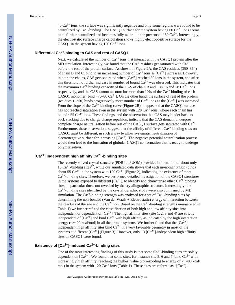
40 Ca2+ ions, the surface was significantly negative and only some regions were found to beneutralized by Ca2+-binding. The CASQ1 surface for the system having 60 Ca2+ ions seemsto be further neutralized and becomes fully neutral in the presence of 80 Ca2+. Interestingly,the electrostatic surface charge calculation shows highly electropositive surface for theCASQ1 in the system having 120 Ca2+ ions.
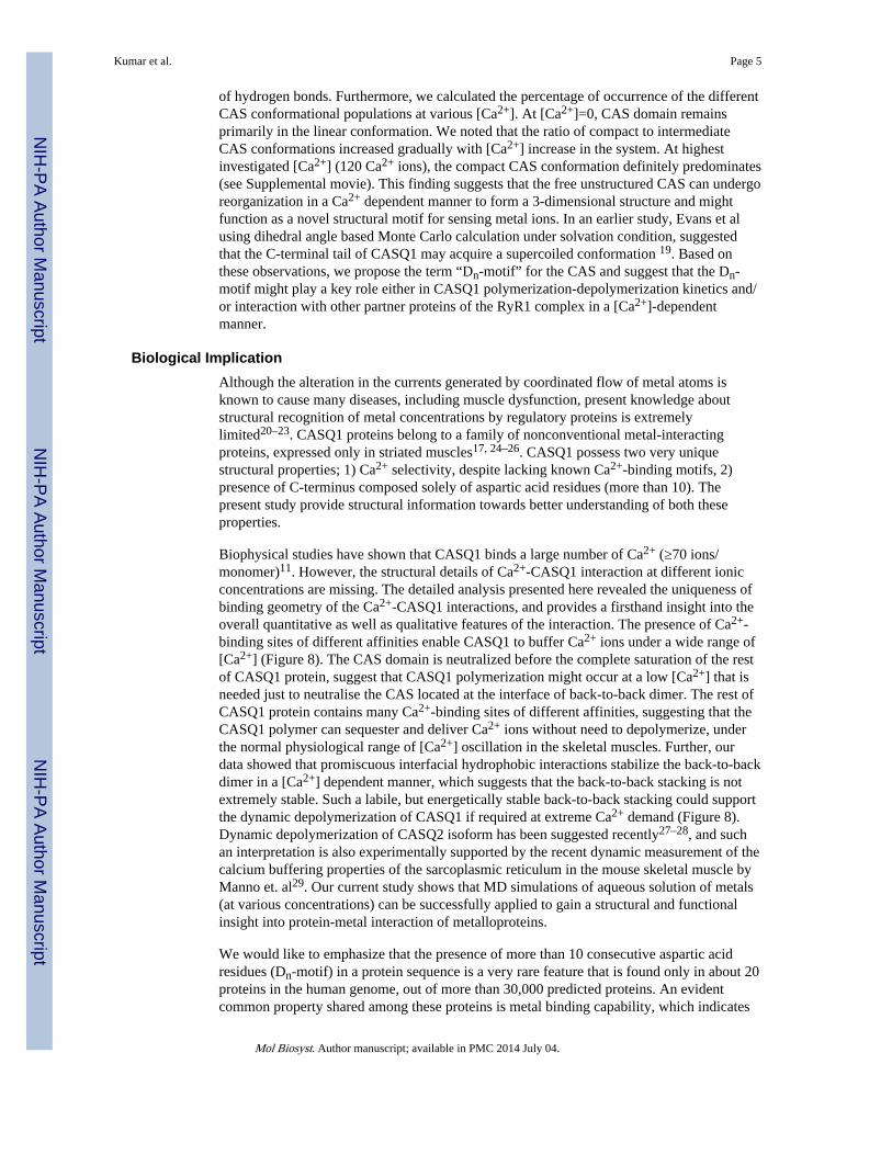
Differential Ca2+-binding to CAS and rest of CASQ1Next, we calculated the number of Ca2+ ions that interact with the CASQ1 protein after theMD simulation. Interestingly, we found that the CAS residues get saturated with Ca2+
before the rest of the protein surface. As shown in Figure 2A, the CAS residues (350–364)of chain B and C, bind to an increasing number of Ca2+ ions as [Ca2+] increases. However,in both the chains, CAS gets saturated when [Ca2+] reached 80 ions in the system, and afterthis threshold no further increase in number of bound Ca2+ was observed. This indicates thatthe maximum Ca2+ binding capacity of the CAS of chain B and C is ~6 and ~8 Ca2+ ionsrespectively, and the CAS cannot account for more than 10% of the Ca2+ binding of eachCASQ1 monomer (bind ~70–80 Ca2+). On the other hand, the surface of rest of the protein(residues 1–350) binds progressively more number of Ca2+ ions as the [Ca2+] was increased.From the slope of the Ca2+-binding curve (Figure 2B), it appears that the CASQ1 surfacehas not reached saturation even in the system with 120 Ca2+ ions, where each chain hasbound ~55 Ca2+ ions. These findings, and the observation that CAS may hinder back-to-back stacking due to charge-charge repulsion, indicate that the CAS domain undergoescomplete charge neutralization before rest of the CASQ1 surface gets saturated with Ca2+.Furthermore, these observations suggest that the affinity of different Ca2+-binding sites onCASQ1 must be different, in such a way to allow systematic neutralization ofelectronegative surface for increasing [Ca2+]. The negative potential neutralization processwould then lead to the formation of globular CASQ1 conformation that is ready to undergopolymerization.
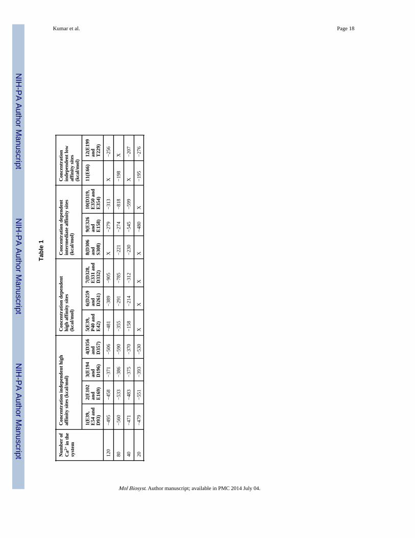
[Ca2+] independent high affinity Ca2+-binding sitesThe recently solved crystal structure (PDB Id: 3UOM) provided information of about only15 Ca2+-binding sites12, while our simulated data shows that each monomer (chain) bindsabout 55 Ca2+ in the system with 120 Ca2+ (Figure 2), indicating the existence of moreCa2+-binding sites. Therefore, we performed detailed investigation of the CASQ1 structuresin the systems exposed to different [Ca2+], to identify and characterize other Ca2+ bindingsites, in particular those not revealed by the crytallographic structure. Interestingly, theCa2+-binding sites identified by the crystallographic study were also confirmed by MDsimulation. The Ca2+-binding strength was analysed for a set of Ca2+-binding sites bydetermining the non-bonded (Van der Waals + Electrostatic) energy of interaction betweenthe residues of the site and the Ca2+ ion. Based on the Ca2+-binding strength (summarized inTable 1) we further refined the classification of both high and low affinity sites intoindependent or dependent of [Ca2+]. The high affinity sites (site 1, 2, 3 and 4) are strictlyindependent of [Ca2+] and bind Ca2+ with high affinity as indicated by the high interactionenergy (<−400 kcal/mol) in all the protein systems. We further found that the [Ca2+]-independent high affinity sites bind Ca2+ in a very favorable geometry in most of thesystems at different [Ca2+] (Figure 3). However, only 13 [Ca2+]-independent high affinitysites on CASQ1 were found.
Existence of [Ca2+]-induced Ca2+-binding sitesOne of the most interesting findings of this study is that some Ca2+-binding sites are solelydependent on [Ca2+]. We found that some sites, for instance site 5, 6 and 7, bind Ca2+ withincreasingly high affinity, reaching the highest value (correspoding to energy of <−400 kcal/mol) in the system with 120 Ca2+ ions (Table 1). These sites are referred as “[Ca2+]-
Kumar et al. Page 3
Mol Biosyst. Author manuscript; available in PMC 2014 July 04.
NIH
-PA Author Manuscript
NIH
-PA Author Manuscript
NIH
-PA Author Manuscript

dependent high affinity sites” and do not bind Ca2+ when the system has only 20 Ca2+ ions.Their interaction energy gradually increases as [Ca2+] increases, reaching a maximum value(<−400 kcal/mol) for the system containing 120 Ca2+ ions. Moreover, sites 5, 6 and 7 showa more favorable geometry of Ca2+-binding when [Ca2+] increases, demonstrating that thesesites are induced upon Ca2+-binding (Figure 4). Our data suggest that the [Ca2+]-independent high affinity sites gets occupied first and then the binding events initiatesstructural alterations, bringing the residues involved in the [Ca2+]-dependent high affinitysites in a favorable geometry for subsequent Ca2+-binding.
Interestingly, some inducible sites (site 8, 9 and 10) were observed to bind Ca2+ with highaffinity only at intermediate [Ca2+] (Table 1), thus we refer to them as “[Ca2+]-dependentintermediate affinity sites”. The geometry of site 10 in systems with different [Ca2+] isshown in Figure 5. As can be seen in Figure 5, the residues in site 10 are not in a properorientation to support Ca2+ recognition in 20 Ca2+ ions. system. Thereafter, up to the systemhaving 80 Ca2+, these residues gradually reorient to a correct geometry for ion Ca2+-binding, with all three acidic residues involved in bidentate interaction with the Ca2+ ion.However, further increase in [Ca2+] in the system disrupted the optimal geometry of the site10, thus leading to a sharp decline of the interaction energy (Figure 5 and Table 1). Distanceprobability distributions also showed that in the system having 80 Ca2+ ions the distancebetween the residues and Ca2+ is the least value (Supplemental Figure 1). As a whole, wehave found 25 inducible Ca2+-binding sites in CASQ1. Additionally, we also found thatCASQ1 has many low affinity sites (for instance site 11 and 12) that are formed by either anacidic residue (Asp or Glu) alone or in combination with any other supporting residue(Supplemental Figure 1D). These sites bind Ca2+ with low affinity, with an interactionenergy higher than −250 kcal/mol independent of [Ca2+] in the system.
[Ca2+]-mediated dimer stabilization by hydrophobic interactionsWe investigated the structure of the back-to-back dimeric interface in all the proteinsystems. Importantly, we found only one hydrogen bond at the interface, while interestinglythe number of hydrophobic interactions increased with the rise in [Ca2+] (Figure 6). Then,on increasing the number of Ca2+ ions, we observed a gradual increase of hydrophobicinteractions, and maximum of four hydrophobic interactions were found in the system with120 Ca2+ ions. However, we did not observe any committed residue-to-residue contact,rather a set of interfacial residues that promiscuously established hydrophobic contacts as[Ca2+] increases. Nevertheless, all the hydrophobic interactions were found between acidic(glutamic and aspartic) residues. This suggests that the Ca2+-binding mediated chargeneutralization of these interfacial acidic residues provide them the ability to establishhydrophobic interactions. We also observed that the number of bound water molecules inthe interfacial region and to the CAS of chain B is unchanged in all the protein systemsindicating that water plays only a minor role in back-to-back stacking. On the other hand,the free CAS of chain C binds more water with increasing [Ca2+] in the system(Supplemental Figure 2).
Ca2+-induced CAS foldingNext, we investigated the alteration in structure of CAS upon Ca2+-binding throughout theMD trajectory in all the systems. One of the most exciting finding of this study is that theCAS acquires a compact 3-dimensional structure upon Ca2+-binding. Analysis of MDsimulation trajectory suggest that CAS domain can exist in three distinct conformations;linear, intermediate and compact, as shown in Figure 7. The linear conformation of CASforms 2–3 hydrogen bonds, the intermediate conformation 4–5 hydrogen bonds, and thecompact conformation is stabilized with 6–8 hydrogen bonds. Then, we calculated theenergy of the different CAS conformational populations, which is pertinent with the number
Kumar et al. Page 4
Mol Biosyst. Author manuscript; available in PMC 2014 July 04.
NIH
-PA Author Manuscript
NIH
-PA Author Manuscript
NIH
-PA Author Manuscript
of hydrogen bonds. Furthermore, we calculated the percentage of occurrence of the differentCAS conformational populations at various [Ca2+]. At [Ca2+]=0, CAS domain remainsprimarily in the linear conformation. We noted that the ratio of compact to intermediateCAS conformations increased gradually with [Ca2+] increase in the system. At highestinvestigated [Ca2+] (120 Ca2+ ions), the compact CAS conformation definitely predominates(see Supplemental movie). This finding suggests that the free unstructured CAS can undergoreorganization in a Ca2+ dependent manner to form a 3-dimensional structure and mightfunction as a novel structural motif for sensing metal ions. In an earlier study, Evans et alusing dihedral angle based Monte Carlo calculation under solvation condition, suggestedthat the C-terminal tail of CASQ1 may acquire a supercoiled conformation 19. Based onthese observations, we propose the term “Dn-motif” for the CAS and suggest that the Dn-motif might play a key role either in CASQ1 polymerization-depolymerization kinetics and/or interaction with other partner proteins of the RyR1 complex in a [Ca2+]-dependentmanner.
Biological ImplicationAlthough the alteration in the currents generated by coordinated flow of metal atoms isknown to cause many diseases, including muscle dysfunction, present knowledge aboutstructural recognition of metal concentrations by regulatory proteins is extremelylimited20–23. CASQ1 proteins belong to a family of nonconventional metal-interactingproteins, expressed only in striated muscles17, 24–26. CASQ1 possess two very uniquestructural properties; 1) Ca2+ selectivity, despite lacking known Ca2+-binding motifs, 2)presence of C-terminus composed solely of aspartic acid residues (more than 10). Thepresent study provide structural information towards better understanding of both theseproperties.
Biophysical studies have shown that CASQ1 binds a large number of Ca2+ (≥70 ions/monomer)11. However, the structural details of Ca2+-CASQ1 interaction at different ionicconcentrations are missing. The detailed analysis presented here revealed the uniqueness ofbinding geometry of the Ca2+-CASQ1 interactions, and provides a firsthand insight into theoverall quantitative as well as qualitative features of the interaction. The presence of Ca2+-binding sites of different affinities enable CASQ1 to buffer Ca2+ ions under a wide range of[Ca2+] (Figure 8). The CAS domain is neutralized before the complete saturation of the restof CASQ1 protein, suggest that CASQ1 polymerization might occur at a low [Ca2+] that isneeded just to neutralise the CAS located at the interface of back-to-back dimer. The rest ofCASQ1 protein contains many Ca2+-binding sites of different affinities, suggesting that theCASQ1 polymer can sequester and deliver Ca2+ ions without need to depolymerize, underthe normal physiological range of [Ca2+] oscillation in the skeletal muscles. Further, ourdata showed that promiscuous interfacial hydrophobic interactions stabilize the back-to-backdimer in a [Ca2+] dependent manner, which suggests that the back-to-back stacking is notextremely stable. Such a labile, but energetically stable back-to-back stacking could supportthe dynamic depolymerization of CASQ1 if required at extreme Ca2+ demand (Figure 8).Dynamic depolymerization of CASQ2 isoform has been suggested recently27–28, and suchan interpretation is also experimentally supported by the recent dynamic measurement of thecalcium buffering properties of the sarcoplasmic reticulum in the mouse skeletal muscle byManno et. al29. Our current study shows that MD simulations of aqueous solution of metals(at various concentrations) can be successfully applied to gain a structural and functionalinsight into protein-metal interaction of metalloproteins.
We would like to emphasize that the presence of more than 10 consecutive aspartic acidresidues (Dn-motif) in a protein sequence is a very rare feature that is found only in about 20proteins in the human genome, out of more than 30,000 predicted proteins. An evidentcommon property shared among these proteins is metal binding capability, which indicates
Kumar et al. Page 5
Mol Biosyst. Author manuscript; available in PMC 2014 July 04.
NIH
-PA Author Manuscript
NIH
-PA Author Manuscript
NIH
-PA Author Manuscript
that the Dn-motif might be important as metal concentration sensors or for the metal sensingand binding, and to best of our knowledge, has not been investigated so far. For instance,these 20 proteins include; zinc finger protein castor homolog 1 (genebank accession id:NP_001073312), Histidine-rich calcium-binding protein (AAI12356), ring finger protein 34(EAW98263), E3 ubiquitin-protein ligase RNF34 isoform 3 (NP_001243787), and asporin(CAI16698). Searches in PDB indicate that the structure of Dn-motif has also not beendetermined through experimental methods for any protein and functional role of Dn-motif inmetalloproteins is currently unknown. Our result showed that Dn-motif of CASQ1undergoes folding at increasing [Ca2+], which suggests that this motif might have a metal-sensing function.
Materials and methodCASQ1 Model Building with CAS motif
The recent crystal structure of the hexameric model of skeletal calsequestrin (PDB Id:3UOM30), was adopted as a base model for our MD studies. The protein preparation wizardof the Schrodinger suite was used to optimize the model structure for it chemicalcorrectness.31 In general, before simulations, the crystal structures requires fixing thecommon issues related to the experimental adopted methodology, that is missing hydrogenatoms, missing side chains, bond order assignments, charge states, and conformationalorientations of symmetrical groups. During the structure refinement the orientation ofhydroxyl (or thiol) groups, the terminal amide groups in asparagine (Asn) and glutamine(Gln), and the ring of histidine (His), those not already determined in the X-ray structure,were corrected. Flipping the terminal amide groups and the histidine ring can improvecharge-charge interactions with neighboring groups as well as improving hydrogen bonding.The 180° flips preserve the heavy-atom placement deduced from the X-ray electron density.In addition, the protonation state of histidine is varied to optimize hydrogen bonding andcharge interactions. The optimization was done using exhaustive sampling, which allowedincreasing the number of iteration to an adequate level. The protein was further minimizedusing protein preparation wizard using OPLS2005 force field with the maximumconvergence of 0.3 Å of heavy atoms. As shown in Supplemental Figure 3A, the CASQ1monomers (labeled A, B, C, D, E and F) are arranged in a repetitive manner. AB, CD andEF are the front-to-front dimers and back-to-back interactions occur between B and Cchains. In this study we are interested to understand the dynamics of back-to-back stackingand C-terminus-to-Ca2+ interaction, thus we chose to consider chains B and C only. Themissing C-terminal residues were added using prime and builder module of Schrodinger tothe chain B and C, without disturbing the structure of the remaining regions of the chains.32
After C-terminus addition, the residues were corrected by extended serial loop samplingmethod, using loop refinement module of Prime (Supplemental Figure 3B).33 The dimericstructure with the CAS region was subjected to global minimization through theMacromodel34 module of the Schrodinger suite using the OPLS 2005 force field with a GB/SA continuum water solvation model. This was followed by a Polak-Ribiere ConjugateGradient energy minimization stopped after 5000 steps or when the energy differencebetween two subsequent structures was inferior to 0.05 kJ/mol. Finally, varying number ofCa2+ ions were added randomly to the system to build up the final CASQ1-Ca2+ complexesusing System Building Module of the Desmond suite to perform the following MDsimulations.35
Molecular Dynamics SimulationsThe parameters for the protein and ions were assigned using CHARMM27 force-field (C27-FF) parameters36. The protein-protein complex with different concentration of Ca2+ ionswere alternatively inserted in water box, and additional counter-ions were added to
Kumar et al. Page 6
Mol Biosyst. Author manuscript; available in PMC 2014 July 04.
NIH
-PA Author Manuscript
NIH
-PA Author Manuscript
NIH
-PA Author Manuscript
neutralize the system, using the modules in VMD software37. We used the TIP3Pparameters for water molecules38. Standard protonation states were assigned to all residuesusing the propKa software39. Each of the resulting molecular system was energy minimizedand slowly heated to 300 K in steps of 30 K, with positional restraints of 50 kcal/(mol Å2)on C-alpha atoms for a simulation time of 0.2 ns. The positional restraints on the C-alphaatoms were then slowly released in steps of 10 kcal/(mol Å2) and after 0.3 ns of simulationall positional restraints on the C-alpha atoms were completely released. Then, equilibrationof the molecular system was done for a simulation time of 3 ns. Subsequently, all productionruns of 30 ns simulations were performed at 300 K and 1 atm pressure (NPT ensemble),using periodic boundary conditions. The initial dimension of the simulation box edges were[138, 110, 95] Å, for a total system of ~130.000 atoms. All bonds involving hydrogen atomswere constrained using SHAKE40, which allowed using an integration time step of 2 fs. TheLong-range electrostatic interactions were evaluated using particle mesh Ewald with a [148128 96] Å grid dimension41. We used a 12 Å cut-off radius for both Van der Waals andelectrostatic interactions along with smooth particle mesh Ewald41. We used the NAMDsoftware package42 to perform all-atom molecular dynamics (MD) simulations on 96processors cluster.
Structure Analysis and Energy CalculationsTo monitor the stability of protein structure during MD simulations, we calculated the rootmean square deviation (RMSD) on heavy protein atoms using VMD software. Theconfigurational entropy for CAS domain during MD simulations was calculated fromcovariance matrices of the heavy atom fluctuations at 200 ps time step, using CARMAsoftware43–45. The interaction energy for the Ca2+ ion in the many sites we identified wasdone by evaluating non-bonded interactions (that is, Van der Waals and electrostatics)between the Ca2+ and the protein residues in the identified sites, using a cutoff of 12.0 Å.For the electrostatic interactions, we adopted the same scheme adopted for the whole MDsimulations. The total number of Ca2+ ions around the CAS residues (residue id 350–364)and the rest of the protein (residue id 1–350) for the chains B, C was monitored usingdistance cutoff of 5.0 Å, at every 20 ps time step of each MD trajectory. Similarly, thenumber of water molecules around the CAS residues and the rest of the protein for thechains B and C was calculated using distance cutoff of 3.0 Å. The H-bond and hydrophobicinteractions between the interface residues belonging to chain B, C were calculated usingappropriate VMD scripts on average structure of the protein calculated at intervals of 2 ns.The two dimensional schematic representation for the interaction network were finallygenerated by using the software LIGPLOT46.
Supplementary MaterialRefer to Web version on PubMed Central for supplementary material.
AcknowledgmentsAS acknowledges for modelling software support from Department of Biotechnology (DBT), Delhi, through grantno BT/PR14237/MED/29/196/2010. AK thanks the computational facility at CRS4 (Polaris), Pula, Italy. HCsupported by institute research fellowship from Birla Institute of Technology, Mesra. TB thanks CSIR, Delhi forproviding Senior Research Fellowship. N.C.B. was supported by postdoctoral fellowship from the American HeartAssociation. This work was supported in part, by National Institutes of Health Grant R01 HL64014 to M. P.
References1. Beard NA, Laver DR, Dulhunty AF. Prog Biophys Mol Biol. 2004; 85:33–69. [PubMed: 15050380]
2. Caudwell B, Antoniw JF, Cohen P. Eur J Biochem. 1978; 86:511–518. [PubMed: 95949]
Kumar et al. Page 7
Mol Biosyst. Author manuscript; available in PMC 2014 July 04.
NIH
-PA Author Manuscript
NIH
-PA Author Manuscript
NIH
-PA Author Manuscript
3. Maurer A, Tanaka M, Ozawa T, Fleischer S. Proc Natl Acad Sci U S A. 1985; 82:4036–4040.[PubMed: 3858861]
4. Cala SE, Jones LR. J Biol Chem. 1983; 258:11932–11936. [PubMed: 6619149]
5. Franzini-Armstrong C, Kenney LJ, Varriano-Marston E. J Cell Biol. 1987; 105:49–56. [PubMed:3497158]
6. Wang S, Trumble WR, Liao H, Wesson CR, Dunker AK, Kang CH. Nat Struct Biol. 1998; 5:476–483. [PubMed: 9628486]
7. Park H, Wu S, Dunker AK, Kang C. J Biol Chem. 2003; 278:16176–16182. [PubMed: 12594204]
8. Cozens B, Reithmeier RA. J Biol Chem. 1984; 259:6248–6252. [PubMed: 6725251]
9. Aaron BM, Oikawa K, Reithmeier RA, Sykes BD. J Biol Chem. 1984; 259:11876–11881. [PubMed:6480588]
10. Ikemoto N, Ronjat M, Meszaros LG, Koshita M. Biochemistry. 1989; 28:6764–6771. [PubMed:2790030]
11. Park H, Park IY, Kim E, Youn B, Fields K, Dunker AK, Kang C. J Biol Chem. 2004; 279:18026–18033. [PubMed: 14871888]
12. Sanchez EJ, Lewis KM, Danna BR, Kang C. J Biol Chem. 2012
13. Fliegel L, Ohnishi M, Carpenter MR, Khanna VK, Reithmeier RA, MacLennan DH. Proc NatlAcad Sci U S A. 1987; 84:1167–1171. [PubMed: 3469659]
14. Zarain-Herzberg A, Fliegel L, MacLennan DH. J Biol Chem. 1988; 263:4807–4812. [PubMed:2832408]
15. Mishra A, Suman SK, Srivastava SS, Sankaranarayanan R, Sharma Y. J Mol Biol. 2012; 415:75–91. [PubMed: 22099475]
16. Kim E, Youn B, Kemper L, Campbell C, Milting H, Varsanyi M, Kang C. J Mol Biol. 2007;373:1047–1057. [PubMed: 17881003]
17. Gaburjakova M, Bal NC, Gaburjakova J, Periasamy M. Cell Mol Life Sci. 2012
18. Bal NC, Kumar A, Chakravarty A, Balaraju T, Bal C, Jena N, Sharon A, Periasamy M.Biophysical Journal. 2013; 104:173a. [PubMed: 23332070]
19. Evans JS, Chan SI, Goddard WA 3rd. Protein Sci. 1995; 4:2019–2031. [PubMed: 8535238]
20. Priori SG, Chen SR. Circ Res. 2011; 108:871–883. [PubMed: 21454795]
21. Berg JM. Cold Spring Harb Symp Quant Biol. 1987; 52:579–585. [PubMed: 3454279]
22. Ozawa T. Mol Med Rep. 2010; 3:199–204. [PubMed: 21472222]
23. Dulhunty AF, Beard NA, Hanna AD. J Gen Physiol. 2012; 140:87–92. [PubMed: 22851673]
24. Ikemoto N, Nagy B, Bhatnagar GM, Gergely J. J Biol Chem. 1974; 249:2357–2365. [PubMed:4856651]
25. Bal NC, Jena N, Sopariwala D, Balaraju T, Shaikh S, Bal C, Sharon A, Gyorke S, Periasamy M.Biochem J. 2011; 435:391–399. [PubMed: 21265816]
26. Scriven DR, Asghari P, Moore ED. Cardiovasc Res. 2013
27. Bal NC, Sharon A, Gupta SC, Jena N, Shaikh S, Gyorke S, Periasamy M. J Biol Chem. 2010;285:17188–17196. [PubMed: 20353949]
28. Lee KW, Maeng JS, Choi JY, Lee YR, Hwang CY, Park SS, Park HK, Chung BH, Lee SG, KimYS, Jeon H, Eom SH, Kang C, Kim do H, Kwon KS. J Biol Chem. 2012; 287:1679–1687.[PubMed: 22123818]
29. Manno C, Sztretye M, Figueroa L, Allen PD, Rios E. J Physiol. 2012
30. Sanchez EJ, Lewis KM, Danna BR, Kang C. J Biol Chem. 2012; 287:11592–11601. [PubMed:22337878]
31. Park KW, Goo JH, Chung HS, Kim H, Kim DH, Park WJ. Gene. 1998; 217:25–30. [PubMed:9795116]
32. Arai M, Alpert NR, Periasamy M. Gene. 1991; 109:275–279. [PubMed: 1662658]
33. Fliegel L, Leberer E, Green NM, MacLennan DH. FEBS Lett. 1989; 242:297–300. [PubMed:2914612]
34. Damiani E, Volpe P, Margreth A. J Muscle Res Cell Motil. 1990; 11:522–530. [PubMed:2084148]
Kumar et al. Page 8
Mol Biosyst. Author manuscript; available in PMC 2014 July 04.
NIH
-PA Author Manuscript
NIH
-PA Author Manuscript
NIH
-PA Author Manuscript

35. Biral D, Volpe P, Damiani E, Margreth A. FEBS Lett. 1992; 299:175–178. [PubMed: 1544490]
36. MacKerell AD Jr, Banavali N, Foloppe N. Biopolymers. 2000; 56:257–265. [PubMed: 11754339]
37. Humphrey W, Dalke A, Schulten K. J Mol Graph. 1996; 14:33–38. 27–38. [PubMed: 8744570]
38. Jorgensen WL, Chandrasekhar J, Madura JD, Impey RW, Klein ML. J Chem Phys. 1983; 79:926–935.
39. Li H, Robertson AD, Jensen JH. Proteins. 2005; 61:704–721. [PubMed: 16231289]
40. Ryckaert JP, Ciccotti G, Berendsen HJC. J Comput Phys. 1977; 23:327–341.
41. Essmann U, Perera L, Berkowitz ML, Darden T, Lee H, Pedersen LG. J Chem Phys. 1995;103:8577–8593.
42. Phillips JC, Braun R, Wang W, Gumbart J, Tajkhorshid E, Villa E, Chipot C, Skeel RD, Kale L,Schulten K. J Comput Chem. 2005; 26:1781–1802. [PubMed: 16222654]
43. Andricioaei I, Karplus M. J Chem Phys. 2001; 115:6289–6292.
44. Glykos NM. J Comput Chem. 2006; 27:1765–1768. [PubMed: 16917862]
45. Balaraju T, Kumar A, Bal C, Chattopadhyay D, Jena N, Bal NC, Sharon A. Structural Chemistry.2012 In Press.
46. Wallace AC, Laskowski RA, Thornton JM. Protein Eng. 1995; 8:127–134. [PubMed: 7630882]
Kumar et al. Page 9
Mol Biosyst. Author manuscript; available in PMC 2014 July 04.
NIH
-PA Author Manuscript
NIH
-PA Author Manuscript
NIH
-PA Author Manuscript

Figure 1. Changes in the surface charge pattern of CASQ1 with increasing Ca2+ concentrationsSurface charge was calculated for the average structure for each system indicated in thefigure. Red, blue and white indicate negative, positive and neutral charge on the molecularsurface. The color scale used to represent the Poisson Boltzmann electrostatic potentialisosurface range is from −4 to +4 KT/e (where K - Boltzmann constant, T - temperature, ande - electron charge. CASQ1 gains electro-positivity on the surface in the 120 Ca2+-system,even if the surface negative charge is almost neutralized already in the 80 Ca2+-system.
Kumar et al. Page 10
Mol Biosyst. Author manuscript; available in PMC 2014 July 04.
NIH
-PA Author Manuscript
NIH
-PA Author Manuscript
NIH
-PA Author Manuscript

Figure 2. Number of bound Ca2+ in the presence of increasing [Ca2+](A) Ca2+ bound to the Dn-motif (from amino acid 350 to 364 on the C-terminus) ofeach monomer. The Dn-motif at the dimeric interface binds less Ca2+ than the free Dn-motif. (B) Total number of Ca2+ ions bound to the rest of the protein (amino acids 1–350) surface of each monomer. Both the monomers bind almost similar number of Ca2+
ions although at intermediate [Ca2+] the monomer “chain B” binds more Ca2+ ions than“chain C”. Observing the slope of the curves it seems that both the monomers still possessthe ability to bind even a higher number of Ca2+ ions, if provided.
Kumar et al. Page 11
Mol Biosyst. Author manuscript; available in PMC 2014 July 04.
NIH
-PA Author Manuscript
NIH
-PA Author Manuscript
NIH
-PA Author Manuscript
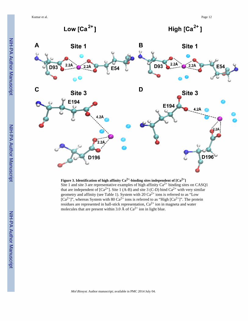
Figure 3. Identification of high affinity Ca2+-binding sites independent of [Ca2+]Site 1 and site 3 are representative examples of high affinity Ca2+ binding sites on CASQ1that are independent of [Ca2+]. Site 1 (A-B) and site 3 (C-D) bind Ca2+ with very similargeometry and affinity (see Table 1). System with 20 Ca2+ ions is referred to as “Low[Ca2+]”, whereas System with 80 Ca2+ ions is referred to as “High [Ca2+]”. The proteinresidues are represented in ball-stick representation, Ca2+ ion in magneta and watermolecules that are present within 3.0 Å of Ca2+ ion in light blue.
Kumar et al. Page 12
Mol Biosyst. Author manuscript; available in PMC 2014 July 04.
NIH
-PA Author Manuscript
NIH
-PA Author Manuscript
NIH
-PA Author Manuscript

Figure 4. Identification of [Ca2+] dependent high affinity Ca2+-binding sitesRepresentative examples of Ca2+ binding sites on CASQ1 that are induced by an increase in[Ca2+] in the system. Site 5 (A-B) and site 7 (C-D) show progressively a more favorableCa2+-binding geometry and better binding affinity increasing Ca2+ in the system (see Table1).
Kumar et al. Page 13
Mol Biosyst. Author manuscript; available in PMC 2014 July 04.
NIH
-PA Author Manuscript
NIH
-PA Author Manuscript
NIH
-PA Author Manuscript

Figure 5. [Ca2+] dependent intermediate affinity Ca2+ binding sites on CASQ1Some Ca2+ binding sites on CASQ1, like site 10, are induced to bind Ca2+ with high affinityonly at intermediate [Ca2+]. (A) Site 10 is constituted by residues D319, E350 and E354,that show no sensitivity to Ca2+ when only 20 Ca2+ ions are present in the system. (B) Site10 binds Ca2+ when 40 Ca2+ ions are present in the system. (C) Site 10 recognizes Ca2+ andbinds with very high affinity and an optimal geometry when [Ca2+] increases to 80 ions. (D)With further increase in [Ca2+] to 120 ions, we note disruption of the optimal bindingtopology of site 10 and thus the interaction energy drastically reduces.
Kumar et al. Page 14
Mol Biosyst. Author manuscript; available in PMC 2014 July 04.
NIH
-PA Author Manuscript
NIH
-PA Author Manuscript
NIH
-PA Author Manuscript
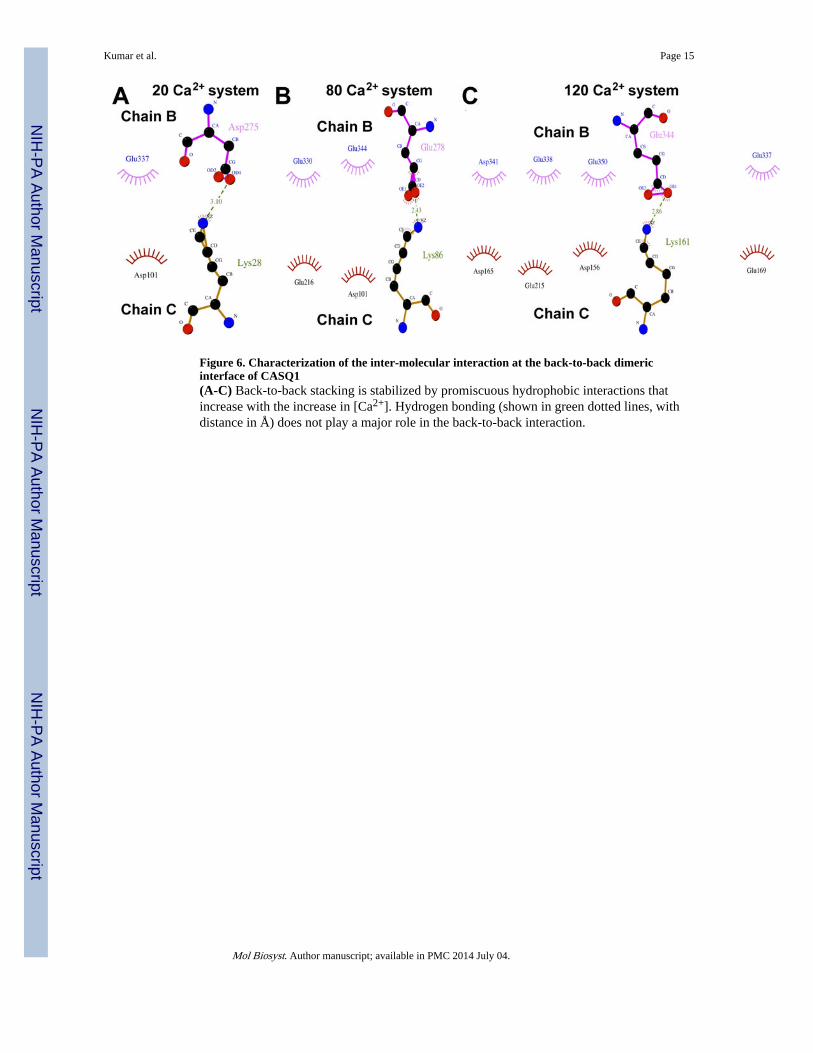
Figure 6. Characterization of the inter-molecular interaction at the back-to-back dimericinterface of CASQ1(A-C) Back-to-back stacking is stabilized by promiscuous hydrophobic interactions thatincrease with the increase in [Ca2+]. Hydrogen bonding (shown in green dotted lines, withdistance in Å) does not play a major role in the back-to-back interaction.
Kumar et al. Page 15
Mol Biosyst. Author manuscript; available in PMC 2014 July 04.
NIH
-PA Author Manuscript
NIH
-PA Author Manuscript
NIH
-PA Author Manuscript

Figure 7. The Dn-motif of CASQ1 assumes compact conformation upon binding of Ca2+ ions(A-C) The CAS or the Dn-motif is observed to exist in three main conformations. The linearconformation of CAS is characterized by the presence of the least number of Ca2+-bound aswell as by the energetically least stable state. Additional Ca2+-binding leads to to anintermediate conformer, which was found to be energetically more stable. Finally, the Dn-motif saturation by Ca2+ resulted in the most energetically stable and a very compactstructure.
Kumar et al. Page 16
Mol Biosyst. Author manuscript; available in PMC 2014 July 04.
NIH
-PA Author Manuscript
NIH
-PA Author Manuscript
NIH
-PA Author Manuscript

Figure 8. Proposed model for Ca2+-CASQ1 interaction and polymerizationThe centre of each domain of the CASQ1 molecule is hydrophobic and must undergohydrophobic collapse to form the domains. The core region of CASQ1 at the interface,between the three domains is acidic, which is characteristic feature of the CASQ-proteinfamily. The inter-domain charge neutralization by Ca2+ is necessary for the three domains tocome to close proximity to form a compact monomer. The Dn-motif of CASQ1 is composedof 13 aspartic acids that can not bind more than 8 Ca2+ ions, whereas CASQ1 monomer hascapacity to bind 70–80 Ca2+ ions. Therefore, the CASQ1 structural domains can bind morethan 50 Ca2+. Existence of sites that can switch to an high affinity sites at varying [Ca2+]allows CASQ1 to release Ca2+ without the necessity of undergoing depolymerization. Thisobservation holds in most part of the “physiological range of Ca2+ variations”. Dotted linesindicate the extreme Ca2+ concentrations that might be important only during higherphysiological Calcium demand.
Kumar et al. Page 17
Mol Biosyst. Author manuscript; available in PMC 2014 July 04.
NIH
-PA Author Manuscript
NIH
-PA Author Manuscript
NIH
-PA Author Manuscript
NIH
-PA Author Manuscript
NIH
-PA Author Manuscript
NIH
-PA Author Manuscript
Kumar et al. Page 18
Tabl
e 1
Num
ber
ofC
a2+ in
the
syst
em
Con
cent
rati
on in
depe
nden
t hi
ghaf
fini
ty s
ites
(kc
al/m
ol)
Con
cent
rati
on d
epen
dent
high
aff
init
y si
tes
(kca
l/mol
)
Con
cent
rati
on d
epen
dent
inte
rmed
iate
aff
init
y si
tes
(kca
l/mol
)
Con
cent
rati
onin
depe
nden
t lo
waf
fini
ty s
ites
(kca
l/mol
)
1(E
39,
E54
and
D93
)
2(E
102
and
E16
9)
3(E
194
and
D19
6)
4(D
356
and
D35
7)
5(E
39,
P40
and
E42
)
6(D
259
and
D26
1)
7(D
328,
E33
1 an
dD
332)
8(D
306
and
S308
)
9(E
326
and
E15
8)
10(D
319,
E35
0 an
dE
354)
11(E
66)
12(E
199
and
T22
9)
120
−49
5−
458
−37
1−
506
−48
1−
389
−90
5X
−27
9−
313
X−
256
80−
560
−53
3−
386
−59
0−
355
−29
1−
785
−22
1−
274
−81
8−
198
X
40−
471
−48
3−
375
−37
0−
158
−21
4−
312
−23
0−
545
−59
9X
−20
7
20−
479
−55
1−
393
−53
0X
XX
X−
480
X−
195
−27
6
Mol Biosyst. Author manuscript; available in PMC 2014 July 04.