Identification of a Novel in Vivo Virus-targeted Phosphorylation Site in Interferon Regulatory...
15
Identification of a Novel in Vivo Virus-targeted Phosphorylation Site in Interferon Regulatory Factor-3 (IRF3) * □ S Received for publication, November 13, 2009, and in revised form, May 4, 2010 Published, JBC Papers in Press, May 28, 2010, DOI 10.1074/jbc.M109.084822 Bjarte Bergstroem ‡§ , Ingvild B. Johnsen ‡ , Thuy Thanh Nguyen ‡ , Lars Hagen ¶ , Geir Slupphaug ¶ , Liv Thommesen §¶ , and Marit W. Anthonsen ‡1 From the ‡ Department of Laboratory Medicine, Children’s and Women’s Health, Faculty of Medicine, ¶ Department of Cancer Research and Molecular Medicine, and Functional Genomics (FUGE) Proteomics Node, Norwegian University of Science and Technology, 7006 Trondheim and the § Faculty of Technology, Sør-Trøndelag University College (HiST), 7004 Trondheim, Norway The transcription factor interferon regulatory factor-3 (IRF3) regulates expression of type I interferon- and plays an impor- tant role in antiviral immunity. Despite the biological impor- tance of IRF3, its in vivo phosphorylation pattern has not been reported. In this study, we have identified residues in IRF3 that are phosphorylated in vivo after infection with Sendai virus. We found that Sendai virus induced phosphorylation of the C-ter- minal residues Thr 390 and Ser 396 , in addition to either Ser 385 or Ser 386 . Moreover, Ser 173 and Ser 175 were constitutively phos- phorylated. Ser 396 has previously been suggested to be the major target of the IRF3-activating kinase TBK1 (TANK-binding kinase-1), whereas Thr 390 has not previously been implicated in IRF3 regulation. Mutagenesis studies indicated that phosphor- ylation of Thr 390 promotes Ser 396 phosphorylation and binding to the coactivator cAMP-response element-binding protein. Taken together, our results show that IRF3 is subject to multiple interdependent phosphorylations, and we identify Thr 390 as a novel in vivo phosphorylation site that modulates the phos- phorylation status of TBK1-targeted Ser 396 . Innate immune responses upon viral infections include pro- duction of antiviral cytokines and type I interferons (IFNs). 2 The transcription factor interferon regulatory factor-3 (IRF3) is critical for IFN production and directs expression of several diverse genes that are implicated in the antiviral immune response (1). IRF3 is constitutively expressed in multiple tissues and shuttles between the cytoplasm and nucleus in resting cells. Activation of IRF3 involves its virus-induced phosphorylation at several sites in the C-terminal IRF3 dimerization and trans- location to the nucleus. In the nucleus, IRF3 associates with cAMP-response element-binding protein-binding protein (CBP) or the closely related p300, promoting binding to promoters containing interferon-stimulated response elements to initiate transcription of target genes (2). Activation of IRF3 is initiated after recognition of viral nucleic acids by Toll-like receptors or by the cytoplasmic RNA helicases RIG-I and MDA5 (3, 4). These receptors recruit dis- tinct adapter proteins to initiate signaling, which induce ubiq- uitination of the cytoplasmic adapter protein TRAF3, leading to activation of the IB kinase-related kinase TANK-binding kinase-1 (TBK1) and phosphorylation of IRF3 (5, 6). The acti- vation mechanism of TBK1 is still poorly understood, but it involves phosphorylation of Ser 172 in the mitogen-activated protein kinase activation loop of TBK1 (7, 8). From studies based on IRF3 mutagenesis, functional in vitro assays, and in vitro phosphorylations of IRF3 with TBK1 (9, 10), it has been proposed that IRF3 is regulated by phosphorylation of multiple residues that are clustered in its C terminus. The IRF3 residues that are phosphorylated in vivo upon viral infec- tion have not been determined directly, but based on an anti- body recognizing phosphorylated Ser 396 , this residue was sug- gested to be the main target of the IRF3-activating kinase TBK1 (11). It has been advocated that sequential phosphorylation of residues in site 1 (Ser 385 and Ser 386 ) and site 2 (Ser 396 , Ser 398 , Ser 402 , Thr 404 , and Ser 405 ) directs unfolding of an autoinhibi- tory state and promotes IRF3 dimerization (12–16). However, the sequential order of site 1 and site 2 phosphorylations has been debated. In a recent study, Panne et al. (9) examined IRF3 phosphorylated in vitro with TBK1 and proposed that phos- phorylation of residues in site 2 alleviates autoinhibition and promotes site 1 phosphorylation and resultant IRF3 dimeriza- tion. However, proof for such a mechanism is still lacking, because the phosphorylation pattern of IRF3 in vivo after viral infection remains elusive. In recent years, mass spectrometry (MS)-based analysis has emerged as a major tool to identify specific post-translational protein modifications, including phosphorylation. In this study, we have utilized MS analysis to identify phosphorylated residues in IRF3 after Sendai virus (SV) infection and to demonstrate several phosphorylated sites in the C-terminal regulatory domain of IRF3. In addition to the previously proposed phosphorylation sites Ser 385 , Ser 386 , and Ser 396 , we found that Thr 390 was phosphorylated in response to SV infection. Moreover, Ser 173 and Ser 175 were found to be constitutively phosphorylated. Functional analysis after in vitro mutagenesis showed that Thr 390 , in addition to Ser 396 , contrib- utes to IRF3 activation. This is the first study to identify in vivo * This work was supported by the National Programme for Research in Func- tional Genomics (FUGE) in Norway (to M. W. A.), FUGE Mid-Norway (to M. W. A.), the Research Council of Norway (to M. W. A.), the Faculty of Med- icine, Norwegian University of Science and Technology (to I. B. J. and M. W. A.), the Cancer Fund at St. Olavs Hospital (to M. W. A.), and the Nor- wegian Cancer Society. □ S The on-line version of this article (available at http://www.jbc.org) contains supplemental Figs. 1 and 2. 1 To whom correspondence should be addressed. Tel.: 47-72-573351; Fax: 47-72-576400; E-mail: [email protected]. 2 The abbreviations used are: IFN, interferon; TLR, Toll-like receptors; SV, Sen- dai virus; TRAF, tumor necrosis receptor-associated factor; IRF, interferon regulatory factor; CBP, CREB-binding protein; MS, mass spectrometry; BN, blue native; MOPS, 4-morpholinepropanesulfonic acid; MALDI-TOF’/TOF, matrix-assisted laser desorption ionization time-of-flight/time-of-flight. THE JOURNAL OF BIOLOGICAL CHEMISTRY VOL. 285, NO. 32, pp. 24904 –24914, August 6, 2010 © 2010 by The American Society for Biochemistry and Molecular Biology, Inc. Printed in the U.S.A. 24904 JOURNAL OF BIOLOGICAL CHEMISTRY VOLUME 285 • NUMBER 32 • AUGUST 6, 2010 by guest on May 24, 2016 http://www.jbc.org/ Downloaded from by guest on May 24, 2016 http://www.jbc.org/ Downloaded from by guest on May 24, 2016 http://www.jbc.org/ Downloaded from by guest on May 24, 2016 http://www.jbc.org/ Downloaded from by guest on May 24, 2016 http://www.jbc.org/ Downloaded from
-
Upload
independent -
Category
Documents
-
view
0 -
download
0
Transcript of Identification of a Novel in Vivo Virus-targeted Phosphorylation Site in Interferon Regulatory...

Identification of a Novel in Vivo Virus-targeted PhosphorylationSite in Interferon Regulatory Factor-3 (IRF3)*□S
Received for publication, November 13, 2009, and in revised form, May 4, 2010 Published, JBC Papers in Press, May 28, 2010, DOI 10.1074/jbc.M109.084822
Bjarte Bergstroem‡§, Ingvild B. Johnsen‡, Thuy Thanh Nguyen‡, Lars Hagen¶�, Geir Slupphaug¶�, Liv Thommesen§¶,and Marit W. Anthonsen‡�1
From the ‡Department of Laboratory Medicine, Children’s and Women’s Health, Faculty of Medicine, ¶Department of CancerResearch and Molecular Medicine, and �Functional Genomics (FUGE) Proteomics Node, Norwegian University of Science andTechnology, 7006 Trondheim and the §Faculty of Technology, Sør-Trøndelag University College (HiST), 7004 Trondheim, Norway
The transcription factor interferon regulatory factor-3 (IRF3)regulates expression of type I interferon-� and plays an impor-tant role in antiviral immunity. Despite the biological impor-tance of IRF3, its in vivo phosphorylation pattern has not beenreported. In this study, we have identified residues in IRF3 thatare phosphorylated in vivo after infection with Sendai virus.Wefound that Sendai virus induced phosphorylation of the C-ter-minal residues Thr390 and Ser396, in addition to either Ser385 orSer386. Moreover, Ser173 and Ser175 were constitutively phos-phorylated. Ser396 has previously been suggested to be themajortarget of the IRF3-activating kinase TBK1 (TANK-bindingkinase-1), whereas Thr390 has not previously been implicated inIRF3 regulation. Mutagenesis studies indicated that phosphor-ylation of Thr390 promotes Ser396 phosphorylation and bindingto the coactivator cAMP-response element-binding protein.Taken together, our results show that IRF3 is subject tomultipleinterdependent phosphorylations, and we identify Thr390 as anovel in vivo phosphorylation site that modulates the phos-phorylation status of TBK1-targeted Ser396.
Innate immune responses upon viral infections include pro-duction of antiviral cytokines and type I interferons (IFNs).2The transcription factor interferon regulatory factor-3 (IRF3) iscritical for IFN production and directs expression of severaldiverse genes that are implicated in the antiviral immuneresponse (1). IRF3 is constitutively expressed inmultiple tissuesand shuttles between the cytoplasm and nucleus in resting cells.Activation of IRF3 involves its virus-induced phosphorylationat several sites in the C-terminal IRF3 dimerization and trans-location to the nucleus. In the nucleus, IRF3 associates withcAMP-response element-binding protein-binding protein (CBP)
or the closely related p300, promoting binding to promoterscontaining interferon-stimulated response elements to initiatetranscription of target genes (2).Activation of IRF3 is initiated after recognition of viral
nucleic acids by Toll-like receptors or by the cytoplasmic RNAhelicases RIG-I and MDA5 (3, 4). These receptors recruit dis-tinct adapter proteins to initiate signaling, which induce ubiq-uitination of the cytoplasmic adapter proteinTRAF3, leading toactivation of the I�B kinase-related kinase TANK-bindingkinase-1 (TBK1) and phosphorylation of IRF3 (5, 6). The acti-vation mechanism of TBK1 is still poorly understood, but itinvolves phosphorylation of Ser172 in the mitogen-activatedprotein kinase activation loop of TBK1 (7, 8).From studies based on IRF3 mutagenesis, functional in vitro
assays, and in vitro phosphorylations of IRF3with TBK1 (9, 10),it has been proposed that IRF3 is regulated by phosphorylationof multiple residues that are clustered in its C terminus. TheIRF3 residues that are phosphorylated in vivo upon viral infec-tion have not been determined directly, but based on an anti-body recognizing phosphorylated Ser396, this residue was sug-gested to be themain target of the IRF3-activating kinase TBK1(11). It has been advocated that sequential phosphorylation ofresidues in site 1 (Ser385 and Ser386) and site 2 (Ser396, Ser398,Ser402, Thr404, and Ser405) directs unfolding of an autoinhibi-tory state and promotes IRF3 dimerization (12–16). However,the sequential order of site 1 and site 2 phosphorylations hasbeen debated. In a recent study, Panne et al. (9) examined IRF3phosphorylated in vitro with TBK1 and proposed that phos-phorylation of residues in site 2 alleviates autoinhibition andpromotes site 1 phosphorylation and resultant IRF3 dimeriza-tion. However, proof for such a mechanism is still lacking,because the phosphorylation pattern of IRF3 in vivo after viralinfection remains elusive. In recent years, mass spectrometry(MS)-based analysis has emerged as a major tool to identifyspecific post-translational protein modifications, includingphosphorylation. In this study, we have utilized MS analysis toidentify phosphorylated residues in IRF3 after Sendai virus (SV)infection and to demonstrate several phosphorylated sites inthe C-terminal regulatory domain of IRF3. In addition to thepreviously proposed phosphorylation sites Ser385, Ser386, andSer396, we found that Thr390 was phosphorylated in response toSV infection. Moreover, Ser173 and Ser175 were found to beconstitutively phosphorylated. Functional analysis after in vitromutagenesis showed that Thr390, in addition to Ser396, contrib-utes to IRF3 activation. This is the first study to identify in vivo
* This work was supported by the National Programme for Research in Func-tional Genomics (FUGE) in Norway (to M. W. A.), FUGE Mid-Norway (toM. W. A.), the Research Council of Norway (to M. W. A.), the Faculty of Med-icine, Norwegian University of Science and Technology (to I. B. J. andM. W. A.), the Cancer Fund at St. Olavs Hospital (to M. W. A.), and the Nor-wegian Cancer Society.
□S The on-line version of this article (available at http://www.jbc.org) containssupplemental Figs. 1 and 2.
1 To whom correspondence should be addressed. Tel.: 47-72-573351; Fax:47-72-576400; E-mail: [email protected].
2 The abbreviations used are: IFN, interferon; TLR, Toll-like receptors; SV, Sen-dai virus; TRAF, tumor necrosis receptor-associated factor; IRF, interferonregulatory factor; CBP, CREB-binding protein; MS, mass spectrometry; BN,blue native; MOPS, 4-morpholinepropanesulfonic acid; MALDI-TOF’/TOF,matrix-assisted laser desorption ionization time-of-flight/time-of-flight.
THE JOURNAL OF BIOLOGICAL CHEMISTRY VOL. 285, NO. 32, pp. 24904 –24914, August 6, 2010© 2010 by The American Society for Biochemistry and Molecular Biology, Inc. Printed in the U.S.A.
24904 JOURNAL OF BIOLOGICAL CHEMISTRY VOLUME 285 • NUMBER 32 • AUGUST 6, 2010
by guest on May 24, 2016
http://ww
w.jbc.org/
Dow
nloaded from
by guest on May 24, 2016
http://ww
w.jbc.org/
Dow
nloaded from
by guest on May 24, 2016
http://ww
w.jbc.org/
Dow
nloaded from
by guest on May 24, 2016
http://ww
w.jbc.org/
Dow
nloaded from
by guest on May 24, 2016
http://ww
w.jbc.org/
Dow
nloaded from

phosphorylation sites in IRF3. Our findings show that severalphosphorylation sites contribute to regulation of IRF3 activityin vivo. We propose that sequential phosphorylation of distinctsites (Ser385/Ser3863 Ser3963 Thr3903 Ser396, in a feedbackmechanism) in the IRF3 C-terminal domain is instrumental forIRF3 activation.
EXPERIMENTAL PROCEDURES
Cell Culture and Reagents—HEK293 cells were obtainedfrom ATCC. Cells were grown in Dulbecco’s modified Eagle’smedium supplemented with 10% fetal calf serum, 5 mM L-glu-tamine, gentamicin, and G418 for selection of stable transfec-tants. Sendai virus, Cantell strain, was from Charles River Lab-oratories (Wilmington, MA).Plasmids and Transfection Assays—The Gal4-IRF3 system
has been described (12). The expression construct encodingIRF3FLAG was kindly provided by K. Fitzgerald (University ofMassachusetts). Mutants were made using the QuikChange kit(Stratagene), and mutagenesis primers were found by employ-ing the QuikChange primer design program. DNA sequencingwas performed using the BigDye 1.1 and 3.1 kit (Applied Bio-systems), DyeEx columns (Qiagen), and the ABI3130 capillaryelectrophoresis station (Applied Biosystems). Transfection wasperformed with GeneJuice (Novagen), using a reagent/DNAratio of 4:1. For transfection in a 96-well format, 15,000 cellswere seeded per well and allowed to attach before transfection.Cells were stimulated 24 h post-transfection. For 6-well assays,300,000 cells were seeded per well, whereas 4.8 million cellswere seeded per 15-cm plate for immunoprecipitation and sub-sequent MS analysis of IRF3FLAG.Immunoprecipitation—After transfection and stimulation,
cells were harvested in lysis buffer (50 mMTris, pH 7.5, 150mM
NaCl, 10% glycerol, 0.5% Triton X-100, 2 mM EDTA, 40 mM
�-glycerophosphate, 100 mM NaF, 200 �M Na3VO4, 10 �g/mlleupeptin, 1 �M pepstatin, and 1 mM phenylmethylsulfonylfluoride) and homogenized through a 23-gauge needle, and celldebris was removed by centrifugation. Anti-FLAGM2 AffinityGel (Sigma) was added to the clarified extracts and left on rota-tion overnight. The pelleted (8200 � g) affinity gels werewashed twice with lysis buffer, and bound proteins eluted with4� lithium dodecyl sulfate (Invitrogen) or 3� FLAG peptide(Sigma). Representative results from two to four separateexperiments are shown.Gel Electrophoresis and Immunoblotting—Protein samples
were electrophoresed using the NuPAGE or blue nativeNuPAGE system (Invitrogen) and blotted onto Hybond-Pnitrocellulose or polyvinylidene difluoride membranes (GEHealthcare) according to the manufacturer’s descriptions.Prior to MS analysis, gels were stained with SimplyBlueSafeStain (Invitrogen) according to the maximum sensitivityprotocol. Antibodies used in immunoblot analysis were anti-FLAGM2 (Sigma), anti-CBPA-22 (Santa Cruz Biotechnology),anti-IRF3 4962, anti-�-actin 4967, and anti-IRF3 S396-P 4947(all from Cell Signaling Technology).Reporter Assays—Luciferase assays were performed in a
96-well format using the luciferase assay system (Promega).After transfection and stimulation, medium was removed, andcells were lysed in 25 �l of diluted luciferase cell culture lysis
5� reagent (Promega). After freezing and thawing, luciferaseactivity was measured on a Victor luminescence plate reader(PerkinElmer Life Sciences) according to the manufacturer’sinstruction.Production of Recombinant Full-length IRF3—Full-length
GST-IRF3 in pGEX 6P1 was kindly provided by S. Sankar (Cel-gene). GST-IRF3 was produced in Escherichia coli BL21 DE3RIPL (Stratagene), purified on GSTrap HP 1-ml columns (GEHealthcare), and eluted with PreScission protease (GE Health-care). Aliquots were snap-frozen in liquid nitrogen and storedat �80 °C. Products were verified by MS/MS.In Vitro Kinase Assay—2 �g of purified IRF3 and 0.5 �g of
TBK1 (Upstate Biotechnology, Inc.) was incubated with1 �M of unlabeled ATP in 40mMMOPS, pH 7.0, 1 mM EDTA,5 mM dithiothreitol, 100 mM �-glycerophosphate, 5 mM
Na3VO4, 50 mM MgCl2 at 30 °C for 30 min. The reaction wasthen subjected to immediate proteolysis or snap-frozen andstored at �80 °C.Proteolytic Digestion and MS Analysis—Immunoprecipi-
tated or recombinant in vitro phosphorylated IRF3 was sub-jected to proteolysis. Protein bands after gel electrophoresiswere digested with trypsin as described previously (17). Pep-tides were desalted using in-house made C18 reversed phasecolumns (18), eluted on a stainless steel MALDI plate, andmixedwithmatrix (7 g/liter�-cyano-4-hydroxycinnamic acid).Peptide mass fingerprinting and MS/MS analysis were per-formedon aUltraflex IIIMALDITOF/TOF (BrukerDaltonics).For MS/MS analysis of phosphorylated peptides, the phos-phopeptide fraction was enriched on titanium dioxide (TiO2)columns and eluted at pH �10.5 as described previously (19).
RESULTS
Mass Spectrometry Analysis of in Vivo IRF3 Phosphoforms—To determine the in vivo phosphorylation pattern of IRF3,HEK293 cells were transfectedwith FLAG-tagged IRF3 prior toinfection with SV and immunoprecipitation using anti-FLAGantibody. Proteins were subjected to blue native (BN)-PAGE toseparate proteins in a native state. Several bands were observedin the immunoprecipitates fromboth untreated and SV-treatedcells, including two major bands corresponding to the mole-cular weights of monomeric and dimeric forms of IRF3(supplemental Fig. 1A). Parallel immunoblot analysis of BNgels using anti-IRF3 demonstrated that the putative dimer-containing band in the native gel indeed contained IRF3 andthat dimerization was induced subsequent to SV infection(supplemental Fig. 1B). To analyze potential phosphorylationsin IRF3, bands representing IRF3-containing complexes withmolecular weights corresponding to IRF3monomer and higheroligomers (as indicated in supplemental Fig. 1A) were excisedand subjected to phosphopeptide analysis. A representativeMS/MS spectrum of a double phosphorylated peptide encom-passing residues 384–399 is shown in Fig. 1A. The MALDI-TOF/TOF analyses revealed a complex phosphorylation pat-tern in IRF3 (Table 1). First, phosphorylation of either Ser173 orSer175 was observed in all forms of IRF3, indicating that a singlephosphorylation at either of these two serines is a constitutivemodification in the protein, at least in HEK cells. Constitutive
In Vivo Phosphorylation of IRF3
AUGUST 6, 2010 • VOLUME 285 • NUMBER 32 JOURNAL OF BIOLOGICAL CHEMISTRY 24905
by guest on May 24, 2016
http://ww
w.jbc.org/
Dow
nloaded from

FIGURE 1. Mass spectrometric identification of in vivo phosphorylation sites in IRF3. A, MS/MS spectrum of the doubly phosphorylated peptide 384–399. HEK293cells were transfected with IRF3FLAG. 24 h later cells were infected with SV for 12 h. Lysates were immunoprecipitated with anti-FLAG-Sepharose and separated byBN-PAGE. Bands corresponding to IRF3 monomer or dimer were excised, and IRF3 was digested with trypsin prior to isolation of phosphopeptides using TiO2. Theisolated peptides were analyzed by MALDI/TOF MS/MS. B, schematic overview of IRF3. The DNA-binding domain (DBD), proline-rich domain (PRO), IRF associationdomain (IAD), and regulatory domain (RD) are indicated. Residues in site 1 and site 2 are indicated (red line). C, alignment of IRF3 sequences from different species usingJalView. The amino acids adjacent to Ser173 and Ser175 (upper panel) and amino acids in the C-terminal regulatory domain (lower panel) are shown.
In Vivo Phosphorylation of IRF3
24906 JOURNAL OF BIOLOGICAL CHEMISTRY VOLUME 285 • NUMBER 32 • AUGUST 6, 2010
by guest on May 24, 2016
http://ww
w.jbc.org/
Dow
nloaded from

phosphorylation of IRF3 is also corroborated by previous find-ings from Whathelet et al. (20), who detected partially phos-phorylated IRF3 in HEC-1B cells in the absence of virus infec-tion. Ser173 and Ser175 are located within the IRF3 proline-richdomain (schematically depicted in Fig. 1B), and Ser173 wasrecently proposed to be a target of c-Jun N-terminal kinase(JNK) (21). Sequence alignment of IRF3 from different species
(Fig. 1C) demonstrated that Ser173 is highly conserved, whereasSer175 is less well conserved. Second, in the C-terminal reg-ulatory domain (comprising amino acids 380–427 in humanIRF3), several phosphorylations were observed that wereinduced by SV infection. One of the two consecutive serinesat position 385 or 386 was found to be phosphorylated inuninfected cells, although we could not unequivocally iden-tify which was targeted. Finally, phosphorylation at Thr390and Ser396 was only observed in the SV-infected cells. Ofthese, Ser396 phosphorylation has been described previouslyand was recently suggested to mediate exposure of Ser339 tophosphorylation and subsequently hyperphosphorylation,dimerization, and association with CBP (10). Notably, phos-phorylation of Thr390 has not been described previously. Inour analyses, we find this phosphorylation always to occurconcomitantly with phosphorylation of either Ser386 orSer396, and we also identified a triply phosphorylated peptideat Ser385/Ser386/Thr390.Mass Spectrometric Analysis of IRF3 Phosphorylated in Vitro
with TBK1—It has been reported that the IRF3-activatingkinase TBK1 phosphorylates the C-terminal cluster of Ser andThr residues (5, 9, 10). As we found that either Ser173 or Ser175was phosphorylated in all identified phosphoforms of IRF3(Table 1), we next examined if these residues could be phosphor-ylated by TBK1. Hence, full-length human GST-IRF3 wasexpressed in E. coli, and the glutathione S-transferase tag wasproteolytically removed during purification. The IRF3 prepara-tion was relatively pure, as assessed by SDS-PAGE and stainingwith Coomassie Blue (Fig. 2A). Purified IRF3 protein was coin-cubated with purified TBK1 for in vitro phosphorylation priorto protease digestion, enrichment of phosphopeptides by TiO2,and MS analysis. We found that Ser173 and Ser175 were phos-phorylated one by one. Notably, Ser173 and Ser175 were notphosphorylated in bacterially expressed IRF3 that had beenincubated in the absence of TBK1. This indicates that TBK1may directly target Ser173 and Ser175, at least in vitro. Regardingthe kinase that phosphorylates Thr390, following in vitrophosphorylation with recombinant purified IRF3 and TBK1,
we found up to five simultaneousphosphorylations in the tryptic pep-tide encompassing amino acids381–409 in human IRF3 (VGGAS-SLENTVDLHISNSHPLSLTSDQ-YK). Heavily phosphorylated pep-tides ionize poorly thus renderingthese peptides more difficult tostudy by MS. Moreover, because ofthe complexity and close proximityof these five phosphorylations, wewere unable to precisely determinewhich amino acids were phosphor-ylated, but most likely one ofthese is Thr390. However, furtherstudies are needed to strictlydetermine whether TBK1 phos-phorylates Thr390 in vitro and invivo upon virus infection. Immu-noblotting confirmed that incuba-
FIGURE 2. Identification of phosphorylated peptides in IRF3 phosphorylated in vitro with TBK1. A, full-length GST-IRF3 was produced in E. coli and purified on GSTrap HP before elution and removal of the glutathi-one S-transferase tag with PreScission protease. The purity was examined by SDS-PAGE and staining withCoomassie Blue. B, IRF3 purified from E. coli was phosphorylated in vitro with TBK1 and separated by BN-PAGEprior to immunoblotting (IB) with antibodies recognizing IRF3 phosphorylated on Ser396, Ser386, or total IRF3.
TABLE 1In vivo phosphorylation targets IRF3 from SV- and mock-infectedcellsFLAG-tagged IRF3 was isolated from either SV- or mock-infected cells and sub-jected to in-gel digestion andMS analysis as described under “Experimental Proce-dures.” The 2nd column (PO4) indicates the number of phosphates present on eachpeptide. The “Sample” column indicates different molecular weights of IRF3-con-taining complexes that were isolated after BN-PAGE. NS1 and SV1 correspond tomonomeric IRF3. NS2–4 and SV2–4 represent IRF3-containing complexes ofhigher molecular weights.
IRF3 peptide No. ofPO4
Amino acid Sample
BN-PAGE173–193 1 Mix 173 and 175a NS1173–193 1 Mix 173 and 175a NS2173–193 1 Mix 173 and 175a NS3381–409 1 Ser385 or Ser386
173–193 1 Mix 173 and 175a NS4381–409 1 Ser385 or Ser386
173–193 1 Mix 173 and 175a SV1381–409 1 Ser386381–409 2173–193 1 Mix 173 and 175a SV2381–409 1 Ser386381–409 1 Ser386 and Thr390
173–193 1 Mix 173 and 175a SV3381–409 1 Ser386381–409 2 Ser386 and Thr390
173–193 1 Mix 173 and 175a SV4381–409 1 Ser386381–409 2 Ser386 and Thr390
SDS-PAGE173–193 1 Mix 173 and 175a NS381–409 1 Ser385, Ser386, or (Thr390)173–193 1 Mix 173 and 175a SV381–409 1 Ser385 or Ser386381–409 2 (Ser386 and Thr390), (Thr390 and Ser396),
(Ser385 and Ser386)a Either of the two phosphorylations (Ser173 or Ser175) is present, but the positioncould not be unequivocally determined.
In Vivo Phosphorylation of IRF3
AUGUST 6, 2010 • VOLUME 285 • NUMBER 32 JOURNAL OF BIOLOGICAL CHEMISTRY 24907
by guest on May 24, 2016
http://ww
w.jbc.org/
Dow
nloaded from
tion of IRF3 with TBK1 induced phosphorylation of Ser386and Ser396 and IRF3 dimerization (Fig. 2B).Transcriptional Activity of IRF3 Phosphoinhibiting Mutants—
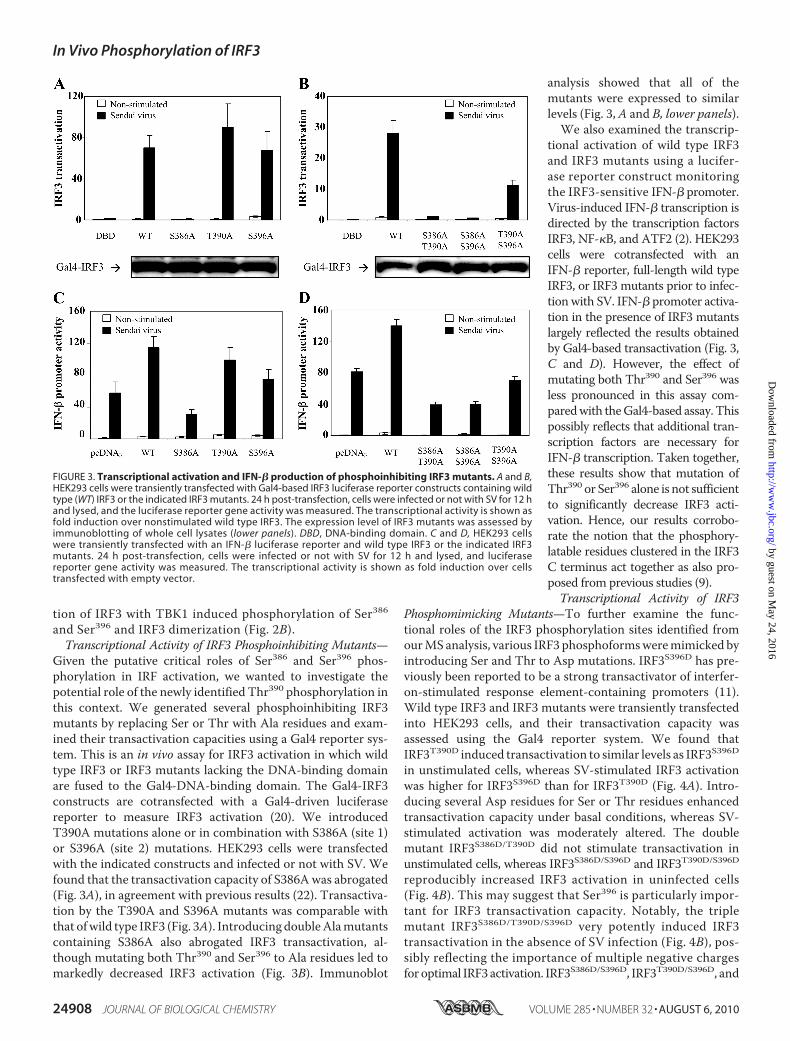
Given the putative critical roles of Ser386 and Ser396 phos-phorylation in IRF activation, we wanted to investigate thepotential role of the newly identified Thr390 phosphorylation inthis context. We generated several phosphoinhibiting IRF3mutants by replacing Ser or Thr with Ala residues and exam-ined their transactivation capacities using a Gal4 reporter sys-tem. This is an in vivo assay for IRF3 activation in which wildtype IRF3 or IRF3 mutants lacking the DNA-binding domainare fused to the Gal4-DNA-binding domain. The Gal4-IRF3constructs are cotransfected with a Gal4-driven luciferasereporter to measure IRF3 activation (20). We introducedT390A mutations alone or in combination with S386A (site 1)or S396A (site 2) mutations. HEK293 cells were transfectedwith the indicated constructs and infected or not with SV. Wefound that the transactivation capacity of S386Awas abrogated(Fig. 3A), in agreement with previous results (22). Transactiva-tion by the T390A and S396A mutants was comparable withthat ofwild type IRF3 (Fig. 3A). Introducing doubleAlamutantscontaining S386A also abrogated IRF3 transactivation, al-though mutating both Thr390 and Ser396 to Ala residues led tomarkedly decreased IRF3 activation (Fig. 3B). Immunoblot
analysis showed that all of themutants were expressed to similarlevels (Fig. 3,A and B, lower panels).
We also examined the transcrip-tional activation of wild type IRF3and IRF3 mutants using a lucifer-ase reporter construct monitoringthe IRF3-sensitive IFN-� promoter.Virus-induced IFN-� transcription isdirected by the transcription factorsIRF3, NF-�B, and ATF2 (2). HEK293cells were cotransfected with anIFN-� reporter, full-length wild typeIRF3, or IRF3 mutants prior to infec-tion with SV. IFN-� promoter activa-tion in the presence of IRF3 mutantslargely reflected the results obtainedby Gal4-based transactivation (Fig. 3,C and D). However, the effect ofmutating both Thr390 and Ser396 wasless pronounced in this assay com-paredwith theGal4-based assay. Thispossibly reflects that additional tran-scription factors are necessary forIFN-� transcription. Taken together,these results show that mutation ofThr390 or Ser396 alone is not sufficientto significantly decrease IRF3 acti-vation. Hence, our results corrobo-rate the notion that the phosphory-latable residues clustered in the IRF3C terminus act together as also pro-posed from previous studies (9).Transcriptional Activity of IRF3
Phosphomimicking Mutants—To further examine the func-tional roles of the IRF3 phosphorylation sites identified fromourMS analysis, various IRF3 phosphoformsweremimicked byintroducing Ser and Thr to Asp mutations. IRF3S396D has pre-viously been reported to be a strong transactivator of interfer-on-stimulated response element-containing promoters (11).Wild type IRF3 and IRF3 mutants were transiently transfectedinto HEK293 cells, and their transactivation capacity wasassessed using the Gal4 reporter system. We found thatIRF3T390D induced transactivation to similar levels as IRF3S396Din unstimulated cells, whereas SV-stimulated IRF3 activationwas higher for IRF3S396D than for IRF3T390D (Fig. 4A). Intro-ducing several Asp residues for Ser or Thr residues enhancedtransactivation capacity under basal conditions, whereas SV-stimulated activation was moderately altered. The doublemutant IRF3S386D/T390D did not stimulate transactivation inunstimulated cells, whereas IRF3S386D/S396D and IRF3T390D/S396Dreproducibly increased IRF3 activation in uninfected cells(Fig. 4B). This may suggest that Ser396 is particularly impor-tant for IRF3 transactivation capacity. Notably, the triplemutant IRF3S386D/T390D/S396D very potently induced IRF3transactivation in the absence of SV infection (Fig. 4B), pos-sibly reflecting the importance of multiple negative chargesfor optimal IRF3 activation. IRF3S386D/S396D, IRF3T390D/S396D, and
FIGURE 3. Transcriptional activation and IFN-� production of phosphoinhibiting IRF3 mutants. A and B,HEK293 cells were transiently transfected with Gal4-based IRF3 luciferase reporter constructs containing wildtype (WT) IRF3 or the indicated IRF3 mutants. 24 h post-transfection, cells were infected or not with SV for 12 hand lysed, and the luciferase reporter gene activity was measured. The transcriptional activity is shown asfold induction over nonstimulated wild type IRF3. The expression level of IRF3 mutants was assessed byimmunoblotting of whole cell lysates (lower panels). DBD, DNA-binding domain. C and D, HEK293 cellswere transiently transfected with an IFN-� luciferase reporter and wild type IRF3 or the indicated IRF3mutants. 24 h post-transfection, cells were infected or not with SV for 12 h and lysed, and luciferasereporter gene activity was measured. The transcriptional activity is shown as fold induction over cellstransfected with empty vector.
In Vivo Phosphorylation of IRF3
24908 JOURNAL OF BIOLOGICAL CHEMISTRY VOLUME 285 • NUMBER 32 • AUGUST 6, 2010
by guest on May 24, 2016
http://ww
w.jbc.org/
Dow
nloaded from

IRF3S386D/T390D/S396D could not be further activated by SVinfection. The various IRF3 phosphomimicking mutants wereexpressed to similar levels as wild type IRF3 (Fig. 4, A and B,lower panels), thus precluding differential expression as a causeof altered activity.We next examined if the effect of Asp mutations on SV elic-
ited IFN-� transcription (Fig. 4, C and D). Indeed, IRF3T390Dand IRF3S396D stimulated IFN-� transcription in uninfectedcells, thus corroborating Gal4 reporter-based results. Surpris-ingly, however, IFN-� transcription after transfection of doubleAsp mutants differed from data from Gal4-based IRF3 activa-tion for these mutants. As mentioned previously, this mightreflect the involvement of several transcription factors in IFN-�promoter activation as compared with the Gal4-based IRF3activation assay. Thus specific phosphomimicking mutationsmay mediate both increased and decreased association to suchtranscription factors in the IFN-� system. Nevertheless, takentogether, our results show that Thr390 contributes to IRF3 tran-scriptional activation. However, the precise mechanism likelydepends on the availability and status of other transcriptionmodulators as well as the post-translationalmodification statusat other sites in IRF3 itself.
Effect of Mutations in the IRF3C-terminal Domain on Interactionbetween IRF3 and CBP—The coac-tivator CBP and the closely relatedp300 associate with IRF3 causingincreased IRF3-mediated transcrip-tion (12). CBP acts to recruit RNApolymerase II and chromatin re-modeling proteins. Ser396 has previ-ously been implicated in binding toCBP (11, 15). Servant et al. (11)found that replacement of Ser396with Ala abrogated binding of IRF3to CBP, whereas replacement of thisresidue with Asp leads to CBP bind-ing in the absence of IRF3-trigger-ing stimulus. We examined therole of IRF3 C-terminal phosphory-lations for interaction with CBPby coimmunoprecipitation assaysusing an anti-FLAG antibody forimmunoprecipitation of FLAG-tagged IRF3 and detecting copre-cipitated endogenous CBP. First, weinvestigated the kinetics of IRF3 andCBP association after SV infection.We found that CBP and IRF3 inter-acted after 6 and 12hof SV infection(Fig. 5A). Mutating Thr390 to Alareduced CBP-IRF3 interaction to70% that of wild type IRF3, whereasmutating Ser396 to Ala decreasedthe association of IRF3 with CBP to40% of wild type IRF3 (Fig. 5B).Interestingly, both IRF3T390D andIRF3S396D were able to bind CBP in
uninfected cells, exhibiting CBP binding of 50 and 72% that ofSV-stimulated wild type IRF3 (Fig. 5C). Moreover, SV-stimu-lated CBP binding of IRF3T390D and IRF3S396D was markedlyincreased to 199 and 186% that of wild type IRF3 (Fig. 5C). Thisindicates that Thr390 and Ser396 contribute to IRF3-mediatedCBP binding. Mutating Ser386 to either Ala or Asp completelyabolished CBP binding, thus corroborating previous resultsin which Ser385 and Ser386 were replaced with Ala (13). Veryweak binding was observed between the double mutantsIRF3S386A/T390A, IRF3S386A/S396A, or IRF3T390A/S396A andCBP in resting or SV-infected cells (Fig. 5D). Collectively,these results suggest that both Thr390 and Ser396 mediatebinding of IRF3 to the cofactor CBP. Introducing two Aspresidues for Ser or Thr residues leads to decreased SV-stim-ulated CBP binding of IRF3S386D/T390D, IRF3S386D/S396D, andIRF3T390D/S396D relative to wild type IRF3 (giving reductionsto 24, 5, and 16% of wild type CBP binding, respectively; Fig.5E). Immunoblotting showed that the double Asp mutantswere expressed at levels comparable with that of wtIRF3(Fig. 6D). Notably, IRF3T390D/S396D exhibited considerablyreduced CBP binding. Hence, although Asp residues intro-duced separately at either Thr390 or Ser396 promote CBP
FIGURE 4. Transcriptional activation and IFN-� production of phosphomimicking IRF3 mutants. A and B,HEK293 cells were transiently transfected with Gal4-based IRF3 luciferase reporter constructs containing wildtype (WT) IRF3 or the indicated IRF3 mutants. 24 h post-transfection, cells were infected or not with SV for 12 hand lysed, and luciferase reporter gene activity was measured. The transcriptional activity is shown as foldinduction over nonstimulated wild type IRF3. The expression level of IRF3 mutants was assessed byimmunoblotting of whole cell lysates (lower panels). DBD, DNA-binding domain. C and D, HEK293 cellswere transiently transfected with an IFN-� luciferase reporter and wild type IRF3 or the indicated IRF3mutants. 24 h post-transfection, cells were infected or not with SV for 12 h and lysed, and luciferasereporter gene activity was measured. The transcriptional activity is shown as fold induction over cellstransfected with empty vector.
In Vivo Phosphorylation of IRF3
AUGUST 6, 2010 • VOLUME 285 • NUMBER 32 JOURNAL OF BIOLOGICAL CHEMISTRY 24909
by guest on May 24, 2016
http://ww
w.jbc.org/
Dow
nloaded from
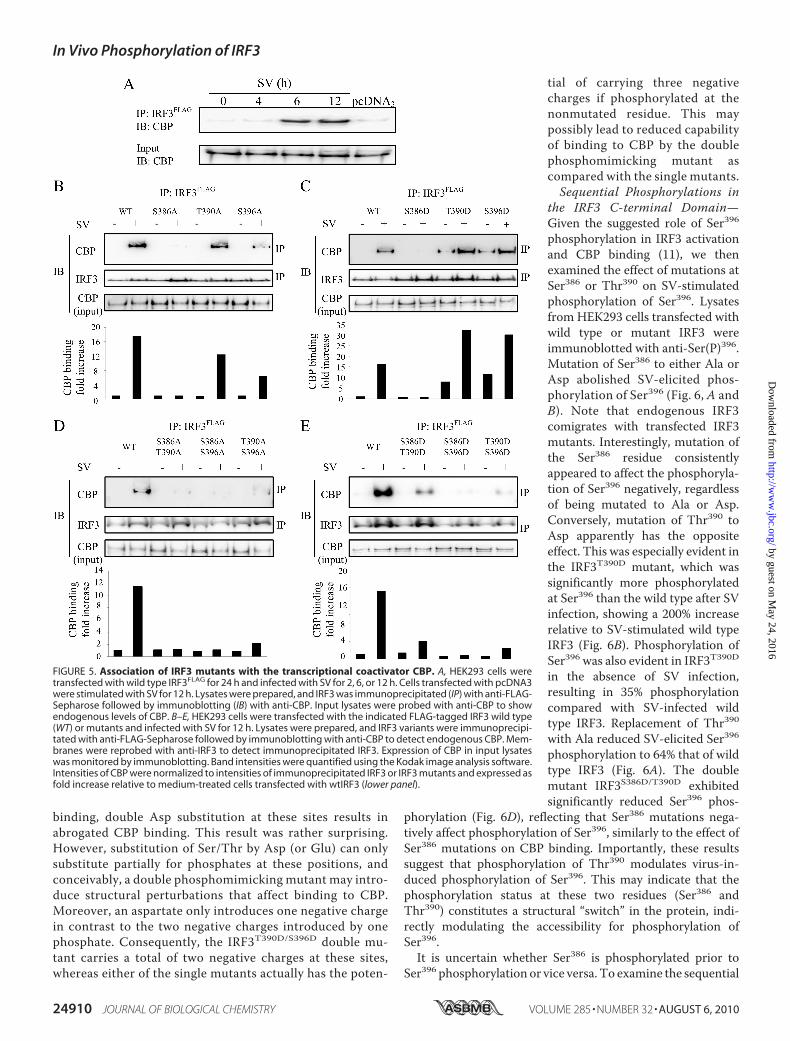
binding, double Asp substitution at these sites results inabrogated CBP binding. This result was rather surprising.However, substitution of Ser/Thr by Asp (or Glu) can onlysubstitute partially for phosphates at these positions, andconceivably, a double phosphomimicking mutant may intro-duce structural perturbations that affect binding to CBP.Moreover, an aspartate only introduces one negative chargein contrast to the two negative charges introduced by onephosphate. Consequently, the IRF3T390D/S396D double mu-tant carries a total of two negative charges at these sites,whereas either of the single mutants actually has the poten-
tial of carrying three negativecharges if phosphorylated at thenonmutated residue. This maypossibly lead to reduced capabilityof binding to CBP by the doublephosphomimicking mutant ascompared with the single mutants.Sequential Phosphorylations in
the IRF3 C-terminal Domain—Given the suggested role of Ser396phosphorylation in IRF3 activationand CBP binding (11), we thenexamined the effect of mutations atSer386 or Thr390 on SV-stimulatedphosphorylation of Ser396. Lysatesfrom HEK293 cells transfected withwild type or mutant IRF3 wereimmunoblotted with anti-Ser(P)396.Mutation of Ser386 to either Ala orAsp abolished SV-elicited phos-phorylation of Ser396 (Fig. 6, A andB). Note that endogenous IRF3comigrates with transfected IRF3mutants. Interestingly, mutation ofthe Ser386 residue consistentlyappeared to affect the phosphoryla-tion of Ser396 negatively, regardlessof being mutated to Ala or Asp.Conversely, mutation of Thr390 toAsp apparently has the oppositeeffect. This was especially evident inthe IRF3T390D mutant, which wassignificantly more phosphorylatedat Ser396 than the wild type after SVinfection, showing a 200% increaserelative to SV-stimulated wild typeIRF3 (Fig. 6B). Phosphorylation ofSer396 was also evident in IRF3T390Din the absence of SV infection,resulting in 35% phosphorylationcompared with SV-infected wildtype IRF3. Replacement of Thr390with Ala reduced SV-elicited Ser396phosphorylation to 64% that of wildtype IRF3 (Fig. 6A). The doublemutant IRF3S386D/T390D exhibitedsignificantly reduced Ser396 phos-
phorylation (Fig. 6D), reflecting that Ser386 mutations nega-tively affect phosphorylation of Ser396, similarly to the effect ofSer386 mutations on CBP binding. Importantly, these resultssuggest that phosphorylation of Thr390 modulates virus-in-duced phosphorylation of Ser396. This may indicate that thephosphorylation status at these two residues (Ser386 andThr390) constitutes a structural “switch” in the protein, indi-rectly modulating the accessibility for phosphorylation ofSer396.
It is uncertain whether Ser386 is phosphorylated prior toSer396 phosphorylation or vice versa. To examine the sequential
FIGURE 5. Association of IRF3 mutants with the transcriptional coactivator CBP. A, HEK293 cells weretransfected with wild type IRF3FLAG for 24 h and infected with SV for 2, 6, or 12 h. Cells transfected with pcDNA3were stimulated with SV for 12 h. Lysates were prepared, and IRF3 was immunoprecipitated (IP) with anti-FLAG-Sepharose followed by immunoblotting (IB) with anti-CBP. Input lysates were probed with anti-CBP to showendogenous levels of CBP. B–E, HEK293 cells were transfected with the indicated FLAG-tagged IRF3 wild type(WT) or mutants and infected with SV for 12 h. Lysates were prepared, and IRF3 variants were immunoprecipi-tated with anti-FLAG-Sepharose followed by immunoblotting with anti-CBP to detect endogenous CBP. Mem-branes were reprobed with anti-IRF3 to detect immunoprecipitated IRF3. Expression of CBP in input lysateswas monitored by immunoblotting. Band intensities were quantified using the Kodak image analysis software.Intensities of CBP were normalized to intensities of immunoprecipitated IRF3 or IRF3 mutants and expressed asfold increase relative to medium-treated cells transfected with wtIRF3 (lower panel).
In Vivo Phosphorylation of IRF3
24910 JOURNAL OF BIOLOGICAL CHEMISTRY VOLUME 285 • NUMBER 32 • AUGUST 6, 2010
by guest on May 24, 2016
http://ww
w.jbc.org/
Dow
nloaded from

order of Ser386 and Ser396 phosphorylations, lysates from cellsinfected with SV were separated by native PAGE and immuno-blotted with antibodies recognizing phosphorylated Ser386 orSer396. We found that phosphorylation of Ser386 and Ser396appeared to have similar kinetics. However, Ser386 was phos-phorylated in both monomeric and dimeric IRF3, whereasSer396 was only phosphorylated in IRF3 dimers (Fig. 6E). Theseresults strongly indicate that phosphorylation of Ser386 occursfirst, inducing IRF3 dimerization and Ser396 phosphorylation.We have previously reported the kinetics of IFN-� transcrip-tion (23), and phosphorylation of Ser386 and Ser396 coincidedwith IFN-� induction in these cells.
DISCUSSION
Despite the important role of IRF3 in antiviral signaling, its invivo phosphorylation pattern has not been reported. The role ofphosphorylation in IRF3 activation has to date been studied byindirect methods using in vitromutagenesis and deletion map-
ping, phosphospecific antibodies, orin vitro phosphorylation studieswith IRF3 (purified from bacteria orinsect cells) and recombinant TBK1(9–11, 14, 22). In this study weimmunoprecipitated FLAG-taggedIRF3 from SV-infected cells and byMS analysis directly identified sitesthat are phosphorylated in IRF3.Our results show that Ser386,Thr390, and Ser396, which arelocated in the C-terminal regulatorydomain of IRF3 (amino acids 380–427), are phosphorylated after SVinfection.Moreover, ourmutagene-sis studies indicate that Thr390,which has not previously beenimplicated in IRF3 activation, con-tributes to IRF3-mediated tran-scription through positively affect-ing SV-induced phosphorylation ofSer396, binding of the cofactor CBPand IRF3 transcriptional activation.We found that substitutions ofeither Thr390 or Ser396 with Ala didnot ablate binding to CBP and didnot significantly affect IRF3 transac-tivation. However, in a double IRF3mutant in which both Thr390 andSer396 were mutated to Ala, CBPbinding was abrogated, and IRF3transactivation was significantlyimpaired. This likely reflects thatIRF3 regulation is an intricate proc-ess controlled by multisite hierar-chical phosphorylations that aremutually dependent to achieve opti-mal IRF3 activation.The residue corresponding to
Thr390 is evolutionarily strictly con-served in mammals, whereas the eight residues upstream anddownstream of Thr390 are highly conserved. One previousstudy has suggested a role for threonine phosphorylation inIRF3 regulation in vivo. Weaver et al. (24) isolated 32P-labeledIRF3 fromNewcastle disease virus-infected cells prior to phos-phoamino acid analysis of IRF3. The authors found that IRF3was phosphorylated mainly on serine residues but also in parton threonine residues (24), suggesting a role for threoninephosphorylation in IRF3 responses in vivo. In this studywe usedSV, which is a single-stranded RNA virus that is recognized bythe cytoplasmicRNAhelicaseRIG-I (25). It is possible that IRF3phosphorylation may be subject to quantitative and qualitativechanges depending on the nature of the virus and host recogni-tion mechanisms. Indeed, Noyce et al. (26) reported thathuman cytomegalovirus (double-stranded DNA virus) andNewcastle disease virus (single-stranded RNA virus) produceddifferent migration patterns after two-dimensional PAGE sep-aration of IRF3. This indicates that distinct IRF3 post-transla-
FIGURE 6. SV-elicited phosphorylation of Ser396 and Ser386. A–D, HEK293 cells were transfected with theindicated IRF3 wild type (WT) or mutants and infected or not with SV for 12 h. Lysates were prepared, andphosphorylation of Ser396 was examined by immunoblotting with an antibody recognizing phosphorylatedSer396. Membranes were reprobed with anti-FLAG. Band intensities were quantified using the Kodak imageanalysis software. Intensities of phosphorylated Ser396 were normalized to intensities of FLAG-tagged IRF3 orIRF3 mutants and expressed as fold increase relative to medium-treated cells transfected with wtIRF3 (lowerpanels; A and B). E, lysates were prepared from SV-infected HEK293 cells and separated by native PAGE orSDS-PAGE prior to immunoblotting (IB) with anti-Ser(P)386, anti-Ser(P)396, or total IRF3.
In Vivo Phosphorylation of IRF3
AUGUST 6, 2010 • VOLUME 285 • NUMBER 32 JOURNAL OF BIOLOGICAL CHEMISTRY 24911
by guest on May 24, 2016
http://ww
w.jbc.org/
Dow
nloaded from
tional modifications may prevail after infection with specificviruses. From MS analysis, we observed that Ser173 and Ser175were phosphorylated both in uninfected and in SV-infectedcells, but the results do not disclose the stoichiometry of phos-phorylation at these sites, i.e. if phosphorylation is positively ornegatively affected by viral exposure. The sequence aroundSer173 resembles SP-phosphodegron motifs, and IRF3 proteinlevels have been reported to be sensitive to proteasome inhibi-tors. Hence, phosphorylation of Ser173 and Ser175may be impli-cated in ubiquitin-mediated IRF3 degradation. These aspectsare currently under investigation in our laboratory.In this study, we transfected IRF3 and examined its phosphor-
ylation pattern in medium- and SV-treated cells. We alsoattempted to map the phosphorylation sites of endogenousIRF3. However, endogenous levels of IRF3 are low and consistof multiple, distinct phosphoforms. The inherent problem oflower detection limit for phosphorylated peptides comparedwith nonphosphorylated peptides necessitates significantamounts of IRF3 for identification of phosphopeptides by MS.We did not succeed to efficiently affinity purify IRF3 for thispurpose. Difficulties with effective affinity enrichment ofendogenous IRF3 might rely on intrinsic biological mecha-nisms such as shielding of the most immunogenic epitopes ofthe proteinwithin IRF3 dimers/multimers or evenwithin largermultiprotein complexes. Nevertheless, we examined activationproperties of transfected IRF3 and found that it does not bindCBP or induce IFN-� production in the absence of viral infec-tion, whereas transfection of IRF3 leads to enhanced SV-in-duced IFN-� production (Figs. 3–5). Moreover, transfectedIRF3 is not spontaneously phosphorylated at Ser396, but itsSer396 phosphorylation is induced by SV and follows compara-ble kinetics as endogenous IRF3. Also, SV-induced Ser396 phos-phorylation is increased for transfected IRF3 (compared withendogenous IRF3; supplemental Fig. 2). Taken together, thissuggests that overexpressed IRF3 is not activated aberrantly butfollows the similar virus-induced regulatory switches that applyto endogenous IRF3. Thus, our findings on phosphorylationsites in (transfected) IRF3 should be valuable and contribute tounderstanding of IRF3 activity regulation.There have been numerous reports on IRF3 phosphoryla-
tion, but these are based on mutation of presumed phosphory-lation sites, in vitro phosphorylations (using partially purifiedIRF3 and TBK1), or phosphorylation state-specific antibodiestoward Ser386 or Ser396 (9–11). This is the first study to directlydetermine sites that are simultaneously phosphorylated in IRF3after a viral infection (Table 1). Our findings implicate Thr390(which has not previously been identified as an IRF3 phosphor-ylation site) in IRF3 activation and IFN-� transcription. Ourresults show that the phosphorylation site considered to be ofhighest importance for IRF3 antiviral activity, Ser396, is regu-lated by the phosphorylation status of Thr390. Additionally, bymaking use of a phosphoproteomic approach, we were able toidentify multisite phosphorylated peptides, thus providinginformation on multisite, hierarchical phosphorylations. Wefound that phosphorylation ofThr390 always occurred concom-itantly with phosphorylation of either Ser386 or Ser396, and wealso identified a triply phosphorylated peptide with Ser385/Ser386 and Thr390(Table 1). Although the implication of this is
presently not clear, it has been shown in other settings thatspecific combinations of phosphorylations and their timelyoccurrence within a biological pathway are crucial for modula-tion of activity.We have recently demonstrated this for anotherprotein, the uracil-DNA glycosylase UNG2 (27).A functional role for phosphorylation of the IRF3C terminus
has been proposed from the cocrystal structure of IRF3with theIRF3-binding domain of CBP. Based on this structure, it hasbeen suggested that IRF3 in unstimulated cells exists in a latentform that is autoinhibited by interactions within the IRF asso-ciation domain of eachmonomer (15). This autoinhibited stateof IRF3 masks a hydrophobic surface, which upon unfolding ofIRF3mediates IRF3-CBP binding as illustrated in Fig. 7A. Thus,it has been suggested that phosphorylation of the IRF3 C termi-nus leads to unfolding of the regulatory domain because ofrepulsions between negative charges and the hydrophobicregion. Hence, initial phosphorylation(s) in the C-terminal reg-ulatory domainmay trigger conformational changes (e.g.partialrelief of the autoinhibitory folding of IRF3) that facilitate phos-phorylation at other sites in IRF3.In an attempt to rationalize our results obtained with IRF3
phosphorylation site mutants in terms of structural insight, weexamined the published crystal structures of IRF3 and theclosely related IRF5 (15, 16, 28, 29). In two tertiary structures ofIRF3 (16, 28), the phosphorylation targets in the C-terminalregulatory domain are partially surface-exposed. This isespecially evident for Ser386 that is located in a solvent-ac-cessible turn and should thus be readily available for phos-phorylation. This is illustrated in two different structuralviews of IRF3 (Fig. 7, B and C). Previous results and ourfindings presented herein suggest that phosphorylation ofSer396 is of particular importance for IRF3 activation. Thestructure of IRF3 shows that Ser396 is located in the proxim-ity of the negatively charged residues Glu200 and Glu201 (Fig.7D). Hence, as suggested by Qin et al. (16), phosphorylationof Ser396 would introduce a negative charge that could causeconformational changes because of electrostatic repulsion.This provides a structural basis for the importance of Ser396phosphorylation in IRF3 activation.Based on results obtained with various Ala and Asp muta-
tions, it has been discussedwhether Ser386 (site 1) or Ser396 (site2) is the critical/initial virus-induced phosphorylation residue(11, 14, 22). Panne et al. (9) suggested that phosphorylation ofsite 2 residues alleviated IRF3 autoinhibition, allowing for CBPbinding and facilitating phosphorylation at site 1 (Ser385/Ser386). Mori et al. (22) proposed that phosphorylation of site 1was the initial event and was instrumental for IRF3 dimeriza-tion. In our study, MS analysis revealed that Ser385 or Ser386, incontrast to Ser396, was phosphorylated in noninfected cells.Also, we found that Ser386 was phosphorylated in monomericand dimeric IRF3, whereas Ser396 was predominantly phosphor-ylated in IRF3 dimers (Fig. 6E). Hence, from our results wepropose that Ser386 phosphorylation is the primary event. Thisalso agrees with the higher solvent accessibility of Ser386 com-pared with Ser396. Phosphorylation of Ser386 might, however,promote partial unfolding of the autoinhibitory IRF3 structureand increase the accessibility of Ser396 to facilitate its phos-phorylation, as suggested by Qin et al. (16).
In Vivo Phosphorylation of IRF3
24912 JOURNAL OF BIOLOGICAL CHEMISTRY VOLUME 285 • NUMBER 32 • AUGUST 6, 2010
by guest on May 24, 2016
http://ww
w.jbc.org/
Dow
nloaded from
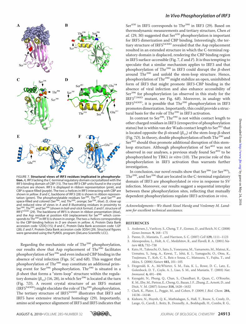
Regarding the mechanistic role of Thr390 phosphorylation,our results show that Asp replacement of Thr390 facilitatesphosphorylation of Ser396 and even induced CBP binding in theabsence of viral infection (Figs. 5C and 6B). This suggest thatphosphorylation of Thr390 may constitute an additional prim-ing event for Ser396 phosphorylation. Thr390 is situated in a�-sheet that forms a “stem-loop” structure within the regula-tory domain (�12) (16, 28), in which Ser396 is located at the turn(Fig. 7D). A recent crystal structure of an IRF5 mutant(IRF5S430D)might elucidate the role of Thr390 phosphorylation.The tertiary structure of IRF5S430D illustrates that IRF3 andIRF5 have extensive structural homology (29). Importantly,amino acid sequence alignment of IRF3 and IRF5 indicates that
Ser430 in IRF5 corresponds to Thr390 in IRF3 (29). Based onthermodynamic measurements and tertiary structure, Chen etal. (29, 30) suggested that Ser430 phosphorylation is importantfor IRF5 dimerization and CBP binding. Interestingly, the ter-tiary structure of IRF5S430D revealed that the Asp replacementresulted in an extended structure in which the C-terminal reg-ulatory domain is displaced, rendering the CBP binding regionin IRF5 surface-accessible (Fig. 7,E and F). It is thus tempting tospeculate that a similar mechanism applies to IRF3 and thatphosphorylation of Thr390 in IRF3 could disrupt the �-sheetaround Thr390 and unfold the stem-loop structure. Hence,phosphorylation of Thr390 might stabilize an open, uninhibitedform of IRF3 that might promote IRF3-CBP binding in theabsence of viral infection and also enhance accessibility ofSer396 for phosphorylation (as observed in this study for theIRF3T390D mutant, see Fig. 6B). Moreover, in analogy withIRF5S430D, it is possible that Thr390 phosphorylation in IRF3promotes dimerization. Importantly, this could provide a struc-tural basis for the role of Thr390 in IRF3 activation.In contrast to Ser396, Thr390 is not within contact length to
other charged residues in IRF3 (irrespective of phosphorylationstatus) but is within van derWaals contact length to Ser402 thatis located opposite the �-strand (�13) of the stem-loop �-sheet(Fig. 7D). In theory, double phosphorylation of both Thr390 andSer402 should thus promote additional disruption of this stem-loop structure. Although phosphorylation of Ser402 was notobserved in our analyses, a previous study found Ser402 to bephosphorylated by TBK1 in vitro (10). The precise role of thisphosphorylation in IRF3 activation thus warrants furtherinvestigation.In conclusion, our novel results show that Ser386 (or Ser385),
Thr390, and Ser396 that are located in the C-terminal regulatorydomain of IRF3 are phosphorylated in vivo in response to SVinfection. Moreover, our results suggest a sequential interplaybetween these phosphorylation sites, reflecting that mutuallydependent phosphorylations regulate IRF3 activation in vivo.
Acknowledgments—We thank Sissel Havåg and Veslemøy M. Land-sem for excellent technical assistance.
REFERENCES1. Andersen, J., VanScoy, S., Cheng, T. F., Gomez, D., and Reich, N. C. (2008)
Genes Immun. 9, 168–1752. Panne, D., Maniatis, T., and Harrison, S. C. (2007) Cell 129, 1111–11233. Alexopoulou, L., Holt, A. C., Medzhitov, R., and Flavell, R. A. (2001) Na-
ture 413, 732–7384. Kato, H., Takeuchi, O., Sato, S., Yoneyama,M., Yamamoto,M.,Matsui, K.,
Uematsu, S., Jung, A., Kawai, T., Ishii, K. J., Yamaguchi, O., Otsu, K.,Tsujimura, T., Koh, C. S., Reis e Sousa, C., Matsuura, Y., Fujita, T., andAkira, S. (2006) Nature 441, 101–105
5. Fitzgerald, K. A., McWhirter, S. M., Faia, K. L., Rowe, D. C., Latz, E.,Golenbock, D. T., Coyle, A. J., Liao, S. M., and Maniatis, T. (2003) Nat.Immunol. 4, 491–496
6. Kayagaki, N., Phung, Q., Chan, S., Chaudhari, R., Quan, C., O’Rourke,K.M., Eby,M., Pietras, E., Cheng,G., Bazan, J. F., Zhang, Z., Arnott, D., andDixit, V. M. (2007) Science 318, 1628–1632
7. Clark, K., Plater, L., Peggie, M., and Cohen, P. (2009) J. Biol. Chem. 284,14136–14146
8. Kishore, N., Huynh, Q. K., Mathialagan, S., Hall, T., Rouw, S., Creely, D.,Lange, G., Caroll, J., Reitz, B., Donnelly, A., Boddupalli, H., Combs, R. G.,
FIGURE 7. Structural views of IRF3 residues implicated in phosphoryla-tion. A, IRF3 lacking the C-terminal regulatory domain cocrystallized with theIRF3-binding domain of CBP (15). The two IRF3-CBP units found in the crystalstructure are shown. IRF3 is displayed in ribbon representation (pink), andCBP is space-filled (purple). The two �-helices in IRF3 interacting with CBP areshown in yellow. B and C, backbone of IRF3 (28) is shown in ribbon represen-tation (green). The phosphorylatable residues Ser386, Thr390, and Ser396 arespace-filled and colored (Ser386, red; Thr390, orange; Ser396, blue). D, close upand reduced view of views in A and B illustrating residues in proximity toSer386, Thr390, and Ser396 (shown in ball-and-stick format). E and F, structure ofIRF5S430D (29). The backbone of IRF5 is shown in ribbon presentation (blue),and the Asp residue at position 430 (replacement for Ser430 which corre-sponds to Thr390 in IRF3) is shown in orange. The two �-helices correspondingto the CBP-binding helices in D are shown in yellow. A, Protein Data Bankaccession code 1ZOQ (15); B and C, Protein Data Bank accession code 1J2F(28); E and F, Protein Data Bank accession code 3DSH (29). Structural figureswere generated using the PyMOL program (DeLano Scientific LCC).
In Vivo Phosphorylation of IRF3
AUGUST 6, 2010 • VOLUME 285 • NUMBER 32 JOURNAL OF BIOLOGICAL CHEMISTRY 24913
by guest on May 24, 2016
http://ww
w.jbc.org/
Dow
nloaded from

Kretzmer, K., and Tripp, C. S. (2002) J. Biol. Chem. 277, 13840–138479. Panne, D., McWhirter, S. M., Maniatis, T., and Harrison, S. C. (2007)
J. Biol. Chem. 282, 22816–2282210. Clement, J. F., Bibeau-Poirier, A., Gravel, S. P., Grandvaux, N., Bonneil, E.,
Thibault, P., Meloche, S., and Servant, M. J. (2008) J. Virol. 82, 3984–399611. Servant, M. J., Grandvaux, N., tenOever, B. R., Duguay, D., Lin, R., and
Hiscott, J. (2003) J. Biol. Chem. 278, 9441–944712. Yoneyama, M., Suhara, W., Fukuhara, Y., Fukuda, M., Nishida, E., and
Fujita, T. (1998) EMBO J. 17, 1087–109513. Lin, R., Mamane, Y., and Hiscott, J. (1999)Mol. Cell. Biol. 19, 2465–247414. Lin, R., Heylbroeck, C., Pitha, P. M., and Hiscott, J. (1998)Mol. Cell. Biol.
18, 2986–299615. Qin, B. Y., Liu, C., Srinath, H., Lam, S. S., Correia, J. J., Derynck, R., and Lin,
K. (2005) Structure 13, 1269–127716. Qin, B. Y., Liu, C., Lam, S. S., Srinath,H., Delston, R., Correia, J. J., Derynck,
R., and Lin, K. (2003) Nat. Struct. Biol. 10, 913–92117. Shevchenko, A., Wilm, M., Vorm, O., and Mann, M. (1996) Anal. Chem.
68, 850–85818. Rappsilber, J., Ishihama, Y., and Mann, M. (2003) Anal. Chem. 75,
663–67019. Thingholm, T. E., Jørgensen, T. J., Jensen, O. N., and Larsen, M. R. (2006)
Nat. Protoc. 1, 1929–193520. Wathelet, M. G., Lin, C. H., Parekh, B. S., Ronco, L. V., Howley, P. M., and
Maniatis, T. (1998)Mol. Cell 1, 507–518
21. Zhang, B., Li, M., Chen, L., Yang, K., Shan, Y., Zhu, L., Sun, S., Li, L., andWang, C. (2009) Cell Res. 19, 412–428
22. Mori, M., Yoneyama, M., Ito, T., Takahashi, K., Inagaki, F., and Fujita, T.(2004) J. Biol. Chem. 279, 9698–9702
23. Johnsen, I. B., Nguyen, T. T., Bergstroem, B., Fitzgerald, K. A., and An-thonsen, M. W. (2009) J. Biol. Chem. 284, 19122–19131
24. Weaver, B. K., Kumar, K. P., and Reich, N. C. (1998) Mol. Cell. Biol. 18,1359–1368
25. Rothenfusser, S., Goutagny, N., DiPerna, G., Gong, M., Monks, B. G.,Schoenemeyer, A., Yamamoto, M., Akira, S., and Fitzgerald, K. A. (2005)J. Immunol. 175, 5260–5268
26. Noyce, R. S., Collins, S. E., and Mossman, K. L. (2009) J. Virol. 83,4013–4022
27. Hagen, L., Kavli, B., Sousa, M. M., Torseth, K., Liabakk, N. B., Sundheim,O., Pena-Diaz, J., Otterlei, M., Hørning, O., Jensen, O. N., Krokan, H. E.,and Slupphaug, G. (2008) EMBO J. 27, 51–61
28. Takahasi, K., Suzuki, N. N., Horiuchi,M.,Mori,M., Suhara,W., Okabe, Y.,Fukuhara, Y., Terasawa, H., Akira, S., Fujita, T., and Inagaki, F. (2003)Nat.Struct. Biol. 10, 922–927
29. Chen, W., Lam, S. S., Srinath, H., Jiang, Z., Correia, J. J., Schiffer, C. A.,Fitzgerald, K. A., Lin, K., and Royer,W. E., Jr. (2008)Nat. Struct. Mol. Biol.15, 1213–1220
30. Chen, W., Srinath, H., Lam, S. S., Schiffer, C. A., Royer, W. E., Jr., and Lin,K. (2008) J. Mol. Biol. 379, 251–260
In Vivo Phosphorylation of IRF3
24914 JOURNAL OF BIOLOGICAL CHEMISTRY VOLUME 285 • NUMBER 32 • AUGUST 6, 2010
by guest on May 24, 2016
http://ww
w.jbc.org/
Dow
nloaded from

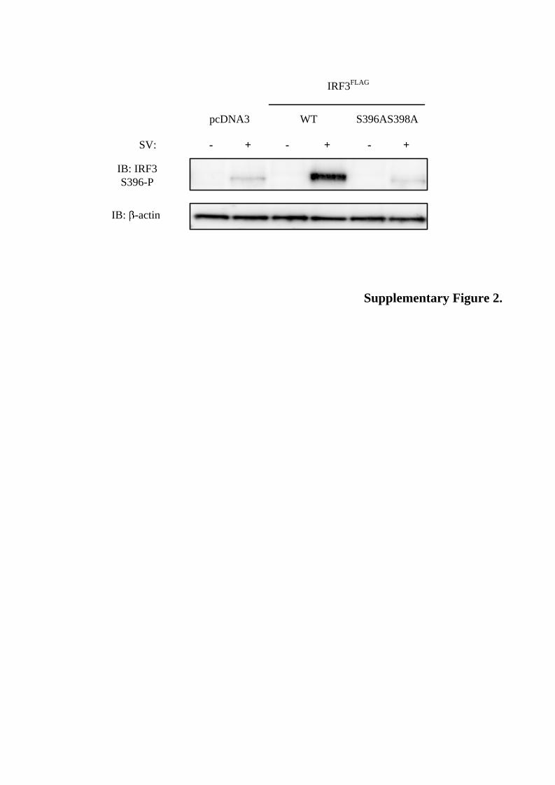
SUPPLEMENTAL DATA Supplementary Fig. 1. Blue Native PAGE resolution of IRF3. HEK293 cells were transiently transfected with IRF3FLAG. 24 hours post-transfection cells were infected or not with SV (12 hours). Lysates were immunoprecipitated with anti-FLAG-Sepharose and proteins were separated by BN-PAGE and stained with Coomassie-based SimplyBlue Safestain (A) or separated by BN-PAGE followed by immunoblotting with anti-IRF3 (B). Bands excised and analysed by MS are indicated in A. NS: non-stimulated, SV: SV-stimulated cells. NS1 and SV1 correspond to IRF3 monomers. NS2-4 and SV2-4 represent IRF3-containing complexes of higher molecular weights. Supplementary Fig. 2. HEK293 cells were transfected with FLAG-tagged IRF3, IRF3S396AS398A or the empty vector and infected with SV for 12 hours. Lysates were prepared and separated by denaturing gel electrophoresis. Phosphorylation of Ser396 was examined by immunoblotting with an antibody recognizing phosphorylated Ser396. Membranes were reprobed with anti-β-actin.

Coomassie
BA
NS4
NS1
NS2
NS3
Medium
SV4
SV1
SV2
SV3
SV
SV
IB: IRF3
Medium
Monomer
Dimer
Supplementary Figure 1.
SV:
IB: IRF3
S396-P
S396AS398AWTpcDNA3
IRF3FLAG
+ ++- --
IB: -actin
Supplementary Figure 2.

Slupphaug, Liv Thommesen and Marit W. AnthonsenBjarte Bergstroem, Ingvild B. Johnsen, Thuy Thanh Nguyen, Lars Hagen, Geir
Regulatory Factor-3 (IRF3) Virus-targeted Phosphorylation Site in Interferonin VivoIdentification of a Novel
doi: 10.1074/jbc.M109.084822 originally published online May 28, 20102010, 285:24904-24914.J. Biol. Chem.
10.1074/jbc.M109.084822Access the most updated version of this article at doi:
Alerts:
When a correction for this article is posted•
When this article is cited•
to choose from all of JBC's e-mail alertsClick here
Supplemental material:
http://www.jbc.org/content/suppl/2010/05/28/M109.084822.DC1.html
http://www.jbc.org/content/285/32/24904.full.html#ref-list-1
This article cites 30 references, 14 of which can be accessed free at
by guest on May 24, 2016
http://ww
w.jbc.org/
Dow
nloaded from