Identification of a deep intronic mutation in the COL6A2 gene by a novel custom oligonucleotide CGH...
12
RESEARCH ARTICLE Open Access Identification of a deep intronic mutation in the COL6A2 gene by a novel custom oligonucleotide CGH array designed to explore allelic and genetic heterogeneity in collagen VI-related myopathies Matteo Bovolenta 1† , Marcella Neri 1† , Elena Martoni 1 , Anna Urciuolo 2 , Patrizia Sabatelli 3 , Marina Fabris 1 , Paolo Grumati 2 , Eugenio Mercuri 4 , Enrico Bertini 5 , Luciano Merlini 1,6 , Paolo Bonaldo 2 , Alessandra Ferlini 1 , Francesca Gualandi 1* Abstract Background: Molecular characterization of collagen-VI related myopathies currently relies on standard sequencing, which yields a detection rate approximating 75-79% in Ullrich congenital muscular dystrophy (UCMD) and 60-65% in Bethlem myopathy (BM) patients as PCR-based techniques tend to miss gross genomic rearrangements as well as copy number variations (CNVs) in both the coding sequence and intronic regions. Methods: We have designed a custom oligonucleotide CGH array in order to investigate the presence of CNVs in the coding and non-coding regions of COL6A1, A2, A3, A5 and A6 genes and a group of genes functionally related to collagen VI. A cohort of 12 patients with UCMD/BM negative at sequencing analysis and 2 subjects carrying a single COL6 mutation whose clinical phenotype was not explicable by inheritance were selected and the occurrence of allelic and genetic heterogeneity explored. Results: A deletion within intron 1A of the COL6A2 gene, occurring in compound heterozygosity with a small deletion in exon 28, previously detected by routine sequencing, was identified in a BM patient. RNA studies showed monoallelic transcription of the COL6A2 gene, thus elucidating the functional effect of the intronic deletion. No pathogenic mutations were identified in the remaining analyzed patients, either within COL6A genes, or in genes functionally related to collagen VI. Conclusions: Our custom CGH array may represent a useful complementary diagnostic tool, especially in recessive forms of the disease, when only one mutant allele is detected by standard sequencing. The intronic deletion we identified represents the first example of a pure intronic mutation in COL6A genes. Background Mutations in the genes encoding collagen VI (COL6A1, COL6A2 and COL6A3) result in two major phenotypes: Bethlem myopathy [BM, OMIM #158810] and Ullrich congenital muscular dystrophy [UCMD, OMIM #254090]. Despite BM being classically reported as an autosomal dominant condition due to heterozygous COL6 mutations [1,2], we and others have recently described autosomal recessive BM patients [3,4]. In con- trast, the allelic form UCMD was initially considered to be an autosomal recessive disorder, with homozygous or compound heterozygous mutations occurring in all three COL6 genes [2], although a few double heterozy- gous mutations in two different COL6 genes have also been described [5]. Recently, however, up to 50% of UCMD cases have been found to carry only one mutated allele, indicating autosomal dominant inheri- tance [5-7]. Thus far, roughly 100 different mutations in COL6 genes have been associated with either UCMD or * Correspondence: [email protected] † Contributed equally 1 Department of Experimental and Diagnostic Medicine - Section of Medical Genetics, University of Ferrara, Ferrara, Italy Bovolenta et al. BMC Medical Genetics 2010, 11:44 http://www.biomedcentral.com/1471-2350/11/44 © 2010 Bovolenta et al; licensee BioMed Central Ltd. This is an Open Access article distributed under the terms of the Creative Commons Attribution License (http://creativecommons.org/licenses/by/2.0), which permits unrestricted use, distribution, and reproduction in any medium, provided the original work is properly cited.
Transcript of Identification of a deep intronic mutation in the COL6A2 gene by a novel custom oligonucleotide CGH...

RESEARCH ARTICLE Open Access
Identification of a deep intronic mutation in theCOL6A2 gene by a novel custom oligonucleotideCGH array designed to explore allelic and geneticheterogeneity in collagen VI-related myopathiesMatteo Bovolenta1†, Marcella Neri1†, Elena Martoni1, Anna Urciuolo2, Patrizia Sabatelli3, Marina Fabris1,Paolo Grumati2, Eugenio Mercuri4, Enrico Bertini5, Luciano Merlini1,6, Paolo Bonaldo2, Alessandra Ferlini1,Francesca Gualandi1*
Abstract
Background: Molecular characterization of collagen-VI related myopathies currently relies on standard sequencing,which yields a detection rate approximating 75-79% in Ullrich congenital muscular dystrophy (UCMD) and 60-65%in Bethlem myopathy (BM) patients as PCR-based techniques tend to miss gross genomic rearrangements as wellas copy number variations (CNVs) in both the coding sequence and intronic regions.
Methods: We have designed a custom oligonucleotide CGH array in order to investigate the presence of CNVs inthe coding and non-coding regions of COL6A1, A2, A3, A5 and A6 genes and a group of genes functionally relatedto collagen VI. A cohort of 12 patients with UCMD/BM negative at sequencing analysis and 2 subjects carrying asingle COL6 mutation whose clinical phenotype was not explicable by inheritance were selected and theoccurrence of allelic and genetic heterogeneity explored.
Results: A deletion within intron 1A of the COL6A2 gene, occurring in compound heterozygosity with a smalldeletion in exon 28, previously detected by routine sequencing, was identified in a BM patient. RNA studiesshowed monoallelic transcription of the COL6A2 gene, thus elucidating the functional effect of the intronicdeletion. No pathogenic mutations were identified in the remaining analyzed patients, either within COL6A genes,or in genes functionally related to collagen VI.
Conclusions: Our custom CGH array may represent a useful complementary diagnostic tool, especially in recessiveforms of the disease, when only one mutant allele is detected by standard sequencing. The intronic deletion weidentified represents the first example of a pure intronic mutation in COL6A genes.
BackgroundMutations in the genes encoding collagen VI (COL6A1,COL6A2 and COL6A3) result in two major phenotypes:Bethlem myopathy [BM, OMIM #158810] and Ullrichcongenital muscular dystrophy [UCMD, OMIM#254090]. Despite BM being classically reported as anautosomal dominant condition due to heterozygousCOL6 mutations [1,2], we and others have recently
described autosomal recessive BM patients [3,4]. In con-trast, the allelic form UCMD was initially considered tobe an autosomal recessive disorder, with homozygous orcompound heterozygous mutations occurring in allthree COL6 genes [2], although a few double heterozy-gous mutations in two different COL6 genes have alsobeen described [5]. Recently, however, up to 50% ofUCMD cases have been found to carry only onemutated allele, indicating autosomal dominant inheri-tance [5-7]. Thus far, roughly 100 different mutations inCOL6 genes have been associated with either UCMD or
* Correspondence: [email protected]† Contributed equally1Department of Experimental and Diagnostic Medicine - Section of MedicalGenetics, University of Ferrara, Ferrara, Italy
Bovolenta et al. BMC Medical Genetics 2010, 11:44http://www.biomedcentral.com/1471-2350/11/44
© 2010 Bovolenta et al; licensee BioMed Central Ltd. This is an Open Access article distributed under the terms of the CreativeCommons Attribution License (http://creativecommons.org/licenses/by/2.0), which permits unrestricted use, distribution, andreproduction in any medium, provided the original work is properly cited.

BM and most of them are confined to single families[5,8].The distribution of mutations along COL6 genes is
rather uniform and lacks mutation hot spots, thereforethese patients require extensive genotyping, which is cur-rently performed by genomic or cDNA sequencing [1,5].Nevertheless, a relevant proportion of patients clinicallydiagnosed as having a collagen-VI related myopathy stilllack molecular characterization. In fact, with currentlyavailable diagnostic tools, the detection rate of mutationsof COL6 genes varies from 60-65% in BM cases and 75-79% in UCMD patients [5]. The majority of these muta-tions are small variations like missense, frame-shifting,ins-del or point mutations which lead to a splicing defect.Large, multi-exon deletions of the COL6A1 gene, invol-ving both exonic and intronic regions and thus detectableby mRNA analysis, have been reported as causativemutations in three patients [9,10]. One limitation ofPCR-based genome analysis techniques is their inabilityto detect gross CNVs, as well as atypical mutations,which could account for a significant proportion of unde-tected COL6 mutations. On the other hand, the relativelylow rate of mutation detection in COL6 genes could bedue to the genetic heterogeneity of these diseases. Thus,mutations in genes functionally related to collagen VIcould theoretically underlie UCMD and BM phenocopiesand/or be responsible for secondary collagen VI defects[11]. In order to test this hypothesis, we selected 12UCMD/BM patients who were found to be negativeupon extensive sequence analysis of the three COL6genes, and two patients carrying only one mutation,deemed insufficient to explain the clinical phenotype, itbeing inherited from a healthy parent.The occurrence of both allelic and genetic heterogene-
ity was explored in these patients by using an innovativeoligonucleotide array-based comparative genomic hybri-dization (CGH) approach able to detect CNVs in COL6genes, as well as in other genes functionally related tocollagen VI.A deep intronic deletion in the COL6A2 gene was dis-
covered in one BM patient, with a single mutationinherited from the healthy mother identified at sequen-cing analysis. Subsequently, the functional effect of theidentified mutation was demonstrated via RNA studies.In the remaining patients, only non pathogenic CNVswere identified.
MethodsGenome sequence analysisPatients’ genomic DNA was extracted from peripheralblood lymphocytes after informed consent and approvalby the local ethics committee was obtained (approvalnumber 7/2009). PCR primers (sequences are available
upon request) were designed to amplify all the 107 exonsof the COL6 genes, as well as their flanking intronicregions. Amplified fragments were directly sequencedusing a BigDye Terminator v3.1 Cycle sequencing systemon the automated ABI 3130 Genetic Analyzer (AppliedBiosystems, Foster City, CA).In order to try to attempt amplification across the
deletion within COL6A2 intron 1A, identified by theCGH-array, the following oligonucleotides were posi-tioned outside the maximum theoretical deleted region:F1 CCCTGAATTCCTGGACATGAT; R1 GAACGTC-CATCCTCCCTGAT; and flanking the region identifiedby the two deleted probes (F2 AGATCCACAGCCAC-GACTT; R2 GGCCTCACTGTGCTGCTG) (Figure 1).Long-range PCR was performed using LA-Taq polymer-ase and 40 cycles at 64°C.
Micro-array design, hybridization and data analysisCOL6-CGH array design was performed using the high-density aCGH search function of the web-based AgilenteArray database, version 4.5 [12].The genomic regions corresponding to COL6A1-A2-
A3-A5 and -A6 genes as well as a group of genes func-tionally related to collagen VI (Table 1) [13-21] weremasked for repetitive elements and converted into a10.197 probe set by selecting the maximum number ofexonic and intronic 60 mer oligonucleotide probes avail-able in the Agilent database. This probe set was enrichedwith 377 probes in triplicate, covering the regions ofCOL6A1, 2 and 3 genes not investigated by the AgilentCGH probe database. The final mean resolution for thesegenes was one probe every 320 bp. In order to reach the15K array format, each array was filled with controlprobes from all the chromosomes (2851).The array format utilized was 8 × 15K, made up of
eight identical 15K arrays on a single slide, thereby per-mitting simultaneous analysis of eight different samples.Genomic DNA was extracted from the patients’ wholeblood or cultured fibroblasts by a Nucleon™ BACCGenomic DNA Extraction Kit (GE Healthcare). Labelingand hybridization were performed following the proto-cols provided by Agilent (Agilent OligonucleotideArray-Based CGH for Genomic DNA Analysis protocolv5.0). The array was analyzed with the Agilent scannerand the Feature Extraction software (v9.1). A graphicaloverview and analysis of the data were obtained usingthe CGH analytics software (v3.5). For identifying dupli-cations and deletions we used the statistical calculationsbased on ADM-2 algorithm provided by the CGH analy-tics software. According to this set-up and in the case ofautosomal genes, deletions are visualized with values of-1 if in heterozygosity and with values of minus infinite(-4 in CGH analytics) if in homozygosity. For three
Bovolenta et al. BMC Medical Genetics 2010, 11:44http://www.biomedcentral.com/1471-2350/11/44
Page 2 of 12
copies, the value would be approximately +0.6 and forfour copies the value would be +1.The platform informations have been submitted to the
online data repository Gene Expression Omnibus (GEO)[22], under accession number GPL9972.
Real-Time PCRAd hoc Real-Time PCR assays were designed for theCOL6A2 intron 1A deletion and the CNVs identifiedwithin the ITGB1 and ITGA5/ITGA genomic regions(Table 1 and Additional file 1). For confirming COL6A2
intron 1A deletion, a TaqMan assay was firstly used;MGB probe and primer design was performed by Pri-mer Express Software 2.0 (Applied Biosystems) (FWTTGGTCACAGGTTATGCAACA, Rev GGTGAGTTT-CACAGCTTCAAGGA; Probe 6-FAM-AACAAGT-TAAATAGCATGAAGTG) (see Additional file 2).Real-Time PCR was performed in triplicate in 96-well
plates using 50 ng genomic DNA and default parameterson the Applied Biosystems Prism 7300 system. For rela-tive quantification, the ΔΔCT Method (Applied Biosys-tems User Bulletin #2) was utilized. CFTR exon 15 was
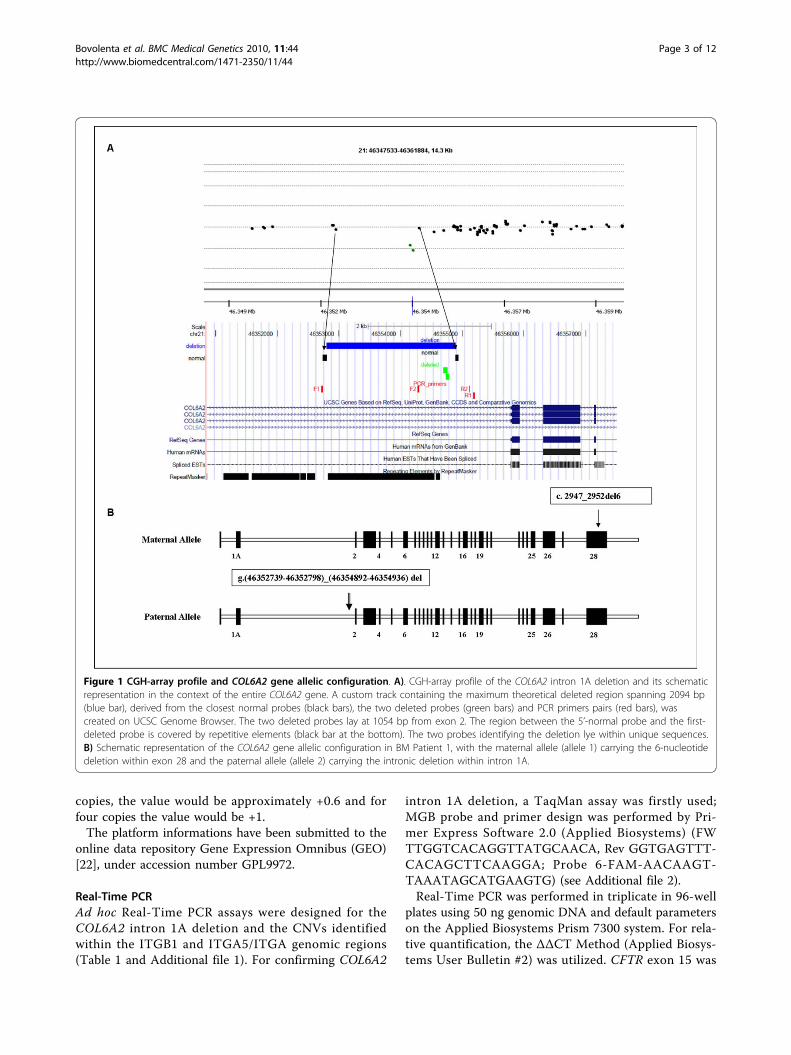
Figure 1 CGH-array profile and COL6A2 gene allelic configuration. A). CGH-array profile of the COL6A2 intron 1A deletion and its schematicrepresentation in the context of the entire COL6A2 gene. A custom track containing the maximum theoretical deleted region spanning 2094 bp(blue bar), derived from the closest normal probes (black bars), the two deleted probes (green bars) and PCR primers pairs (red bars), wascreated on UCSC Genome Browser. The two deleted probes lay at 1054 bp from exon 2. The region between the 5’-normal probe and the first-deleted probe is covered by repetitive elements (black bar at the bottom). The two probes identifying the deletion lye within unique sequences.B) Schematic representation of the COL6A2 gene allelic configuration in BM Patient 1, with the maternal allele (allele 1) carrying the 6-nucleotidedeletion within exon 28 and the paternal allele (allele 2) carrying the intronic deletion within intron 1A.
Bovolenta et al. BMC Medical Genetics 2010, 11:44http://www.biomedcentral.com/1471-2350/11/44
Page 3 of 12
employed as a reference gene, and three control DNAswere used as calibrators for each experiment. Specificassays based on SYBR-green chemistry were alsoemployed in order to further verify the intron 1A dele-tion and to confirm the CNVs identified in the ITGB1and ITGA5/ITGA7 genomic regions, (Chr10_FWCGTGGAGATGGGATTAGTGTG, Chr10_Rev TTTGTTGGGAATTTACTTGGTG; Chr12_FW AATTTGCTGTTGCTGGGTCT, Chr12_Rev TCCCATACTCTC-CATTGTCC; Chr21_FW GCCTGTCTGCCTCTTCCA,Chr21_Rev TGTTGCATAACCTGTGACCAA) (seeTable 1 and Additional file 2).
Transcript analysis in BM Patient 1Total RNA was isolated from confluent fibroblasts byusing an RNeasy Kit (QIAGEN, Chatsworth, CA), andreverse transcribed using a High Capacity cDNAReverse Transcription Kit (Applied Biosystem, FosterCity CA). RT-PCR was performed with a forward primerwithin COL6A2 exon 25 and a reverse primer within the5’ of exon 28, as well as with a forward primer withinexon 26 and a reverse within the 3’ of exon 28, as pre-viously described [23]. The sequence of primersemployed is available upon request.
Immunohistochemistry, electron microscopy and WesternBlot in BM Patient 1Unfixed frozen sections of the tibialis anterior musclefrom BM Patient 1 and control were labeled with anti-collagen VI antibody (Chemicon MAB1944) diluted1:100, followed by FITC-conjugated anti-mouse antibody(DAKO); sections were double-labeled with anti-perle-can antibody (Chemicon) diluted 1:100, and revealedwith a TRITC conjugated anti-rat antibody (SIGMA).Muscle sections were also labeled with anti-caveolin 3
(BD-Transduction), collagen IV, laminin a2 and lamininb1 chains (Chemicon), followed by FITC-conjugatedsecondary antibody, while fibronectin (Sigma) andalpha-dystroglycan (Upstate Biotechnologies) were fol-lowed by TRITC-conjugated antibody (Sigma, MO). Thefibroblast cultures from Patient 1 and from a controlwere obtained by mechanical means from bioptic skinfragments and set up as previously described [24].A mouse monoclonal anti-collagen VI antibody(MAB1944, Chemicon) was employed, and the resultingimmunoreaction was detected with a secondary FITC-conjugated anti-mouse antibody (DAKO). For immu-noelectron microscopy analysis, cells were incubatedwith a monoclonal anti-collagen VI antibody (Chemi-con) diluted 1:25 with Dulbecco’s modified Eagle’s med-ium and with 5 or 15-nm colloidal gold-labeled IgG(Amersham). Rotary shadowing electron microscopy wasperformed as described [25].Samples derived from skin fibroblast cultures and
muscle biopsies were prepared for Western Blot analysisas previously described [23]. Collagen VI was detectedby immunoblotting with antibodies recognizing eitherall collagen VI chains (Fitzgerald 70XR95) or the a1(VI)chain (Santa Cruz sc-20649) alone. Antibodies recogniz-ing AKT (Cell Signaling) and myosin (Sigma) were usedfor cell culture loading and muscle samples.
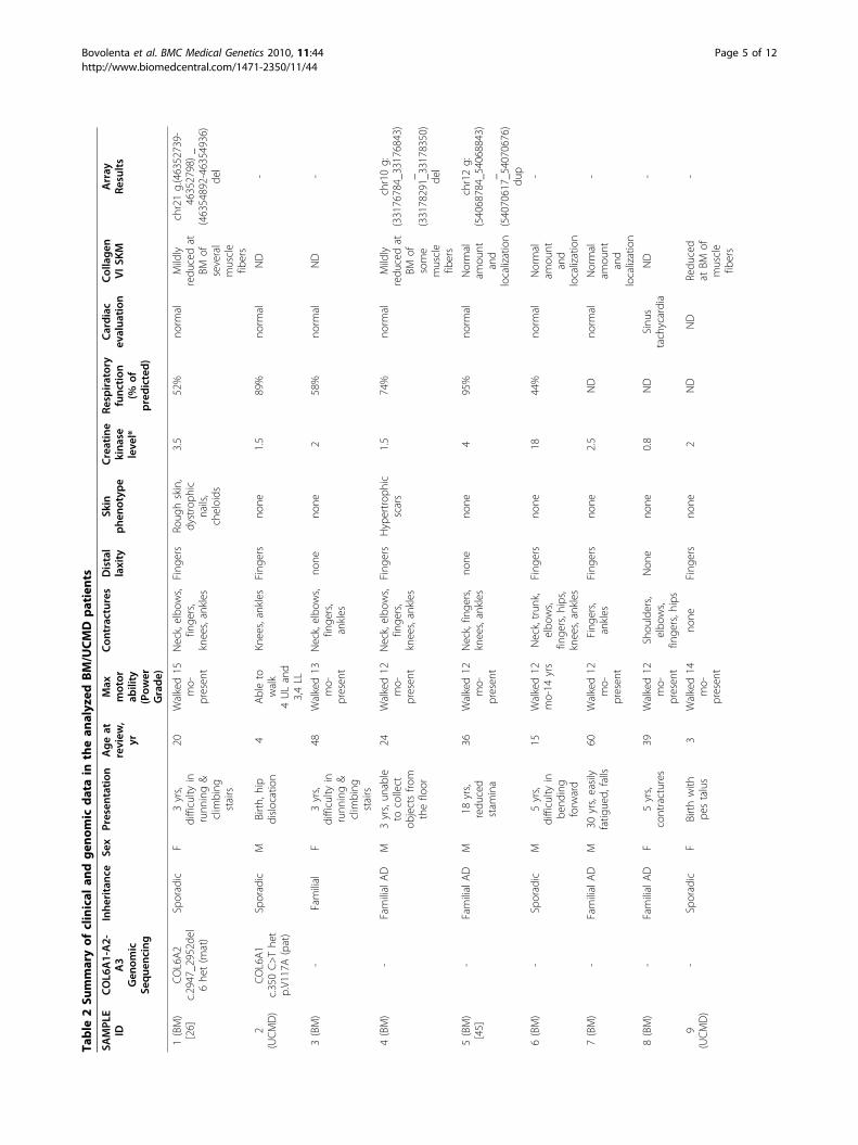
ResultsPatient selection and COL6-CGH array validationTwelve patients clinically diagnosed as possessingUCMD (6 patients) or BM (6 patients) phenotypes nega-tive at genomic sequence analysis, and 2 patients(1 UCMD, 1 BM) in whom a sequence analysis positivefor COL6 mutations failed to fully explain the clinicalphenotype were selected (Table 2).
Table 1 Genes, corresponding genomic regions and protein products included in the COL6-CGH micro-array design
Genes Chromosomal Coordinates (NCBI Build 35) Proteins
HSPG2 chr1:21894043-22010053 Heparan sulfate proteoglycan 2
COL6A3 Chr2:237914662-238204820 Collagen, type VI, alpha-3 chain
COL29A1;COL6A6
Chr3:131447049-131978580 Collagen, type XXIX, alpha 1Collagen type VI alpha 6
ITGA1;ITGA2
chr5:52019893-52526365 Integrin, alpha 1Integrin, alpha 2
TNXB chr6:32116611-32186183 Tenascin XB
ITGB1 chr10:33130501-33364492 Integrin, beta 1
DCN chr12:90041504-90075827 Decorin
ITGA5;ITGA7
chr12:52975314-54487894 Integrin, alpha 5;Integrin, alpha 7
CSPG4 chr15:73654019-73892151 Chondroitin sulfate proteoglycan 4
COL6A1;COL6A2
chr21:46126091-46474147 Collagen, type VI, alpha 1 chain Collagen, type VI, alpha 2 chain
BGN chrX:152181258-152395851 Biglycan
Bovolenta et al. BMC Medical Genetics 2010, 11:44http://www.biomedcentral.com/1471-2350/11/44
Page 4 of 12
Table
2Su
mmaryof
clinical
andgen
omic
datain
thean
alyzed
BM/UCMD
patients
SAMPL
EID
COL6
A1-A2-
A3
Gen
omic
Sequ
encing
Inhe
ritanc
eSe
xPresen
tation
Ageat
review
,yr
Max
motor
ability
(Pow
erGrade)
Con
tractures
Distal
laxity
Skin
phe
notype
Creatine
kina
seleve
l*
Respiratory
func
tion
(%of
predicted
)
Cardiac
evalua
tion
Collage
nVISK
MArray
Results
1(BM)
[26]
COL6A2
c.2947_2952d
el6he
t(m
at)
Sporadic
F3yrs,
difficulty
inrunn
ing&
clim
bing
stairs
20Walked15
mo-
presen
t
Neck,elbo
ws,
finge
rs,
knees,ankles
Fing
ers
Roug
hskin,
dystroph
icnails,
cheloids
3.5
52%
norm
alMildly
redu
cedat
BMof
several
muscle
fibers
chr21g.(46352739-
46352798)_
(46354892-46354936)
del
2(UCMD)
COL6A1
c.350C>The
tp.V117A(pat)
Sporadic
MBirth,
hip
dislocation
4Ableto
walk
4ULand
3,4LL
Knees,ankles
Fing
ers
none
1.5
89%
norm
alND
-
3(BM)
-Familial
F3yrs,
difficulty
inrunn
ing&
clim
bing
stairs
48Walked13
mo-
presen
t
Neck,elbo
ws,
finge
rs,
ankles
none
none
258%
norm
alND
-
4(BM)
-FamilialAD
M3yrs,un
able
tocollect
objectsfro
mthefloor
24Walked12
mo-
presen
t
Neck,elbo
ws,
finge
rs,
knees,ankles
Fing
ers
Hypertrop
hic
scars
1.5
74%
norm
alMildly
redu
cedat
BMof
some
muscle
fibers
chr10g:
(33176784_33176843)
_(33178291_33178350)
del
5(BM)
[45]
-FamilialAD
M18
yrs,
redu
ced
stam
ina
36Walked12
mo-
presen
t
Neck,finge
rs,
knees,ankles
none
none
495%
norm
alNormal
amou
ntand
localization
chr12g:
(54068784_54068843)
_(54070617_54070676)
dup
6(BM)
-Sporadic
M5yrs,
difficulty
inbe
nding
forw
ard
15Walked12
mo-14
yrs
Neck,trun
k,elbo
ws,
finge
rs,h
ips,
knees,ankles
Fing
ers
none
1844%
norm
alNormal
amou
ntand
localization
-
7(BM)
-FamilialAD
M30
yrs,easily
fatig
ued,
falls
60Walked12
mo-
presen
t
Fing
ers,
ankles
Fing
ers
none
2.5
ND
norm
alNormal
amou
ntand
localization
-
8(BM)
-FamilialAD
F5yrs,
contractures
39Walked12
mo-
presen
t
Shou
lders,
elbo
ws,
finge
rs,h
ips
Non
eno
ne0.8
ND
Sinu
stachycardia
ND
-
9(UCMD)
-Sporadic
FBirthwith
pestalus
3Walked14
mo-
presen
t
none
Fing
ers
none
2ND
ND
Redu
ced
atBM
ofmuscle
fibers
-
Bovolenta et al. BMC Medical Genetics 2010, 11:44http://www.biomedcentral.com/1471-2350/11/44
Page 5 of 12
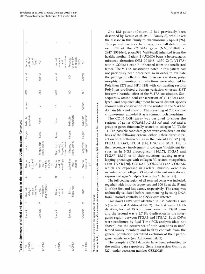
One BM patient (Patient 1) had previously beendescribed by Demir et al. (# 10, Family 8), who linkedthe disease in this family to chromosome 21q22.3 [26].This patient carries a heterozygous small deletion inexon 28 of the COL6A2 gene (NM_001849, c.2947_2952del6, p.Asp983_Val984del) inherited from thehealthy mother. Patient 2 (UCMD) bears a heterozygousmissense alteration (NM_001848, c.350 C>T, V117A)within COL6A1 exon 3, inherited from the unaffectedfather. The V117A substitution noted in this patient hadnot previously been described, so in order to evaluatethe pathogenic effect of this missense variation, poly-morphism phenotyping predictions were obtained byPolyPhen [27] and SIFT [28] with contrasting results:PolyPhen predicted a benign variation whereas SIFTforesaw a harmful effect of the V117A substitution. Sub-sequently, amino acid conservation of V117 was ana-lyzed, and sequence alignment between distant speciesshowed high conservation of the residue in the VWFA1domain (data not shown). The screening of 200 controlchromosomes excluded it as a common polymorphism.The COL6-CGH array was designed to cover the
regions of genes COL6A1-A2-A3-A5 and -A6 and agroup of genes functionally related to collagen VI (Table1). Ten possible candidate genes were considered on thebasis of the following criteria: either i) their direct inter-action with collagen VI, as in the case of HSPG2 [13],ITGA1, ITGA2, ITGB1 [14], DNC and BGN [15]; ii)their secondary involvement in collagen VI-deficient tis-sues, as in NG2-proteoglycan [16,17], ITGA5 andITGA7 [18,19], or iii) their mutations causing an over-lapping phenotype with collagen VI-related myopathies,as in TNXB [20]. COL6A5 (COL29A1) and COL6A6,which are expressed in skeletal muscle, were alsoincluded since collagen VI alpha1-deficient mice do notexpress collagen VI alpha 5 or alpha 6 chains [21].The full coding region of all selected genes was included,
together with intronic sequences and 100 kb at the 5’ and3’ of the first and last exons, respectively. The array wastechnically validated before commencing by using DNAfrom 8 normal controls; no CNVs were detected.Two novel CNVs were identified in BM patients 4 and
5 (Table 1 and Additional File 2). The first was a 1.4 Kbdeletion, located 35 Kb downstream the ITGB1 geneand the second was a 1.7 Kb duplication in the inter-genic region between ITGA5 and ITGA7. Both CNVswere confirmed by Real-Time PCR analysis (data notshown), but the occurrence of both variations in unaf-fected family members and healthy controls from thegeneral population permitted exclusion of their patho-genic significance (see Additional File 2).The complete CGH datasets have been submitted to
the online data repository Gene Expression Omnibus[22], under accession number GSE20025.Ta
ble
2:Su
mmaryof
clinical
andgen
omic
datain
thean
alyzed
BM/UCMD
patients
(Con
tinued)
10(UCMD)
[11]
-Sporadic
FBirthwith
delayin
motor
mileston
es
8Ableto
walk
4ULand
LL
none
Fing
ers
Follicular
hype
rplasia
norm
al85%
norm
alRedu
ced
atBM
ofmuscle
fibers
-
11(UCMD)
-Sporadic
M2yrs,
hype
rlaxity
andhype
rCK
8Ableto
walk
1hipflex-
3knees
ext
Fing
ers,hip,
knees,ankles
Non
eno
ne2
33%
norm
alND
-
12(UCMD)
-Sporadic
F18
mo,
unable
towalk
31Ableto
walk24
mo-12
yrs
Neck,trun
k,elbo
ws,
finge
rs,h
ips,
knees,ankles
Non
eno
ne1.5
62%
norm
alNormal
amou
ntand
localization
-
13(UCMD)
-Sporadic
FBirth,
flopp
iness
3Walked18
mo-
presen
t
Fing
ers,
knees,ankles
Fing
ers
Follicular
hype
rplasia
2ND
ND
Redu
ced
attheBM
ofmuscle
fibers
-
14(UCMD)
-Sporadic
MBirth
4Ableto
walkat
3yrs
Con
genital
kyph
osis
Fing
ers
Follicular
hype
rplasia
norm
alND
norm
alND
-
*Creatinekina
selevel:tim
estheup
pervalueof
norm
al.
UL:
uppe
rlim
bs;L
L:lower
limbs;A
D:a
utosom
aldo
minan
t;yrs:years;mo:
mon
ths;ND:n
otdo
ne;S
kM:skeletalmuscle;
BM:b
asem
entmem
bran
e
Bovolenta et al. BMC Medical Genetics 2010, 11:44http://www.biomedcentral.com/1471-2350/11/44
Page 6 of 12

Identification of a pure intronic deletion in intron 1A ofthe COL6A2 gene in BM patient 1In early infancy, Patient 1 (BM) (Table 2) attained nor-mal motor milestones and walked at 15 months of age.However, aged 3 and a half, she began to have difficultyrunning. Creatine kinase was 3.5 times the upper limitof normal, and examination at age 21 revealed a shortstature, excessive weight and bilateral club foot. Thepatient was able to walk and climb stairs without sup-port, but was unable to run. Mild facial and limb/girdleweakness and moderate axial and distal weakness werenoted. The patient was unable to bury the eyelashescompletely or to flex neck against gravity, and digitorumextension weakness was evident. The patient sufferedcontractures of the neck, elbows, fingers, knees andankles, combined with finger extension hypermobilitywhen the wrist was flexed. Her skin was rough and che-loidal, and nails were dystrophic. Forced vital capacitywas 52%, and cardiac examination evidenced noabnormality. Examination at age 30 years revealed arelatively stable condition, with only the ability to get upfrom the floor having been further compromised.In BM Patient 1, CGH analysis revealed the presence
of a deletion within intron 1A of the COL6A2 gene(NM_001849). This deletion was detected by two over-lapping probes which covered 95 base pairs: the5’-deleted probe lying 12.237 nucleotides from exon 1A,and the 3’-deleted probe located 999 nucleotides fromexon 2. The two flanking probes, which showed normalhybridization levels, were localized respectively, 1900nucleotides from the proximal 5’-deleted probe and 194nucleotides from the distal 3’-deleted probe. Thus, theidentified deletion spans a maximum theoretical regionof 2094 bp, and is located 1 kb upstream of the first
COL6A2 coding exon (exon 2). Comparative sequenceanalysis using the two deleted probes identified a geno-mic region lying adjacent to a cluster of repetitive ele-ments (Figure 1A) [29].Real-Time PCR analysis performed using two different
assays, one based on SYBR-green and the other on Taq-Man chemistries, confirmed the occurrence of the dele-tion, which was inherited from the healthy father (seeAdditional File 3). Analysis of 100 control subjects fromthe normal population by Real-Time PCR failed to iden-tify the COL6A2 intron deletion.Genotypic analysis of the proband’s parents showed
the intronic deletion and the exon 28 small mutationsin trans, denoting autosomal recessive transmission.An attempt to amplify by junction PCR the deletion
breakpoint with primers outside the maximum theoreti-cal deleted region was unsuccessful, and sequencinganalysis of the PCR products obtained using differentsets of primers failed to identify the mutated allele. Thissuggests the possibility of a complex rearrangement/inversion of the involved genomic region (Figure 1A).
Occurrence of monoallelic COL6A2 transcription in BMPatient 1 fibroblastsIn order to assess the pathogenic meaning of the identifiedintronic deletion found in BM Patient 1, fibroblasts wereharvested and cultured prior to RNA analysis. This analy-sis was focused on the terminal region of the COL6A2transcript, from exon 26 (harboring two common poly-morphisms (Ala698Ala and Gly699Gly), heterozygous atthe genomic analysis) to exon 28 (the site of the maternalheterozygous 6-nucleotide deletion). Direct sequencing ofthe amplified fragments revealed the presence of apseudo-homozygosity for the exon-28 deletion, as well as
Figure 2 COL6A2 genomic and transcript analysis in BM Patient 1 and his father. A) Sequence chromatograms of BM Patient 1 showingthe maternal deletion within exon 28 (GACGTG) (left) and two polymorphisms (c.2094 G>A - A698A; C.2097 C>T - G699G) within exon 26 (blackvertical arrows) (right) occurring heterozygously at the DNA level (upper panel) and pseudo-homozygously at the RNA level (lower panel). B)Sequence chromatograms in the BM Patient 1 father showing that the exon 26 polymorphisms (c.2094 G>A - A698A; C.2097 C>T - G699G) occurheterozygously at the DNA level (upper panel). At the RNA level (lower panel) the nucleotide variants that are undetectable in the BM Patient 1(c.2094 G and c.2097 C), are only barely visible in the father.
Bovolenta et al. BMC Medical Genetics 2010, 11:44http://www.biomedcentral.com/1471-2350/11/44
Page 7 of 12
for the polymorphisms within exon 26 (Figure 2). Thisimplies that the allele carrying the intronic deletion is nottranscribed at appreciable levels.In order to strengthen the case for a relationship
between the identified intronic deletion and the tran-scriptional behavior, RNA analysis was performed on cul-tured skin fibroblasts from the patient’s father. The twoexon 26 polymorphisms (Ala698Ala; Gly699Gly) werefound to be heterozygous at the genomic level, althoughtheir transcription was strongly imbalanced. The
nucleotide variant of the polymorphisms (c.2094 G>A -A698A; C.2097 C>T - G699G) that was completely unde-tectable in the proband’s RNA (G and C, respectively),was only barely visible in cells from the father (Figure 2).
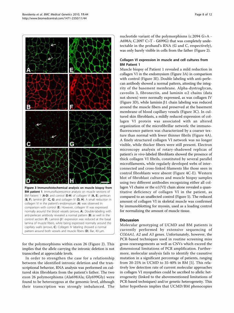
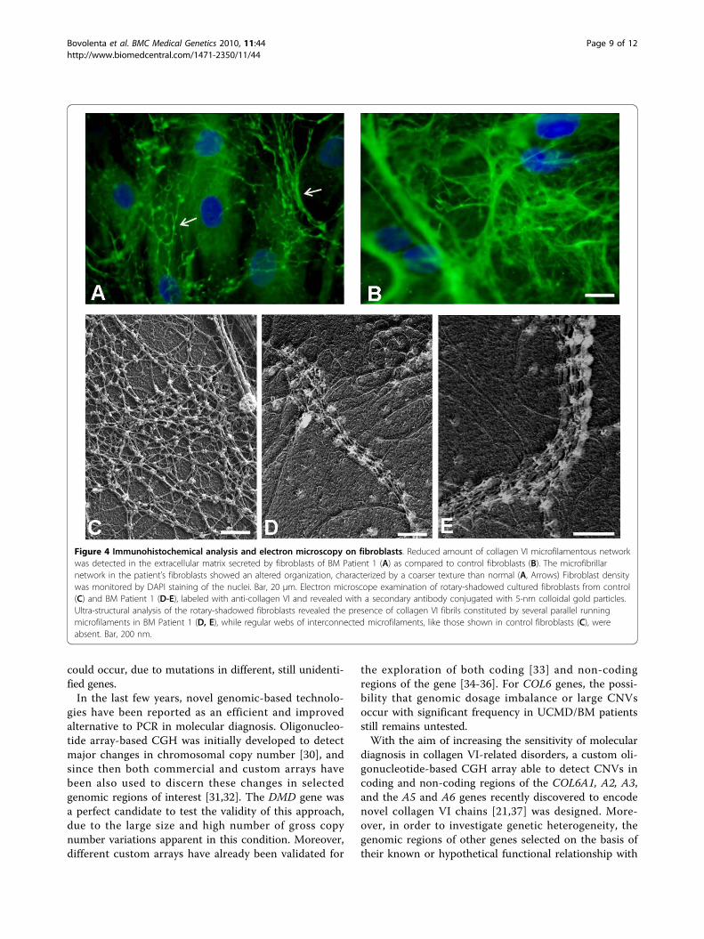
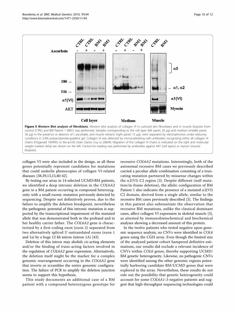
Collagen VI expression in muscle and cell cultures fromBM Patient 1Muscle biopsy of Patient 1 revealed a mild reduction incollagen VI in the endomysium (Figure 3A) in comparisonwith control (Figure 3E). Double labeling with anti-perle-can antibody showed a normal pattern, attesting the integ-rity of the basement membrane. Alpha-dystroglycan,caveolin 3, fibronectin, and laminin a2 chains (datanot shown) were normally expressed, as was collagen IV(Figure 3D), while laminin b1 chain labeling was reducedaround the muscle fibers and preserved at the basementmembrane of blood capillary vessels (Figure 3C). In cul-tured skin fibroblasts, a mildly reduced expression of col-lagen VI protein was associated with an alteredorganization of the microfibrillar network: the immuno-fluorescence pattern was characterized by a coarser tex-ture than normal with fewer thinner fibrils (Figure 4A).A finely structured collagen VI network was no longervisible, while thicker fibers were still present. Electronmicroscopy analysis of rotary-shadowed replicas ofpatient’s in vivo-labeled fibroblasts showed the presence ofthick collagen VI fibrils, constituted by several parallelmicrofilaments, while regularly developed webs of inter-connected and cross-linked filaments like those seen incontrol fibroblasts were absent (Figure 4C-E). Westernblot of fibroblast cultures and muscle biopsy samplesusing two different antibodies recognizing either all col-lagen VI chains or the a1(VI) chain alone revealed a quan-titative deficiency of collagen VI in the patient, ascompared to an unaffected control (Figure 5). The reducedamount of collagen VI in skeletal muscle was confirmedby immunoblotting for myosin, used as a loading controlfor normalizing the amount of muscle tissue.
DiscussionMolecular genotyping of UCMD and BM patients iscurrently performed by extensive sequencing ofCOL6A1, A2 and A3 genes. Unfortunately, however, thePCR-based techniques used in routine screening missgross rearrangements as well as CNVs which exceed thedimensional limitations of PCR amplification. Further-more, molecular analysis fails to identify the causativemutation in a significant percentage of patients, rangingfrom 20-25% in UCMD to 35-40% in BM [5]. This rela-tively low detection rate of current molecular approachesin collagen VI myopathies could be ascribed to allelic het-erogeneity (linked to the abovementioned limitations ofPCR-based techniques) and/or genetic heterogeneity. Thislatter hypothesis implies that UCMD/BM phenocopies
Figure 3 Immunohistochemical analysis on muscle biopsy fromBM patient 1. Immunofluorescence analysis on muscle sections ofBM Patient 1 (A-D) and control (E-H) of collagen VI (A, E), perlecan(B, F), laminin b1 (C, G) and collagen IV (D, H). A small reduction incollagen VI in the patient’s endomysium (A) was observed incomparison with control (E). However, collagen VI was expressednormally around the blood vessels (arrows, A). Double-labeling withanti-perlecan antibody revealed a normal pattern (B) as well in thecontrol section (F). Laminin b1 expression was reduced at the basallamina of muscle fibers, while being expressed normally around thecapillary walls (arrows, C). Collagen IV labeling showed a normalpattern around both vessels and muscle fibers (D). Bar, 40 μm.
Bovolenta et al. BMC Medical Genetics 2010, 11:44http://www.biomedcentral.com/1471-2350/11/44
Page 8 of 12
could occur, due to mutations in different, still unidenti-fied genes.In the last few years, novel genomic-based technolo-
gies have been reported as an efficient and improvedalternative to PCR in molecular diagnosis. Oligonucleo-tide array-based CGH was initially developed to detectmajor changes in chromosomal copy number [30], andsince then both commercial and custom arrays havebeen also used to discern these changes in selectedgenomic regions of interest [31,32]. The DMD gene wasa perfect candidate to test the validity of this approach,due to the large size and high number of gross copynumber variations apparent in this condition. Moreover,different custom arrays have already been validated for
the exploration of both coding [33] and non-codingregions of the gene [34-36]. For COL6 genes, the possi-bility that genomic dosage imbalance or large CNVsoccur with significant frequency in UCMD/BM patientsstill remains untested.With the aim of increasing the sensitivity of molecular
diagnosis in collagen VI-related disorders, a custom oli-gonucleotide-based CGH array able to detect CNVs incoding and non-coding regions of the COL6A1, A2, A3,and the A5 and A6 genes recently discovered to encodenovel collagen VI chains [21,37] was designed. More-over, in order to investigate genetic heterogeneity, thegenomic regions of other genes selected on the basis oftheir known or hypothetical functional relationship with
Figure 4 Immunohistochemical analysis and electron microscopy on fibroblasts. Reduced amount of collagen VI microfilamentous networkwas detected in the extracellular matrix secreted by fibroblasts of BM Patient 1 (A) as compared to control fibroblasts (B). The microfibrillarnetwork in the patient’s fibroblasts showed an altered organization, characterized by a coarser texture than normal (A, Arrows) Fibroblast densitywas monitored by DAPI staining of the nuclei. Bar, 20 μm. Electron microscope examination of rotary-shadowed cultured fibroblasts from control(C) and BM Patient 1 (D-E), labeled with anti-collagen VI and revealed with a secondary antibody conjugated with 5-nm colloidal gold particles.Ultra-structural analysis of the rotary-shadowed fibroblasts revealed the presence of collagen VI fibrils constituted by several parallel runningmicrofilaments in BM Patient 1 (D, E), while regular webs of interconnected microfilaments, like those shown in control fibroblasts (C), wereabsent. Bar, 200 nm.
Bovolenta et al. BMC Medical Genetics 2010, 11:44http://www.biomedcentral.com/1471-2350/11/44
Page 9 of 12
collagen VI were also included in the design, as all thesegenes potentially represent candidates for mutationsthat could underlie phenocopies of collagen VI-relateddiseases [38,39,13,15,40-42].By testing our array in 14 selected UCMD/BM patients,
we identified a deep intronic deletion in the COL6A2gene in a BM patient occurring in compound heterozyg-osity with a small exonic mutation previously detected bysequencing. Despite not definitively proven, due to thefailure to amplify the deletion breakpoint, neverthelessthe pathogenic potential of this intronic mutation is sup-ported by the transcriptional impairment of the mutatedallele that was demonstrated both in the proband and inher healthy carrier father. The COL6A2 gene is charac-terized by a first-coding exon (exon 2) separated fromtwo alternatively spliced 5’-untranslated exons (exon 1and 1a) by a huge 12 kb intron (intron 1A) [43].Deletion of this intron may abolish cis-acting elements
and/or the binding of trans-acting factors involved inthe regulation of COL6A2 gene expression. Alternatively,the deletion itself might be the marker for a complexgenomic rearrangement occurring in the COL6A2 genethat inverts or scrambles the entire genomic configura-tion. The failure of PCR to amplify the deletion junctionseems to support this hypothesis.This study documents an additional case of a BM
patient with a compound heterozygous genotype for
recessive COL6A2 mutations. Interestingly, both of theautosomal recessive BM cases we previously describedcarried a peculiar allele combination consisting of a trun-cating mutation partnered by missense changes withinthe a2(VI) C2 region [3]. Despite different (null muta-tion/in-frame deletion), the allelic configuration of BMPatient 1 also indicates the presence of a mutated a2(VI)C2 domain, derived from a single allele, similar to therecessive BM cases previously described [3]. The findingsin this patient also substantiate the observation thatrecessive BM mutations, unlike the classical dominantcases, affect collagen VI expression in skeletal muscle [3],as attested by immunohistochemical and biochemicalanalyses showing a decreased amount of this protein.In the twelve patients who tested negative upon geno-
mic sequence analysis, no CNVs were identified in COL6genes using the CGH array. Even though the limited sizeof the analyzed patient cohort hampered definitive esti-mations, our results did exclude a relevant incidence ofCNVs within COL6 genes, thereby supporting UCMD/BM genetic heterogeneity. Likewise, no pathogenic CNVswere identified among the other genomic regions poten-tially harboring candidate BM/UCMD genes that wereexplored in the array. Nevertheless, these results do notrule out the possibility that genetic heterogeneity couldaccount for some COL6A1-3-negative patients and sug-gest that high-throughput sequencing technologies could
Figure 5 Western Blot analysis of fibroblasts. Western blot analysis of collagen VI in cultured skin fibroblasts and in muscle biopsies fromcontrol (CTRL) and BM Patient 1 (BM1) was performed. Samples corresponding to the cell layer (left panel, 20 μg) and medium (middle panel,30 μg) in the presence or absence of L-ascorbate, and muscle extracts (right panel, 15 μg), were separated by electrophoresis under reducingconditions in 3-8% polyacrylamide-gradient gel. Collagen VI was detected by immunoblotting with antibodies recognizing either all collagen VIchains (Fitzgerald 70XR95) or the a1(VI) chain (Santa Cruz sc-20649). Migration of the collagen VI chains is indicated on the right and molecularweight markers (kDa) are shown on the left. Control for loading was performed by antibodies against AKT (cell layers) or myosin (musclebiopsies).
Bovolenta et al. BMC Medical Genetics 2010, 11:44http://www.biomedcentral.com/1471-2350/11/44
Page 10 of 12
represent more appropriate future approaches for detec-tion of point mutations. In fact, these innovative toolscould allow significant enlargement of the spectrum offunctionally related collagen VI genes to be exploredsequentially via CNV identification and re-sequencing fordetection of point mutations.
ConclusionsThe described COL6-CGH array could represent a use-ful complementary diagnostic test, useful for increasingthe sensitivity of molecular analysis in patients with aclinical diagnosis of collagen VI-related disorders. Thelimited size of the patient cohort we analyzed hamperedestimation of copy number variation frequency, but ana-lysis of larger populations could well permit conclusionsto be drawn. In fact, this novel tool allowed us for thefirst time to identify a COL6A2 mutation affecting genetranscription deeply located within an intronic region,and thus undetectable with all other techniques cur-rently available. Furthermore, in recent years specificgenetic diagnosis has become mandatory for a patient tobe eligible for upcoming therapeutic trials [44], and thusthe lack of molecular diagnosis in a large percentage ofpatients with collagen-VI related phenotypes makes thesearch and the validation of novel diagnostic tools anever-more pressing issue.
Additional file 1: Gene Symbols, chromosomal coordinates, transcriptand protein identifiers for the genes included in the COL6-CGH micro-array design.
Additional file 2: CNVs identified in BM Patients 4 and 5. A) COL6-CGH array result in BM Patient 4, showing the deletion of about 1.4 kbidentified on chromosome 10, 35 Kb downstream of the ITGB1 gene. B)The CNV on chromosome 10 was validated by Real-Time PCR, and itssegregation was analyzed in Patient 4’s family; the deletion was presentin three unaffected subjects (2-ΔΔCT values of 0.44, 0.41, 0.45) and absentin the symptomatic proband’s mother and cousin (2-ΔΔCT values of 0.99and 0.86), thus not linked to the disease. C) COL6-CGH array result in BMPatient 5, showing the 1.7 Kb duplication occurring in the intergenicregion between ITGA5 and ITGA7 on chromosome 12.
Additional file 3: Real Time PCR experiments confirming COL6A2intron 1A deletion in BM Patient 1 and in the father. In the upperpanel, the results obtained with an intron 1A specific TaqMan assay areshown. Red plots correspond to utilized reference gene (CFTR exon 15)whereas green plots refer to target sequence within intron 1A. The Ct(threshold cycle) values of the target sequence are in line with thereference in control sample (unaffected subject) and in proband’smother, whereas the target Ct values are higher than reference in BMPatient 1 and in the father, attesting the deletion (2-ΔΔCT values were0.47 and 0.53 in BM Patient 1 and in the father respectively, whereas the2-ΔΔCT value was 0.97 in the proband’s mother). In the lower panel theposition of the primers utilized in the SYBR green assay (blu) and ofprimers and probe utilized in the TaqMan assay (pink), are shown inrespect to the deleted region (in bold).
AcknowledgementsThis study was supported by the Italian Telethon Foundation grantsGUP07004 (to GF). The Telethon grant GGP08107 (awarded to AF and PB)
and the TREAT-NMD Network of Excellence of EU FP7 (awarded 036825 toLM and Telethon-Italy), are also acknowledged.
Author details1Department of Experimental and Diagnostic Medicine - Section of MedicalGenetics, University of Ferrara, Ferrara, Italy. 2Department of Histology,Microbiology and Medical Biotechnologies, University of Padua, Padua, Italy.3IGM-CNR, Unit of Bologna c/o IOR, Bologna, Italy. 4Department of ChildNeurology and Psychiatry, Catholic University, Rome, Italy. 5Unit of MolecularMedicine, Department of Laboratory Medicine, Bambino Gesu’ Hospital,Rome, Italy. 6Laboratory of Biology, IOR, Bologna, Italy.
Authors’ contributionsMB designed the array and carried out DNA extraction and hybridization ofsamples, Real-time PCR experiments, and analysis and interpretation of data;MN performed DNA extraction and hybridization of samples, Real-time PCRexperiments, and contributed to preparation of the manuscript; EMperformed genomic and transcript analyses; MF prepared fibroblasts cellcultures; AU and PG carried out Western Blotting; PS performed ICC analysis;EM and EB carried out clinical evaluation of the patients; LM clinicallyevaluated the patients and participated in revision of the manuscript; PBassisted in the interpretation of data and revision of the manuscript; AFcontributed to conception of the study and preparation of the manuscript;FG conceived and designed the study, prepared and revised the manuscriptand approved the final version.
Competing interestsThe authors declare that they have no competing interests.
Received: 4 September 2009 Accepted: 19 March 2010Published: 19 March 2010
References1. Lampe AK, Bushby KM: Collagen VI related muscle disorders. J Med Genet
2005, 42:673-85.2. Bertini E, Pepe G: Collagen type VI and related disorders: Bethlem
myopathy and Ullrich scleroatonic muscular dystrophy. Eur J PaediatrNeurol 2002, 6:193-198.
3. Gualandi F, Urciuolo A, Martoni E, Sabatelli P, Squarzoni S, Bovolenta M,Messina S, Mercuri E, Franchella A, Ferlini A, Bonaldo P, Merlini L:Autosomal recessive Bethlem myopathy. Neurology 2009, 73(22):1883-91.
4. Foley AR, Hu Y, Zou Y, Columbus A, Shoffner J, Dunn DM, Weiss RB,Bönnemann CG: Autosomal recessive inheritance of classic Bethlemmyopathy. Neuromuscul Disord 2009, 19:813-7.
5. Lampe AK, Dunn DM, von Niederhausern AC, Hamil C, Aoyagi A, Laval SH,Marie SK, Chu ML, Swoboda K, Muntoni F, Bonnemann CG, Flanigan KM,Bushby KM, Weiss RB: Automated genomic sequence analysis of thethree collagen VI genes: applications to Ullrich congenital musculardystrophy and Bethlem myopathy. J Med Genet 2005, 42:108-20.
6. Baker NL, Morgelin M, Peat R, Goemans N, North KN, Bateman JF,Lamande SR: Dominant collagen VI mutations are a common cause ofUllrich congenital muscular dystrophy. Hum Mol Genet 2005, 14:279-93.
7. Giusti B, Lucarini L, Pietroni V, Lucioli S, Bandinelli B, Sabatelli P, Squarzoni S,Petrini S, Gartioux C, Talim B, Roelens F, Merlini L, Topaloglu H, Bertini E,Guicheney P, Pepe G: Dominant and recessive COL6A1 mutations inUllrich scleroatonic muscular dystrophy. Ann Neurol 2005, 58:400-10.
8. Leiden Pages. [http://www.dmd.nl/].9. Pepe G, Lucarini L, Zhang RZ, Pan TC, Giusti B, Quijano-Roy S, Gartioux C,
Bushby KM, Guicheney P, Chu ML: COL6A1 genomic deletions in Bethlemmyopathy and Ullrich muscular dystrophy. Ann Neurol 2006, 59:190-5.
10. Pan TC, Zhang RZ, Sudano DG, Marie SK, Bonnemann CG, Chu ML: Newmolecular mechanism for Ullrich congenital muscular dystrophy: aheterozygous in-frame deletion in the COL6A1 gene causes a severephenotype. Am J Hum Genet 2003, 73:355-69.
11. Petrini S, D’Amico A, Sale P, Lucarini L, Sabatelli P, Tessa A, Giusti B,Verardo M, Carrozzo R, Mattioli E, Scarpelli M, Chu ML, Pepe G, Russo MA,Bertini E: Ullrich myopathy phenotype with secondary ColVI defectidentified by confocal imaging and electron microscopy analysis.Neuromuscul Disord 2007, 17:587-96.
12. Agilent Technologies eArray. [https://earray.chem.agilent.com/earray].
Bovolenta et al. BMC Medical Genetics 2010, 11:44http://www.biomedcentral.com/1471-2350/11/44
Page 11 of 12

13. Tillet E, Wiedemann H, Golbik R, Pan TC, Zhang RZ, Mann K, Chu ML,Timpl R: Recombinant expression and structural and binding propertiesof alpha 1(VI) and alpha 2(VI) chains of human collagen type VI. Eur JBiochem 1994, 221:177-85.
14. Pfaff M, Aumailley M, Specks U, Knolle J, Zerwes HG, Timpl R: Integrin andArg-Gly-Asp dependence of cell adhesion to the native and unfoldedtriple helix of collagen type VI. Exp Cell Res 1993, 206:167-76.
15. Wiberg C, Hedbom E, Khairullina A, Lamandé SR, Oldberg A, Timpl R,Mörgelin M, Heinegård D: Biglycan and decorin bind close to the n-terminal region of the collagen VI triple helix. J Biol Chem 2001,276:18947-52.
16. Petrini S, Tessa A, Stallcup WB, Sabatelli P, Pescatori M, Giusti B, Carrozzo R,Verardo M, Bergamin N, Columbaro M, Bernardini C, Merlini L, Pepe G,Bonaldo P, Bertini E: Altered expression of the MCSP/NG2 chondroitinsulfate proteoglycan in collagen VI deficiency. Mol Cell Neurosci 2005,30:408-17.
17. Higashi K, Higuchi I, Niiyama T, Uchida Y, Shiraishi T, Hashiguchi A, Saito A,Horikiri T, Suehara M, Arimura K, Osame M: Abnormal expression ofproteoglycans in Ullrich’s disease with collagen VI deficiency. MuscleNerve 2006, 33:120-6.
18. Hu J, Higuchi I, Shiraishi T, Suehara M, Niiyama T, Horikiri T, Uchida Y,Saito A, Osame M: Fibronectin receptor reduction in skin and fibroblastsof patients with Ullrich’s disease. Muscle Nerve 2002, 26:696-701.
19. Pepe G, Bertini E, Bonaldo P, Bushby K, Giusti B, de Visser M, Guicheney P,Lattanzi G, Merlini L, Muntoni F, Nishino I, Nonaka I, Yaou RB, Sabatelli P,Sewry C, Topaloglu H, Kooi van der A: Bethlem myopathy (BETHLEM) andUllrich scleroatonic muscular dystrophy: 100th ENMC internationalworkshop 2001, 23-24 November Naarden, The Netherlands.Neuromuscul Disord 2002, 12:984-93.
20. Voermans NC, Jenniskens GJ, Hamel BC, Schalkwijk J, Guicheney P, vanEngelen BG: Ehlers-Danlos syndrome due to tenascin-X deficiency:muscle weakness and contractures support overlap with collagen VImyopathies. Am J Med Genet A 2007, 143A:2215-9.
21. Gara SK, Grumati P, Urciuolo A, Bonaldo P, Kobbe B, Koch M, Paulsson M,Wagener R: Three novel collagen VI chains with high homology to thealpha3 chain. J Biol Chem 2008, 283:10658-70.
22. Gene Expression Omnibus. [http://www.ncbi.nlm.nih.gov/geo/].23. Merlini L, Martoni E, Grumati P, Sabatelli P, Squarzoni S, Urciuolo A, Ferlini A,
Gualandi F, Bonaldo P: Autosomal recessive myosclerosis myopathy is acollagen VI disorder. Neurology 2008, 71:1245-53.
24. Martoni E, Urciuolo A, Sabatelli P, Fabris M, Bovolenta M, Neri M, Grumati P,D’Amico A, Pane M, Mercuri E, Bertini E, Merlini L, Bonaldo P, Ferlini A,Gualandi F: Identification and characterization of novel collagen VI non-canonical splicing mutations causing ullrich congenital musculardystrophy. Hum Mutat 2009, 30:E662-72.
25. Zhang RZ, Sabatelli P, Pan TC, Squarzoni S, Mattioli E, Bertini E, Pepe G,Chu ML: Effects on collagen VI mRNA stability and microfibrillarassembly of three COL6A2 mutations in two families with Ullrichcongenital muscular dystrophy. J Biol Chem 2002, 277:43557-64.
26. Demir E, Ferreiro A, Sabatelli P, Allamand V, Makri S, Echenne B, Maraldi M,Merlini L, Topaloglu H, Guicheney P: Collagen VI status and clinicalseverity in Ullrich congenital muscular dystrophy: phenotype analysis of11 families linked to the COL6 loci. Neuropediatrics 2004, 35:103-12.
27. Polyphen. [http://genetics.bwh.harvard.edu/pph/].28. SIFT. [http://sift.jcvi.org/www/SIFT_chr_coords_submit.html].29. Repeat Masker. [http://www.repeatmasker.org/].30. Lu X, Shaw CA, Patel A, Li J, Cooper ML, Wells WR, Sullivan CM, Sahoo T,
Yatsenko SA, Bacino CA, Stankiewicz P, Ou Z, Chinault AC, Beaudet AL, Lupski JR,Cheung SW, Ward PA: Clinical implementation of chromosomal microarrayanalysis: summary of 2513 postnatal cases. PLoS One 2007, 2:e327.
31. Wong LJ, Dimmock D, Geraghty MT, Quan R, Lichter-Konecki U, Wang J,Brundage EK, Scaglia F, Chinault AC: Utility of oligonucleotide array-basedcomparative genomic hybridization for detection of target genedeletions. Clin Chem 2008, 54:1141-8.
32. Gunn SR, Robetorye RS, Mohammed MS: Comparative genomichybridization arrays in clinical pathology: progress and challenges. MolDiagn Ther 2007, 11:73-7.
33. Dhami P, Coffey AJ, Abbs S, Vermeesch JR, Dumanski JP, Woodward KJ,Andrews RM, Langford C, Vetrie D: Exon array CGH: detection of copy-number changes at the resolution of individual exons in the humangenome. Am J Hum Genet 2005, 76:750-762.
34. Hegde MR, Chin EL, Mulle JG, Okou DT, Warren ST, Zwick ME: Microarray-based mutation detection in the dystrophin gene. Human mutation 2008,29:1091-9.
35. Saillour Y, Cossée M, Leturcq F, Vasson A, Beugnet C, Poirier K, Commere V,Sublemontier S, Viel M, Letourneur F, Barbot JC, Deburgrave N, Chelly J,Bienvenu T: Detection of exonic copy-number changes using a highlyefficient oligonucleotide-based comparative genomic hybridization-arraymethod. Human mutation 2008, 29:1083-90.
36. Bovolenta M, Neri M, Fini S, Fabris M, Trabanelli C, Venturoli A, Martoni E,Bassi E, Spitali P, Brioschi S, Falzarano MS, Rimessi P, Ciccone R, Ashton E,McCauley J, Yau S, Abbs S, Muntoni F, Merlini L, Gualandi F, Ferlini A: Anovel custom high density-comparative genomic hybridization arraydetects common copy number variations as well as deep intronicmutations in dystrophinopathies. BMC genomics 2008, 9:572.
37. Fitzgerald J, Rich C, Zhou FH, Hansen U: Three novel collagen VI chains,alpha4(VI), alpha5(VI), and alpha6(VI). J Biol Chem 2008, 283:20170-80.
38. Bonaldo P, Russo V, Bucciotti F, Doliana R, Colombatti A: Structural andfunctional features of the alpha 3 chain indicate a bridging role forchicken collagen VI in connective tissues. Biochemistry 1990, 29:1245-54.
39. Bidanset DJ, Guidry C, Rosenberg LC, Choi HU, Timpl R, Hook M: Binding ofthe proteoglycan decorin to collagen type VI. J Biol Chem 1992,267:5250-6.
40. Merlini L, Villanova M, Sabatelli P, Malandrini A, Maraldi NM: Decreasedexpression of laminin beta 1 in chromosome 21-linked Bethlemmyopathy. Neuromuscul Disord 1999, 9:326-9.
41. Sabatelli P, Bonaldo P, Lattanzi G, Braghetta P, Bergamin N, Capanni C,Mattioli E, Columbaro M, Ognibene A, Pepe G, Bertini E, Merlini L,Maraldi NM, Squarzoni S: Collagen VI deficiency affects the organizationof fibronectin in the extracellular matrix of cultured fibroblasts. MatrixBiol 2001, 20:475-86.
42. Scacheri PC, Gillanders EM, Subramony SH, Vedanarayanan V, Crowe CA,Thakore N, Bingler M, Hoffman EP: Novel mutations in collagen VI genes:expansion of the Bethlem myopathy phenotype. Neurology 2002,58:593-602.
43. Saitta B, Timpl R, Chu ML: Human alpha 2(VI) collagen gene.Heterogeneity at the 5’-untranslated region generated by an alternateexon. J Biol Chem 1992, 267:6188-96.
44. Merlini L, Angelin A, Tiepolo T, Braghetta P, Sabatelli P, Zamparelli A,Ferlini A, Maraldi NM, Bonaldo P, Bernardi P: Cyclosporin A correctsmitochondrial dysfunction and muscle apoptosis in patients withcollagen VI myopathies. Proc Natl Acad Sci USA 2008, 105:5225-9.
45. Merlini L, Morandi L, Granata C, Ballestrazzi A: Bethlem myopathy: early-onset benign autosomal dominant myopathy with contractures.Description of two new families. Neuromuscul Disord 1994, 4:503-11.
Pre-publication historyThe pre-publication history for this paper can be accessed here:[http://www.biomedcentral.com/1471-2350/11/44/prepub]
doi:10.1186/1471-2350-11-44Cite this article as: Bovolenta et al.: Identification of a deep intronicmutation in the COL6A2 gene by a novel custom oligonucleotide CGHarray designed to explore allelic and genetic heterogeneity in collagenVI-related myopathies. BMC Medical Genetics 2010 11:44.
Submit your next manuscript to BioMed Centraland take full advantage of:
• Convenient online submission
• Thorough peer review
• No space constraints or color figure charges
• Immediate publication on acceptance
• Inclusion in PubMed, CAS, Scopus and Google Scholar
• Research which is freely available for redistribution
Submit your manuscript at www.biomedcentral.com/submit
Bovolenta et al. BMC Medical Genetics 2010, 11:44http://www.biomedcentral.com/1471-2350/11/44
Page 12 of 12