Molecular mechanism for distinct neurological phenotypes conveyed by allelic truncating mutations
9
ARTICLES Different mutations in the same gene often cause distinct disease phe- notypes in humans. Generally, such variations in the clinical pheno- types have been considered to be a consequence of the function or dysfunction of mutant proteins. Thus, a primary emphasis in geno- type-phenotype correlation studies has been placed on determining the unique functional properties of encoded mutant proteins. But in vitro functional assays of mutant proteins often show discordance between predicted protein function and clinical outcome. Little is known about the many factors that are potentially involved in this dis- crepancy, but loss-of-function versus gain-of-function effects are often invoked as a possible mechanism. We previously identified two unrelated individuals with an unusual phenotype that combined four distinct syndromesperipheral demyelinating neuropathy, central dysmyelinating leukodystrophy, Waardenburg syndrome and Hirschsprung disease—that are charac- terized by deficiencies of Schwann cells, oligodendrocytes, melanocytes and enteric ganglia neurons, respectively 1,2 . Here we describe four more individuals and propose that this complex dis- order is a newly described neurocristopathy called PCWH. We previously identified mutations in SOX10 in all affected individ- uals 1,2 . SOX10 is a transcription factor that contains a central high mobility group (HMG) DNA-binding domain and a transactivation domain at its C terminus 3 . SOX10 is essential for the development of cells in the neural crest lineage, including melanocytes and enteric ganglia neurons 4,5 ; it also controls the proliferation and differentiation of Schwann cells and oligodendrocytes 6–8 . Notably, some mutations in SOX10 also cause a distinct and more restricted disease that does not involve either the peripheral (PNS) or the central (CNS) nervous sys- tems 9–11 . This less complicated neurocristopathy, called WS4, com- bines Waardenburg and Hirschsprung diseases 12 . Most SOX10 disease-associated mutations, regardless of whether they cause PCWH or WS4, result in premature termination codons (PTCs). As in SOX10, different mutations in MPZ are responsible for dis- tinct neurological diseases, which each affect the myelin of the PNS. These neuropathies include early onset congenital hypomyelinating neuropathy (CHN; OMIM 605253), Dejerine-Sottas neuropathy (DSN; OMIM 145900) and the less severe, adult onset Charcot-Marie- Tooth disease type 1B (CMT1B; OMIM 118200; ref. 13). It has been suggested that the severity of alleles in CHN and DSN is due to domi- nant-negative effects, whereas the reduced severity of alleles in CMT1B is due to loss of function. But although some nonsense and frameshift alleles cause CMT1B, several truncating mutations have been reported that convey either a CHN or a DSN phenotype. We investigated the molecular mechanisms underlying the neuro- logical phenotypes of the PCWH and WS4 neurocristopathies result- ing from allelic SOX10 truncating mutations, as well as those underlying the CHN, DSN and CMT1B myelinopathies caused by allelic MPZ truncating mutations. Unexpectedly, we found that the function of the truncated SOX10 proteins has little effect on clinical outcome. Essentially all truncated SOX10 proteins, irrespective of their associated phenotypes, had a similar dominant-negative effect in vitro. 1 Department of Molecular and Human Genetics, Baylor College of Medicine, One Baylor Plaza, Room 604B, Houston, Texas 77030, USA. 2 Division of Pediatric Neurology, Nagano Children’s Hospital, Nagano, Japan. 3 Department of Neurology, Great Ormond Street Children’s Hospital, London, UK. 4 Meritcare Neuroscience Clinic, Fargo, North Dakota, USA. 5 Department of Neurology, The University of Texas Medical School at Houston, Houston, Texas, USA. 6 Department of Immunology, The University of Texas M.D. Anderson Cancer Center, Houston, Texas, USA. 7 Institut für Biochemie, Universität Erlangen-Nürnberg, Erlangen, Germany. 8 Department of Pediatrics, Baylor College of Medicine and 9 Texas Children’s Hospital, Houston, Texas, USA. 10 Present address: Department of Mental Retardation and Birth Defect Research, National Institute of Neuroscience, National Center of Neurology and Psychiatry, 4-1-1 Ogawahigashi, Kodaira, Tokyo 187-8502, Japan. 11 These authors contributed equally to this work. Correspondence should be addressed to J.R.L. ([email protected]). Published online 7 March 2004; doi:10.1038/ng1322 Molecular mechanism for distinct neurological phenotypes conveyed by allelic truncating mutations Ken Inoue 1,10,11 , Mehrdad Khajavi 1,11 , Tomoko Ohyama 1 , Shin-ichi Hirabayashi 2 , John Wilson 3 , James D Reggin 4 , Pedro Mancias 5 , Ian J Butler 5 , Miles F Wilkinson 6 , Michael Wegner 7 & James R Lupski 1,8,9 The molecular mechanisms by which different mutations in the same gene can result in distinct disease phenotypes remain largely unknown. Truncating mutations of SOX10 cause either a complex neurocristopathy designated PCWH or a more restricted phenotype known as Waardenburg-Shah syndrome (WS4; OMIM 277580). Here we report that although all nonsense and frameshift mutations that cause premature termination of translation generate truncated SOX10 proteins with potent dominant- negative activity, the more severe disease phenotype, PCWH, is realized only when the mutant mRNAs escape the nonsense- mediated decay (NMD) pathway. We observe similar results for truncating mutations of MPZ that convey distinct myelinopathies. Our experiments show that triggering NMD and escaping NMD may cause distinct neurological phenotypes. NATURE GENETICS VOLUME 36 | NUMBER 4 | APRIL 2004 361 © 2004 Nature Publishing Group http://www.nature.com/naturegenetics
-
Upload
independent -
Category
Documents
-
view
4 -
download
0
Transcript of Molecular mechanism for distinct neurological phenotypes conveyed by allelic truncating mutations
A R T I C L E S
Different mutations in the same gene often cause distinct disease phe-notypes in humans. Generally, such variations in the clinical pheno-types have been considered to be a consequence of the function ordysfunction of mutant proteins. Thus, a primary emphasis in geno-type-phenotype correlation studies has been placed on determiningthe unique functional properties of encoded mutant proteins. But invitro functional assays of mutant proteins often show discordancebetween predicted protein function and clinical outcome. Little isknown about the many factors that are potentially involved in this dis-crepancy, but loss-of-function versus gain-of-function effects are ofteninvoked as a possible mechanism.
We previously identified two unrelated individuals with an unusualphenotype that combined four distinct syndromes peripheraldemyelinating neuropathy, central dysmyelinating leukodystrophy,Waardenburg syndrome and Hirschsprung disease—that are charac-terized by deficiencies of Schwann cells, oligodendrocytes,melanocytes and enteric ganglia neurons, respectively1,2. Here wedescribe four more individuals and propose that this complex dis-order is a newly described neurocristopathy called PCWH.
We previously identified mutations in SOX10 in all affected individ-uals1,2. SOX10 is a transcription factor that contains a central highmobility group (HMG) DNA-binding domain and a transactivationdomain at its C terminus3. SOX10 is essential for the development ofcells in the neural crest lineage, including melanocytes and entericganglia neurons4,5; it also controls the proliferation and differentiationof Schwann cells and oligodendrocytes6–8. Notably, some mutations in
SOX10 also cause a distinct and more restricted disease that does notinvolve either the peripheral (PNS) or the central (CNS) nervous sys-tems9–11. This less complicated neurocristopathy, called WS4, com-bines Waardenburg and Hirschsprung diseases12. Most SOX10disease-associated mutations, regardless of whether they cause PCWHor WS4, result in premature termination codons (PTCs).
As in SOX10, different mutations in MPZ are responsible for dis-tinct neurological diseases, which each affect the myelin of the PNS.These neuropathies include early onset congenital hypomyelinatingneuropathy (CHN; OMIM 605253), Dejerine-Sottas neuropathy(DSN; OMIM 145900) and the less severe, adult onset Charcot-Marie-Tooth disease type 1B (CMT1B; OMIM 118200; ref. 13). It has beensuggested that the severity of alleles in CHN and DSN is due to domi-nant-negative effects, whereas the reduced severity of alleles inCMT1B is due to loss of function. But although some nonsense andframeshift alleles cause CMT1B, several truncating mutations havebeen reported that convey either a CHN or a DSN phenotype.
We investigated the molecular mechanisms underlying the neuro-logical phenotypes of the PCWH and WS4 neurocristopathies result-ing from allelic SOX10 truncating mutations, as well as thoseunderlying the CHN, DSN and CMT1B myelinopathies caused byallelic MPZ truncating mutations. Unexpectedly, we found that thefunction of the truncated SOX10 proteins has little effect on clinicaloutcome. Essentially all truncated SOX10 proteins, irrespective oftheir associated phenotypes, had a similar dominant-negative effectin vitro.
1Department of Molecular and Human Genetics, Baylor College of Medicine, One Baylor Plaza, Room 604B, Houston, Texas 77030, USA. 2Division of PediatricNeurology, Nagano Children’s Hospital, Nagano, Japan. 3Department of Neurology, Great Ormond Street Children’s Hospital, London, UK. 4Meritcare NeuroscienceClinic, Fargo, North Dakota, USA. 5Department of Neurology, The University of Texas Medical School at Houston, Houston, Texas, USA. 6Department of Immunology,The University of Texas M.D. Anderson Cancer Center, Houston, Texas, USA. 7Institut für Biochemie, Universität Erlangen-Nürnberg, Erlangen, Germany. 8Departmentof Pediatrics, Baylor College of Medicine and 9Texas Children’s Hospital, Houston, Texas, USA. 10Present address: Department of Mental Retardation and Birth DefectResearch, National Institute of Neuroscience, National Center of Neurology and Psychiatry, 4-1-1 Ogawahigashi, Kodaira, Tokyo 187-8502, Japan. 11These authorscontributed equally to this work. Correspondence should be addressed to J.R.L. ([email protected]).
Published online 7 March 2004; doi:10.1038/ng1322
Molecular mechanism for distinct neurologicalphenotypes conveyed by allelic truncating mutationsKen Inoue1,10,11, Mehrdad Khajavi1,11, Tomoko Ohyama1, Shin-ichi Hirabayashi2, John Wilson3, James D Reggin4,Pedro Mancias5, Ian J Butler5, Miles F Wilkinson6, Michael Wegner7 & James R Lupski1,8,9
The molecular mechanisms by which different mutations in the same gene can result in distinct disease phenotypes remain largelyunknown. Truncating mutations of SOX10 cause either a complex neurocristopathy designated PCWH or a more restrictedphenotype known as Waardenburg-Shah syndrome (WS4; OMIM 277580). Here we report that although all nonsense andframeshift mutations that cause premature termination of translation generate truncated SOX10 proteins with potent dominant-negative activity, the more severe disease phenotype, PCWH, is realized only when the mutant mRNAs escape the nonsense-mediated decay (NMD) pathway. We observe similar results for truncating mutations of MPZ that convey distinct myelinopathies.Our experiments show that triggering NMD and escaping NMD may cause distinct neurological phenotypes.
NATURE GENETICS VOLUME 36 | NUMBER 4 | APRIL 2004 361
©20
04 N
atur
e P
ublis
hing
Gro
up
http
://w
ww
.nat
ure.
com
/nat
ureg
enet
ics
A R T I C L E S
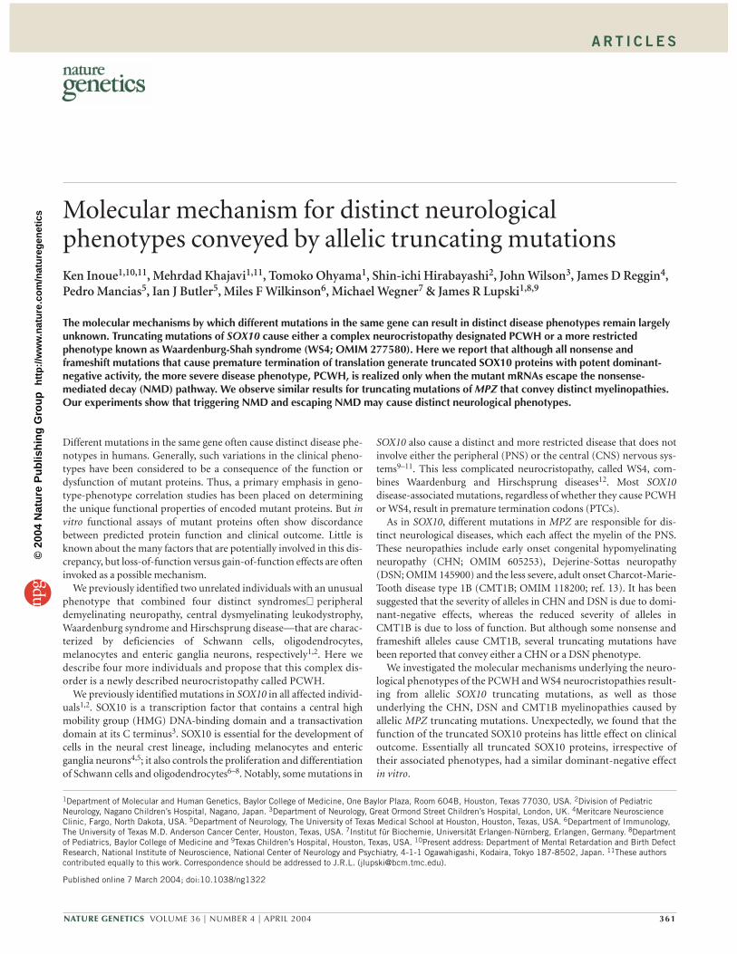
We also examined SOX10 and MPZ mRNAs that contain truncatingmutations resulting in distinct neurological phenotypes. Our findingsshow that mutant mRNAs that result in a less severe disease haddecreased stability, suggesting that the underlying mechanism is hap-loinsufficiency. By contrast, the more severe neurological diseaseseemed to result from a stable mRNA that is translated into a mutantprotein with potent dominant-negative activity. We show that theunstable mRNAs are degraded by the NMD pathway. Thus, NMD isresponsible for distinct neurological phenotypes arising from allelictruncating mutations and, by extension, may contribute to allelicaffinity where multiple diseases have been associated with differentmutations in the same gene.
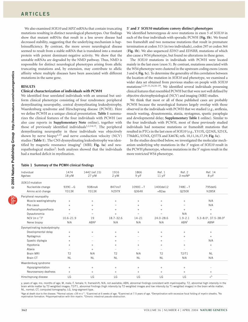
RESULTSClinical characterization of individuals with PCWHWe identified four unrelated individuals with an unusual but uni-form clinical phenotype consisting of four syndromes: peripheraldemyelinating neuropathy, central dysmyelinating leukodystrophy,Waardenburg syndrome and Hirschsprung disease (PCWH). Herewe define PCWH as a unique clinical presentation. Table 1 summa-rizes the clinical features of the four individuals with PCWH (seealso case reports in Supplementary Note online), together withthose of previously identified individuals1,2,14,15. The peripheraldemyelinating neuropathy in these individuals was objectivelyshown by nerve biopsy2,15 and nerve conduction velocity (NCV)studies (Table 1). The CNS dysmyelinating leukodystrophy was iden-tified by magnetic resonance imaging1 (MRI; Fig. 1a) and neu-ropathological studies2: both analyses showed that the individualshad a marked deficit in myelination.
5′ and 3′ SOX10 mutations convey distinct phenotypesWe identified heterozygous de novo mutations in exon 5 of SOX10 ineach of the four individuals with sporadic PCWH (Fig. 1b). We foundtwo frameshift and two nonsense mutations that result in prematuretermination at codon 313 (in two individuals), codon 297 or codon 364(Fig. 1b). We also sequenced EDN3 and EDNRB, mutations of whichalso cause a WS4 phenotype, but found no alterations in these genes.
The SOX10 mutations in individuals with PCWH were locatedmainly in the last exon (exon 5). By contrast, mutations associated withthe WS4 phenotype were clustered in the upstream coding exons (exons3 and 4; Fig. 1c). To determine the generality of this correlation betweenthe location of the mutation in SOX10 and phenotype, we examined awider data set obtained from previous studies on people with SOX10mutations1,2,9–11,14,16–19. We identified several individuals possessingclinical features that resembled PCWH but that were not well defined byobjective electrophysiological (NCV) and imaging (MRI) studies.
We think that most or all of these published cases are probablyPCWH because the neurological features largely overlap with thoseobserved in the individuals with PCWH that we studied (e.g., areflexia,muscle wasting, dysautonomia, ataxia, nystagmus, spastic paraplegiaand developmental delay; Supplementary Table 1 online). Similar tothe four individuals with PCWH, most of these previously studiedindividuals had nonsense mutations or frameshift mutations thatresulted in PTCs in the last exon of SOX10 (e.g., Y313X, Q234X, S251X,778delG, S376X, Q377X and X467K; refs. 10,11,16,17,19; Fig. 1c).
In the studies described below, we investigated the molecular mech-anism underlying why mutations in the 3′ region of SOX10 result inthe PCWH phenotype, whereas mutations in the 5′ region result in themore restricted WS4 phenotype.
362 VOLUME 36 | NUMBER 4 | APRIL 2004 NATURE GENETICS
Table 1 Summary of the PCWH clinical findings
Individual 1474 1442 (ref.15) 1916 1866 Ref. 1 Ref. 2 Ref. 14Age/sex 18 y/M 27 y/M 2 y/M 9 y/F 11 y/F 3 mo/Ma 8 y/F
SOX10 mutation
Nucleotide change 939C→G 938insA 847insT 1090C→T 1400del12 748C→T 795delG
Amino acid change Y313X Y313X fr297X Q364X +82aa Q250X fr285X
Peripheral neuropathy
Muscle wasting/atrophy + + + + + + N/A
Pes cavus + + − + + − N/A
Areflexia/hyporeflexia + + + + + + +
Dysautonomia − − + − − N/A +
NCV (m s–1)b 10.6–21.9 19 18.7–32.6 14–21 24.0–28.6 1.9–2.1 5.3–8.0c, 37.5–38.0d
Nerve biopsy N/A ABNe N/A N/A N/A ABNf ABNg
Dysmyelinating leukodystrophy
Developmental delay + + + + + + +
Nystagmus + – + – + + +
Spastic diplegia + – + – + – N/A
Hypotonia + + + + + + +
Ataxia – + – – – – –
Brain MRI T2 N/A T2 N/A T2 T2/T1 NL
Brain CT NL NL NL NL NL N/A N/A
Waardenburg syndrome
Hypopigmentation + + + + + + –
Neurosensory deafness + + + + + + +
Hirschsprung disease LG LG LG LG LG LG +h
y, years of age; mo, months of age; M, male; F, female; fr, frameshift; N/A, not available; ABN, abnormal findings consistent with myelinopathy; T2, abnormal high intensity in thebrain white matter by T2-weighted images; T2/T1, abnormal findings (high intensity by T2-weighted images and low intensity by T1-weighted images) in the brain white matter;NL, normal; CT, computed tomography; LG, long-segment type.aAge at death due to the disease. bNormal values >39 m s–1. cExamined at 6 weeks of age. dExamined at 7.5 years of age. eDemyelination with excessive focal folding of myelin sheaths. fNomyelination formation. gHypomyelination with thin myelin. hChronic intestinal pseudo-obstruction.
©20
04 N
atur
e P
ublis
hing
Gro
up
http
://w
ww
.nat
ure.
com
/nat
ureg
enet
ics
A R T I C L E S
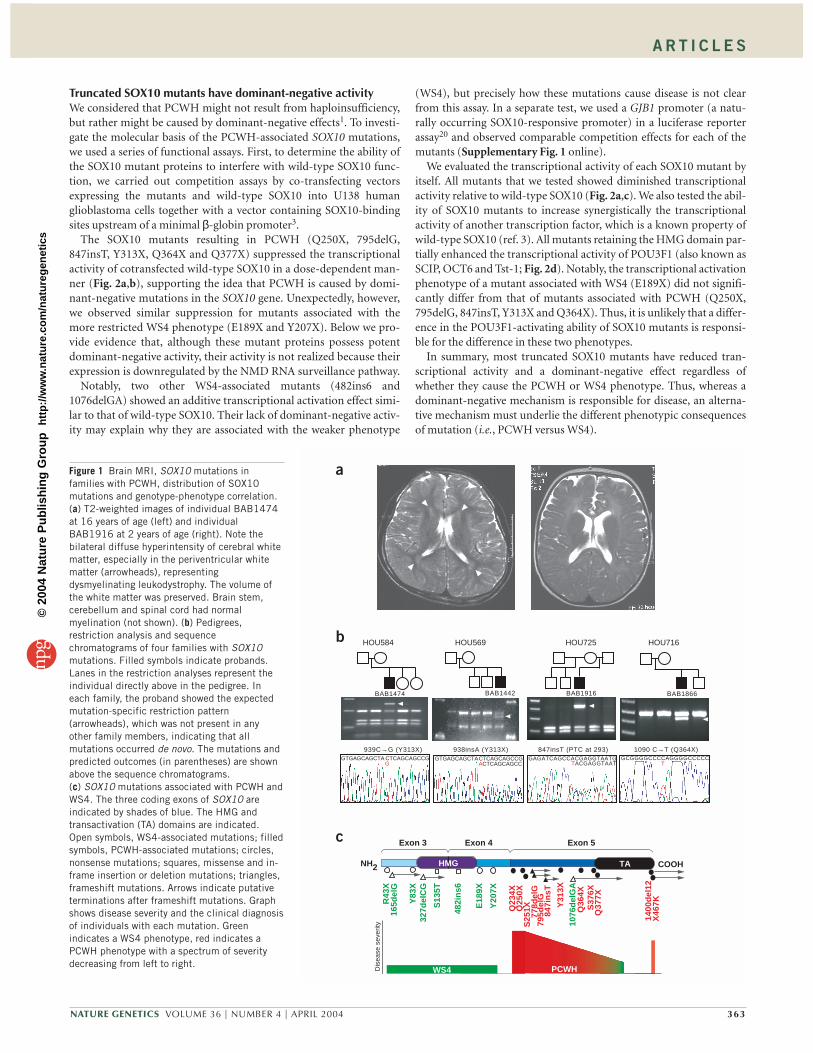
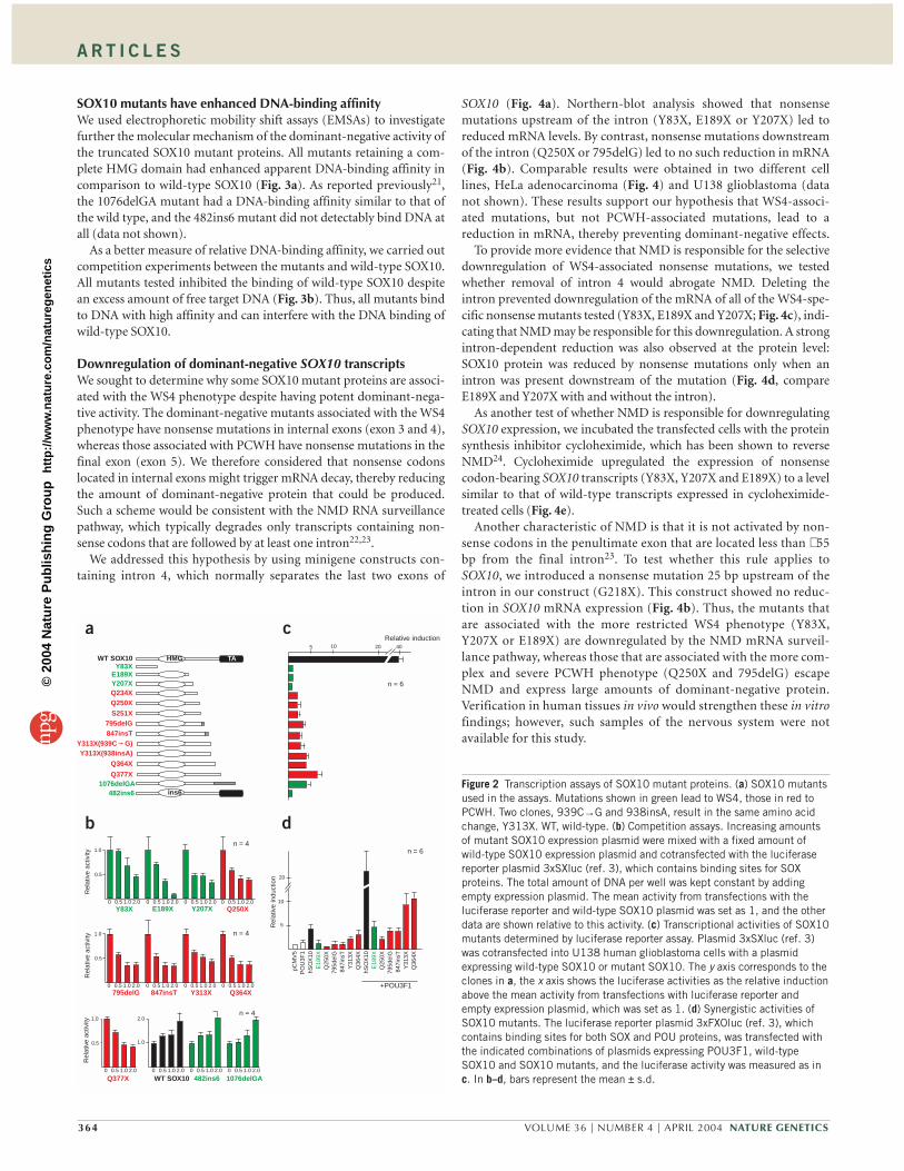
Truncated SOX10 mutants have dominant-negative activityWe considered that PCWH might not result from haploinsufficiency,but rather might be caused by dominant-negative effects1. To investi-gate the molecular basis of the PCWH-associated SOX10 mutations,we used a series of functional assays. First, to determine the ability ofthe SOX10 mutant proteins to interfere with wild-type SOX10 func-tion, we carried out competition assays by co-transfecting vectorsexpressing the mutants and wild-type SOX10 into U138 humanglioblastoma cells together with a vector containing SOX10-bindingsites upstream of a minimal β-globin promoter3.
The SOX10 mutants resulting in PCWH (Q250X, 795delG,847insT, Y313X, Q364X and Q377X) suppressed the transcriptionalactivity of cotransfected wild-type SOX10 in a dose-dependent man-ner (Fig. 2a,b), supporting the idea that PCWH is caused by domi-nant-negative mutations in the SOX10 gene. Unexpectedly, however,we observed similar suppression for mutants associated with themore restricted WS4 phenotype (E189X and Y207X). Below we pro-vide evidence that, although these mutant proteins possess potentdominant-negative activity, their activity is not realized because theirexpression is downregulated by the NMD RNA surveillance pathway.
Notably, two other WS4-associated mutants (482ins6 and1076delGA) showed an additive transcriptional activation effect simi-lar to that of wild-type SOX10. Their lack of dominant-negative activ-ity may explain why they are associated with the weaker phenotype
(WS4), but precisely how these mutations cause disease is not clearfrom this assay. In a separate test, we used a GJB1 promoter (a natu-rally occurring SOX10-responsive promoter) in a luciferase reporterassay20 and observed comparable competition effects for each of themutants (Supplementary Fig. 1 online).
We evaluated the transcriptional activity of each SOX10 mutant byitself. All mutants that we tested showed diminished transcriptionalactivity relative to wild-type SOX10 (Fig. 2a,c). We also tested the abil-ity of SOX10 mutants to increase synergistically the transcriptionalactivity of another transcription factor, which is a known property ofwild-type SOX10 (ref. 3). All mutants retaining the HMG domain par-tially enhanced the transcriptional activity of POU3F1 (also known asSCIP, OCT6 and Tst-1; Fig. 2d). Notably, the transcriptional activationphenotype of a mutant associated with WS4 (E189X) did not signifi-cantly differ from that of mutants associated with PCWH (Q250X,795delG, 847insT, Y313X and Q364X). Thus, it is unlikely that a differ-ence in the POU3F1-activating ability of SOX10 mutants is responsi-ble for the difference in these two phenotypes.
In summary, most truncated SOX10 mutants have reduced tran-scriptional activity and a dominant-negative effect regardless ofwhether they cause the PCWH or WS4 phenotype. Thus, whereas adominant-negative mechanism is responsible for disease, an alterna-tive mechanism must underlie the different phenotypic consequencesof mutation (i.e., PCWH versus WS4).
NATURE GENETICS VOLUME 36 | NUMBER 4 | APRIL 2004 363
GTGAGCAGCTACTCAGCAGCCGACTCAGCAGCC
GTGAGCAGCTA CTCAGCAGCCGG
GAGATCAGCCACGAGGTAATGTACGAGGTAAT
GCGGGGCCCCAGGGGCCCCCT
BAB1474 BAB1442 BAB1916 BAB1866
939C→G (Y313X) 938insA (Y313X) 847insT (PTC at 293) 1090 C→T (Q364X)
b
a
HOU584 HOU569 HOU725 HOU716
c
WS4 PCWH
R43
X16
5del
G
Y83
X32
7del
CG
482i
ns6
E18
9X
Y20
7X
Q23
4X
S25
1XQ25
0X
778d
elG
795d
elG
847i
nsT
Y31
3X
Q36
4X10
76d
elG
A
Q37
7XS
376X
X46
7K14
00d
el12
HMG TA
Dis
ease
sev
erity
Exon 3 Exon 4 Exon 5
NH2 COOH
S13
5T
Figure 1 Brain MRI, SOX10 mutations infamilies with PCWH, distribution of SOX10mutations and genotype-phenotype correlation.(a) T2-weighted images of individual BAB1474at 16 years of age (left) and individualBAB1916 at 2 years of age (right). Note thebilateral diffuse hyperintensity of cerebral whitematter, especially in the periventricular whitematter (arrowheads), representingdysmyelinating leukodystrophy. The volume ofthe white matter was preserved. Brain stem,cerebellum and spinal cord had normalmyelination (not shown). (b) Pedigrees,restriction analysis and sequencechromatograms of four families with SOX10mutations. Filled symbols indicate probands.Lanes in the restriction analyses represent theindividual directly above in the pedigree. Ineach family, the proband showed the expectedmutation-specific restriction pattern(arrowheads), which was not present in anyother family members, indicating that allmutations occurred de novo. The mutations andpredicted outcomes (in parentheses) are shownabove the sequence chromatograms.(c) SOX10 mutations associated with PCWH andWS4. The three coding exons of SOX10 areindicated by shades of blue. The HMG andtransactivation (TA) domains are indicated.Open symbols, WS4-associated mutations; filledsymbols, PCWH-associated mutations; circles,nonsense mutations; squares, missense and in-frame insertion or deletion mutations; triangles,frameshift mutations. Arrows indicate putativeterminations after frameshift mutations. Graphshows disease severity and the clinical diagnosisof individuals with each mutation. Greenindicates a WS4 phenotype, red indicates aPCWH phenotype with a spectrum of severitydecreasing from left to right.
©20
04 N
atur
e P
ublis
hing
Gro
up
http
://w
ww
.nat
ure.
com
/nat
ureg
enet
ics
A R T I C L E S
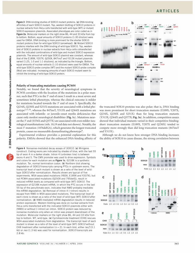
SOX10 mutants have enhanced DNA-binding affinityWe used electrophoretic mobility shift assays (EMSAs) to investigatefurther the molecular mechanism of the dominant-negative activity ofthe truncated SOX10 mutant proteins. All mutants retaining a com-plete HMG domain had enhanced apparent DNA-binding affinity incomparison to wild-type SOX10 (Fig. 3a). As reported previously21,the 1076delGA mutant had a DNA-binding affinity similar to that ofthe wild type, and the 482ins6 mutant did not detectably bind DNA atall (data not shown).
As a better measure of relative DNA-binding affinity, we carried outcompetition experiments between the mutants and wild-type SOX10.All mutants tested inhibited the binding of wild-type SOX10 despitean excess amount of free target DNA (Fig. 3b). Thus, all mutants bindto DNA with high affinity and can interfere with the DNA binding ofwild-type SOX10.
Downregulation of dominant-negative SOX10 transcriptsWe sought to determine why some SOX10 mutant proteins are associ-ated with the WS4 phenotype despite having potent dominant-nega-tive activity. The dominant-negative mutants associated with the WS4phenotype have nonsense mutations in internal exons (exon 3 and 4),whereas those associated with PCWH have nonsense mutations in thefinal exon (exon 5). We therefore considered that nonsense codonslocated in internal exons might trigger mRNA decay, thereby reducingthe amount of dominant-negative protein that could be produced.Such a scheme would be consistent with the NMD RNA surveillancepathway, which typically degrades only transcripts containing non-sense codons that are followed by at least one intron22,23.
We addressed this hypothesis by using minigene constructs con-taining intron 4, which normally separates the last two exons of
SOX10 (Fig. 4a). Northern-blot analysis showed that nonsensemutations upstream of the intron (Y83X, E189X or Y207X) led toreduced mRNA levels. By contrast, nonsense mutations downstreamof the intron (Q250X or 795delG) led to no such reduction in mRNA(Fig. 4b). Comparable results were obtained in two different celllines, HeLa adenocarcinoma (Fig. 4) and U138 glioblastoma (datanot shown). These results support our hypothesis that WS4-associ-ated mutations, but not PCWH-associated mutations, lead to areduction in mRNA, thereby preventing dominant-negative effects.
To provide more evidence that NMD is responsible for the selectivedownregulation of WS4-associated nonsense mutations, we testedwhether removal of intron 4 would abrogate NMD. Deleting theintron prevented downregulation of the mRNA of all of the WS4-spe-cific nonsense mutants tested (Y83X, E189X and Y207X; Fig. 4c), indi-cating that NMD may be responsible for this downregulation. A strongintron-dependent reduction was also observed at the protein level:SOX10 protein was reduced by nonsense mutations only when anintron was present downstream of the mutation (Fig. 4d, compareE189X and Y207X with and without the intron).
As another test of whether NMD is responsible for downregulatingSOX10 expression, we incubated the transfected cells with the proteinsynthesis inhibitor cycloheximide, which has been shown to reverseNMD24. Cycloheximide upregulated the expression of nonsensecodon-bearing SOX10 transcripts (Y83X, Y207X and E189X) to a levelsimilar to that of wild-type transcripts expressed in cycloheximide-treated cells (Fig. 4e).
Another characteristic of NMD is that it is not activated by non-sense codons in the penultimate exon that are located less than ∼ 55bp from the final intron23. To test whether this rule applies toSOX10, we introduced a nonsense mutation 25 bp upstream of theintron in our construct (G218X). This construct showed no reduc-tion in SOX10 mRNA expression (Fig. 4b). Thus, the mutants thatare associated with the more restricted WS4 phenotype (Y83X,Y207X or E189X) are downregulated by the NMD mRNA surveil-lance pathway, whereas those that are associated with the more com-plex and severe PCWH phenotype (Q250X and 795delG) escapeNMD and express large amounts of dominant-negative protein.Verification in human tissues in vivo would strengthen these in vitrofindings; however, such samples of the nervous system were notavailable for this study.
364 VOLUME 36 | NUMBER 4 | APRIL 2004 NATURE GENETICS
WT SOX10
E189XY207X
Y313X(939C G)
Q377X
TA
Q234XQ250XS251X
Y83X
Y313X(938insA)
795delG847insT
Q364X
Relative induction10 205 40
a c
d
n = 6
E18
9XQ
250X
Y31
3X
795d
elG
847i
nsT
Q36
4X
10
20
5
pCM
V5
PO
U3F
1hS
OX
10
E18
9XQ
250X
Y31
3X
795d
elG
847i
nsT
Q36
4X
hSO
X10
+POU3F1
Rel
ativ
e in
duct
ion
n = 6
0 0.5 1.0 2.0Y83X
0 0.5 1.0 2.0Y207X Q250X
0 0.5 1.0 2.0
0 0.5 1.0 2.0 0 0.5 1.0 2.0847insT Y313X
0 0.5 1.0 2.0E189X
Rel
ativ
e ac
tivity 1.0
0.5
Rel
ativ
e ac
tivity 1.0
0.5
0 0.5 1.0 2.0
n = 4
n = 4
0 0.5 1.0 2.0795delG
0 0.5 1.0 2.0 0 0.5 1.0 2.0
WT SOX10 482ins6
Rel
ativ
e ac
tivity 1.0
0.5
2.0
0 0.5 1.0 2.0
1076delGA
n = 4
0 0.5 1.0 2.0
Q377X
Q364X
1.0
HMG
ins6482ins61076delGA
b
Figure 2 Transcription assays of SOX10 mutant proteins. (a) SOX10 mutantsused in the assays. Mutations shown in green lead to WS4, those in red toPCWH. Two clones, 939C→G and 938insA, result in the same amino acidchange, Y313X. WT, wild-type. (b) Competition assays. Increasing amountsof mutant SOX10 expression plasmid were mixed with a fixed amount ofwild-type SOX10 expression plasmid and cotransfected with the luciferasereporter plasmid 3xSXluc (ref. 3), which contains binding sites for SOXproteins. The total amount of DNA per well was kept constant by addingempty expression plasmid. The mean activity from transfections with theluciferase reporter and wild-type SOX10 plasmid was set as 1, and the otherdata are shown relative to this activity. (c) Transcriptional activities of SOX10mutants determined by luciferase reporter assay. Plasmid 3xSXluc (ref. 3)was cotransfected into U138 human glioblastoma cells with a plasmidexpressing wild-type SOX10 or mutant SOX10. The y axis corresponds to theclones in a, the x axis shows the luciferase activities as the relative inductionabove the mean activity from transfections with luciferase reporter andempty expression plasmid, which was set as 1. (d) Synergistic activities ofSOX10 mutants. The luciferase reporter plasmid 3xFXOluc (ref. 3), whichcontains binding sites for both SOX and POU proteins, was transfected withthe indicated combinations of plasmids expressing POU3F1, wild-typeSOX10 and SOX10 mutants, and the luciferase activity was measured as inc. In b–d, bars represent the mean ± s.d.
©20
04 N
atur
e P
ublis
hing
Gro
up
http
://w
ww
.nat
ure.
com
/nat
ureg
enet
ics
A R T I C L E S
Polarity of truncating mutations causing PCWHNotably, we found that the severity of neurological symptoms inPCWH correlates with the location of the mutations in a polar man-ner, such that PTCs in the 5′ end of exon 5 result in a most severe andsometimes lethal phenotype, whereas the clinical outcome is milderfor mutations located towards the 3′ end of exon 5. Specifically, theQ234X, Q250X and S251X mutations are associated with a lethal phe-notype2,11,16, whereas the 847insT, Y313X and Q364X mutations areassociated with infantile- or juvenile-onset myelin deficiencies thatcause only modest neurological disabilities (Fig. 1c). Mutations near-est the 3′ end (S376X and Q377X) are associated with even milder neu-rological manifestations (refs. 10,19 and data not shown). Notably, themost 3′ mutation (1076delGA), which generates the longest truncatedprotein, causes no measurable dysmyelinating phenotype9.
Experimental evidence provides a potential explanation for thispolarity. EMSAs showed that the enhanced DNA-binding affinity of
the truncated SOX10 proteins was also polar; that is, DNA bindingwas more prominent for short truncation mutants (E189X, Y207X,Q234X, Q250X and S251X) than for long truncation mutants(Y313X, Q364X and Q377X; Fig. 3a). In addition, competition assaysshowed that individual mutants varied in their competitive binding:short truncation mutants (E189X, Y207X and Q250X) tended tocompete more strongly than did long truncation mutants (847insTand Y313X).
Although we do not know how stronger DNA binding increasesthe ability of SOX10 to cause disease, the strong correlation between
NATURE GENETICS VOLUME 36 | NUMBER 4 | APRIL 2004 365
WTE189X
WT
Mut
++––
+++
WT847insT+
+––
+++
WTY207X+
+––
+++
WTY313X+
+––
+++
Q23
4X
795d
elG
847i
nsT
Y31
3X
Q25
0X
Q37
7X
Q36
4X
WT
E18
9X
Y20
7X
S25
1X
HeL
a
WT
Mut
WT
Mut WT
Mut
a bWTQ250X+
+–
–+++
WT
Mut
Figure 3 DNA-binding studies of SOX10 mutant proteins. (a) DNA-bindingaffinities of each SOX10 mutant. Top, western blotting of SOX10 proteins innuclear extracts from HeLa cells transfected with wild-type (WT) or mutantSOX10 expression plasmids. Associated phenotypes are color coded as inFigure 2a. Molecular markers on the right show 86, 44 and 33 kDa from topto bottom. Bottom, equal amounts of SOX10 proteins (1:30 dilution) wereused for EMSA. DNA binding is more prominent for the shorter SOX10mutant proteins than for wild-type SOX10 (arrowhead). (b) Mutant SOX10proteins interfere with the DNA binding of wild-type SOX10. Top, westernblot of SOX10 proteins in nuclear extracts from HeLa cells cotransfectedwith the indicated combinations of wild-type and mutant SOX10 expressionplasmids. The amount of wild-type SOX10 plasmid was constant, whereasthat of the E189X, Y207X, Q250X, 847insT and Y313X mutant plasmidsvaried (1:25, 1:5 and 1:1 dilutions), as indicated by the triangle. Bottom,equal amounts of nuclear extracts (1:10 dilution) were used for EMSA. Thewild-type SOX10-probe complex (WT) and the mutant SOX10-probe complex(Mut) are indicated. Increasing amounts of each SOX10 mutant seem toinhibit the binding of wild-type SOX10 protein.
Figure 4 Nonsense-mediated decay assays of SOX10. (a) Minigeneconstruct. Coding exons are indicated by shades of blue, with the last 55bp of exon 4 in a darker shade. Intron 4 (unbroken line) is betweenexons 4 and 5. The CMV promoter was used to drive expression. Symbolsand colors for each mutation are as Figure 1c. G218X is a syntheticmutation. Ter, normal termination codon. (b) Northern blot showingdegradation of SOX10 transcripts carrying PTCs in upstream exons. Thetranscript level of each mutant is shown as a ratio of the level of wild-type SOX10 after normalization. Results shown are typical of fiveexperiments. WS4-associated mutations (Y83X, E189X and Y207X), butnot PCWH-associated mutations (Q250X and 795delG), result inreduced mRNA levels as compared with wild-type (WT) SOX10. Theexpression of G218X mutant mRNA, in which the PTC occurs in the last55 bp of the penultimate exon, indicates that NMD probably mediatesthe mRNA degradation. (c) Removal of intron 4 (−intron) results inescape from NMD in WS4-associated mutations. The transcript level ofeach clone is shown as a ratio of the level of wild-type (WT) SOX10 afternormalization. (d) NMD-mediated mRNA degradation results in reducedprotein expression. Western blotting was done on nuclear extracts fromHeLa cells transfected with the indicated SOX10 plasmids either with(NMD) or without (−intron) an intron. SOX10 protein was reduced bynonsense mutations only when an intron was present downstream of themutation. Molecular markers on the right show 86, 44 and 33 kDa fromtop to bottom. WT, wild-type. (e) Cycloheximide treatment (CHX) rescuesWS4-associated mutations from degradation. The transcript level of eachmutant is shown as a ratio of the level of wild-type (WT) SOX10 withoutCHX treatment after normalization (n = 2). In each blot, either lacZ (3.1kb) or neo (1.3 kb) was used for normalization. SOX10 transcripts are2.8 kb.
Exon 3 Exon 5CMV
Ter
Y83
X
E18
9XY
207X
G21
8X
Q25
0X79
5del
GY
207X
–in
tro
n
WT
SO
X10
–in
tro
n
WT
SO
X10
Y20
7X
Y83
X
a
Y83
X
WT
SO
X10
– + – + – + – +
E18
9X
Y20
7X
CHX
Y83
X –
intr
on
SOX10
lacZ
SOX10
neo
E18
9X
E18
9X –
intr
on
b
c d
Ratio
Ratio
0.4 1.4 0.3 0.51.6 1.3 1.0 1.6
1.0 1.0 0.2 0.41.0 1.2 0.3 1.0
WT
SO
X10
WT
SO
X10
–in
tro
n
E18
9X –
intr
on
Y20
7X –
intr
on
E18
9X
Y20
7X
G21
8X
795d
elG
Q25
0X
e –intron NMD
795d
elG
Q25
0X
G21
8X
WT
SO
X10
Y20
7X
E18
9X
Y83
X
SOX10
neo
Ratio 1.0 1.5 1.21.30.2 0.30.3
XExon 4
©20
04 N
atur
e P
ublis
hing
Gro
up
http
://w
ww
.nat
ure.
com
/nat
ureg
enet
ics

Figure 5 Genotype-phenotype correlation and NMD assays of MPZ. (a) MPZtruncating mutations associated with inherited peripheral neuropathies26. Thesix coding exons of MPZ are indicated by shades of blue. The transmembranedomain (TM) is encoded by exon 4. Open symbols, mutations associated withCMT1B; filled symbols, mutations associated with DSN/CHN; circles,nonsense mutations; triangles, frameshift mutations. Arrows indicate putativeterminations after frameshift mutations. Green indicates a CMT1B phenotype,red indicates a DSN/CHN phenotype. (b) MPZ genomic expression constructused for northern blotting. Six mutations associated with either CMT1B orDSN/CHN are shown (symbols as in a). Ter, normal termination codon.(c) Northern blots showing degradation of MPZ transcripts carrying PTCs ininternal exons. The transcript level of each mutant is shown as a ratio of thelevel of wild-type (WT) MPZ after normalization. Results shown are typical ofthree experiments. The 5′ PTCs (223delG, Y154X, Y181X and Q206X), butnot the 3′ PTCs (Q215X and 662insGC), result in reduced mRNA levels ascompared with wild-type MPZ. The DSN/CHN-associated Q215X PTC located2 bp from the end of exon 4 escaped mRNA downregulation, whereas theCMT1B-associated G206X PTC located 28 bp upstream of Q215X in the sameexon underwent mRNA downregulation. These data suggest that MPZ does notobey the ‘−55 bp rule’23, as has been observed for some genes44. (d) Removalof intron 4 (−intron) recovers the expression of CMT1B-associated mutations.Experiments were done as in Figure 4c. WT, wild-type. (e) Cycloheximidetreatment (CHX) rescues mRNAs carrying CMT1B-associated mutations fromdegradation. Experiments were done as in Figure 4e (n = 2). WT, wild-type.(f) RNAi-mediated suppression of UPF1 rescues mRNAs carrying CMT1B-associated PTCs from degradation (n = 3). Western blot (inset) showsdecreased expression of UPF1 protein after siRNA treatment. In the northernblots, neo (1.3 kb) was used for normalization. MPZ transcripts are 1.8 kb. Forall constructs that showed mRNA instability owing to NMD, quantitative RT-PCR experiments were done independently to validate the results of thenorthern blots (not shown). WT, wild-type.
the DNA-binding affinity of SOX10 and its disease phenotype indi-cates that this variable may be important. Either stronger DNA bind-ing may enhance the dominant-negative activity of SOX10 by amechanism that is not detectable in our assays (for example, pro-moter-specific dominant-negative effects) or it may elicit effects thatremain to be defined.
NMD downregulates PTC-bearing MPZ transcriptsTo examine whether NMD can explain why other distinct neurologicaldiseases result from allelic truncating mutations, we tested PTCs in thegene MPZ by similar expression assays. Allelic point mutations ofMPZ cause a spectrum of genotypically related, dominantly inherited,peripheral neuropathies with variable severity, including the moresevere, early onset CHN and DSN, as well as the less severe, adult onsetCMT1B. Most disease-associated mutations in MPZ mutations aremissense alterations, but 18 truncating MPZ mutations have beenreported to cause either mild or severe forms of peripheral neuropathy(ref. 25 and Inherited Peripheral Neuropathies Mutation Database; seeURL in Methods). Molecular studies of mutant MPZ have not pro-vided a plausible explanation for the association between mutationand disease severity (that is, genotype-phenotype correlations). As inSOX10, we identified an apparent correlation between phenotype andthe location of PTCs in MPZ. The PTCs in the 5′ end result in mildneuropathy, whereas most of those in the 3′ end cause severe diseases,with the exception of a few cases for which detailed clinical informa-tion was not available (Fig. 5a).
We examined the mRNA stability of MPZ PTC mutations that con-vey distinct peripheral neuropathies. Of six mutations (Fig. 5b), two(Y181X and G206X) cause truncation at a position similar to trunca-tion clones that have been shown in vitro to behave as dominant-nega-tive alleles, despite causing mild CMT1B (ref. 26). Each PTCassociated with a mild phenotype is located in an internal exon and
resulted in a downregulation of transcripts (Fig. 5c). This downregula-tion was prevented by each of the following: first, elimination of allintrons by using a cDNA construct; second, cycloheximide treatment;and third, suppression of UPF1 (ref. 27), an essential factor in theNMD pathway, by RNA-mediated interference (RNAi; Fig. 5d–f).
These findings suggest that the MPZ transcripts carrying thesePTCs selectively undergo NMD and are likely to be degraded in vivo.By contrast, the PTCs associated with severe diseases are all locatedin the last exon (except for one that is located at the distal end of thepenultimate exon) and resulted in an accumulation of mRNA at lev-els equivalent to the wild-type allele. These finding suggest that theMPZ transcripts containing these PTCs escape NMD and are proba-bly translated into truncated proteins that may have a dominant-negative function26.
DISCUSSIONWe propose that the phenotypes of genetic diseases are commonlyinfluenced by NMD. First, PTCs are common: nonsense andframeshift mutations are present in approximately one-third of muta-tions that cause human genetic disease28,29. Second, nonsense codonsin all internal exons are capable of triggering NMD23, which meansthat mRNA levels will be reduced by most nonsense and frameshiftmutations28–30; therefore, PTCs generally result in milder phenotypesas compared with missense mutations, as has been postulated in sev-eral human diseases including osteogenesis imperfecta31, Stickler syn-drome32 and Marfan syndrome33,34. Third, on the basis that NMD hasa potential role in most disease-causing PTCs, it has been proposedthat haploinsufficiency is the most predictable pathogenetic mecha-nism underlying heterozygous nonsense alleles29. Last, PTCs locatedin the last coding exon specifically escape NMD23 and so are processeddistinctly from those in internal exons, leading to the stable translationof truncated proteins.
A R T I C L E S
366 VOLUME 36 | NUMBER 4 | APRIL 2004 NATURE GENETICS
MPZneo
bCMV
a
c d
______ ______ ______ ______ – +
e
CHX
siRNA
β-actin
UPF1
Q215X
G206X
Y181X
Y154X
Q141X
223d
elG
306d
elA
614in
s17
496d
elC;4
99G
C
522d
el4
554d
elG
563in
s4
f
Ter
X X
E71X
NH2 COOH
XX X
XX
XX
X
XX
506d
elT
550d
el3in
sG
XX
662in
sGC
676in
sCA
699d
el4
WT
MP
Z
0.3 0.2 0.3 1.0 0.9
223d
elG
Y15
4X
Y18
1X
WT
MP
Z
1.0
662i
nsG
C
Q21
5X
1.0
223d
elG
Y15
4XY
181X
G20
6XQ
215X
662i
nsG
C
MPZneo
WT
MP
Z
Y15
4X
Y18
1X
223d
elG
1.0 1.1 0.4 0.9 0.3 1.01.0 0.3Ratio
Ratio
______ ______ ______ ______ WT
MP
Z
Y15
4X
Y18
1X
223d
elG
SiRNA
MPZneoRatio 1.0 1.00.9 0.30.20.31.1 0.8
WT
MP
Z
Q20
6X
1.0 0.4
MPZneo
Ratio
WT
MP
Z
Y15
4X
Y18
1X
223d
elG
WT
MP
Z –
intr
on
s
Y15
4X –
intr
on
s
Y18
1X –
intr
on
s
223d
elG
–in
tro
ns
1.0 1.0 1.10.3 0.2 1.2 1.00.3
1 63 41 63 TM
– + – + – + – + – + – + – +
– +
2 52 5
©20
04 N
atur
e P
ublis
hing
Gro
up
http
://w
ww
.nat
ure.
com
/nat
ureg
enet
ics

A R T I C L E S
β-thalassemia is a classic example in which 5′ PTCs in the geneencoding β-globin result in a recessive trait, whereas 3′ PTCs result in aspecific dominant form of disease, because 5′ PTCs but not 3′ PTCstrigger β-globin NMD35,36. We think that this previously underappre-ciated mechanism has an important role not only in phenotypic varia-tion, but also in the expression of distinct phenotypes from allelictruncating mutations. In support of this idea, we have shown experi-mentally that PTCs that trigger NMD versus those that escape NMDresult in distinct neurological phenotypes in two different disease-causing genes, SOX10 and MPZ.
A corollary to our study is that many genetic diseases that were pre-viously thought to be the result of dominant-negative mutations onthe basis of functional assays may actually result from haploinsuffi-ciency in vivo because of NMD. In fact, previous genotype-phenotypestudies of MPZ mutations could not explain the mild phenotype asso-ciated with dominant-negative truncated proteins26,37,38. InvokingNMD not only delineates the genotype-phenotype correlation fortruncating mutations, but it also facilitates a functional analysis ofmissense alterations by comparison with clinical phenotypes. Thus,NMD must be considered when formulating and testing explanationsfor phenotypic differences associated with disease-causing mutations.We have shown that triggering NMD versus escaping NMD may causedistinct neurological phenotypes in addition to contributing to thevariability of expression in human genetic diseases.
Three case reports previously identified an association betweenSOX10 mutations and deficiencies in peripheral and/or centralmyelin1,2,14. Here, by combining those data with data from four addi-tional individuals, we have provided evidence for the existence of a dis-tinct genetic disease, PCWH, caused by SOX10 mutations. Weconsider that PCWH is a distinct neurocristopathy because it can bedistinguished from other neurocristopathies, including WS4, on thebasis of the following observations.
First, there are no other syndromes that simultaneously show theclinical findings of peripheral myelinopathy, central dysmyelination,Waardenburg syndrome and Hirschsprung disease. In particular, theinvolvement of both the PNS and the CNS is a distinguishing charac-teristic of PCWH. These neurological findings predominate in deter-mining the degree of disability in individuals and also significantlyinfluence the prognosis: severe hypomyelination in the PNS and CNShas even resulted in early death in some cases2,11,16. Second, PCWH isunique because so far it is known only to be caused by a single gene,whereas WS4 is associated with at least two other genes, EDN3 andEDNRB (refs 9,39,40). Notably, neither EDN3 nor EDNRB has beenassociated with some of the symptoms characteristic of PCWH, suchas peripheral and central myelin deficiencies. Last, we have shown thatthe SOX10 mutations that cause PCWH represent a subset that are dis-tinct from those that cause WS4.
Our study indicates that the two phenotypes associated with SOX10mutations, PCWH and WS4, may be caused by two distinct molecularmechanisms. The more severe phenotype, PCWH, is caused by non-sense mutations that generate truncated SOX10 mutant proteins pos-sessing enhanced DNA-binding affinity and potent dominant-negativeactivity. The more modest phenotype, WS4, is caused by nonsensemutations that activate the NMD RNA surveillance pathway, therebyreducing dominant-negative expression and resulting in SOX10 hap-loinsufficiency.
This interpretation of the data from SOX10-deficient humans isconsistent with the finding that Sox10−/− homozygous mice have pro-found defects in both peripheral and central myelin lineages that resultin embryonic lethality, whereas heterozygotes show a phenotype cor-responding to the WS4 phenotype8. The dominant-negative activity of
the SOX10 mutant proteins that cause PCWH probably does not blockSOX10 function completely, because the PCWH phenotype is milderthan that seen in homozygous Sox10–/– mice. It is also possible, how-ever, that mice and humans differ subtly in their reliance on SOX10 forneurological development. Although our data are most consistentwith dominant-negative interference, a gain-of-function effect cannotbe absolutely ruled out.
We found that for mutations in SOX10 exon 5, the severity of thePCWH phenotype inversely correlates with the length of SOX10 pro-tein (Fig. 1c). Our transfection studies identified a potential molecularmechanism for this correlation: the shorter SOX10 mutants hadstronger DNA-binding affinity and dominant-negative activity thandid the longer SOX10 mutants (Figs. 2b and 3a,b). This polarity mayexplain the differential severity observed in individuals with PCWH;that is, it may explain why Q250X, one of the early truncating muta-tions, completely diminishes myelin development in both the PNS andthe CNS2, whereas a mutant that is 63 residues longer, Y313X, causes amuch milder phenotype (patients 1 and 2). In addition, later trunca-tions (i.e., longer SOX10 mutants) might also have a smaller interfer-ence effect and thus would not result in the PCWH phenotype9 (e.g.,1076delGA in Figs. 1c and 2a).
Much evidence indicates that the more restricted WS4 pheno-type is caused by SOX10 nonsense mutations that activate the pro-tective NMD RNA surveillance pathway. Two WS4-associatedmutations in exon 4, E189X and Y207X, resulted in truncated pro-teins possessing strong dominant-negative activity when expressedfrom cDNAs lacking introns. However, cellular mRNAs bearingthese mutations were diminished when transcribed from a con-struct with an intron-containing minigene, suggesting that NMDdegrades mRNAs containing PTCs before translation, which seemsto mitigate the dominant-negative action of these mutations. As aresult, these mutant alleles become equivalent to null alleles and thushaploinsufficiency is probably the underlying mechanism causing thedisease phenotype in vivo. By contrast, mRNAs with nonsense muta-tions in the final exon escape NMD because there is no intron down-stream of the stop codon. Large amounts of dominant-negativeprotein are therefore produced, leading to the severe (PCWH) dis-ease phenotype. Consistent with this interpretation, all truncatingmutations associated with PCWH are located in exon 5 and areanticipated to escape NMD (Fig. 1c).
Both SOX10 and MPZ PTCs encode proteins with dominant-nega-tive effects, but only those PTCs that escape NMD realize that poten-tial. This finding, in conjunction with previous results31–36, suggeststhat one of the physiological roles of NMD may be to protect againstsevere disease phenotypes by converting dominant-negative effects tohaploinsufficiency. The extent of the beneficial effect may varydepending on both the toxicity of truncated proteins encoded by dif-ferent genes and the nature of traits. Rarely, as exemplified by the hem-izygous X-linked DMD gene mutated in muscular dystrophy, NMDmay result in more severe disease by abrogating the hypomorphic pro-tein function41. Nevertheless, individuals potentially carry severalsilent PTCs in their genome and the suppression of these PTC-carry-ing alleles by NMD may be crucial for preventing disease expression,as implied by the embryonic lethality caused by congenital suppres-sion of NMD in mice42.
METHODSSOX10 mutation analysis. Peripheral blood was obtained from individuals andfamily members with informed consent approved by the Institutional ReviewBoard at Baylor College of Medicine. Genomic DNA was extracted from whiteblood cells by a standard method. The genomic DNA from each individual was
NATURE GENETICS VOLUME 36 | NUMBER 4 | APRIL 2004 367
©20
04 N
atur
e P
ublis
hing
Gro
up
http
://w
ww
.nat
ure.
com
/nat
ureg
enet
ics
A R T I C L E S
used as a template for PCR to amplify each coding exon of the SOX10 gene1. Wesequenced PCR products directly by using DyePrimer chemistry and anABI377 sequencer (Applied Biosystems). Gene mutations were examined ineach family member by restriction enzyme digestion of PCR products to deter-mine the de novo occurrence or segregation within the family. DdeI, MwoI,BssSI and StyI were used to determine each mutation. Using the same restric-tion analyses, we also examined at least 180 normal chromosomes to excludethe possibility of benign polymorphism. Paternity was confirmed in each fam-ily by microsatellite haplotyping analyses done by IdentiGene.
Recombinant constructs. We incorporated each mutation into pCMV-huSOX10, which contains human wild-type SOX10 cDNA under acytomegalovirus (CMV) promoter, either by PCR-directed mutagenesis or bysubcloning PCR products generated from patient DNA. The POU3F1 expres-sion plasmid pCMV/Tst-1 and the luciferase reporter plasmids 3xSXluc and3xFXOluc have been described3. A 750-bp fragment from the GJB1 promoterwas subcloned into the luciferase reporter plasmid pGL3 (Promega) asdescribed20. To construct intron-containing SOX10 minigenes, we used PCRto fuse two overlapping fragments, one from cDNA spanning exons 3–4 andthe other from genomic DNA spanning a portion of exon 4, intron 4 and exon5, and subcloned the product into the mammalian expression vectorpcDNA3.1 (Invitrogen) to generate pcDNASOX10NMD. We also subclonedthe human SOX10 cDNA into pcDNA3.1 to generate pcDNASOX10-intron.Full-length human MPZ cDNA (IMAGE 3926008) was obtained(OpenBiosystems) and subcloned into pcDNA3.1 (Invitrogen) to generatepcDNAMPZ. The whole genomic coding region was also subcloned fromgenomic DNA by PCR to generate pcDNAMPZNMD. Mutations were gener-ated in each construct with the QuikChange Site-Directed mutagenesis kit(Stratagene). A β-galactosidase expression plasmid, pCMVβ (Clontech), wasused as a reference for transfections.
Functional assays. U138 human glioblastoma cells and HeLa cells were grownin DMEM medium supplemented with 10% fetal bovine serum and weretransfected by using PolyFect or Effectene transfection reagents (Qiagen). Forthe luciferase assay, we collected U138 cells from 24-well trays after 48 h oftransfection and then assayed luciferase activity3. Nuclear extract was obtainedfrom HeLa cells after 48 h of transfection and used for western blotting andEMSA studies. Rabbit antiserum to SOX10 (1:3,000 dilution; Chemicon) wasused as a primary antibody for western blots. This antiserum recognizes anepitope at amino acids 165–181 of human SOX10; therefore, the signals werequantitative for both wild-type SOX10 and the truncated proteins used in thisstudy. We used oligonucleotide probes containing a single SOX10-binding site(site B)43 for EMSAs.
For northern blotting, total RNA was extracted from HeLa cells after 24 h oftransfection using 0.1 µg of SOX10 or MPZ expression plasmids along with 0.9µg of pCMVβ reference plasmid per 60-mm dish. Two hours before the cells werecollected, cycloheximide was added to a concentration of 1 mg/ml. Knockdownof UPF1 by RNAi was done as described27. Polyclonal antibody to UPF1 was a giftfrom J. Lykke-Andersen (University of Colorado, Boulder). Expression of theneomycin phosphotransferase gene (neo) gene from pcDNA3.1 or the cotrans-fected β-galactosidase gene (lacZ) was used as a reference control for normaliza-tion. After electrophoretic separation and transfer to a nylon membrane, mRNAwas probed by 32P-labeled SOX10, neo and lacZ probes. The value for each signalwas measured with a Personal Densitometer SI (Molecular Dynamics).
URL. The Inherited Peripheral Neuropathies Mutation Database is available athttp://molgen-www.uia.ac.be/CMTMutations/.
Note: Supplementary information is available on the Nature Genetics website.
ACKNOWLEDGMENTSWe thank the affected individuals and their families for their cooperation;M. Quanrud for collecting blood samples from family members; T. Shimotakeand Y. Tsuchida for providing clinical information; R. Peirano and E. Sock foradvice and assistance; and A. Beaudet, H. Bellen, K. Szigeti and H. Zoghbi forcritical reviews. This study was supported in part by grants from the USNational Institute for Neurological Disorders and Strokes, the US NationalInstitutes of Health and the Muscular Dystrophy Association to J.R.L. K.I. was a
fellow of the Charcot-Marie-Tooth Association when this study was initiatedand is currently supported by a development grant from the MuscularDystrophy Association.
COMPETING INTERESTS STATEMENTThe authors declare that they have no competing financial interests.
Received 18 December 2003; accepted 30 January 2004Published online at http://www.nature.com/naturegenetics/
1. Inoue, K., Tanabe, Y. & Lupski, J.R. Myelin deficiencies in both the central and theperipheral nervous systems associated with a SOX10 mutation. Ann. Neurol. 46,313–318 (1999).
2. Inoue, K. et al. Congenital hypomyelinating neuropathy, central dysmyelination, andWaardenburg-Hirschsprung disease: phenotypes linked by SOX10 mutation. Ann.Neurol. 52, 836–842 (2002).
3. Kuhlbrodt, K., Herbarth, B., Sock, E., Hermans-Borgmeyer, I. & Wegner, M. Sox10, anovel transcriptional modulator in glial cells. J. Neurosci. 18, 237–250 (1998).
4. Herbarth, B. et al. Mutation of the Sry-related Sox10 gene in Dominant megacolon, amouse model for human Hirschsprung disease. Proc. Natl. Acad. Sci. USA 95,5161–5165 (1998).
5. Southard-Smith, E.M., Kos, L. & Pavan, W.J. Sox10 mutation disrupts neural crestdevelopment in Dom Hirschsprung mouse model. Nat. Genet. 18, 60–64 (1998).
6. Sonnenberg-Riethmacher, E. et al. Development and degeneration of dorsal root gan-glia in the absence of the HMG-domain transcription factor Sox10. Mech. Dev. 109,253–265 (2001).
7. Stolt, C.C. et al. Terminal differentiation of myelin-forming oligodendrocytes dependson the transcription factor Sox10. Genes Dev. 16, 165–170 (2002).
8. Britsch, S. et al. The transcription factor Sox10 is a key regulator of peripheral glialdevelopment. Genes Dev. 15, 66–78 (2001).
9. Pingault, V. et al. SOX10 mutations in patients with Waardenburg-Hirschsprung dis-ease. Nat. Genet. 18, 171–173 (1998).
10. Southard-Smith, E.M. et al. The Sox10Dom mouse: modeling the genetic variation ofWaardenburg-Shah (WS4) syndrome. Genome Res. 9, 215–225 (1999).
11. Pingault, V. et al. SOX10 mutations in chronic intestinal pseudo-obstruction suggesta complex physiopathological mechanism. Hum. Genet. 111, 198–206 (2002).
12. Omenn, G.S. & McKusick, V.A. The association of Waardenburg syndrome andHirschsprung megacolon. Am. J. Med. Genet. 3, 217–223 (1979).
13. Warner, L.E. et al. Clinical phenotypes of different MPZ (P0) mutations may includeCharcot-Marie-Tooth type 1B, Dejerine-Sottas, and congenital hypomyelination.Neuron 17, 451–460 (1996).
14. Pingault, V. et al. Peripheral neuropathy with hypomyelination, chronic intestinalpseudo-obstruction and deafness: a developmental ‘neural crest syndrome’ related toa SOX10 mutation. Ann. Neurol. 48, 671–676 (2000).
15. Jacobs, J.M. & Wilson, J. An unusual demyelinating neuropathy in a patient withWaardenburg’s syndrome. Acta Neuropathol. 83, 670–674 (1992).
16. Touraine, R.L. et al. Neurological phenotype in Waardenburg syndrome type 4 corre-lates with novel SOX10 truncating mutations and expression in developing brain. Am.J. Hum. Genet. 66, 1496–1503 (2000).
17. Sham, M.H., Lui, V.C.H., Chen, B.L.S., Fu, M. & Tam, P.K.H. Novel mutations ofSOX10 suggest a dominant negative role in Waardenburg-Shah syndrome. J. Med.Genet. 38, E30 (2001).
18. Bondurand, N. et al. A molecular analysis of the Yemenite deaf-blind hypopigmenta-tion syndrome: SOX10 dysfunction causes different neurocristopathies. Hum. Mol.Genet. 8, 1785–1789 (1999).
19. Toki, F. et al. Intestinal agangliosis associated with the Waardenburg syndrome: reportof two cases and review of the literature. Pediatr. Surg. Int. 19, 725–728 (2003).
20. Bondurand, N. et al. Human Connexin 32, a gap junction protein altered in the X-linked form of Charcot-Marie-Tooth disease, is directly regulated by the transcriptionfactor SOX10. Hum. Mol. Genet. 10, 2783–2795 (2001).
21. Kuhlbrodt, K. et al. Functional analysis of Sox10 mutations found in humanWaardenburg-Hirschsprung patients. J. Biol. Chem. 273, 23033–23038 (1998).
22. Carter, M.S., Li, S. & Wilkinson, M.F. A splicing-dependent regulatory mechanismthat detects translation signals. EMBO J. 15, 5965–5975 (1996).
23. Nagy, E. & Maquat, L.E. A rule for termination-codon position within intron-contain-ing genes: when nonsense affects RNA abundance. Trends Biochem. Sci. 23,198–199 (1998).
24. Carter, M.S. et al. A regulatory mechanism that detects premature nonsense codonsin T-cell receptor transcripts in vivo is reversed by protein synthesis inhibitors in vitro.J. Biol. Chem. 270, 28995–29003 (1995).
25. Saifi, G.M., Szigeti, K., Snipes, G.J., Garcia, C.A. & Lupski, J.R. Molecular mecha-nisms, diagnosis, and rational approaches to management and therapy for Charcot-Marie-Tooth disease and related peripheral neuropathies. J. Investig. Med. 51,261–283 (2003).
26. Wong, M-H. & Filbin, M.T. Dominant-negative effect on adhesion by myelin Po pro-tein truncated in its cytoplasmic domain. J. Cell Biol. 134, 1531–1541 (1996).
27. Mendell, J.T., ap Rhys, C.M.J. & Dietz, H.C. Separable roles for rent1/hUpf1 inaltered splicing and decay of nonsense transcripts. Science 298, 419–422(2002).
28. Frischmeyer, P.A. & Dietz, H.C. Nonsense-mediated mRNA decay in health and dis-ease. Hum. Mol. Genet. 8, 1893–1900 (1999).
368 VOLUME 36 | NUMBER 4 | APRIL 2004 NATURE GENETICS
©20
04 N
atur
e P
ublis
hing
Gro
up
http
://w
ww
.nat
ure.
com
/nat
ureg
enet
ics
A R T I C L E S
29. Mendell, J.T. & Dietz, H.C. When the message goes awry: disease-producing muta-tions that influence mRNA content and performance. Cell 107, 411–414 (2001).
30. Byers, P.H. Killing the messenger: new insights into nonsense-mediated mRNAdecay. J. Clin. Invest. 109, 3–6 (2002).
31. Körkkö, J. et al. Analysis of the COL1A1 and COL1A2 genes by PCR amplificationand scanning by conformation-sensitive gel electrophoresis identifies only COL1A1mutations in 15 patients with osteogenesis imperfecta type I: identification of com-mon sequences of null-allele mutations. Am. J. Hum. Genet. 62, 98–110 (1998).
32. Snead, M.P. & Yates, J.R.W. Clinical and Molecular genetics of Stickler syndrome. J.Med. Genet. 36, 353–359 (1999).
33. Dietz, H.C. et al. Four novel FBN1 mutations: significance for mutant transcript leveland EGF-like domain calcium binding in the pathogenesis of Marfan syndrome.Genomics 17, 468–475 (1993).
34. Schrijver, I. et al. Premature termination mutations in FBN1: distinct effects on dif-ferential allelic expression and on protein and clinical phenotypes. Am. J. Hum.Genet. 71, 223–237 (2002).
35. Baserga, S.J. & Benz, E.J. Jr. Nonsense mutations in the human β-globin gene affectmRNA metabolism. Proc. Natl. Acad. Sci. USA 85, 2056–2060 (1988).
36. Forget, B.G., Benz, E.J. Jr., Skoultchi, A., Baglioni, C. & Housman, D. Absence ofmessenger RNA for β globin chain in β0-thalassaemia. Nature 247, 379–381 (1974).
37. Shames, I., Fraser, A., Colby, J., Orfali, W. & Snipes, G.J. Phenotypic differencesbetween peripheral myelin protein-22 (PMP22) and myelin protein zero (P0) muta-tions associated with Charcot-Marie-Tooth-related diseases. J. Neuropathol. Exp.Neurol. 62, 751–764 (2003).
38. Yoshida, M. & Colman, D.R. Rapid functional analysis in Xenopus oocytes of Po pro-tein adhesive interactions. Neurochem. Res. 26, 703–712 (2001).
39. Edery, P. et al. Mutation of the endothelin-3 gene in the Waardenburg-Hirschsprungdisease (Shah-Waardenburg syndrome). Nat. Genet. 12, 442–444 (1996).
40. Puffenberger, E.G. et al. A missense mutation of the endothelin-B receptor gene inmultigenic Hirschsprung’s disease. Cell 79, 1257–1266 (1994).
41. Kerr, T.P., Sewry, C.A., Robb, S.A. & Roberts, R.G. Long mutant dystrophins and vari-able phenotypes: evasion of nonsense-mediated decay? Hum. Genet. 109, 402–407(2001).
42. Medghalchi, S.M. et al. Rent1, a trans-effector of nonsense-mediated mRNA decay, isessential for mammalian embryonic viability. Hum. Mol. Genet. 10, 99–105 (2001).
43. Peirano, R.I., Goerich, D.E., Riethmacher, D. & Wegner, M. Protein zero gene expres-sion is regulated by the glial transcription factor Sox10. Mol. Cell. Biol. 20,3198–3209 (2000).
44. Wang, J., Gudikote, J.P., Olivas, O.R. & Wilkinson, M.F. Boundary-independent polarnonsense-mediated decay. EMBO Rep. 3, 274–279 (2002).
NATURE GENETICS VOLUME 36 | NUMBER 4 | APRIL 2004 369
©20
04 N
atur
e P
ublis
hing
Gro
up
http
://w
ww
.nat
ure.
com
/nat
ureg
enet
ics