TSGA10 prevents nuclear localization of the hypoxia-inducible factor (HIF)-1α
Upload
independentCategory
view
2download
0
Hypoxia Inducible Factor Signaling ModulatesSusceptibility to Mycobacterial Infection via a NitricOxide Dependent MechanismPhilip M. Elks1¤*, Sabrina Brizee1, Michiel van der Vaart1, Sarah R. Walmsley2, Fredericus J. van Eeden3,4,
Stephen A. Renshaw2,3., Annemarie H. Meijer1.*
1 Institute of Biology, Leiden University, Leiden, The Netherlands, 2 Academic Unit of Respiratory Medicine, Department of Infection and Immunity, University of Sheffield,
Western Bank, Sheffield, United Kingdom, 3 Medical Research Council Centre for Developmental and Biomedical Genetics, University of Sheffield, Western Bank, Sheffield,
United Kingdom, 4 Department of Biomedical Science, University of Sheffield, Western Bank, Sheffield, United Kingdom
Abstract
Tuberculosis is a current major world-health problem, exacerbated by the causative pathogen, Mycobacterium tuberculosis(Mtb), becoming increasingly resistant to conventional antibiotic treatment. Mtb is able to counteract the bactericidalmechanisms of leukocytes to survive intracellularly and develop a niche permissive for proliferation and dissemination.Understanding of the pathogenesis of mycobacterial infections such as tuberculosis (TB) remains limited, especially for earlyinfection and for reactivation of latent infection. Signaling via hypoxia inducible factor a (HIF-a) transcription factors haspreviously been implicated in leukocyte activation and host defence. We have previously shown that hypoxic signaling viastabilization of Hif-1a prolongs the functionality of leukocytes in the innate immune response to injury. We sought tomanipulate Hif-a signaling in a well-established Mycobacterium marinum (Mm) zebrafish model of TB to investigate effectson the host’s ability to combat mycobacterial infection. Stabilization of host Hif-1a, both pharmacologically and genetically,at early stages of Mm infection was able to reduce the bacterial burden of infected larvae. Increasing Hif-1a signalingenhanced levels of reactive nitrogen species (RNS) in neutrophils prior to infection and was able to reduce larvalmycobacterial burden. Conversely, decreasing Hif-2a signaling enhanced RNS levels and reduced bacterial burden,demonstrating that Hif-1a and Hif-2a have opposing effects on host susceptibility to mycobacterial infection. Theantimicrobial effect of Hif-1a stabilization, and Hif-2a reduction, were demonstrated to be dependent on inducible nitricoxide synthase (iNOS) signaling at early stages of infection. Our findings indicate that induction of leukocyte iNOS bystabilizing Hif-1a, or reducing Hif-2a, aids the host during early stages of Mm infection. Stabilization of Hif-1a thereforerepresents a potential target for therapeutic intervention against tuberculosis.
Citation: Elks PM, Brizee S, van der Vaart M, Walmsley SR, van Eeden FJ, et al. (2013) Hypoxia Inducible Factor Signaling Modulates Susceptibility to MycobacterialInfection via a Nitric Oxide Dependent Mechanism. PLoS Pathog 9(12): e1003789. doi:10.1371/journal.ppat.1003789
Editor: David S. Schneider, Stanford University, United States of America
Received March 20, 2013; Accepted October 11, 2013; Published December 19, 2013
Copyright: � 2013 Elks et al. This is an open-access article distributed under the terms of the Creative Commons Attribution License, which permits unrestricteduse, distribution, and reproduction in any medium, provided the original author and source are credited.
Funding: PME is the recipient of a European Respiratory Society Fellowship (LTRF fellowship nu176-2011) (http://www.ersnet.org/). SAR is an MRC Senior ClinicalFellowship (G0701932) (http://www.mrc.ac.uk/). AHM is funded by the Smart Mix Program of the Netherlands Ministry of Economic Affairs and the Ministry ofEducation, Culture and Science (http://www.agentschapnl.nl/subsidies-regelingen/smart-mix). AHM and PME are further supported by the EuropeanCommission 7th framework project ZF-HEALTH (HEALTH-F4-2010-242048) (http://zf-health.org/), and AHM and SAR by the European Marie-Curie Initial TrainingNetwork FishForPharma (PITN-GA-2011-289209) (http://www.fishforpharma.com/). The funders had no role in study design, data collection and analysis, decisionto publish, or preparation of the manuscript.
Competing Interests: The authors have declared that no competing interests exist.
* E-mail: [email protected] (PME); [email protected] (AHM)
. These authors contributed equally to this work.
¤ Current address: Department of Infection and Immunity, The University of Sheffield Medical School, Sheffield, United Kingdom.
Introduction
Pulmonary tuberculosis (TB), caused by the pathogen Mycobac-
terium tuberculosis (Mtb), is a major world health problem and is a
key priority for infectious disease research. The burden of TB has
been exacerbated by the increasing occurrence of Mtb strains with
resistance to multiple drug treatments, prioritising the need for
understanding of the mechanistic basis of host-pathogen interac-
tions during pathogenesis of disease in order to identify novel
therapeutic strategies [1]. Upon infection Mtb are rapidly
phagocytosed by host leukocytes, but are able to evade bacterial
killing mechanisms and utilize the leukocytes as a niche in which to
proliferate and disseminate [2]. Leukocyte infection initiates the
recruitment of uninfected macrophages, neutrophils and T-cells,
to form highly organised structures known as granulomas [3,4].
Mtb within granulomas can persist for many years and may
eventually escape and disseminate during clinical reactivation,
causing active disease [5]. The pathogenesis of both initial
infection and reactivation of latent infection are not well
understood, and further research into host signaling pathways at
these stages may uncover novel, host-derived targets for thera-
peutic intervention against Mtb.
Mycobacterial disease and hypoxia are intimately related.
Human tuberculous granulomas are hypoxic environments, and
it has been suggested that the relative hypoxia of granulomas
contributes to the latent infection phenotype and the associated
PLOS Pathogens | www.plospathogens.org 1 December 2013 | Volume 9 | Issue 12 | e1003789
relative resistance of Mtb to host and pharmacological killing
[6,7]. Hypoxia exerts its effects on host cell signaling predomi-
nantly through stabilization of Hypoxia Inducible Factor alpha
(HIF-a) transcription factor. HIF-a is stability and activity is
regulated by a group of oxygen sensitive enzymes: prolyl
hydroxylases (PHDs) and Factor Inhibiting HIF (FIH) [8–10].
Oxygen dependent PHD activity leads to degradation of HIF-a,
while hypoxia reduces PHD activity, stabilizing HIF-a, which joins
a nuclear complex and transduces the hypoxic cellular response
[11]. Three HIF-a isoforms have been identified in humans to
date, of which HIF-1a is a key regulator of leukocyte function
during both inflammation and a range of bacterial infections [12–
15].
Normal host defense is dependent on HIF-1a expression, which
activates and enhances leukocyte functionality [12]. We have
previously shown in a zebrafish model of inflammation that
stabilized Hif-1a delays inflammation resolution by reducing
neutrophil apoptosis and reverse migration at the inflammation
site [16]. Existing evidence suggests that the successful clearance of
bacterial infections depends on normal HIF-a signaling, and
furthermore, immune cell HIF-a is activated by bacterial
challenge in normal oxygen levels, demonstrating the fundamental
importance of this pathway to immune cell response to invading
pathogens [13,17]. Despite the extensive work done on the effects
of hypoxia on Mtb phenotype, the effects of HIF-a stabilization or
downregulation in determining the outcome of host-mycobacterial
interaction remains unknown, and presents a major research
challenge requiring a combination of modern cell biology and
genetic approaches in animal models.
The zebrafish is a well-established model organism used to study
a wide variety of human diseases [18]. Zebrafish embryos are
easily manipulated genetically and their translucency allows for
detailed microscopy studies. Mycobacterium marinum (Mm), a natural
fish pathogen and a close relative of Mtb, causes an infection in
zebrafish that mimics key features of human TB, including the
formation of caseating granulomas and development of latency
[19,20]. Mm infection of zebrafish embryos has been successfully
used to understand both host cell signaling and mycobacterial
virulence determinants [21–24]. The Hif-a pathway can be
manipulated in vivo in the zebrafish, both pharmacologically, using
non-specific inhibitors of PHD enzymes such as dimethyloxaloyl-
glycine (DMOG), and genetically, by expression of dominant Hif-
1a variants [16,25].
Using the zebrafish Mm model and Hif-a manipulation we
aimed to understand the relationship between Hif-a signaling and
the outcome of mycobacterial infection. We show that Hif
isoforms, Hif-1a and Hif-2a have opposing effects on the host
susceptibility to mycobacterial infection by demonstrating that
stabilization of Hif-1a and downregulation of Hif-2a signaling
decreases bacterial burden of Mm infection. Furthermore, using
both genetic and pharmaceutical approaches, we show that this
effect acts via a nitric oxide (NO) dependent mechanism. Our
findings identify both Hif-a and NO signaling components as
potential host-derived targets for therapeutic intervention against
TB.
Results
Hif-a Signaling Is Detectable at Early Stages of MmInfection
HIF-a signaling and its role during mycobacterial infection
remain unclear. Upon Mm infection, zebrafish larvae develop
early stage granulomas within several days, but levels of Hif-asignaling in early infection and larval granulomas are unknown.
To detect levels of Hif-a signaling in Mm infection we utilized in
situ hybridization for the known Hif-a target phd3 (Figure S1A and
S1B) and the transgenic line Tg(phd3:GFP)i144 (Figure 1A and 1B)
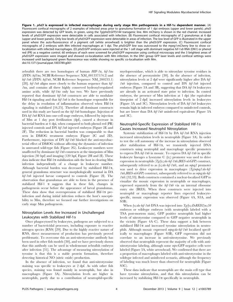
[16,25]. At 1 day post infection (dpi) phd3:GFP expression was
observed in infected leukocytes (Figure 1A). Larval granulomas at
6dpi showed only very low levels of phd3:GFP expression and are
therefore unlikely to have stabilized Hif-a at this later stage
(Figure 1B and S1B). By crossing the Tg(phd3:GFP)i144 line with a
line marking macrophages with a membrane targeted mCherry we
showed that the phd3:GFP expression was found in infected
macrophages at 1 dpi (Figure 1C). This macrophage-specific
upregulation of phd3:GFP expression by Mm infection was blocked
by injection of RNA for dominant negative (DN) hif-1ab,
indicating this is a Hif-1a dependent host response to Mm
infection (Figure 1D).
Stabilization of Hif-a by DMOG treatment between 5 and
6 dpi did not reduce the bacterial burden at 6 dpi (Figure 2A and
2B), suggesting that it is specifically early Hif-a stabilization which
may represent a novel cellular defense mechanism to arm the
leukocytes and cause Mm killing.
Stabilization of Hif-1a Signaling at Early Stages ofInfection Decreases Mm Burden
Using a combination of pharmacological and genetic approach-
es to upregulate Hif-1a signaling we aimed to determine the effects
on pathogenesis in the zebrafish Mm model. Treatment with
DMOG from 4 hours pre-infection to 24 hours post infection
significantly decreased bacterial burden compared to DMSO
negative control embryos, assessed at 4 dpi by fluorescent imaging
and pixel count analysis (Figure 2C and 2D). This is consistent
with stabilized Hif-1a aiding the host to combat Mm infection. To
eliminate the possibility that DMOG affects bacterial growth we
performed an in vitro assay for 24 hours of treatment, which
showed that DMOG does not affect bacterial growth in culture,
assessed by both OD600 reading and by plating to check viability
(Figure S2A and S2B).
Hif-1a signaling was manipulated genetically by injecting RNA
for dominant active (DA) and dominant negative (DN) hif-1ab
variants [16,26,27]. Due to a genome duplication event in
Author Summary
Tuberculosis is a mycobacterial disease that was a majorcause of death until the discovery of antibiotics in the mid-twentieth century. However, TB is once again on the rise,with the emergence of strains that are multi-drug resistant.Mycobacteria are specialists in evading immune cell killingand use host immune cells as a niche in which they canproliferate and survive latently, until subsequent re-activation and spreading causing life-threatening disease.Pharmaceutical reprogramming of the immune system tokill intracellular mycobacteria would represent a therapeu-tic strategy, effective against currently untreatable strainsand less susceptible to drug resistance. Here we use an invivo zebrafish model of TB to show that manipulation ofthe host genetic pathway responsible for detecting lowoxygen levels (hypoxia) causes a decrease in mycobacterialinfection. This antimicrobial effect was due to a priming ofimmune cells with increased levels of nitric oxide, amolecule that is used by immune cells to kill bacteria. Herewe show in vivo manipulation of a host-signaling pathwayaids the host in combatting mycobacteria infection,identifying hypoxic signaling as a potential target forfuture therapeutics against TB.
HIF Signaling Modulates Mycobacterial Infection
PLOS Pathogens | www.plospathogens.org 2 December 2013 | Volume 9 | Issue 12 | e1003789

HIF Signaling Modulates Mycobacterial Infection
PLOS Pathogens | www.plospathogens.org 3 December 2013 | Volume 9 | Issue 12 | e1003789
zebrafish there are two homologues of human HIF-1a, hif-1aa
(ZFIN: hif1aa, NCBI Reference Sequence: XM_001337574.2) and
hif-1ab (ZFIN: hif1ab, NCBI Reference Sequence: NM_200233.1)
[28]. hif-1ab aligns more closely to the human sequence than hif-
1aa, and contains all three highly conserved hydroxyl-regulated
amino acids, while hif-1aa only has two. We have previously
reported that dominant hif-1ab constructs are able to modulate
Hif-1a signaling and that hif-1ab is the homologue responsible for
the delay in resolution of inflammation observed when Hif-1asignaling is stabilized [16,25]. Therefore all dominant constructs
used in this study are based on the hif-1ab homologue. Injection of
DA hif-1ab RNA into one-cell stage embryos, followed by injection
of Mm at 1 day post fertilization (dpf), caused a decrease in
bacterial burden at 4 dpi, when compared to both phenol red (PR)
injected controls and DN hif-1ab injected embryos (Figure 2E and
2F). The reduction in bacterial burden was comparable to that
seen in DMOG treatment embryos (Figure 2C and 2D).
Furthermore, injection of DN hif-1ab blocked the anti-mycobac-
terial effect of DMOG without affecting the dynamics of infection
in untreated wild-type fish (Figure 2G). Leukocyte numbers were
unaffected by dominant hif-1ab constructs at the timepoints of Mm
injection (30 hpf) and imaging (120 hpf, Figure 2H and 2I). These
data indicate that Hif-1a stabilization aids the host in clearing Mm
infection independently of a change in leukocyte number.
Although bacterial burden was decreased by stabilized Hif-1a,
general granuloma structure was morphologically normal in DA
hif-1ab injected larvae compared to controls (Figure 2I). The
observation that granulomas are able to form in the presence of
stabilized Hif-1 a suggests that in these fish, changes in
pathogenesis occur before the appearance of larval granulomas.
These data show that overexpression of stabilized Hif-1a pre-
infection and during initial infection reduces the host’s suscepti-
bility to Mm, therefore we focused our further investigations on
early stage Mm pathogenesis.
Nitrosylation Levels Are Increased in UnchallengedLeukocytes with Stabilized Hif-1a
Once phagocytosed by leukocytes, pathogens are subjected to a
number of bactericidal processes, including exposure to reactive
nitrogen species (RNS) [29]. Due to the highly reactive nature of
RNS, direct measurement of production has previously proved
problematic. To overcome this an anti-nitrotyrosine antibody has
been used in other fish models [30], and we have previously shown
that this antibody can be used in wholemount zebrafish embryos
after infection [31]. The advantage of measuring nitrosylation of
tyrosine is that this is a stable protein formation, therefore
detecting historical NO (nitric oxide) production.
In the absence of infection, we found that anti-nitrotyrosine
staining was specific to leukocytes at 2 dpf. As with other fish
species, staining was found mainly in neutrophils, but also in
macrophages (Figure 3A). Nitrosylation levels are higher in
neutrophils, partly due to a contribution of neutrophil-specific
myeloperoxidase, which is able to nitrosylate tyrosine residues in
the absence of peroxynitrite [30]. In the absence of infection,
nitrosylation levels at 2 dpf were significantly higher after DA hif-
1ab injection, compared to control and DN hif-1ab injected
embryos (Figure 3A and 3B), suggesting that DA hif-1a leukocytes
are already in an activated state prior to infection. In control
embryos, the presence of Mm infection at 1 dpi (the equivalent
timepoint of 2 dpf) increased nitrosylation levels in leukocytes
(Figure 3A and 3C). Nitrosylation levels of DA hif-1ab leukocytes
remain high in infected embryos compared to uninfected controls,
but are lower than DA hif-1ab uninfected equivalents (Figure 3A
and 3C).
Neutrophil-Specific Expression of Stabilized Hif-1aCauses Increased Neutrophil Nitrosylation
Systemic stabilization of Hif-1a by DA hif-1ab RNA injection
increased nitrosylation levels in neutrophils (Figure 3A). In order
to test the cell autonomy of the increase in nitrosylation observed
after stabilization of Hif-1a, we transiently injected DNA
constructs using neutrophil and macrophage specific promoters
to express DA hif-1ab in mosaic. To express DA hif-1ab in specific
leukocyte lineages a lysozyme C (lyz) promoter was used to drive
expression in neutrophils (Tg(lyz:da-hif-1ab,IRES-nlsGFP) construct,
subsequently referred to as lyz:da-hif-1ab), and an mpeg1 promoter
was used to drive expression in macrophages (Tg(mpeg1:da-hif-
1ab,IRES-nlsGFP) construct, subsequently referred to as mpeg:da-hif-
1ab) [32,33]. Both constructs contained a nuclear-localized GFP to
visualize the mosaic expression in neutrophils or macrophages,
expressed separately from the hif-1ab via an internal ribosome
entry site (IRES). When these constructs were injected into
neutrophil or macrophage reporter lines expected leukocyte-
specific, mosaic expression was observed (Figure 4A, S3A, and
S3B).
When lyz:da-hif-1ab DNA was injected into Tg(lyz:DsRED2)nz50
embryos or wildtype embryos (with neutrophils labeled with a
TSA post-mortem stain), GFP positive neutrophils had higher
levels of nitrotyrosine compared to GFP negative neutrophils in
the vicinity (Figure 4A–C). These data support a link between
stabilized Hif-1a and increased tyrosine nitrosylation in neutro-
phils. Although mosaic expressed mpeg:da-hif-1ab localized specif-
ically to macrophages (Figure S3B), GFP expression did not
correlate to an increase in anti-nitrotyrosine. We previously
observed that neutrophils represent the majority of cells with anti-
nitrotyrosine labeling, although some mpx:GFP negative cells were
labeled (Figure 3A, white arrowhead). We confirmed that there are
a proportion of macrophages labeled with anti-nitrotyrosine in the
wildtype infected and uninfected scenario, although the frequency
of labeling was much lower than observed for neutrophils (Figure
S3C).
These data indicate that neutrophils are the main cell type that
have tyrosine nitrosylation, and that this nitrosylation can be
increased by stabilizing Hif-1a specifically in neutrophils.
Figure 1. phd3 is expressed in infected macrophages during early stage Mm pathogenesis in a Hif-1a dependent manner. (A)Fluorescent confocal micrographs of 2 examples of infected areas prior to granuloma formation of 1 dpi embryos (upper and lower panels). phd3expression was detected by GFP levels, in green, using the Tg(phd3:GFP)i144 transgenic line. Mm mCherry is shown in the red channel. Increasedlevels of phd3:GFP expression were detectable in cells associated with infection. (B) Fluorescent confocal micrographs of 2 granulomas at 6 dpi(upper and lower panels). Only low levels of phd3:GFP expression were detectable in areas of infection. The low level of GFP is illustrated in the upperpanel where the auto-fluorescence of a pigment cell (white arrowhead) is brighter than the phd3:GFP expression. (C) Fluorescent confocalmicrographs of 2 embryos with Mm infected macrophages at 1 dpi. The phd3:GFP line was outcrossed to the mpeg1:mCherry line to show co-localization with infected macrophages. (D) phd3:GFP embryos were injected at the 1 cell stage with dominant negative hif-1ab RNA (DN1) or phenolred (PR) as a negative control. 60 embryos of each were screened for phd3:GFP expression using confocal microscopy and the 3 brightest areas ofphd3:GFP expression were imaged and showed co-localization with Mm infection. In the DN1 group GFP laser levels and confocal settings wereincreased until background green fluorescence was visible showing no specific co-localisation with Mm.doi:10.1371/journal.ppat.1003789.g001
HIF Signaling Modulates Mycobacterial Infection
PLOS Pathogens | www.plospathogens.org 4 December 2013 | Volume 9 | Issue 12 | e1003789

Figure 2. Stabilization of Hif-1a at early stages of infection leads to a decrease in bacterial burden. (A) Quantification of bacterial burdenby fluorescent pixel count after DMOG treatment between 5 and 6 dpi. No significant difference was observed between groups. Data shown aremean 6 SEM, n = 106–121 accumulated from 3 independent experiments. (B) Stereo-fluorescence micrographs of Mm mCherry infected 6 dpi larvaefrom (C). (C) Bacterial pixel counts of larvae treated with DMOG between -4 and 24 hpi, imaged at 4 dpi. DMOG treated embryos have significantlylower levels of bacterial burden. Data shown are mean 6 SEM, n = 109–114 accumulated from 3 independent experiments. *P,.05, **P,.01, and***P,.001. (D) Fluorescence micrographs of representative infected larvae for data shown in (E). (E) Bacterial pixel counts of RNA injected larvae at
HIF Signaling Modulates Mycobacterial Infection
PLOS Pathogens | www.plospathogens.org 5 December 2013 | Volume 9 | Issue 12 | e1003789

Decrease in Mm Burden Caused by Stabilized Hif-1a IsDependent on iNOS
RNS are produced by the activity of the nitric oxide synthase
(NOS) enzymes. There are three characterized forms of NOS,
namely endothelial-NOS (eNOS), neural-NOS (nNOS), and the
leukocyte specific inducible-NOS (iNOS). iNOS expression has
been shown to be increased in infected leukocytes, and is present
in zebrafish leukocytes [34].
Increase of nitrosylation levels in neutrophils after infection is
likely to be due to increased iNOS as the morpholino against
nos2a, the zebrafish gene for iNOS [34], was able to abrogate the
increase in nitrotyrosine levels following infection (Figure 3D). To
confirm that the increase in nitrosylation observed after DA hif-1ab
is due to iNOS we used a biochemical probe for NO and an
antibody stain for iNOS. DAF-FM DA is a probe that measures
NO levels directly [35]. In the absence of Mm, DAF-FM DA
staining was increased after RNA injection of DA hif-1ab
compared to controls (Figure 5A). The signal of DAF-FM DA
staining increased after Mm infection, indicating iNOS activity
(Figure 5B). Both the increasing effect on DAF-FM DA of DA hif-
1ab in the absence of infection, and the increase after infection in
controls, could be partially blocked using the nos2a morpholino,
illustrating the iNOS specificity of these effects (Figure 5A and 5B).
Furthermore, using an iNOS antibody [36], we were able to detect
higher levels of iNOS protein in the DA hif-ab neutrophils in non-
infected embryos, which were present only at low levels in controls
(Figure 5C).
To assess the effect of blocking iNOS activity at the 1 dpi stage
on bacterial burden at 4 dpi, we treated early infected embryos
with NOS inhibitors. NOS activity was inhibited using the pan-
NOS inhibitor L-NG-Nitroarginine methyl ester (L-NAME) and
the iNOS specific inhibitor N6-(1-iminoethyl)-L-lysine (L-NIL) at
early stages of Mm infection [34,37,38]. Bacterial burden at 4 dpi
was not significantly affected by either treatment in control
embryos, although there was a trend towards increased infection
levels after NOS inhibition (Figure 5D and 5E). Both inhibitors
were able to block the decreasing bacterial burden effect of DA hif-
1ab at 4 dpi (Figure 5D and 5E). Morpholino knockdown of nos2a
was also able to block the decreased bacterial burden in DA hif-1ab
injected embryos compared to PR injected controls (Figure 5F).
These data confirm that the positive effect of stabilized Hif-1a on
the host to combat Mm infection is dependent on iNOS.
Hif-1a and Hif-2a Have Opposing Effects on Mm Burdenin an iNOS Dependent Mechanism
In humans there are three different HIF-a transcription factors:
HIF-1a, HIF-2a, and HIF-3a. It is becoming clear that HIF-2a is
important in leukocyte biology [39,40]. In order to investigate the
effects of hif-2a modulation, we synthesized dominant variants for
hif-2a with the equivalent hydroxylation site mutations to the hif-
1a variants [16]. As with Hif-1a, Hif-2a has two homologues in
the zebrafish, hif-2aa (ZFIN: epas1a, NCBI Reference Sequence:
XM_690170.5) and hif-2ab (ZFIN: epas1b, GenBank:DQ375242).
Unlike hif-1a homologues, hif-2a sequences are highly similar and
both contain all three conserved hydroxylation sites. DA hif-2aa
increased phd3 expression by in situ hybridization and phd3:GFP
expression at 1 dpf, whilst DN hif-2aa decreased expression levels
(Figure S4A,B). DN hif-2aa also blocked the increase in phd3
expression associated with early Mm infection at the 1 dpi stage of
pathogenesis (Figure S4C). These data illustrate that zebrafish Hif-
2a has similar effects on a well characterized target of the Hif-atranscription factor, phd3, as the previously characterized zebrafish
Hif-1a [16,25].
DA hif-2aa, while able to increase phd3 expression, had no effect
on bacterial burden compared to controls (Figure 6A and
Figure 6B). DN hif-2aa reduced bacterial burden at 4 dpi to a
similar level to that of DA hif-1ab (Figure 6C and 6D). As was the
case with dominant hif-1ab constructs, dominant hif-2aa variants
had no effect on leukocyte numbers (Figure 6E and 6F).
Nitrosylation levels in DN hif-2aa were found to be high in non-
infected leukocytes, as with DA hif-1ab, with no effect of DA hif-
2aa (Figure 7A and 7B). Mm infection caused a decrease in
nitrosylation levels in DN hif-2aa leukocyte, as with DA hif-1ab
(Figure 7C). The reduction in bacterial burden seen with DN hif-
2aa could be blocked by inhibition of iNOS (Figure 7D–7F)
indicating that the reciprocal effects of Hif-1a and Hif-2amodulate susceptibility to mycobacterial infection via an iNOS
dependent mechanism (Figure 7G).
Discussion
The rise in prevalence of multi-drug resistant TB creates an
urgent need for novel, host-targeting therapies to complement
existing antibiotics and to combat currently untreatable strains of
Mtb [41,42]. Using a well-established zebrafish/Mm infection
model of TB, we have identified a new anti-mycobacterial
leukocyte phenotype being driven by Hif-1a stabilization and
consequent iNOS activity. Our data identify that in vivo
manipulation of Hif-a, a ubiquitous host-signaling pathway, can
affect specific cellular mechanisms of pathogen handling, tipping
the host-pathogen balance in favour of the host to decrease
mycobacterial infection.
Mtb infection and Hif-a signaling have only previously been
linked in the context of the hypoxic, necrotic center of the fully
formed caseating granuloma. The necrotic center has been
previously identified in adult zebrafish granulomas, however larval
granulomas are not necrotic, and to our knowledge the levels of
Hif-a signaling in larval granulomas had not previously been
investigated [19]. We did not detect high levels of Hif-a signaling,
via elevated phd3 target gene expression, in larval granulomas.
However, upregulated expression of phd3 was observed in infected
macrophages at early stages of pathogenesis, before the formation
of larval granulomas, and we were able to demonstrate that this
increase in expression is Hif-a dependent. Upregulation and
stabilization of HIF-1a is a known consequence of leukocyte
activation during onset of infection, even in normoxia [13], and
remains high during pathogenesis of other types of bacterial
4 dpi. Dominant active hif-1ab (DA1) significantly decreased bacterial burden in infected larvae compared to both phenol red injected controls (PR)and dominant negative hif-1ab (DN1). Data shown are mean 6 SEM, n = 115–127 as accumulated from 3 independent experiments. (F) Examplefluorescence micrographs from the data found in (E). (G) Bacterial pixel counts of RNA injected larvae at 4 dpi after DMSO/DMOG treatment for24 hours at 4 hours before Mm injection. Dominant negative hif-1ab was able to block the antimicrobial effect of DMOG treatment. Data shown aremean 6 SEM, n = 83–100 as accumulated from 3 independent experiments. (H, upper panel) L-plastin (macrophages and neutrophils) and TSA(neutrophils only) wholebody counts at 30 hpf. No significant difference was observed between groups. Data shown are mean 6 SEM, n = 90accumulated from 3 independent experiments. (H, lower panel) L-plastin and TSA wholebody counts at 120 hpf. Data shown are mean 6 SEM, n = 90as accumulated from 3 independent experiments. (I) Example fluorescence confocal micrographs of 4 dpi granuloma structures from phenol red (PR)and dominant active hif-1ab (DA1) injected larvae. Leukocytes are identified by Alexa-488 (green) labeled L-plastin antibody.doi:10.1371/journal.ppat.1003789.g002
HIF Signaling Modulates Mycobacterial Infection
PLOS Pathogens | www.plospathogens.org 6 December 2013 | Volume 9 | Issue 12 | e1003789

Figure 3. Nitrosylation levels are increased in unchallenged leukocytes with stabilized Hif-1a. (A) Example fluorescence confocal z-stacksof the caudal vein region of embryos stained with Alexa-633 labeled anti-nitrotyrosine antibody (blue), imaged at 1 dpi in the presence or absence ofMm infection. Embryos were injected with phenol red (PR), dominant active hif-1ab (DA1), dominant negative hif-1ab (DN1), or nos2a morpholino(NOS2MO). Anti-nitrotyrosine mainly co-localized with neutrophils (labeled with mpx:GFP), however, some cells without GFP (white arrowhead),possibly macrophages, were also labeled. (B) Corrected fluorescence intensity levels of anti-nitrotyrosine antibody confocal z-stacks in uninfectedlarvae at 2 dpf (1 dpi equivalent). Dominant active hif-1ab (DA1) had significantly increased anti-nitrotyrosine levels in the absence of Mm bacterialchallenge compared to phenol red (PR) injected controls. Data shown are mean 6 SEM, n = 67–92 cells accumulated from 5 embryos per group.Graph shown is a representative dataset of 3 independent experiments. (C) Corrected fluorescence intensity levels of anti-nitrotyrosine antibodyconfocal z-stacks of dominant active hif-1ab (DA1), or phenol red (PR) control injected embryos in the presence or absence of Mm infection at 1 dpi.Data shown are mean 6 SEM, n = 233–270 cells accumulated from 15 embryos. Graph shown is combined data from 365 embryos from independentexperiments. (D) Corrected fluorescence intensity levels of anti-nitrotyrosine antibody confocal z-stacks of nos2a morpholino (NMO) or standardcontrol morpholino (SCMO) injected embryos imaged at 1 dpi (2 dpf) in the presence or absence of Mm. Data shown are mean 6 SEM, n = 46–92cells accumulated from 5 embryos. Graph shown is a representative dataset of 3 independent experiments.doi:10.1371/journal.ppat.1003789.g003
HIF Signaling Modulates Mycobacterial Infection
PLOS Pathogens | www.plospathogens.org 7 December 2013 | Volume 9 | Issue 12 | e1003789

HIF Signaling Modulates Mycobacterial Infection
PLOS Pathogens | www.plospathogens.org 8 December 2013 | Volume 9 | Issue 12 | e1003789

infections leading to enhanced leukocyte function [13,15]. In
contrast, we observed levels of Hif-a signaling in early Mm
infection, but not at the granuloma stage, suggestive of a silencing
of Hif-a signaling over the course of Mm pathogenesis. This
observation is consistent with leukocyte transcriptional reprogram-
ing observed in human mycobacterial disease and with murine
macrophages having upregulated HIF-1a in the presence of heat-
killed Mtb but not in the presence of viable Mtb [43–45]. A
dampening effect on Hif-a signaling could be a mechanism by
which mycobacteria are able to form a protected niche in which to
proliferate and disseminate, and this may be via manipulation of
transcription factors such as HIF-1a. This possibility is supported
by dominant negative hif-1ab having no effect on Mm bacterial
burden, suggesting that Hif-1a signaling does not play a major role
in leukocyte anti-mycobacterial activity during the normal
pathogenesis of infection. Stabilization of HIF-1a during the
pathogenesis of mycobacterial infection may represent a thera-
peutic opportunity to re-arm leukocytes and to inhibit further
infection.
The study of the zebrafish/Mm larval granuloma model has led
to key discoveries for TB pathogenesis causing changes in
treatment practises in the clinic [42,46]. However, the zebrafish
embryo is an untapped resource for study of mycobacterial
pathogenesis at the earliest stages of infection, before granuloma
structures form. Genetic manipulation of Hif-1a during myco-
bacterial infection has not previously been explored, and further
understanding of the roles of this critical host-signaling pathway
may uncover Hif and its signaling components as future
therapeutic targets for intervention against TB. A critical role of
HIF-1a during bacterial infection was demonstrated in murine
knockout studies showing that HIF-1a signaling is required for
proper response to Streptococcal bacterial challenge [47]. Further-
more localized treatment with a drug that stabilized HIF-1a led to
a decrease in proliferation of the skin pathogens, Pseudomonas
aeruginosa and Acinetobacter baumanii, in a mouse abscess model [48].
Our data demonstrate, in vivo, that pharmacological or genetic
stabilization of Hif-1a can aid the host in the fight against
mycobacterial infection. Importantly, these data suggest that
therapeutic upregulation of HIF-1a signaling could complement
current antibiotic treatments in the fight against Mtb infection.
In the zebrafish Mm model, the early treatment window of
DMOG (treatment between -4 hpi and 24 hpi, followed by wash-
off then bacterial burden assessment at 4 dpi) and the DA hif-1ab
RNA injection at the one-cell stage, (an effect that will be diluted
as the embryo develops), indicate that Hif-1a stabilization at early
stages of Mm infection, pre-granuloma formation, is causing the
decrease bacterial burden at 4 dpi. This is further supported by
the observation that embryo granulomas are able to form after DA
hif-1ab injection despite a decrease in bacterial burden. Therefore,
to understand the mechanism of action of early Hif-1a stabiliza-
tion, we focused on the role of bacterial killing by the leukocytes
during early stage infection. A major mechanism of bacterial
killing is the use of RNS by leukocytes [29,49]. The leukocyte
enzymatic producer of NO, iNOS, is a tightly regulated enzyme,
and is a known target of a number of immune transcription
factors, including HIF-1a [40]. We found that levels of NO, as
assessed by protein nitrosylation, were consistently higher in
neutrophils of infected embryos at early stages of infection.
Morpholino reduction of iNOS confirmed that increased nitrosy-
lation in neutrophils after infection was iNOS dependent. We
showed that stabilization of Hif-1a is able to activate neutrophils to
produce NO in the absence of bacterial challenge and that this
could be achieved through neutrophil specific expression of DA
hif-1ab, demonstrating a cell-autonomous effect. The increased
level of NO in non-infected embryos with stabilized Hif-1aindicates a priming of neutrophils to bacterial challenge, leading to
greater levels of RNS pre-infection. Early bacterial killing would
lead to lower levels of bacterial survival, decreased dissemination
and the observed decrease in bacterial burden at the granuloma
stages of infection. This early priming of neutrophils could be
blocked using iNOS inhibition, confirming that the reduced
susceptibility to Mm infection due to Hif-1a stabilization is
dependent on iNOS activity.
The role of neutrophils in early mycobacterial infection is not
fully understood. Macrophages have previously been thought the
major leukocyte involved in the pathogenesis of mycobacterial
infection [50] and are also the main leukocyte-type involved in the
phagocytosis of intravenously injected mycobacteria and are
present in abundance in the granuloma. However, more recently,
potentially important roles of neutrophils during mycobacterial
infection are becoming evident. Neutrophils are known to be able
to undertake oxidative killing of Mm phagocytosed by macro-
phages in embryonic granulomas, a mechanism which may be
important to control infection, however their role at early stages of
infection has not been addressed [51,52]. We observed that
enhanced nitrotyrosine levels early after infection were mostly
detected in neutrophils, although in rare instances nitrosylation
levels were detectable in both uninfected and infected macro-
phages. This was confirmed using the iNOS antibody, which also
showed a mainly neutrophil localization. In the absence of
infection there are detectable levels of nitrotyrosine in neutrophils,
likely due to myeloperoxidase activity, an enzyme that is
neutrophil-specific in zebrafish [53]. Myeloperoxidase can form
NO-derived inflammatory oxidants and it has been shown that
myeloperoxidase is responsible for the majority of tissue nitrosyla-
tion in fish in the absence of infection [30,54].
The anti-microbial effect of stabilized Hif-1a was abrogated
after pharmacological and morpholino inhibition of iNOS.
Neutrophils form the major leukocyte population that display
protein nitrosylation after stabilization of Hif-1a, therefore we
hypothesize that through an unknown mechanism, increased
neutrophil iNOS levels leads to increased Mm killing at early
Figure 4. Neutrophil-specific stabilization of Hif-1a causes increased neutrophil nitrosylation. (A) Confocal photomicrographs ofexpression of lyz driven DA hif-1ab and IRES- nlseGFP (lyz:da-hif-1a) in lyz:dsRed embryos stained with anti-nitrotyrosine antibody at 2 dpf. Cells withnuclear localized eGFP colocalized with lyz:dsRed expression showing neutrophil specificity of the transgenic construct. Mosaic labeled neutrophils(white arrowheads) had a higher level of anti nitrotyrosine signal compared to GFP negative ones (black arrowheads). (B) Confocal photomicrographsof a Tg(lyz:da-hif-1ab:IRES-nlsegfp) (lyz:da-hif-1a) injected ABTL embryo at 2 dpf. TSA staining of endogenous myeloperoxidase was used to stainneutrophils. Due to the nature of the myeloperoxidase staining, the whole cell is not marked, so the cell boundaries have been traced (dotted line)using brightfield z-stacks. The upper panel shows an example of a lyz:da-hif-1a negative neutrophil with low levels of anti-nitrotyrosine. The lowerpanels show two examples of lyz:da-hif-1a positive neutrophils in the same embryo, exhibiting a greater level of anti-nitrotyrosine compared to thenegative neutrophils. (C) Corrected fluorescence intensity levels of anti-nitrotyrosine antibody confocal z-stacks of GFP negative or positiveneutrophils in embryos transiently expressing Tg(lyz:da-hif-1ab:IRES-nlsegfp). Embryos were imaged at 2 dpf. For each GFP positive neutrophilobserved, a neighboring GFP negative neutrophil was also imaged. Data shown are mean 6 SEM, n = 20 cells per group accumulated from 13embryos from 4 independent experiments. P values were calculated using a paired T-test.doi:10.1371/journal.ppat.1003789.g004
HIF Signaling Modulates Mycobacterial Infection
PLOS Pathogens | www.plospathogens.org 9 December 2013 | Volume 9 | Issue 12 | e1003789

Figure 5. Hif-1a mediated anti-mycobacterial effect is iNOS dependent. (A) Fluorescent confocal micrographs of DAF-FM DA stainedembryos at 2 dpf in the absence of infection. Neutrophils are identified by lyz:dsRed expression. DAF-FM DA staining between the timepoint ofinfection and 1 dpi produced varying levels of background and stained cells of the central nervous system (neurons and notochord) as well as havingleukocyte-associated staining. The line of staining at the top of each image is DAF-FM DA staining in the notochord. Punctae of DAF-FM DA showupregulation of NO signaling Dominant active hif-1ab (DA1) embryos had more punctae than phenol red (PR) controls. nos2a morpholino (NOS2MO)reduced the punctae in both the PR and DA1 background. (B) Fluorescent confocal micrographs of DAF-FM DA stained embryos at 2 dpf in thepresence of Mm infection. In phenol red (PR) controls DAF-FM DA punctae are increased. DAF-FM DA staining is not specific for iNOS (it is a pan-NOSprobe), and the nos2a morpholino (NOS2MO) was not able to downregulate all of the DAF-FM DA staining after Mm infection, although punctaenumber were reduced. The number of punctae was also reduced in the dominant active hif-1ab (DA1) injected embryos after Mm infection.Dominant negative hif-1ab (DN1) caused no change in punctae in the presence of Mm infection compared to PR controls. (C) Fluorescent confocalmicrographs of iNOS antibody staining in mpx:GFP embryos. Phenol red (PR) injected controls had very low levels of anti-iNOS antibody staining.Dominant active hif-1ab (DA1) had increased levels of anti-iNOS antibody, a stain which was mainly neutrophil specific. (D) Bacterial burden at 4 dpiafter injection of DA hif-1ab (DA1) or phenol red control (PR) and treatment with the pan-NOS inhibitor L-NAME. Data shown are mean 6 SEM,
HIF Signaling Modulates Mycobacterial Infection
PLOS Pathogens | www.plospathogens.org 10 December 2013 | Volume 9 | Issue 12 | e1003789

stages of infection, ultimately decreasing bacterial burden. As
discussed above, macrophages are the major leukocyte involved in
the phagocytosis of Mm in this model [50]. However we found
many instances where neutrophils contain internalized Mm in the
first 24 hours post infection. Our observations indicate that
neutrophils are able to internalize Mm before the presence of
granuloma structures, and that they have elevated nitrosylation
levels during infection. Therefore, a potential mechanism is that
neutrophils are able to phagocytose Mm and increase iNOS after
Hif-1a stabilization leading to enough early bactericidal activity to
n = 62–89 as accumulated from 3 independent experiments. (E) Bacterial burden at 4 dpi after injection of DA hif-1ab (DA1) and treatment with theiNOS inhibitor L-NIL. Data shown are mean 6 SEM, n = 60–87 as accumulated from 3 independent experiments. (F) Bacterial burden at 4 dpi after co-injection of DA hif-1ab and the nos2a morpholino, using the standard control (SCMO) morpholino as a negative control. Data shown are mean 6 SEM,n = 109–116 as accumulated from 4 independent experiments.doi:10.1371/journal.ppat.1003789.g005
Figure 6. Hif-2a has opposing effects on bacterial burden than Hif-1a. (A) Bacterial pixel counts of dominant active hif-2aa (DA2) bacterialburden levels in 4 dpi infected embryos compared to phenol red (PR) and dominant active hif-1ab (DA1) injected controls. Data shown are mean 6SEM, n = 52–79 as accumulated from 3 independent experiments. (B) Example fluorescence micrographs of the data shown in (A). (C) Bacterial pixelcounts of dominant negative hif-2aa (DN2) bacterial burden levels in 4 dpi infected embryos compared to phenol red (PR) and dominant active hif-1ab (DA1) injected controls. Data shown are mean 6 SEM, n = 74–82 performed as 3 independent experiments. (D) Example fluorescencemicrographs of the data shown in (C). (E) L-plastin (macrophages and neutrophils) and TSA (neutrophils only) wholebody counts at 30 hpf afterinjection of dominant active (DA2) and dominant negative (DN2) hif-2aa RNA. No significant difference was observed between groups. Data shownare mean 6 SEM, n = 77–82 as accumulated from 3 independent experiments. (F) L-plastin and TSA wholebody counts at 120 hpf after injection ofdominant active (DA2) and dominant negative (DN2) hif-2aa RNA. Data shown are mean 6 SEM, n = 87–90 as accumulated from 3 independentexperiments.doi:10.1371/journal.ppat.1003789.g006
HIF Signaling Modulates Mycobacterial Infection
PLOS Pathogens | www.plospathogens.org 11 December 2013 | Volume 9 | Issue 12 | e1003789

Figure 7. Dominant negative Hif-2a decreases bacterial burden via an iNOS dependent mechanism. (A) Example fluorescence confocalz-stacks of the caudal vein region of embryos stained with anti-nitrotyrosine antibody, imaged at 1 dpi (2 dpf), in the absence and presence of Mminfection. Embryos were injected with dominant negative hif-2aa (DN2), dominant active hif-2aa (DA2), or phenol red (PR). (B) Corrected fluorescenceintensity levels of anti-nitrotyrosine antibody confocal z-stacks in uninfected larvae. Dominant negative hif-2aa (DN2) had significantly increased anti-nitrotyrosine levels in the absence of Mm bacterial challenge compared to phenol red (PR) injected controls. Data shown are mean 6 SEM, n = 42–92cells accumulated from 5 embryos. Graph shown is a representative dataset of 3 independent experiments. (C) Corrected fluorescence intensity levelsof anti-nitrotyrosine antibody confocal z-stacks of dominant active hif-2aa (DA2), or phenol red (PR) control injected embryos in the presence orabsence of Mm infection at 1 dpi (2 dpf). Data shown are mean 6 SEM, n = 46–92 cells accumulated from 5 embryos. Graph shown is a representative
HIF Signaling Modulates Mycobacterial Infection
PLOS Pathogens | www.plospathogens.org 12 December 2013 | Volume 9 | Issue 12 | e1003789

significantly reduce bacterial burden at later timepoints. However,
it is clear that in the zebrafish embryo model, as in other models,
macrophages are the major cell type with internalized Mm at early
timepoints of infection. Therefore, a more likely hypothesis is that
neutrophils with activated iNOS are able to interact with infected
macrophages, either by transfer of live bacteria or transfer of
reactive nitrogen species, leading to increased bactericidal activity.
We therefore hypothesize that neutrophils play an important role
alongside macrophages in early Mm bacterial killing, but the
bactericidal mechanisms of this interaction are yet to be
uncovered.
The role of Hif-2a isoform stabilization in leukocytes during
inflammation and infection has not been widely investigated in any
in vivo model of infection. We set out to identify what effect Hif-2amodulation had on the outcome of Mm infection. Although
stabilization of Hif-2a has the same increasing effect as Hif-1astabilization on expression of the major Hif-a target gene, phd3, we
observed an opposing effect on Mm bacterial burden. We
demonstrate that the decrease in bacterial burden was due to an
increase in neutrophil nitrosylation after downregulation of Hif-
2a. The decrease in bacterial burden after Hif-2a downregulation
could be blocked by early iNOS inhibition and we hypothesize
that this effect is mediated by differential roles of the Hif-aisoforms on the iNOS genetic pathway. HIF-1a and HIF-2a have
been previously been shown to have opposing effects on iNOS in
mammalian cultured macrophages, where HIF-1a stabilization
transcriptionally upregulated iNOS, while HIF-2a stabilization
decreased NO levels [40]. Our findings confirm that Hif-2a is able
to upregulate the well-characterised Hif-a target phd3 while having
opposing effects on nitrosylation, corroborating previous in vitro
observations [40]. These data demonstrate the in vivo consequence
on bacterial infection of the differential regulation of iNOS by Hif-
a variants. The opposing effects of Hif-a isoforms on bacterial
burden highlight the tight control of NO homeostasis in
leukocytes. The potential of the HIF-a pathway for therapeutic
intervention in other diseases, including cancer and ischemia, is
widely recognized, however differential roles of HIF-a isoforms in
these diseases are only recently coming to light [55,56]. These
regulatory mechanisms, in part mediated by HIF-a, are complex
and further studies are required before the regulation of HIF-aisoforms and iNOS during infection is fully understood.
In conclusion, our data demonstrate that in vivo modulation of
host Hif-a signaling during early Mm pathogenesis can lead to
decreased burden of mycobacterial infection. Stabilization of Hif-
1a, or reduction of Hif-2a, results in priming of neutrophil NO
bactericidal activity leading to lower mycobacterial burden after
challenge with infection. Our data highlight the delicate balance of
HIF-a and iNOS signaling in leukocyte function during infection
and highlight the important role of neutrophils during early stage
Mm infection. Further understanding of the complex crosstalk
between Hif-a and iNOS pathways during Mtb infection will help
identify novel, host-targeted, therapeutic strategies against TB.
NO priming of neutrophils by targeted Hif-a modulation, may
decrease the level of initial Mtb infection and act to block the
development of acute TB disease caused by re-activation and
dissemination of latent Mtb infection. Host targeted strategies
would be predicted to be beneficial against all types of TB,
including multiple drug resistant strains, and may be less
susceptible to therapy-resistance than antibiotic strategies, thereby
reducing the global burden of TB.
Materials and Methods
Ethics StatementZebrafish lines were handled in compliance with the local
animal welfare regulations and maintained according to standard
protocols (zfin.org). The breeding of adult fish was approved by
the local animal welfare committee (DEC) of the University of
Leiden. All protocols adhered to the international guidelines
specified by the EU Animal Protection Directive 2010/63/EU.
Zebrafish and Bacterial StrainsZebrafish were maintained according to standard proto-
cols [57] and local animal welfare regulations. Strains used were
ABTL (wildtype), Tg(phd3:GFP)i144, Tg(mpx:GFP)i114, Tg(lyz:Ds-
RED2)nz50 and Tg(mpeg1:mCherryF)ump2 [25,32,58].
Infection experiments were performed using M. marinum strain
M (ATCC #BAA-535), containing the pSMT3-mCherry or
pSMT3-Crimson vector [59]. Liquid cultures were prepared from
bacterial plates [59]. Injection inoculum was prepared in 2%
polyvinylpyrrolidone40 (PVP40) solution (CalBiochem) as previ-
ously described [60,61]. 100 colony-forming units (CFU) of
bacteria were injected into the caudal vein at 28 hpf as previously
described [61].
Whole Mount in situ Hybridization of phd3Whole mount in situ hybridization of phd3 was carried out as
previously described [16,25].
Confocal Microscopy of Tg(phd3:GFP)i144 Larvae6 dpi and 1 dpi Tg(phd3:GFP)i144 [25] larvae infected with Mm
were embedded in 1% low melting point agarose (Sigma Aldrich)
and transferred to a Leica DMIRBE inverted microscope with a
Leica SP1 confocal scanhead for imaging with 40 or 63 times
lenses.
Pharmacological Stabilization of Hif-a with DMOGThe pan hydroxylase inhibitor, DMOG (dimethyloxaloylgly-
cine, Enzo Life Sciences), was used at a 100 mM concentration as
previously described [16]. DMSO solvent controls were used.
Unless otherwise stated embryos were treated from 4 hours pre
Mm infection to 24 hpi by addition to the embryo water. The
inhibitors were then washed off with fresh embryo water and
grown to 4 dpi for assessment of bacterial load as described below.
RNA Injections of Hif-a VariantsEmbryos were injected with dominant Hif-a RNA at the one
cell stage as previously described [16]. hif-a variants used were
dominant active (DA) and dominant negative (DN) hif-1ab (ZFIN:
hif1ab) and hif-2aa (ZFIN: epas1a) (primer sequences in Table S1).
Phenol red (Sigma Aldrich) was used as a vehicle control.
dataset of 3 independent experiments. (D) Bacterial burden at 4 dpi after injection of dominant negative hif-2aa (DN2) or phenol red control (PR) andtreatment with the pan-NOS inhibitor L-NAME. Data shown are mean 6 SEM, n = 67–85 as accumulated from 3 independent experiments. (E)Bacterial burden at 4 dpi after injection of dominant negative hif-2aa (DN2) and treatment with the iNOS inhibitor L-NIL. Data shown are mean 6
SEM, n = 52–58 as accumulated from 3 independent experiments. (F) Bacterial burden at 4 dpi after co-injection of dominant negative hif-2aa (DN2)and the nos2a morpholino, using the standard control (SC) morpholino as a negative control. Data shown are mean 6 SEM, n = 103–109 asaccumulated from 4 independent experiments. (G) Schematic of the effect of early Hif-a modulation during early Mm infection.doi:10.1371/journal.ppat.1003789.g007
HIF Signaling Modulates Mycobacterial Infection
PLOS Pathogens | www.plospathogens.org 13 December 2013 | Volume 9 | Issue 12 | e1003789

Stereoimaging and Bacterial Pixel CountEmbryos were imaged at 4 dpi on a Leica MZ16FA Fluores-
cence Stereo Microscope. Brightfield and fluorescence images
were generated with a Leica DC500 (DFC420C) camera. Bacterial
loads were analysed using dedicated pixel counting software as
previously described [23].
Leukocyte StainingLarvae were fixed in 4% paraformaldehyde in PBS overnight at
4uC and leukocytes were immune-labeled using the l-plastin
antibody as previously described [60,62]. Neutrophils were labeled
with TSA (TSAplus kit, Fluorescence Systems, Perkin Elmer Inc)
staining labeled neutrophils in fluorescein green fluorescence as
previously described [63]. Two timepoints were chosen for this
analysis, the timepoint of Mm injection (28–30 hpf) and the
timepoint of bacterial burden assessment (5 dpf). RNA groups
were blinded prior to counting. Neutrophils and leukocytes in
embryos and larvae were counted in the tail region using a Leica
MZ16FA Fluorescence Stereo Microscope.
Morpholino Knockdown of nos2aThe nos2a morpholino (Genetools) was used as previously
reported [34]. A standard control morpholino (Genetools) was
used as a negative control.
Antibody Staining to Detect NO MarkersLarvae were fixed in 4% paraformaldehyde in PBS overnight at
4uC and nitrotyrosine levels were immune-labeled with a rabbit
polyclonal anti-nitrotyrosine antibody (Merck Millipore 06-284) at
a 1:200 dilution of primary antibody, and were detected using an
Alexa Fluor (Invitrogen Life Technologies) secondary antibody.
Larvae were fixed in 4% paraformaldehyde in PBS overnight at
4uC and iNOS was immune-labeled with a rabbit polyclonal
iNOS antibody (BD Biosciences) as previously described [36].
Detection was with goat anti-rabbit HRP-conjugated antibody
(Abcam, 1:500 dilution) and Cy3Plus TSA kit (Perkin-Elmer).
Confocal Microscopy and Quantification of Corrected CellFluorescence of Anti-Nitrotyrosine Levels
Embryos were imaged at 1 dpi, in the presence or absence of
infection, embedded in 1% low melting point agarose (Sigma
Aldrich) and transferred to a Leica DMIRBE inverted microscope
with a Leica SP1 confocal scanhead for imaging with 40 or 63
times lenses. For quantification purposes acquisition settings and
area of imaging (in the caudal vein region) were kept the same
across groups. Corrected total cell fluorescence was calculated for
each immune-stained cell using Image J as previously described
[64]. The GFP of the Tg(mpx:GFP)i114 [58] was used to assess the
leukocyte cell boundaries.
Mosaic Expression of DA hif-1ab in Neutrophils andMacrophages
The Tol2kit multisite gateway-based transposon system was
used to make transgenic constructs to transiently and mosaic
express DA hif-1a specifically in neutrophils and macrophages
[65]. DA hif-1ab was recombined into the middle entry
pDONR221 using BP Clonase (Invitrogen). An LR Clonase
(Invitrogen) Gateway reaction was performed with p5E-lyz
(neutrophil specific promoter) or p5E-mpeg1 (macrophage specific
promoter, [33]), pDONR221-da-hif-1ab and p3E-IRES-
nlsEGFPpA inserted into pDestTol2pA2. The resulting plasmids,
Tg(lyz:da-hif-1ab,IRES-nlsGFP) and Tg(mpeg1:da-hif-1ab,IRES-
nlsGFP), were microinjected with tol2 transposase RNA into the
one cell stage embryo to express in transient, mosaically. Positive
fish were screened for the transgene using the heart marker eGFP
expression (found in the Gateway vector), and positive cells were
screened by confocal microscopy for the nuclear localized eGFP
signal showing the expression of the transgene.
DAF-FM DA StainingEmbryos were injected with the relevant hif-a construct and
infected with Mm at 1 dpf. At the timepoint of infection DAF-FM
DA was applied to the embryo water as previously described [35].
DAF-FM DA was washed off using embryo water at 1 dpi and
imaged using confocal microscopy.
Pharmacological Inhibition of NOSThe pan-NOS inhibitor L-NAME, (NG-Nitro-L-arginine
methyl ester, Tocris Bioscience), was used at 200 mM as previously
described [34,37]. The iNOS inhibitor L-NIL (N6-(1-iminoethyl)-
L-lysine, dihydrochloride, Tocris Bioscience) was used at a
200 mM concentration [38]. DMSO solvent controls were used
at corresponding concentrations for each treatment. Unless
otherwise stated embryos were treated from 4 hours pre Mm
infection to 24 hpi by addition to the embryo water. The
inhibitors were then washed off with fresh embryo water and
grown to 4 dpi for assessment of bacterial load as described above.
Dominant hif-2aa CloningDominant active zebrafish hif-2aa was generated by successive
rounds of site directed mutagenesis, each mutating a hydroxylation
site into a non-hydroxylatable form as previously described for hif-
1ab [16]. Dominant negative hif-2aa was generated by a truncation
at the equivalent amino acid to the 330th amino acid in the human
sequence, as previously described for hif-1ab [16].
Statistical AnalysisAll data were analysed (Prism 5.0, GraphPad Software) using
unpaired, two-tailed t-tests for comparisons between two groups
and one-way ANOVA (with Bonferonni post-test adjustment) for
other data. P values shown are: *P,.05, **P,.01, and ***P,.001.
Supporting Information
Figure S1 Later stage larval infection showed nodetectable levels of Hif-1a signaling. (A) In situ hybridiza-
tion using a phd-3 antisense probe indicated no detectable
expression in granulomas in 6 dpi zebrafish larvae. Lower panels
show larvae treated with DMOG that have upregulated expression
of phd3 indicating that the in situ detection of phd3 is functional
and dependent on activated Hif-a signaling. (B) Micrographs of 4
individual granulomas (encircled with dotted lines) of 6 dpi larvae
taken with DIC light microscopy. Hif-a signaling is labeled in the
larvae by phd3 in situ hybridization, however, no levels of
expression were detectable in granulomas.
(TIF)
Figure S2 DMOG did not affect MM bacterial growth invitro. (A) Bacterial pixel count of plated out serial dilutions of Mm
liquid cultures after overnight incubation in DMSO/DMOG. The
bacterial culture was split into two after inoculation and treated with
100 mM DMOG, or DMSO. After treatment and growth overnight,
serial dilutions of the liquid culture were plated out and grown for 4
days before imaging. Data shown are mean 6 SEM, n = 15 as
accumulated from 3 independent experiments. (B) Example
fluorescence photomicrographs from the data shown in (A).
(TIF)
HIF Signaling Modulates Mycobacterial Infection
PLOS Pathogens | www.plospathogens.org 14 December 2013 | Volume 9 | Issue 12 | e1003789

Figure S3 Leukocyte cell-type specific expression ofstabilized Hif-1a and macrophage labeling withanti-nitrotyrosine. (A) Confocal photomicrographs of
Tg(lyz:da-hif-1ab:ires-nlsegfp) (lyz:da-hif-1a) injected mpeg1:mCherry
embryos at 2 dpf. IRES-nlsGFP is expressed in cells in the caudal
haematopoetic tissue associated with leukocytes and mpeg1 positive
macrophages, but is not present within the same cell. (B) Confocal
photomicrographs of Tg(mpeg1:da-hif-1ab:ires-nlsegfp) (mpeg:da-hif-1a)
injected Tg(mpeg1:mCherryF)ump2 line (mpeg:mCherry) at 2 dpf.
IRES-nlsGFP is found expressed in the same cells as mpeg:mCherry
indicating macrophage expression. (C) Confocal micrographs
showing anti nitrotyrosine staining in macrophages in the
mpeg:mCherry at 2 dpf. Upper panels show a nitrotyrosine
negative macrophage, which is representative of the majority of
the macrophage population. Middle panels show a nitrotyrosine
positive macrophage in the absence of infection. Lower panels
show a nitrotyrosine positive macrophage in the presence of
infection. In both the absence and presence of infection
nitrotyrosine positive macrophages are a rare event (approximate-
ly ,5% of the population).
(TIF)
Figure S4 Dominant hif-2aa variants exhibit the sameeffects on phd3 expression as the equivalent dominanthif-1ab variants. (A) Photomicrographs of 24 hpf embryos after
injection with dominant active (DA2) and dominant negative
(DN2) hif-2aa constructs or phenol red (PR) as a control, showing
expression of the Hif-a target gene phd3 by in situ hybridization.
(B) Fluorescent photomicrographs of 48 hpf phd3:GFP embryos
injected with dominant active with dominant active (DA2) and
dominant negative (DN2) hif-2aa constructs or phenol red (PR) as
a control. Upper panels show that DA2 increases the expression of
phd3:GFP compared to PR and DN2 (white arrows). Lower
panels show that DN2 can block the increased expression of
phd3:GFP in the yolk (white arrows) after DMOG treatment. (C)
phd3:GFP embryos were injected at the 1 cell stage with dominant
negative hif-2aa RNA (DN2) or phenol red (PR) as a negative
control. 60 embryos of each were screened for phd3:GFP
expression using confocal microscopy and the 3 brightest areas
of phd3:GFP expression were imaged and showed co-localization
Mm infection. In the DN2 group GFP laser levels and confocal
settings were increased until background green fluorescence was
visible showing no specific co-localisation with Mm.
(TIF)
Table S1 hif-2aa primers used for cloning and site-directed mutagenesis. Primers used to PCR amplify the
zebrafish HIF-2a homologue, hif-2aa (ZFIN: epas1a), and to make
the dominant constructs. Dominant active primers are longer as
site directed mutagenesis was performed to introduce each
mutation individually in separate PCR reactions. PCR products
were transformed into the pCR- II-TOPO vector (Invitrogen) and
sequence verified. Each hif-2aa construct was then inserted into the
pCS2+ vector (Invitrogen) from which RNA was transcribed using
SP6 enzyme and the mMessage-Machine kit (Ambion).
(DOCX)
Acknowledgments
The authors would like to thank Professor Herman Spaink (University of
Leiden) for valuable discussions and critically reading the manuscript,
Professor Paul Martin (University of Bristol) for the provision of the l-
plastin antibody, Dr. Maria Forlenza (University of Wageningen) for NO
expertise, Dr. Georges Lutfalla (University of Montpellier) for the
Tg(mpeg1:mCherryF)ump2 line and Mr Vincent van Hensbergen (University
of Leiden) for help with immunostaining.
Author Contributions
Conceived and designed the experiments: PME AHM SAR FJvE SRW.
Performed the experiments: PME SB. Analyzed the data: PME AHM
SAR. Contributed reagents/materials/analysis tools: MvdV FJvE. Wrote
the paper: PME AHM SAR.
References
1. Koul A, Arnoult E, Lounis N, Guillemont J, Andries K (2011) The challenge of
new drug discovery for tuberculosis. Nature 469: 483–490.
2. Podinovskaia M, Lee W, Caldwell S, Russell DG (2012) Infection ofmacrophages with Mycobacterium tuberculosis induces global modifications to
phagosomal function. Cell Microbiol 15: 843–59.
3. Dannenberg AM, Jr. (1993) Immunopathogenesis of pulmonary tuberculosis.
Hosp Pract (Off Ed) 28: 51–58.
4. Flynn JL, Chan J, Lin PL (2011) Macrophages and control of granulomatous
inflammation in tuberculosis. Mucosal Immunol 4: 271–278.
5. Friedrich N, Hagedorn M, Soldati-Favre D, Soldati T (2012) Prison break:pathogens’ strategies to egress from host cells. Microbiol Mol Biol Rev 76: 707–720.
6. Boshoff HI, Barry CE, 3rd (2005) Tuberculosis - metabolism and respiration inthe absence of growth. Nat Rev Microbiol 3: 70–80.
7. Via LE, Lin PL, Ray SM, Carrillo J, Allen SS, et al. (2008) Tuberculousgranulomas are hypoxic in guinea pigs, rabbits, and nonhuman primates. Infect
Immun 76: 2333–2340.
8. Epstein AC, Gleadle JM, McNeill LA, Hewitson KS, O’Rourke J, et al. (2001)C. elegans EGL-9 and mammalian homologs define a family of dioxygenases
that regulate HIF by prolyl hydroxylation. Cell 107: 43–54.
9. Bruick RK, McKnight SL (2001) A conserved family of prolyl-4-hydroxylases
that modify HIF. Science 294: 1337–1340.
10. Mahon PC, Hirota K, Semenza GL (2001) FIH-1: a novel protein that interactswith HIF-1alpha and VHL to mediate repression of HIF-1 transcriptional
activity. Genes Dev 15: 2675–2686.
11. Wenger RH (2002) Cellular adaptation to hypoxia: O2-sensing protein
hydroxylases, hypoxia-inducible transcription factors, and O2-regulated geneexpression. FASEB J 16: 1151–1162.
12. Cramer T, Yamanishi Y, Clausen BE, Forster I, Pawlinski R, et al. (2003) HIF-
1alpha is essential for myeloid cell-mediated inflammation. Cell 112: 645–657.
13. Peyssonnaux C, Datta V, Cramer T, Doedens A, Theodorakis EA, et al. (2005)
HIF-1alpha expression regulates the bactericidal capacity of phagocytes. J ClinInvest 115: 1806–1815.
14. Walmsley SR, Chilvers ER, Whyte MK (2009) Hypoxia. Hypoxia, hypoxiainducible factor and myeloid cell function. Arthritis Res Ther 11: 219.
15. Nizet V, Johnson RS (2009) Interdependence of hypoxic and innate immune
responses. Nat Rev Immunol 9: 609–617.
16. Elks PM, van Eeden FJ, Dixon G, Wang X, Reyes-Aldasoro CC, et al. (2011)
Activation of hypoxia-inducible factor-1alpha (Hif-1alpha) delays inflammation
resolution by reducing neutrophil apoptosis and reverse migration in a zebrafish
inflammation model. Blood 118: 712–722.
17. Anand RJ, Gribar SC, Li J, Kohler JW, Branca MF, et al. (2007) Hypoxia causes
an increase in phagocytosis by macrophages in a HIF-1alpha-dependent
manner. J Leukoc Biol 82: 1257–1265.
18. Santoriello C, Zon LI (2012) Hooked! Modeling human disease in zebrafish.
J Clin Invest 122: 2337–2343.
19. Swaim LE, Connolly LE, Volkman HE, Humbert O, Born DE, et al. (2006)
Mycobacterium marinum infection of adult zebrafish causes caseatinggranulomatous tuberculosis and is moderated by adaptive immunity. Infect
Immun 74: 6108–6117.
20. Parikka M, Hammaren MM, Harjula SK, Halfpenny NJ, Oksanen KE, et al.
(2012) Mycobacterium marinum causes a latent infection that can be reactivated
by gamma irradiation in adult zebrafish. PLoS Pathog 8: e1002944.
21. Berg RD, Ramakrishnan L (2012) Insights into tuberculosis from the zebrafish
model. Trends in molecular medicine 18: 689–690.
22. Alibaud L, Rombouts Y, Trivelli X, Burguiere A, Cirillo SL, et al. (2011) A
Mycobacterium marinum TesA mutant defective for major cell wall-associated
lipids is highly attenuated in Dictyostelium discoideum and zebrafish embryos.Mol Microbiol 80: 919–934.
23. Stoop EJ, Schipper T, Huber SK, Nezhinsky AE, Verbeek FJ, et al. (2011)
Zebrafish embryo screen for mycobacterial genes involved in the initiation of
granuloma formation reveals a newly identified ESX-1 component. Dis Model
Mech 4: 526–536.
24. van der Vaart M, van Soest JJ, Spaink HP, Meijer AH (2013) Functional analysis
of a zebrafish myd88 mutant identifies key transcriptional components of the
innate immune system. Disease models & mechanisms 6: 841–54.
25. Santhakumar K, Judson EC, Elks PM, McKee S, Elworthy S, et al. (2012) A
zebrafish model to study and therapeutically manipulate hypoxia signaling in
tumorigenesis. Cancer Res 72: 4017–4027.
HIF Signaling Modulates Mycobacterial Infection
PLOS Pathogens | www.plospathogens.org 15 December 2013 | Volume 9 | Issue 12 | e1003789

26. Manotham K, Tanaka T, Ohse T, Kojima I, Miyata T, et al. (2005) A biologic
role of HIF-1 in the renal medulla. Kidney Int 67: 1428–1439.27. Linke S, Stojkoski C, Kewley RJ, Booker GW, Whitelaw ML, et al. (2004)
Substrate requirements of the oxygen-sensing asparaginyl hydroxylase factor-
inhibiting hypoxia-inducible factor. J Biol Chem 279: 14391–14397.28. Rojas DA, Perez-Munizaga DA, Centanin L, Antonelli M, Wappner P, et al.
(2007) Cloning of hif-1alpha and hif-2alpha and mRNA expression patternduring development in zebrafish. Gene Expr Patterns 7: 339–345.
29. Summersgill JT, Powell LA, Buster BL, Miller RD, Ramirez JA (1992) Killing of
Legionella pneumophila by nitric oxide in gamma-interferon-activated macro-phages. J Leukoc Biol 52: 625–629.
30. Forlenza M, Scharsack JP, Kachamakova NM, Taverne-Thiele AJ, RomboutJH, et al. (2008) Differential contribution of neutrophilic granulocytes and
macrophages to nitrosative stress in a host-parasite animal model. Mol Immunol45: 3178–3189.
31. van der Vaart M, Spaink HP, Meijer AH (2012) Pathogen recognition and
activation of the innate immune response in zebrafish. Adv Hematol 2012:159807.
32. Hall C, Flores MV, Storm T, Crosier K, Crosier P (2007) The zebrafishlysozyme C promoter drives myeloid-specific expression in transgenic fish. BMC
Dev Biol 7: 42.
33. Ellett F, Pase L, Hayman JW, Andrianopoulos A, Lieschke GJ (2011) mpeg1promoter transgenes direct macrophage-lineage expression in zebrafish. Blood
117: e49–56.34. Hall CJ, Flores MV, Oehlers SH, Sanderson LE, Lam EY, et al. (2012)
Infection-responsive expansion of the hematopoietic stem and progenitor cellcompartment in zebrafish is dependent upon inducible nitric oxide. Cell Stem
Cell 10: 198–209.
35. Lepiller S, Laurens V, Bouchot A, Herbomel P, Solary E, et al. (2007) Imagingof nitric oxide in a living vertebrate using a diamino-fluorescein probe. Free
Radic Biol Med 43: 619–627.36. Clay H, Volkman HE, Ramakrishnan L (2008) Tumor necrosis factor signaling
mediates resistance to mycobacteria by inhibiting bacterial growth and
macrophage death. Immunity 29: 283–294.37. North TE, Goessling W, Peeters M, Li P, Ceol C, et al. (2009) Hematopoietic
stem cell development is dependent on blood flow. Cell 137: 736–748.38. Christiansen B, Wellendorph P, Brauner-Osborne H (2006) Known regulators
of nitric oxide synthase and arginase are agonists at the human G-protein-coupled receptor GPRC6A. Br J Pharmacol 147: 855–863.
39. Roda JM, Sumner LA, Evans R, Phillips GS, Marsh CB, et al. (2011) Hypoxia-
inducible factor-2alpha regulates GM-CSF-derived soluble vascular endothelialgrowth factor receptor 1 production from macrophages and inhibits tumor
growth and angiogenesis. J Immunol 187: 1970–1976.40. Takeda N, O’Dea EL, Doedens A, Kim JW, Weidemann A, et al. (2010)
Differential activation and antagonistic function of HIF-{alpha} isoforms in
macrophages are essential for NO homeostasis. Genes Dev 24: 491–501.41. Kuijl C, Savage ND, Marsman M, Tuin AW, Janssen L, et al. (2007)
Intracellular bacterial growth is controlled by a kinase network around PKB/AKT1. Nature 450: 725–730.
42. Tobin DM, Roca FJ, Oh SF, McFarland R, Vickery TW, et al. (2012) Hostgenotype-specific therapies can optimize the inflammatory response to
mycobacterial infections. Cell 148: 434–446.
43. Tailleux L, Waddell SJ, Pelizzola M, Mortellaro A, Withers M, et al. (2008)Probing host pathogen cross-talk by transcriptional profiling of both Mycobac-
terium tuberculosis and infected human dendritic cells and macrophages. PLoSOne 3: e1403.
44. Masaki T, Qu J, Cholewa-Waclaw J, Burr K, Raaum R, et al. (2013)
Reprogramming Adult Schwann Cells to Stem Cell-like Cells by Leprosy BacilliPromotes Dissemination of Infection. Cell 152: 51–67.
45. Ehrt S, Schnappinger D, Bekiranov S, Drenkow J, Shi S, et al. (2001)Reprogramming of the macrophage transcriptome in response to interferon-
gamma and Mycobacterium tuberculosis: signaling roles of nitric oxide synthase-
2 and phagocyte oxidase. J Exp Med 194: 1123–1140.
46. Tobin DM, Vary JC, Jr., Ray JP, Walsh GS, Dunstan SJ, et al. (2010) The lta4hlocus modulates susceptibility to mycobacterial infection in zebrafish and
humans. Cell 140: 717–730.
47. Peyssonnaux C, Boutin AT, Zinkernagel AS, Datta V, Nizet V, et al. (2008)Critical role of HIF-1alpha in keratinocyte defense against bacterial infection.
The Journal of investigative dermatology 128: 1964–1968.
48. Okumura CY, Hollands A, Tran DN, Olson J, Dahesh S, et al. (2012) A newpharmacological agent (AKB-4924) stabilizes hypoxia inducible factor-1 (HIF-1)
and increases skin innate defenses against bacterial infection. Journal of
molecular medicine 90: 1079–1089.
49. Evans TJ, Buttery LD, Carpenter A, Springall DR, Polak JM, et al. (1996)
Cytokine-treated human neutrophils contain inducible nitric oxide synthase that
produces nitration of ingested bacteria. Proc Natl Acad Sci U S A 93: 9553–9558.
50. Clay H, Davis JM, Beery D, Huttenlocher A, Lyons SE, et al. (2007)
Dichotomous role of the macrophage in early Mycobacterium marinuminfection of the zebrafish. Cell Host Microbe 2: 29–39.
51. Yang CT, Cambier CJ, Davis JM, Hall CJ, Crosier PS, et al. (2012) Neutrophilsexert protection in the early tuberculous granuloma by oxidative killing of
mycobacteria phagocytosed from infected macrophages. Cell Host Microbe 12:
301–312.
52. Lowe DM, Redford PS, Wilkinson RJ, O’Garra A, Martineau AR (2012)
Neutrophils in tuberculosis: friend or foe? Trends Immunol 33: 14–25.
53. Lieschke GJ, Oates AC, Crowhurst MO, Ward AC, Layton JE (2001)Morphologic and functional characterization of granulocytes and macrophages
in embryonic and adult zebrafish. Blood 98: 3087–3096.
54. Eiserich JP, Hristova M, Cross CE, Jones AD, Freeman BA, et al. (1998)Formation of nitric oxide-derived inflammatory oxidants by myeloperoxidase in
neutrophils. Nature 391: 393–397.
55. Misson P, van den Brule S, Barbarin V, Lison D, Huaux F (2004) Markers ofmacrophage differentiation in experimental silicosis. J Leukoc Biol 76: 926–932.
56. Keith B, Johnson RS, Simon MC (2012) HIF1alpha and HIF2alpha: sibling
rivalry in hypoxic tumour growth and progression. Nature reviews Cancer 12:9–22.
57. Nusslein-Volhard C DR (2002) Zebrafish: A Practical Approach. Oxford:Oxford University Press.
58. Renshaw SA, Loynes CA, Trushell DM, Elworthy S, Ingham PW, et al. (2006) A
transgenic zebrafish model of neutrophilic inflammation. Blood 108: 3976–3978.
59. van der Sar AM, Spaink HP, Zakrzewska A, Bitter W, Meijer AH (2009)
Specificity of the zebrafish host transcriptome response to acute and chronic
mycobacterial infection and the role of innate and adaptive immunecomponents. Mol Immunol 46: 2317–2332.
60. Cui C, Benard EL, Kanwal Z, Stockhammer OW, van der Vaart M, et al.
(2011) Infectious disease modeling and innate immune function in zebrafishembryos. Methods Cell Biol 105: 273–308.
61. Benard EL, van der Sar AM, Ellett F, Lieschke GJ, Spaink HP, et al. (2012)
Infection of zebrafish embryos with intracellular bacterial pathogens. J Vis Exp61: pii: 3781.
62. Redd MJ, Cooper L, Wood W, Stramer B, Martin P (2004) Wound healing and
inflammation: embryos reveal the way to perfect repair. Philos Trans R SocLond B Biol Sci 359: 777–784.
63. Loynes CA, Martin JS, Robertson A, Trushell DM, Ingham PW, et al. (2010)Pivotal Advance: Pharmacological manipulation of inflammation resolution
during spontaneously resolving tissue neutrophilia in the zebrafish. J Leukoc Biol
87: 203–212.
64. Burgess A, Vigneron S, Brioudes E, Labbe JC, Lorca T, et al. (2010) Loss of
human Greatwall results in G2 arrest and multiple mitotic defects due to
deregulation of the cyclin B-Cdc2/PP2A balance. Proc Natl Acad Sci U S A107: 12564–12569.
65. Kwan KM, Fujimoto E, Grabher C, Mangum BD, Hardy ME, et al. (2007) The
Tol2kit: a multisite gateway-based construction kit for Tol2 transposontransgenesis constructs. Dev Dyn 236: 3088–3099.
HIF Signaling Modulates Mycobacterial Infection
PLOS Pathogens | www.plospathogens.org 16 December 2013 | Volume 9 | Issue 12 | e1003789
Copyright © 2022 FDOKUMEN


![[The expression and significance of hypoxia-inducible factor-1 alpha and related genes in abdominal aorta aneurysm]](https://static.fdokumen.com/doc/165x107/6333061e576b626f850dad15/the-expression-and-significance-of-hypoxia-inducible-factor-1-alpha-and-related.jpg)