Human versus mouse eosinophils: "that which we call an eosinophil, by any other name would stain as...
13
Mechanisms of allergic diseases Series editors: Joshua A. Boyce, MD, Fred Finkelman, MD, and William T. Shearer, MD, PhD Human versus mouse eosinophils: ‘‘That which we call an eosinophil, by any other name would stain as red’’ James J. Lee, PhD, a Elizabeth A. Jacobsen, PhD, a Sergei I. Ochkur, PhD, a Michael P. McGarry, PhD, a Rachel M. Condjella, PhD, b Alfred D. Doyle, BS, a Huijun Luo, PhD, a Katie R. Zellner, BS, b Cheryl A. Protheroe, BA, b Lian Willetts, BS, a,c William E. LeSuer, BS, b Dana C. Colbert, MS, b Richard A. Helmers, MD, d Paige Lacy, PhD, c Redwan Moqbel, PhD, FRCPath, e and Nancy A. Lee, PhD b Scottsdale, Ariz, and Edmonton, Alberta, and Winnipeg, Manitoba, Canada The respective life histories of human subjects and mice are well defined and describe a unique story of evolutionary conservation extending from sequence identity within the genome to the underpinnings of biochemical, cellular, and physiologic pathways. As a consequence, the hematopoietic lineages of both species are invariantly maintained, each with identifiable eosinophils. This canonical presence nonetheless does not preclude disparities between human and mouse eosinophils, their effector functions, or both. Indeed, many books and reviews dogmatically highlight differences, providing INFORMATION FOR CATEGORY 1 CME CREDIT Credit can now be obtained, free for a limited time, by reading the review articles in this issue. Please note the following instructions. Method of Physician Participation in Learning Process: The core ma- terial for these activities can be read in this issue of the Journal or online at the JACI Web site: www.jacionline.org. The accompanying tests may only be submitted online at www.jacionline.org. Fax or other copies will not be accepted. Date of Original Release: September 2012. Credit may be obtained for these courses until August 31, 2014. Copyright Statement: Copyright Ó 2012-2014. All rights reserved. Overall Purpose/Goal: To provide excellent reviews on key aspects of allergic disease to those who research, treat, or manage allergic disease. Target Audience: Physicians and researchers within the field of allergic disease. Accreditation/Provider Statements and Credit Designation: The American Academy of Allergy, Asthma & Immunology (AAAAI) is ac- credited by the Accreditation Council for Continuing Medical Education (ACCME) to provide continuing medical education for physicians. The AAAAI designates these educational activities for a maximum of 1 AMA PRA Category 1 Creditä. Physicians should only claim credit commensu- rate with the extent of their participation in the activity. List of Design Committee Members: James J. Lee, PhD, Elizabeth A. Jacobsen, PhD, Sergei I. Ochkur, PhD, Michael P. McGarry, PhD, Rachel M. Condjella, PhD, Alfred D. Doyle, BS, Huijun Luo, PhD, Katie R. Zell- ner, BS, Cheryl A. Protheroe, BA, Lian Willetts, BS, William E. LeSuer, BS, Dana C. Colbert, MS, Richard A. Helmers, MD, Paige Lacy, PhD, Redwan Moqbel, PhD, FRCPath, and Nancy A. Lee, PhD Activity Objectives 1. To define the cellular characteristics of human and mouse eosinophils. 2. To differentiate the mechanisms of degranulation of eosinophils in human subjects and mice in the setting of allergic disease. 3. To describe the ways that eosinophils act as a part of the innate im- mune system and as a regulator of the adaptive immune system. 4. To recognize the similarities between human and mouse eosinophils, which support the translational application of mice as a model for human disease. Recognition of Commercial Support: This CME activity has not re- ceived external commercial support. Disclosure of Significant Relationships with Relevant Commercial Companies/Organizations: J. J. Lee has received research support from the National Institutes of Health (NIH) and the American Heart Association; has received consultancy fees from Amgen; and receives royalties from the Mayo Foundation. E. A. Jacobsen, S. I. Ochkur, M. P. McGarry, R. M. Con- djella, A. D. Doyle, H. Luo, K. R. Zellner, C. A. Protheroe, W. E. LeSuer, D. C. Colbert, R. A. Helmers, and N. A. Lee have received research support from the NIH and the American Heart Association; and receive royalties from the Mayo Foundation. L. Willetts has received research support from the NIH, American Heart Association, Lung Association of Alberta, and Canadian Institutes of Health Research. P. Lacy has received research support from the Canadian Institutes of Health Research, the Lung Associ- ation of Albert & NWT, Alberta Innovates-Health Solutions CRIO, National Sanitarium Association, and the Australian Research Council; and is em- ployed by the University of Alberta. R. Moqbel has received research sup- port from the Canadian Institutes of Health Research, the Lung Association of Alberta, and CIHR Canada MHRC, MICH; has received consultancy fees from GlaxoSmithKline Canada; is employed by the University of Manitoba; and has a patent in diagnosing respiratory diseases with urine metabolites identified by nuclear magnetic resonance. From the Divisions of a Pulmonary Medicine and b Hematology/Oncology, Department of Biochemistry and Molecular Biology, and d the Division of Pulmonary Medicine, De- partment of Critical Care Medicine, Mayo Clinic Arizona, Scottsdale; c the Pulmonary Research Group, Department of Medicine, University of Alberta, Edmonton; and e the Department of Immunology, University of Manitoba, Winnipeg. Supported by the Mayo Foundation and grants from the United States National Institutes of Health (to N.A.L. [HL058723] and J.J.L. [HL065228 and RR0109709]), the Amer- ican Heart Association (to N.A.L. [05556392], J.J.L. [0855703], and E.A.J. [11SDG7510043]), the Canadian Institutes of Health Research (to R.M. [MOP89748]), and the Lung Association of Alberta (to L.W.). Received for publication June 22, 2012; revised July 25, 2012; accepted for publication July 25, 2012. Corresponding author: James J. Lee, PhD, Division of Pulmonary Medicine, Department of Biochemistry and Molecular Biology, Mayo Clinic Collaborative Research Building, 2-206, Mayo Clinic Arizona, 13400 E Shea Blvd, Scottsdale, AZ 85259. E-mail: [email protected]. 0091-6749/$36.00 Ó 2012 American Academy of Allergy, Asthma & Immunology http://dx.doi.org/10.1016/j.jaci.2012.07.025 Terms in boldface and italics are defined in the glossary on page 573. 572
Transcript of Human versus mouse eosinophils: "that which we call an eosinophil, by any other name would stain as...
Mechanisms of allergic diseases
Series editors: Joshua A. Boyce, MD, Fred Finkelman, MD, and William T. Shearer, MD, PhD
Human versus mouse eosinophils: ‘‘That which we callan eosinophil, by any other name would stain as red’’
James J. Lee, PhD,a Elizabeth A. Jacobsen, PhD,a Sergei I. Ochkur, PhD,a Michael P. McGarry, PhD,a
Rachel M. Condjella, PhD,b Alfred D. Doyle, BS,a Huijun Luo, PhD,a Katie R. Zellner, BS,b Cheryl A. Protheroe, BA,b
Lian Willetts, BS,a,c William E. LeSuer, BS,b Dana C. Colbert, MS,b Richard A. Helmers, MD,d Paige Lacy, PhD,c
Redwan Moqbel, PhD, FRCPath,e and Nancy A. Lee, PhDb Scottsdale, Ariz, and Edmonton, Alberta, and Winnipeg,
Manitoba, Canada
INFORMATION FOR CATEGORY 1 CME CREDIT
Credit can now be obtained, free for a limited time, by reading the review
articles in this issue. Please note the following instructions.
Method of Physician Participation in Learning Process: The core ma-
terial for these activities can be read in this issue of the Journal or online at
the JACI Web site: www.jacionline.org. The accompanying tests may only
be submitted online at www.jacionline.org. Fax or other copies will not be
accepted.
Date of Original Release: September 2012. Credit may be obtained for
these courses until August 31, 2014.
Copyright Statement: Copyright � 2012-2014. All rights reserved.
Overall Purpose/Goal: To provide excellent reviews on key aspects of
allergic disease to those who research, treat, or manage allergic disease.
Target Audience: Physicians and researchers within the field of allergic
disease.
Accreditation/Provider Statements and Credit Designation: The
American Academy of Allergy, Asthma & Immunology (AAAAI) is ac-
credited by the Accreditation Council for Continuing Medical Education
(ACCME) to provide continuing medical education for physicians. The
AAAAI designates these educational activities for a maximum of 1 AMA
PRA Category 1 Credit�. Physicians should only claim credit commensu-
rate with the extent of their participation in the activity.
List of Design Committee Members: James J. Lee, PhD, Elizabeth A.
Jacobsen, PhD, Sergei I. Ochkur, PhD, Michael P. McGarry, PhD, Rachel
M. Condjella, PhD, Alfred D. Doyle, BS, Huijun Luo, PhD, Katie R. Zell-
ner, BS, Cheryl A. Protheroe, BA, LianWilletts, BS,WilliamE. LeSuer, BS,
Dana C. Colbert, MS, Richard A. Helmers, MD, Paige Lacy, PhD, Redwan
Moqbel, PhD, FRCPath, and Nancy A. Lee, PhD
Activity Objectives
1. To define the cellular characteristics of human andmouse eosinophils.
2. To differentiate the mechanisms of degranulation of eosinophils in
human subjects and mice in the setting of allergic disease.
3. To describe the ways that eosinophils act as a part of the innate im-
mune system and as a regulator of the adaptive immune system.
4. To recognize the similarities between human and mouse eosinophils,
which support the translational application of mice as a model for
human disease.
Recognition of Commercial Support: This CME activity has not re-
ceived external commercial support.
Disclosure of Significant Relationships with Relevant Commercial
Companies/Organizations: J. J. Lee has received research support from
theNational Institutes of Health (NIH) and theAmericanHeart Association;
has received consultancy fees from Amgen; and receives royalties from the
Mayo Foundation. E. A. Jacobsen, S. I. Ochkur, M. P. McGarry, R. M. Con-
djella, A. D. Doyle, H. Luo, K. R. Zellner, C. A. Protheroe,W. E. LeSuer, D.
C. Colbert, R. A. Helmers, and N. A. Lee have received research support
from the NIH and the American Heart Association; and receive royalties
from the Mayo Foundation. L. Willetts has received research support
from the NIH, American Heart Association, Lung Association of Alberta,
and Canadian Institutes of Health Research. P. Lacy has received research
support from the Canadian Institutes of Health Research, the Lung Associ-
ation of Albert&NWT,Alberta Innovates-Health SolutionsCRIO,National
Sanitarium Association, and the Australian Research Council; and is em-
ployed by the University of Alberta. R. Moqbel has received research sup-
port from the Canadian Institutes of Health Research, the Lung Association
of Alberta, and CIHRCanadaMHRC,MICH; has received consultancy fees
fromGlaxoSmithKline Canada; is employed by theUniversity ofManitoba;
and has a patent in diagnosing respiratory diseases with urine metabolites
identified by nuclear magnetic resonance.
The respective life histories of human subjects and mice are welldefined and describe a unique story of evolutionaryconservation extending from sequence identity within thegenome to the underpinnings of biochemical, cellular, andphysiologic pathways. As a consequence, the hematopoietic
From the Divisions of aPulmonaryMedicine and bHematology/Oncology, Department of
Biochemistry and Molecular Biology, and dthe Division of Pulmonary Medicine, De-
partment of Critical Care Medicine, Mayo Clinic Arizona, Scottsdale; cthe Pulmonary
Research Group, Department of Medicine, University of Alberta, Edmonton; and ethe
Department of Immunology, University of Manitoba, Winnipeg.
Supported by the Mayo Foundation and grants from the United States National Institutes
of Health (to N.A.L. [HL058723] and J.J.L. [HL065228 and RR0109709]), the Amer-
ican Heart Association (to N.A.L. [05556392], J.J.L. [0855703], and E.A.J.
[11SDG7510043]), the Canadian Institutes of Health Research (to R.M.
[MOP89748]), and the Lung Association of Alberta (to L.W.).
572
lineages of both species are invariantly maintained, each withidentifiable eosinophils. This canonical presence nonethelessdoes not preclude disparities between human and mouseeosinophils, their effector functions, or both. Indeed, manybooks and reviews dogmatically highlight differences, providing
Received for publication June 22, 2012; revised July 25, 2012; accepted for publication
July 25, 2012.
Corresponding author: James J. Lee, PhD, Division of Pulmonary Medicine, Department
of Biochemistry and Molecular Biology, Mayo Clinic Collaborative Research
Building, 2-206, Mayo Clinic Arizona, 13400 E Shea Blvd, Scottsdale, AZ 85259.
E-mail: [email protected].
0091-6749/$36.00
� 2012 American Academy of Allergy, Asthma & Immunology
http://dx.doi.org/10.1016/j.jaci.2012.07.025
Terms in boldface and italics are defined in the glossary on page 573.
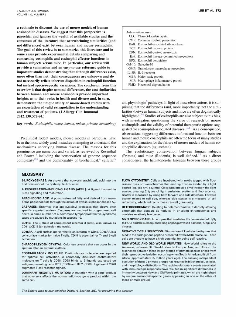
Abbreviations used
CLC: Charcot-Leyden crystal
CMP: Common myeloid progenitor
EAR: Eosinophil-associated ribonuclease
ECP: Eosinophil cationic protein
EDN: Eosinophil-derived neurotoxin
EoP: Eosinophil lineage–committed progenitors
EPX: Eosinophil peroxidase
Gal-10: Galectin-10
GMP: Granulocyte macrophage progenitor
IL-5R: IL-5 receptor
MBP: Major basic protein
MIP: Macrophage inflammatory protein
PMD: Piecemeal degranulation
J ALLERGY CLIN IMMUNOL
VOLUME 130, NUMBER 3
LEE ET AL 573
a rationale to discount the use of mouse models of humaneosinophilic diseases. We suggest that this perspective isparochial and ignores the wealth of available studies and theconsensus of the literature that overwhelming similarities (andnot differences) exist between human and mouse eosinophils.The goal of this review is to summarize this literature and insome cases provide experimental details comparing andcontrasting eosinophils and eosinophil effector functions inhuman subjects versus mice. In particular, our review willprovide a summation and an easy-to-use reference guide toimportant studies demonstrating that although differences exist,more often than not, their consequences are unknown and donot necessarily reflect inherent disparities in eosinophil functionbut instead species-specific variations. The conclusion from thisoverview is that despite nominal differences, the vast similaritiesbetween human and mouse eosinophils provide importantinsights as to their roles in health and disease and, in turn,demonstrate the unique utility of mouse-based studies withan expectation of valid extrapolation to the understandingand treatment of patients. (J Allergy Clin Immunol2012;130:572-84.)
Key words: Eosinophils, mouse, human, rodent, primate, hematology
Preclinical rodent models, mouse models in particular, havebeen the most widely used in studies attempting to understand themechanisms underlying human disease. The reasons for thisprominence are numerous and varied, as reviewed by Rosenthaland Brown,1 including the conservation of genome sequencecomplexity2,3 and the commonality of biochemical,4 cellular,5
GLOSSARY
5-LIPOXYGENASE: An enzyme that converts arachidonic acid into the
first precursor of the cysteinyl leukotrienes.
A PROLIFERATION-INDUCING LIGAND (APRIL): A ligand involved in
B-cell signaling and maturation.
ARACHIDONIC ACID: A polyunsaturated fatty acid derived from mem-
brane phospholipids through the action of cytosolic phospholipase A2.
CASPASES: Enzymes that are cysteinyl proteases that cleave after
specific aspartyl residues. Caspases are involved in programmed cell
death. A small number of autoimmune lymphoproliferative syndrome
cases are caused by mutations in caspase 10.
CD11B: The a chain of complement receptor 3 (CR3), also known as
CD11b/CD18 (an adhesion molecule).
CD45RA: A cell-surface marker that is an isoform of CD45. CD45RA is a
cell-surface marker for naive T cells. CD45 is essential for T- and B-cell
activation.
CHARCOT-LEYDEN CRYSTAL: Colorless crystals that can occur in the
sputum after an asthmatic attack.
COSTIMULATORY MOLECULE: Costimulatory molecules are required
for optimal cell activation. A commonly discussed costimulatory
molecule on T cells is CD28. CD28 binds to 2 ligands expressed on
antigen-presenting cells: B7.1 (CD80) and B7.2 (CD86). Ligation of CD28
augments T-cell receptor signals.
DOMINANT NEGATIVE MUTATION: A mutation with a gene product
that adversely affects the normal wild-type gene product within the
same cell.
The Editors wish to acknowledge Daniel A. Searing, MD, for preparing this
and physiologic6 pathways. In light of these observations, it is sur-prising that the differences (and, more importantly, not the simi-larities) between human subjects and mice are often dogmaticallyhighlighted.7-9 Studies of eosinophils are also subject to this bias,with investigators questioning the value of research on mouseeosinophils and the validity of potential therapeutic options sug-gested for eosinophil-associated diseases.10,11 As a consequence,observations suggesting differences in form and function betweenhuman and mouse eosinophils are often the focus of many studiesand the explanation for the failure of mouse models of human eo-sinophilic diseases (eg, asthma).The evolutionary conservation between human subjects
(Primata) and mice (Rodentia) is well defined.12 As a directconsequence, the hematopoietic lineages between these groups
FLOW CYTOMETRY: Cells are incubated with mAbs tagged with fluo-
rescent dyes or fluorochromes that emit light when excited by a light
source (eg, 488 nm, 633 nm). Cells pass one at a time through the light
source, creating 2 types of light emission: scatter and fluorescence.
Scatter is measured by using both forward and side detectors. Forward
scatter relates to cell size, whereas side scatter is a measure of cell
refractivity, which indirectly measures cell granularity.
HETEROCHROMATIC: Relating to heterochromatin, a densely staining
chromatin that appears as nodules in or along chromosomes and
contains relatively few genes.
MYELOPEROXIDASE: An enzyme that mediates the conversion of H2O2
to HOCl and the subsequent killing of phagocytosed bacteria, fungi, and
viruses.
NEGATIVE T-CELL SELECTION: Elimination of T cells in the thymus that
bind to the endogenous peptide presented by theMHCmolecule. These
cells are thought to have a high potential for being self-reactive.
NEW WORLD AND OLD WORLD PRIMATES: New World refers to the
Americas, whereas Old World refers to Europe, Asia, and Africa. The
distinction between these larger groups of primate species arises from
their reproductive isolation occurringwhen South America split off from
Africa (approximately 85 million years ago). The ensuing independent
evolution of these 2primate groupshas resulted in biochemical, cellular,
andmorphologic distinctions. The rapid evolutionary events associated
with immunologic responses have resulted in significant differences in
immunity between New and OldWorld primates, which are highlighted
by unique eosinophil-specific genes appearing in one or the other of
these primate groups.
glossary.
TABLE I. Human versus mouse eosinophils: review at a glance
1. Eosinophilopoiesis: Hematologic origin and development/maturation
2. Defining characteristics of mature eosinophils in peripheral circulation
d Cell morphology
d Staining properties using Romanowsky dye sets
d Electron microscopic morphology
d Cell-Surface inventory
d Evolutionary conservation, divergence, or both of characteristic
eosinophil-associated genes/proteins
3. Eosinophil location and relative abundance at homeostatic baseline and
disease
4. Eosinophil trafficking and recruitment
5. Eosinophil degranulation and mediator release
6. Eosinophils as regulators of the immune microenvironment
7. The end game: Eosinophil turnover
8. Conclusions/Summary
Supplementary tables
Table E1: Cell-surface phenotype of human and mouse eosinophils (total
references: 98*)
Table E2: Cell-surface phenotype of myeloid cells in human (total
references: 98*)
Table E3: Cell-surface phenotype of myeloid cells in mouse (total
references: 98*)
Table E4: Eosinophils in human diseases and mouse models (total
references: 151)
Table E5: Adhesion molecules used by eosinophils (total references: 43)
Table E6: Eosinophil-specific chemokines and chemokine receptors (total
references: 59)
Table E7: Eosinophil-specific chemoattractants: nonchemokines and
nonchemokine receptors (total references: 53)
Table E8: Modes of eosinophil degranulation (total references: 19)
Table E9: Mediators released from human and mouse eosinophils (total
references: 85)
*Shared reference database of Tables E1 through E3.
J ALLERGY CLIN IMMUNOL
SEPTEMBER 2012
574 LEE ET AL
are invariantly maintained in type, relative numbers, and generalmorphology.13 In particular, the eosinophil lineage–committedgranulocyte is a clearly identifiable component of both humanand mouse blood.14 For that matter, identifiable eosinophilsare found in virtually every vertebrate species, with evenmany invertebrates possessing a cell or cells with eosinophiliccharacter.14,15 The omnipresence of eosinophils nonetheless be-lies observations that disparities between human and mouse eo-sinophils exist and that these observed differences might havesignificance regarding eosinophil activities. However, we be-lieve that in the absence of only a rudimentary understandingof the roles of eosinophils in human subjects, it would be ex-ceedingly shortsighted to focus on small human versus mousedifferences, as well as to suggest that these differences are ofmore significance than the commonalities observed between hu-man and mouse eosinophils.The goal of this review is to provide both a global perspective
and a detailed review of data summarizing what is known abouteosinophils in these 2 species, including both similarities anddifferences. Table I provides the reader with a summary of theconcepts addressed in this review of human versus mouse eosin-ophils, as well as a summation of the extensive supplementary ta-bles linked to this review (ie, titles and numbers of referencescited in a given table/topic area; see Tables E1-E9 in this article’sOnline Repository at www.jacionline.org). Our objectives will be2-fold: (1) to provide an easy-to-use reference guide to importantstudies contrasting the hematopoietic origins, morphology,
proposed effector functions, and role of eosinophils in humansubjects and mice and (2) to demonstrate that although differ-ences exist, more often than not, their consequences are unknownand do not necessarily reflect inherent differences in granulocytefunction but instead species-specific variations. More signifi-cantly, despite these differences, the vast similarities between hu-man and mouse eosinophils tell us far more regarding the role ofeosinophils in health and disease.
EOSINOPHILOPOIESIS: HEMATOLOGIC ORIGIN
AND DEVELOPMENT/MATURATIONThe generation of terminally differentiated eosinophils from
pluripotent hematopoietic stem cells follows a similar path inhuman subjects and mice (Fig 1). Specifically, the isolation of aprogenitor population of eosinophils in mice has led directly tothe recent identification of eosinophil lineage–committed progen-itors (EoPs) in human subjects.16,17 In both species hematopoieticstem cells in the bone marrow progress through the multipotentprogenitor stage followed by the common myeloid progenitor(CMP) stage before branching into EoPs in human subjects orthrough granulocyte macrophage progenitors (GMPs) and intoEoPs inmice (Fig 1, reviewed byUhm et al18). The surface pheno-typeused to identifyEoPs in human subjectswasLin2, IL-5 recep-tor (IL-5R) a1, CD341, CD381, IL-3Ra1, CD45RA- whereas inmice it was Lin2, IL-5Ra1, Sca2, CD341, ckitlo.16,17Mori et al17
have revised the human CMP population to account for an IL-5Ra1 population of CMPs (ie, EoPs) from which eosinophilscan develop. Furthermore, the fact that human GMPs, unlikemouse GMPs, do not differentiate into eosinophils might suggestthat a revision of the human GMP definition is possible as well.Transcription factors important to eosinophil development are
shared by both species. In particular, GATA-1 and C/EBP-aexpression are critical to eosinophil differentiation. Deletion ofGATA-1 or C/EBP-a in mice results in loss of the eosinophillineage. Expression of a dominant negative mutation form ofGATA-1 in human cord blood progenitor cells prevented eosino-phil differentiation in culture, and ectopic expression of eitherGATA-1 or C/EBP-a in cord blood progenitors led to eosinophildifferentiation.19 Promisingly, EoPs from both species display asimilar pattern of expression of GATA-1 and C/EBP-a, as wellas other transcription factors thought to play key roles in eosino-phil differentiation in both species, including PU.1, FOG-1, andGATA-2.16,17
Cytokines important to the survival, expansion, and terminaldifferentiation of eosinophils from EoPs include IL-3, IL-5, andGM-CSF. Of these, IL-5 is the most specific to eosinophildevelopment in both species.20 Accordingly, IL-5 effectively sup-ports proliferation and survival of eosinophils from bone marrowculture in human subjects21 and mice.22
Once eosinophils have matured, they either remain in themarrow or exit into the circulation and then quickly ‘‘home’’ toselected compartments/tissues. Under inflammatory conditions,eosinophil progenitor levels are increased in the marrow and arefound in the circulation, as well as in inflamed tissues of eachspecies. Moreover, inflammation can also promote eosinophilo-poiesis at extramedullary sites in both human subjects andmice.23
In summary, evolutionary conservation of eosinophilopoiesisamong mammals ultimately represents an experimental strengthas investigators strive to understand the significance of hemato-logic changes occurring in mouse models.
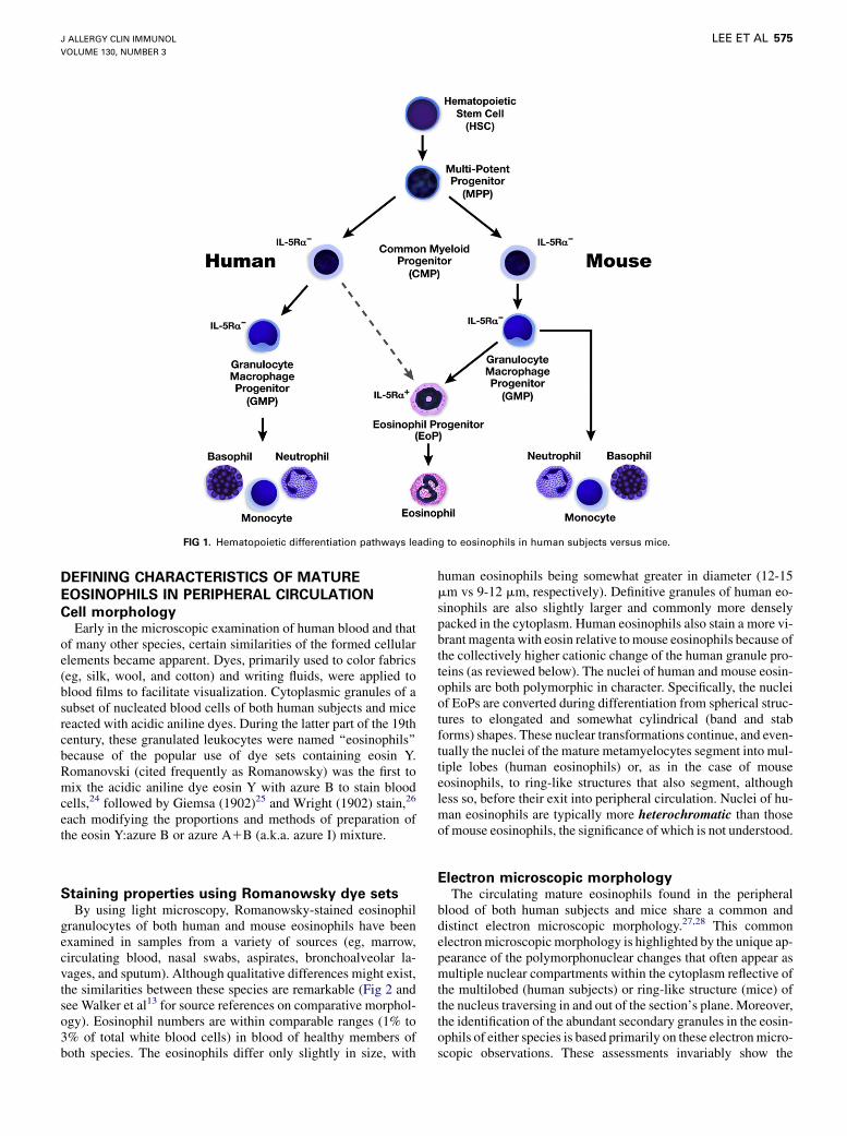
FIG 1. Hematopoietic differentiation pathways leading to eosinophils in human subjects versus mice.
J ALLERGY CLIN IMMUNOL
VOLUME 130, NUMBER 3
LEE ET AL 575
DEFINING CHARACTERISTICS OF MATURE
EOSINOPHILS IN PERIPHERAL CIRCULATION
Cell morphologyEarly in the microscopic examination of human blood and that
of many other species, certain similarities of the formed cellularelements became apparent. Dyes, primarily used to color fabrics(eg, silk, wool, and cotton) and writing fluids, were applied toblood films to facilitate visualization. Cytoplasmic granules of asubset of nucleated blood cells of both human subjects and micereacted with acidic aniline dyes. During the latter part of the 19thcentury, these granulated leukocytes were named ‘‘eosinophils’’because of the popular use of dye sets containing eosin Y.Romanovski (cited frequently as Romanowsky) was the first tomix the acidic aniline dye eosin Y with azure B to stain bloodcells,24 followed by Giemsa (1902)25 and Wright (1902) stain,26
each modifying the proportions and methods of preparation ofthe eosin Y:azure B or azure A1B (a.k.a. azure I) mixture.
Staining properties using Romanowsky dye setsBy using light microscopy, Romanowsky-stained eosinophil
granulocytes of both human and mouse eosinophils have beenexamined in samples from a variety of sources (eg, marrow,circulating blood, nasal swabs, aspirates, bronchoalveolar la-vages, and sputum). Although qualitative differences might exist,the similarities between these species are remarkable (Fig 2 andsee Walker et al13 for source references on comparative morphol-ogy). Eosinophil numbers are within comparable ranges (1% to3% of total white blood cells) in blood of healthy members ofboth species. The eosinophils differ only slightly in size, with
human eosinophils being somewhat greater in diameter (12-15mm vs 9-12 mm, respectively). Definitive granules of human eo-sinophils are also slightly larger and commonly more denselypacked in the cytoplasm. Human eosinophils also stain a more vi-brant magenta with eosin relative tomouse eosinophils because ofthe collectively higher cationic change of the human granule pro-teins (as reviewed below). The nuclei of human and mouse eosin-ophils are both polymorphic in character. Specifically, the nucleiof EoPs are converted during differentiation from spherical struc-tures to elongated and somewhat cylindrical (band and stabforms) shapes. These nuclear transformations continue, and even-tually the nuclei of the mature metamyelocytes segment into mul-tiple lobes (human eosinophils) or, as in the case of mouseeosinophils, to ring-like structures that also segment, althoughless so, before their exit into peripheral circulation. Nuclei of hu-man eosinophils are typically more heterochromatic than thoseof mouse eosinophils, the significance of which is not understood.
Electron microscopic morphologyThe circulating mature eosinophils found in the peripheral
blood of both human subjects and mice share a common anddistinct electron microscopic morphology.27,28 This commonelectronmicroscopicmorphology is highlighted by the unique ap-pearance of the polymorphonuclear changes that often appear asmultiple nuclear compartments within the cytoplasm reflective ofthe multilobed (human subjects) or ring-like structure (mice) ofthe nucleus traversing in and out of the section’s plane. Moreover,the identification of the abundant secondary granules in the eosin-ophils of either species is based primarily on these electronmicro-scopic observations. These assessments invariably show the
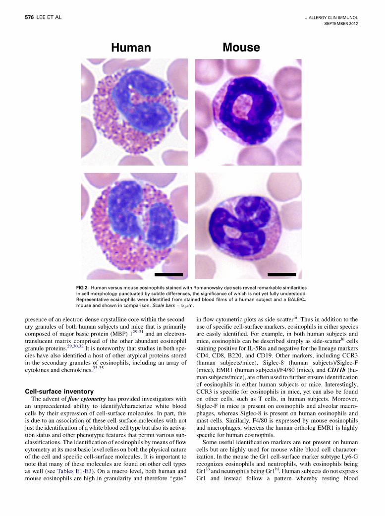
FIG 2. Human versus mouse eosinophils stained with Romanowsky dye sets reveal remarkable similarities
in cell morphology punctuated by subtle differences, the significance of which is not yet fully understood.
Representative eosinophils were identified from stained blood films of a human subject and a BALB/CJ
mouse and shown in comparison. Scale bars 5 5 mm.
J ALLERGY CLIN IMMUNOL
SEPTEMBER 2012
576 LEE ET AL
presence of an electron-dense crystalline core within the second-ary granules of both human subjects and mice that is primarilycomposed of major basic protein (MBP) 129-31 and an electron-translucent matrix comprised of the other abundant eosinophilgranule proteins.29,30,32 It is noteworthy that studies in both spe-cies have also identified a host of other atypical proteins storedin the secondary granules of eosinophils, including an array ofcytokines and chemokines.33-35
Cell-surface inventoryThe advent of flow cytometry has provided investigators with
an unprecedented ability to identify/characterize white bloodcells by their expression of cell-surface molecules. In part, thisis due to an association of these cell-surface molecules with notjust the identification of a white blood cell type but also its activa-tion status and other phenotypic features that permit various sub-classifications. The identification of eosinophils by means of flowcytometry at its most basic level relies on both the physical natureof the cell and specific cell-surface molecules. It is important tonote that many of these molecules are found on other cell typesas well (see Tables E1-E3). On a macro level, both human andmouse eosinophils are high in granularity and therefore ‘‘gate’’
in flow cytometric plots as side-scatterhi. Thus in addition to theuse of specific cell-surface markers, eosinophils in either speciesare easily identified. For example, in both human subjects andmice, eosinophils can be described simply as side-scatterhi cellsstaining positive for IL-5Ra and negative for the lineage markersCD4, CD8, B220, and CD19. Other markers, including CCR3(human subjects/mice), Siglec-8 (human subjects)/Siglec-F(mice), EMR1 (human subjects)/F4/80 (mice), and CD11b (hu-man subjects/mice), are often used to further ensure identificationof eosinophils in either human subjects or mice. Interestingly,CCR3 is specific for eosinophils in mice, yet can also be foundon other cells, such as T cells, in human subjects. Moreover,Siglec-F in mice is present on eosinophils and alveolar macro-phages, whereas Siglec-8 is present on human eosinophils andmast cells. Similarly, F4/80 is expressed by mouse eosinophilsand macrophages, whereas the human ortholog EMR1 is highlyspecific for human eosinophils.Some useful identification markers are not present on human
cells but are highly used for mouse white blood cell character-ization. In the mouse the Gr1 cell-surface marker subtype Ly6-Grecognizes eosinophils and neutrophils, with eosinophils beingGr1lo and neutrophils being Gr1hi. Human subjects do not expressGr1 and instead follow a pattern whereby resting blood
J ALLERGY CLIN IMMUNOL
VOLUME 130, NUMBER 3
LEE ET AL 577
eosinophils would be CD14lo/CD16lo and then CD14med/CD16med on activation. These changes in expression could haveconsequences in various studies given that peripheral blood eosin-ophils are generally isolated away from neutrophils by mean ofmagnetic depletion of CD16hi neutrophils.
As just described, cell-surface markers are also an indicator ofactivation status of the cell. In particular, eosinophils exposed toactivating agents, such as platelet-activating factor, increase theirexpression of CD69 in both human subjects and mice. Addition-ally, mouse and human eosinophils will upregulate chemokinereceptors (eg, CX3CR1), costimulatory molecules, and antigenpresentation molecules (eg, CD86 and MHC class II) on activa-tion. The study of eosinophil surface markers is expanding butnonetheless remains behind studies of other leukocytes, such asneutrophils and mononuclear cells, including monocytes, lym-phocytes, and dendritic cells, in both human subjects and mice(see Table E2 vs Table E3). It is likely that similar to the increasesin subclassifications associated with other white blood cells, eo-sinophil subtype classifications will grow significantly in thenext few years.36We anticipate that this growth in eosinophil sub-type classifications will likely occur in both species, with moresimilarities rather than differences noted.
Evolutionary conservation, divergence, or both of
characteristic eosinophil-associated genes/proteinsDiscussions of eosinophil-associated genes/proteins invariably
focus on the genes encoding either the prominent groups ofsecondary granule proteins (ie, eosinophil MBPs, eosinophil-associated ribonucleases [EARs], and eosinophil peroxidase[EPX]37) or the abundant and primarily cytoplasmic proteinthat composes the Charcot-Leyden crystal (CLC), a structurecharacteristically found at sites of eosinophil-mediated inflamma-tion.38 The commonalities and lack thereof of these genes/pro-teins between human subjects and mice will be used tohighlight general principles and concepts. However, it is impor-tant to keep in mind that the significance of either the reported dif-ferences or similarities of these genes/proteins between humansubjects and mice is often obscured by the confounding lack ofa clear understanding of the function of the encoded proteins(MBPs and CLC), the role the identified protein function has ineosinophil-mediated activities (EARs and EPX), or the relativeimportance of degranulation and the site-specific release of theseproteins.33 In addition, it is also noteworthy that other eosinophil-associated genes/proteins of significance exist and that in manycases these are noted in other areas of this review.Eosinophil MBPs. Orthologous gene pairs (MBP-1 and
MBP-2) are found in both human subjects and mice.39-42 In bothspecies the abundance of the protein encoded by MBP-1 is thehighest on a molar basis among the eosinophil secondary granuleproteins and is greater than an order of magnitude higher relativeto the protein encoded by the MBP-2 gene.41,42 Nonetheless, sig-nificant differences between human subjects and mice exist interms of both the expression of these genes and the biochemistryof the encoded proteins. HumanMBP-1, unlike mouse MBP-1, istranscribed from multiple promoters.43 Human and mouseMBP-1 appear to be predominantly expressed in eosinophils(composing the electron-dense cores characteristic of the eosino-phil secondary granule). However, in human subjects (unknownin the mouse) expression extends to other leukocytes.44,45 In hu-man subjects MBP-1 is highly cationic, whereas MBP-2 has lost
this cationic character.42 In contrast, these 2 proteins retain theircationic character in the mouse.41 Finally, in human subjects ex-pression of MBP-1 extends to nonleukocytes, such as placental Xcells, which leads to the accumulation of the unprocessed ‘‘pro’’-form of this protein during pregnancy.46 This placental expressionand serum accumulation of pro–MBP-1 does not occur in themouse.41
EARs. The EARs are a rapidly evolving group of genes thatencode multiple family members stored in the electron-translucent matrix of the secondary granules of both human(eosinophil cationic protein [ECP]47 and eosinophil-derived neu-rotoxin [EDN]48) and mouse (Ear-1, Ear-2, Ear-6/7, and Ear-5/1149,50) eosinophils. These stored ribonucleases are second onlyto MBP-1 in abundance on a molar basis in both species. ECPis a very cationic protein with weak ribonuclease activity48 foundonly in human subjects and other Old World primates.51 In com-parison, EDN is a nominally cationic protein that has a strong ri-bonuclease activity,51 with orthologs present in both Old WorldandNewWorld primates.51 The mouse EARs are not orthologouswith human ECP and EDN and instead represent distant paralo-gous genes the expansion of which occurred well after the diver-gence of Primata and Rodentia.50 In the mouse all of the EARs arenominally cationic proteins, with each retaining strong ribonu-cleolytic activities.49,50 In both human subjects andmice, these ri-bonucleases are stored as mature proteins retaining activity ineosinophil secondary granules. Moreover, in both species thisfamily of genes is expressed in many cell types other than eosin-ophils, including other leukocytes52,53 and nonhematopoieticcells.49,54
EPX. EPX genes in human subjects and mice are orthologousgenes encoding proteins that display significant peroxidaseenzymatic activities and an extraordinarily high level of sequenceidentity at the amino acid level (>94%55,56). In both species EPXis found as part of a closely linked leukocyte-specific pair of per-oxidase genes (myeloperoxidase-EPX) the duplication event ofwhich occurred before the divergence of Primata and Rodentia.12
Similar to the encoded EARs, EPX in both human subjects andmice is stored as a mature functional protein in the electron-translucent matrix of eosinophil secondary granules and is themost abundant proteins (by mass) found in the granules of bothspecies. Among all of the eosinophil granule proteins in either hu-man subjects or mice, EPX appears to be the most eosinophil spe-cific, with no published reports demonstrating expression in otherleukocytes or nonhematopoietic cell types.CLC protein. Human CLC protein was originally identified
as a lysophospholipase expressed predominantly in the cytoplasmbut also present in the primary granules of eosinophils.57 How-ever, subsequent studies have demonstrated that human CLC isnot a lysophospholipase and instead is a member of the larger ga-lectin family that is also described as galectin-10 (Gal-10).38
Studies of CLC/Gal-10 in human subjects demonstrated that itis one of the most highly expressed genes in eosinophils45 andthat this expression was not limited to eosinophils, with tran-scripts also noted in regulatory T cells.58 The activities mediatedby CLC/Gal-10 in either eosinophils or T cells are unclear, and inturn, the role or roles of CLC/Gal-10 in human subjects remainobscure. Surprisingly, despite its prominent level of expressionin eosinophils and the relative conservation of the galectin genefamily between human subjects and mice, genome sequencinghas demonstrated unambiguously that CLC/Gal-10 is absentfrom the mouse genome,3 which is consistent with the lack of
J ALLERGY CLIN IMMUNOL
SEPTEMBER 2012
578 LEE ET AL
reports in the literature noting the presence of this structure inmouse models of human disease.
EOSINOPHIL LOCATION AND RELATIVE
ABUNDANCE AT HOMEOSTATIC BASELINE AND
DISEASEThe 20th century saw many significant attempts to expand on
the observations of Paul Erlich59 and correlate eosinophil infiltra-tion of organs/tissues with health and disease, such as the work ofHuber and Koessler.60 These ongoing studies continue today withever-increasing scope and complexity.61 A commonality that sur-rounds all of these studies (despite the hyperbole associated withsome reviews in the literature9,10) is that the identification of eo-sinophils in tissues/organs during health and disease has beenstrikingly similar between human subjects and mice. For exam-ple, both human subjects and mice have similar peripheral bloodand marrow distribution of eosinophils relative to other whiteblood cells; as such, eosinophils comprise approximately 2% ofthe peripheral white blood cells and approximately 8% of bonemarrow leukocytes.13 Eosinophils are also found at homeostaticbaseline of otherwise healthy human subjects andmice in the gas-trointestinal tract (ie, stomach to rectum), thymus, secondarylymphoid tissues, uterus, and adipose tissue.61 The specific andunique role or roles of eosinophils in the tissues/organs of healthysubjects in both species is the subject of current debates and inves-tigations. However, the commonality of this tissue distribution be-tween human subjects and mice suggests eosinophils contributeto tissue/organ-specific pathways mediating the maintenance ofhomeostasis15 and that these roles have been conserved sincethe radiation of the major extant mammalian orders.12
An equally provocative conservation between human subjectsand mice occurs when examining the relative increase in eosin-ophil numbers and location as part of the changes associated withdifferent diseases (see Table E4). In many cases the specific andunique role or roles of eosinophils in these tissues remains to bedefined; yet again, the overwhelming consensus is that far moresimilarities between human subjects and mice exist relative tosmall variations that have been observed (many of which remainunclear, unresolved, or both).Interestingly, this list of eosinophil-associated diseases in
human subjects has expanded dramatically over the last 2decades. Thus eosinophils are now linked with an expansivenumber of diseases in human subjects beyond simply parasiticinfections and allergic diseases, such as asthma.62 More impor-tantly, as documented in Table E4, this expanding number ofeosinophil-associated human diseases has been accompanied bya concomitant increase in mouse models of human disease withsimilar eosinophil associations. This commonality again suggestsan underlying conservation of eosinophil effector functionsamong mammals. In turn, this lack of substantive differences be-tween human subjects and mice raises expectations for the utilityand ultimately the validity of mouse models in studies ofeosinophil-associated diseases in human subjects.
EOSINOPHIL TRAFFICKING AND RECRUITMENTInflammatory cytokines are released in response to tissue
injury, infection, or both, stimulating the surrounding cells toproduce adhesion molecules and chemotactic factors that signalthe recruitment of various leukocytes, including eosinophils.
The movement of eosinophils out of the circulation to sites ofinflammation is a well-orchestrated process that involves adhe-sion to the vascular endothelium, diapedesis into surroundingtissues, and trafficking to inflammatory sites. This process hasbeen well studied in both human subjects and mouse models,with very little disparity. Tables E5, E6, and E7 provide detailsavailable regarding the similarities/differences between humansubjects and mice associated with eosinophil diapedesis and re-cruitment into inflamed tissues. Specifically, they address simi-larities/differences in cell adhesion molecules (see Table E5),chemokines and their corresponding receptors (see Table E6),and nonchemokines and their receptors (see Table E7). As is ap-parent from these tables, there are only a few differences be-tween human and mouse eosinophils with respect totrafficking and recruitment. Despite this overwhelming similar-ity in human versus mouse recruitment/trafficking, several areasof debate and/or notable difference in eosinophil chemotaxisexist.First, although CCR3-mediated chemotactic events between
human subjects and mice are conserved, differences exist in theexpression of the various eotaxin chemokines. There are 3 knownhuman eotaxin chemokines (eotaxin-1 [CCL11], eotaxin-2[CCL24], and eotaxin-3 [CCL26]) expressed in response toinflammatory events. Mice, on the other hand, only expresseotaxin-1 (CCL11) and eotaxin-2 (CCL24) and possess only aneotaxin-3 (CCL26) pseudogene (ie, a nonfunctional gene). Inaddition, mice do not display eosinophil chemotaxis in responseto human eotaxin-3.63
Second, RANTES is an eosinophil agonist chemokine (CCL5)that is also involved in recruitment of monocytes, eliciting effectsthrough interactions with either CCR3 or CCR1 receptors. Inhuman subjects RANTES (CCL5) binds directly to appropriatereceptors on eosinophils to signal their recruitment.64 Miceexpress a RANTES ortholog (CCL5), and in vivo experiments ap-pear to show that it also elicits the recruitment of eosinophils.65-68
However, purified mouse eosinophils do not chemotax in directresponse to RANTES (CCL5), likely because of the inability ofmouse RANTES (CCL5) to bind to mouse CCR3.63 This suggeststhat although RANTES (CCL5) might mediate eosinophil che-motaxis in the mouse, it occurs through an unknown mechanismormechanisms of activationmediated by other concurrent inflam-matory pathways. Thus species-specific differences in cytokine/chemokine-mediated trafficking events might exist in detail, butother selective pressures have maintained a degree of commonfunctionality.Third, as noted above, small molecule–mediated chemotaxis of
eosinophils is a central component of recruitment and trafficking,particularly products of arachidonic acid metabolism, such asleukotriene B4.
69 However, many other lipid mediators withvery robust eosinophil chemotactic properties have been identi-fied, with 5-oxo-ETE as a representative example. This lipid me-diator is generated from its precursor, 5-HETE, through oxidationin the 5-lipoxygenase pathway of arachidonic acid metabolism.In human subjects it is a strong chemoattractant that specificallybinds to the oxoeicosanoid receptor 1, which is highly expressedon eosinophils.70-73 In mice its role in eosinophil chemotaxis isnot yet fully elucidated. Specifically, studies have not demon-strated a strong chemotactic response (our unpublished observa-tions), possibly because of the lack of the specific receptor (ie,oxoeicosanoid receptor 1) that has yet to be identified on thecell surface of mouse eosinophils.74
J ALLERGY CLIN IMMUNOL
VOLUME 130, NUMBER 3
LEE ET AL 579
EOSINOPHIL DEGRANULATION AND MEDIATOR
RELEASE
Eosinophil degranulationHuman and mouse eosinophils are, by definition, granulocytes,
and for obvious reasons, the process of degranulation (ie, theextracellular release of granule contents) has been suggested tobe singularly important to eosinophil effector function or func-tions. Specifically, after 1 or more events characterized as‘‘priming’’ or ‘‘activation,’’ eosinophils are capable of rapid andstimulus-specific release of intact granules, preformed granularcontents, or both independent of the classical ER-Golgi secretionpathway. Clinical studies, in vitro/ex vivo experimentation, andassessments of mouse models of human disease suggest that 4mechanisms, pathways, or both leading to degranulation existthat can vary between human subjects and mice (see Table E8).These mechanisms include (1) classical exocytosis (ie, the releaseof granule contents through the fusion of individual granules andthe plasma membrane); (2) compound exocytosis (ie, homotypicfusion of multiple secretory granules to form ‘‘supergranules’’ be-fore fusion with the plasma membrane); (3) piecemeal degranula-tion (PMD; ie, gradual stepwise release of granule contentsthrough secretory vesicles budding, mobilizing, and fusing withthe plasma membrane); and (4) cytolytic release of otherwise in-tact granules (ie, the release of otherwise intact cytoplasmic gran-ules as a consequence of rupturing the plasma membrane as partof necrotic cell death).Studies of both human subjects and mouse models have
demonstrated that PMD is a common mechanism of eosinophildegranulation. Secretory vesicles fusing with the plasma mem-brane have been detailed elegantly in several in vitro/ex vivo stud-ies using human eosinophils.75-80 PMD in human subjects hasalso been shown to occur in response to defined environmentallyrelevant stimuli (eg, antibody-mediated cross-linking of Fc recep-tors81). However, such demonstrations are limited when investi-gating mouse eosinophils. Although the available data suggestthat mouse eosinophils are capable of undergoing PMD, exam-ples of this phenomenon in the mouse are rare by comparisonand only mechanistically suggestive. For example, exposure ofmouse eosinophils in vitro to 1 or more agonists appears to elicitthe piecemeal release of eosinophil granule proteins,82,83 and insome cases these activated eosinophils display ultrastructuralchanges detectable by using electron microscopy suggestive ofPMD.83 In vivo studies of mouse disease models also provide ev-idence suggestive of PMD, including assessments of eosinophilsrecruited to the lung in response of allergen provocation84,85 andeosinophils accumulating in the lung as a consequence of consti-tutive expression of IL-5 and eotaxin-2 (CCL24).86
Studies of patients highlight the surprisingly wide scope andextensive character of ECL as a mode of release of humaneosinophil granule components. The extracellular deposition ofotherwise intact eosinophil secondary granules within tissues arereadily detectable in patients with allergic asthma,87 rhinitis,88
atopic dermatitis,89 acute lung injury,90 and eosinophilic esopha-gitis.91,92 The commonality of this phenomenon has led investiga-tors to give a name to these released and largely intact eosinophilgranules: clusters of free eosinophil granules.93 Indeed, recentstudies in both human subjects andmice have suggested that theseextracellular granules might even function autonomously,responding to physiologically relevant stimuli in their tissuemicroenvironment.83,94 However, in contrast to human subjects,
documented cases of ECL occurring in mouse models ofhuman disease are very limited, and even these cases are moresubjective interpretation than demonstrable evidence of thismechanism.86,95
Collectively, the data show that the release of eosinophilgranule proteins (ie, degranulation) is a phenomenon occurring inboth human subjects and mice. Nonetheless, species-specificmechanisms achieving this degranulation in vivo are also evident.This dichotomy is highlighted as the easily observable eosinophildegranulation that occurs in human asthmatic patients96,97 incontrast to only sporadic reports of nominal (if any)eosinophil degranulation occurring in the lungs in mousemodels.31,32,82,86,98-100 These observations demonstrate that theresponses of human and mouse eosinophils to physiologic stimuliin all cases are not necessarily the same, and in regard to asthmastudies, these differences have led some investigators to questionthe relevance/importance of mouse models.10,11 Unfortunately,the resolution of this and other debates regarding the utility ofmousemodels of human disease has been slow in coming becausethe significance of these species-specific differences has often re-mained unclear. Moreover, the role of degranulation itself in eo-sinophil effector function or functions in either human subjectsor mice is still debatable and awaits a more complete experimen-tal definition.33
Eosinophil mediator releaseThe importance and critical character of the release by eosin-
ophils of various mediators is highlighted by the wealth ofoutstanding reviews with in-depth discussions published re-cently.83,95,101-105 They describe and review efforts to elucidatetriggers, mechanisms, and subsequent pathways resulting frommediator release in the context of homeostasis and disease pathol-ogy. Table E9 is a comprehensive summary of mediators secretedfrom eosinophils, stimuli/conditions that cause/aid this release,and exhaustive references to the respective studies. This summaryis inclusive of both in vitro/ex vivo studies of isolated populationsof eosinophils (thus avoiding ambiguity as to the source of themediators), as well as examples of eosinophil degranulation andmediator release in vivo. For the most part, the available studiesdo not necessarily allow exact comparisons between human andmouse eosinophils; few complementary studies of these granulo-cytes exist using the same experimental conditions, agonist stim-uli (ie, in most cased responses were achieved with differentsecretagogues), or identical in vivo provocations (a rare exceptionis Meeusen and Balic106). Despite the comparative difficulties, itis extraordinarily informative that even though experimental con-ditions have often varied in detail, the composition of granule pro-teins and cellular mediators released from either human or mouseeosinophils in response to comparable stimuli have largelyoverlapped.As shown in Table E9, and as discussed earlier, human and
mouse eosinophils are each capable of (1) secreting cationic pro-teins from their secondary granules that are often orthologous inorigin; (2) releasing reactive oxygen species on priming/activa-tion; (3) secreting cytokines, chemokines, growth factors, andlipid mediators as part of innate and acquired immune re-sponses/inflammation; and (4) releasing mitochondrial DNAand other antibacterial agents as part of innate host defense.The mediators and secretagogues studied in one species were
J ALLERGY CLIN IMMUNOL
SEPTEMBER 2012
580 LEE ET AL
often not studied in the other, and thus it is noteworthy that appar-ent human versus mouse variations are not necessarily differencesbut simply the lack of overlapping datasets (see Table E9). For ex-ample, the secretion of b-glucuronidase, soluble CD16, ENA-78,GM-CSF, IL-8, kynurenins, macrophage inflammatory protein(MIP) 3, matrix metalloproteinase 9, nerve growth factor, osteo-pontin, stem cell factor, and TGF-a has been studied extensivelyin human subjects. However, similar studies have not been re-ported in the mouse or at best have only tangentially been ad-dressed. Alternatively, the release of C10, ELC, IL-17, IL-2,interferon-inducible protein 10, monocyte chemoattractant pro-teins 1 and 5, macrophage-derived chemokine, monokine inducedby IFN-g, MIP-1a, MIP-1g, MIP-2, thymus and activation-regulated chemokine, and TGF-b secretion was studied in farmore detail in mouse eosinophils in comparison with their humancounterparts.It is also instructive to recognize that the studies available in
the literature have noted species-specific differences inmediator release between human and mouse eosinophils. Thesedifferences include secretagogues that commonly initiate degran-ulation in human eosinophils (IL-5, IL-3, GM-CSF, and eotaxin-1 [CCL-11]) but have little to no effect on mouse eosinophils. Inaddition, some stimuli, such as platelet-activating factor, elicit thesecretion of IL-9, IL-13, IL-1 receptor a, basic fibroblast growthfactor, RANTES, and platelet-derived growth factor b fromhuman eosinophils but did not have similar effects on mouseeosinophils derived ex vivo from bone marrow cultures.106 As isoften the case in comparisons of human and mouse eosinophils,the significance (if any) of differences noted in mediator releaseare unknown. Thus although these differences exist and are exper-imentally repeatable, they might represent only nominal varia-tions between distinct mammalian species and not necessarilyfundamental divergences in eosinophil effector functions.
EOSINOPHILS AS REGULATORS OF THE IMMUNE
MICROENVIRONMENTAs noted earlier, hematologic systems and immune pathways
are very similar between human subjects and mice. This isinherently evident by the use of the mouse as a model of humandisease not only in academic research but also in pharmaceuticalindustry studies. However, only recently have studies of bothhuman and mouse eosinophils in health and disease enabled anappreciation of the eosinophil as a specific immune regulatorycell on par with the functionality of other regulatory cells (eg,dendritic cells).15,103,107,108
Eosinophils from both human subjects and mice are similar intheir capacities to function as both innate cells and regulators ofadaptive immunity. This includes the expression of surfacemarkers of immunologic relevance (see Table E1), the expressionof immune-modulating cytokines (TH1: IL-12 and IFN-g; TH2:IL-4, IL-5, IL-9, IL-13, IL-25; proinflammatory: IL-6, IL-1b,and TNF-a; suppressive: TGF-b, IL-10, and indolamine-2,3,oxy-genase35,109), proteases (eg, matrix metalloproteinase 9110,111),and lipid mediators (eg, leukotrienes and prostaglandins112,113),the activation of receptors eliciting innate responses to selectpathogens (eg, Toll-like receptors114,115 and proteinase-activated receptor 2116-118), the generation of reactive oxygen spe-cies,119 and the release of genomic/mitochondria/DNA.120 Someof these immune mediators have been described in human sub-jects but do not exclude their expression in mice. Thus many of
the differences between human subjects and mice are either a re-sult of insufficient research, an inability to assess eosinophil activ-ities in human subjects, or true interspecies variations.The role of eosinophils as immune regulators of the adaptive
immune system was originally realized on the isolation ofeosinophils from human subjects that were found to haveincreased MHC class II and costimulatory receptor expres-sion.121-123Mousemodels enabled the elucidation of this functionand demonstrated a similar capacity to process and present anti-gens,124,125 migrate to the secondary lymph nodes,126 and inducecytokine release from T cells.127,128 Through the use ofeosinophil-deficient animals, eosinophils were demonstrated tomodulate directly the local immune polarization of TH2 versusTH1/TH17 T-cell responses.
129 These complex systemic pathwaysare not possible to measure in human subjects with current meth-odologies but likely occur based on correlations found betweeneosinophils and immune polarization in various disease states,such as viral exacerbation in asthmatic patients,130 transplant re-jection,131 and autoimmune diseases,132 as well as potentially re-lated/similar inflammatory events (eg, inflammatory boweldisease133).
Additional immune responses that have only been demon-strated to date in themouse but are not excluded to occur in humansubjects include the role of eosinophils in plasma B-cell survivalthrough the release of IL-6 and a proliferation-inducing ligand(APRIL).134 This might have particular significance in humansubjects, who also rely on IL-6 and a proliferation-inducing lig-and for plasma B-cell survival.135Moreover, in themouse glucosehomeostasis has also been demonstrated to be mediated byeosinophil-dependent immune regulation of macrophages in adi-pose tissue.136 These immune regulatory activities of eosinophilson macrophages in mice might represent a novel inflammatoryprocess linked to metabolic diseases of human subjects, inwhom inflammatory macrophages are known to be significantmediators of disease.137 In addition, mouse eosinophils havebeen shown to modulate immune environments for remodelingand repair in health and disease, such as reproductive develop-ment138,139 and neuronal branching and growth.140 In the latterexample parallels between human and mouse eosinophils arehighlighted by similar capabilities to express neuromediators,suggesting these mechanisms are translational.141
Eosinophil associations with underlying immune events in alarger context have also been described in both human subjectsand mice. For example, human and mouse eosinophils have beenshown to localize to the thymus and are hypothesized to have acontributory role in the process of negative T-cell selection,142,143
TH2 polarization of the thymus,142,144 or both. In both human sub-jects and mice, eosinophil Toll-like receptor activation is associ-ated with antiviral activities.145-147 Moreover, eosinophilimmune/remodeling pathways are likely similar between humansubjects and mice in response to cancer and transplant rejection,particularly because eosinophils are found in parallel tissue sitesin equivalent numbers (see Table E4).
In summary, just as dendritic cells, T lymphocytes, macro-phages, B cells, and mast cells, for example, have similar immuneactivities between human subjects and mice, eosinophils fromthese species also appear to display similar immune-mediatingfunctions. Differences that do arise are likely due to evolutionaryevents that result in paralogous functions or alternative pathwaysleading to the same end point. Occasionally, evolutionary diver-gences do exist, yet they appear to be limited in number and
J ALLERGY CLIN IMMUNOL
VOLUME 130, NUMBER 3
LEE ET AL 581
represent only a minor portion of the total immune response orresponses. Thus the further eosinophils are studied in bothpatients and mouse models of human disease, the more similartheir respective immune activities and functions becomeapparent.
THE END GAME: EOSINOPHIL TURNOVEREosinophil survival and death are regulated by the local
immune microenvironment. The cytokines IL-3, IL-5, and GM-CSF generally enable survival of eosinophils in both humansubjects148 and mice.149 Withdrawal of these prosurvival cyto-kines is often sufficient for cellular stress-induced cell deaththrough activation of caspases. Although the signaling pathwaysleading to passive cell death in human and mouse eosinophilshave not been compared side by side, they likely rely on similarmechanisms and play a similar role in basal turnover. The causesof actively induced apoptosis vary between human and mouse eo-sinophils, but there are also many similarities. For example, hu-man150 and mouse151 eosinophils undergo apoptosis on ligationof Fas (CD95), particularly in the presence of TNF-a andIFN-g, leading to activation of caspase-3 and caspase-8. Simi-larly, human and mouse eosinophils express paralogous sialicacid–binding immunoglobulin-like lectins linked with eosinophilapoptosis (see Table E1). The presence of IL-5 enhances the ap-optotic mediating effects of these lectins in human subjects andresults in activation of reactive oxygen species and mitochondrialdysfunction152; similar effects on mouse eosinophils have yet tobe reported.153 The identification of additional targets, such asCD30154 and CD300a,155 described on human eosinophils doesnot preclude their presence or function on mouse eosinophils. Fi-nally, both human and mouse eosinophils are susceptible tocorticosteroid-induced cell death. Corticosteroid binding to re-ceptors on eosinophils is proposed to lead to downregulation ofIL-5, inhibition of nuclear factor kB, and the activation of cas-pases.156-158 This highlights a key feature for the translational ap-plication of mice as a model of human disease: the majority ofcellular targets for eosinophil-induced death are retained betweenthese 2 mammalian species.
CONCLUSIONS/SUMMARYThe exhaustive summary presented here and in Tables E1
through E9 provides a clear demonstration of the thesis articu-lated in the introductory paragraphs. Specifically, genetic, bio-chemical, molecular, and morphologic differences betweenhuman and mouse eosinophils exist. However, most of these dif-ferences are often either of unknown significance or ‘‘cosmetic’’in character, leaving what we believe is an inherent underlyingtruth: eosinophils and their associated effector functions are activ-ities all mammals gained from a protomammalian ancestor, withspecific differences subsequently arising because of selectivepressures on each extant mammalian order and, in turn, individualfamilies, species, or both. Thus the structural and, more impor-tantly, functional similarities between human and mouse eosino-phils simply reflect the amazing level of propinquitydemonstrated from the available genomic sequence studies.This conclusion has significant logistic consequences on our un-derstanding of human disease and the development of therapeuticoptions. Specifically, the significant overlap and similarities be-tween human and mouse eosinophils suggested by the literature
highlights the unique utility of mouse-based studies with an ex-pectation of valid extrapolation to the understanding and treat-ment of human eosinophil-associated diseases.
We thank the editors of the Journal of Allergy and Clinical Immunology for
this incredible opportunity to summarize the difficulties and complexities as-
sociated with interspecies comparisons of human and mouse eosinophils. In
manyways this was a chance for our group to reflect on a lifetime of eosinophil
studies that were predicated on the belief that mice are important and valuable
models of human disease. In addition, we thank everyone in and out of the re-
search and clinical communities who put up with our pathologic interests in
both eosinophils and the mouse as a model system. Indeed, the discussions
and arguments over the years in our pursuit of all things eosinophilsmore often
than not are the source of the ideas and insights noted in this article. Moreover,
we acknowledge the tireless efforts of Mandana Minai to compile and orga-
nize the wealth of references included in the article, as well as in Tables E1
through E9. We also thank the Mayo Clinic Arizona medical graphic artist
Marv Ruona and Joseph Esposito of Research Library Services for their un-
ending efforts and support. Finally, we express our gratitude to the Lee Labo-
ratories administrative staff (Linda Mardel and Charlie Kern), without whom
we could neither function as an integrated group nor achieve the degree of suc-
cess that we have experienced.
REFERENCES
1. Rosenthal N, Brown S. The mouse ascending: perspectives for human-disease
models. Nat Cell Biol 2007;9:993-9.
2. Church DM, Goodstadt L, Hillier LW, Zody MC, Goldstein S, She X, et al. Lin-
eage-specific biology revealed by a finished genome assembly of the mouse.
PLoS Biol 2009;7:e1000112.
3. Waterston RH, Lindblad-Toh K, Birney E, Rogers J, Abril JF, Agarwal P, et al.
Initial sequencing and comparative analysis of the mouse genome. Nature
2002;420:520-62.
4. Evsikov AV, Dolan ME, Genrich MP, Patek E, Bult CJ. MouseCyc: a curated bi-
ochemical pathways database for the laboratory mouse. Genome Biol 2009;10:
R84.
5. Mestas J, Hughes CC. Of mice and not men: differences between mouse and hu-
man immunology. J Immunol 2004;172:2731-8.
6. Kararli TT. Comparison of the gastrointestinal anatomy, physiology, and bio-
chemistry of humans and commonly used laboratory animals. Biopharm Drug
Dispos 1995;16:351-80.
7. Rangarajan A, Weinberg RA. Opinion: comparative biology of mouse versus hu-
man cells: modelling human cancer in mice. Nat Rev Cancer 2003;3:952-9.
8. de Jong M, Maina T. Of mice and humans: are they the same?-implications in
cancer translational research. J Nucl Med 2010;51:501-4.
9. Wenzel S, Holgate ST. The mouse trap: it still yields few answers in asthma. Am
J Respir Crit Care Med 2006;174:1173-8.
10. Persson CG, Erjef€alt JS, Korsgren M, Sundler F. The mouse trap. Trends Pharma-
col Sci 1997;18:465-7.
11. Wenzel SE. Eosinophils in asthma—closing the loop or opening the door? N Engl
J Med 2009;360:1026-8.
12. McKenna MG. The origin and early differentiation of therian mammals. Ann N Y
Acad Sci 1969;167:217-40.
13. McGarry MP, Protheroe CA, Lee JJ. Mouse hematology. 1st ed. New York: Cold
Spring Harbor Laboratory Press; 2009.
14. McGarry MP. The evolutionary origins and presence of eosinophils in extant spe-
cies. In: Lee JJ, Rosenberg H, editors. Eosinophils in health and disease. Waltham
(MA): Elsevier; 2012.
15. Lee JJ, Jacobsen EA, McGarry MP, Schleimer RP, Lee NA. Eosinophils in health
and disease: the LIAR hypothesis. Clin Exp Allergy 2010;40:563-75.
16. Iwasaki H, Mizuno S, Mayfield R, Shigematsu H, Arinobu Y, Seed B, et al. Iden-
tification of eosinophil lineage-committed progenitors in the murine bone mar-
row. J Exp Med 2005;201:1891-7.
17. Mori Y, Iwasaki H, Kohno K, Yoshimoto G, Kikushige Y, Okeda A, et al. Iden-
tification of the human eosinophil lineage-committed progenitor: revision of phe-
notypic definition of the human common myeloid progenitor. J Exp Med 2009;
206:183-93.
18. Uhm TG, Kim BS, Chung IY. Eosinophil development, regulation of eosinophil-
specific genes, and role of eosinophils in the pathogenesis of asthma. Allergy
Asthma Immunol Res 2012;4:68-79.
19. McNagny K, Graf T. Making eosinophils through subtle shifts in transcription
factor expression. J Exp Med 2002;195:F43-7.

J ALLERGY CLIN IMMUNOL
SEPTEMBER 2012
582 LEE ET AL
20. Sanderson CJ. Interleukin-5, eosinophils, and disease. Blood 1992;79:3101-9.
21. Clutterbuck EJ, Hirst EM, Sanderson CJ. Human interleukin-5 (IL-5) regulates
the production of eosinophils in human bone marrow cultures: comparison and
interaction with IL-1, IL-3, IL-6, and GMCSF. Blood 1989;73:1504-12.
22. Dyer KD, Moser JM, Czapiga M, Siegel SJ, Percopo CM, Rosenberg HF. Func-
tionally competent eosinophils differentiated ex vivo in high purity from normal
mouse bone marrow. J Immunol 2008;181:4004-9.
23. Radinger M, Lotvall J. Eosinophil progenitors in allergy and asthma—do they
matter? Pharmacol Ther 2009;121:174-84.
24. Romanovski DL. On the question of parasitology and therapy of malaria. St Pe-
tersburg (Russia): Imperial Medical Military Academy; 1891.
25. Giemsa G. F€arbemethoden f€ur malariaparasiten. Zentralbl Bakteriol 1902;31:
429-30.
26. Wright JH. A rapid method for the differential staining of blood films and malar-
ial parasites. J Med Res 1902;7:138-44.
27. Dvorak AM, Ishizaka T. Human eosinophils in vitro. An ultrastructural morphol-
ogy primer. Histol Histopathol 1994;9:339-74.
28. Miller F, de Harven E, Palade GE. The structure of eosinophil leukocyte granules
in rodents and in man. J Cell Biol 1966;31:349-62.
29. Dvorak AM, Furitsu T, Estrella P, Letourneau L, Ishizaka T, Ackerman SJ. Ultra-
structural localization of major basic protein in the human eosinophil lineage
in vitro. J Histochem Cytochem 1994;42:1443-51.
30. Egesten A, Alumets J, von Mecklenburg C, Palmegren M, Olsson I. Localization
of eosinophil cationic protein, major basic protein, and eosinophil peroxidase in
human eosinophils by immunoelectron microscopic technique. J Histochem Cy-
tochem 1986;34:1399-403.
31. Denzler KL, Farmer SC, Crosby JR, Borchers MT, Cieslewicz G, Larson KA,
et al. Eosinophil major basic protein-1 does not contribute to allergen-induced
airway pathologies in mouse models of asthma. J Immunol 2000;165:
5509-17.
32. Denzler KL, Borchers MT, Crosby JR, Cieslewicz G, Hines EM, Justice JP, et al.
Extensive eosinophil degranulation and peroxidase-mediated oxidation of airway
proteins do not occur in a mouse ovalbumin-challenge model of pulmonary in-
flammation. J Immunol 2001;167:1672-82.
33. Lee JJ, Lee NA. Eosinophil degranulation: an evolutionary vestige or a univer-
sally destructive effector function? Clin Exp Allergy 2005;35:986-94.
34. Melo RC, Spencer LA, Perez SA, Ghiran I, Dvorak AM, Weller PF. Human eo-
sinophils secrete preformed, granule-stored interleukin-4 through distinct vesicu-
lar compartments. Traffic 2005;6:1047-57.
35. Spencer LA, Szela CT, Perez SA, Kirchhoffer CL, Neves JS, Radke AL, et al. Hu-
man eosinophils constitutively express multiple Th1, Th2, and immunoregulatory
cytokines that are secreted rapidly and differentially. J Leukoc Biol 2009;85:
117-23.
36. Wen T, Mingler MK, Blanchard C, Wahl B, Pabst O, Rothenberg ME. The pan-B
cell marker CD22 is expressed on gastrointestinal eosinophils and negatively reg-
ulates tissue eosinophilia. J Immunol 2012;188:1075-82.
37. Gleich GJ, Adolphson CR, Leiferman KM. The biology of the eosinophilic leu-
kocyte. Ann Rev Med 1993;44:85-101.
38. Ackerman SJ, Liu L, Kwatia MA, Savage MP, Leonidas DD, Swaminathan GJ,
et al. Charcot-Leyden crystal protein (galectin-10) is not a dual function galectin
with lysophospholipase activity but binds a lysophospholipase inhibitor in a novel
structural fashion. J Biol Chem 2002;277:14859-68.
39. Barker RL, Gundel RH, Gleich GJ, Checkel JL, Loegering DA, Pease LR, et al.
Acidic polyamino acids inhibit human eosinophil granule major basic protein tox-
icity. Evidence of a functional role for ProMBP. J Clin Invest 1991;88:798-805.
40. Larson KA, Horton MA, Madden BJ, Gleich GJ, Lee NA, Lee JJ. The identifica-
tion and cloning of a murine major basic protein gene expressed in eosinophils.
J Immunol 1995;155:3002-12.
41. Macias MP, Welch KC, Denzler KL, Larson KA, Lee NA, Lee JJ. The identifica-
tion of a new murine eosinophil major basic protein (mMBP) gene: cloning and
characterization of mMBP-2. J Leukoc Biol 2000;67:567-76.
42. Plager DA, Loegering DA, Weiler DA, Checkel JL, Wagner JM, Clarke NJ, et al.
A novel and highly divergent homolog of human eosinophil granule major basic
protein. J Biol Chem 1999;274:14464-73.
43. Li MS, Sun L, Satoh T, Fisher LM, Spry CJ. Human eosinophil major basic pro-
tein, a mediator of allergic inflammation, is expressed by alternative splicing from
two promoters. Biochem J 1995;305:921-7.
44. Ackerman SJ, Kephart GM, Habermann TM, Greipp PR, Gleich GJ. Localization
of eosinophil granule major basic protein in human basophils. J Exp Med 1983;
158:946-61.
45. Nakajima T, Matsumoto K, Suto H, Tanaka K, Ebisawa M, Tomita H, et al. Gene
expression screening of human mast cells and eosinophils using high-density ol-
igonucleotide probe arrays: abundant expression of major basic protein in mast
cells. Blood 2001;98:1127-34.
46. Wagner JM, Hustin J, Bonno M, Kephart GM, Gurian KV, Gleich GJ. Pregnancy-
associated major basic protein: deposition of protein and expression of mRNA at
the maternal-fetal junction in early and late gestation. Placenta 1994;15:625-40.
47. Rosenberg HF, Ackerman SJ, Tenen DG. Human eosinophil cationic protein. Mo-
lecular cloning of a cytotoxin and helminthotoxin with ribonuclease activity.
J Exp Med 1989;170:163-76.
48. Rosenberg HF, Tenen DG, Ackerman SJ. Molecular cloning of the human
eosinophil-derived neurotoxin: a member of the ribonuclease gene family. Proc
Natl Acad Sci U S A 1989;86:4460-4.
49. Cormier SA, Larson KA, Yuan S, Mitchell TL, Lindenberger K, Carrigan P, et al.
Mouse Eosinophil-associated ribonucleases: a unique subfamily expressed during
hematopoiesis. Mamm Genome 2001;12:352-61.
50. Larson KA, Olson EV, Madden BJ, Gleich GJ, Lee NA, Lee JJ. Two highly ho-
mologous ribonuclease genes expressed in mouse eosinophils identify a larger
subgroup of the mammalian ribonuclease superfamily. Proc Natl Acad Sci U S
A 1996;93:12370-5.
51. Rosenberg HF, Dyer KD, Tiffany HL, Gonzalez M. Rapid evolution of a unique
family of primate ribonuclease genes. Nat Genet 1995;10:219-23.
52. Cormier SA, Yuan S, Crosby JR, Protheroe CA, Dimina DM, Hines EM, et al.
T(H)2-mediated pulmonary inflammation leads to the differential expression of
ribonuclease genes by alveolar macrophages. Am J Respir Cell Mol Biol 2002;
27:678-87.
53. Sur S, Glitz DG, Kita H, Kujawa SM, Peterson EA, Weiler DA, et al. Localization
of eosinophil-derived neurotoxin and eosinophil cationic protein in neutrophilic
leukocytes. J Leukoc Biol 1998;63:715-22.
54. Shapiro R, Vallee BL. Interaction of human placental ribonuclease with placental
ribonuclease inhibitor. Biochemistry 1991;30:2246-55.
55. Horton MA, Larson KA, Lee JJ, Lee NA. Cloning of the murine eosinophil per-
oxidase gene (mEPO): characterization of a conserved subgroup of mammalian
hematopoietic peroxidases. J Leukoc Biol 1996;60:285-94.
56. Ten RM, Pease LR, McKean DJ, Bell MP, Gleich GJ. Molecular cloning of the
human eosinophil peroxidase. J Exp Med 1989;169:1757-69.
57. Weller PF, Goetzl EJ, Austen KF. Identification of human eosinophil lysophos-
pholipase as the constituent of Charcot-Leyden crystals. Proc Natl Acad Sci
U S A 1980;77:7440-3.
58. Kubach J, Lutter P, Bopp T, Stoll S, Becker C, Huter E, et al. Human
CD41CD251 regulatory T cells: proteome analysis identifies galectin-10 as a
novel marker essential for their anergy and suppressive function. Blood 2007;
110:1550-8.
59. Erlich P. Ueber die Specifischen granulationen des Blutes [in German]. Arch
Anant Physiol 1879;3:571-9.
60. Huber HL, Koessler KK. The pathology of bronchial asthma. Arch Intern Med
1922;30:689-760.
61. Lee JJ, Rosenberg H, editors. Eosinophils in health and disease. Waltham (MA):
Elsevier; 2012.
62. Jacobsen EA, Lee JJ, Lee NA. Eosinophils in health and disease . did you
know? Blood. In press 2012.
63. Borchers MT, Ansay TL, DeSalle R, Daugherty BL, Shen HH, Metzger M, et al.
In vitro assessment of chemokine receptor-ligand interactions mediating mouse
eosinophil migration. J Leukoc Biol 2002;71:1033-41.
64. Rot A, Krieger M, Brunner T, Bischoff SC, Schall TJ, Dahinden CA. RANTES
and macrophage inflammatory protein 1 alpha induce the migration and activation
of normal human eosinophil granulocytes. J Exp Med 1992;176:1489-95.
65. Das AM, Ajuebor MN, Flower RJ, Perretti M, McColl SR. Contrasting roles for
RANTES and macrophage inflammatory protein-1 alpha (MIP-1 alpha) in a mu-
rine model of allergic peritonitis. Clin Exp Immunol 1999;117:223-9.
66. Gonzalo JA, Lloyd CM, Wen D, Albar JP, Wells TN, Proudfoot A, et al. The co-
ordinated action of CC chemokines in the lung orchestrates allergic inflammation
and airway hyperresponsiveness. J Exp Med 1998;188:157-67.
67. Lee JB, Matsumoto T, Shin YO, Yang HM, Min YK, Timothy O, et al. The role of
RANTES in a murine model of food allergy. Immunol Invest 2004;33:27-38.
68. Schall TJ, Simpson NJ, Mak JY. Molecular cloning and expression of the murine
RANTES cytokine: structural and functional conservation between mouse and
man. Eur J Immunol 1992;22:1477-81.
69. Jampilek J, Dolezal M, Opletalova V, Hartl J. 5-Lipoxygenase, leukotrienes bio-
synthesis and potential antileukotrienic agents. Curr Med Chem 2006;13:117-29.
70. Grant GE, Rokach J, Powell WS. 5-Oxo-ETE and the OXE receptor. Prostaglan-
dins Other Lipid Mediat 2009;89:98-104.
71. Jones CE, Holden S, Tenaillon L, Bhatia U, Seuwen K, Tranter P, et al. Expres-
sion and characterization of a 5-oxo-6E,8Z,11Z,14Z-eicosatetraenoic acid recep-
tor highly expressed on human eosinophils and neutrophils. Mol Pharmacol 2003;
63:471-7.
72. Powell WS, Chung D, Gravel S. 5-oxo-6,8,11,14-eicosatetraenoic acid is a potent
stimulator of human eosinophil migration. J Immunol 1995;154:4123-32.
J ALLERGY CLIN IMMUNOL
VOLUME 130, NUMBER 3
LEE ET AL 583
73. Powell WS, Rokach J. Biochemistry, biology and chemistry of the 5-lipoxygenase
product 5-oxo-ETE. Prog Lipid Res 2005;44:154-83.
74. Hevko JM, Bowers RC, Murphy RC. Synthesis of 5-oxo-6,8,11,14-
eicosatetraenoic acid and identification of novel omega-oxidized metabolites in
the mouse macrophage. J Pharmacol Exp Ther 2001;296:293-305.
75. Lacy P, Moqbel R. Immune effector functions of eosinophils in allergic airway
inflammation. Curr Opin Allergy Clin Immunol 2001;1:79-84.
76. Lacy P, Moqbel R. Eosinophil cytokines. Chem Immunol 2000;76:134-55.
77. Logan MR, Lacy P, Bablitz B, Moqbel R. Expression of eosinophil target
SNAREs as potential cognate receptors for vesicle-associated membrane
protein-2 in exocytosis. J Allergy Clin Immunol 2002;109:299-306.
78. Logan MR, Lacy P, Odemuyiwa SO, Steward M, Davoine F, Kita H, et al. A crit-
ical role for vesicle-associated membrane protein-7 in exocytosis from human eo-
sinophils and neutrophils. Allergy 2006;61:777-84.
79. Moqbel R, Lacy P. Molecular mechanisms in eosinophil activation. Chem Immu-
nol 2000;78:189-98.
80. Stanley AC, Lacy P. Pathways for cytokine secretion. Physiology (Bethesda)
2010;25:218-29.
81. Abu-Ghazaleh RI, Fujisawa T, Mestecky J, Kyle RA, Gleich GJ. IgA-induced eo-
sinophil degranulation. J Immunol 1989;142:2393-400.
82. Ochkur SI, Kim JD, Protheroe CA, Colbert D, Moqbel R, Lacy P, et al. The de-
velopment of a sensitive and specific ELISA for mouse eosinophil peroxidase: as-
sessment of eosinophil degranulation ex vivo and in models of human disease.
J Immunol Methods 2012;375:138-47.
83. Shamri R, Melo RC, Young KM, Bivas-Benita M, Xenakis JJ, Spencer LA, et al.
CCL11 elicits secretion of RNases from mouse eosinophils and their cell-free
granules. FASEB J 2012;26:2084-93.
84. Mould AW, Ramsay AJ, Matthaei KI, Young IG, Rothenberg ME, Foster PS. The
effect of IL-5 and eotaxin expression in the lung on eosinophil trafficking and de-
granulation and the induction of bronchial hyperreactivity. J Immunol 2000;164:
2142-50.
85. Clark K, Simson L, Newcombe N, Koskinen AM, Mattes J, Lee NA, et al. Eosin-
ophil degranulation in the allergic lung of mice primarily occurs in the airway lu-
men. J Leukoc Biol 2004;75:1001-9.
86. Ochkur SI, Jacobsen EA, Protheroe CA, Biechele TL, Pero RS, McGarry MP,
et al. Co-expression of IL-5 and eotaxin-2 in mice creates an eosinophil-
dependent model of respiratory inflammation with characteristics of severe
asthma. J Immunol 2007;178:7879-89.
87. Erjefalt JS, Persson CG. New aspects of degranulation and fates of airway muco-
sal eosinophils. Am J Respir Crit Care Med 2000;161:2074-85.
88. Greiff L, Erjef€alt JS, Andersson M, Svensson C, Persson CG. Generation of clus-
ters of free eosinophil granules (Cfegs) in seasonal allergic rhinitis. Allergy 1998;
53:200-3.
89. Leiferman KM. Eosinophils in atopic dermatitis. Allergy 1989;44(suppl 9):20-6.
90. Willetts L, Parker K, Wesselius LJ, Protheroe CA, Jaben E, Graziano P, et al. Im-
munodetection of occult eosinophils in lung tissue biopsies may help predict sur-
vival in acute lung injury. Respir Res 2011;12:116.
91. Protheroe CA, Woodruff SA, DePetris G, Mukkada V, Ochkur SI, Janarthanan S,
et al. A novel histological scoring system to evaluate mucosal biopsies from pa-
tients with eosinophilic esophagitis. Clin Gastroenterol Hepatol 2009;7:749-55.
92. Aceves SS, Newbury RO, Dohil R, Bastian JF, Broide DH. Esophageal remodel-
ing in pediatric eosinophilic esophagitis. J Allergy Clin Immunol 2007;119:
206-12.
93. Persson CG, Erjefalt JS. ‘‘Ultimate activation’’ of eosinophils in vivo: lysis
and release of clusters of free eosinophil granules (Cfegs). Thorax 1997;52:
569-74.
94. Neves JS, Perez SA, Spencer LA, Melo RC, Reynolds L, Ghiran I, et al. Eosin-
ophil granules function extracellularly as receptor-mediated secretory organelles.
Proc Natl Acad Sci U S A 2008;105:18478-83.
95. Hogan SP, Mishra A, Brandt EB, Royalty MP, Pope SM, Zimmermann N, et al. A
pathological function for eotaxin and eosinophils in eosinophilic gastrointestinal
inflammation. Nat Immunol 2001;2:353-60.
96. Erjef€alt JS, Andersson M, Greiff L, Korsgren M, Gizycki M, Jeffery PK, et al.
Cytolysis and piecemeal degranulation as distinct modes of activation of airway
mucosal eosinophils. J Allergy Clin Immunol 1998;102:286-94.
97. Filley WV, Holley KE, Kephart GM, Gleich GJ. Identification by immunofluores-
cence of eosinophil granule major basic protein in lung tissues of patients with
bronchial asthma. Lancet 1982;2:11-6.
98. Bink K, Walch A, Feuchtinger A, Eisenmann H, Hutzler P, Hofler H, et al. TO-
PRO-3 is an optimal fluorescent dye for nuclear counterstaining in dual-colour
FISH on paraffin sections. Histochem Cell Biol 2001;115:293-9.
99. Malm-Erjef€alt M, Persson CGA, Erjefalt JS. Degranulation status of airway tissue
eosinophils in mouse models of allergic airway inflammation. Am J Respir Cell
Mol Biol 2001;24:352-9.
100. Stelts D, Egan RW, Falcone A, Garlisi CG, Gleich GJ, Kreutner W, et al. Eosin-
ophils retain their granule major basic protein in a murine model of allergic pul-
monary inflammation. Am J Respir Cell Mol Biol 1998;18:9463-70.
101. Rothenberg ME, Hogan SP. The eosinophil. Annu Rev Immunol 2006;24:147-74.
102. Ackerman SJ, Bochner BS. Mechanisms of eosinophilia in the pathogenesis
of hypereosinophilic disorders. Immunol Allergy Clin North Am 2007;27:357-75.
103. Blanchard C, Rothenberg ME. Biology of the eosinophil. Adv Immunol 2009;
101:81-121.
104. Kita H. Eosinophils: multifaceted biological properties and roles in health and
disease. Immunol Rev 2011;242:161-77.
105. Spencer L, Weller P. Eosinophils and Th2 immunity: contemporary insights. Im-
munol Cell Biol 2010;88:250-6.
106. Dyer KD, Percopo CM, Xie Z, Yang Z, Kim JD, Davoine F, et al. Mouse and hu-
man eosinophils degranulate in response to platelet-activating factor (PAF) and
lysoPAF via a PAF-receptor-independent mechanism: evidence for a novel recep-
tor. J Immunol 2010;184:6327-34.
107. Shamri R, Xenakis JJ, Spencer LA. Eosinophils in innate immunity: an evolving
story. Cell Tissue Res 2011;343:57-83.
108. Akuthota P, Wang HB, Spencer LA, Weller PF. Immunoregulatory roles of eosin-
ophils: a new look at a familiar cell. Clin Exp Allergy 2008;38:1254-63.
109. Moqbel R, Levi-Schaffer F, Kay AB. Cytokine generation by eosinophils.
J Allergy Clin Immunol 1994;94:1183-8.
110. Ohno I, Ohtani H, Nitta Y, Suzuki J, Hoshi H, Honma M, et al. Eosinophils as a
source of matrix metalloproteinase-9 in asthmatic airway inflammation. Am J Re-
spir Cell Mol Biol 1997;16:212-9.
111. Tseng YK, Tu WC, Lee HH, Chen KM, Chou HL, Lai SC. Ultrastructural local-
ization of matrix metalloproteinase-9 in eosinophils from the cerebrospinal fluid
of mice with eosinophilic meningitis caused by Angiostrongylus cantonensis. Ann
Trop Med Parasitol 2004;98:831-41.
112. Kanaoka Y, Boyce JA. Cysteinyl leukotrienes and their receptors: cellular distri-
bution and function in immune and inflammatory responses. J Immunol 2004;173:
1503-10.
113. Christie PE, Henderson WR Jr. Lipid inflammatory mediators: leukotrienes,
prostaglandins, platelet-activating factor. Clin Allergy Immunol 2002;16:
233-54.
114. Wong CK, Cheung PF, Ip WK, Lam CW. Intracellular signaling mechanisms reg-
ulating toll-like receptor-mediated activation of eosinophils. Am J Respir Cell
Mol Biol 2007;37:85-96.
115. Takeda K, Akira S. Toll-like receptors in innate immunity. Int Immunol 2005;17:
1-14.
116. Matsuwaki Y, Wada K, Moriyama H, Kita H. Human eosinophil innate response
to Alternaria fungus through protease-activated receptor-2. Int Arch Allergy Im-
munol 2011;155(suppl 1):123-8.
117. Schmidlin F, Amadesi S, Dabbagh K, Lewis DE, Knott P, Bunnett NW, et al. Pro-
tease-activated receptor 2 mediates eosinophil infiltration and hyperreactivity in
allergic inflammation of the airway. J Immunol 2002;169:5315-21.
118. Coughlin SR. Thrombin signalling and protease-activated receptors. Nature 2000;
407:258-64.
119. Giembycz MA, Lindsay MA. Pharmacology of the eosinophil. Pharmacol Rev
1999;51:213-340.
120. Yousefi S, Gold JA, Andina N, Lee JJ, Kelly AM, Kozlowski E, et al. Catapult-
like release of mitochondrial DNA by eosinophils contributes to antibacterial de-
fense. Nat Med 2008;14:949-53.
121. Weller PF, Rand TH, Barrett T, Elovic A, Wong DT, Finberg RW. Accessory cell
function of human eosinophils. HLA-DR-dependent, MHC-restricted antigen-
presentation and IL-1 alpha expression. J Immunol 1993;150:2554-62.
122. Lucey DR, Nicholson-Weller A, Weller PF. Mature human eosinophils have the
capacity to express HLA-DR. Proc Natl Acad Sci U S A 1989;86:1348-51.
123. Akuthota P, Wang H, Weller PF. Eosinophils as antigen-presenting cells in aller-
gic upper airway disease. Curr Opin Allergy Clin Immunol 2010;10:14-9.
124. ShiHZ, Humbles A,Gerard C, Jin Z,Weller PF. Lymph node trafficking and antigen
presentation by endobronchial eosinophils. J Clin Invest 2000;105:945-53.
125. Padigel UM, Lee JJ, Nolan TJ, Schad GA, Abraham D. Eosinophils can function
as antigen-presenting cells to induce primary and secondary immune responses to
Strongyloides stercoralis. Infect Immun 2006;74:3232-8.
126. Wang H-B, Ghiran I, Matthaei K, Weller PF. Airway eosinophils: allergic inflam-
mation recruited professional antigen-presenting cells. J Immunol 2007;179:
7585-92.
127. Shi H-Z, Xiao C-Q, Li C-Q, Mo X-Y, Yang Q-L, Leng J, et al. Endobronchial eo-
sinophils preferentially stimulate T helper cell type 2 responses. Allergy 2004;59:
428-35.
128. MacKenzie JR, Mattes J, Dent LA, Foster PS. Eosinophils promote allergic dis-
ease of the lung by regulating CD41 Th2 lymphocyte function. J Immunol 2001;
167:3146-55.
J ALLERGY CLIN IMMUNOL
SEPTEMBER 2012
584 LEE ET AL
129. Jacobsen EA, Zellner KR, Colbert D, Lee NA, Lee JJ. Eosinophils regulate den-
dritic cells and Th2 pulmonary immune responses following allergen provocation.
J Immunol 2011;187:6059-68.
130. Percopo CM, Qiu Z, Phipps S, Foster PS, Domachowske JB, Rosenberg HF. Pul-
monary eosinophils and their role in immunopathologic responses to formalin-
inactivated pneumonia virus of mice. J Immunol 2009;183:604-12.
131. Goldman M, Le Moine A, Braun M, Flamand V, Abramowicz D. A role for eo-
sinophils in transplant rejection. Trends Immunol 2001;22:247-51.
132. Correale J, Fiol M. Activation of humoral immunity and eosinophils in neuromy-
elitis optica. Neurology 2004;63:2363-70.
133. Masterson JC, Furuta GT, Lee JJ. Update on clinical and immunological features
of eosinophilic gastrointestinal diseases. Curr Opin Gastroenterol 2011;27:
515-22.
134. Chu VT, Frohlich A, Steinhauser G, Scheel T, Roch T, Fillatreau S, et al. Eosin-
ophils are required for the maintenance of plasma cells in the bone marrow. Nat
Immunol 2011;12:151-9.
135. Tokoyoda K, Hauser AE, Nakayama T, Radbruch A. Organization of immunolog-
ical memory by bone marrow stroma. Nat Rev Immunol 2010;10:193-200.
136. Wu D, Molofsky AB, Liang HE, Ricardo-Gonzalez RR, Jouihan HA, Bando JK,
et al. Eosinophils sustain adipose alternatively activated macrophages associated
with glucose homeostasis. Science 2011;332:243-7.
137. Chawla A, Nguyen KD, Goh YP. Macrophage-mediated inflammation in meta-
bolic disease. Nat Rev Immunol 2011;11:738-49.
138. Gouon-Evans V, Rothenberg ME, Pollard JW. Postnatal mammary gland devel-
opment requires macrophages and eosinophils. Development 2000;127:
2269-82.
139. Timmons BC, Fairhurst AM, Mahendroo MS. Temporal changes in myeloid cells
in the cervix during pregnancy and parturition. J Immunol 2009;182:2700-7.
140. Foster EL, Simpson EL, Fredrikson LJ, Lee JJ, Lee NA, Fryer AD, et al. Eosin-
ophils increase neuron branching in human and murine skin and in vitro. PLoS
One 2011;6:e22029.
141. Raap U, Wardlaw AJ. A new paradigm of eosinophil granulocytes: neuroimmune
interactions. Exp Dermatol 2008;17:731-8.
142. Throsby M, Herbelin A, Pleau JM, Dardenne M. CD11c1 eosinophils in the mu-
rine thymus: developmental regulation and recruitment upon MHC class I-re-
stricted thymocyte deletion. J Immunol 2000;165:1965-75.
143. Kim HJ, Alonzo ES, Dorothee G, Pollard JW, Sant’Angelo DB. Selective deple-
tion of eosinophils or neutrophils in mice impacts the efficiency of apoptotic cell
clearance in the thymus. PLoS One 2010;5:e11439.
144. Tulic MK, Sly PD, Andrews D, Crook M, Davoine F, Odemuyiwa SO, et al. Thy-
mic indoleamine 2,3-dioxygenase-positive eosinophils in young children:
potential role in maturation of the naive immune system. Am J Pathol 2009;
175:2043-52.
145. Nagase H, Okugawa S, Ota Y, Yamaguchi M, Tomizawa H, Matsushima K, et al.
Expression and function of Toll-like receptors in eosinophils: activation by Toll-
like receptor 7 ligand. J Immunol 2003;171:3977-82.
146. Kvarnhammar AM, Cardell LO. Pattern-recognition receptors in human eosino-
phils. Immunology 2012;136:11-20.
147. Phipps S, Lam CE, Mahalingam S, Newhouse M, Ramirez R, Rosenberg HF, et al.
Eosinophils contribute to innate antiviral immunity and promote clearance of res-
piratory syncytial virus. Blood 2007;110:1578-86.
148. Yousefi S, Hoessli DC, Blaser K, Mills GB, Simon HU. Requirement of Lyn and
Syk tyrosine kinases for the prevention of apoptosis by cytokines in human eosin-
ophils. J Exp Med 1996;183:1407-14.
149. Geijsen N, Koenderman L, Coffer PJ. Specificity in cytokine signal transduction:
lessons learned from the IL-3/IL-5/GM-CSF receptor family. Cytokine Growth
Factor Rev 2001;12:19-25.
150. Letuve S, Druilhe A, Grandsaigne M, Aubier M, Pretolani M. Involvement of cas-
pases and of mitochondria in Fas ligation-induced eosinophil apoptosis: modula-
tion by interleukin-5 and interferon-gamma. J Leukoc Biol 2001;70:767-75.
151. Tong J, Bandulwala HS, Clay BS, Anders RA, Shilling RA, Balachandran DD,
et al. Fas-positive T cells regulate the resolution of airway inflammation in a mu-
rine model of asthma. J Exp Med 2006;203:1173-84.
152. Nutku E, Aizawa H, Hudson SA, Bochner BS. Ligation of Siglec-8: a selective
mechanism for induction of human eosinophil apoptosis. Blood 2003;101:5014-20.
153. Zimmermann N, McBride ML, Yamada Y, Hudson SA, Jones C, Cromie KD,
et al. Siglec-F antibody administration to mice selectively reduces blood and tis-
sue eosinophils. Allergy 2008;63:1156-63.
154. Berro AI, Perry GA, Agrawal DK. Increased expression and activation of CD30
induce apoptosis in human blood eosinophils. J Immunol 2004;173:2174-83.
155. Munitz A, Bachelet I, Eliashar R, Moretta A, Moretta L, Levi-Schaffer F. The in-
hibitory receptor IRp60 (CD300a) suppresses the effects of IL-5, GM-CSF, and
eotaxin on human peripheral blood eosinophils. Blood 2006;107:1996-2003.
156. Chauhan S, Leach CH, Kunz S, Bloom JW, Miesfeld RL. Glucocorticoid regula-
tion of human eosinophil gene expression. J Steroid Biochem Mol Biol 2003;84:
441-52.
157. Walsh GM, Sexton DW, Blaylock MG. Corticosteroids, eosinophils and bronchial
epithelial cells: new insights into the resolution of inflammation in asthma.
J Endocrinol 2003;178:37-43.
158. Bloom JW, Chacko J, Lohman IC, Halonen M, Martinez FD, Miesfeld RL. Dif-
ferential control of eosinophil survival by glucocorticoids. Apoptosis 2004;9:
97-104.