Human placenta and bone marrow derived MSC cultured in serum-free, b-FGF-containing medium express...
13
ORIGINAL ARTICLE Venkata Lokesh Battula . Petra M. Bareiss . Sabrina Treml . Sabine Conrad . Ingrid Albert . Sigrid Hojak . Harald Abele . Bernhard Schewe . Lothar Just . Thomas Skutella . Hans-Jo¨rg Bu¨hring Human placenta and bone marrow derived MSC cultured in serum-free, b-FGF-containing medium express cell surface frizzled-9 and SSEA-4 and give rise to multilineage differentiation Received July 7, 2006; accepted in revised form August 31, 2006 Abstract Conventionally, mesenchymal stem cells (MSC) are generated by plating cells from bone mar- row (BM) or other sources into culture flasks and selecting plastic-adherent cells with fibroblastoid morphology. These cells express CD9, CD10, CD13, CD73, CD105, CD166, and other markers but show only a weak or no expression of the embryonic markers stage-specific embryonic antigen-4 (SSEA-4), Oct-4 and nanog-3. Using a novel protocol we prepared MSC from BM and non-amniotic placenta (PL) by culture of Ficoll-selected cells in gelatin-coated flasks in the pres- ence of a serum-free, basic fibroblast growth factor (b-FGF)-containing medium that was originally de- signed for the expansion of human embryonic stem cells (ESC). MSC generated in gelatin-coated flasks in the presence of ESC medium revealed a four-to fivefold higher proliferation rate than conventionally prepared MSC which were grown in uncoated flasks in serum- containing medium. In contrast, the colony forming unit fibroblast number was only 1.5- to twofold in- creased in PL-MSC and not affected in BM-MSC. PL- MSC grown in ESC medium showed an increased sur- face expression of SSEA-4 and frizzled-9 (FZD-9), an increased Oct-4 and nestin mRNA expression, and an induced expression of nanog-3. BM-MSC showed an induced expression of FZD-9, nanog-3, and Oct-4. In contrast to PL-MSC, only BM-MSC expressed the MSC-specific W8B2 antigen. When cultured under appropriate conditions, these MSC gave rise to func- tional adipocytes and osteoblast-like cells (mesoderm), glucagon and insulin expressing pancreatic-like cells (endoderm), as well as cells expressing the neuronal markers neuron-specific enolase, glutamic acid decarboxylase-67 (GAD), or class III b-tubulin, and the astrocyte marker glial fibrillary acidic protein (ectoderm). In conclusion, using a novel protocol we demonstrate that adult BM-and neonatal PL-derived MSC can be induced to express high levels of FZD-9, Oct-4, nanog-3, and nestin and are able of multi-lineage differentiation. Key words mesenchymal stem cells MSC bone marrow placenta frizzled-9 SSEA-4 Venkata Lokesh Battula Sabrina Treml Hans-Jo¨ rg Bu¨ hring ( . *) Department of Internal Medicine II Division of Hematology, Immunology, Oncology and Rheumatology, University Clinic of Tu¨bingen Tu¨bingen, Germany Tel: 149 7 0712982730 Fax: 149 7 071292730 E-mail: [email protected] Petra M. Bareiss Sabine Conrad Lothar Just Thomas Skutella Department of Experimental Embryology Division of Tissue Engineering, Institute of Anatomy University of Tu¨bingen Tu¨bingen, Germany Ingrid Albert Sigrid Hojak m-phasys GmbH, Tu¨bingen, Germany Harald Abele Department of Obstetrics and Gynecology University Clinic of Tu¨bingen Tu¨bingen, Germany Bernhard Schewe Hospital for Workers Compensation Tu¨bingen Tu¨bingen, Germany U.S. Copyright Clearance Center Code Statement: 0301–4681/2007/7504–279 $ 15.00/0 Differentiation (2007) 75:279–291 DOI: 10.1111/j.1432-0436.2006.00139.x r 2006, Copyright the Authors Journal compilation r 2007, International Society of Differentiation
-
Upload
independent -
Category
Documents
-
view
0 -
download
0
Transcript of Human placenta and bone marrow derived MSC cultured in serum-free, b-FGF-containing medium express...

ORIGINAL ARTICLE
Venkata Lokesh Battula . Petra M. Bareiss . Sabrina
Treml . Sabine Conrad . Ingrid Albert . Sigrid Hojak .
Harald Abele . Bernhard Schewe . Lothar Just .
Thomas Skutella . Hans-Jorg Buhring
Human placenta and bone marrow derived MSC cultured in serum-free,b-FGF-containing medium express cell surface frizzled-9 and SSEA-4and give rise to multilineage differentiation
Received July 7, 2006; accepted in revised form August 31, 2006
Abstract Conventionally, mesenchymal stem cells(MSC) are generated by plating cells from bone mar-row (BM) or other sources into culture flasks andselecting plastic-adherent cells with fibroblastoidmorphology. These cells express CD9, CD10, CD13,CD73, CD105, CD166, and other markers but showonly a weak or no expression of the embryonic markersstage-specific embryonic antigen-4 (SSEA-4), Oct-4 and
nanog-3. Using a novel protocol we prepared MSCfrom BM and non-amniotic placenta (PL) by culture ofFicoll-selected cells in gelatin-coated flasks in the pres-ence of a serum-free, basic fibroblast growth factor(b-FGF)-containing medium that was originally de-signed for the expansion of human embryonic stem cells(ESC). MSC generated in gelatin-coated flasks in thepresence of ESC medium revealed a four-to fivefoldhigher proliferation rate than conventionally preparedMSC which were grown in uncoated flasks in serum-containing medium. In contrast, the colony formingunit fibroblast number was only 1.5- to twofold in-creased in PL-MSC and not affected in BM-MSC. PL-MSC grown in ESC medium showed an increased sur-face expression of SSEA-4 and frizzled-9 (FZD-9), anincreased Oct-4 and nestin mRNA expression, andan induced expression of nanog-3. BM-MSC showed aninduced expression of FZD-9, nanog-3, and Oct-4. Incontrast to PL-MSC, only BM-MSC expressed theMSC-specific W8B2 antigen. When cultured underappropriate conditions, these MSC gave rise to func-tional adipocytes and osteoblast-like cells (mesoderm),glucagon and insulin expressing pancreatic-like cells(endoderm), as well as cells expressing the neuronalmarkers neuron-specific enolase, glutamic aciddecarboxylase-67 (GAD), or class III b-tubulin, andthe astrocyte marker glial fibrillary acidic protein(ectoderm). In conclusion, using a novel protocol wedemonstrate that adult BM-and neonatal PL-derivedMSC can be induced to express high levels of FZD-9,Oct-4, nanog-3, and nestin and are able of multi-lineagedifferentiation.
Key words mesenchymal stem cells � MSC � bonemarrow � placenta � frizzled-9 � SSEA-4
Venkata Lokesh Battula � Sabrina Treml � Hans-Jorg Buhring( .*)Department of Internal Medicine IIDivision of Hematology, Immunology, Oncology andRheumatology,University Clinic of TubingenTubingen, GermanyTel: 149 7 0712982730Fax: 149 7 071292730E-mail: [email protected]
Petra M. Bareiss � Sabine Conrad � Lothar Just � ThomasSkutellaDepartment of Experimental EmbryologyDivision of Tissue Engineering, Institute of AnatomyUniversity of TubingenTubingen, Germany
Ingrid Albert � Sigrid Hojakm-phasys GmbH, Tubingen, Germany
Harald AbeleDepartment of Obstetrics and GynecologyUniversity Clinic of TubingenTubingen, Germany
Bernhard ScheweHospital for Workers Compensation TubingenTubingen, Germany
U.S. Copyright Clearance Center Code Statement: 0301–4681/2007/7504–279 $ 15.00/0
Differentiation (2007) 75:279–291 DOI: 10.1111/j.1432-0436.2006.00139.xr 2006, Copyright the AuthorsJournal compilation r 2007, International Society of Differentiation

Introduction
Mesenchymal stem cells (MSC) are self-renewing cellsthat can give rise to mesodermally derived tissues in-cluding bone, cartilage, muscle, stromal cells, tendon,and connective tissue (Friedenstein et al., 1976; Pereiraet al., 1995; Deans and Moseley, 2000; Erices et al.,2000; Liechty et al., 2000; Zhao et al., 2002). Originally,MSC were derived from plastic-adherent bone marrow(BM) cells (Friedenstein et al., 1976; Caplan, 1991;Jones et al., 2002), but more recent studies have iden-tified a number of other sources that contain MSC atvarying frequencies and varying differentiation capac-ities. These sources include adipose tissue, peripheralblood, umbilical cord blood, fetal hepatic and pulmon-ary tissue, and placenta (PL; Erices et al., 2000; Zvaifleret al., 2000; Mareschi et al., 2001). Although MSC fromall sources are negative for CD34 and CD45, and uni-formly express the cell-surface markers CD9, CD10,CD13, CD29, CD44, CD73, CD90, CD105, andCD166, a more heterogeneous expression of other mol-ecules has been reported. While markers like the low-affinity nerve growth receptor (CD271), W8B2 antigen,or CD109 are heterogeneously and/or developmentallyexpressed on BM-MSC (Vogel et al., 2003), othermarkers like stage-specific embryonic antigen-4 (SSEA-4) or TRA-1-81 were described on PL-derived MSC butnot on BM-MSC (Vogel et al., 2003; Yen et al., 2005).
Very recently, human term PL has been described asan attractive source to generate MSC with multi-lineagedifferentiation capacity (Fukuchi et al., 2004; In’t An-ker et al., 2004; Zhang et al., 2004; Yen et al., 2005).Miki et al. (2005) reported that MSC derived from am-niotic epithelium expressed the embryonic stem cell(ESC) markers Oct-4, SSEA-4, and nanog. These MSCcould be further differentiated into cells expressing ec-todermal, mesodermal, and endodermal markers. Ac-cording to their protocol, the MSC cultures were firstdriven to form spheroid structures that expressed simi-lar markers and displayed similar morphological fea-tures as ES-derived embryoid bodies (EB). Mature,lineage-restricted cells were derived directly from thesecells, demonstrating a high differentiation capacity ofPL-derived MSC.
Wnt signaling is known to be involved in stem-cellrenewal (Manoukian and Woodgett, 2002; Moon et al.,2002; Behrens and Lustig, 2004; Gaspar and Fodde,2004; Teo et al., 2006) and to play key functions in earlydevelopmental morphogenetic processes, such as gas-trulation (Schneider et al., 1996; Deardorff et al., 1998;Baker et al., 1999; Niehrs, 1999; Brown et al., 2000;Niehrs, 2004). Wnt ligands bind to one or more of the10 known frizzled (FZD) receptors that are not onlyexpressed on stem cells but also on a variety of humancancers (Logan and Nusse, 2004). Thus, FZD1/2 mR-NA was detected in breast and colon cancer, FZD3 inovarian cancer and melanoma, FZD4 in melanoma,
FZD5 in melanoma and ovarian cancer, FZD6 inmelanoma and renal cancer, FZD7 in colon and blad-der cancer, FZD8 in glioblastoma, FZD9 in squamouscell carcinoma, and FZD10 in colon and ovarian car-cinoma (Vincan, 2004).
In this study, we compared the phenotype and thedifferentiation potential of non-amniotic, PL-derivedMSC with BM-MSC using a novel culture protocol. Weshow that MSC from both sources cultured in gelatin-coated dishes in a serum-free medium designed to ex-pand ESC posses a higher proliferation rate and ahigher expression of FZD-9, SSEA-4, Oct-4, nanog, andCDCP1 (CD318; Scherl-Mostageer et al., 2001; Conzeet al., 2003; Buhring et al., 2004; Zola et al., 2005) thanMSC grown in conventional, serum-containing me-dium. Finally, we demonstrate that these MSC can dif-ferentiate into cells expressing markers of endodermal,mesodermal, and ectodermal differentiation lineage.
Methods
Isolation of primary cells
PL: PL was obtained at the Department of Obstetrics andGynecology from women undergoing elective cesarean section un-der the approval of the ethics committee of the University ofTubingen. Immediately after surgery, chorion plate with amnioticepithelium and stem villi including large blood vessels were re-moved by dissection from PL. The remaining placental tissue wascut into small pieces and extensively washed three times withHank’s balanced salt solution (PAA, Pasching, Austria) containing1U/ml penicillin and 100 mg/ml streptomycin, (HBSS-Pen/Strep,PAA). In the next step, the tissue was digested with a combinationof collagenase (750U/ml; Sigma-Aldrich, Taufkirchen, Germany)and dispase II (250 mg/ml; Roche Diagnostics, Mannheim, Ger-many) in HBSS at 371C for 1 hr in a shaking water bath. Theremaining tissue was separated by filtration through a 100 mm pore-sized sieve. In the final step, erythrocytes were removed by Histopac(Sigma-Aldrich) density gradient fractionation at 750� g for 20minat room temperature (RT) and remaining erythrocytes in the in-terphase cells were lysed in ammonium chloride solution (0.8%NH4Cl and 0.1mM EDTA; Cell Systems, Remagen, Germany) for10min at 41C.
BM: BM was harvested at the Hospital for Workers Compen-sation from the femur shaft of patients undergoing a total hip re-placement after approval of the ethics committee of the Universityof Tubingen. Approximately 25ml of BM cells were collected andmixed with 5,000U of heparin (Sigma-Aldrich). Mononuclear cellswere recovered by Ficoll Histopac density gradient fractionation(750� g for 20min at RT) and remaining erythrocytes lysed inammonium chloride solution for 10min at 41C.
Culture of primary cells
Formation of MSC: 1� 107 Ficoll-separated cells from BM orPL were transferred either to uncoated or to gelatine-coated (gel-atin� or gelatin1) 75 cm2 cell culture flasks and grown at 371C and5% CO2 in 20ml of either RPMI-1640 (PAA) containing 10% fetalbovine serum (FBS; PAA), 2mM L-glutamine (PAA), and penicil-lin/streptomycin solution (PAA; ‘‘conventional’’ medium; serum1).Alternatively, the cells were cultured in a serum-free medium de-signed to expand human ESC (serum� ). This medium was pre-pared as described (Xu et al., 2001) using Dulbecco’s modified
280

Eagle’s medium, (Knockoutt DMEM; Invitrogen, Karlsruhe,Germany) containing 20% Knockoutt serum replacement(Invitrogen), 1mM L-glutamine, 0.1mM b-mercaptoethanol (Sigma-Aldrich), 1% non-essential amino acids and 5 ng/ml human basicfibroblast growth factor (bFGF; CellSystems). Two milliliters of theserum-free medium was added every second day to the flasks.Coating of flasks was performed by adding 5ml of 0.1% gelatin/H2O solution (CellSystems) and incubation, for 30min at RT. Afterincubation the remaining solution was removed and gelatin airdried over night at RT. Adherent cells were grown in serum�gel-atin1 conditions for 14 (BM) or 7 days (PL) to reach approximately80%–90% confluency. Cells cultured under serum1gelatin� con-ditions were grown for 28 (BM) or 14 days (PL) to generate acomparable number of MSC.
Cell proliferation and Colony Forming Unit Fibroblast (CFU-F)assay: Cell proliferation was analyzed by cell counting using Neu-bauer chamber (Assistent, Sondheim, Germany). Freshly isolatedBM and PL mononuclear cells (5� 105 each) were cultured inserum1gelatine� , serum1gelatine1, serum�gelatine� , orserum�gelatin1 conditions. The adherent MSC were detached bytrypsinization and counted 14 days (BM) or 7 days (PL) after cul-ture. The CFU-F assay was performed by plating 1� 105 BM or PLmononuclear cells into T-25 flasks either under serum1gelatine� ,serum1gelatine1, or serum�gelatine� , or serum�gelatin1 condi-tions. After 7 days (PL) or 14 days (BM) of culture, adherent cellswere washed twice with PBS, fixed with methanol (5min, RT) andair dried. To visualize and enumerate MSC CFU-F, the cells werestained with Giemsa solution (1–20 diluted with deionized water,5min, RT), washed twice with deionized water and air dried. CFU-F colonies were typically between 1 and 8mm in diameter and werescored macroscopically.
Mesodermal differentiation of MSC grown under serum�gelatin1
conditions: For the differentiation of MSC into adipocytes andosteoblasts, cells were cultured in the presence of NH AdipoDifftmedium and NH OsteoDifft medium, respectively (both fromMiltenyi Biotec, Bergisch Gladbach, Germany), according to themanufacturer’s instructions. In brief, 12� 104 (adipogenesis) or4.5� 104 MSC (osteogenesis) were resuspended in 1.5ml of theappropriate medium and transferred into six-well plates (Falcon,Heidelberg, Germany). The medium was replaced every 3 days. Theformation of adipocytes was evaluated on day 18 of culture byfixing the cells with methanol for 5min at � 201C and subsequentstaining of intracellular neutral lipids with Oil Red O dye (Sigma-Aldrich) for 30min at RT. The formation of osteogenic cells wasanalyzed on day 10 of culture by staining the alkaline phosphataseactivity of methanol-fixed cells (� 201C, 5min) with 5-bromo-4-chloro-3-indolyl phosphate/nitro blue tetrazolium (FASTt BCIP/NBT substrate; Sigma-Aldrich) for 10min at RT.
Neuronal differentiation of MSC grown under serum�gelatin1
conditions: For the differentiation of MSC into the neuronal lin-eage, 2.5� 104 cells were cultured for 6 days on poly-D-lysine coat-ed or uncoated glass chamber slides or in the six-well cell culturedishes in the presence of neuronal proliferation medium (Neuro-Cults NS-A basal medium containing NeuroCults NS-A prolif-eration supplements, 20 ng/ml rh-EGF, 10 ng/ml rh-bFGF, 2 mg/mlheparin; CellSystems). The medium was renewed every 2 days.After 6 days of culture, the neuronal proliferation medium wasreplaced with ‘‘complete’’ NeuroCults NS-A differentiation me-dium (NS-A basal medium with NS-A differentiation supplements;CellSystems). The medium was changed every 2 days with freshneuronal differentiation medium. After 7 days of culture, the cellswere used for immunofluorescent staining and reverse transcriptasepolymerase chain reaction (RT-PCR).
Pancreatic differentiation of MSC grown under serum�gelatin1
conditions: For the differentiation of MSC into insulin expressingpancreatic-like cells, 2.5� 104 cells were first expanded in pancre-atic proliferation medium and then cultured in pancreatic differen-tiation medium. In the first step, cells were suspended in pancreaticES-Cults basal medium containing N2-A and B27 supplementsand 25 ng/ml rh-bFGF (pancreatic proliferation medium; CellSys-
tems) and cultured for 6 days on glass chamber slides (BD Bio-sciences, Heidelberg, Germany) or in the six-well cell culture dishes(for RNA extraction). After medium replacement, the cells werecultured for additional 6 days in pancreatic differentiation medium(rh-bFGF-free pancreatic proliferation medium containing 10mMnicotinamide; CellSystems) and used for immunofluorescence stain-ing and RT-PCR analysis.
Transfection of HEK-293 cells
Transient transfection of HEK-293 cells (purchased by the DSMZ,Braunschweig, Germany) with plasmids containing the cDNA ofhuman FZD receptors 9 or 10 (Supplementary Fig. S1A) wasperformed according to the manufacturer’s instructions using theFuGene transfection reagent (Roche Diagnostics). 200,000 cellswere seeded in a Falcon six-well dish and cultured in 5ml RPMI-1,640110% FBS until they reached 80% confluence. Transfectionwas conducted by applying 200 ml of a mixture of varying concen-trations of FuGene (3, 6, 12ml) and empty vectors or vectorscontaining FZD receptor cDNA (1, 2, 4mg), respectively.
Construction of FZD gene containing plasmids
A synthetic DNA fragment encoding the signal sequence of theV-J2-C region of the mouse Ig k-chain followed by a FLAG tag wasintroduced into the pIRES2-EGFP vector (Clontech, MountainView, CA). 50 phosphorylated oligonucleotides NheSiFXho 50
(5 0-CTAGCATGGAGACAGACACACTCCTGCTATGGGTACTGCTGCTCTGGGTTCCAGGTTCCACTGGTGACGATTATAAGGATGACGATGACAAGC-3 0) and NheSiFXho 30 (50-TCGAGCTTGTCATCGTCATCCTTATAATCGTCACCAGTGGAACCTGGAACCCAGGCAGCAGTACCCATAGCAGGAGTGTGTCTGTCTCCATG-3 0) were hybridized and ligated into NdeIand XhoI digested pIRES2-EGFP. The multiple cloning site(MCS) was replaced by a novel MCS containing a new set ofrestriction sites by ligating a second pair of hybridized oligo-nucleotides termed XhoMCSBam-50 (50-TCGAGGGATCCGAATTCGACGTCCCCGGGTCTAGAGAGC TCGGTACCACTAGTA-30) and XhoMCSBam-30 (50-GATCTACTAGTGGTACCGAGCTCTCTAGACCCGGGGACGTCGAATTCGGATCCC-3 0)into the XhoI and BamHI sites. The resulting expression vector,termed pIRES2-Si-F-EGFP, was verified on the basis of restrictiondigestion analysis and nucleotide sequencing.The expression vector pGEX2aHis-FZD9 containing the FZD9
cDNA was purchased from Cytomyx (Cambridge, UK). The FZD9cDNA, starting at position Leu23, was obtained by restriction di-gestion of this vector with EcoRI and XbaI. The DNA fragmentswere ligated into the analogously digested pIRES2-Si-F-EGFPvector to yield the plasmid pIRES2-EGFP-Si-F-FZD9 (supple-mentary Fig. S1A). Positive clones were verified by nucleotide se-quencing.The cDNA of FZD10 was purchased from Origene (Rockville,
MD). The coding sequence of FZD10 was PCR amplified usingprimer pairs FZD10BamH1 50 (50-CGGGATCCATCAGC TCCATGGACATGGAGCGC-3 0)/FZD10SpeI 30 (50-GGACTAGTCACGCAGGTGGGCGACTGGG-3 0). The amplified product, start-ing at position Ile21, was digested with BamHI and SpeI and clonedinto the corresponding sites of pIRES2-Si-F-EGFP. The resultingexpression vector pIRES2-EGFP-Si-F-FZD10 (Supplementary Fig.S1A) was verified by nucleotide sequencing.
Generation of monoclonal antibodies (mAb) against human FZD9and FZD10
The human FZD9-reactive mAb W3C4E11 was raised by immun-ization of a 6-week-old female Balb/c mouse (Charles River WIGA,Sulzfeld, Germany) with the retinoblastoma cell line WERI-RB-1
281

(DSMZ). The FZD10-specific mAb 1/4C4 was raised by immun-ization with HEK-293 cells transfected with a plasmid containingthe full-length coding sequence of human FZD10 using. Both an-tibodies were generated according to previously described proced-ures (Rappold et al., 1997). The specificities of mAbs W3C4E11 and1/4C4 for human FZD9 and FZD10, respectively, were verified bytheir selective recognition of human embryonic kidney cells (HEK-293) transfected with plasmids containing the full-length codingsequences of the corresponding human FZD genes. The isotypes ofmAbs W3C4E11 (IgM) and 1/4C4 (IgG2a) were determined byELISA (Boehringer Mannheim, Mannheim, Germany). Both anti-bodies were clustered at the recent Workshop on Human Cell Dif-ferentiation Molecules in Quebec, May 22, 2006, during IASCXXIII International congress in Quebec City, May 20–24, FZD9and FZD10 were allocated to CD349 CD350, respectively (Zolaet al., 2006).
Antibodies, cell surface staining, and flow cytometry analysis
Antibodies against the following specificities were used for flowcytometry analysis: CD318 (CDCP1; clone CUB2; Conze et al.,2003; Vogel et al., 2003; Buhring et al., 2004; Zola et al., 2005),CD105 (clone 1G2C2; Vogel et al., 2003), W8B2 antigen (cloneW8B2B10; Vogel et al., 2003), CD349 (FZD9; clone W3C4E11),and CD350 (FZD10; clone 1/4C4) were produced in our own lab-oratory. The antibody against SSEA-4 (clone MC-813-70) waspurchased from Chemicon (Hampshire, UK). The anti-Flag anti-body (Bio-M2) was purchased from Sigma. For staining of HEK-293 cells transfected with full-length expressing plasmids of FZD9and FZD10, cells were labeled as described previously (Vogel et al.,2003). In brief the transfectant cell lines were washed twice withphosphate-buffered saline (PBS) containing 1% of FBS and 0.01%of NaN3 (FACS buffer) and incubated with polyglobin to block thenon-specific binding. In the next step, the cells were incubated withFZD9- or FZ10-reactive monoclonal antibody (W3C4E11 or 1/4C4) or with anti-FLAG antibody (1:10 diluted) for 15min on ice.After washing, the cells were incubated with a F(ab)2 fragment ofgoat anti-mouse secondary antibody conjugated with R-phycoe-rythrin (PE; Dako Cytomations, Glostrup, Denmark) for addi-tional 15min. After washing the cells were analyzed on a
FACSCanto flow cytometer (Becton Dickinson, Heidelberg, Ger-many) using the FlowJo software (TreeStar Inc., Ashland, OR).For staining of the BM- or PL-derived MSC, the cultured cells weredetached with trypsin-EDTA (PAA). After incubation for 5min at371C, the cells were washed twice with FACS buffer. The cell werethen stained by the above-described procedure and analyzed on aFACSCanto flow cytometer.
RT-PCR analysis
Cultured or primary cells were analyzed by RT-PCR for mRNAexpression of the following molecules: FZD9, FZD10, Oct-4, na-nog-3, and nestin. To analyze exogenous expression of FZD9 andFZD10, HEK-293 cells were transfected with the full-length codingsequence of FZD9 and FZD10. To examine the differentiation ofBM and PL MSC into pancreatic and neuronal like cells the totalRNA was extracted from both cell types after differentiation pro-cedure as explained before. RNA extracted by RNeasy Mini Kit(Qiagen, Hilden, Germany) was used as a template in reverse tran-scriptase PCR (Promega, Mannheim, Germany) at 0.1 mg per re-action. Because of the high GC content of the template, 3% DMSOwas added to the FZD9 and FZD10 PCRs along with Taq DNApolymerase (Applied Biosystems) and a mix of dNTP (Promega).To analyze the neuronal differentiation, cDNA generated fromdifferentiated PL and BM-MSC was amplified by primers againstneuron-specific enolase (NSE) and glutamic acid decarboxylase-67(GAD). To analyze the pancreatic differentiation, primers againstinsulin and glucagon were used. The primer sequences and PCRconditions for the studied genes are shown in Table 1. The PCRproducts were run on a 1% agarose (Peqlab, Erlangen, Germany)gel with ethidium bromide (Roth, Karlsruhe, Germany) and theDNA bands were visualized on a Vilber Lourmat UV-transillu-minator (Vilber Lourmat, Marne La Vallee, France).
Western blot analysis
HEK 293 cells transiently transfected with pIRES2-EGFP-Si-F-FZD9 or pIRES2-EGFP-Si-F-FZD10 were used to analyze exo-genous protein expression. Twenty-four hours after transfection the
Table 1 Primer sequences and PCR conditions
Primer Sequence (50–30) Product size Annual temperature Cycles
Oct-4 CAGGAGTCCCAGGACATGAAGTGGTCTGGCTGAACACCTT
151 61 35
Nestin CAGCTGGCGCACCTCAAGATGAGGGAAGTTGGGCTCAGGACTGG
208 61 35
Nanog-3 CTGTGATTTGTGGGCCTGAATGTTTGCCTTTGGGACTGGT
153 61 35
Frizzled 9 TGTGCTACAACGTCTACTGGCAGTGCGTGCCACGTCCGTAGC
885 62 35
Frizzled 10 AACAAGAACGACCCCAACTACCTGTGTAGCAGGCCAGGGGAATGAG
801 62 35
Neuron-specific enolase (NSE) CCCACTGATCCTTCCCGATACATCCGATCTGGTTGACCTTGAGCA
254 57 40
Glutamate decarboxylase-67 (GAD) GCGCCATATCCAACAGTGACAGGCCAGCAGTTGCATTGACATAA
284 57 40
Glucagon GAGGGCTTGCTCTCTCTTCAGTGAATGTGCCCTGTGAATG
236 59 40
Insulin GCCTTTGTGAACCAACACCTGGTTGCAGTAGTTCTCCAGCTG
1,047 59 40
GAPDH GCCAAGGTCATCCATGACAACTTTGGGCCTGCTTCACCACCTTCTTGATGTC
313 60 35
Primer sequences for Oct-4, nestin, nanog 3, glucagon, insulin, NSE, and GAD-67 were the same as described by Miki et al. (2005); primersequences of FZD10 were described by Etheridge et al. (2004); FZD9 and GAPDH primers were derived from the gene bank sequences IDNM_003508 and BT006893, respectively.
282

cells were lysed by sonication in sodium dodecyl sulfate (SDS)protein loading buffer (125mM Tris-Cl, pH 6.8%, 4% w/v SDS,0.005% bromophenol blue (w/v), 20% v/v glycerol) containing200mM dithiothreitol (DTT; Sigma), 2mM PMSF (Santa CruzBiotechnologies, Heidelberg, Germany) and protease inhibitorcocktail (1:100, Santa Cruz). The protein lysates were separatedby 10% SDS-polyacryilamide gel electrophoresis (SDS-PAGE) andthen transferred to a nitrocellulose membrane (Biorad, Munchen,Germany). To block non-specific binding the membranes weresoaked with blocking buffer containing 5% non-fat dry milk (Na-turaflor, Dietmannsried, Germany) in 1� tris-buffered saline (TBS)and 0.05% Tween 20, and then incubated either with a 1:1,500diluted anti-FLAG antibody (Sigma, clone M2) or with 1:1,000diluted FZD9- or FZD10-reactive antisera (Acris, Hiddenhausen,Germany). The bound antibodies were visualized with HRP con-jugated secondary antibodies (Dako) and ECL Western blottingsubstrate (Pierce, Bonn, Germany) according to the manufacturer’sdirections.
Double staining of PL tissues by immunohistochemistry
Paraformaldehyde fixed (4% in PBS) and paraffin-embedded pla-cental tissue was cut into 5-mm-thick sections. After rehydration,sections were boiled for 10min in citrate buffer (2.1 g sodium cit-rate/l, pH 6.0) in a microwave oven. Endogenous peroxidase reac-tion was blocked with 3% H2O2 in TBS for 15min. After blockingof non-specific immunoglobulin binding with 10% normal porcineserum in TBS containing 0.1% bovine serum albumin (BSA) and0.3% Triton X-100 for 30min, sections were incubated withprimary antibodies diluted in TBS (0.1% BSA, 0.3% Triton X-100)overnight at 41C. Primary mAbs with the following specificitieswere used: SSEA-4 (Chemicon, dilution 1:100), as well as the un-diluted supernatants of the in-house antibodies against CD318(CUB2), FZD9 (W3C4E11), and FZD10 (1/4C4). After threewashing cycles in TBS buffer, the sections were incubated withbiotinylated secondary antibodies for 30min at room temperature(1:400, swine anti-mouse IgG; Dako Cytomations, Glostrup, Den-mark). After three washing steps with TBS buffer, the staining ofcells was carried out with 3–30 diaminobenzidine tetrahydrochloride(DAB, Sigma-Aldrich) using the ABC staining system according tothe manufacturer’s recommendations (Dako). For double stainingexperiments sections were then incubated with a smooth muscle cellactin-reactive antibody (rabbit anti-human IgG, Dako, dilution1:100). After three washing cycles with TBS buffer, cells werestained for 30min with alkaline phosphatase-conjugated ABCcomplex (Dako) diluted 1:400 in TBS (0.1% BSA). Cells werevisualized by adding Fast-Blue BB salt chromogen-substrate solu-tion (Sigma-Aldrich). The sections were then dried and coveredwith Kaiser’s glycerol gelatine (Merck, Darmstadt, Germany). Forimmunohistochemistry on cryostat sections, frozen PL tissue wascut into 12-mm-thick sections on a freezing microtome. After fix-ation in ice-cold acetone and blocking with pork serum, the stainingwas performed, as described above.
Immunocytochemistry
Cells of the indicated differentiation stages were washed two timeswith PBS and fixed with 4% paraformaldehyde (Sigma-Aldrich) for15min at room temperature. In the next step, cells were permea-bilized and blocked for non-specific binding by incubation in 0.1%BSA, 10% porcine and 0.3% TritonX-100 in PBS (Sigma) for30min at room temperature. To analyze neuronal differentiation,the cells were labeled with astrocyte-specific and neuron-specificantibodies, by incubating the cells with a 1:100 diluted rabbitanti-human glial fibrillary acidic protein (GFAP), and a 1:1,000diluted mouse anti-human neuronal class III-b-tubulin antibody(both antibodies from CellSystems) overnight in 1% porcine serum.After four times washing with TBS10.05% Tween 20 (Sigma-
Aldrich), the cells were incubated for1 hr with a CY3-conjugatedporcine anti-rabbit and anti-mouse secondary antibody (JacksonImmuno Research, Cambridge, UK). After staining, the cells werewashed four times with TBS containing 0.05% Tween 20. To visu-alize nuclear structures, the cells were counter stained with DAPIfor 10min.For staining of cells of the pancreatic lineage, cells were labeled
for 1 hr with a 1:100 diluted rabbit anti-human glucagon antibody(Dako Cytomations). After washing, the cells were stained as de-scribed above. The staining of the glucagon-specific antibody wasperformed using a CY3-conjugated porcine anti-rabbit antibody.The stained cells were photographed with a Zeiss Axiovert 200microscope (Carl Zeiss GmbH; Gottinger, Germany) using theAxiovision software.
Statistics
Statistical analysis was performed using student t-test and a p-valueless than 0.05 was considered as significant.
Results
Growth characteristics of PL- and BM-derived MSC
To generate adherent MSC, 10 million cells of BM or PLwere cultured either in uncoated (gelatin� ) or gelatin-coated (gelatin1) flasks in the presence of either 10%FBS (serum1) or 5 ng/ml b-FGF containing serum-freemedium (serum� ). After culture of cells for 7 days (PL)or 14 days (BM) in serum�gelatin1 conditions, and for28 days (BM) or 14 (PL) days in serum1gelatin� con-ditions, a confluent layer of 7.5� 1 million fibroblastoidcells was obtained (Fig. 1A). Compared with the morph-ology of MSC grown under conventional conditions(serum1gelatin� ), MSC generated under serum�gel-atin1 conditions showed a decreased cell size indicatingthat these cells are less differentiated (Fig. 1A). Inaddition, these MSC showed a four- to fivefoldincrease in the proliferation rate compared withMSC cultured under serum1gelatin� conditions(Figs. 1B–1D). In line with this observation is the fact,that after 14 days of culture the colony size of BM-de-rived CFU-F was much larger when cells were grownunder serum�gelatin1 conditions (Fig. 1B). Underserum1gelatin� conditions, it was mandatory to growthe MSC for 28 days to reach a similar colony size and toallow an accurate determination of colony numbers (notshown). In BM-MSC, the higher proliferation rate wasnot correlated with a higher CFU-F capacity after cul-ture of cells under serum�gelatin1 conditions (Fig. 1E),whereas in PL-MSC, a slight increase of CFU-F num-bers was detected (Fig. 1D). When cells were grown un-der serum�gelatin� conditions, the proliferation rateand the CFU-F capacity was 10–12-fold reduced com-pared with serum�gelatin1 conditions (Figs. 1E–4F),suggesting that one or more gelatin component(s) play acritical role for the growth of MSC. Because the prolif-eration rate and CFU-F capacity of cells grown in
283

serum1gelatin1 conditions was similar to that of cellsgrown in serum1gelatin� conditions (Figs. 1C–4F), itcan be deduced that the serum contains componentsthat can be replaced by or are identical to gelatincompounds.
Expression pattern of FZD9, FZD10, CD318, andSSEA-4 in PL tissue
PL tissue sections were analyzed by immunohistochem-istry for the expression of the ESC antigen SSEA-4, as
A BBM Placenta
BM Placenta
C D
0
1
2
3
4
5
6
Cell proliferation
CFU-F
0
0.2
0.4
0.6
0.8
1
1.2
1.4
0
0.5
1
1.5
2
2.5
E F − Gelatin
+Gelatin
− Gelatin
+Gelatin
− Gelatin
+Gelatin
− Gelatin
+Gelatin
1cm
BM
serum+ serum−serum+ serum−
BM Placenta
serum+
serum+serum−
serum−
gelatin−
seru
m+
gela
tin− gelatin+
seru
m−
gela
tin+
serum+ serum−F
old
indu
ctio
n
Fol
d in
duct
ion
Fol
d in
duct
ion
0
1
2
3
4
5
6
Fol
d in
duct
ion
*
* *
*
*
*
*
*
Fig. 1 Morphology, proliferative and colony forming unit fibro-blast (CFU-F) capacity of bone marrow-mesenchymal stem cells(BM-MSC) and placenta-MSC (PL-MSC). Primary cells from BMor placenta were cultured under serum1gelatin� or serum�gel-atin1 conditions (described in the methods) in T-75 cell cultureflasks. (A) 1� 107 mononuclear cells derived from BM or placentawere cultured in serum1gelatin� or serum�gelatin1 conditions forseven (placenta) or 14 (BM) days. Photographs show confluentlayers of fibroblastoid cells representing 7.5� 106 cells/ flask; scalebar: 100 mm. (B) 1� 105 mononuclear cells derived from BM orplacenta were cultured in serum1gelatin� or serum�gelatin1 con-ditions for 14 days. For enumeration, the resulting colonies werestained with Giemsa’s azur eosin methylene blue staining solution(Merck, Darmstadt, Germany) and photographed using a ZeissAxiovert Microscope. Note the different colony size of serum1gel-
atin� and serum�gelatin1-derived CFU-F. (C–F) Cell prolifera-tion and CFU-F capacity of BM-MSC and PL-MSC were analyzedas described in the methods after culture under the indicated con-ditions (serum1gelatin1, serum�gelatin1, serum1gelatin� , andserum�gelatin� ). To determine cellular proliferation 5� 105 BMor placenta mononuclear cells were grown in T-25 flasks andcounted 14 days (BM) or 7 days (placenta) after culture. To de-termine the CFU-F capacity 1� 105 BM or placenta mononuclearcells were grown in T-25 flasks and counted 14 days (BM) or 7 days(placenta) after culture. The mean cell number of three prolifera-tion experiments in serum1gelatin� conditions was 1.6� 104 (BM)and 1.8� 104 (placenta). The mean colony numbers of three CFU-F experiments in serum1gelatin� were 89 (BM) and 30 (placenta).The calculation of the induction rate was based on the serum1gel-atin� values. �po0.002.
284

well as for the hematopoietic/neural/ MSC markerCD318 and the potential stem cell markers FZD9 andFZD10. The generation of FZD-specific monoclonal
antibodies is described in the methods and their specificreactivity with transfectant lines, as well as the specificprotein and mRNA expression is shown online in sup-
Fig. 2 Characterization of placenta tissue by immunohistochemis-try. (A) Immunohistological staining of CD318 on aceton fixedcryostat sections. Positive cells were mainly stained in the par-enchym of small placenta villi; star: blood vessel; scale bar: 100 mm.(B) Higher magnification of (A). Arrows point to CD318 positivecells; scale bar: 25 mm. (C) Immunohistological staining of frizzled-10 (FZD10) on a paraffin section, V, villi; IS, intervillus space; SV,stem villus; scale bar: 100 mm. (D) Higher magnification of (C);arrows point to FZD10 positive syncytiotrophoblast; star: bloodvessel; scale bar: 25 mm. (E) Combined immunostaining of FZD9(brown) and smooth muscle actin (blue) on aceton fixed cryostatsections. Arrow points to stained area surrounding a large bloodvessel; scale bar: 200 mm. (F) Higher magnification of (E); strong
staining (brown) of the media of a blood vessel and of cells sur-rounding the vessel (arrow); arrowhead marks stained smoothmuscle positive media of blood vessel (blue); scale bar: 100 mm. (G)High magnification of FZD9-stained cells (arrow) in the par-enchym of a small villus; star: blood vessel; scale bar: 25 mm. (H)Immunohistological staining of stage-specific embryonic antigen-4(SSEa-4) on aceton fixed cryostat sections. Note the positive stain-ing of syncytiotrophoblast (arrow); star: blood vessel; scale bar:50mm. (I) Higher magnification of (H). Strong staining of syncy-tiotrophoblast and single cells in the parenchym of the villus(arrowhead); star: blood vessel; scale bar: 25 mm. (J) Highmagnification of SSEA-4 positive syncytiotrophoblast in the smallvillus; star: blood vessel; scale bar: 25mm.
285

nanog-3
Oct-4
GAPDH
Fzd-9
Placenta BM
nestin
A
CD105 FZD9
FZD10 W8B2B10
CD318 SSEA-4
CD105 FZD9
FZD10 W8B2B10
CD318 SSEA-4
CD105 FZD9
FZD10 W8B2B10
CD318 SSEA-4
CD105 FZD9
FZD10 W8B2B10
CD318 SSEA-4
B
BM
Pla
cent
a
serum+gelatin−− serum−gelatin+
−
g−
S−g+
S+ −g−
S−g+
S+ −
286
plementary Figures S1B–1D. CD318 was mainly ex-pressed in cells of small PLl villi (Figs. 2A,2B). Positivestaining of FZD10 was observed in the syncytiotropho-blast of stem villi as well as small placental villi (Figs.2C,2D). In contrast to FZD10, FZD9 showed a strongexpression in parenchymatic cells surrounding largeblood vessels and in the vessel media, which was doublestained with an antibody against smooth muscle actin(Figs. 2E–2G). Immunohistochemistry for the ESCmarker SSEA-4 shows a strong staining of syncytiotro-phoblast as well as single cells in the parenchym of thevillus (Figs. 2H–2J).
MSC cultured under serum�gelatin1 conditionsexpress a more immature phenotype than MSC culturedunder serum1gelatin� conditions
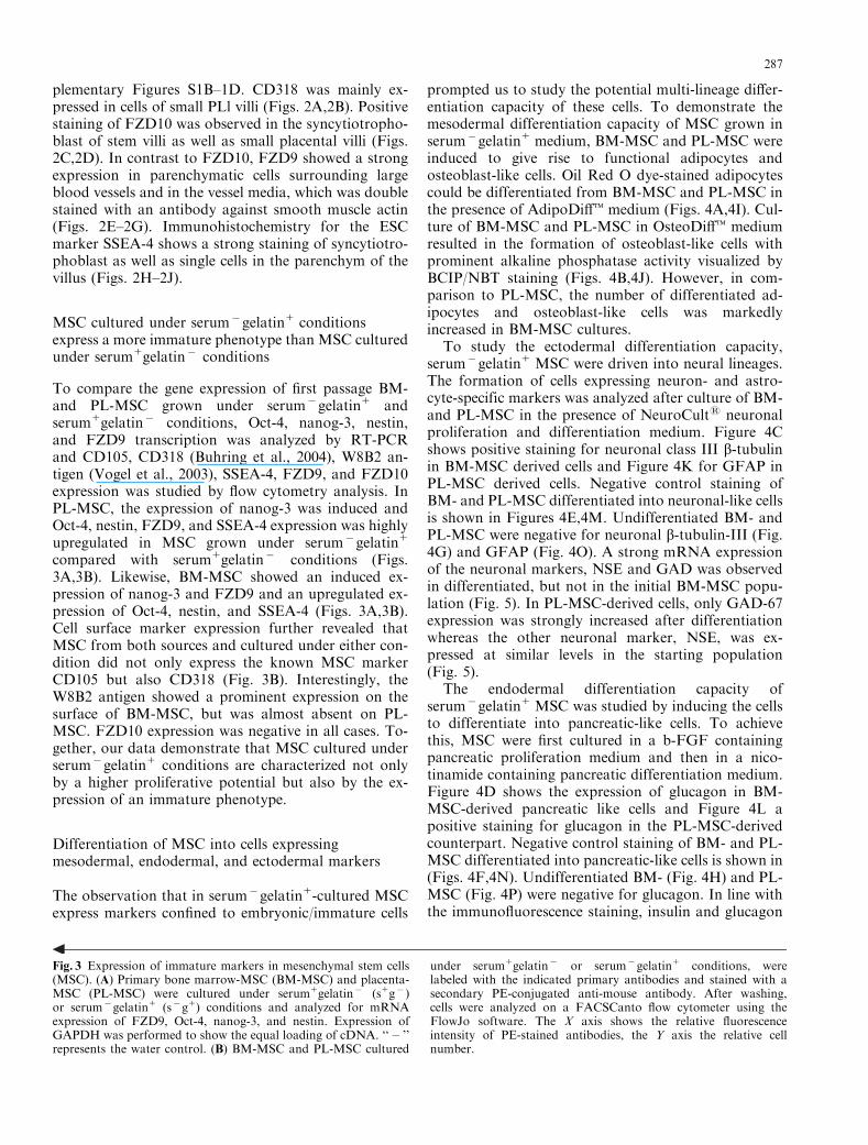
To compare the gene expression of first passage BM-and PL-MSC grown under serum�gelatin1 andserum1gelatin� conditions, Oct-4, nanog-3, nestin,and FZD9 transcription was analyzed by RT-PCRand CD105, CD318 (Buhring et al., 2004), W8B2 an-tigen (Vogel et al., 2003), SSEA-4, FZD9, and FZD10expression was studied by flow cytometry analysis. InPL-MSC, the expression of nanog-3 was induced andOct-4, nestin, FZD9, and SSEA-4 expression was highlyupregulated in MSC grown under serum�gelatin1
compared with serum1gelatin� conditions (Figs.3A,3B). Likewise, BM-MSC showed an induced ex-pression of nanog-3 and FZD9 and an upregulated ex-pression of Oct-4, nestin, and SSEA-4 (Figs. 3A,3B).Cell surface marker expression further revealed thatMSC from both sources and cultured under either con-dition did not only express the known MSC markerCD105 but also CD318 (Fig. 3B). Interestingly, theW8B2 antigen showed a prominent expression on thesurface of BM-MSC, but was almost absent on PL-MSC. FZD10 expression was negative in all cases. To-gether, our data demonstrate that MSC cultured underserum�gelatin1 conditions are characterized not onlyby a higher proliferative potential but also by the ex-pression of an immature phenotype.
Differentiation of MSC into cells expressingmesodermal, endodermal, and ectodermal markers
The observation that in serum�gelatin1-cultured MSCexpress markers confined to embryonic/immature cells
prompted us to study the potential multi-lineage differ-entiation capacity of these cells. To demonstrate themesodermal differentiation capacity of MSC grown inserum�gelatin1 medium, BM-MSC and PL-MSC wereinduced to give rise to functional adipocytes andosteoblast-like cells. Oil Red O dye-stained adipocytescould be differentiated from BM-MSC and PL-MSC inthe presence of AdipoDifft medium (Figs. 4A,4I). Cul-ture of BM-MSC and PL-MSC in OsteoDifft mediumresulted in the formation of osteoblast-like cells withprominent alkaline phosphatase activity visualized byBCIP/NBT staining (Figs. 4B,4J). However, in com-parison to PL-MSC, the number of differentiated ad-ipocytes and osteoblast-like cells was markedlyincreased in BM-MSC cultures.
To study the ectodermal differentiation capacity,serum�gelatin1 MSC were driven into neural lineages.The formation of cells expressing neuron- and astro-cyte-specific markers was analyzed after culture of BM-and PL-MSC in the presence of NeuroCults neuronalproliferation and differentiation medium. Figure 4Cshows positive staining for neuronal class III b-tubulinin BM-MSC derived cells and Figure 4K for GFAP inPL-MSC derived cells. Negative control staining ofBM- and PL-MSC differentiated into neuronal-like cellsis shown in Figures 4E,4M. Undifferentiated BM- andPL-MSC were negative for neuronal b-tubulin-III (Fig.4G) and GFAP (Fig. 4O). A strong mRNA expressionof the neuronal markers, NSE and GAD was observedin differentiated, but not in the initial BM-MSC popu-lation (Fig. 5). In PL-MSC-derived cells, only GAD-67expression was strongly increased after differentiationwhereas the other neuronal marker, NSE, was ex-pressed at similar levels in the starting population(Fig. 5).
The endodermal differentiation capacity ofserum�gelatin1 MSC was studied by inducing the cellsto differentiate into pancreatic-like cells. To achievethis, MSC were first cultured in a b-FGF containingpancreatic proliferation medium and then in a nico-tinamide containing pancreatic differentiation medium.Figure 4D shows the expression of glucagon in BM-MSC-derived pancreatic like cells and Figure 4L apositive staining for glucagon in the PL-MSC-derivedcounterpart. Negative control staining of BM- and PL-MSC differentiated into pancreatic-like cells is shown in(Figs. 4F,4N). Undifferentiated BM- (Fig. 4H) and PL-MSC (Fig. 4P) were negative for glucagon. In line withthe immunofluorescence staining, insulin and glucagon
Fig. 3 Expression of immature markers in mesenchymal stem cells(MSC). (A) Primary bone marrow-MSC (BM-MSC) and placenta-MSC (PL-MSC) were cultured under serum1gelatin� (s1g� )or serum�gelatin1 (s�g1) conditions and analyzed for mRNAexpression of FZD9, Oct-4, nanog-3, and nestin. Expression ofGAPDH was performed to show the equal loading of cDNA. ‘‘� ’’represents the water control. (B) BM-MSC and PL-MSC cultured
under serum1gelatin� or serum�gelatin1 conditions, werelabeled with the indicated primary antibodies and stained with asecondary PE-conjugated anti-mouse antibody. After washing,cells were analyzed on a FACSCanto flow cytometer using theFlowJo software. The X axis shows the relative fluorescenceintensity of PE-stained antibodies, the Y axis the relative cellnumber.
287

mRNA expression was induced after differentiation ofMSC from both sources into the pancreatic lineage(Fig. 5). The BM-MSC and PL-MSC starting popula-tion was negative for insulin and glucagon, confirmingthe specific induction of pancreatic differentiation. Insummary, we could demonstrate that PL- and BM-de-rived MSC that are generated according to our novelprotocol can give rise to cells expressing markers of allthree germ lineages.
Discussion
In this paper we have described a novel procedure togenerate MSC with an immature phenotype. Primarycells from PL and BM cultured in gelatine-coated flasksin b-FGF containing serum-free medium (serum�gel-atin1) gave rise to MSC with a four to five times higherproliferation rate than cells cultured under conventional(serum1gelatin� ) conditions. Compared with conven-
L
N
J
Placenta
I
D
F
A B
BM
C
E
K
M
G H O P
Fig. 4 Differentiation of mesenchymal stem cells (MSC) into cellsexpressing markers of different germ lines. For adipogenic differ-entiation, bone marrow-MSC (BM-MSC) (A) or placenta-Pl (PL-MSC) (I) were cultured for 18 days in the presence of AdipoDifftmedium. Functional adipocytes show Oil Red O dye uptake; scalebar: 100 mm. For osteogenic differentiation, BM-MSC (B) or PL-MSC (J) were cultured for 10 days in OsteoDifft medium. Theresulting osteoblast-like cells are identified by their prominentstaining of alkaline phosphatase activity with BCIP/NBT substrate(scale bar: 100 mm). The formation of neuron-like and astrocyte-like cells derived from BM (C) and placenta (K) were cultured inNeurocults neuronal proliferation medium and then in Neuro-cults differentiation medium. The graph in C shows that BM-MSCgave rise to cells with long spindle-like morphology that stain forneuronal b-tubulin-III-reactive antibody; scale bar: 100 mm. PL-
MSC (K) cultured in the same medium gave raise to cells stain for(GFAP); scale bar: 50mm. (E) and (M) represent the negative con-trols (without the primary antibody) of BM- and PL-MSC differ-entiated into neuronal-like cells. (G) shows the staining of neuronalb-tubulin-III and (O) shows the staining of GFAP in undifferen-tiated BM and PL-derived MSC; scale bar for E and G: 100 mm,scale bar for M and O: 50 mm. For pancreatic differentiation, MSCwere first cultured in a b-FGF-containing pancreatic proliferationmedium and then in a nicotinamide-containing pancreatic differ-entiation medium (CellSystems). The graphs reveal a few BM-MSC(D) and PL-MSC-derived cells (L) that show a positive staining forglucagon-specific antibody; scale bar: 50 mm. (F) and (N) representthe negative controls of BM- and PL-MSC differentiated into pan-creatic-like cells. (H) and (P) show the staining of glucagon inundifferentiated BM and PL-derived MSC; scale bar: 50 mm.
288

tionally prepared MSC, these MSC showed an up-regu-lated expression of the stem/progenitor cell markersSSEA-4, Oct-4, nanog-3, and nestin as well as FZD9. Inaddition, the resulting MSC exhibited multi-lineage dif-ferentiation capacity as demonstrated by their potentialto give rise to cells of the mesodermal (osteoblast, ad-ipocytes), ectodermal (neuron-like), and endodermal(pancreatic-like) differentiation lineage. The describedapproach, which was initially designed to promote thegrowth of human ES cells (Xu et al., 2001), is thereforea powerful method to prepare MSC with high prolif-erative and multi-lineage differentiation capacity.
Because of their increasing therapeutic potential,MSC are now used for a number of medical applica-tions such as tissue engineering and cell-based therapies.Although BM was the first described source of MSCisolation, later studies have shown that MSC are alsopresent in adult organs such as liver, kidney, and adi-pose tissue, as well as in peripheral blood and cordblood (Erices et al., 2000; Zvaifler et al., 2000; Mareschiet al., 2001). More recently, other groups have shownthat human term PL is a powerful source of MSC withmulti-lineage differentiation capability (Fukuchi et al.,2004; In’t Anker et al., 2004; Yen et al., 2005). Yen et al.
(2005) have demonstrated that PL-derived MSC expressthe ESC markers SSEA-4, TRA-1-81, and TRA-1-60.Li et al. have reported that MSC with these phenotypiccharacteristics gave rise to cells of the mesodermal dif-ferentiation lineage including adipocytes, osteoblastsand chondrocytes (Yen et al., 2005). Very recently, Mikiet al. (2005) have shown that MSC derived from am-niotic epithelium were positive for the ESC markersOct-4, nanog-3, SSEA-4. By induction of differentiationin defined media, the resulting cells expressed markersfor neural, muscle, and liver tissues. Our study revealedthat the embryonic stem/progenitor cell marker SSEA-4as well as FZD9, FZD10, and CD318 are expressed ondistinct cell types in PL suggesting the presence of cellswith immature phenotype in the placental tissue.
Comparison of morphology and cell proliferationrate of BM- and PL-MSC revealed an increase in thecell size and reduced proliferation rate in cultures underconventional (serum1gelatin� ) conditions comparedwith serum�gelatin1 conditions. The increase in the cellsize and reduced proliferation rate that is observed inserum1gelatin� conditions is generally observed inmore differentiated MSC suggesting that serum1gel-atin� conditions favor differentiation and reduce pro-liferation. In contrast, BM-MSC and PL-MSC grownunder serum�gelatin1 conditions were not only char-acterized by a smaller cell size but also by a higherproliferation rate and a more immature gene expressionprofile demonstrating the superior potential of thesecells. Notably, the CFU-F capacity of BM-MSC andPL-MSC was not significantly altered in the differentculture conditions, suggesting that the stem cell pool ofMSC is not affected. Gelatin coating did not alter thegrowth characteristics of MSC grown under conven-tional conditions. However, when cells were grown un-der serum-free conditions and in uncoated flasks(serum�gelatin� ), the proliferation rate of MSC wasmore than 10-fold decreased. This indicates that colla-gen, the major component of gelatin, is crucial for op-timal MSC growth and that this compound is present inserum. Receptor blocking experiments with anti-colla-gen receptor antibodies (for example, against b-integrin,discoidin receptors or Endo-180) may facilitate theidentification of the cognate receptor. Our data furtherdemonstrate that the presence of serum compensates forthe lack of gelatin that is necessary for MSC growthunder the novel culture conditions. This indicates thatthe serum contains compounds/molecules that are ableto substitute gelatin.
Comparative mRNA expression analysis of BM-MSC and PL-MSC generated in serum1gelatin� orserum�gelatin1 conditions revealed that only the lattercells expressed Oct4, nanog-3, nestin, and FZD9 at themRNA level and only these cells expressed elevatedlevels of the ESC marker SSEA-4 and of FZD9 on thecell surface, demonstrating the more immature pheno-type of the cells. Thus, FZD9 represents a novel key
Glucagon
Insulin
GAPDH
Glucagon
Insulin
GAPDH
GAPDH
NSE
GAD
GAPDH
NSE
GADM
SC
Pan
crea
tic
Dif
f
Con
trol
MSC
Con
trol
Neu
rona
l Dif
f
BM
Pla
cent
a
Endodermaldifferentiation(Pancreatic)
Ectodermaldifferentiation(Neuronal)
Fig. 5 Expression of pancreatic and neuronal markers in bonemarrow-mesenchymal stem cells (BM-MSC) and placenta-MSC(PL-MSC) before and after differentiation. BM-MSC and PL-MSCwere cultured either in Neurocults neuronal proliferation and dif-ferentiation medium or in pancreatic proliferation and differenti-ation medium. The mRNA expression was then analyzed by reversetranscriptase polymerase chain reaction for neuronal markers be-fore and after differentiation of MSC. The neuronal markers con-sisted of neuron-specific enolase (NSE) and glutamic aciddecarboxylase-67 (GAD) and the pancreatic markers of insulinand glucagon. Expression of GAPDH was performed to show theequal loading of cDNA. Right lanes represent water controls.
289

marker of primitive MSC that is suitable to distinguishthem from MSC, which are produced by conventionalmethods. Our preliminary data further suggest thatFZD9 is a suitable marker to isolate primary MSC ofBM and PL (manuscript in preparation). Interestingly,the recently described W8B2 antigen, a novel and se-lective MSC marker of unknown identity (Vogel et al.,2003), was only expressed on BM-MSC, but not on PL-derived MSC. This antigen is therefore suitable to dis-criminate between BM-MSC and PL-MSC.
Wnt signaling is a key process in organogenesis andstem cell renewal (Baker et al., 1999; Niehrs, 1999;Logan and Nusse, 2004; Teo et al., 2006). So far, theanalysis of this process was hampered by the lack ofsuitable tools to study FZD molecules, the cognate re-ceptors for Wnt ligands. To circumvent this obstacle,mAbs against these receptors were generated. Employ-ing mAbs against FZD9- and FZD10, we could showfor the first time a selective FZD9 and FZD10 expres-sion in PL tissues. While the FZD9-reactive mAb showsstrong staining of parenchymatic cells surroundinglarge blood vessels and cells of vessel media, FZD10expression was mainly restricted to the syncytiotropho-blast of PL villi. Upon differentiation of PL cells intoMSC, the expression of FZD9 protein and mRNA in-creased, whereas FZD10 expression declined. A similarprofile was observed when BM cells were induced todifferentiate into MSC. Ongoing studies address the in-volvement of Wnt ligands on the upregulation of FZD9expression during the differentiation of primary cellsinto MSC.
In conclusion, we have developed a novel approachto isolate and propagate BM-MSC and PL-MSC. Com-pared with conventionally prepared MSC, these cellsexpressed FZD9 and elevated levels of the stem cellmarker SSEA-4 and could be induced to multi-lineagedifferentiation. The exclusive expression of FZD9 inthese MSC suggests that this is a candidate molecule forMSC isolation in both BM and PL. Our preliminarydata suggest that FZD9 is indeed a suitable marker forMSC isolation in both, PL and BM. This also suggeststhat Wnt-FZD9 signaling is important for stem-cell re-newal. In these studies, we could also show that PL is anattractive source for MSC production because of theirpotent proliferation and differentiation capacity, theireasy, abundant, and ethically unbiased availability.When prepared according to our new protocol, PL-de-rived MSC may represent a feasible alternative for fu-ture applications in regenerative medicine and mayextend the known applications of hematopoietic stemcell transplantation and tissue repair to novel fields inregenerative medicine.
Acknowledgments The authors thank Dr. Barbel Spring and theengaged midwives of the Department of Obstetrics and Gynecologyfor their indispensable help in PL collection. This work was sup-ported by the Federal Ministry for Education and Research
(BMBF), project 0313048A: Generierung von Antikorpern gegenFrizzled Rezeptoren (GPCRs) zur Isolierung und Charakterisie-rung von Stammzellen.
References
Baker, J.C., Beddington, R.S. and Harland, R.M. (1999) Wnt sig-naling in Xenopus embryos inhibits bmp4 expression and acti-vates neural development. Genes Dev 13:3149–3159.
Behrens, J. and Lustig, B. (2004) The Wnt connection to tumor-igenesis. Int J Dev Biol 48:477–487.
Brown, J.D., Hallagan, S.E., McGrew, L.L., Miller, J.R. andMoon, R.T. (2000) The maternal Xenopus beta-catenin signalingpathway, activated by frizzled homologs, induces goosecoid in acell non-autonomous manner. Dev Growth Differ 42:347–357.
Buhring, H.J., Kuci, S., Conze, T., Rathke, G., Bartolovic, K.,Grunebach, F., Scherl-Mostageer, M., Brummendorf, T.H., Sch-weifer, N. and Lammers, R. (2004) CDCP1 identifies a broadspectrum of normal and malignant stem/progenitor cell subsetsof hematopoietic and nonhematopoietic origin. Stem Cells22:334–343.
Caplan, A.I. (1991) Mesenchymal stem cells. J Orthop Res 9:641–650.
Conze, T., Lammers, R., Kuci, S., Scherl-Mostageer, M., Schwei-fer, N., Kanz, L. and Buhring, H.J. (2003) CDCP1 is a novelmarker for hematopoietic stem cells. Ann N Y Acad Sci 996:222–226.
Deans, R.J. and Moseley, A.B. (2000) Mesenchymal stem cells:biology and potential clinical uses. Exp Hematol 28:875–884.
Deardorff, M.A., Tan, C., Conrad, L.J. and Klein, P.S. (1998)Frizzled-8 is expressed in the spemann organizer and plays a rolein early morphogenesis. Development 125:2687–2700.
Erices, A., Conget, P. and Minguell, J.J. (2000) Mesenchymal pro-genitor cells in human umbilical cord blood. Br J Haematol109:235–242.
Etheridge, S.L., Spencer, G.J., Heath, D.J. and Genever, P.G.(2004) Expression profiling and functional analysis of wnt sig-naling mechanisms in mesenchymal stem cells. Stem Cells22:849–860.
Friedenstein, A.J., Gorskaja, J.F. and Kulagina, N.N. (1976) Fi-broblast precursors in normal and irradiated mouse hematopoi-etic organs. Exp Hematol 4:267–274.
Fukuchi, Y., Nakajima, H., Sugiyama, D., Hirose, I., Kitamura, T.and Tsuji, K. (2004) Human placenta-derived cells havemesenchymal stem/progenitor cell potential. Stem Cells 22:649–658.
Gaspar, C. and Fodde, R. (2004) APC dosage effects in tumori-genesis and stem cell differentiation. Int J Dev Biol 48:377–386.
In ‘t Anker, P.S., Scherjon, S.A., Kleijburg-van der Keur, C.,Groot-Swings, G.M., Claas, F.H., Fibbe, W.E. and Kanhai,H.H. (2004) Isolation of mesenchymal stem cells of fetal or ma-ternal origin from human placenta. Stem Cells 22:1338–1345.
Jones, E.A., Kinsey, S.E., English, A., Jones, R.A., Straszynski, L.,Meredith, D.M., Markham, A.F., Jack, A., Emery, P. andMcGonagle, D. (2002) Isolation and characterization of bonemarrow multipotential mesenchymal progenitor cells. ArthritisRheum 46:3349–3360.
Liechty, K.W., MacKenzie, T.C., Shaaban, A.F., Radu, A., Mose-ley, A.M., Deans, R., Marshak, D.R. and Flake, A.W. (2000)Human mesenchymal stem cells engraft and demonstrate site-specific differentiation after in utero transplantation in sheep.Nat Med 6:1282–1286.
Logan, C.Y. and Nusse, R. (2004) The Wnt signaling pathway indevelopment and disease. Annu Rev Cell Dev Biol 20:781–810.
Manoukian, A.S. and Woodgett, J.R. (2002) Role of glycogen syn-thase kinase-3 in cancer: regulation by Wnts and other signalingpathways. Adv Cancer Res 84:203–229.
290

Mareschi, K., Biasin, E., Piacibello, W., Aglietta, M., Madon, E.and Fagioli, F. (2001) Isolation of human mesenchymal stemcells: bone marrow versus umbilical cord blood. Haematologica86:1099–1100.
Miki, T., Lehmann, T., Cai, H., Stolz, D.B. and Strom, S.C. (2005)Stem cell characteristics of amniotic epithelial cells. Stem Cells23:1549–1559.
Moon, R.T., Bowerman, B., Boutros, M. and Perrimon, N. (2002)The promise and perils of Wnt signaling through beta-catenin.Science 296:1644–1646.
Niehrs, C. (1999) Head in the WNT: the molecular nature of Spe-mann’s head organizer. Trends Genet 15:314–319.
Niehrs, C. (2004) Regionally specific induction by the Spemann-Mangold organizer. Nat Rev Genet 5:425–434.
Pereira, R.F., Halford, K.W., O’Hara, M.D., Leeper, D.B., Sokol-ov, B.P., Pollard, M.D., Bagasra, O. and Prockop, D.J. (1995)Cultured adherent cells from marrow can serve as long-lastingprecursor cells for bone, cartilage, and lung in irradiated mice.Proc Natl Acad Sci USA 92:4857–4861.
Rappold, I., Ziegler, B.L., Kohler, I., Marchetto, S., Rosnet, O.,Birnbaum, D., Simmons, P.J., Zannettino, A.C., Hill, B., Neu,S., Knapp, W., Alitalo, R., Alitalo, K., Ullrich, A., Kanz, L. andBuhring, H.J. (1997) Functional and phenotypic characterizationof cord blood and bone marrow subsets expressing FLT3(CD135) receptor tyrosine kinase. Blood 90:111–125.
Scherl-Mostageer, M., Sommergruber, W., Abseher, R., Haupt-mann, R., Ambros, P. and Schweifer, N. (2001) Identification ofa novel gene, CDCP1, overexpressed in human colorectal cancer.Oncogene 20:4402–4408.
Schneider, S., Steinbeisser, H., Warga, R.M. and Hausen, P. (1996)Beta-catenin translocation into nuclei demarcates the dorsalizingcenters in frog and fish embryos. Mech Dev 57:191–198.
Teo, R., Mohrlen, F., Plickert, G., Muller, W.A. and Frank, U.(2006) An evolutionary conserved role of Wnt signaling in stemcell fate decision. Dev Biol 289:91–99.
Vincan, E. (2004) Frizzled/WNT signalling: the insidious promoterof tumour growth and progression. Front Biosci 9:1023–1034.
Vogel, W., Grunebach, F., Messam, C.A., Kanz, L., Brugger, W.and Buhring, H.J. (2003) Heterogeneity among human bonemarrow-derived mesenchymal stem cells and neural progenitorcells. Haematologica 88:126–133.
Xu, C., Inokuma, M.S., Denham, J., Golds, K., Kundu, P., Gold,J.D. and Carpenter, M.K. (2001) Feeder-free growth of undif-ferentiated human embryonic stem cells. Nat Biotechnol 19:971–974.
Yen, B.L., Huang, H.I., Chien, C.C., Jui, H.Y., Ko, B.S., Yao, M.,Shun, C.T., Yen, M.L., Lee, M.C. and Chen, Y.C. (2005) Isol-ation of multipotent cells from human term placenta. Stem Cells23:3–9.
Zhang, Y., Li, C., Jiang, X., Zhang, S., Wu, Y., Liu, B., Tang, P.and Mao, N. (2004) Human placenta-derived mesenchymal pro-genitor cells support culture expansion of long-term culture-ini-tiating cells from cord blood CD341 cells. Exp Hematol 32:657–664.
Zhao, L.R., Duan, W.M., Reyes, M., Keene, C.D., Verfaillie, C.M.and Low, W.C. (2002) Human bone marrow stem cells exhibitneural phenotypes and ameliorate neurological deficits aftergrafting into the ischemic brain of rats. Exp Neurol 174:11–20.
Zola, H., Swart, B., Nicholson, I., Aasted, B., Bensussan, A., Bo-umsell, L., Buckley, C., Clark, G., Drbal, K., Engel, P., Hart, D.,Horejsi, V., Isacke, C., Macardle, P., Malavasi, F., Mason, D.,Olive, D., Saalmueller, A., Schlossman, S.F., Schwartz-Albiez,R., Simmons, P., Tedder, T.F., Uguccioni, M. and Warren, H.
(2005) CD molecules 2005: human cell differentiation molecules.Blood 106:3123–3126.
Zola, H., et al. (2006) HCDM workshop 2006. Quebec City, Ca-nada, May 22, 2006. Http://www.hcdm.org/CD1toCD350.htm
Zvaifler, N.J., Marinova-Mutafchieva, L., Adams, G., Edwards,C.J., Moss, J., Burger, J.A. and Maini, R.N. (2000) Mesenchy-mal precursor cells in the blood of normal individuals. ArthritisRes 2:477–488.
Supplementary material
The following supplementary material is available forthis article:Figure S1. Vector construction, transfection of HEK-293 cells with pIRES2-EGFP-Si-F-FZD9/FZ10, andFZD9 and FZD10 expression in transfected cells. (A)Vector constructs used for expression of FZD9 andFZD10 receptors (FZD) in HEK293 cells are based onpIRES-EGFP (Clontech). The construct contains botha N-terminal signal sequence for membrane insertionand a FLAG tag. HEK-293 cells were transfected witheither pIRES2-EGFP-Si-F-FZD9 or with pIRES2-EGFP-Si-F-FZD10 as described in the methods. (B)Flow cytometry analysis of transfected cells. Cells werelabeled with either an anti-Flag or an anti-FZD anti-body and stained with a secondary PE-conjugated anti-mouse antibody. After washing, cells were analyzed ona FACSCanto flow cytometer using the FlowJo soft-ware. Note the specific reaction of mAb W3C4 and 1/4C4 with the transfectant lines. (C) Exogenous frizzledreceptor expression in HEK-293/huFZD9 and HEK-293/huFZD10 cells was confirmed by immuno-blottingthe SDS-PAGE separated lysates of these cells (trans-fected with 1, 2, and 4 mg of plasmids) with commer-cially available anti-FZD antisera and anti-Flagantibody. The figure shows detectable expression ofFZD9 or FZD10 only in transfected but not in parentalcells. (D) RT-PCR analysis of FZD9 and FZD10 ex-pression in parental and transfected HEK293 cells.Transfected cells show an increased mRNA expressionof FZD9 and FZD10, respectively.
This material is available as part of the online articlefrom: http://www.blackwell-synergy.com/doi/abs/10.1111/j.1432-0436.2006.00139.x
(This link will take you to the article abstract.)Please note: Blackwell Publishing is not responsible
for the content or functionality of any supplementarymaterials supplied by the authors. Any queries (otherthan missing material) should be directed to the corre-sponding author for the article.
291