Human epicardial adipose tissue induces fibrosis of the atrial myocardium through the secretion of...
12
..................................................................................................................................................................................... ..................................................................................................................................................................................... BASIC SCIENCE Human epicardial adipose tissue induces fibrosis of the atrial myocardium through the secretion of adipo-fibrokines Nicolas Venteclef 1,2,3† , Valeria Guglielmi 1,2,3† , Elise Balse 1,3,4 , Be ´ne ´ dicte Gaborit 1,2,3,5,6 , Aure ´ lie Cotillard 1,2,3 , Fabrice Atassi 1,3,4 , Julien Amour 1,5 , Pascal Leprince 1,5 , Anne Dutour 6 , Karine Cle ´ ment 1,2,3,5 * , and Ste ´phane N. Hatem 1,3,4,5 * 1 Institute of Cardiometabolism and Nutrition, Paris, France; 2 INSERM, UMR_S 872, Team 7 Nutriomique, Paris, France; 3 Universite ´ Pierre et Marie Curie-Paris 6, Cordeliers Research Center, Paris, France; 4 Universite ´ Pierre et Marie Curie-Paris 6, INSERM UMR_S956, Paris, France; 5 Heart and metabolism division, Assistance Publique-Ho ˆ pitaux de Paris, Pitie ´-Salpe ˆtrie `re Hospital, Paris; and 6 Faculte ´ de Me ´decine, INSERM NORT UMR 1062, INRA1260, Aix Marseille Universite ´, Marseille, France Received 15 November 2012; revised 8 February 2013; accepted 4 March 2013; online publish-ahead-of-print 27 February 2014 Aims Recent studies have reported a relationship between the abundance of epicardial adipose tissue (EAT) and the risk of cardiovascular diseases including atrial fibrillation (AF). However, the underlying mechanisms are unknown. The aim of this study was to examine the effects of the secretome of human EAT on the histological properties of the myocardium. Methods and results Samples of EAT and subcutaneous adipose (SAT), obtained from 39 patients undergoing coronary bypass surgery, were analysed and tested in an organo-culture model of rat atria to evaluate the fibrotic properties of human fat depots. The EAT secretome induced global fibrosis (interstitial and peripheral) of rat atria in organo-culture condi- tions. Activin A was highly expressed in EAT compared with SAT and promoted atrial fibrosis, an effect blocked using neutralizing antibody. In addition, Activin A levels were enhanced in patients with low left-ventricular function. In sec- tions of human atrial and ventricular myocardium, adipose and myocardial tissues were in close contact, together with fibrosis. Conclusion This study provides the first evidence that the secretome from EAT promotes myocardial fibrosis through the secre- tion of adipo-fibrokines such as Activin A. ----------------------------------------------------------------------------------------------------------------------------------------------------------- Keywords Myocardial fibrosis † Epicardial adipose tissue † Atrial fibrillation † Atrial fibrosis † Activin A Introduction Recently, the epicardial adipose tissue (EAT) has emerged as an im- portant factor in the pathogenesis of metabolic-related cardiac dis- eases. This ectopic fat depot, distinct from paracardial fat, surrounds the myocardium and coronary arteries and is in direct contact with cardiomyocytes and adventitia. The EAT is a metabol- ically active tissue that produces pro-atherogenic and pro- inflammatory adipokines. 1 – 4 In other adipose-tissue depots, a para- crine dialog between adipocytes, pre-adipocytes, endothelial, and inflammatory cells has been described. This results in the release of a myriad of pro- and anti-inflammatory molecules, leading to a low-grade inflammatory and pro-fibrotic environment. 5,6 The bio- logical activity of the EAT is related to the severity of coronary artery disease (CAD), 4,7,8 the degree of cardiac hypertrophy, 9,10 and is enhanced in type-2 diabetic and obese patients. 11 In addition, † These authors contributed equally to the work. * Corresponding author. Tel: +33 1 40 77 95 84 (S.N.H.), +33 1 42 34 69 93 (K.C.); Fax: +33 1 40 77 96 49 (S.N.H.), +33 1 42 34 69 93 (K.C.); Email: [email protected] (S.N.H.), [email protected] (K.C.) Published on behalf of the European Society of Cardiology. All rights reserved. & The Author 2014. For permissions please email: [email protected] European Heart Journal (2015) 36, 795–805 doi:10.1093/eurheartj/eht099 by guest on February 9, 2016 http://eurheartj.oxfordjournals.org/ Downloaded from
-
Upload
independent -
Category
Documents
-
view
0 -
download
0
Transcript of Human epicardial adipose tissue induces fibrosis of the atrial myocardium through the secretion of...

. . . . . . . . . . . . . . . . . . . . . . . . . . . . . . . . . . . . . . . . . . . . . . . . . . . . . . . . . . . . . . . . . . . . . . . . . . . . . . . . . . . . . . . . . . . . . . . . . . . . . . . . . . . . . . . . . . . . . . . . . . . . . . . . . . . . . . . . . . . . . . . . . . . . . . . . . . . . . . . . . . . . . . . . . . . . . . . . . . . . .
. . . . . . . . . . . . . . . . . . . . . . . . . . . . . . . . . . . . . . . . . . . . . . . . . . . . . . . . . . . . . . . . . . . . . . . . . . . . . . . . . . . . . . . . . . . . . . . . . . . . . . . . . . . . . . . . . . . . . . . . . . . . . . . . . . . . . . . . . . . . . . . . . . . . . . . . . . . . . . . . . . . . . . . . . . . . . . . . . . . . .
BASIC SCIENCE
Human epicardial adipose tissue induces fibrosisof the atrial myocardium through the secretionof adipo-fibrokinesNicolas Venteclef1,2,3†, Valeria Guglielmi1,2,3†, Elise Balse1,3,4, Benedicte Gaborit1,2,3,5,6,Aurelie Cotillard1,2,3, Fabrice Atassi1,3,4, Julien Amour1,5, Pascal Leprince1,5,Anne Dutour6, Karine Clement1,2,3,5*, and Stephane N. Hatem1,3,4,5*
1Institute of Cardiometabolism and Nutrition, Paris, France; 2INSERM, UMR_S 872, Team 7 Nutriomique, Paris, France; 3Universite Pierre et Marie Curie-Paris 6, CordeliersResearch Center, Paris, France; 4Universite Pierre et Marie Curie-Paris 6, INSERM UMR_S956, Paris, France; 5Heart and metabolism division, Assistance Publique-Hopitaux de Paris,Pitie-Salpetriere Hospital, Paris; and 6Faculte de Medecine, INSERM NORT UMR 1062, INRA1260, Aix Marseille Universite, Marseille, France
Received 15 November 2012; revised 8 February 2013; accepted 4 March 2013; online publish-ahead-of-print 27 February 2014
Aims Recent studies have reported a relationship between the abundance of epicardial adipose tissue (EAT) and the risk ofcardiovascular diseases including atrial fibrillation (AF). However, the underlying mechanisms are unknown. The aimof this study was to examine the effects of the secretome of human EAT on the histological properties of themyocardium.
Methodsand results
Samples of EAT and subcutaneous adipose (SAT), obtained from 39 patients undergoing coronary bypass surgery,were analysed and tested in an organo-culture model of rat atria to evaluate the fibrotic properties of human fatdepots. The EAT secretome induced global fibrosis (interstitial and peripheral) of rat atria in organo-culture condi-tions. Activin A was highly expressed in EAT compared with SAT and promoted atrial fibrosis, an effect blocked usingneutralizing antibody. In addition, Activin A levels were enhanced in patients with low left-ventricular function. In sec-tions of human atrial and ventricular myocardium, adipose and myocardial tissues were in close contact, togetherwith fibrosis.
Conclusion This study provides the first evidence that the secretome from EAT promotes myocardial fibrosis through the secre-tion of adipo-fibrokines such as Activin A.
- - - - - - - - - - - - - - - - - - - - - - - - - - - - - - - - - - - - - - - - - - - - - - - - - - - - - - - - - - - - - - - - - - - - - - - - - - - - - - - - - - - - - - - - - - - - - - - - - - - - - - - - - - - - - - - - - - - - - - - - - - - - - - - - - - - - - - - - - - - - - - - - - - - - - - - - - - -Keywords Myocardial fibrosis † Epicardial adipose tissue † Atrial fibrillation † Atrial fibrosis † Activin A
IntroductionRecently, the epicardial adipose tissue (EAT) has emerged as an im-portant factor in the pathogenesis of metabolic-related cardiac dis-eases. This ectopic fat depot, distinct from paracardial fat,surrounds the myocardium and coronary arteries and is in directcontact with cardiomyocytes and adventitia. The EAT is a metabol-ically active tissue that produces pro-atherogenic and pro-
inflammatory adipokines.1– 4 In other adipose-tissue depots, a para-crine dialog between adipocytes, pre-adipocytes, endothelial, andinflammatory cells has been described. This results in the releaseof a myriad of pro- and anti-inflammatory molecules, leading to alow-grade inflammatory and pro-fibrotic environment.5,6 The bio-logical activity of the EAT is related to the severity of coronaryartery disease (CAD),4,7,8 the degree of cardiac hypertrophy,9,10
and is enhanced in type-2 diabetic and obese patients.11 In addition,
† These authors contributed equally to the work.
* Corresponding author. Tel: +33 1 40 77 95 84 (S.N.H.), +33 1 42 34 69 93 (K.C.); Fax: +33 1 40 77 96 49 (S.N.H.), +33 1 42 34 69 93 (K.C.); Email: [email protected](S.N.H.), [email protected] (K.C.)
Published on behalf of the European Society of Cardiology. All rights reserved. & The Author 2014. For permissions please email: [email protected]
European Heart Journal (2015) 36, 795–805doi:10.1093/eurheartj/eht099
by guest on February 9, 2016http://eurheartj.oxfordjournals.org/
Dow
nloaded from

the abundance of EAT is increased in patients with chronic atrialfibrillation (AF) or with atrial dilatation.12– 16
To date, the mechanisms underlying the effect of EAT on themyocardium have been poorly studied. The fact that EAT is con-tiguous with the heart without fascia boundaries enables paracrineprocesses to occur, through the release of adipo-fibrokines. In thisstudy, the hypothesis that adipose tissue secretes factors with pro-fibrotic properties was tested using an original organo-culturemodel of rat atria. We observed that the secretome from humanEAT, but not from subcutaneous adipose tissue (SAT), has amarked pro-fibrotic effect on the rat atrial myocardium. Thiseffect is mediated in part by Activin A, a member of the TGFbfamily. Moreover, we confirmed in human myocardium that theadipose tissue is in close proximity with the myocardium, and isassociated with an increased fibrosis.
MethodsAn expanded version of the Methods is provided in the Supplementarymaterial online.
Human adipose and cardiac tissuePaired samples of EAT and SAT were collected from 39 patientsundergoing routine cardiac bypass surgery. The SAT biopsies weretaken from the parasternal region, while EAT samples were obtainedfrom the left-interventricular groove before cardiopulmonary bypass(paracardial fat was not collected). The clinical parameters of patientsare listed in Table 1. Twelve paired samples were dedicated for tran-scriptomic study (Group 1); 13 for the organo-culture experiments(Group 2), and 14 for the proteomic analysis of the conditionedmedia (Group 3). Samples of right (n ¼ 15) or left (n ¼ 3) atriawere obtained for histological study from 18 patients. Human ventriclesamples (n ¼ 5) were obtained from autopsy. All clinical investigationswere approved by the Ethical Committee of Pitie-Salpetriere Hospital.All patients undergoing bypass surgery gave written informed consentafter individual explanation.
Organo-culture of adult rat atriaTo study the effect of adipose-tissue-conditioned medium, we devel-oped an organo-culture model of adult rat atria. All experimentswere conducted in accordance with the Guide for the Care and Useof Laboratory Animals published by the US National Institute ofHealth.
Rat cardiac fibroblast culturesAtria-derived fibroblast cultures were prepared as previouslydescribed.17
Immunohistological analysisFrozen atrial organo-culture tissue sections were used for immunohis-tological analysis of atrial fibrosis.
Multiplexed proteomics and enzyme-linkedimmunosorbent assayCytometric bead arrays were used to analyse protein composition ofEAT- and SAT-conditioned media.
Statistical analysisValues are expressed as mean+ SEM. The Gaussian distribution of allparameters was tested. Differences between variables were deter-mined by the non-parametric Wilcoxon signed-rank test and theMann–Whitney–Wilcoxon test for paired and unpaired data, respect-ively. Statistical tests were two-tailed. Correlative analyses were deter-mined by Spearman statistical test. In all analysis, P , 0.05 wasconsidered statistically significant. To study the relationship betweenclinical parameters and the composition of the EAT secretome, a mul-tiple factor analysis was used [(Escofier and Pages, 1994, MultipleFactor Analysis (AFMULT package)].
Results
Epicardial, but not subcutaneous,adipose-tissue secretome inducesatrial fibrosisThe EAT was collected in patients with coronary insufficiency, acondition known to be associated with biologically active EAT2,8
(Table 1). The secretome was obtained by harvesting the condi-tioned media of adipose-tissue samples maintained in culture aspreviously described.18 EAT secretomes from two patients werepooled in order to obtain enough volume. Atrial organo-cultureincubated for 1 week with EAT showed global fibrosis(Figure 1A–F). Picrosirius-red staining indicated a two-fold increasein fibrosis in EAT-treated atria (33.78%+ 1.33, P ¼ 0.001) com-pared with control-treated ones (15.67%+ 0.78). The SAT-conditioned media did not induce fibrosis (15.33%+1.60)(Figure 1J). The atrial myocardium preserved its sarcomeric organ-ization throughout culture (Figure 1G–I). Fibrosis was composed ofcollagen types I, III, and VI (Supplementary material online, FigureS2A and B). At higher magnification and using combined phase con-trast and high resolution 3-dimensional deconvolution microscopy,collagen accumulation could be seen at the endocardial (peritrabe-culae) and epicardial faces of the atria. Collagen accumulation was
. . . . . . . . . . . . . . . . . . . . . . . . . . . . . . . . . . . . . . . . . . . . . . . . . . . . . . . . . . . . . . . . . . . . . . . . . . . . . . . .
Table 1 Patients characteristics
Group 1 Group 2 Group 3
n 12 13 14
Age 60.1+6.7 66.7+8.8 62.5+8.7
Sex (M/F) 10/2 8/5 11/3
BMI (kg/m2) 26.5+3.1 29.6+6.6 26.4+4.4
Diabetes (n) 5 5 6
Dyslipidemia (n) 10 9 11
Hypertension (n) 8 6 10
LEVF (,45%) 2 3 4
BMI, body mass index; LVEF, left-ventricular ejection fraction.Group 1: 12 subjects whose EAT and SAT samples where used for transcriptomicanalysis; Group 2: 13 subjects whose EAT and SAT samples were used fororgano-culture experiments; Group 3: 14 subjects whose EAT and SAT sampleswere used for proteomic analysis.
N. Venteclef et al.796
by guest on February 9, 2016http://eurheartj.oxfordjournals.org/
Dow
nloaded from

also observed between myocytes (interstitial areas), and was morepronounced in EAT-treated atria (Figure 2A and B).
Fibroblasts are the primary source of extracellular matrix pro-teins. Indeed, collagen I gene expression was up-regulated inatrial fibroblasts cultured with EAT-conditioned medium for4 days. The a-smooth muscle actin was also up-regulated in thiscondition, suggesting differentiation of fibroblasts into myofibro-blasts (Supplementary material online, Figure S3B). Collectively,
these results indicate that EAT secretome induces a global fibrosisof the atrial myocardium ex vivo.
Adipo-fibrokines secreted by humanepicardial adipose tissueThe adipo-cytokines secreted by human EAT were identified usingcytometric bead arrays and ELISA. The EAT secreted angiogenic
Figure 1 Epicardial adipose tissue induces atrial fibrosis. Masson’s trichrome (A–C; collagen in blue) and Picrosirius red (D–F; collagen in red)were used to visualize fibrosis in rat atrial sections after incubation with epicardial (EAT), subcutaneous (SAT) adipose-tissue-conditionedmedia, or with control medium (Ctrl) (10× magnification, scale bar ¼ 100 mm). Arrowheads show atrial fibrotic areas. (G– I ) Sarcomerica-actinin immunostaining of atrial sections confirmed that samples maintained their typical organization in organo-culture (60× magnification,scale bar ¼ 10 mm). (J ) Total fibrosis in atrial sections: data depict the amount of fibrotic area as a percentage of the total tissue surface (fourseparate experiments, *P , 0.05).
Cardiac fat depot and myocardial fibrosis 797
by guest on February 9, 2016http://eurheartj.oxfordjournals.org/
Dow
nloaded from

factors (VEGF and Thrombospondin-2) and matrix metalloprotei-nases (MMPs) such as MMP1, MMP8, and MMP9, more abundantlythan SAT (Table 2). In addition, EAT expressed Activin A, amember of the TGF-b superfamily. In contrast, the level ofTGF-b1 was unchanged between EAT and SAT, and otherTGF-b members were not detectable. There was a three-fold in-crease in Activin A concentration in EAT-conditioned media(3.5+ 0.8 ng/mL; ranging from 1.3 to 11 ng/mL, median concentra-tion: 2.3 ng/mL) compared with SAT-conditioned media (1.3+
0.3 ng/mL; ranging from 0.3 to 3.6 ng/mL, median concentration:0.8 ng/mL) (Table 2, P ¼ 0.0002). This increase was also confirmedat the tissue level by immunostaining (Figure 3E and F ). At themRNA level, Activin A was higher in EAT than in SAT depots, al-though the difference did not reach significance (relative expres-sion, 2.58+0.65 vs. 1.15+ 0.28; P ¼ 0.07) (Figure 3A). Inaddition, Follistatin, the endogenous inhibitor of Activin A, wasnot enhanced in EAT resulting in an increase in Activin A/Follistatinratio at both the transcriptional and protein levels (Figure 3C and D).
Figure 2 Distribution of fibroblasts and collagen fibres in rat atrial explants treated with EAT-conditioned medium. Collagen 1 (A) and 3 (B)immunostainings of rat atrial organo-cultures treated with control or/and EAT-conditioned medium. Arrow heads indicate perimyocyte (orperitrabecula) and epicardial collagen fibres, stars point fibroblast cells bodies. Note that collagen fibres are also found between two adjacentmyocytes, deeply inserted into the tissue (arrows).
N. Venteclef et al.798
by guest on February 9, 2016http://eurheartj.oxfordjournals.org/
Dow
nloaded from

Multiple factor and correlative analysis of individual secretomeprofile of EAT revealed a positive correlation between the levelof Activin A and MMP8 (rho ¼ 0.6689, P ¼ 0.00894). Moreover,the composition of the secretome varied markedly betweenindividuals; the level of Activin A being higher in patients withreduced left-ventricular function (LVEF , 45%) (Figure 4A and B).
Activin A promotes myocardial fibrosisGiven the known pro-fibrotic effect of Activin A,19 we went on totest the hypothesis that Activin A is involved in EAT-induced atrialfibrosis. First, organo-cultures treated for 1 week with controlmedium supplemented with Activin A showed a marked fibrosis(2 ng/mL: 39.40%+3.9, P , 0.001; 5 ng/mL: 36.36%+2.04, P ,
0.001) compared with controls (15.67%+0.78) (Figure 5). In add-ition, Activin A induced a de novo synthesis of collagen types I, III,and VI (Figure 5D and Supplementary material online, Figure S4).Collagen accumulation was observed at the endocardial and epi-cardial faces of the myocardium and between myocytes(Figure 5D). Secondly, when EAT-conditioned medium was pre-
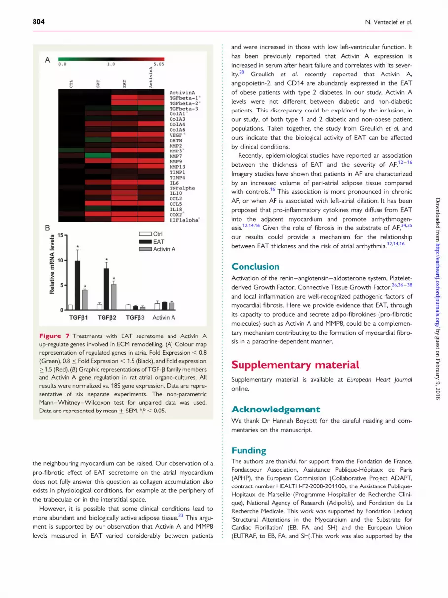
incubated with Activin A neutralizing antibodies, myocardial fibro-sis was not observed (EAT+Activin A-neutralizing Ab: 11.83%+0.4; EAT+IgG: 24%+0.93; P ¼ 0.002) (Figure 6A–C). Thirdly,EAT secretome and Activin A induced similar changes in atrialgene expression profiles (Figure 7A). Both EAT and Activin Aincreased the expression of TGF-b1 and b2 (Figure 7B).However, EAT-conditioned media did not induce additionalActivin A expression in the atrial organo-culture, suggesting thatEAT may be the primary source of Activin A (Figure 7B). Theseresults indicate that atrial fibrosis induced by EAT is primarilymediated by Activin A.
Tight interaction between epicardialadipose tissue and human atrialmyocardiumTo study whether the presence of fat depots could be associatedwith fibrosis in human myocardium, histological analysis was per-formed in left (n ¼ 3) and right atria (n ¼ 15) as well as in ventricu-lar myocardium (n ¼ 5). Figure 8 shows representative examples of
. . . . . . . . . . . . . . . . . . . . . . . . . . . . . . . . . . . . . . . . . . . . . . . . . . . . . . . . . . . . . . . . . . . . . . . . . . . . . . . . . . . . . . . . . . . . . . . . . . . . . . . . . . . . . . . . . . . . . . . . . . . . . . . . . . . . . . . . . . . . . . . . . . . . . . . . . . . . . . . . . . . . . . . . . . . . . .
. . . . . . . . . . . . . . . . . . . . . . . . . . . . . . . . . . . . . . . . . . . . . . . . . . . . . . . . . . . . . . . . . . . . . . . . . . . . . . . . . . . . . . . . . . . . . . . . . . . . . . . . . . . . . . . . . . . . . . . . . . . . . . . . . . . . . . . . . . . . . . . . . . . . . . . . . . . . . . . . . . . . . . . . . . . . . .
Table 2 Quantification of adipo-fibrokines secreted by human epicardial adipose tissue
EAT (ng/mL) SAT (ng/mL) P-value*
Angiogenic factors
Angiogenin 7.8+1.5 9.4+1.7 0.34
Endostatin 3.8+0.5 3.4+0.4 0.61
Angiopoietin ND ND NA
Thrombospondin-2 2.6+0.12 1.2+0.15 ,0.0001
VEGF-D ND ND NA
VEGF 0.4+0.02 0.09+0.01 ,0.0001
Remodelling factors
Activin A 3.5+0.8 1.2+0.2 0.0002
Follistatin 0.54+0.2 0.61+0.1 0.73
TGF-b1 0.4+0.2 0.7+0.1 0.007
TGF-b2 ND ND NA
TGF-b3 ND ND NA
MMP1 10+0.9 5+0.6 0.0002
MMP2 2.6+0.3 5.2+0.3 ,0.0001
MMP3 0.6+0.1 4.1+0.6 ,0.0001
MMP7 ND ND NA
MMP8 1.3+0.07 0.4+0.03 ,0.0001
MMP9 2.4+0.2 0.6+0.06 ,0.0001
MMP12 ND ND NA
MMP13 0.09+0.01 0.1+0.01 0.03
NA, not applicable; ND, non-detectable.*P-value for the difference between EAT and SAT, Mann–Whitney test.
Cardiac fat depot and myocardial fibrosis 799
by guest on February 9, 2016http://eurheartj.oxfordjournals.org/
Dow
nloaded from

dense fat tissue infiltrating the atrial and ventricular myocardium(Figure 8A and B). This tissue was composed of well-organized adi-pocytes surrounded by peri-cellular fibrosis as described else-where.5 At a higher magnification, fibrosis could be seen at theinterface between adipose and myocardial tissues (Figure 8A and B).
A marked interstitial fibrosis of the neighbouring myocardiumwas also observed (Figure 8B). These results indicate that myocar-dial fibrosis and EAT can be associated in situ and support the pos-sibility of a paracrine effect of the EAT secretome on theneighbouring myocardium.
Figure 3 Activin A is increased in epicardial adipose tissue. (A and B) Activin A and Follistatin gene expression were measured in pairedsamples of EAT and SAT adipose tissue from 12 subjects (Study population: Group 1). All results are normalized vs. 18S gene expressionlevels. (C and D) Ratios of Activin A/Follistatin have been measured at the mRNA and protein levels. (E) Activin A immunostaining ofpaired samples of EAT and SAT adipose tissue from 12 subjects (Study population: Group 1). (F ) Quantification of Activin A staining (signalintensity in pixels). The non-parametric Mann–Whitney–Wilcoxon test for paired and unpaired data was used. Data are represented bymean+ SEM. *P , 0.05.
N. Venteclef et al.800
by guest on February 9, 2016http://eurheartj.oxfordjournals.org/
Dow
nloaded from

DiscussionThis study constitutes the first evidence that the secretome fromhuman EAT can induce fibrosis of the myocardium and thatActivin A, an adipo-fibrokine, may be the mediator of this pro-fibrotic effect.
Using an original atrial organo-culture model, it was possible tostudy directly the effects of the secretome of human adipose
tissues on the myocardium, regardless of co-morbid factors asso-ciated with increased body fat. We observed that conditionedmedium from human EAT, but not from SAT, obtained frompatient suffering mainly from coronary insufficiency, rapidlyinduces marked myocardial fibrosis ex vivo. This may indicate thatdeep adipose tissues such as EAT have particular pro-fibroticeffects. Low-grade inflammation of adipose tissue is associatedwith the release of several adipokines and cytokines. For instance,
Figure 4 EAT secretome profile and clinical parameters. (A) Multiple factor analysis was used to establish the relation between Activin A,MMP8, and clinical parameters. (B) Histogram of Activin A level in patients with LVEF . 45% (n ¼ 8) and LVEF , 45% (n ¼ 4). The non-parametric Mann–Whitney–Wilcoxon test for unpaired data was used. Data are represented by mean+ SEM. *P , 0.05.
Cardiac fat depot and myocardial fibrosis 801
by guest on February 9, 2016http://eurheartj.oxfordjournals.org/
Dow
nloaded from

pre-adipocytes, which display strong similarities with fibroblasts,produce Activin A in macrophage-induced inflammatory micro-environment.3 Activin A, a member of the TGF-b superfamily, issecreted by various cell types and exerts pleiotropic activities.For instance, Activin A has been involved in reproductive andstem-cells biology, inflammation, erythroid differentiation, andwound healing. Moreover, Activin A has pathogenic roles in lung,kidney, and liver fibrotic diseases.19 –23
Here we found that Activin A is abundantly secreted by humanEAT, a finding that was consistent with the recent study of Greulichet al.11 on the EAT of type 2 diabetic and obese patients. TheEAT of obese guinea pig also secretes Activin A.24 We showedthat supplementation of culture media with recombinant humanActivin A reproduced the atrial myocardial fibrosis observed
with EAT secretome, and that anti-Activin A antibody neutralizedthe pro-fibrotic effects induced by the EAT secretome. Taken to-gether, these observations provide compelling evidence thatActivin A is an important mediator of the pro-fibrotic effect ofEAT on atrial myocardium. Other members of the TGF-b super-family exhibit fibrotic properties.25 Interestingly, both EAT-conditioned medium and Activin A induced the expression ofTGF-b1 and -b2 in rat atria, which could indirectly contributeto the pro-fibrotic effect of Activin A. The high level of MMPsin EAT secretome is likely to contribute to extracellular matrixremodelling of the myocardium.26 We found that Activin A andMMP8 from EAT secretomes were modulated in the same way.Of note, an up-regulation of MMP8 has been reported duringheart failure.27
Figure 5 Activin A promotes atrial remodelling. (A) Picrosirius red was used to visualize collagen deposition in rat atrial sections after 1-weekincubation with human recombinant Activin A (5 ng/mL, middle and right) or control medium (Ctrl, left). (B) Preservation of cardiac tissuecharacteristics after treatment with Activin A (left: Ctrl; middle: Activin A-treated). (C) Total fibrosis in atrial sections: data depict theamount of fibrotic area as percentage of the total tissue area. The non-parametric Mann–Whitney–Wilcoxon test for unpaired data wasused. Data are represented by mean+ SEM. *P , 0.05. (D) Distribution of fibroblasts and collagen fibres in rat atrial organo-culturestreated with Activin A. Immunostaining of collagen 1 in rat atrial sections after incubation with Activin A or control medium (Ctrl) (×60 mag-nification, scale bar ¼ 10 mm). Data are representative of four separate experiments.
N. Venteclef et al.802
by guest on February 9, 2016http://eurheartj.oxfordjournals.org/
Dow
nloaded from

Additional cardiac effects have been reported for Activin A. Forinstance, Activin A (i) induces the expression of mediators involvedin post-infarction healing in rats,28,29 (ii) has anti-hypertrophic andanti-apoptotic properties on the myocardium exposed to ischae-mia/reperfusion and pressure overload injuries,30,31 and (iii) has anegative inotropic effect on adult rat cardiomyocytes.11
The lack of fascia between the EAT and the myocardium enablesmolecules secreted by the EAT to diffuse into the myocardium, in aparacrine-like manner. The close contact between adipose tissueand myocardium observed in human samples supports this
possibility. It is also likely that cytokines secreted by the EAT accu-mulate in the pericardial fluid and contribute to the local effect ofthe EAT secretome on the heart.32
Pathological consequences and potentialclinical significanceIn the present study, we show that the EAT of all patients of thestudy expressed numerous adipo-fibrokines. Therefore, the ques-tion of whether EAT consistently exerts pathological effects on
Figure 6 Neutralization of Activin A prevents atrial fibrosis. Atria were co-incubated for 1 week with epicardial adipose tissue (EAT)-conditioned medium or control medium (Ctrl) pre-incubated with Activin A-neutralizing antibody or IgG. (A) Fibrosis was visualized usingPicrosirius red. Sarcomeric a-actinin immunostaining confirmed that the samples were well preserved (right panel). (B) Total fibrosis: datadepict the amount of fibrotic area as the percentage of the total tissue surface. *P , 0.05 (C) Data are expressed in the percentage of fibroticarea compared with EAT treatment taken as reference. The non-parametric Mann–Whitney–Wilcoxon test for unpaired data was used. Dataare represented by mean+ SEM. *P , 0.05 Data are representative of three separate experiments.
Cardiac fat depot and myocardial fibrosis 803
by guest on February 9, 2016http://eurheartj.oxfordjournals.org/
Dow
nloaded from
the neighbouring myocardium can be raised. Our observation of apro-fibrotic effect of EAT secretome on the atrial myocardiumdoes not fully answer this question as collagen accumulation alsoexists in physiological conditions, for example at the periphery ofthe trabeculae or in the interstitial space.
However, it is possible that some clinical conditions lead tomore abundant and biologically active adipose tissue.33 This argu-ment is supported by our observation that Activin A and MMP8levels measured in EAT varied considerably between patients
and were increased in those with low left-ventricular function. Ithas been previously reported that Activin A expression isincreased in serum after heart failure and correlates with its sever-ity.28 Greulich et al. recently reported that Activin A,angiopoietin-2, and CD14 are abundantly expressed in the EATof obese patients with type 2 diabetes. In our study, Activin Alevels were not different between diabetic and non-diabeticpatients. This discrepancy could be explained by the inclusion, inour study, of both type 1 and 2 diabetic and non-obese patientpopulations. Taken together, the study from Greulich et al. andours indicate that the biological activity of EAT can be affectedby clinical conditions.
Recently, epidemiological studies have reported an associationbetween the thickness of EAT and the severity of AF.12– 16
Imagery studies have shown that patients in AF are characterizedby an increased volume of peri-atrial adipose tissue comparedwith controls.16 This association is more pronounced in chronicAF, or when AF is associated with left-atrial dilation. It has beenproposed that pro-inflammatory cytokines may diffuse from EATinto the adjacent myocardium and promote arrhythmogen-esis.12,14,16 Given the role of fibrosis in the substrate of AF,34,35
our results could provide a mechanism for the relationshipbetween EAT thickness and the risk of atrial arrhythmia.12,14,16
ConclusionActivation of the renin–angiotensin–aldosterone system, Platelet-derived Growth Factor, Connective Tissue Growth Factor,26,36 –38
and local inflammation are well-recognized pathogenic factors ofmyocardial fibrosis. Here we provide evidence that EAT, throughits capacity to produce and secrete adipo-fibrokines (pro-fibroticmolecules) such as Activin A and MMP8, could be a complemen-tary mechanism contributing to the formation of myocardial fibro-sis in a paracrine-dependent manner.
Supplementary materialSupplementary material is available at European Heart Journalonline.
AcknowledgementWe thank Dr Hannah Boycott for the careful reading and com-mentaries on the manuscript.
FundingThe authors are thankful for support from the Fondation de France,Fondacoeur Association, Assistance Publique-Hopitaux de Paris(APHP), the European Commission (Collaborative Project ADAPT,contract number HEALTH-F2-2008-201100), the Assistance Publique-Hopitaux de Marseille (Programme Hospitalier de Recherche Clini-que), National Agency of Research (Adipofib), and Fondation de LaRecherche Medicale. This work was supported by Fondation Leducq‘Structural Alterations in the Myocardium and the Substrate forCardiac Fibrillation’ (EB, FA, and SH) and the European Union(EUTRAF, to EB, FA, and SH).This work was also supported by the
Figure 7 Treatments with EAT secretome and Activin Aup-regulate genes involved in ECM remodelling. (A) Colour maprepresentation of regulated genes in atria. Fold Expression , 0.8(Green), 0.8 ≤ Fold Expression , 1.5 (Black), and Fold expression≥1.5 (Red). (B) Graphic representations of TGF-b family membersand Activin A gene regulation in rat atrial organo-cultures. Allresults were normalized vs. 18S gene expression. Data are repre-sentative of six separate experiments. The non-parametricMann–Whitney–Wilcoxon test for unpaired data was used.Data are represented by mean+ SEM. *P , 0.05.
N. Venteclef et al.804
by guest on February 9, 2016http://eurheartj.oxfordjournals.org/
Dow
nloaded from

French National Agency through the national program ‘Investissementsd’avenir’ with the reference ANR-10-IAHU-05.
Conflict of interest: SNH received travel fee from EUTRAF FP7European Network.
References1. Iacobellis G, Corradi D, Sharma AM. Epicardial adipose tissue: anatomic, biomo-
lecular and clinical relationships with the heart. Nat Clin Pract Cardiovasc Med 2005;2:536–543.
2. Mazurek T, Zhang L, Zalewski A, Mannion JD, Diehl JT, Arafat H, Sarov-Blat L,O’Brien S, Keiper EA, Johnson AG, Martin J, Goldstein BJ, Shi Y. Human epicardialadipose tissue is a source of inflammatory mediators. Circulation 2003;108:2460–2466.
3. Keophiphath M, Achard V, Henegar C, Rouault C, Clement K, Lacasa D.Macrophage-secreted factors promote a profibrotic phenotype in human preadi-pocytes. Mol Endocrinol 2009;23:11–24.
4. Ouwens DM, Sell H, Greulich S, Eckel J. The role of epicardial and perivascularadipose tissue in the pathophysiology of cardiovascular disease. J Cell Mol Med2010;14:2223–2234.
5. Divoux A, Tordjman J, Lacasa D, Veyrie N, Hugol D, Aissat A, Basdevant A,Guerre-Millo M, Poitou C, Zucker JD, Bedossa P, Clement K. Fibrosis in humanadipose tissue: composition, distribution, and link with lipid metabolism and fatmass loss. Diabetes 2010;59:2817–2825.
6. Dalmas E, Clement K, Guerre-Millo M. Defining macrophage phenotype and func-tion in adipose tissue. Trends Immunol 2011;32:307–314.
7. Eroglu S, Sade LE, Yildirir A, Bal U, Ozbicer S, Ozgul AS, Bozbas H, Aydinalp A,Muderrisoglu H. Epicardial adipose tissue thickness by echocardiography is amarker for the presence and severity of coronary artery disease. Nutr Metab Car-diovasc Dis 2009;19:211–217.
8. Dutour A, Achard V, Sell H, Naour N, Collart F, Gaborit B, Silaghi A, Eckel J,Alessi MC, Henegar C, Clement K. Secretory type II phospholipase A2 is
produced and secreted by epicardial adipose tissue and overexpressed in patientswith coronary artery disease. J Clin Endocrinol Metab 2010;95:963–967.
9. Iacobellis G, Ribaudo MC, Zappaterreno A, Iannucci CV, Leonetti F. Relationbetween epicardial adipose tissue and left ventricular mass. Am J Cardiol 2004;94:1084–1087.
10. Fox CS, Gona P, Hoffmann U, Porter SA, Salton CJ, Massaro JM, Levy D,Larson MG, D’Agostino RB Sr, O’Donnell CJ, Manning WJ. Pericardial fat,intrathoracic fat, and measures of left ventricular structure and function: the Fra-mingham Heart Study. Circulation 2009;119:1586–1591.
11. Greulich S, Maxhera B, Vandenplas G, de Wiza DH, Smiris K, Mueller H,Heinrichs J, Blumensatt M, Cuvelier C, Akhyari P, Ruige JB, Ouwens DM,Eckel J. Secretory products from epicardial adipose tissue of patients with type2 diabetes mellitus induce cardiomyocyte dysfunction. Circulation 2012;126:2324–2334.
12. Al Chekakie MO, Welles CC, Metoyer R, Ibrahim A, Shapira AR, Cytron J,Santucci P, Wilber DJ, Akar JG. Pericardial fat is independently associated withhuman atrial fibrillation. J Am Coll Cardiol 2010;56:784–788.
13. Nagashima K, Okumura Y, Watanabe I, Nakai T, Ohkubo K, Kofune T, Kofune M,Mano H, Sonoda K, Hirayama A. Association between epicardial adipose tissuevolumes on 3-dimensional reconstructed CT images and recurrence of atrial fib-rillation after catheter ablation. Circ J 2011;75:2559–2565.
14. Thanassoulis G, Massaro JM, O’Donnell CJ, Hoffmann U, Levy D, Ellinor PT,Wang TJ, Schnabel RB, Vasan RS, Fox CS, Benjamin EJ. Pericardial fat is associatedwith prevalent atrial fibrillation: the Framingham Heart Study. Circ Arrhythm Elec-trophysiol 2010;3:345–350.
15. Tsao HM, Hu WC, Wu MH, Tai CT, Lin YJ, Chang SL, Lo LW, Hu YF, Tuan TC,Wu TJ, Sheu MH, Chang CY, Chen SA. Quantitative analysis of quantity and dis-tribution of epicardial adipose tissue surrounding the left atrium in patients withatrial fibrillation and effect of recurrence after ablation. Am J Cardiol 2011;107:1498–1503.
16. Wong CX, Abed HS, Molaee P, Nelson AJ, Brooks AG, Sharma G, Leong DP,Lau DH, Middeldorp ME, Roberts-Thomson KC, Wittert GA, Abhayaratna WP,Worthley SG, Sanders P. Pericardial fat is associated with atrial fibrillation severityand ablation outcome. J Am Coll Cardiol 2011;57:1745–1751.
Figure 8 EAT infiltration in human myocardium is associated with myocardial fibrosis. Picrosirius-red staining of left atrial (A) and ventricularmyocardial (B) sections. High magnification images show that adipocytes inclusions are associated with myocardial fibrosis (right panels).
Cardiac fat depot and myocardial fibrosis 805
by guest on February 9, 2016http://eurheartj.oxfordjournals.org/
Dow
nloaded from

17. Balse E, El-Haou S, Dillanian G, Dauphin A, Eldstrom J, Fedida D, Coulombe A,Hatem SN. Cholesterol modulates the recruitment of Kv1.5 channels fromRab11-associated recycling endosome in native atrial myocytes. Proc Natl AcadSci USA 2009;106:14681–6.
18. Keophiphath M, Rouault C, Divoux A, Clement K, Lacasa D. CCL5 promotesmacrophage recruitment and survival in human adipose tissue. ArteriosclerThromb Vasc Biol 2010;30:39–45.
19. Werner S, Alzheimer C. Roles of activin in tissue repair, fibrosis, and inflamma-tory disease. Cytokine Growth Factor Rev 2006;17:157–171.
20. Matsuse T, Ikegami A, Ohga E, Hosoi T, Oka T, Kida K, Fukayama M, Inoue S,Nagase T, Ouchi Y, Fukuchi Y. Expression of immunoreactive activin A proteinin remodeling lesions associated with interstitial pulmonary fibrosis. Am J Pathol1996;148:707–713.
21. Yamashita S, Maeshima A, Kojima I, Nojima Y. Activin A is a potent activator ofrenal interstitial fibroblasts. J Am Soc Nephrol 2004;15:91–101.
22. Ohnishi N, Miyata T, Ohnishi H, Yasuda H, Tamada K, Ueda N, Mashima H,Sugano K. Activin A is an autocrine activator of rat pancreatic stellate cells: poten-tial therapeutic role of follistatin for pancreatic fibrosis. Gut 2003;52:1487–1493.
23. De Bleser PJ, Niki T, Xu G, Rogiers V, Geerts A. Localization and cellular sourcesof activins in normal and fibrotic rat liver. Hepatology 1997;26:905–912.
24. Greulich S, de Wiza DH, Preilowski S, Ding Z, Mueller H, Langin D, Jaquet K,Ouwens DM, Eckel J. Secretory products of guinea pig epicardial fat induceinsulin resistance and impair primary adult rat cardiomyocyte function. J CellMol Med 2011;15:2399–2410.
25. Verheule S, Sato T, Everett Tt, Engle SK, Otten D, Rubart-von der Lohe M,Nakajima HO, Nakajima H, Field LJ, Olgin JE. Increased vulnerability to atrial fib-rillation in transgenic mice with selective atrial fibrosis caused by overexpressionof TGF-beta1. Circ Res 2004;94:1458–1465.
26. Boixel C, Fontaine V, Rucker-Martin C, Milliez P, Louedec L, Michel JB, Jacob MP,Hatem SN. Fibrosis of the left atria during progression of heart failure is associatedwith increased matrixmetalloproteinases in the rat. J AmColl Cardiol 2003;42:336–344.
27. Polyakova V, Loeffler I, Hein S, Miyagawa S, Piotrowska I, Dammer S, Risteli J,Schaper J, Kostin S. Fibrosis in endstage human heart failure: severe changes incollagen metabolism and MMP/TIMP profiles. Int J Cardiol 2011;151:18–33.
28. Yndestad A, Ueland T, Oie E, Florholmen G, Halvorsen B, Attramadal H,Simonsen S, Froland SS, Gullestad L, Christensen G, Damas JK, Aukrust P. Ele-vated levels of activin A in heart failure: potential role in myocardial remodeling.Circulation 2004;109:1379–1385.
29. Ohmura K, Ishimori N, Ohmura Y, Tokuhara S, Nozawa A, Horii S, Andoh Y,Fujii S, Iwabuchi K, Onoe K, Tsutsui H. Natural killer T cells are involved inadipose tissues inflammation and glucose intolerance in diet-induced obesemice. Arterioscler Thromb Vasc Biol 2010;30:193–199.
30. Shimano M, Ouchi N, Nakamura K, Oshima Y, Higuchi A, Pimentel DR, Panse KD,Lara-Pezzi E, Lee SJ, Sam F, Walsh K. Cardiac myocyte-specific ablation offollistatin-like 3 attenuates stress-induced myocardial hypertrophy. J Biol Chem2011;286:9840–9848.
31. Oshima Y, Ouchi N, Shimano M, Pimentel DR, Papanicolaou KN, Panse KD,Tsuchida K, Lara-Pezzi E, Lee SJ, Walsh K. Activin A and follistatin-like 3 deter-mine the susceptibility of heart to ischemic injury. Circulation 2009;120:1606–1615.
32. Bechtloff R, Goette A, Bukowska A, Kahne T, Peters B, Huth C, Wolke C,Lendeckel U. Gender and age-dependent differences in the bradykinin-degradation within the pericardial fluid of patients with coronary artery disease.Int J Cardiol 2011;146:164–170.
33. Gaborit B, Kober F, Jacquier A, Moro PJ, Cuisset T, Boullu S, Dadoun F, Alessi MC,Morange P, Clement K, Bernard M, Dutour A. Assessment of epicardial fatvolume and myocardial triglyceride content in severely obese subjects: relation-ship to metabolic profile, cardiac function and visceral fat. Int J Obes (Lond)2012;36:422–430.
34. Burstein B, Nattel S. Atrial fibrosis: mechanisms and clinical relevance in atrial fib-rillation. J Am Coll Cardiol 2008;51:802–809.
35. Rucker-Martin C, Milliez P, Tan S, Decrouy X, Recouvreur M, Vranckx R,Delcayre C, Renaud JF, Dunia I, Segretain D, Hatem SN. Chronic hemodynamicoverload of the atria is an important factor for gap junction remodeling inhuman and rat hearts. Cardiovasc Res 2006;72:69–79.
36. Li D, Shinagawa K, Pang L, Leung TK, Cardin S, Wang Z, Nattel S. Effects ofangiotensin-converting enzyme inhibition on the development of the atrial fibril-lation substrate in dogs with ventricular tachypacing-induced congestive heartfailure. Circulation 2001;104:2608–2614.
37. Milliez P, Deangelis N, Rucker-Martin C, Leenhardt A, Vicaut E, Robidel E,Beaufils P, Delcayre C, Hatem SN. Spironolactone reduces fibrosis of dilatedatria during heart failure in rats with myocardial infarction. Eur Heart J 2005;26:2193–2199.
38. Leask A. Potential therapeutic targets for cardiac fibrosis: TGFbeta, angiotensin,endothelin, CCN2, and PDGF, partners in fibroblast activation. Circ Res 2010;106:1675–1680.
N. Venteclef et al.805a
by guest on February 9, 2016http://eurheartj.oxfordjournals.org/
Dow
nloaded from