Hsp72 is an early and sensitive biomarker to detect acute kidney injury
16
Hsp72 is an early and sensitive biomarker to detect acute kidney injury Jonatan Barrera-Chimal 1,2 , Rosalba Pe ´rez-Villalva 1,2 , Cesar Corte ´s-Gonza ´lez 1,2 , Marcos Ojeda-Cervantes 2 , Gerardo Gamba 1,2,3 , Luis E. Morales-Buenrostro 2 , Norma A. Bobadilla 1,2 * Keywords: early biomarker; stratifying biomarker; spironolactone renoprotection; patients with AKI DOI 10.1002/emmm.201000105 Received May 17, 2010 Revised November 01, 2010 Accepted November 08, 2010 This study was designed to assess whether heat shock protein Hsp72 is an early and sensitive biomarker of acute kidney injury (AKI) as well as to monitor a renoprotective strategy. Seventy-two Wistar rats were divided into six groups: sham-operated and rats subjected to 10, 20, 30, 45 and 60 min of bilateral ischemia (I) and 24 h of reperfusion (R). Different times of reperfusion (3, 6, 9, 12, 18, 24, 48, 72, 96 and 120 h) were also evaluated in 30 other rats subjected to 30 min of ischemia. Hsp72 messenger RNA (mRNA) and protein levels were determined in both kidney and urine. Hsp72-specificity as a biomarker to assess the success of a renoprotective intervention was evaluated in rats treated with different doses of spironolactone before I/R. Renal Hsp72 mRNA and protein, as well as urinary Hsp72 levels, gradually increased relative to the extent of renal injury induced by different periods of ischemia quantified by histomorphometry as a benchmark of kidney damage. Urinary Hsp72 increased significantly after 3 h and continued rising until 18 h, followed by restoration after 120 h of reperfusion in accord with histopathological findings. Spironolactone renoprotection was associated with normalization of urinary Hsp72 levels. Accordingly, urinary Hsp72 was significantly increased in patients with clinical AKI before serum creatinine elevation. Our results show that urinary Hsp72 is a useful biomarker for early detection and stratification of AKI. In addition, urinary Hsp72 levels are sensitive enough to monitor therapeutic interventions and the degree of tubular recovery following an I/R insult. INTRODUCTION Ischemia/reperfusion (I/R) and nephrotoxic injuries are the major causes of acute kidney injury (AKI) in native and transplanted kidneys (Friedewald & Rabb, 2004). AKI occurs in about 5% of hospitalized patients and up to 40–60% of intensive care unit (ICU) patients (Kelly, 2006). Despite technical improvements in clinical care and the development of preventive strategies, the prevalence of AKI has risen significantly in the last 15 years due to population aging and the rising pandemics of obesity, diabetes and hypertension (Liano & Pascual, 1996; Waikar et al, 2006). Despite efforts and advances in the development of new therapeutics, the mortality rate of AKI remains between 40 and 80% and has not been reduced in the last four decades, mainly because current tools used for the early detection of AKI are not adequately sensitive or specific (Wu & Parikh, 2008). In current clinical practice, AKI is typically diagnosed by a rise in serum creatinine (SCr). Indeed, acute kidney injury network (AKIN) and risk injury failure lost end stage renal disease (RIFLE) classifications for the detection of AKI are based on elevation of SCr (Bagshaw, 2010; Lopes et al, 2008; Wu & Parikh, 2008). It is generally accepted, however, that SCr is an unreliable and delayed marker of kidney injury. Substantial injury to the kidney may occur without affecting glomerular filtration; for Research Article Hsp72 as a novel biomarker to detect AKI (1) Molecular Physiology Unit, Instituto de Investigaciones Biome ´dicas, Universidad Nacional Auto ´noma de Me ´xico, Mexico City, Mexico. (2) Department of Nephrology, Instituto Nacional de Ciencias Me ´dicas y Nutricio ´n Salvador Zubira ´n, Tlalpan, Mexico. (3) Department of Nephrology, Instituto Nacional de Cardiologı ´a Ignacio Cha ´vez, Mexico City, Mexico. *Corresponding author: Tel: þ5255 5485 2676; Fax: þ5255 5655 0382; E-mail: [email protected] www.embomolmed.org EMBO Mol Med 3, 5–20 ß 2011 EMBO Molecular Medicine 5
Transcript of Hsp72 is an early and sensitive biomarker to detect acute kidney injury

Research ArticleHsp72 as a novel biomarker to detect AKI
Hsp72 is an early and sensitive biomarker todetect acute kidney injury
Jonatan Barrera-Chimal1,2, Rosalba Perez-Villalva1,2, Cesar Cortes-Gonzalez1,2,Marcos Ojeda-Cervantes2, Gerardo Gamba1,2,3, Luis E. Morales-Buenrostro2, Norma A. Bobadilla1,2*
Keywords: early biomarker; stratifying
biomarker; spironolactone
renoprotection; patients with AKI
DOI 10.1002/emmm.201000105
Received May 17, 2010
Revised November 01, 2010
Accepted November 08, 2010
(1) Molecular Physiology Unit, Instituto de Investig
Universidad Nacional Autonoma de Mexico, Mexico
(2) Department of Nephrology, Instituto Nacional de
Nutricion Salvador Zubiran, Tlalpan, Mexico.
(3) Department of Nephrology, Instituto Nacional de
Chavez, Mexico City, Mexico.
*Corresponding author: Tel: þ5255 5485 2676; Fax:
E-mail: [email protected]
www.embomolmed.org EM
This study was designed to assess whether heat shock protein Hsp72 is an early
and sensitive biomarker of acute kidney injury (AKI) as well as to monitor a
renoprotective strategy. Seventy-two Wistar rats were divided into six groups:
sham-operated and rats subjected to 10, 20, 30, 45 and 60min of bilateral
ischemia (I) and 24h of reperfusion (R). Different times of reperfusion (3, 6, 9, 12,
18, 24, 48, 72, 96 and 120h) were also evaluated in 30 other rats subjected to
30min of ischemia. Hsp72 messenger RNA (mRNA) and protein levels were
determined in both kidney and urine. Hsp72-specificity as a biomarker to assess
the success of a renoprotective intervention was evaluated in rats treated with
different doses of spironolactone before I/R. Renal Hsp72 mRNA and protein, as
well as urinary Hsp72 levels, gradually increased relative to the extent of renal
injury induced by different periods of ischemia quantified by histomorphometry
as a benchmark of kidney damage. Urinary Hsp72 increased significantly after 3 h
and continued rising until 18 h, followed by restoration after 120 h of reperfusion
in accord with histopathological findings. Spironolactone renoprotection was
associated with normalization of urinary Hsp72 levels. Accordingly, urinary
Hsp72 was significantly increased in patients with clinical AKI before serum
creatinine elevation. Our results show that urinary Hsp72 is a useful biomarker
for early detection and stratification of AKI. In addition, urinary Hsp72 levels are
sensitive enough to monitor therapeutic interventions and the degree of tubular
recovery following an I/R insult.
INTRODUCTION
Ischemia/reperfusion (I/R) and nephrotoxic injuries are the
major causes of acute kidney injury (AKI) in native and
transplanted kidneys (Friedewald & Rabb, 2004). AKI occurs in
about 5% of hospitalized patients and up to 40–60% of
intensive care unit (ICU) patients (Kelly, 2006). Despite
technical improvements in clinical care and the development
aciones Biomedicas,
City, Mexico.
Ciencias Medicas y
Cardiologıa Ignacio
þ5255 5655 0382;
BO Mol Med 3, 5–20
of preventive strategies, the prevalence of AKI has risen
significantly in the last 15 years due to population aging and the
rising pandemics of obesity, diabetes and hypertension (Liano
& Pascual, 1996; Waikar et al, 2006). Despite efforts and
advances in the development of new therapeutics, the
mortality rate of AKI remains between 40 and 80% and has
not been reduced in the last four decades, mainly because
current tools used for the early detection of AKI are not
adequately sensitive or specific (Wu & Parikh, 2008). In current
clinical practice, AKI is typically diagnosed by a rise in serum
creatinine (SCr). Indeed, acute kidney injury network (AKIN)
and risk injury failure lost end stage renal disease (RIFLE)
classifications for the detection of AKI are based on elevation of
SCr (Bagshaw, 2010; Lopes et al, 2008; Wu & Parikh, 2008). It is
generally accepted, however, that SCr is an unreliable and
delayed marker of kidney injury. Substantial injury to the
kidney may occur without affecting glomerular filtration; for
� 2011 EMBO Molecular Medicine 5

Research ArticleHsp72 as a novel biomarker to detect AKI
6
example, extensive kidney mass reduction may occur without
changes in SCr levels, and urinary obstruction caused by post-
renal factors may be associated with an elevation in SCr
without renal tubular injury (Coca & Parikh, 2008). Moreover,
it has been demonstrated that SCr rises too late to facilitate
early diagnosis of AKI; creatinine elevation is detected after
48 h of ischemic insult (Parikh et al, 2006).
It is known that early initiation of treatment substantially
improves the prognosis for patients with AKI. Therefore, the
development of novel, sensitive renal biomarkers is crucial for
the identification of new therapeutic strategies for AKI; such
biomarkers will facilitate early treatment and monitoring of the
disease course (Yamamoto et al, 2007). Ideally, a sensitive
biomarker for AKI should comprise the following character-
istics: easy detection, non-invasive and capable of detecting AKI
early in the clinical course. Because acute tubular necrosis
(ATN), which is characterized by loss of brush border and
polarity in the tubular epithelium with necrosis and apoptosis as
well as cell tubular detachment (Devarajan, 2006; Liu &
Brakeman, 2008; Price et al, 2009), is a common feature of
most AKI, a good biomarker should also possess the ability to
predict the extent of tubular injury. The detection of the extent of
insult would identify those patients with severe tubular damage
and who could be at risk for developing end stage chronic
kidney disease. Consequently, a good biomarker could also be
used to identify patients that will require subsequent follow-up
to decrease or slow the progression of chronic renal failure. In
addition to early diagnosis and prognosis, it would also be
desirable to identify biomarkers capable of discerning AKI
subtypes, identifying etiologies, predicting clinical outcomes,
allowing for risk stratification and monitoring the response to
interventions (Coca & Parikh, 2008).
It is well known that, during AKI, several mechanisms are
activated to compensate for the resultant cell injury; one of these
compensatory mechanisms is the up-regulation of heat shock
proteins (Hsp) (Csermely et al, 2007; Ritossa, 1962), which help
to restore cell homeostasis. These proteins, which have
molecular weights ranging from 10 to 150 kDa (Lindquist &
Craig, 1988), are encoded by different genes. The Hsp70
subfamily is composed of four isoforms: Hsc70, the inducible
isoform Hsp72, mHsp75 and Grp78. Hsp72 is expressed in
response to cell stress, and its induction can be as great as 15%
of the total cell protein (Fan et al, 2003). Several studies have
shown that Hsp72 is up-regulated in damaged tubules after
ischemic and toxic kidney injury (Hernadez-Pando et al, 1995;
Kelly, 2002; Kelly et al, 2001; Molinas et al, 2010; Mueller et al,
2003; Turman & Rosenfeld, 1999; Zhipeng et al, 2006). Given
that Hsp72 is induced in renal tubules during AKI and that
proximal tubular detachment is projected to the urinary space,
we reasoned that the urinary Hsp72 level could serve as an early
biomarker to detect, monitor and/or stratify AKI. The
performance of Hsp72 as a sensitive biomarker to detect
different degrees of renal injury and recovery was corroborated
by using histopathological analysis as a benchmark of kidney
damage. Here, we provide evidence that Hsp72 is an early and
sensitive biomarker of AKI in both rats subjected to I/R and in
humans with clinical AKI.
� 2011 EMBO Molecular Medicine
RESULTS
Different degrees of renal injury were induced in five groups of
rats that underwent various periods of bilateral renal ischemia
(10 to 60 min); all rats were studied after 24 h of reperfusion.
Figure 1 shows the renal function parameters in the six groups
studied together with the quantification of two classic tubular
injury markers. Although increases in SCr were only significant
after 30 min of bilateral ischemia (Fig 1A), all rats that
underwent I/R exhibited renal dysfunction characterized by
gradual reduction in creatinine clearance (Fig 1B) and renal
plasma flow (Fig 1C). These alterations were not associated with
changes in mean arterial pressure (MAP) (Fig 1D). As expected,
the worst renal dysfunction was observed in the animals
subjected to 60 min of bilateral ischemia. Tubular injury
induced by different periods of ischemia was assessed by the
elevation of urinary N-acetyl-b-D-glucosaminidase (NAG) and
proteinuria. Statistical differences in NAG were seen only after
30 min of ischemia, suggesting that NAG is not adequately
sensitive to detect slight or mild renal injury.
Tubular injury was also detected by light microscopy, the
gold standard in evaluating acute renal injury. The left panels of
Fig 2 show representative images of kidney sections from rats
subjected to different periods of ischemia, and the right panels
show quantified cast numbers and the percentage of injured
tubular area quantified by morphometry. Tubular histopathol-
ogy induced by I/R was characterized by brush border loss,
lumen dilatation or collapse and cellular detachment from
tubular basement membranes (Fig 2A–F). Thus, a proportional
increase in tubular injury correlates to a longer period of
induced renal ischemia. After 24 h of reperfusion, the smallest
degree of tubular injury was observed in rats that underwent
10 min of bilateral ischemia, and the worst injury was observed
in rats subjected to 60 min of ischemia (Fig 2 G–H). Therefore,
progressive increases in tubular damage are proportional to the
ischemia period provoked.
I/R induced renal Hsp72 up-regulation
To evaluate if Hsp72 is proportionally induced with different
degrees of ischemic injury, messenger RNA (mRNA) and protein
levels were detected in the renal cortex from rats subjected to
different periods of ischemia. As shown in Fig 3A, after 24 h of
reperfusion, Hsp72 mRNA levels were significantly and
progressively increased following 10 min of bilateral ischemia.
These findings were reflected at protein level, as is shown in
Fig 3B. Renal injury induced by different periods of renal
ischemia was associated with a gradual and significant increase
in Hsp72 expression, compared with almost undetectable levels
in sham-operated rats. The greatest expression of Hsp72 was
observed in the group with severe tubular injury, around 30-fold
greater compared to control levels. These results show that
Hsp72 is gradually increased relative to the intensity of induced
renal injury.
Urinary Hsp72 levels as a biomarker of AKI
Consequently, we addressed whether Hsp72 could be detected
in the urine of animals that suffered from AKI. First, Hsp72
EMBO Mol Med 3, 5–20 www.embomolmed.org

Research ArticleJonatan Barrera-Chimal et al.
Figure 1. Renal functional parameters in rats
underwent which different periods of bilateral
renal ischemia (10, 20, 30, 45, and 60min) and
24 h of reperfusion compared to sham-
operated rats (white bars).
A. Serum creatinine levels.
B. Creatinine clearance.
C. Renal blood flow.
D. Mean arterial pressure.
E. Urinary NAG excretion.
F. Protein excretion.
n¼ 6, �p<0.05 versus sham-operated rats and¥p< 0.05 versus 45min I/R group.
mRNA levels were analyzed in collected urine samples of the
different groups studied. As shown in Fig 4A, Hsp72 mRNA
levels were increased following 10 min of ischemia compared to
the control group, and the incremental increase in Hsp72 was
progressively enhanced relative to the duration of ischemia.
When these values were compared to the number of casts, a
significant correlation was found (r2¼ 0.72 and p< 0.0001, data
not shown). Similar results were observed when Hsp72 mRNA
levels and tubular affected area were correlated, as is shown in
Fig 4B (r2¼ 0.82 and p< 0.0001).
Urinary protein levels of Hsp72 were assessed by two
immunoassays: enzyme-linked immunosorbent assay (ELISA)
and Western blot analysis. The urinary determination of Hsp72
by ELISA is shown in Fig 4C and revealed that this protein can be
detected in urine samples and that it is an excellent marker of
acute renal injury as it is capable of differentiating scant (since
10 min), moderate (20 and 30 min) and severe renal injury (45
and 60 min of ischemia). Thus, Hsp72 correlates with the extent
of renal injury induced by different periods of ischemia. The
amount of Hsp72 detected was the greatest in the group that
underwent 60 min of ischemia; it increased by 23-fold compared
to the control group. As demonstrated in Fig 4D, the progressive
www.embomolmed.org EMBO Mol Med 3, 5–20
urinary elevation of Hsp72 correlated with the intensity of
renal injury, as measured by tubular injured area, r2¼ 0.62
(p< 0.0001). Similar results, but with a greater sensitivity, were
observed when urinary Hsp72 protein levels were detected by
Western blot. The upper panel of Fig 4E shows Western blots of
the urine from four different rats from each group studied.
Hsp72 was undetectable in the urine of sham-operated rats; in
contrast, Hsp72 was progressively increased in the urine from
rats that suffered increasing periods of ischemia. As shown in
Fig 4E, Hsp72 protein levels, assessed by Western blot, were
significantly enhanced after 10 min of ischemia and increased in
animals subjected to longer ischemia periods. However, the
sensitivity to detect different degrees of renal damage was
greater with Western blot analysis than with ELISA. Urinary
Hsp72 was 40-fold increased in the group subjected to 10 min of
ischemia and 535-fold in the group with severe renal damage
(60 min of ischemia) compared to sham-operated rats. As shown
in Fig 4F, stronger correlations were found between the amount
of Hsp72 in the urine and the % affected tubular area, r2¼ 0.89
(p< 0.0001). These findings show that Hsp72 is found in the
urine of ischemic rats and its urinary detection is adequately
sensitive to assess the extent of tubular injury.
� 2011 EMBO Molecular Medicine 7

Research ArticleHsp72 as a novel biomarker to detect AKI
Figure 2. Representative images and morphometry of subcortical histopathological lesions induced by different periods of ischemia and 24h of
reperfusion.
A. Sham-operated rats.
B. 10min.
C. 20min.
D. 30min.
E. 45min.
F. 60min.
G. Mean cast number per field.
H. Percentage of tubular affected area.
�p<0.05 versus sham-operated rats, ¤p< 0.05 versus 10min, sp< 0.05 versus 20min, £p<0.05 versus 30min and ¥p<0.05 versus 45min of ischemia group.
8
Urinary Hsp72 as an early biomarker of AKI
To evaluate if Hsp72 could be utilized as an early biomarker of
AKI and if it is adequately sensitive to detect tubular recovery
after an ischemic insult, urinary Hsp72 concentrations were
assessed in animals subjected to 30 min of bilateral ischemia
followed by 3–120 h of reperfusion. Figure 5 shows representa-
tive microphotographs from kidneys after reperfusion periods of
3, 6, 9, 12, 18, 24, 48, 72 and 120 h (Fig 5A–J). In the right panels,
quantification of injured areas and casts number per field
are shown. As the morphometric analysis shows in Fig 5K, there
was a progressive increment in tubular injury, reaching the
maximal degree after 18 h of reperfusion, at which point
more than 50% of the tubular area was damaged. After this
point, there was progressive tubular recovery until 120 h of
reperfusion, at which point only 12% of tubular area was
� 2011 EMBO Molecular Medicine
injured. The same pattern was observed when casts number
were quantified, as depicted in Fig 5L.
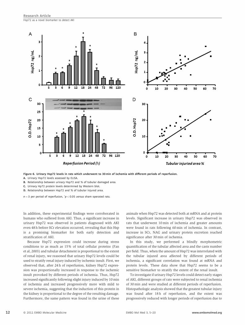
In Fig 6A (urinary detection of Hsp72 by ELISA), a significant
elevation was detected from 3 h after reperfusion. Similarly to
histological injury, the greatest urinary Hsp72 was found after 18h
and return to basal values after 96 h of reperfusion. Again a
significant correlation between urinary Hsp72 and % of tubular
injured area was observed, r2¼ 0.65 (p< 0.0001). Insets in the top
of Fig 6C show Western blots from three rats of each group;
densitometric analysis is shown below. Urinary Hsp72 was almost
undetectable in sham-operated rats; in contrast, there was a
significant increase in Hsp72 in groups subjected to renal ischemia
followed by 3 h of reperfusion and a progressive increase in the
amount of Hsp72 detected in the urine was observed relative to the
reperfusion period, peaking at 18 h of reperfusion with a
EMBO Mol Med 3, 5–20 www.embomolmed.org

Research ArticleJonatan Barrera-Chimal et al.
Figure 3. Renal Hsp72 expression in rats subjected to different periods of ischemia.
A. Total RNA was individually extracted from the renal cortex of all studied groups (n¼5) and Hsp72 mRNA levels were determined by real time RT-PCR.
B. Renal cortex proteins were individually extracted from three rats of each group, and Hsp72 protein levels were assessed by western blot. Upper inset shows a
representative image of the autoradiography of the membrane and the lower graph depicts densitometric analyses of the ratio of Hsp72 to b-actin. �p<0.05
versus sham-operated rats, £p<0.05 versus 30min and ¥p<0.05 versus 45min of ischemia group.
subsequent decrease in excretion of this protein. Intriguingly,
there was a significant correlation between urinary Hsp72 and the
% area of tubular injury, r2¼ 0.55 (p< 0.0001) as depicted in
Fig 6D. A better correlation was found between Hsp72 and cast
formation, r2¼ 0.73 (p< 0.0001, data not shown). These results
demonstrate that Hsp72 is an early biomarker to detect AKI and
suggest that the amount of Hsp72 detected in the urine might
reveal the state of tubular injury and recovery.
Kim-1, NGAL, IL-18 as biomarkers for stratifying and early
detecting AKI
The performance of the kidney injury molecule 1 (Kim-1),
neutrophil gelatinase-associated lipocalin (NGAL) and Inter-
leukin 18 (IL-18) for stratification and early detection of AKI was
also investigated to compare it with performance of Hsp72. As
shown in figure Fig 7A, urinary Kim-1 levels significantly
increased after 20 min of ischemia, and these values reached the
maximum increment at 30 min; thus Kim-1 was unable to
differentiate greater renal injury induced by 45 and 60 min of
ischemia. As a result, a lower correlation between percent of
tubular injured area and Kim-1 levels was found (r2¼ 0.46).
NGAL levels progressively increased with the extent of renal
injury induced by different periods of ischemia, r2¼ 0.61
(p< 0.001) as shown in Fig 7B. However, statistical differences
between different periods of ischemia were not found. Thus,
NGAL was not as good a biomarker as Hsp72 to predict different
degrees of renal damage. Similarly to Hsp72 and NGAL, IL-18
increased proportionally to the renal injury induced from 20 min
of ischemia (r2¼ 0.59, p< 0.001), as depicted in Fig 7C.
The usefulness of these biomarkers for early diagnosis of AKI
was also assessed. Figure 7D shows the urinary Kim-1 values
www.embomolmed.org EMBO Mol Med 3, 5–20
from rats exposed to different periods of reperfusion. A significant
elevation of Kim-1 was observed after 9 h of reperfusion, reaching
the maximal value after 48h. As a result, a low correlation
between tubular affected area and urinary Kim-1 levels (r2¼ 0.27)
was found. In the case of urinary NGAL, a significant elevation
was found after 3 h of reperfusion; however, NGAL levels reached
the maximum peak at 6 h and did not reflect the greater renal
injury that is observed after 18 h. As a result, a lower correlation
between urinary NGAL levels and tubular affected area was
observed, r2¼ 0.48 versus r2¼ 0.65 observed with Hsp72. When
IL-18 was assessed, a significant increase was observed after 6 h
and levels remained elevated after 24h of reperfusion. As a result,
the correlation between tubular affected area and urinary IL-18
was r2¼ 0.51, p< 0.001 (Fig 7F), which was similar to NGAL
(r2¼ 0.48), but lower than Hsp72 (r2¼ 0.65).
Urinary Hsp72 levels as a monitor of a renoprotective
intervention in AKI
Because we previously reported that aldosterone blockade is a
helpful treatment to prevent renal injury induced by I/R (Mejia-
Vilet et al, 2007; Ramirez et al, 2009), we assessed if this
renoprotective effect could be reflected by the prevention of
Hsp72 elevation. Figure 8A and B shows the SCr and creatinine
clearance in two groups of rats subjected to ischemia and 24 h of
reperfusion without treatment (I/R) and pre-treated with
spironolactone 3 days before I/R (I/Rþ Sp). Spironolactone
administration prevented renal dysfunction. This renoprotec-
tive effect was associated with a significant reduction in urinary
Hsp72 levels detected by ELISA and Western blot, as shown in
Fig 8C and D, respectively. In order to evaluate if Hsp72 might
allow to monitor different degrees of renoprotection in the
� 2011 EMBO Molecular Medicine 9

Research ArticleHsp72 as a novel biomarker to detect AKI
Figure 4. Urinary Hsp72 mRNA and protein
levels from rats subjected to different periods
of ischemia.
A. Total RNA was individually extracted from
the urine of six rats per group and Hsp72
mRNA levels were determined by real time
RT-PCR.
B. Relationship between mRNA levels and
tubular affected area.
C. Urinary Hsp72 levels assessed by ELISA.
D. Relationship between urinary Hsp72 levels
and the % of tubular affected area.
E. Urinary Hsp72 levels assessed byWB analysis
from four rats of each group.
F. Relationship between urinary Hsp72 levels
detected by WB and tubular injured area.�p< 0.05 versus sham-operated rats,¤p<0.05 versus 10min, sp<0.05 versus
20min, £p<0.05 versus 30min and¥p< 0.05 versus 45min of ischemia group.
10
absence of creatinine elevation, different groups with I/R were
pre-treated with lower doses of spironolactone. As is shown in
Fig 8E, rats pre-treated with spironolactone at 10 and 5 mg/kg
exhibited normal values of SCr that contrast with the 2.5 mg/kg
dose, in which the values were similar to untreated I/R group.
Interestingly, urinary Hsp72 levels were significantly increased
from 10 mg/kg of spironolactone and progressively enhanced
when the lower doses of were administrated, as is depicted in
Fig 8F. These findings suggest that the lower the dose of
spironolactone, the lower the renoprotection. This can be
efficiently detected by urinary Hsp72.
Urinary Hsp72 levels in healthy kidney donors and patients
with AKI
To determine whether Hsp72 is a sensitive biomarker to detect
AKI in humans, the levels of this protein were assessed by
Western blot in the urine of normal subjects and compared to
� 2011 EMBO Molecular Medicine
those patients who developed AKI in the ICU of our institution.
AKI was defined as an increase in SCr by at least 0.3 mg/dL, or
urine output �0.5 ml/kg/h over 6 h. The Supplementary Table 1
shows general characteristics and renal function of five healthy
kidney donors and nine patients with septic AKI. AKI patients
included five females and four males aged between 24 and 84
years. At admission to the ICU, all patients exhibited normal
creatinine levels; however, increases in SCr from 0.55� 0.05 to
2.30� 0.52 mg/dl were subsequently observed (p¼ 0.004).
Urinary Hsp72 levels are depicted in Fig 9A. In the urine of
healthy kidney donors, Hsp72 was almost undetectable
(3.4� 0.7 arbitrary units), whereas urinary Hsp72 levels
significantly increased by 17.3-fold in patients with clinical
AKI (58.3� 8.5 arbitrary units). These results were confirmed
by ELISA. The basal levels of urinary Hsp72 from healthy living
donors were 0.22� 0.07, contrasting with the values in patients
diagnosed with AKI of 4.90� 1.5 ng/ml (22-fold increase). Of
EMBO Mol Med 3, 5–20 www.embomolmed.org

Research ArticleJonatan Barrera-Chimal et al.
Figure 5. Representative images and morphometry of subcortical histopathological lesions induced by 30min of ischemia and different periods of
reperfusion.
A. 3h, B. 6 h, C. 9 h, D. 12 h, E. 18 h, F. 24 h, G. 48 h, H. 72 h, I. 96 h and J. 120 h of reperfusion.
K. Morphometric quantification of affected tubular area.
L. Mean cast number per field �p< 0.05 versus sham-operated rats.
note, two patients diagnosed with AKI died during hospitaliza-
tion; these patients exhibited greater urinary values of Hsp72 as
assessed by either Western blot or ELISA.
To identify if Hsp72 is an early biomarker to detect AKI in
humans, daily urine samples were collected from patients who
were admitted to the ICU exhibited and respiratory failure
(mechanical ventilation) complicated with other organ failure.
These patients had normal renal function at the admission. Five
patients that developed AKI and five patients with no evidence
of clinical AKI were included. Urine samples taken every day
were analyzed to investigate if an elevation of Hsp72 could be
detected before AKI criteria was fulfilled. Figure 9C and 9D show
the mean daily serum creatinine and urine output from patients
with or without AKI. Day 0 means the day at which AKI was
diagnosed based on AKI criteria. Figure 9E depicts Western blots
showing the daily urinary Hsp72 levels from the patients
www.embomolmed.org EMBO Mol Med 3, 5–20
diagnosed with AKI, compared with the Western blots from
patients without AKI (Fig 9F). The basal levels of Hsp72 were
minimal in patients without AKI and three days before AKI was
diagnosed in AKI patients. In agreement with our experimental
studies, one or two days before AKI diagnosis, there was a
significant increase in urinary Hsp72 levels, suggesting that,
indeed, Hsp72 can be an early marker of AKI in humans.
DISCUSSION
In this study, we found that urinary Hsp72 is a reliable
biomarker for the early detection of AKI. This novel biomarker
was adequately sensitive for stratifying different degrees of
tubular injury and recovery, as well as for monitoring a
renoprotective intervention in an experimental rat model of AKI.
� 2011 EMBO Molecular Medicine 11
Research ArticleHsp72 as a novel biomarker to detect AKI
Figure 6. Urinary Hsp72 levels in rats which underwent to 30min of ischemia with different periods of reperfusion.
A. Urinary Hsp72 levels assessed by ELISA.
B. Relationship between urinary Hsp72 and % of tubular damaged area.
C. Urinary Hp72 protein levels determined by Western blot.
D. Relationship between Hsp72 and % of tubular injured area.
n¼ 3 per period of reperfusion, �p< 0.05 versus sham operated rats.
12
In addition, these experimental findings were corroborated in
humans who suffered from AKI. Thus, a significant increase in
urinary Hsp72 was observed in patients diagnosed with AKI
even 48 h before SCr elevation occurred, revealing that this Hsp
is a promising biomarker for both early detection and
stratification of AKI.
Because Hsp72 expression could increase during stress
conditions to as much as 15% of total cellular proteins (Fan
et al, 2003) and tubular detachment is proportional to the extent
of renal injury, we reasoned that urinary Hsp72 levels could be
used to stratify renal injury induced by ischemic insult. First, we
observed that, after 24 h of reperfusion, kidney Hsp72 expres-
sion was proportionally increased in response to the ischemic
insult provoked by different periods of ischemia. Thus, Hsp72
increased significantly following slight injury induced by 10 min
of ischemia and increased progressively more with mild to
severe ischemia, suggesting that the induction of this protein in
the kidney is proportional to the degree of the resulting damage.
Furthermore, the same pattern was found in the urine of these
� 2011 EMBO Molecular Medicine
animals when Hsp72 was detected both at mRNA and at protein
levels. Significant increase in urinary Hsp72 was observed in
rats that underwent 10 min of ischemia and greater amounts
were found in rats following 60 min of ischemia. In contrast,
increase in SCr, NAG and urinary protein excretion reached
significance after 30 min of ischemia.
In this study, we performed a blindly morphometric
quantification of the tubular affected area and the casts number
per field. Thus, when the amount of Hsp72 was interrelated with
the tubular injured area affected by different periods of
ischemia, a significant correlation was found at mRNA and
protein levels. These data show that Hsp72 seems to be a
sensitive biomarker to stratify the extent of the renal insult.
To investigate if urinary Hsp72 levels could detect early stages
of AKI, different groups of rats were subjected to renal ischemia
of 30 min and were studied at different periods of reperfusion.
Histopathologic analysis showed that the greatest tubular injury
was found after 18 h of reperfusion, and the extent was
progressively reduced with longer periods of reperfusion due to
EMBO Mol Med 3, 5–20 www.embomolmed.org

Research ArticleJonatan Barrera-Chimal et al.
Figure 7. Urinary levels of Kim-1, NGAL and IL-
18 in rats which underwent different periods
of ischemia and reperfusion (n¼6).
A, B, C. Urinary levels of Kim-1, NGAL, and IL-18
in different degrees of renal injury
induced by increasing periods of
ischemia (10, 20, 30, 45 and 60min),
respectively.
D, E, F. Urinary concentrations of Kim-1, NGAL
and IL-18 after several times of reper-
fusion (3, 6, 9, 12, 18, 24, 48, 72, 96 and
120h), respectively.¤p< 0.05 versus 10min, sp<0.05 versus 20 -
min, £p<0.05 versus 30min and ¥p<0.05 ver-
sus 45min of ischemia group.
the concomitant tubular recovery observed after 72 h. Interest-
ingly, the amount of urinary Hsp72 detected also reflected the
extent of tubular epithelium injury during different periods of
reperfusion and the regeneration of tubular epithelium. Urinary
Hsp72 increased significantly after 3 h and continually rose until
18 h of reperfusion. After this time, a progressive reduction was
detected until 120 h of reperfusion, at which point Hsp72
returned to basal levels. The pattern of urinary Hsp72
expression observed with different periods of reperfusion
significantly correlated with the tubular injury quantified by
morphometry. This finding suggests that this protein was not
only able to detect early AKI but that it could also reflect the
tubular recovery processes that occur after the epithelium is
exposed following ischemic/reperfusion insult.
Recently, a sequence of studies which evaluated seven
different biomarkers of glomerular or tubular injury induced
by different nephrotoxic drugs in the rat was reported. These
biomarkers included urinary total protein, cystatin C, a2-
microglobulin, Kim-1, urinary trefoil factor 3 (TFF3), clusterin
www.embomolmed.org EMBO Mol Med 3, 5–20
(CLU) and albumin urinary excretion. Renal histopathology was
used as the benchmark or gold standard to define renal injury
(Dieterle et al, 2010a; Vaidya et al, 2010; Yu et al, 2010). As was
discussed by the FDA-EMEA and Predictive Safety Testing
Consortium, in these rat toxicology studies, Kim-1, CLU, urinary
albumin excretion and TFF3 were helpful to diagnose drug-
induced acute kidney tubular injury in the rat and were superior
to the traditional markers such as SCr and blood urea nitrogen
(BUN), whereas urinary total protein, cystatin C, and a2-
microglobulin were useful to identify acute drug-induced
glomerular damage or impairment of kidney tubular reabsorption
(Dieterle et al, 2010b). This consortium emphasized, however,
that these findings must wait validation in order to be employed
for clinical practice. The renal injury induced by I/R, as the most
common cause of AKI in ICUs and during cardiovascular surgery,
was not addressed in most of these studies (Dieterle et al, 2010;
Vaidya et al, 2010; Yu et al, 2010).
Regarding AKI induced by renal ischemia in animals and
humans, several urinary biomarkers have been identified over
� 2011 EMBO Molecular Medicine 13

Research ArticleHsp72 as a novel biomarker to detect AKI
Figure 8. Urinary Hsp72 as a biomarker of
renoprotection conferred by spironolactone in
I/R rats (n¼5).
A. Serum creatinine in I/R group without
treatment (gray bar) and rats pre-treated
with spironolactone (black bar),
B. Creatinine clearance,
C. Urinary Hsp72 assessed by ELISA and
D. By WB analysis, the inset shows the indi-
vidual analysis from five individual urines.
E. Serum creatinine in I/R group and in rats pre-
treated with lower doses of spironolactone
(10, 5 and 2.5mg/kg) and
F. WBanalysis of Hsp72 in rats pre-treatedwith
lower doses of spironolactone, the superior
inset shows the individual Hsp72 level from
five different animals. �p< 0.05 versus I/R
group.
14
the past few years. These biomarkers include the following:
NAG (Tsutsumi & Neckers, 2007), neutrophil gelatinase
associated lipocalin (NGAL) (McIlroy et al, 2010; Mishra et
al, 2003, 2005), Kim-1 (Han et al, 2009; Vaidya et al, 2006,
2009), cystatin C (Nejat et al, 2010; Uchida & Gotoh, 2002),
interleukin-18 (IL-18) (Melnikov et al, 2001; Parikh et al, 2005,
2006), hepatocyte growth factor (HGF) (Taman et al, 1997) and
liver fatty acid binding protein (L-FABP) (Ferguson et al, 2010;
Negishi et al, 2009; Yamamoto et al, 2007). In humans with AKI
induced by an ischemic insult, NGAL, NAG and IL-18 have
shown to be early biomarkers (Han et al, 2009; Parikh et al,
2006; Wagener et al, 2006), whereas Kim-1 elevation is seen
several hours after AKI has occurred and remains elevated along
the time (Han et al, 2009). In addition, it is still unknown if these
biomarkers individually possess the features that an ideal
biomarker should have, such as the ability to detect AKI early in
the disease course, to stratify different degrees of renal injury,
to identify tubular recovery, to predict patients at risk of
� 2011 EMBO Molecular Medicine
developing end stage renal disease (ESRD) and those at
increased risk of death. In fact, it has been proposed that a
panel of biomarkers might be a powerful tool to diagnose AKI
before SCr elevation occurs; however, this strategy represents
greater effort and cost (Han et al, 2009; Liangos et al, 2007;
Vaidya et al, 2008). In this study, we compared the performance
of Kim-1, NGAL and IL-18 for stratifying and early detecting AKI
compared to Hsp72. As was shown before (Han et al, 2009),
although Kim-1 differentiated low and moderate renal injury, it
was unable to stratify severe renal injury degrees (Fig 7A). This
is supported by the low correlation between tubular affected
area and urinary Kim-1 levels (r2¼ 0.27). In addition, Kim-1 did
not perform well as an early biomarker, since a significant
elevation of Kim-1 was observed only after 9 h of reperfusion.
With respect to NGAL, this biomarker increased progressively
with the extent of renal injury induced by different periods of
ischemia, but statistical differences between different periods of
ischemia were not found. Although NGAL was an early
EMBO Mol Med 3, 5–20 www.embomolmed.org

Research ArticleJonatan Barrera-Chimal et al.
Healthy volunteers AK I
Hs p72
A
0
250
500
750
1000
No AKI AKI
O.D
. Hsp
72
0
5
10
15
No AKI AKI
Hsp
72(n
g/m
L)
B
C
-2 -1 0 1 2 3 4 5-3
D
E
F
SCr
(mg/
dL)
-3 -2 -1 0 1 2 3 4 50.0
0.4
0.8
1.2
1.6 *
Day
U. V
ol (L
)
0
1
2
3
*
-3 -2 -1 0 1 2 3 4 5
1 3 5 8 10
0
(ng/
mL)
-2 -1 0 1 2 3 4 5-3 - --
- - 1 0
- - -
1 3 5 8 101 3 5 8 101 3 5 8 10
Figure 9. Urinary Hsp72 levels as a biomarker
of AKI in humans.
A, B. Hsp72 levels assessed by Western blot and
ELISA, respectively, in five healthy kidney
donors (&) and nine patients that were
diagnosed with AKI (~).
C. Serum creatinine from five patients with
no evidence of AKI (*) or in five patients
with AKI diagnosed with AKIN criteria (�).D. Urine output from five patients with no
evidence of AKI (*) or in five patients with
AKI diagnosed with AKIN criteria (�).E. Daily urinary Hsp72 levels from patients
with diagnosed AKI.
F. Daily urinary Hsp72 levels in patients
without AKI.
The samples were collected during three days
before and 5 days after AKI was diagnosed.�p< 0.05 versus 3 days before AKI.
biomarker as Hsp72, it reached the maximum peak at 6 h and
thus did not reflect the greater renal injury that is observed after
18 h of reperfusion. In the case of IL-18, it showed a similar
pattern to Hsp72 for stratifying renal injury induced by different
periods of ischemia; however, it was unable to detect slight renal
injury induced by 10 min of ischemia. In addition, IL-18 was not
useful for early AKI detection.
It has been reported that a sensitive biomarker must sense if a
therapeutic strategy could reduce AKI (Yamamoto et al, 2007).
In this regard, we have previously shown that mineralocorticoid
receptor blockade is an effective intervention to prevent renal
injury induced by I/R (Mejia-Vilet et al, 2007; Ramirez et al,
2009). Therefore, in this study, we pre-treated rats with
spironolactone 3 days before induction of ischemia. Then,
renal function and urinary Hsp72 were detected. In untreated
rats, renal function was significantly reduced, and a significant
rise in urinary Hsp72 was observed. The renoprotection
conferred by spironolactone was associated with the prevention
www.embomolmed.org EMBO Mol Med 3, 5–20
of this Hsp72 increase, and this effect was consistent with the
preservation of tubular architecture, as we previously reported
(Mejia-Vilet et al, 2007; Ramirez et al, 2009). In this study, we
also evaluated urinary Hsp72 performance for monitoring
different degrees of renoprotection. For this purpose, rats
subjected to I/R were pre-treated with different doses of
spironolactone. The lower dose employed (2.5 mg/kg) was not
able to protect the kidneys from ischemic injury, as demon-
strated by a similar increase in SCr in rats treated with this dose
as compared to those untreated (Fig 8E). In contrast, some
degree of protection was observed with administration of 5 or
10 mg/kg. Consistent with urinary Hsp72 being a good
biomarker, the lower the renoprotection, the higher the
Hsp72 amount in urine. These data suggest that urinary
Hsp72 is a helpful tool for monitoring the effectiveness of a
pharmacological intervention to prevent ischemic insult.
Acute kidney injury remains a common syndrome in
hospitalized patients and has consistently been associated
� 2011 EMBO Molecular Medicine 15

Research ArticleHsp72 as a novel biomarker to detect AKI
16
with increased morbidity and mortality. However, advances in
reducing this complication have long been hindered by the lack
of early and sensitive biomarkers. In spite of creatinine
limitations, current AKIN and RIFLE classifications for
diagnosing AKI are based on the elevation of SCr or urine
output reduction. In this study, we included patients that
developed AKI and were diagnosed by the AKIN classification.
Once the diagnosis was made, a urine sample was taken to
evaluate urinary Hsp72 levels to compare with healthy living
donors. To determine Hsp72 concentration, the urine sample
was analyzed by Western blot analysis and ELISA. Hsp72
levels were almost undetectable in healthy subjects; in
contrast, patients with clinical AKI exhibited an abrupt
increase in this protein. The variability in the AKI patients
observed might result from different degrees and extension of
the renal injury. In this regard, two of three patients that
exhibited high urinary Hsp72 died during their hospitalization,
suggesting that this protein could also serve as a biomarker of
AKI-associated death. However, more studies are necessary to
evaluate this issue.
To evaluate the potential of urinary Hsp72 as an early
biomarker in humans, daily urine samples from ten patients
with normal renal function at the admission to ICU in severe
conditions (mechanical ventilation and other organ failure)
were included. In accord with our experimental data, an
increase in urinary Hsp72 was found even two days before AKI
criteria were fulfilled (SCr elevation or urine output reduction),
suggesting that urinary Hsp72 could be a promising early
biomarker of AKI in humans. Most of the biomarkers studies
include patients that are expected to develop AKI, such as after
cardiovascular surgery or organ transplantation (Han et al,
2009; McIlroy et al, 2010; Mishra et al, 2005; Parikh et al, 2006;
Yamamoto et al, 2007), however, in the daily clinical setting, we
cannot predict which patients will develop AKI. In this study,
urinary Hsp72 had the potential to predict AKI 48 h before AKIN
criteria were fulfilled. Further clinical studies will be required to
validate its use in multiple cohort studies. In this regard, it has
been reported that there are five critical phases in a biomarker
development that include: (1) experimental studies that identify
proteins that are up-regulated in AKI mice and rat models,
(2) development of clinical assays to detect the biomarker in
samples from patients with clinical AKI that should be obtained
non-invasively, (3) evaluation of the biomarker’s ability to
detect preclinical disease prior to clinical diagnosis, and
(4) phase IV and V studies that involve large-scale biomarker
validation (Coca & Parikh, 2008).
In summary, in an experimental model of AKI, our results
revealed that Hsp72 possesses most of the specific characteristics
of an ideal sensitive biomarker. Hsp72 is an early, non-invasive
and easily detected biomarker that can be used to stratify renal
injury that correlates with tubular injury and recovery. It also
seems to be a tool to monitor the effectiveness of a
pharmacological intervention. In addition, the preliminary data
that we found in humans support the notion that Hsp72 could be
an early predictor of AKI in the clinical setting. Taken together,
these results suggest that urinary Hsp72 detection is a promising
and sensitive biomarker in a clinical translational context.
� 2011 EMBO Molecular Medicine
MATERIALS AND METHODS
All experiments involving animals were conducted in accordance with
the Guide for the Care and Use of Laboratory Animals (National Academy
Press, Washington, DC, 1996) and were approved by the Animal Care
and Use Committee of our Institution. One hundred and five male
Wistar rats, weighing 270–300g, were included in this study and
divided into two different experimental protocols. In the first protocol,
seventy-two rats were randomly divided into six groups: sham-operated
(sham) or subjected to bilateral ischemia of 10, 20, 30, 45 or 60min. All
of these groups were studied after 24h of reperfusion. One half of each
group was used only for the determination of urinary Hsp72 mRNA
levels, as is described below. In the second protocol, thirty rats were
subjected to 30min of bilateral ischemia and divided in ten different
groups, which differed by reperfusion time: 3, 6, 9, 12, 18, 24, 48, 72, 96
and 120h. A sham-operated group was also included in this protocol.
Kidney I/R injury animal model
Rats were anesthetized with an intraperitoneal injection of sodium
pentobarbital (30mg/kg), placed on a heating pad to maintain core
body temperature at 378C and monitored with a rectal thermometer. A
midline abdominal incision was made, and both kidneys were exposed.
Renal pedicles were isolated, and bilateral ischemia was induced by
non-traumatic clamps over the pedicles for different times, as we
previously described (Mejia-Vilet et al, 2007; Ramirez et al, 2009).
Ischemia was verified visually by change in kidney colour. Reperfusion
was achieved by release of the clips and confirmed by return of
oxygenated blood to the kidney. After reperfusion, the incision was
closed in two layers with 3–0 sutures. Sham-operated rats underwent
anesthesia, laparotomy and renal pedicle dissection only.
Functional studies
Rats were placed in metabolic cages at 228C with a 12:12 h light–dark
cycle and allowed free access to water. Individual urine samples were
collected from all studied rats. For urine ribonucleic acid (RNA)
isolation, metabolic cages were previously cleaned with RNAse ZAP
(Ambion) and 300mL of RNAlater (Ambion) added to the urine
recipient. In both cases, urine samples were aliquoted and quickly
frozen at 808C until biomarker analysis.
After the reperfusion period, rats were anesthetized with an intraper-
itoneal injection of sodium pentobarbital (30mg/kg) and placed on a
homoeothermic table. The trachea and femoral arteries were catheter-
ized with polyethylene tubing (PE-240 and PE-50). The rats were
maintained under euvolemic conditions by infusing 10ml of rat plasma
per kg of body weight during surgery. MAP was monitored with a
pressure transducer (model p23 db, Gould) and recorded on a polygraph
(Grass Instruments, Quincy, MA). An ultrasound transit-time flow probe
was placed around the left artery and filled with ultrasonic coupling gel
(HR Lubricating Jelly, Carter-Wallace, New York, NY) to record renal blood
flow. Blood samples were taken at the end of the study. Serum and
creatinine concentrations were measured with an autoanalyzer (Tech-
nicon RA-1000, Bayer, Tarrytown, NY), and renal creatinine clearance was
calculated by the standard formula C¼ (U� V)/P, where U is the
concentration in urine, V is the urine flow rate and P is the serum
concentration. Urinary protein excretion was measured by a trichlor-
oacetic acid (TCA) turbidimetric method, and NAG was measured
spectrophotometrically.
EMBO Mol Med 3, 5–20 www.embomolmed.org

Research ArticleJonatan Barrera-Chimal et al.
The paper explained
PROBLEM:
Acute kidney injury is a common and serious complication in
critically ill patients, especially in the ICU in both native and
transplanted kidneys and may also be associated with long-term
chronic kidney disease development. AKI may occur in about 5%
of hospitalized patients and up to 40–60% patients in ICU. In
addition, over the last decade the incidence of AKI has increased
mainly due to population aging and the rising pandemics of
obesity, diabetes and hypertension. Despite efforts and
advances in new therapeutics, the mortality rate of AKI remains
between 40 and 80% and has not been reduced in the last four
decades, mainly because current tools used for early AKI
detection are not adequately sensitive or specific. Therefore,
novel renal early biomarkers indicating tubular injury extent are
needed.
RESULTS:
In an experimental model of AKI, we found that Hsp72 possess
most of the specific characteristics of an ideal biomarker should
have such as: non-invasive, easily detected, to be able to early
detect AKI, to stratify renal injury that correlates with tubular
injury and recovery, and to be a tool to monitor the effectiveness
of a pharmacological intervention. In addition, urinary Hsp72
levels performance to detect and stratify renal injury induced by
different periods of ischemia was better than urinary NGAL, Kim-
1 and IL-18. Our preliminary data in humans support the notion
that Hsp72 could be an early predictor of AKI in the clinical
setting.
IMPACT:
Hsp72 may be a useful tool to early AKI diagnose in patients
admitted to ICU or with high risk of developing renal injury such
as those patients undergoing cardiac surgery, renal transplant or
under nephrotoxic treatment. The opportune AKI diagnosis would
permit the clinician to make an adequate pharmacologic
intervention to improve patient survival and to avoid end stage
renal disease development.
Histopathologic studies
At the end of the experiment, the right kidney was removed and
quickly frozen for molecular studies, and the left kidney was perfused
through the femoral catheter with phosphate buffered saline (PBS).
Following blanching of the kidney, the perfusate was replaced by a
freshly prepared 4% formalin buffer, and perfusion was continued
until fixation was completed. After appropriate dehydration, kidney
slices were embedded in paraffin, sectioned at 4mm and stained via a
periodic acid-Schiff (PAS) technique. Ten subcortical and juxtamedul-
lary fields were randomly recorded from each kidney slide using a
digital camera incorporated in a Nikon Light microscope. In each
microphotograph, tubular cast per field were counted, and the results
were expressed as the average of fields observed. The affected tubular
area was analyzed blindly. Tubular damage was characterized by a loss
of brush border, lumen dilatation or collapse and detachment from
basement membrane. Digital microphotographs were recorded for
each rat to assess, by morphometric analysis, the total tubular area
(excluding luminal, interstitial and glomerular areas) and damaged
tubular area, delimited by using eclipse net software (magnification
400�). The damaged tubular area was expressed as a proportion of
the affected tubular area and total tubular area.
Hsp72 mRNA levels
Each renal cortex was isolated and snap frozen in liquid nitrogen. Total
RNA was isolated with the TRIzol method (Invitrogen) and checked for
integrity by 1% agarose gel electrophoresis. To avoid DNA contamina-
tion, all total RNA samples were treated with DNAse (DNAase I,
Invitrogen). For urine RNA isolation, 24 h urine-collection from each
rat was centrifuged at 3000 rpm for 30min at 48C. The sediment was
resuspended in 1ml of PBS; afterward, it was centrifuged at
13,000 rpm for 3min and processed by the TrIzol method for RNA
www.embomolmed.org EMBO Mol Med 3, 5–20
isolation. Reverse transcription (RT) was carried out with 1mg of total
RNA and 200U of Moloney murine leukemia virus reverse tran-
scriptase (Invitrogen). The mRNA levels of Hsp72 were quantified by
real-time PCR on an ABI Prism 7300 Sequence Detection System
(TaqMan, ABI, Foster City, CA). Primer and probe for Hsp72 were
ordered as a kit: Rn00583013_s1 (Assay-on-Demand, ABI). As an
endogenous control, eukaryotic 18S rRNA (predesigned assay reagent
Applied by ABI, external run) was used. Relative quantification of
Hsp72 gene expression was performed with the comparative thresh-
old cycle (Ct) method (Livak & Schmittgen, 2001).
Kidney and urinary Hsp72 protein levels
Hsp72 detected by Western blot
Total renal proteins were isolated from six cortexes from each
group and homogenized separately in lysis buffer (50mM
HEPES pH 7.4, 250mM NaCl, 5mM EDTA, 0.1% NP-40 and
complete protease inhibitor (Roche)). Protein samples containing
20mg of total protein were resolved by 8.5% sodium polyacry-
lamide gel electrophoresis (SDS–PAGE) electrophoresis and
electroblotted onto polyvinylidinediuoride membranes (Amer-
sham). For urinary Hsp72 detection by western blot, urine was
diluted 1:100 in 0.9% saline solution, and 10mL of each dilution
was loaded and resolved by 8.5% SDS–PAGE electrophoresis and
electroblotted, as previously described. Membranes were then
blocked with 5% blotting-grade non-fat dry milk. Membranes were
then incubated in 0.1% blotting-grade non-fat dry milk with their
respective antibodies. For detection of Hsp72 in the renal cortex,
the lower parts of the membranes were incubated with goat anti-
actin antibody (1:5000 dilution) overnight at 4-C. (Santa Cruz
Biotechnology, Santa Barbara CA). Upper membranes were
� 2011 EMBO Molecular Medicine 17

Research ArticleHsp72 as a novel biomarker to detect AKI
18
incubated with monoclonal anti-Hsp72 antibody. Afterward,
membranes for Hsp72 were incubated with a secondary antibody,
HRP-conjugated goat anti-mouse IgG (1:500, Santa Cruz Bio-
technology). Actin detection was performed using donkey anti-goat
IgG-HRP (1:5000, Santa Cruz Biotechnology). Proteins were
detected with an enhanced chemiluminescence kit (Amersham)
and autoradiography, following the manufacturer’s recommenda-
tions. The bands were scanned for densitometric analysis.
Hsp72 detected by ELISA
Urine Hsp72 was analyzed using a commercially available high-
sensitivity ELISA (Assay Designs EKS-715, MI, USA). Briey, samples
and standards were added to wells coated with a mouse
monoclonal antibody. Hsp72 was captured by the antibody and
then detected by adding a rabbit polyclonal detection antibody.
Both antibodies are specic for inducible Hsp72 and do not react
with other members of the HSP70 family. A horseradish peroxidase
conjugate bound to the detection antibody and colour develop-
ment was accomplished by the addition of tetramethylbenzidine
substrate and stopped with an acid stop solution. The optical
density of samples was read at 450 nm by a plate reader and was
overlapped with the standard curve generated from known
concentrations of recombinant Hsp72 that ranged from 0.1 to
12.5 ng/ml.
Urinary Hsp72 levels as a monitor of a renoprotective
intervention in AKI
Because we previously found that aldosterone antagonism with
spironolactone at 20mg/kg prevented renal injury induced by I/R,
urinary Hsp72 levels as a monitor of a renoprotective intervention in
AKI was assessed. Twenty-five Wistar rats, weighing 270–300 g, were
divided in five groups: rats subjected to 30min of ischemia and 24h of
reperfusion and rats that received spironolactone at different doses (20,
10, 5 or 2.5mg/kg, by gastric gavage) 3 days before I/R was induced.
After ischemia, urine was collected in metabolic cages, and blood
samples were taken.
NGAL, Kim-1 and IL-18 detected by ELISA
Urine NGAL, Kim-1 and IL-18 levels were analyzed using commercially
available ELISA kits: rat NGAL ELISA kit (Innovative Research
IRNGALKT), rat Kim-1 ELISA kit (CosmoBIO Co CSB-E08808r) and rat
IL-18 ELISA Kit (Invitrogen KRC2341). All procedures were performed
accordingly manufacturer’s instructions.
Evaluation of urinary Hsp72 in healthy kidney donors and
patients with AKI
Urine samples from five healthy living-kidney donors (controls) and
nine patients with septic AKI from the ICU were tested. AKI was
defined as a 30% or greater increase in SCr from baseline according
to the AKIN criteria (Mehta et al, 2007). In healthy donors, urine
samples were collected from the first morning voids one day previous
to nephrectomy (previously signed consent). All patients with sepsis
in the ICU were followed every day, and, when the AKI criteria were
fulfiled, a fresh urine sample was taken after draining the urine
collection bag. Moreover, daily fresh urine samples were collected
from patients in the ICU that exhibited respiratory failure with
mechanical ventilation and together with other organ failure. These
� 2011 EMBO Molecular Medicine
patients had normal renal function at the admission. From these
patients, we analyzed, five patients that developed AKI and five that
did not. In the urine samples Hsp72 levels were assessed in order to
test if this biomarker could predict AKI criteria was fulfil (SCr
elevation or urine output reduction). All urine samples were frozen
and stored at 808C until Hsp72 levels were evaluated. The human
subjects included in these study form part of two ongoing clinical
studies, which are in accordance with national and international
guidelines and regulations and were approved by our institutional
ethical review board. All subjects or designed surrogate (when ICU
patients could not sign) signed the informed consent form.
Statistical analysis
Results are presented as means� SE. Significance of the differences
between groups was tested by ANOVA using Bonferroni’s correction for
multiple comparisons. All comparisons passed the normality test.
Statistical significance was defined as p-value <0.05.
Author Contributions
JBC, LEM and NAB: Conceived and designed the experiments.
JBC, RPV, CCG and MOC: performed the experiments. JBC, GG,
LEMB and NAB: analyzed the data. GG and NAB: contributed
reagents or analysis tools. JBC, GG and NAB: wrote the paper.
AcknowledgementsWe are grateful to the members of the Molecular Physiology
Unit for their suggestions and to Dr. Octavio Villanueva for his
help with animal care, as well as to Martha Carrrasco for her
technical assistance. The results presented in this paper have
not been published previously neither in whole nor in abstract.
This project was supported by grants from the Mexican Council
of Science and Technology (CONACyT) 101030 and 112780 to
NAB, by a grant from the National University of Mexico
IN200909-3 to NAB and by a grant from Fundacion Miguel
Aleman AC to NAB. JBC is a PhD student supported by
fellowship grant from CONACyT.
Supporting information is available at EMBO Molecular
Medicine online.
The authors declare that they have no conflict of interest.
For more information
Heat Shock 70-kd protein:
http://www.ncbi.nlm.nih.gov/omim/140550
National Kidney Foundation
http://www.kidney.org/
ReferencesBagshaw SM (2010) Acute kidney injury: diagnosis and classification of AKI:
AKIN or RIFLE? Nat Rev Nephrol 6: 71-73
EMBO Mol Med 3, 5–20 www.embomolmed.org

Research ArticleJonatan Barrera-Chimal et al.
Coca SG, Parikh CR (2008) Urinary biomarkers for acute kidney injury:
perspectives on translation. Clin J Am Soc Nephrol 3: 481-490
Csermely P, Soti C, Blatch GL (2007) Chaperones as parts of cellular networks.
Adv Exp Med Biol 594: 55-63
Devarajan P (2006) Update on mechanisms of ischemic acute kidney injury.
J Am Soc Nephrol 17: 1503-1520
Dieterle F, Perentes E, Cordier A, Roth DR, Verdes P, Grenet O, Pantano S,
Moulin P, Wahl D, Mahl, et al (2010) Urinary clusterin, cystatin C, beta2-
microglobulin and total protein as markers to detect drug-induced kidney
injury. Nat Biotechnol 28: 463-469
Dieterle F, Sistare F, Goodsaid F, Papaluca M, Ozer JS, Webb CP, Baer W,
Senagore A, Schipper MJ, Vonderscher J, et al (2010) Renal biomarker
qualification submission: a dialog between the FDA-EMEA and Predictive
Safety Testing Consortium. Nat Biotechnol 28: 455-462
Fan CY, Lee S, Cyr DM (2003) Mechanisms for regulation of Hsp70 function by
Hsp40 cell stress. Chaperones 8: 309-316
Ferguson MA, Vaidya VS, Waikar SS, Collings FB, Sunderland KE, Gioules CJ,
Bonventre JV (2010) Urinary liver-type fatty acid-binding protein predicts
adverse outcomes in acute kidney injury. Kidney Int 77: 708-714
Friedewald JJ, Rabb H (2004) Inflammatory cells in ischemic acute renal
failure. Kidney Int 66: 486-491
Han WK, Wagener G, Zhu Y, Wang S, Lee HT (2009) Urinary biomarkers in the
early detection of acute kidney injury after cardiac surgery. Clin J Am Soc
Nephrol 4: 873-882
Hernadez-Pando R, Pedraza-Chaverri J, Orozco-Estevez H, Silva-Serna P,
Moreno I, Rondan-Zarate A, Elinos M, Correa-Rotter R, Larriva-Sahd J (1995)
Histological and subcellular distribution of 65 and 70 kD heat shock
proteins in experimental nephrotoxic injury. Exp Toxicol Pathol 47: 501-
508
Kelly KJ (2002) Stress response proteins and renal ischemia. Minerva Urol
Nefrol 54: 81-91
Kelly KJ (2006) Acute renal failure: much more than a kidney disease. Semin
Nephrol 26: 105-113
Kelly KJ, Baird NR, Greene AL (2001) Induction of stress response proteins
and experimental renal ischemia/reperfusion. Kidney Int 59: 1798-
1802
Liangos O, Perianayagam MC, Vaidya VS, Han WK, Wald R, Tighiouart H,
MacKinnon RW, Li L, Balakrishnan VS, Pereira BJ, et al (2007) Urinary
N-acetyl-beta-(D)-glucosaminidase activity and kidney injury molecule-1
level are associated with adverse outcomes in acute renal failure. J AmSoc
Nephrol 18: 904-912
Liano F, Pascual J (1996) Epidemiology of acute renal failure: a prospective,
multicenter, community-based study. Madrid Acute Renal Failure Study
Group. Kidney Int 50: 811-818
Lindquist S, Craig EA (1988) The heat-shock proteins. Annu Rev Genet 22: 631-
677
Liu KD, Brakeman PR (2008) Renal repair and recovery. Crit Care Med 36:
S187-S192
Livak KJ, Schmittgen TD (2001) Analysis of relative gene expression data using
real-time quantitative PCR and the 2(-Delta Delta C(T)). Method Methods
25: 402-408
Lopes JA, Fernandes P, Jorge S, Goncalves S, Alvarez A, Costa e S, Franca C, Prata
MM (2008) Acute kidney injury in intensive care unit patients: a comparison
between the RIFLE and the acute kidney injury network classifications. Crit
Care 12: R110
McIlroy DR, Wagener G, Lee HT (2010) Neutrophil gelatinase-associated
lipocalin and acute kidney injury after cardiac surgery: the effect of baseline
renal function on diagnostic performance. Clin J Am Soc Nephrol 5: 211-219
Mehta RL, Kellum JA, Shah SV, Molitoris BA, Ronco C, Warnock DG, Levin A
(2007) Acute kidney injury network: report of an initiative to improve
outcomes in acute kidney injury. Crit Care 11: R31
Mejia-Vilet JM, Ramirez V, Cruz C, Uribe N, Gamba G, Bobadilla NA (2007)
Renal ischemia-reperfusion injury is prevented by the mineralocorticoid
receptor blocker spironolactone. Am J Physiol Renal Physiol 293: F78-F86
www.embomolmed.org EMBO Mol Med 3, 5–20
Melnikov VY, Ecder T, Fantuzzi G, Siegmund B, Lucia MS, Dinarello CA, Schrier
RW, Edelstein CL (2001) Impaired IL-18 processing protects caspase-1-
deficient mice from ischemic acute renal failure. J Clin Invest 107: 1145-
1152
Mishra J, Ma Q, Prada A, Mitsnefes M, Zahedi K, Yang J, Barasch J, Devarajan P
(2003) Identification of neutrophil gelatinase-associated lipocalin as a
novel early urinary biomarker for ischemic renal injury. J Am Soc Nephrol
14: 2534-2543
Mishra J, Dent C, Tarabishi R, Mitsnefes MM, Ma Q, Kelly C, Ruff SM, Zahedi K,
Shao M, Bean J, et al (2005) Neutrophil gelatinase-associated lipocalin
(NGAL) as a biomarker for acute renal injury after cardiac surgery. Lancet
365: 1231-1238
Molinas SM, Rosso M, Wayllace NZ, Pagotto MA, Pisani GB, Monasterolo LA,
Trumper L (2010) Heat shock protein 70 induction and its urinary excretion
in a model of acetaminophen nephrotoxicity. Pediatr Nephrol 25: 1245-
1253
Mueller T, Bidmon B, Pichler P, Arbeiter K, Ruffingshofer D, VanWhy SK,
Aufricht C (2003) Urinary heat shock protein-72 excretion in clinical and
experimental renal ischemia. Pediatr Nephrol 18: 97-99
Negishi K, Noiri E, Doi K, Maeda-Mamiya R, Sugaya T, Portilla D, Fujita T (2009)
Monitoring of urinary L-type fatty acid-binding protein predicts histological
severity of acute kidney injury. Am J Pathol 174: 1154-1159
Nejat M, Pickering JW, Walker RJ, Endre ZH (2010) Rapid detection of acute
kidney injury by plasma cystatin C in the intensive care unit. Nephrol Dial
Transplant 25: 3283-3289
Parikh CR, Abraham E, Ancukiewicz M, Edelstein CL (2005) Urine IL-18 is an
early diagnostic marker for acute kidney injury and predicts mortality in the
intensive care unit. J Am Soc Nephrol 16: 3046-3052
Parikh CR, Mishra J, Thiessen-Philbrook H, Dursun B, Ma Q, Kelly C, Dent C,
Devarajan P, Edelstein CL (2006) Urinary IL-18 is an early predictive
biomarker of acute kidney injury after cardiac surgery. Kidney Int 70: 199-
203
Price PM, Safirstein RL, Megyesi J (2009) The cell cycle and acute kidney injury.
Kidney Int 76: 604-613
Ramirez V, Trujillo J, Valdes R, Uribe N, Cruz C, Gamba G, Bobadilla NA (2009)
Adrenalectomy prevents renal ischemia-reperfusion injury. Am J Physiol
Renal Physiol 297: F932-F942
Ritossa FA (1962) New puffing pattern induced by temperature shoch and
DNP in Drosophila. Experientia 18: 571-573
Taman M, Liu Y, Tolbert E, Dworkin LD (1997) Increase urinary hepatocyte
growth factor excretion in human acute renal failure. Clin Nephrol 48: 241-
245
Tsutsumi S, Neckers L (2007) Extracellular heat shock protein 90: a role for a
molecular chaperone in cell motility and cancer metastasis. Cancer Sci 98:
1536-1539
Turman MA, Rosenfeld SL (1999) Heat shock protein 70 overexpression
protects LLC-PK1 tubular cells from heat shock but not hypoxia. Kidney Int
55: 189-197
Uchida K, Gotoh A (2002) Measurement of cystatin-C and creatinine in urine.
Clin Chim Acta 323: 121-128
Vaidya VS, Ramirez V, Ichimura T, Bobadilla NA, Bonventre JV (2006) Urinary
kidney injury molecule-1: a sensitive quantitative biomarker for early
detection of kidney tubular injury. Am J Physiol Renal Physiol 290: F517-
F529
Vaidya VS, Waikar SS, Ferguson MA, Collings FB, Sunderland K, Gioules C,
Bradwin G, Matsouaka R, Betensky RA, Curhan GC, et al (2008) Urinary
biomarkers for sensitive and specific detection of acute kidney injury in
humans. Clin Transl Sci 1: 200-208
Vaidya VS, Ford GM, Waikar SS, Wang Y, Clement MB, Ramirez V, Glaab WE,
Troth SP, Sistare FD, Prozialeck WC, et al (2009) A rapid urine test for early
detection of kidney injury. Kidney Int 76: 108-114
Vaidya VS, Ozer JS, Dieterle F, Collings FB, Ramirez V, Troth S, Muniappa N,
Thudium D, Gerhold D, Holder DJ, Bobadilla NA, Marrer E, et al (2010) Kidney
injury molecule-1 outperforms traditional biomarkers of kidney injury in
� 2011 EMBO Molecular Medicine 19

Research ArticleHsp72 as a novel biomarker to detect AKI
20
preclinical biomarker qualification studies. Nat Biotechnol 28: 478-
485
Wagener G, Jan M, Kim M, Mori K, Barasch JM, Sladen RN, Lee HT (2006)
Association between increases in urinary neutrophil gelatinase-associated
lipocalin and acute renal dysfunction after adult cardiac surgery.
Anesthesiology 105: 485-491
Waikar SS, Curhan GC, Wald R, McCarthy EP, Chertow GM (2006) Declining
mortality in patients with acute renal failure, 1988 to 2002. J Am Soc
Nephrol 17: 1143-1150
Wu I, Parikh CR (2008) Screening for kidney diseases: older measures versus
novel biomarkers. Clin J Am Soc Nephrol 3: 1895-1901
� 2011 EMBO Molecular Medicine
Yamamoto T, Noiri E, Ono Y, Doi K, Negishi K, Kamijo A, Kimura K, Fujita T,
Kinukawa T, Taniguchi H, et al (2007) Renal L-type fatty acid-
binding protein in acute ischemic injury. J Am Soc Nephrol 18: 2894-
2902
Yu Y, Jin H, Holder D, Ozer JS, Villarreal S, Shughrue P, Shi S, Figueroa DJ, Clouse
H, Su M, et al (2010) Urinary biomarkers trefoil factor 3 and albumin
enable early detection of kidney tubular injury. Nat Biotechnol 28: 470-
477
Zhipeng W, Li L, Qibing M, Linna L, Yuhua R, Rong Z (2006) Increased
expression of heat shock protein (HSP)72 in a human proximal tubular
cell line (HK-2) with gentamicin-induced injury. J Toxicol Sci 31: 61-70
EMBO Mol Med 3, 5–20 www.embomolmed.org