Hippocampal volume and shape analysis in an older adult population
16
HIPPOCAMPAL VOLUME AND SHAPE ANALYSIS IN AN OLDER ADULT POPULATION Tara L. McHugh 1 , Andrew J. Saykin 1,2,3 , Heather A. Wishart 1 , Laura A. Flashman 1 , Howard B. Cleavinger 1 , Laura A. Rabin 1 , Alexander C. Mamourian 2 , and Li Shen 4 1 Department of Psychiatry, Brain Imaging Laboratory, Dartmouth Medical School, Lebanon, NH 2 Department of Radiology, Brain Imaging Laboratory, Dartmouth Medical School, Lebanon, NH 3 Department of Computer Science, Dartmouth College, Hanover, NH 4 Department of Computer and Information Science, University of Massachusetts Dartmouth, MA, USA Abstract This report presents a manual segmentation protocol for the hippocampus that yields a reliable and comprehensive measure of volume, a goal that has proven difficult with prior methods. Key features of this method include alignment of the images in the long axis of the hippocampus and the use of a three-dimensional image visualization function to disambiguate anterior and posterior hippocampal boundaries. We describe procedures for hippocampal volumetry and shape analysis, provide inter- and intra-rater reliability data, and examine correlates of hippocampal volume in a sample of healthy older adults. Participants were 40 healthy older adults with no significant cognitive complaints, no evidence of mild cognitive impairment or dementia, and no other neurological or psychiatric disorder. Using a 1.5 T GE Signa scanner, three-dimensional spoiled gradient recalled acquisition in a steady state (SPGR) sequences were acquired for each participant. Images were resampled into 1 mm isotropic voxels, and realigned along the interhemispheric fissure in the axial and coronal planes, and the long axis of the hippocampus in the sagittal plane. Using the BRAINS program (Andreasen et al., 1993), the boundaries of the hippocampus were visualized in the three orthogonal views, and boundary demarcations were transferred to the coronal plane for tracing. Hippocampal volumes were calculated after adjusting for intracranial volume (ICV). Intra- and inter-rater reliabilities, measured using the intraclass correlation coefficient, exceeded .94 for both the left and right hippocampus. Total ICV-adjusted volumes were 3.48 (±0.43) cc for the left hippocampus and 3.68 (±0.42) for the right. There were no significant hippocampal volume differences between males and females (p > .05). In addition to providing a comprehensive volumetric measurement of the hippocampus, the refinements included in our tracing protocol permit analysis of changes in hippocampal shape. Shape analyses may yield novel information about structural brain changes in aging and dementia that are not reflected in volumetric measurements alone. These and other novel directions in research on hippocampal function and dysfunction will be facilitated by the use of reliable, comprehensive, and consistent segmentation and measurement methods. © 2007 Psychology Press, part of the Taylor & Francis group, an informa business Address correspondence to: Andrew J. Saykin, Psy.D., ABPP-CN, Professor of Radiology, IU Center for Neuroimaging, Division of Imaging Sciences, Department of Radiology, Indiana University School of Medicine, 950 W. Walnut St., R2 E124, Indianapolis, IN 46202, USA., [email protected]. NIH Public Access Author Manuscript Clin Neuropsychol. Author manuscript; available in PMC 2012 October 28. Published in final edited form as: Clin Neuropsychol. 2007 January ; 21(1): 130–145. doi:10.1080/13854040601064534. NIH-PA Author Manuscript NIH-PA Author Manuscript NIH-PA Author Manuscript
Transcript of Hippocampal volume and shape analysis in an older adult population
HIPPOCAMPAL VOLUME AND SHAPE ANALYSIS IN AN OLDERADULT POPULATION
Tara L. McHugh1, Andrew J. Saykin1,2,3, Heather A. Wishart1, Laura A. Flashman1, HowardB. Cleavinger1, Laura A. Rabin1, Alexander C. Mamourian2, and Li Shen4
1Department of Psychiatry, Brain Imaging Laboratory, Dartmouth Medical School, Lebanon, NH2Department of Radiology, Brain Imaging Laboratory, Dartmouth Medical School, Lebanon, NH3Department of Computer Science, Dartmouth College, Hanover, NH4Department of Computer and Information Science, University of Massachusetts Dartmouth, MA,USA
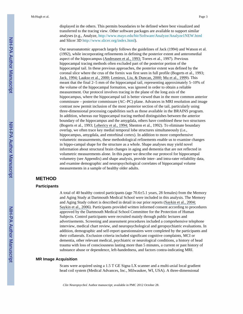
AbstractThis report presents a manual segmentation protocol for the hippocampus that yields a reliable andcomprehensive measure of volume, a goal that has proven difficult with prior methods. Keyfeatures of this method include alignment of the images in the long axis of the hippocampus andthe use of a three-dimensional image visualization function to disambiguate anterior and posteriorhippocampal boundaries. We describe procedures for hippocampal volumetry and shape analysis,provide inter- and intra-rater reliability data, and examine correlates of hippocampal volume in asample of healthy older adults. Participants were 40 healthy older adults with no significantcognitive complaints, no evidence of mild cognitive impairment or dementia, and no otherneurological or psychiatric disorder. Using a 1.5 T GE Signa scanner, three-dimensional spoiledgradient recalled acquisition in a steady state (SPGR) sequences were acquired for eachparticipant. Images were resampled into 1 mm isotropic voxels, and realigned along theinterhemispheric fissure in the axial and coronal planes, and the long axis of the hippocampus inthe sagittal plane. Using the BRAINS program (Andreasen et al., 1993), the boundaries of thehippocampus were visualized in the three orthogonal views, and boundary demarcations weretransferred to the coronal plane for tracing. Hippocampal volumes were calculated after adjustingfor intracranial volume (ICV). Intra- and inter-rater reliabilities, measured using the intraclasscorrelation coefficient, exceeded .94 for both the left and right hippocampus. Total ICV-adjustedvolumes were 3.48 (±0.43) cc for the left hippocampus and 3.68 (±0.42) for the right. There wereno significant hippocampal volume differences between males and females (p > .05). In additionto providing a comprehensive volumetric measurement of the hippocampus, the refinementsincluded in our tracing protocol permit analysis of changes in hippocampal shape. Shape analysesmay yield novel information about structural brain changes in aging and dementia that are notreflected in volumetric measurements alone. These and other novel directions in research onhippocampal function and dysfunction will be facilitated by the use of reliable, comprehensive,and consistent segmentation and measurement methods.
© 2007 Psychology Press, part of the Taylor & Francis group, an informa business
Address correspondence to: Andrew J. Saykin, Psy.D., ABPP-CN, Professor of Radiology, IU Center for Neuroimaging, Division ofImaging Sciences, Department of Radiology, Indiana University School of Medicine, 950 W. Walnut St., R2 E124, Indianapolis, IN46202, USA., [email protected].
NIH Public AccessAuthor ManuscriptClin Neuropsychol. Author manuscript; available in PMC 2012 October 28.
Published in final edited form as:Clin Neuropsychol. 2007 January ; 21(1): 130–145. doi:10.1080/13854040601064534.
NIH
-PA Author Manuscript
NIH
-PA Author Manuscript
NIH
-PA Author Manuscript
KeywordsBoundaries; Hippocampus; Shape; Volume
INTRODUCTIONMedial temporal lobe structures, including the hippocampus, are critical for episodicmemory. Effects of normal aging on hippocampal structural integrity are somewhatcontroversial (Bigler, Blatter, & Andersob, 2002; Raz, Rodrigue, Head, Kennedy, & Acker,2004), but Alzheimer’s disease (AD) is associated with medial temporal atrophy andepisodic memory dysfunction even early in the course of the illness (Jack, Petersen,O’Brien, & Tangalos, 1992; Johnson, Saykin, Flashman, & Riordan, 1998; Killiany, Moss,Albert, Sandor, Tieman, & Jolesz, 1993; Laakso et al., 1995; Scheltens et al., 1992).Magnetic resonance imaging (MRI) is an important tool for examining the size and shape ofthe hippocampus in normal aging, AD, and mild cognitive impairment (MCI) (Jack et al.,2002; Laakso, Hallikainen, & Soininen, 2000; Laakso et al., 1995; Small, Tsai, DeLaPaz,Mayeux, & Stern, 2002), as well as other neuropsychiatric disorders (Meisenzahl &Schlosser, 2001) such as schizophrenia (Bogerts et al., 1993; Chance, Esiri, & Crow, 2002;Csernansky et al., 1998; Joyal et al., 2003; Kalus et al., 2004; Nelson, Saykin, Flashman, &Riordan, 1998; Shenton, Gerig, McCarley, Szekely, & Kikinis, 2002; Torres, Flashman,O’Leary, Swayze, & Andreasen, 1997) and temporal lobe epilepsy (Cendes et al., 1993;Jack, 1994; Scott, DeKosky, & Scheff, 1991; Sims & Williams, 1990; Soininen et al., 1994;Watson et al., 1992).
The majority of hippocampal volumetry studies in aging and AD have employed an MRfield strength of 1.5 Tesla, and derived volumes using coronal slices varying from 1.5 mm to3.1 mm in thickness. Volume acquisition has been calculated using a variety of programsand locally developed software, but time-intensive, individualized manual tracing has beenthe primary method for deriving hippocampal volumes. There now exists a body of literaturewith varying methods for measuring the hippocampus, and other medial temporal lobestructures such as the amygdala, entorhinal cortex, temporal horn, perirhinal cortex, andtemporo-polar cortex, or a combination of the above (Bonilha, Kobayashi, Cendes, & Li,2003; Convit et al., 1999; Golomb et al., 1993; Goncharova, Dickerson, Stoub, & deToledo-Morrell, 2001; Honeycutt & Smith, 1995; Honeycutt et al., 1998; Insausti et al., 1998; Mu,Xie, Wen, Weng, & Shuyun, 1999; Naidich, Valavanis, & Kubik, 1995; Pantel et al., 2000).
Different hippocampal volumetry methods result in different volume measurements. Forexample, Hasboun et al. (1996) compared three scan acquisition and reformatting protocolsand obtained different, though correlated, hippocampal volumes with each. The authorsnoted that three-dimensional processing of scan data helped with visualizing ambiguousboundaries, such as those between the hippocampus and the amygdala. Analyzinghippocampal volume in the plane perpendicular to the long axis was encouraged. Overall,they emphasized the importance of using one protocol throughout a given study to eliminatemethod-related variance.
Our laboratory has developed a protocol for reliably segmenting the entire hippocampususing the software program BRAINS (Brain Research: Analysis of Images, Networks, andSystems; http://www.psychiatry.uiowa.edu/mhcrc/IPL-pages/BRAINS.htm), developed bythe University of Iowa Mental Health Clinical Research Center Imaging Processing Lab(Andreasen et al., 1993). The BRAINS program allows simultaneous visualization andboundary demarcation in any of the three orthogonal views (Figure 1). It also includes a“telegraphing” function in which demarcations made in any view are automatically
McHugh et al. Page 2
Clin Neuropsychol. Author manuscript; available in PMC 2012 October 28.
NIH
-PA Author Manuscript
NIH
-PA Author Manuscript
NIH
-PA Author Manuscript
displayed in the others. This permits boundaries to be defined where best visualized andtransferred to the tracing view. Other software packages are available to support similaranalyses (e.g., Analyze, http://www.mayo.edu/bir/Software/Analyze/Analyze1NEW.htmland Slicer 3D http://www.slicer.org/index.html).
Our neuroanatomic approach largely follows the guidelines of Jack (1994) and Watson et al.(1992), while incorporating refinements in defining the posterior extent and anteromedialaspect of the hippocampus (Andreasen et al., 1993; Torres et al., 1997). Previoushippocampal tracing methods often excluded part of the posterior portion of thehippocampal tail. In these previous approaches, the posterior extent was defined by thecoronal slice where the crus of the fornix was first seen in full profile (Bogerts et al., 1993;Jack, 1994; Laakso et al., 2000; Lemieux, Liu, & Duncan, 2000; Mu et al., 1999). Thismeant that the final 2–5 mm of the hippocampal tail, representing approximately 5–10% ofthe volume of the hippocampal formation, was ignored in order to obtain a reliablemeasurement. Our protocol involves tracing in the plane of the long axis of thehippocampus, where the hippocampal tail is better viewed than in the more common anteriorcommissure – posterior commissure (AC–PC) plane. Advances in MRI resolution and imagecontrast now permit inclusion of the most posterior section of the tail, particularly usingthree-dimensional processing capabilities such as those available in the BRAINS program.In addition, whereas our hippocampal tracing method distinguishes between the anteriorboundary of the hippocampus and the amygdala, others have combined these two structures(Bogerts et al., 1993; Lehericy et al., 1994; Shenton et al., 1992). To eliminate boundaryoverlap, we often trace key medial temporal lobe structures simultaneously (i.e.,hippocampus, amygdala, and entorhinal cortex). In addition to more comprehensivevolumetric measurements, these methodological refinements enable us to examine changesin hippo-campal shape for the structure as a whole. Shape analyses may yield novelinformation about structural brain changes in aging and dementia that are not reflected involumetric measurements alone. In this paper we describe our protocol for hippocampalvolumetry (see Appendix) and shape analysis, provide inter- and intra-rater reliability data,and examine demographic and neuropsychological correlates of hippocampal volumemeasurements in a sample of healthy older adults.
METHODParticipants
A total of 40 healthy control participants (age 70.6±5.1 years, 28 females) from the Memoryand Aging Study at Dartmouth Medical School were included in this analysis. The Memoryand Aging Study cohort is described in detail in our prior reports (Saykin et al., 2004;Saykin et al., 2006). Participants provided written informed consent according to proceduresapproved by the Dartmouth Medical School Committee for the Protection of HumanSubjects. Control participants were recruited mainly through public lectures andadvertisements. Screening and assessment procedures included a comprehensive telephoneinterview, medical chart review, and neuropsychological and geropsychiatric evaluations. Inaddition, demographic and self-report questionnaires were completed by the participants andtheir collaterals. Exclusion criteria included significant cognitive complaints, MCI ordementia, other relevant medical, psychiatric or neurological conditions, a history of headtrauma with loss of consciousness lasting more than 5 minutes, a current or past history ofsubstance abuse or dependence, left-handedness, and factors contra-indicating MRI.
MR Image AcquisitionScans were acquired using a 1.5 T GE Signa LX scanner and a multi-axial local gradienthead coil system (Medical Advances, Inc., Milwaukee, WI, USA). A three-dimensional
McHugh et al. Page 3
Clin Neuropsychol. Author manuscript; available in PMC 2012 October 28.
NIH
-PA Author Manuscript
NIH
-PA Author Manuscript
NIH
-PA Author Manuscript
spoiled gradient recalled acquisition in a steady state (SPGR) sequence was acquired forvolumetric analysis (TR = 25, TE = 3 or min, flip angle = 40, NEX = 1, and slice thickness= 1.5 mm without gaps, yielding 124 contiguous slices with a 24-cm field of view and 256 ×256 matrix with 0.9375-mm in-plane resolution). SPGR scan acquisition time wasapproximately 10 minutes. In addition, a fast spin echo T2-weighted scan (TR = 3000, TE =96, 3-mm contiguous axial slices) was acquired and reviewed along with the SPGR scan bya Board-certified neuror-adiologist to rule out incidental findings.
Image Transfer and PreprocessingEach SPGR scan was transferred via ethernet from the MRI Center in Radiology to a SiliconGraphics Octane workstation (IRIX 6.5) in the Brain Imaging Laboratory. Images wereprepared for use by resampling into 1-mm isotropic voxels. Scans were realigned along theinterhemispheric fissure in the axial and coronal planes and then the long axis of thehippocampus in the sagittal plane.
Hippocampal TracingUsing the BRAINS program (Andreasen et al., 1993), the boundaries of the hippocampuswere visualized in the three orthogonal views. The “telegraphing” function was used totransfer boundary demarcations obtained in the axial and sagittal views to the coronal view,on which the traces were completed and volumetric measurements derived. We included inthe hippocampal formation (HF): the hippocampus proper, dentate gyrus, subicular complex,alveus, and fimbria (Duvernoy, 1988). Our hippocampal boundary definitions and guidelinesare described in detail in the Appendix`.
Volumetric CorrectionTotal intracranial volume (ICV) was obtained by tracing the total brain area, including thecortex, cerebellum, brainstem, and cerebrospinal fluid (CSF), on every other slice of theoriginal SPGR coronal series yielding traces at contiguous 3-mm intervals. All hippocampalvolumes were adjusted for ICV using linear regression (Mathalon, Sullivan, Rawles, &Pfefferbaum, 1993).
Reliability MeasuresOne rater, blinded to diagnostic and demographic data, traced the SPGR scans. Six scanswere used to examine intra- and inter-rater reliability, and were traced independently by asecond rater. Each pair of reliability traces was completed within a 2-week period.
Shape AnalysisThere are numerous methods that can be applied to analysis of the shape of brain structures(Bookstein, 1997; Dryden & Mardia, 1998; Gerig, Styner, Shenton, & Lieberman, 2001;Saykin et al., 2003; Saykin et al., 2001; Shenton et al., 2002; Thompson et al., 2004). In thepresent report, shape analyses were performed using the left and right hippocampal traces,obtained as described above and in the Appendix. A three-dimensional binary image wasreconstructed from each series of two-dimensional slice-by-slice hippocampalsegmentations, with voxel values corresponding to whether each voxel was included (1) orexcluded (0) in the region of interest (ROI). The surface of this three-dimensionalhippocampal image was modeled as a mesh of square faces. Spherical harmonic (SPHARM)description was used to model the hippocampal surfaces (Brechbuhler, Gerig, & Kubler,1995). Briefly, this included parameterizing the hippocampal surfaces using sphericalcoordinates described by three spherical functions. Each surface was reconstructed using theSPHARM coefficients, which were normalized to preserve shape information whileexcluding translation, rotation, and scaling. The shape descriptor was then formed using a
McHugh et al. Page 4
Clin Neuropsychol. Author manuscript; available in PMC 2012 October 28.
NIH
-PA Author Manuscript
NIH
-PA Author Manuscript
NIH
-PA Author Manuscript
set of surface landmarks that were uniformly sampled by normalized reconstruction andhence comparable across participants. Thus, for a given group of hippocampi, the meanshape was estimated by averaging those landmarks.
RESULTSIntra- and Inter-rater Reliability Coefficients
Intra- and inter-rater reliability were assessed using the intraclass correlation coefficient(ICC) (Shrout & Fleiss, 1979). Intra-rater reliability ranged from of 0.94 to 0.99 acrossraters and left and right hippocampal measurements (mean ICC = 0.96). Inter-raterreliability for right hippocampal volume was ICC = 0.96, with a mean volume differencebetween raters of 1.16% (±1.98). Inter-rater reliability for the left hippocampal volume was0.95 with a mean percent difference of −0.58 (±1.40). All inter-rater differences forindividual hippocampal volumes were below 5%.
Volumetric and Morphometric DataTotal ICV-adjusted volumes were 3.48 cc (±0.43) for the left hippocampus and 3.68 (±0.42)for the right hippocampus. There were no significant volume differences between males andfemales for either the left or the right ICV-adjusted hippocampal volumes (p>.05; Table 1).An expected sex difference in ICV was found, with males showing larger brain volumes(p<.01, Table 1). There were no significant correlations between ICV-adjusted hippocampalvolumes and age or memory performance (p>.05). Results of SPHARM analysis of shapefeatures are shown in Figure 2. The surface renderings represent the mean shape of the leftand right hippocampi of the healthy older adult sample. The within-group variability for allsurface locations is represented by the color scale.
DISCUSSIONThis paper describes a standardized protocol for hippocampal volume and shape analysisdeveloped in our laboratory, and presents normative data for an older adult sample. Keyfeatures of the hippocampal tracing methodology include the alignment of the images in theplane of the long axis of the hippocampus, and the use of the “telegraphing” function in theBRAINS software package to maximize visualization and demarcation of otherwiseambiguous anterior and posterior hippocampal boundaries. Together with advances in MRtechnology, these procedures permit manual segmentation of the entire hippocampus withhigh inter- and intra-rater reliability, as well as disambiguation of the hippocampal-amygdalar boundary and extraction of comprehensive hippocampal shape data.
Although manual segmentation of brain structures for volumetry and shape analyses hascontributed extensively to progress in clinical neuroscience, there are significant limitationsto these methods. Manual tracing strategies are time consuming and expert dependent.Robust automated approaches will circumvent these problems and facilitate analysis of datafrom larger scale studies. Software is currently being developed and validated for automatedparcellation of numerous brain structures (e.g., Fischl et al., 2004; Tzourio-Mazoyer et al.,2002) including the hippocampus (Csernansky et al., 2000).
Another limitation has been the spatial resolution, signal to noise, and contrast of relativelysmall ROIs such as the hippocampus and related medial temporal structures located deepwithin the brain. New higher-field MR scanners operating at 3.0 Tesla and beyond arebecoming increasingly available in clinical research settings. These high-field magnetsgenerally yield more signal and better resolution, but may also introduce image artifactsparticularly in the medial temporal region. New phased array and surface coils may help
McHugh et al. Page 5
Clin Neuropsychol. Author manuscript; available in PMC 2012 October 28.
NIH
-PA Author Manuscript
NIH
-PA Author Manuscript
NIH
-PA Author Manuscript
address these issues. Overall, ongoing developments in MR-related hardware and softwareare likely to result in less labor-intensive, more detailed hippocampal measurements.
Improved segmentation methods and enhanced MR technology will also facilitate newdirections in research on hippocampal function and dysfunction. For example, Csernanskyand colleagues recently reported a protocol for structural analysis of hippocampal subfieldsbased on segmented MR data (Csernansky et al., 2005). This appears useful for earlydetection of regionally specific pathology, such as seen in preclinical AD. Related methodscould also enhance understanding of normal hippocampal function. The hippocampalunfolding method developed by Zeineh and colleagues to analyze hippocampal subfieldspecificity of fMRI activation facilitates conjoint structure–function analysis (Zeineh, Engel,& Bookheimer, 2000; Zeineh, Engel, Thompson, & Bookheimer, 2001). Hippocampal ROIscan also be useful for analyses of MR-based diffusion tensor and trace diffusivity data (Westet al., 2005). These precise mapping approaches expand the potential for investigatingstructural integrity and neural activity associated with memory processing within thehippocampus and other memory-critical circuits. It has been a half century since the classicstudies of bilateral temporal lobectomy patient H.M. (Scoville & Milner, 1957) and acentury since Alzheimer (1907) identified the pathology associated with the disease thatbears his name. Memory impairment is a pivotal concern in clinical neuro-psychologicalpractice and neuroimaging now provides unprecedented opportunities for investigation ofthe neural substrates of human memory and its disorders.
AcknowledgmentsThis work was supported, in part, by grants from the National Institutes of Health (NIA R01-AG19771; NCI R01-CA101318; NIBIB=NCBC U54-EB005149), Alzheimer’s Association, Hitchcock Foundation, Ira DeCampFoundation, NARSAD, National Science Foundation, and New Hampshire Hospital. The authors would like tothank the following people for their contributions to the development of analysis methods, data collection, ormanuscript preparation: Leslie C. Baxter, PhD, Annette Beyea, BA, Alice Davison, RT(R) MR, James C. Ford,PhD, Sterling C. Johnson, PhD, Fillia Makedon, PhD, Brenna C. McDonald, PsyD, Katherine Nutter-Upham, BA,Heather Stocks Pixley, BA, Robert M. Roth, PhD, Robert B. Santulli, MD, Molly B. Sparling, BA, Paul Wang, BA,and John West, MS.
ReferencesAlzheimer A. On a peculiar disease of the cerebral cortex. Allgemeine Zeitschrift für Psychiatrie.
1907; 64:146–148.
Andreasen NC, Cizadlo T, Harris G, Swayze V, O’Leary DS, Cohen G, et al. Voxel processingtechniques for the antemortem study of neuroanatomy and neuropathology using magneticresonance imaging. Journal of Neuropsychiatry. 1993; 5:121–130.
Bigler ED, Blatter DD, Andersob CV. Temporal lobe morphology in normal aging and traumatic braininjury. AJNR: American Journal of Neuroradiology. 2002; 23(2):255–266. [PubMed: 11847051]
Bogerts B, Lieberman JA, Ashtari M, Bilder RM, Degreef G, Lerner G, et al. Hippocampus-amygdalavolumes and psychopathology in chronic schizophrenia. Biological Psychiatry. 1993; 33(4):236–246. [PubMed: 8471676]
Bonilha L, Kobayashi E, Cendes F, Li LM. Effects of method and MRI slice thickness on entorhinalcortex volumetry. Neuroreport. 2003; 14(10):1291–1295. [PubMed: 12876459]
Bookstein, FL. Biometrics and brain maps: The promise of the morphometric synthesis. In: Koslow,SH.; Huerta, MF., editors. Neuroinformatics: An overview of the Human Brain Project. Mahwah,NJ: Lawrence Erlbaum Associates Inc; 1997.
Brechbuhler C, Gerig G, Kubler O. Parametrization of closed surfaces for 3D shape description.Computer Vision and Image Understanding. 1995; 61(2):154–170.
Cendes F, Andermann F, Gloor P, Evans A, Jones-Gotman M, Watson C, et al. MRI volumetricmeasurement of amygdala and hippocampus in temporal lobe epilepsy. Neurology. 1993; 43(4):719–725. [PubMed: 8469329]
McHugh et al. Page 6
Clin Neuropsychol. Author manuscript; available in PMC 2012 October 28.
NIH
-PA Author Manuscript
NIH
-PA Author Manuscript
NIH
-PA Author Manuscript
Chance SA, Esiri MM, Crow TJ. Amygdala volume in schizophrenia: Postmortem study and review ofmagnetic resonance imaging findings. British Journal of Psychiatry. 2002; 180:331–338. [PubMed:11925356]
Convit A, McHugh P, Wolf OT, de Leon MJ, Bobinski M, De Santi S, et al. MRI volume of theamygdala: A reliable method allowing separation from the hippocampal formation. PsychiatryResearch: Neuroimaging. 1999; 90(2):113–123.
Csernansky J, Joshi S, Wang L, Haller JW, Fado M, Miller JP, et al. Hippocampal morphometry inschizophrenia by high dimensional brain mapping. Proceedings of the National Academy ofSciences, USA. 1998; 95:11406–11411.
Csernansky JG, Wang L, Joshi S, Miller JP, Gado M, Kido D, et al. Early DAT is distinguished fromaging by high-dimensional mapping of the hippocampus. Neurology. 2000; 55(11):1636–1643.[PubMed: 11113216]
Csernansky JG, Wang L, Swank J, Miller JP, Gado M, McKeel D, et al. Pre-clinical detection ofAlzheimer’s disease: Hippocampal shape and volume predict dementia onset in the elderly.NeuroImage. 2005; 25:783–792. [PubMed: 15808979]
Dryden, IL.; Mardia, KV. Statistical shape analysis. New York: John Wiley & Sons; 1998.
Duvernoy, H. The human hippocampus. New York: J. F. Bergmann Verlag Munchen; 1988.
Fischl B, van der Kouwe A, Destrieux C, Halgren E, Segonne F, Salat DH, et al. Automaticallyparcellating the human cerebral cortex. Cerebral Cortex. 2004; 14(1):11–22. [PubMed: 14654453]
Gerig, G.; Styner, M.; Shenton, ME.; Lieberman, JA. Shape versus size: Improved understanding ofthe morphology of brain structures. Paper presented at the 4th International Conference of MedicalImage Computing and Computer-Assisted Intervention MICCAI; Utrecht, The Netherlands. 2001.p. 24
Golomb J, de Leon MJ, Kluger A, George AE, Tarshish C, Ferris SH. Hippocampal atrophy in normalaging. An association with recent memory impairment. Archives of Neurology. 1993; 50(9):967–973. [PubMed: 8363451]
Goncharova II, Dickerson BC, Stoub TR, deToledo-Morrell L. MRI of human entorhinal cortex: Areliable protocol for volumetric measurement. Neurobiology of Aging. 2001; 22(5):737–745.[PubMed: 11705633]
Hasboun D, Chantome M, Zouaoui A, Sahel M, Deladoeuille M, Sourour N, et al. MR determinationof hippocampal volume: comparison of three methods. AJNR: American Journal ofNeuroradiology. 1996; 17(6):1091–1098. [PubMed: 8791921]
Honeycutt NA, Smith CD. Hippocampal volume measurements using magnetic resonance imaging innormal young adults. Journal of Neuroimaging. 1995; 5(2):95–100. [PubMed: 7718948]
Honeycutt NA, Smith PD, Aylward E, Li Q, Chan M, Barta PE, et al. Mesial temporal lobemeasurements on magnetic resonance imaging scans. Psychiatry Research: Neuroimaging,Section. 1998; 83:85–94.
Insausti R, Juottonen K, Soininen H, Insausti AM, Partanen K, Vainio P, et al. MR volumetric analysisof the human entorhinal, perirhinal, and temporopolar cortices. American Journal ofNeuroradiology. 1998; 19(4):659–671. [PubMed: 9576651]
Jack CR Jr. MRI-based hippocampal volume measurements in epilepsy. Epilepsia. 1994; 35(Suppl6):S21–29. [PubMed: 8206012]
Jack CR Jr, Dickson DW, Parisi JE, Xu YC, Cha RH, O’Brien PC, et al. Antemortem MRI findingscorrelate with hippocampal neuropathology in typical aging and dementia. Neurology. 2002;58(5):750–757. [PubMed: 11889239]
Jack CR Jr, Petersen RC, O’Brien PC, Tangalos EG. MR-based hippocampal volumetry in thediagnosis of Alzheimer’s disease. Neurology. 1992; 42(1):183–188. [PubMed: 1734300]
Johnson SC, Saykin AJ, Flashman LA, Riordan HJ. Reduction of hippocampal formation inAlzheimer’s disease and correlation with memory: A meta-analysis. Journal of the InternationalNeuropsychological Society. 1998; 4:22.
Joyal CC, Laakso MP, Tiihonen J, Syvalahti E, Vilkman H, Laakso A, et al. The amygdala andschizophrenia: A volumetric magnetic resonance imaging study in first-episode, neuroleptic-naivepatients. Biological Psychiatry. 2003; 54(11):1302–1304. [PubMed: 14643099]
McHugh et al. Page 7
Clin Neuropsychol. Author manuscript; available in PMC 2012 October 28.
NIH
-PA Author Manuscript
NIH
-PA Author Manuscript
NIH
-PA Author Manuscript
Kalus P, Buri C, Slotboom J, Gralla J, Remonda L, Dierks T, et al. Volumetry and diffusion tensorimaging of hippocampal subregions in schizophrenia. Neuroreport. 2004; 15(5):867–871.[PubMed: 15073533]
Killiany RJ, Moss MB, Albert MS, Sandor T, Tieman J, Jolesz F. Temporal lobe regions on magneticresonance imaging identify patients with early Alzheimer’s disease. Archives of Neurology. 1993;50(9):949–954. [PubMed: 8363449]
Laakso MP, Hallikainen M, Soininen H. Diagnosis of Alzheimer’s disease: MRI of the hippocampusvs delayed recall. Neuropsychologia. 2000; 38(5):579. [PubMed: 10689035]
Laakso MP, Soininen H, Partanen K, Helkala EL, Hartikainen P, Vainio P, et al. Volumes ofhippocampus, amygdala and frontal lobes in the MRI-based diagnosis of early Alzheimer’sdisease: Correlation with memory functions. Journal of Neural Transmission Parkinson’s Diseaseand Dementia Section. 1995; 9(1):73–86. [PubMed: 7605591]
Lehericy S, Baulac M, Chiras J, Pierot L, Martin N, Pillon B, et al. Amygdalohippocampal MRvolume measurements in the early stages of Alzheimer disease. AJNR: American Journal ofNeuroradiology. 1994; 15(5):929–937. [PubMed: 8059663]
Lemieux L, Liu RS, Duncan JS. Hippocampal and cerebellar volumetry in serially acquired MRIvolume scans. Magnetic Resonance Imaging. 2000; 18(8):1027–1033. [PubMed: 11121708]
Mathalon DH, Sullivan EV, Rawles JM, Pfefferbaum A. Correction for head size in brain-imagingmeasurements. Psychiatry Research. 1993; 50(2):121–139. [PubMed: 8378488]
Meisenzahl EM, Schlosser R. Functional magnetic resonance imaging research in psychiatry.Neuroimaging Clinics of North America. 2001; 11(2):365–374. [PubMed: 11489745]
Mu Q, Xie J, Wen Z, Weng Y, Shuyun Z. A quantitative MR study of the hippocampal formation, theamygdala, and the temporal horn of the lateral ventricle in healthy subjects 40 to 90 years of age.American Journal of Neuroradiology. 1999; 20:207–211. [PubMed: 10094339]
Naidich TP, Valavanis AG, Kubik S. Anatomic relationships along the low-middle convexity: Part I–Normal specimens and magnetic resonance imaging. Neurosurgery. 1995; 36(3):517–532.[PubMed: 7753352]
Nelson MD, Saykin AJ, Flashman LA, Riordan HJ. Hippocampal volume reduction in schizophreniaas assessed by magnetic resonance imaging: A meta-analytic study. Archives of GeneralPsychiatry. 1998; 55:433–440. [PubMed: 9596046]
Pantel J, O’Leary DS, Cretsinger K, Bockholt HJ, Keefe H, Magnotta VA, et al. A new method for thein vivo volumetric measurement of the human hippocampus with high neuroanatomical accuracy.Hippocampus. 2000; 10(6):752–758. [PubMed: 11153720]
Raz N, Rodrigue KM, Head D, Kennedy KM, Acker JD. Differential aging of the medial temporallobe: A study of a five-year change. Neurology. 2004; 62(3):433–438. [PubMed: 14872026]
Saykin AJ, Flashman LA, McHugh T, Pietras C, McAllister TW, Mamourian AC, et al. Principalcomponents analysis of hippocampal shape in schizophrenia. Schizophrenia Research. 2003;60(1):206.
Saykin AJ, Flashman LA, Shen L, Ashburner J, Sparling M, Donnelly A, et al. Hippocampal shape inschizophrenia: A deformation-based morphometric analysis. Neuroimage. 2001; 13(6):S1096.
Saykin AJ, Wishart HA, Rabin LA, Flashman LA, McHugh TL, Mamourian AC, et al. Cholinergicenhancement of frontal lobe activity in mild cognitive impairment. Brain. 2004; 127(7):1574–1583. [PubMed: 15140813]
Saykin AJ, Wishart HA, Rabin LA, Santulli RB, Flashman LA, West JD, et al. Older adults withcognitive complaints show brain atrophy similar to that of amnestic MCI. Neurology. 2006;67:834–842. [PubMed: 16966547]
Scheltens P, Leys D, Barkhof F, Huglo D, Weinstein HC, Vermersch P, et al. Atrophy of medialtemporal lobes on MRI in “probable” Alzheimer’s disease and normal ageing: Diagnostic valueand neuropsychological correlates. Journal of Neurology, Neuro-surgery, and Psychiatry. 1992;55(10):967–972.
Scott SA, DeKosky ST, Scheff SW. Volumetric atrophy of the amygdala in Alzheimer’s disease:Quantitative serial reconstruction. Neurology. 1991; 41(3):351–356. [PubMed: 2006000]
Scoville WB, Milner B. Loss of recent memory after bilateral hippocampal lesions. Journal ofNeurology, Neurosurgery, and Psychiatry. 1957; 20:11–21.
McHugh et al. Page 8
Clin Neuropsychol. Author manuscript; available in PMC 2012 October 28.
NIH
-PA Author Manuscript
NIH
-PA Author Manuscript
NIH
-PA Author Manuscript
Shenton ME, Gerig G, McCarley RW, Szekely G, Kikinis R. Amygdala-hippocampal shapedifferences in schizophrenia: The application of 3D shape models to volumetric MR data.Psychiatry Research. 2002; 115(1–2):15–35. [PubMed: 12165365]
Shenton ME, Kikinis R, Jolesz FA, Pollak SD, LeMay M, Wible CG, et al. Abnormalities of the lefttemporal lobe and thought disorder in schizophrenia. A quantitative magnetic resonance imagingstudy. New England Journal of Medicine. 1992; 327(9):604–612. [PubMed: 1640954]
Shrout PE, Fleiss JL. Intraclass correlations: Uses in assessing rater reliability. Psychological Bulletin.1979; 86(2):420–428. [PubMed: 18839484]
Sims KS, Williams RS. The human amygdaloid complex: A cytologic and histochemical atlas usingNissl, myelin, acetylcholinesterase and nicotinamide adenine dinucleotide phosphate diaphorasestaining. Neuroscience. 1990; 36(2):449–472. [PubMed: 1699167]
Small SA, Tsai WY, DeLaPaz R, Mayeux R, Stern Y. Imaging hippocampal function across thehuman life span: Is memory decline normal or not? Annals of Neurology. 2002; 51(3):290–295.[PubMed: 11891823]
Soininen HS, Partanen K, Pitkanen A, Vainio P, Hanninen T, Hallikainen M, et al. Volumetric MRIanalysis of the amygdala and the hippocampus in subjects with age-associated memoryimpairment: Correlation to visual and verbal memory. Neurology. 1994; 44(9):1660–1668.[PubMed: 7936293]
Thompson PM, Hayashi KM, De Zubicaray GI, Janke AL, Rose SE, Semple J, et al. Mappinghippocampal and ventricular change in Alzheimer disease. Neuro-Image. 2004; 22(4):1754–1766.[PubMed: 15275931]
Torres IJ, Flashman LA, O’Leary DS, Swayze V II, Andreasen NC. Lack of an association betweendelayed memory and hippocampal and temporal lobe size in patients with schizophrenia andhealthy controls. Biological Psychiatry. 1997; 42(12):1087–1096. [PubMed: 9426878]
Tzourio-Mazoyer N, Landeau B, Papathanassiou D, Crivello F, Etard O, Delcroix N, et al. Automatedanatomical labeling of activations in SPM using a macroscopic anatomical parcellation of the MNIMRI single-subject brain. NeuroImage. 2002; 15:273–289. [PubMed: 11771995]
Watson C, Andermann F, Gloor P, Jones-Gotman M, Peters T, Evans A, et al. Anatomic basis ofamygdaloid and hippocampal volume measurement by magnetic resonance imaging. Neurology.1992; 42(9):1743–1750. [PubMed: 1513464]
West, J.; Saykin, A.; Arfkanakis, K.; Wishart, H.; Rabin, L.; Flashman, L., et al. Diffusion tensor MRIin preclinical AD: Diffusivity and anisotropy in MCI and older adults with cognitive complaints.Paper presented at the Alzheimer’s Association International Conference on the Prevention ofDementia; Washington, DC. June 18–21; 2005.
Zeineh MM, Engel SA, Bookheimer SY. Application of cortical unfolding techniques to functionalMRI of the human hippocampal region. NeuroImage. 2000; 11:668–683. [PubMed: 10860795]
Zeineh MM, Engel SA, Thompson PM, Bookheimer SY. Unfolding the human hippocampus with highresolution structural and functional MRI. The Anatomical Record (New Anat). 2001; 265:111–120.
APPENDIX
Hippocampal Boundary DefinitionsAnterior boundary
The anterior is the most difficult of the hippocampal boundaries to visualize. For the lateralaspect of the anterior boundary, the hippocampal formation is separated from the amygdalaby the temporal horn (Jack, 1994). However, the amygdala and hippocampus are frequentlyjuxtaposed to one another, particularly in the medial anterior aspect. The alveus, a thin bandof white matter covering the hippocampus, is usually discernable on the sagittal view andserves as a useful demarcation (Figure 3A). A boundary line can be traced in the sagittalplane and rendered on the coronal view to guide the measurement (Jack, 1994; Watson etal., 1992). In the case where the alveus is not visible, following Jack (1994), a straight linewas drawn from the lateral ventricle to the edge of ambient gyrus, creating telegraph marks
McHugh et al. Page 9
Clin Neuropsychol. Author manuscript; available in PMC 2012 October 28.
NIH
-PA Author Manuscript
NIH
-PA Author Manuscript
NIH
-PA Author Manuscript
onto the coronal view to be used as guides. The anteriomedial edge can usually betelegraphed by tracing on the axial plane as well. The uncus can easily be seen as separatefrom the entorhinal cortex (EC) and coronal nucleus of the amygdala, in either the sagittal oraxial plane (Figure 3B, arrow). This is more striking in the plane of the long axis of thehippocampus than in the AC–PC plane, where the amygdala is larger. The hippocampusgenerally has some digitations on the medial edge that point superiorly toward theamygdala, and we include these digitations in our tracings (Figures 3B, rightside, and 3C).
Posterior boundaryThe tail of the hippocampus is visualized most readily in the sagittal plane, where it issurrounded by white matter on three sides (Duvernoy, 1988). According to our procedures,the boundaries are checked in the axial plane (where the hippocampal tail may appear to tiltdown) (Figure 3E, arrows) and in coronal sections (where it is the only piece of gray matter,surrounded by white matter), and guide tracings can be made as needed (Figure 3F). Oftenthe hippocampal tail extends medially to the third ventricle and should be traced to its fullextent (Figure 3D, arrow). It is important to use all three orthogonal views to see theterminal segment of the hippocampal tail (Figure 3D–F). It should be noted that wegenerally trace the hippocampus posteriorly to anteriorly.
Lateral boundariesThe lateral border of the hippocampus is the CSF of the temporal horn of the lateralventricle (Figure 4A, arrow, and Figure 4B). The dorsolateral aspect of the HF is demarcatedby the temporal horn, the lateral aspect by the contrast between the gray matter of the HFand the white matter of the temporal stem (Jack, 1994).
Inferior boundariesOn the inferior bank, the subicular complex is included in the trace (Figure 4B, arrow).Separation from the entorhinal cortex is a concern and outlining of the subiculum isperformed in the sagittal plane in multiple slices superiorly, adjacent to the lateral ventricle,as well as inferiorly, adjacent to the uncinate fasciculus. The border separating the subicularcomplex from the parahippocampal gyrus can be seen by using the angle formed by the mostmedial extent of the subiculum and parahippocampal cortex as recommended by both Jack(1994) and Watson and colleagues (1992).
Medial boundariesThe medial aspect is bounded by the CSF in the uncal and ambient cisterns, and thedorsomedial aspect is bounded by the choroidal fissure. Occasionally these boundaries aredifficult to visualize, and the hippocampal head is sometimes better observed in the axialview, where the semilunar gyrus can be seen in the uncus (see Figure 3B). A composite ofcoronal hippocampal traces from posterior to anterior is shown in Figure 5. Hippocampallength varies from approximately 40 mm to 52 mm from anterior to posterior extent.Because of this variability, the number of slices traced differs between cases.
McHugh et al. Page 10
Clin Neuropsychol. Author manuscript; available in PMC 2012 October 28.
NIH
-PA Author Manuscript
NIH
-PA Author Manuscript
NIH
-PA Author Manuscript
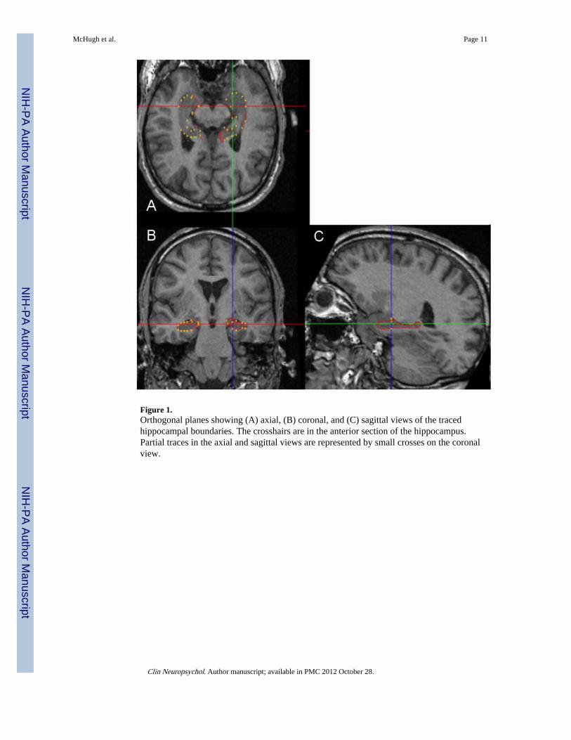
Figure 1.Orthogonal planes showing (A) axial, (B) coronal, and (C) sagittal views of the tracedhippocampal boundaries. The crosshairs are in the anterior section of the hippocampus.Partial traces in the axial and sagittal views are represented by small crosses on the coronalview.
McHugh et al. Page 11
Clin Neuropsychol. Author manuscript; available in PMC 2012 October 28.
NIH
-PA Author Manuscript
NIH
-PA Author Manuscript
NIH
-PA Author Manuscript
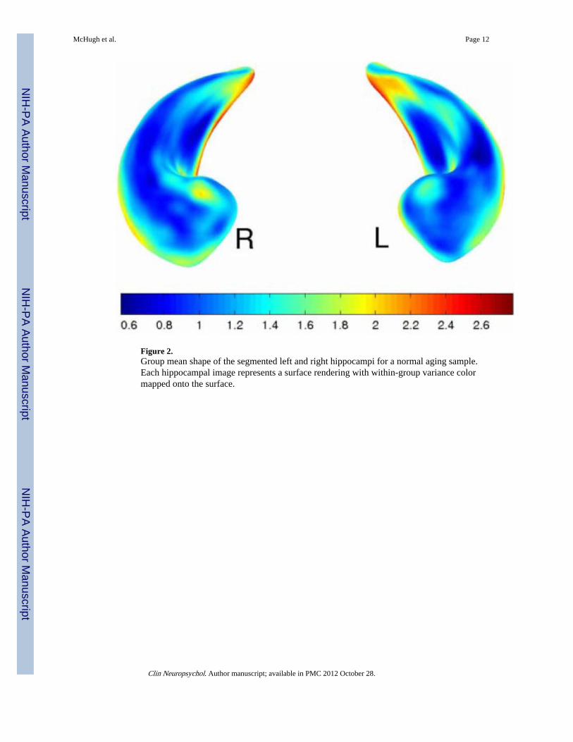
Figure 2.Group mean shape of the segmented left and right hippocampi for a normal aging sample.Each hippocampal image represents a surface rendering with within-group variance colormapped onto the surface.
McHugh et al. Page 12
Clin Neuropsychol. Author manuscript; available in PMC 2012 October 28.
NIH
-PA Author Manuscript
NIH
-PA Author Manuscript
NIH
-PA Author Manuscript
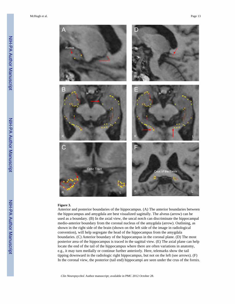
Figure 3.Anterior and posterior boundaries of the hippocampus. (A) The anterior boundaries betweenthe hippocampus and amygdala are best visualized sagittally. The alveus (arrow) can beused as a boundary. (B) In the axial view, the uncal notch can discriminate the hippocampalmedio-anterior boundary from the coronal nucleus of the amygdala (arrow). Outlining, asshown in the right side of the brain (shown on the left side of the image in radiologicalconvention), will help segregate the head of the hippocampus from the amygdalaboundaries. (C) Anterior boundary of the hippocampus in the coronal plane. (D) The mostposterior area of the hippocampus is traced in the sagittal view. (E) The axial plane can helplocate the end of the tail of the hippocampus where there are often variations in anatomy,e.g., it may turn medially or continue further anteriorly. Here, telemarks show the tailtipping downward in the radiologic right hippocampus, but not on the left (see arrows). (F)In the coronal view, the posterior (tail end) hippocampi are seen under the crus of the fornix.
McHugh et al. Page 13
Clin Neuropsychol. Author manuscript; available in PMC 2012 October 28.
NIH
-PA Author Manuscript
NIH
-PA Author Manuscript
NIH
-PA Author Manuscript
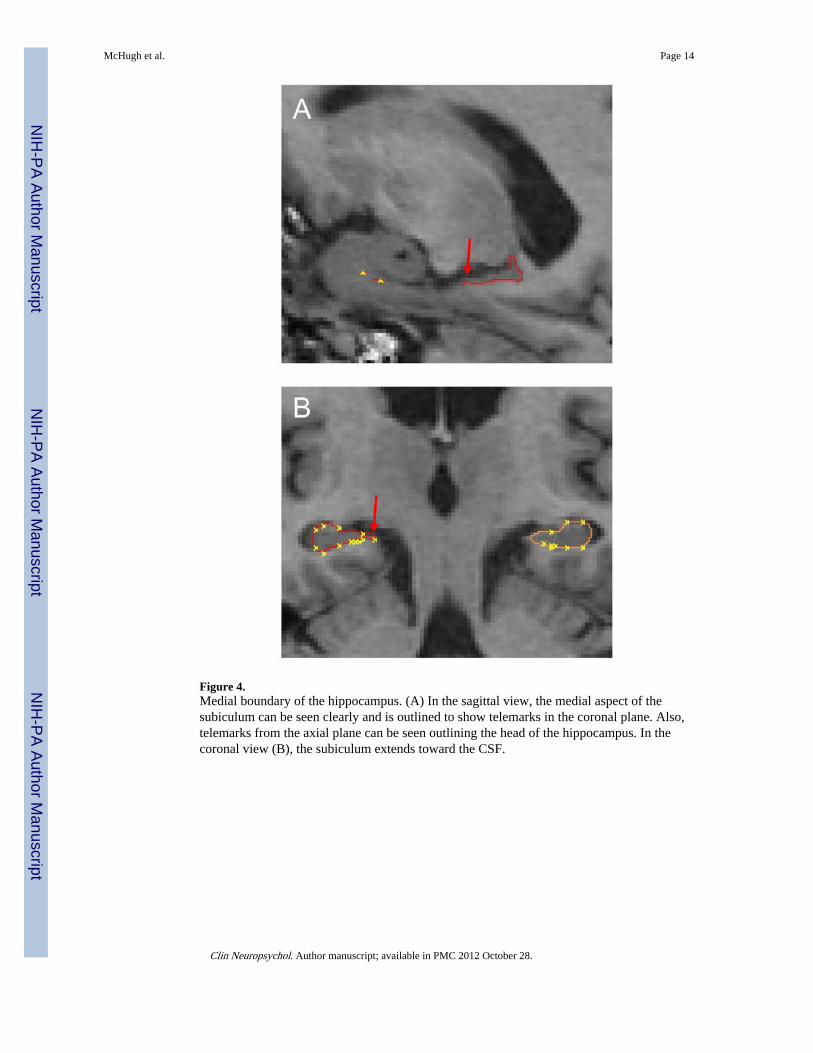
Figure 4.Medial boundary of the hippocampus. (A) In the sagittal view, the medial aspect of thesubiculum can be seen clearly and is outlined to show telemarks in the coronal plane. Also,telemarks from the axial plane can be seen outlining the head of the hippocampus. In thecoronal view (B), the subiculum extends toward the CSF.
McHugh et al. Page 14
Clin Neuropsychol. Author manuscript; available in PMC 2012 October 28.
NIH
-PA Author Manuscript
NIH
-PA Author Manuscript
NIH
-PA Author Manuscript
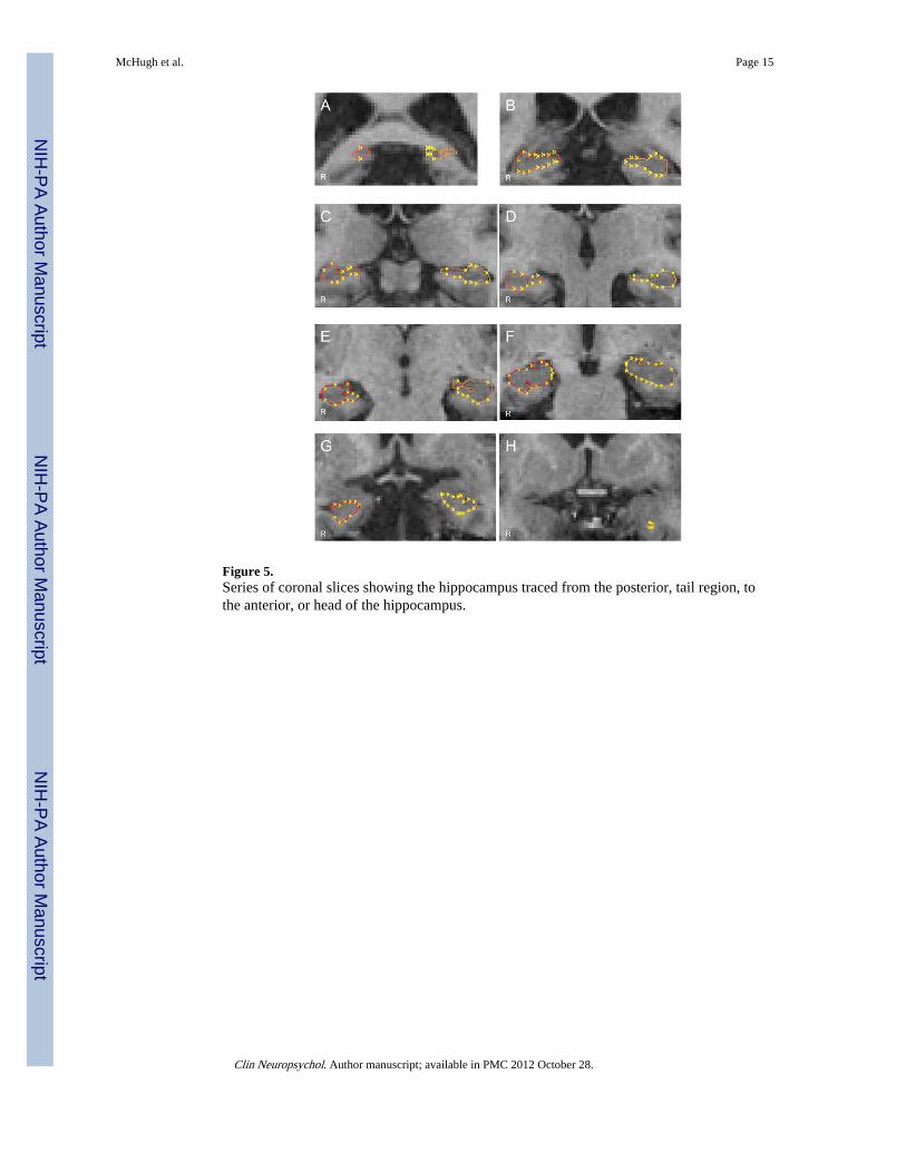
Figure 5.Series of coronal slices showing the hippocampus traced from the posterior, tail region, tothe anterior, or head of the hippocampus.
McHugh et al. Page 15
Clin Neuropsychol. Author manuscript; available in PMC 2012 October 28.
NIH
-PA Author Manuscript
NIH
-PA Author Manuscript
NIH
-PA Author Manuscript

NIH
-PA Author Manuscript
NIH
-PA Author Manuscript
NIH
-PA Author Manuscript
McHugh et al. Page 16
Table 1
Hippocampal and intracranial volumes (cubic centimeters)
Males Females p
Right hippocampusa 3.66 (0.44) 3.70 (0.42) NS
Left hippocampusa 3.52 (0.56) 3.46 (0.38) NS
Intracranial volume 1485.69 (115.16) 1364.51 (108.66) .003
aAdjusted for intracranial volume. Data are mean (SD). NS= not significant.
Clin Neuropsychol. Author manuscript; available in PMC 2012 October 28.