High-frequency stimulation in Parkinson's disease: more or less
8
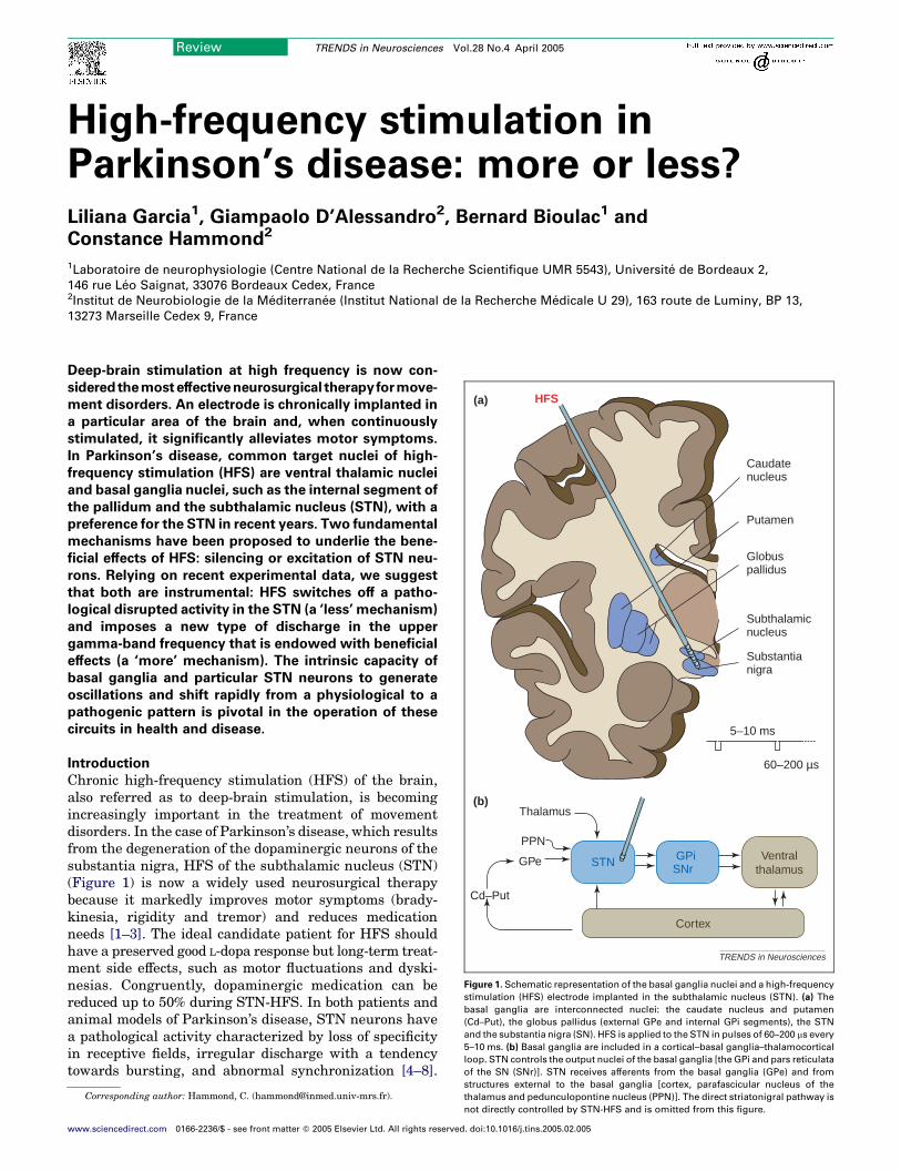
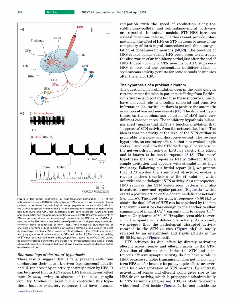
High-frequency stimulation in Parkinson’s disease: more or less? Liliana Garcia 1 , Giampaolo D’Alessandro 2 , Bernard Bioulac 1 and Constance Hammond 2 1 Laboratoire de neurophysiologie (Centre National de la Recherche Scientifique UMR 5543), Universite ´ de Bordeaux 2, 146 rue Le ´ o Saignat, 33076 Bordeaux Cedex, France 2 Institut de Neurobiologie de la Me ´ diterrane ´ e (Institut National de la Recherche Me ´ dicale U 29), 163 route de Luminy, BP 13, 13273 Marseille Cedex 9, France Deep-brain stimulation at high frequency is now con- sidered the most effective neurosurgical therapy for move- ment disorders. An electrode is chronically implanted in a particular area of the brain and, when continuously stimulated, it significantly alleviates motor symptoms. In Parkinson’s disease, common target nuclei of high- frequency stimulation (HFS) are ventral thalamic nuclei and basal ganglia nuclei, such as the internal segment of the pallidum and the subthalamic nucleus (STN), with a preference for the STN in recent years. Two fundamental mechanisms have been proposed to underlie the bene- ficial effects of HFS: silencing or excitation of STN neu- rons. Relying on recent experimental data, we suggest that both are instrumental: HFS switches off a patho- logical disrupted activity in the STN (a ‘less’ mechanism) and imposes a new type of discharge in the upper gamma-band frequency that is endowed with beneficial effects (a ‘more’ mechanism). The intrinsic capacity of basal ganglia and particular STN neurons to generate oscillations and shift rapidly from a physiological to a pathogenic pattern is pivotal in the operation of these circuits in health and disease. Introduction Chronic high-frequency stimulation (HFS) of the brain, also referred as to deep-brain stimulation, is becoming increasingly important in the treatment of movement disorders. In the case of Parkinson’s disease, which results from the degeneration of the dopaminergic neurons of the substantia nigra, HFS of the subthalamic nucleus (STN) (Figure 1) is now a widely used neurosurgical therapy because it markedly improves motor symptoms (brady- kinesia, rigidity and tremor) and reduces medication needs [1–3]. The ideal candidate patient for HFS should have a preserved good L-dopa response but long-term treat- ment side effects, such as motor fluctuations and dyski- nesias. Congruently, dopaminergic medication can be reduced up to 50% during STN-HFS. In both patients and animal models of Parkinson’s disease, STN neurons have a pathological activity characterized by loss of specificity in receptive fields, irregular discharge with a tendency towards bursting, and abnormal synchronization [4–8]. 5–10 ms Subthalamic nucleus Substantia nigra Globus pallidus Putamen Caudate nucleus HFS (a) (b) GPe PPN Thalamus GPi SNr Cortex Cd–Put 60–200 µs STN Ventral thalamus TRENDS in Neurosciences Figure 1. Schematic representation of the basal ganglia nuclei and a high-frequency stimulation (HFS) electrode implanted in the subthalamic nucleus (STN). (a) The basal ganglia are interconnected nuclei: the caudate nucleus and putamen (Cd–Put), the globus pallidus (external GPe and internal GPi segments), the STN and the substantia nigra (SN). HFS is applied to the STN in pulses of 60–200 ms every 5–10 ms. (b) Basal ganglia are included in a cortical–basal ganglia–thalamocortical loop. STN controls the output nuclei of the basal ganglia [the GPi and pars reticulata of the SN (SNr)]. STN receives afferents from the basal ganglia (GPe) and from structures external to the basal ganglia [cortex, parafascicular nucleus of the thalamus and pedunculopontine nucleus (PPN)]. The direct striatonigral pathway is not directly controlled by STN-HFS and is omitted from this figure. Corresponding author: Hammond, C. ([email protected]). Review TRENDS in Neurosciences Vol.28 No.4 April 2005 www.sciencedirect.com 0166-2236/$ - see front matter Q 2005 Elsevier Ltd. All rights reserved. doi:10.1016/j.tins.2005.02.005
-
Upload
independent -
Category
Documents
-
view
1 -
download
0
Transcript of High-frequency stimulation in Parkinson's disease: more or less
High-frequency stimulation inParkinson’s disease: more or less?Liliana Garcia1, Giampaolo D’Alessandro2, Bernard Bioulac1 and
Constance Hammond2
1Laboratoire de neurophysiologie (Centre National de la Recherche Scientifique UMR 5543), Universite de Bordeaux 2,
146 rue Leo Saignat, 33076 Bordeaux Cedex, France2Institut de Neurobiologie de la Mediterranee (Institut National de la Recherche Medicale U 29), 163 route de Luminy, BP 13,
13273 Marseille Cedex 9, France
5–10 ms
Subthalamicnucleus
Substantianigra
Globuspallidus
Putamen
Caudatenucleus
HFS(a)
(b)
GPe
PPN
Thalamus
GPiSNr
Cortex
Cd–Put
60–200 µs
STN Ventralthalamus
TRENDS in Neurosciences
Figure 1. Schematic representation of the basal ganglia nuclei and a high-frequency
stimulation (HFS) electrode implanted in the subthalamic nucleus (STN). (a) The
basal ganglia are interconnected nuclei: the caudate nucleus and putamen
(Cd–Put), the globus pallidus (external GPe and internal GPi segments), the STN
and the substantia nigra (SN). HFS is applied to the STN in pulses of 60–200 ms every
5–10 ms. (b) Basal ganglia are included in a cortical–basal ganglia–thalamocortical
loop. STN controls the output nuclei of the basal ganglia [the GPi and pars reticulata
of the SN (SNr)]. STN receives afferents from the basal ganglia (GPe) and from
Deep-brain stimulation at high frequency is now con-
sideredthemost effectiveneurosurgical therapy formove-
ment disorders. An electrode is chronically implanted in
a particular area of the brain and, when continuously
stimulated, it significantly alleviates motor symptoms.
In Parkinson’s disease, common target nuclei of high-
frequency stimulation (HFS) are ventral thalamic nuclei
and basal ganglia nuclei, such as the internal segment of
the pallidum and the subthalamic nucleus (STN), with a
preference for the STN in recent years. Two fundamental
mechanisms have been proposed to underlie the bene-
ficial effects of HFS: silencing or excitation of STN neu-
rons. Relying on recent experimental data, we suggest
that both are instrumental: HFS switches off a patho-
logical disrupted activity in the STN (a ‘less’ mechanism)
and imposes a new type of discharge in the upper
gamma-band frequency that is endowed with beneficial
effects (a ‘more’ mechanism). The intrinsic capacity of
basal ganglia and particular STN neurons to generate
oscillations and shift rapidly from a physiological to a
pathogenic pattern is pivotal in the operation of these
circuits in health and disease.
Introduction
Chronic high-frequency stimulation (HFS) of the brain,also referred as to deep-brain stimulation, is becomingincreasingly important in the treatment of movementdisorders. In the case of Parkinson’s disease, which resultsfrom the degeneration of the dopaminergic neurons of thesubstantia nigra, HFS of the subthalamic nucleus (STN)(Figure 1) is now a widely used neurosurgical therapybecause it markedly improves motor symptoms (brady-kinesia, rigidity and tremor) and reduces medicationneeds [1–3]. The ideal candidate patient for HFS shouldhave a preserved good L-dopa response but long-term treat-ment side effects, such as motor fluctuations and dyski-nesias. Congruently, dopaminergic medication can bereduced up to 50% during STN-HFS. In both patients andanimal models of Parkinson’s disease, STN neurons havea pathological activity characterized by loss of specificityin receptive fields, irregular discharge with a tendencytowards bursting, and abnormal synchronization [4–8].
Review TRENDS in Neurosciences Vol.28 No.4 April 2005
structures external to the basal ganglia [cortex, parafascicular nucleus of the
thalamus and pedunculopontine nucleus (PPN)]. The direct striatonigral pathway is
not directly controlled by STN-HFS and is omitted from this figure.
Corresponding author: Hammond, C. ([email protected]).
www.sciencedirect.com 0166-2236/$ - see front matter Q 2005 Elsevier Ltd. All rights reserved. doi:10.1016/j.tins.2005.02.005
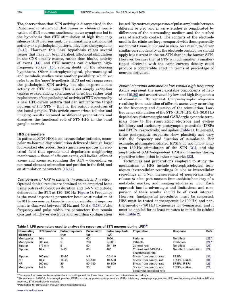
Review TRENDS in Neurosciences Vol.28 No.4 April 2005210
The observations that STN activity is disorganized in theParkinsonian state and that lesion or chemical inacti-vation of STN neurons ameliorate motor symptoms led tothe hypothesis that STN stimulation at high frequencysilences STN neurons and, by eliminating a pathologicalactivity or a pathological pattern, alleviates the symptoms[9–13]. However, this ‘less’ hypothesis raises severalissues that have not been clarified. Electrical stimulationin the CNS usually causes, rather than blocks, activityof axons [14], and STN neurons can discharge high-frequency spikes [15], casting doubt on the silencinghypothesis. Other electrophysiological, pharmacologicaland metabolic studies raise another possibility, which werefer to as the ‘more’ hypothesis: HFS not only suppressesthe pathological STN activity but also imposes a newactivity on STN neurons. This is not simply excitation(spikes evoked among spontaneous ones) but rather totalreplacement of the pathological activity of STN neurons bya new HFS-driven pattern that can influence the targetneurons of the STN – that is, the output structures ofthe basal ganglia. This article summarizes cellular andimaging results obtained in different preparations anddiscusses the functional role of STN-HFS in the basalganglia network.
HFS parameters
In patients, STN-HFS is an extracellular, cathodic, mono-polar 24-hours-a-day stimulation delivered through largefour-contact electrodes. Such stimulation induces an elec-trical field that spreads and depolarizes neighboringmembranes – those of afferent axons, cell bodies, efferentaxons and axons surrounding the STN – depending onneuronal element orientation and position in the field, andon stimulation parameters [16,17].
Comparison of HFS in patients, in animals and in vitro
Optimal clinical results are obtained on an empirical basisusing pulses of 60–200 ms duration and 1–5 V amplitude,delivered in the STN at 120–180 Hz (Figure 1). Frequencyis the most important parameter because stimulation at5–10 Hz worsens parkinsonism and no significant improve-ment is observed between 10 Hz and 50 Hz [3,18]. Pulsefrequency and pulse width are parameters that remainconstant whichever electrode and recording configuration
Table 1. LFS parameters used to analyze the responses of STN neu
Stimulating
electrode
LFS duration Pulse frequency
(Hz)
Pulse width
(ms)
Pulse am
(mA)
Monopolar 20 s 14 60 2000
Monopolar 500 ms 5 200 2–500
Bipolar 1–2 min 5 50 20–150
Bipolar 10 s 1–10 60 400
Bipolar 100 ms 30–80 NR 0.2–1.0
NR 10 s 10–25 50–100 10–500
NR 200 ms 20 50–100 10–500
Monopolar 1–2 h 10 90 500
aThe upper four rows are from extracellular recordings and the lower four rows are frombAbbreviations: 6-OHDA, 6-hydroxydopamine; EPSPs, excitatory postsynaptic potentials
reported; STN, subthalamic nucleus.cParameters for stimulation through large macroelectrodes.
www.sciencedirect.com
is used. By contrast, comparison of pulse amplitude betweendifferent in vivo and in vitro studies is complicated bydifferences of the surrounding medium and the surfacearea of electrode contact. The contacts of the electrodeused in the clinic are large compared with those generallyused in rat tissue in vivo and in vitro. As a result, to deliversimilar current density at the electrode contact, we shouldapply less current in the rat STN than in the human STN.However, because the rat STN is much smaller, a smaller-tipped electrode with the same current density couldproduce a comparable effect in terms of percentage ofneurons activated.
Neural elements activated at low versus high frequency
Axons represent the most excitable components of neu-rons [19,20] and are activated by low and high frequenciesof stimulation. By contrast, the postsynaptic responsesresulting from activation of afferent axons vary accordingto the frequency and duration of the stimulation. Low-frequency stimulation of the STN (STN-LFS, 0.1–30.0 Hz)depolarizes glutamatergic and GABAergic synaptic term-inals close to the stimulating electrode and evokesinhibitory and excitatory postsynaptic potentials (IPSPsand EPSPs, respectively) and spikes (Table 1). In general,these postsynaptic responses show plasticity and varywith the frequency and duration of stimulation. Forexample, glutamate-mediated EPSPs do not follow long-term 130-Hz stimulation of the STN [21], and theamplitude of GABA-dependent IPSPs diminishes duringrepetitive stimulation in other networks [22].
Techniques and preparations employed to study themechanisms of HFS include electrophysiological tech-niques (extracellular recordings in vivo or intracellularrecordings in vitro), measurement of neurotransmitterrelease in vivo, post-mortem immunohistochemistry of ametabolic marker, and imaging studies in vivo. Eachapproach has its advantages and limitations, and com-parison of their results should be of great interest.However, fundamental procedures must be respected.HFS must be tested at therapeutic (R100 Hz) and non-therapeutic (!50 Hz) frequencies for comparison, and itmust be applied for at least minutes to mimic its clinicaluse (Table 2).
rons during LFSa,b
plitude Preparation Response Refs
Patients No effect [25]c
Patients Inhibition [24]c
Control rats No effect [26]
Control and 6-OHDA -
anesthetized rats
No effect or inhibition [27]
Slices from control rats EPSPs [31]
Slices from control rat EPSPs, spikes [34]
Slices from control rats EPSPs, IPSPs [29]
Slices from control and
dopamine-depleted rats
EPSPs, spikes [21]
intracellular recordings.
; IPSPs, inhibitory postsynaptic potentials; LFS, low-frequency stimulation; NR, not
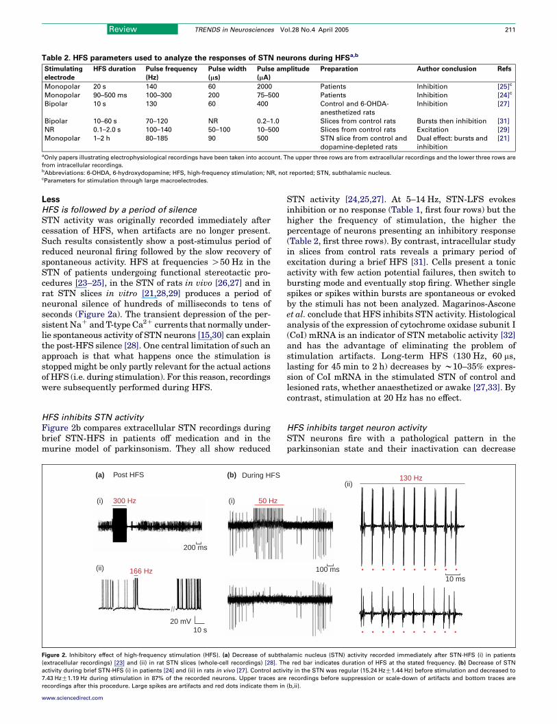
Table 2. HFS parameters used to analyze the responses of STN neurons during HFSa,b
Stimulating
electrode
HFS duration Pulse frequency
(Hz)
Pulse width
(ms)
Pulse amplitude
(mA)
Preparation Author conclusion Refs
Monopolar 20 s 140 60 2000 Patients Inhibition [25]c
Monopolar 90–500 ms 100–300 200 75–500 Patients Inhibition [24]c
Bipolar 10 s 130 60 400 Control and 6-OHDA-
anesthetized rats
Inhibition [27]
Bipolar 10–60 s 70–120 NR 0.2–1.0 Slices from control rats Bursts then inhibition [31]
NR 0.1–2.0 s 100–140 50–100 10–500 Slices from control rats Excitation [29]
Monopolar 1–2 h 80–185 90 500 STN slice from control and
dopamine-depleted rats
Dual effect: bursts and
inhibition
[21]
aOnly papers illustrating electrophysiological recordings have been taken into account. The upper three rows are from extracellular recordings and the lower three rows are
from intracellular recordings.bAbbreviations: 6-OHDA, 6-hydroxydopamine; HFS, high-frequency stimulation; NR, not reported; STN, subthalamic nucleus.cParameters for stimulation through large macroelectrodes.
Review TRENDS in Neurosciences Vol.28 No.4 April 2005 211
Less
HFS is followed by a period of silence
STN activity was originally recorded immediately aftercessation of HFS, when artifacts are no longer present.Such results consistently show a post-stimulus period ofreduced neuronal firing followed by the slow recovery ofspontaneous activity. HFS at frequencies O50 Hz in theSTN of patients undergoing functional stereotactic pro-cedures [23–25], in the STN of rats in vivo [26,27] and inrat STN slices in vitro [21,28,29] produces a period ofneuronal silence of hundreds of milliseconds to tens ofseconds (Figure 2a). The transient depression of the per-sistent NaC and T-type Ca2C currents that normally under-lie spontaneous activity of STN neurons [15,30] can explainthe post-HFS silence [28]. One central limitation of such anapproach is that what happens once the stimulation isstopped might be only partly relevant for the actual actionsof HFS (i.e. during stimulation). For this reason, recordingswere subsequently performed during HFS.
HFS inhibits STN activity
Figure 2b compares extracellular STN recordings duringbrief STN-HFS in patients off medication and in themurine model of parkinsonism. They all show reduced
(a) (b)
200 ms
20 mV10 s
//
Post HFS During HFS
300 Hz
166 Hz
(i)
(ii)
(i) 50 Hz
Figure 2. Inhibitory effect of high-frequency stimulation (HFS). (a) Decrease of subtha
(extracellular recordings) [23] and (ii) in rat STN slices (whole-cell recordings) [28]. Th
activity during brief STN-HFS (i) in patients [24] and (ii) in rats in vivo [27]. Control activ
7.43 HzG1.19 Hz during stimulation in 87% of the recorded neurons. Upper traces are
recordings after this procedure. Large spikes are artifacts and red dots indicate them in
www.sciencedirect.com
STN activity [24,25,27]. At 5–14 Hz, STN-LFS evokesinhibition or no response (Table 1, first four rows) but thehigher the frequency of stimulation, the higher thepercentage of neurons presenting an inhibitory response(Table 2, first three rows). By contrast, intracellular studyin slices from control rats reveals a primary period ofexcitation during a brief HFS [31]. Cells present a tonicactivity with few action potential failures, then switch tobursting mode and eventually stop firing. Whether singlespikes or spikes within bursts are spontaneous or evokedby the stimuli has not been analyzed. Magarinos-Asconeet al. conclude that HFS inhibits STN activity. Histologicalanalysis of the expression of cytochrome oxidase subunit I(CoI) mRNA is an indicator of STN metabolic activity [32]and has the advantage of eliminating the problem ofstimulation artifacts. Long-term HFS (130 Hz, 60 ms,lasting for 45 min to 2 h) decreases by w10–35% expres-sion of CoI mRNA in the stimulated STN of control andlesioned rats, whether anaesthetized or awake [27,33]. Bycontrast, stimulation at 20 Hz has no effect.
HFS inhibits target neuron activity
STN neurons fire with a pathological pattern in theparkinsonian state and their inactivation can decrease
10 ms100 ms
130 Hz(ii)
lamic nucleus (STN) activity recorded immediately after STN-HFS (i) in patients
e red bar indicates duration of HFS at the stated frequency. (b) Decrease of STN
ity in the STN was regular (15.24 HzG1.44 Hz) before stimulation and decreased to
recordings before suppression or scale-down of artifacts and bottom traces are
(b,ii).
Review TRENDS in Neurosciences Vol.28 No.4 April 2005212
activity in STN target structures – the globus pallidus(GP) and substantia nigra (SN). Burbaud et al. [26] andTai et al. [27] have shown that brief STN-HFS(100–130 Hz, lasting for 20–120 s) in control or lesionedrats in vivo either decreases SN pars reticulata (SNr)firing rates or has no effect. In the GP, it causes modestinhibition (12%) in half of the neurons recorded [26].
Shortcomings of the ‘less’ hypothesis
The silencing hypothesis is based on extracellular record-ings during very short periods of HFS (Table 2). Onepotential problem with studies relying on extracellularrecordings is that large stimulus artifacts (of w2 msduration), in particular when the stimulation is close tothe recording site, preclude detection of possible shortlatency (w1 ms) action potentials evoked by direct stimu-lation of nearby cell bodies or axons. This problem isexemplified by results following intranuclear LFS. LFS iscommonly used to evoke EPSPs and spikes in intracellularrecordings in vitro. These evoked spikes have not beenobserved in extracellular recordings in vivo (Table 1),suggesting that artifacts mask them. To solve thisproblem, artifacts are scaled down using diverse pro-cedures; however, whether these procedures reallyunmask spikes or only clean recordings should be testedduring LFS (Figure 2b).
Proposed mechanisms for the silencing are (i) a directeffect of HFS on STN neuronal membranes (the depolar-ization block hypothesis) and (ii) ‘preferential’ activationof GABAergic inhibitory afferents to STN neurons.A depolarizing block means that the membrane is sodepolarized that spikes become smaller and smaller andfinally can no longer be evoked, owing to inactivation ofthe voltage-gated NaC current. Filali et al. [24] and Taiet al. [27] have excluded this hypothesis, because STNspike amplitude does not change in the initial part of thetrain and the firing rate does not increase before activitydecreases. Enhancement of GABAergic currents is alsounlikely because of the usual failure of inhibition duringlong-term repetitive stimulation [22]. Although the CoImRNA results are compatible with inhibition, these obser-vations are conditioned by the possible rapid changes ofSTN activity once HFS is stopped, and by the fact thatHFS-driven activity might need less energy than patho-logical activity. Finally, the analysis of SN responses toSTN-HFS cannot provide direct information about theeffect of HFS on STN neurons, owing to the complexity ofthe intranigral network. In conclusion, there is indeed areductionof the number ofSTNspontaneous spikes betweenstimulation artifacts, but very short-latency evoked spikesclose to the artifacts would not be detected in these experi-ments. Intracellular recordings of STN neuronal activity orextracellular recordings of target neuron responses shouldenable such spikes to be distinguished from stimulationartifacts, owing to the large amplitude of STN intracellularspikes and the latencies of target cell responses.
More
HFS excites STN neurons
Lee et al. [29,34] have reported that STN-HFS involvesexcitation in rat slices in vitro (Table 2). LFS at 20 Hz
www.sciencedirect.com
evokes EPSPs and spikes (Table 1) and HFS at 100–140 Hzincreases action potential firing to its maximum (Table 2).Lee et al. did not analyze precisely the relationship betweenspikes and stimuli, or the behavior of spontaneous activityduring HFS. Because spikes disappear in the presence ofchannel blockers, EPSPs and spikes generated during 2 strains at 100 Hz are proposed to result from the activation ofglutamate-mediated transmission.
HFS has a dual effect
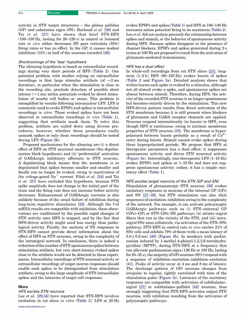
In whole-cell recordings from rat STN slices [21], long-term (1–2 h) HFS (80–185 Hz) evokes bursts of spikes(Table 2 and Figure 3a). Detailed analysis shows thatwithin bursts each spike is evoked by a stimulus, althoughnot all stimuli evoke a spike, and spontaneous spikes areabsent between stimuli. Therefore, during HFS, the acti-vity of the recorded STN neurons is no longer spontaneousbut becomes entirely driven by the stimulation. This newHFS-driven pattern results from direct activation of theSTN membrane because it is still present when blockersof glutamate and GABA receptor channels are applied.Neurons respond intermittently (in bursts) to HFS, eventhough HFS is continuous, owing to intrinsic membraneproperties of STN neurons [35]. The membrane is hyper-polarized between bursts probably as a result of Ca2C
entry during bursts. Stimuli cannot evoke spikes duringthese hyperpolarized periods. We propose that HFS attherapeutic parameters has a dual effect: it suppressesspontaneous activity and drives STN neuronal activity(Figure 3a). Interestingly, non-therapeutic LFS (1–10 Hz)evokes EPSPs and spikes at 1–10 Hz and does not sup-press spontaneous activity; rather, it has a simple exci-tatory effect (Table 1).
HFS excites target neurons of the STN (GP and SN)
Stimulation of glutamatergic STN neurons [36] evokesexcitatory responses in neurons of the internal GP (GPi)and SN [37–39], but STN stimulation can also elicitsequences of excitation–inhibition owing to the complexityof the network. For example, it can activate polysynapticGABAergic pathways such as (i) STN–external GP(GPe)–GPi or STN–GPe–SN pathways, (ii) striato–nigralfibers that run in the vicinity of the STN, and (iii) intra-nigral SNr axon collaterals, via activation of the STN–SNrpathway. STN-HFS in control rats in vivo excites 21% ofSNr cells and inhibits 79% of them (with a mean latency of5.0G0.8 ms) [40] (Figure 3b). In monkeys with parkin-sonism induced by 1-methyl-4-phenyl-1,2,3,6-tetrahydro-pyridine (MPTP), during STN-HFS at a frequency thatcan alleviate parkinsonian signs (136 Hz or 185 Hz, lastingfor 25–35 s), the majority of GPi neurons (85%) respond witha sequence of inhibition–excitation–inhibition–excitation[41]. Peaks of activity occur at 4 ms and 8 ms of latency.The discharge pattern of GPi neurons changes fromirregular to regular, tightly correlated with time of thestimulation pulse (Figure 3c). Latencies of the excitatoryresponses are compatible with activation of subthalamo–nigral [37] or subthalamo–pallidal [42] neurons, thusstrongly suggesting that STN-HFS activates output STNneurons, with inhibition resulting from the activation ofpolysynaptic pathways.
(a) STN recording (b) SN recording
(c) GPi recording20 ms
STN HFS 130 Hz
1000
40
20
00
40
20
0
0 200 400 600
2
1
00 10 20 30
1
0
136 Hz
0 7 msOn-stimulation
a
130 Hz
0 Hz
0 Hz
Pre-stimulation0 7 ms
0.30
0.20
0.10
0
0.30
0.20
0.10
0
100ms
5001500500
60
Time (ms)
ISI (
ms)
Num
.inte
r.(x1
03)
Duration (ms) Duration (ms)
Time in burst (ms)0
Inci
denc
e pe
r st
imul
atio
n
ISI (
ms)
20 ms
(i)
(ii)
(iii)
20m
V
Num
.inte
r.(x1
04)
Figure 3. Dual effect of HFS in the STN and excitation of target neurons. (a) Dual effect of high-frequency stimulation (HFS) of the subthalamic nucleus (STN) in vitro: (i) HFS
forces STN neurons to discharge spikes (single or organized in bursts) that are time-locked to the stimulation [21]. The expanded trace shows that some stimuli (‘a’, a
stimulation artifact) do not evoke spikes and others evoke spikes with no detectable latency. Note the absence of spontaneous spikes. (ii) Interspike intervals (ISIs) as a
function of time in the recording before HFS (left) and as a function of time of occurrence within a burst during HFS at 130 Hz (right). Note that during HFS all spikes are on
average time-locked to one every 2–3 stimuli. (iii) Number of ISIs (Num.inter.) before HFS (left) and within bursts during HFS at 130 Hz (right). (b,c) Excitatory responses
evoked in target neurons of the STN. (b) Extracellular recordings in SNr in the rat in vivo during STN-HFS, showing spikes evoked one every 2–3 stimulations with a mean
latency of 3–6 ms [40]. (c) Overlays of 100 sweeps of extracellular recordings in GPi during STN-HFS in MPTP-treated monkeys, showing the regular pattern of the evoked
discharge [41]. Below are the post-stimulus histograms reconstructed from successive 7-ms periods before (left) and during (right) HFS. Increases significant at P!0.01 are
indicated with ‘C’; decreases significant at P!0.01 are indicated with ‘x’. Red dots indicate stimulation artifacts.
Review TRENDS in Neurosciences Vol.28 No.4 April 2005 213
In patients greatly ameliorated by the stimulation,STN-HFS (pulses at 130–185 Hz, of 60–70 ms duration and1.5–3.4 V amplitude) increases blood flow, regional cere-bral metabolic rates and blood oxygenation [measuredusing positron emission tomography (PET) or functionalmagnetic resonance imaging (fMRI)] in the ipsilateral GPe[43–45]. Similarly, in rats in vivo, STN-HFS (60–130 Hz)provoked a significant increase of glutamate content inboth GPe and SNr, as measured using microdialysis [46,47].This increase was amplified and remained significantthroughout the stimulation period (1 h), with maximaleffect 1 h after the end of stimulation. The glutamate con-centration increase could correspond to activation of STNneurons, as occurs during their pharmacological activationby carbachol [48]. STN-LFS at 10 Hz has no effect, probablybecause the increased release of glutamate caused by therelatively small increase in activity (25%) is not detectable.
HFS and striatal dopamine release
STN-HFS can influence the activity of dopaminergicneurons either directly [37,49] or indirectly via collaterals
www.sciencedirect.com
of SNr cells (Figure 4a). Whenever tested, STN-HFS hasbeen found to increase dopamine content or metabolism inthe ipsilateral striatum in control and partially dopamine-denervated rats [50,51]. By contrast, HFS of the entope-duncular nucleus has no effect [52]. During the STN-HFSperiod (130 Hz for 1 h), the ipsilateral extracellularcontent of dopamine increases by up to 200% in 6-hydroxy-dopamine lesioned rats bearing partial destruction of theSN pars compacta (SNc) and by less in control rats (168%)[50]. Intraneuron dopamine turnover and tyrosine hydroxyl-ase activity also increase [51]. These results have not beenconfirmed in patients. Dopamine release is estimated inpatients as the density of free D2 receptors in the striatum,measured using the reversible ligand [11C] raclopride in PETexperiments. Binding of this tracer is inversely proportionalto levels of extracellular dopamine [53]. After a period whenthe stimulation has been turned off and L-dopa withdrawn,STN-HFS in one side does not induce differences in[11C]raclopride binding between the two striata [54–57].Therefore, there is no evidence for STN stimulation inducingdopamine release in humans.
(a)
(b)
SNc
SNc
SNr
Cortex
GPe
GPe
GPi
Thalamus
PPNPPN
HFS
No stimulation
2 s20 mV
25 ms
ArtifactSpike
1 s
AntiOrtho
HFS (185 Hz)
(i)
(ii)
STN
TRENDS in Neurosciences
Figure 4. The ‘more’ hypothesis. (a) High-frequency stimulation (HFS) of the
subthalamic nucleus (STN) directly activates STN efferent axons or somata. A new
pattern that replaces the pathological one propagates orthodromically (ortho) to
the various target structures of the STN: the external and internal segments of the
pallidum (GPe and GPi), the substantia nigra pars reticulata (SNr) and pars
compacta (SNc) and the pedunculopontine nucleus (PPN). Recurrent collaterals of
SNr neurons terminate on dopaminergic neurons in the SNc and on GABAergic
neurons in the SNr. Notice that in the case of parkinsonism, dopaminergic cells and
terminals have degenerated (broken lines). Pink indicates glutamatergic or
cholinergic terminals, blue indicates GABAergic terminals, and yellow indicates
dopaminergic terminals. When axons are first activated, the HFS-driven pattern
also propagates antidromically (anti) to STN cell bodies. (b) The disrupted activity
of STN neurons recorded in a 6-hydroxydopamine-treated rat in vivo (i) [70] would
be entirely replaced during HFS by a stable HFS-driven pattern consisting of bursts
of evoked spikes (ii). The expanded trace shows the absence of spontaneous spikes
in between stimuli.
Review TRENDS in Neurosciences Vol.28 No.4 April 2005214
Shortcomings of the ‘more’ hypothesis
These results suggest that HFS (i) prevents cells fromdischarging their network-driven (spontaneous) activityand (ii) replaces it by an activity entirely driven by HFS. Itcan be argued that in STN slices, HFS has a different effectfrom in vivo, owing to absence of the basal gangliacircuitry. Studies in target nuclei contradict this hypo-thesis because excitatory responses that have latencies
www.sciencedirect.com
compatible with the speed of conduction along thesubthalamo–pallidal and subthalamo–nigral pathwaysare recorded. In animal models, STN-HFS increasesstriatal dopamine release, but this cannot provide infor-mation on the effect of HFS on STN neurons because of thecomplexity of intra-nigral connections and the autoregu-lation of dopaminergic neurons [58,59]. The presence ofHFS-evoked spikes during HFS could seem to contradictthe observation of an inhibitory period just after the end ofHFS. Indeed, driving of STN neurons by HFS stops onceHFS is over, but the concomitant inhibitory effect onspontaneous activity persists for some seconds or minutesafter the end of HFS.
The hypothesis of a prokinetic rhythm
The question of how stimulation deep in the basal gangliarestores motor function in patients suffering from Parkin-son’s disease is important because these subcortical nucleihave a pivotal role in encoding sensorial and cognitiveinformation (i.e. cortical outflow) to produce the automaticexecution of learned movements [60]. The different hypo-theses on the mechanisms of action of HFS have verydifferent consequences. The inhibitory hypothesis (silenc-ing effect) implies that HFS is a functional ablation that‘suppresses’ STN activity from the network (i.e. ‘less’). Theidea is that no activity at the level of the STN outflow ispreferable to a noisy and disruptive output. The reversehypothesis, an excitatory effect, is that new evoked singlespikes introduced into the STN discharge superimpose onthe network-driven activity. LFS has exactly this effect,and is known to be non-therapeutic [3,18]. The ‘more’hypothesis that we propose is totally different from asimple excitation and appears with stimulation at highfrequency. Following our initial report [21], we proposethat HFS excites the stimulated structure, evokes aregular pattern time-locked to the stimulation, whichoverrides the pathological STN activity. As a consequence,HFS removes the STN deleterious pattern and alsointroduces a new and regular pattern (Figure 3a), whichexerts a positive action on the dopamine-deficient network(i.e. ‘more’). The need for a high frequency (R80 Hz) toobtain the dual effect of HFS can be explained by the factthat stimuli must be close enough to one another to allowsummation of inward Ca2C currents and to trigger Ca2C
bursts. Only bursts of 60–80 Hz spikes seem able to over-come the spontaneous deleterious activity. As a result,we propose that the pathological disrupted activityrecorded in the STN in vivo (Figure 4b,i) is totallyreplaced by an intermittent and stable activity in the60–80 Hz range (Figure 4b,ii).
HFS achieves its dual effect by directly activatingafferent axons, somas and efferent axons in the STN.Activation of afferent axons inside the STN and spon-taneous afferent synaptic activity do not have a role inHFS, because synaptic transmission does not follow long-term HFS and/or because its postsynaptic effects are over-come by direct activation of STN neurons. By contrast,activation of somas and efferent axons gives rise to theHFS-driven activity, which is propagated othodromicallyto STN terminals (Figure 4a). HFS is likely to exert awidespread effect inside (Figures 1, 4a) and outside the
Review TRENDS in Neurosciences Vol.28 No.4 April 2005 215
basal ganglia network, as recently modeled [61], becauseall of the basal ganglia nuclei, not just the STN, dys-function in the parkinsonian state.
The ‘more’ hypothesis is in agreement with the conceptof the prokinetic high-frequency rhythm, first proposedby Brown and Marsden [62]. In untreated patients andprimate models of parkinsonism, local field potentials thatrepresent synchronous activity in many neurons aredominated in the STN and GPi by low-frequency oscil-lations in the 11–30 Hz band [63–65]. Treatment withL-dopa encourages synchronized oscillations at fre-quencies O70 Hz [66] and concomitantly improvesparkinsonism. The reduction of a pathological 11–30 Hzrhythm and the introduction of a high-frequency rhythm[67,68] could provide a common mechanism for thera-peutic effects of L-dopa and deep brain stimulation [69].
Concluding remarks
In keeping with the present understanding of how oscil-lating networks operate, we propose that the improve-ment generated by HFS is due to parallel non-exclusiveactions: silencing of ongoing activity and generation of anactivity pattern in the gamma range. In theory, there isan important advantage in silencing spontaneous activityand imposing a pattern: the signal-to-noise ratio and thefunctional significance of the new signal is enhanced. Thenext step will be to identify how this new HFS-drivenactivity is propagated inside the basal ganglia. The factthat the most beneficial actions are produced in the highgamma range is interesting because it raises the issue oflinks between the integrative actions of this pattern andmotor coordination.
AcknowledgementsWe wish to thank C. Beurrier and Y. Ben-Ari for critical reading of themanuscript.
References
1 Limousin, P. et al. (1995) Effect of parkinsonian signs and symptoms ofbilateral subthalamic nucleus stimulation. Lancet 345, 91–95
2 Limousin, P. et al. (1998) Electrical stimulation of the subthalamicnucleus in advanced Parkinson’s disease. N. Engl. J. Med. 339,1105–1111
3 Moro, E. et al. (2002) The impact on Parkinson’s disease of electricalparameter settings in STN stimulation. Neurology 59, 706–713
4 Bergman, H. et al. (1994) The primate subthalamic nucleus. II.Neuronal activity in the MPTP model of parkinsonism.J. Neurophysiol. 72, 507–520
5 Hassani, O.K. et al. (1996) Increased subthalamic neuronal activityafter nigral dopaminergic lesion independent of disinhibition via theglobus pallidus. Neuroscience 72, 105–115
6 Hutchison, W.D. et al. (1998) Neurophysiological identification of thesubthalamic nucleus in surgery for Parkinson’s disease. Ann. Neurol.44, 622–628
7 Wichmann, T. et al. (1999) Comparison of MPTP-induced changes inspontaneous neuronal discharge in the internal pallidal segment andin the substantia nigra pars reticulata in primates. Exp. Brain Res.125, 397–409
8 Magnin, M. et al. (2000) Single-unit analysis of the pallidum,thalamus and subthalamic nucleus in parkinsonian patients. Neuro-science 96, 549–564
9 Bergman, H. et al. (1990) Reversal of experimental parkinsonism bylesions of the subthalamic nucleus. Science 249, 1436–1438
10 Aziz, T.Z. et al. (1991) Lesion of the subthalamic nucleus for thealleviation of 1-methyl-4-phenyl-1,2,3,6-tetrahydropyridine (MPTP)-induced parkinsonism in the primate. Mov. Disord. 6, 288–292
www.sciencedirect.com
11 Benazzouz, A. et al. (1993) Reversal of rigidity and improvement inmotor performance by subthalamic high-frequency stimulation inMPTP-treated monkeys. Eur. J. Neurosci. 5, 382–389
12 Lang, A.E. (2000) Surgery for Parkinson disease: A critical evaluationof the state of the art. Arch. Neurol. 57, 1118–1125
13 Levy, R. et al. (2001) Lidocaine and muscimol microinjections insubthalamic nucleus reverse parkinsonian symptoms. Brain 124,2105–2118
14 Ranck, J.B. (1975) Which elements are excited in electrical stimu-lation of mammalian central nervous system: a review. Brain Res. 98,417–440
15 Bevan, M.D. and Wilson, C.J. (1999) Mechanisms underlyingspontaneous oscillation and rhythmic firing in rat subthalamicneurons. J. Neurosci. 19, 7617–7628
16 Rattay, F. (1999) The basic mechanism for the electrical stimulation ofthe nervous system. Neuroscience 89, 335–346
17 Grill, W. (2003) Electrically excitable nerve elements: excitation sitesin peripheral and central stimulation. In Brain Stimulation and
Epilepsy (Luders, H., ed.), pp. 55–66, Martin Dunitz18 Rizzone, M. et al. (2001) Deep brain stimulation of the subthalamic
nucleus in Parkinson’s disease: effects of variation in stimulationparameters. J. Neurol. Neurosurg. Psychiatry 71, 215–219
19 Nowak, L.G. and Bullier, J. (1998) Axons, but not cell bodies, areactivated by electrical stimulation in cortical gray matter. I. Evidencefrom chronaxie measurements. Exp. Brain Res. 118, 477–488
20 Gustafsson, B. and Jankowska, E. (1976) Direct and indirectactivation of nerve cells by electrical pulses applied extracellularly.J. Physiol. 258, 33–61
21 Garcia, L. et al. (2003) Dual effect of high-frequency stimulation onsubthalamic neuron activity. J. Neurosci. 23, 8743–8751
22 Thompson, S.M. and Gahwiler, B.H. (1989) Activity-dependentdisinhibition. I. Repetitive stimulation reduces IPSP driving forceand conductance in the hippocampus in vitro. J. Neurophysiol. 61,501–511
23 Lozano, A.M. et al. (2002) Deep brain stimulation for Parkinson’sdisease: disrupting the disruption. Lancet Neurol. 1, 225–231
24 Filali, M. et al. (2004) Stimulation-induced inhibition of neuronalfiring in human subthalamic nucleus. Exp. Brain Res. 156, 274–281
25 Welter, M.L. et al. (2004) Effects of high-frequency stimulation onsubthalamic neuronal activity in parkinsonian patients. Arch. Neurol.61, 89–96
26 Burbaud, P. et al. (1994) Effect of subthalamic high frequencystimulation on substantia nigra pars reticulata and globus pallidusneurons in normal rats. J. Physiol. (Paris) 88, 359–361
27 Tai, C.H. et al. (2003) Electrophysiological and metabolic evidencethat high-frequency stimulation of the subthalamic nucleus bridlesneuronal activity in the subthalamic nucleus and the substantia nigrareticulata. FASEB J. 17, 1820–1830
28 Beurrier, C. et al. (2001) High-frequency stimulation produces atransient blockade of voltage-gated currents in subthalamic neurons.J. Neurophysiol. 85, 1351–1356
29 Lee, K.H. et al. (2004) Neurotransmitter release from high-frequencystimulation of the subthalamic nucleus. J. Neurosurg. 101, 511–517
30 Beurrier, C. et al. (2000) Slowly inactivating sodium current(INap) underlies single-spike activity in rat subthalamic neurons.J. Neurophysiol. 83, 1951–1957
31 Magarinos-Ascone, C. et al. (2002) High-frequency stimulation ofthe subthalamic nucleus silences subthalamic neurons: a possiblecellular mechanism in Parkinson’s disease. Neuroscience 115,1109–1117
32 Hirsch, E.C. et al. (2000) Metabolic effects of nigrostriatal denervationin basal ganglia. Trends Neurosci. 23, S78–S85
33 Salin, P. et al. (2002) High-frequency stimulation of the subthalamicnucleus selectively reverses dopamine denervation-induced cellulardefects in the output structures of the basal ganglia in the rat.J. Neurosci. 22, 5137–5148
34 Lee, K.H. et al. (2003) Effect of high-frequency stimulation of thesubthalamic nucleus on subthalamic neurons: an intracellular study.Stereotact. Funct. Neurosurg. 80, 32–36
35 Beurrier, C. et al. (1999) Subthalamic nucleus neurons switch fromsingle-spike activity to burst-firing mode. J. Neurosci. 19, 599–609
36 Rinvik, E. and Ottersen, O.P. (1993) Terminals of subthalamonigral
Review TRENDS in Neurosciences Vol.28 No.4 April 2005216
fibres are enriched with glutamate-like immunoreactivity: an electronmicroscopic, immunogold analysis in the cat. J. Chem. Neuroanat. 6,19–30
37 Hammond, C. et al. (1978) Electrophysiological demonstration ofan excitatory subthalamonigral pathway in the rat. Brain Res. 151,235–244
38 Nakanishi, H. et al. (1991) Intracellular study of rat entopeduncularnucleus neurons in an in vitro slice preparation: response tosubthalamic stimulation. Brain Res. 549, 285–291
39 Feger, J. et al. (1997) The subthalamic nucleus and its connections.New electrophysiological and pharmacological data. Adv. Neurol. 74,31–43
40 Maurice, N. et al. (2003) Spontaneous and evoked activity ofsubstantia nigra pars reticulata neurons during high-frequencystimulation of the subthalamic nucleus. J. Neurosci. 23, 9929–9936
41 Hashimoto, T. et al. (2003) Stimulation of the subthalamic nucleuschanges the firing pattern of pallidal neurons. J. Neurosci. 23,1916–1923
42 Nambu, A. et al. (2000) Excitatory cortical inputs to pallidal neuronsvia the subthalamic nucleus in the monkey. J. Neurophysiol. 84,289–300
43 Ceballos-Baumann, A.O. et al. (1999) A positron emission tomographicstudy of subthalamic nucleus stimulation in Parkinson disease:enhanced movement-related activity of motor-association cortex anddecreased motor cortex resting activity. Arch. Neurol. 56, 997–1003
44 Hershey, T. et al. (2003) Cortical and subcortical blood flow effects ofsubthalamic nucleus stimulation in PD. Neurology 61, 816–821
45 Zhao, Y.B. et al. (2004) Effects of bilateral subthalamic nucleusstimulation on resting-state cerebral glucose metabolism in advancedParkinson’s disease. Chin Med. J. (Engl.) 117, 1304–1308
46 Windels, F. et al. (2000) Effects of high frequency stimulation of sub-thalamic nucleus on extracellular glutamate and GABA in substantianigra and globus pallidus in the normal rat. Eur. J. Neurosci. 12,4141–4146
47 Windels, F. et al. (2003) Influence of the frequency parameter onextracellular glutamate and g-aminobutyric acid in substantia nigraand globus pallidus during electrical stimulation of subthalamicnucleus in rats. J. Neurosci. Res. 72, 259–267
48 Rosales, M.G. et al. (1997) Reciprocal interaction between glutamateand dopamine in the pars reticulata of the rat substantia nigra:a microdialysis study. Neuroscience 80, 803–810
49 Groenewegen, H.J. and Berendse, H.W. (1990) Connections of thesubthalamic nucleus with ventral striatopallidal parts of the basalganglia in the rat. J. Comp. Neurol. 294, 607–622
50 Bruet, N. et al. (2001) High frequency stimulation of the subthalamicnucleus increases the extracellular contents of striatal dopamine innormal and partially dopaminergic denervated rats. J. Neuropathol.Exp. Neurol. 60, 15–24
51 Meissner, W. et al. (2003) High-frequency stimulation of thesubthalamic nucleus enhances striatal dopamine release and meta-bolism in rats. J. Neurochem. 85, 601–609
52 Meissner, W. et al. (2004) High frequency stimulation of the ento-peduncular nucleus has no effect on striatal dopaminergic trans-mission. Neurochem. Int. 44, 281–286
ScienceDirect collection reache
Elsevier recently announced that six million articles are now available
in electronic scientific, technical andmedical publishingmeans that re
volume of information from the c
The rapid growth of the ScienceDirect collection is due to the integrati
to the Backfiles - heritage collections in a number of disciplines. Th
journals back to volume one, issue one, is the addition of the highly c
online for the first time are six Cell titles’ long-awaited Backfiles, con
developments in the fi
www.science
www.sciencedirect.com
53 Laruelle, M. (2000) Imaging synaptic neurotransmission with in vivobinding competition techniques: a critical review. J. Cereb. Blood FlowMetab. 20, 423–451
54 Abosch, A. et al. (2003) Stimulation of the subthalamic nucleusin Parkinson’s disease does not produce striatal dopamine release.Neurosurgery 53, 1095–1102
55 Hilker, R. et al. (2003) Deep brain stimulation of the subthalamicnucleus does not increase the striatal dopamine concentration inparkinsonian humans. Mov. Disord. 18, 41–48
56 Strafella, A.P. et al. (2003) Subthalamic deep brain stimulation doesnot induce striatal dopamine release in Parkinson’s disease. Neuro-Report 14, 1287–1289
57 Thobois, S. et al. (2003) Chronic subthalamic nucleus stimulationand striatal D2 dopamine receptors in Parkinson’s disease –A [11C]-raclopride PET study. J. Neurol. 250, 1219–1223
58 Smith, I.D. and Grace, A.A. (1992) Role of the subthalamic nucleusin the regulation of nigral dopamine neuron activity. Synapse 12,287–303
59 Bunney, B.S. et al. (1973) Comparison of effects of L-dopa, amphet-amine and apomorphine on firing rate of rat dopaminergic neurones.Nat. New Biol. 245, 123–125
60 Marsden, C.D. (1982) The mysterious motor function of the basalganglia: the Robert Wartenberg Lecture. Neurology 32, 514–539
61 Rubin, J.E. and Terman, D. (2004) High frequency stimulation ofthe subthalamic nucleus eliminates pathological thalamic rhythmi-city in a computational model. J. Comput. Neurosci. 16, 211–235
62 Brown, P. and Marsden, C.D. (1998) What do the basal ganglia do?Lancet 351, 1801–1804
63 Nini, A. et al. (1995) Neurons in the globus pallidus do not showcorrelated activity in the normal monkey, but phase-locked oscil-lations appear in the MPTP model of parkinsonism. J. Neurophysiol.74, 1800–1805
64 Brown, P. et al. (2001) Dopamine dependency of oscillations betweensubthalamic nucleus and pallidum in Parkinson’s disease. J. Neurosci.21, 1033–1038
65 Levy, R. et al. (2002) Synchronized neuronal discharge in the basalganglia of parkinsonian patients is limited to oscillatory activity.J. Neurosci. 22, 2855–2861
66 Williams, D. et al. (2002) Dopamine-dependent changes in the func-tional connectivity between basal ganglia and cerebral cortex inhumans. Brain 125, 1558–1569
67 Montgomery, E.B. and Baker, K.B., Jr. (2000) Mechanisms of deepbrain stimulation and future technical developments. Neurol. Res. 22,259–266
68 McIntyre, C. and Grill, W.M. (2002) Extracellular stimulation ofcentral neurons: influence of stimulus waveform and frequency onneuronal output. J. Neurophysiol. 88, 1592–1604
69 Brown, P. et al. (2004) Effects of stimulation of the subthalamic area onoscillatory pallidal activity in Parkinson’s disease. Exp. Neurol. 188,480–490
70 Hassani, O.K. and Feger, J. (1999) Effects of intrasubthalamicinjection of dopamine receptor agonists on subthalamic neurons innormal and 6-hydroxydopamine-lesioned rats: an electrophysiologicaland c-fos study. Neuroscience 92, 533–543
s six million full-text articles
on its premier electronic platform, ScienceDirect. This milestone
searchers around the globewill be able to access an unsurpassed
onvenience of their desktop.
on of several prestigious publications as well as ongoing addition
e latest step in this ambitious project to digitize all of Elsevier’s
ited Cell Press journal collection on ScienceDirect. Also available
taining more than 12,000 articles highlighting important historic
eld of life sciences.
direct.com