High-Density Targeting of a Viral Multifunctional Nanoplatform to a Pathogenic, Biofilm-Forming...
12
Chemistry & Biology Article High-Density Targeting of a Viral Multifunctional Nanoplatform to a Pathogenic, Biofilm-Forming Bacterium Peter A. Suci, 1,2,3 Deborah L. Berglund, 3 Lars Liepold, 2,4 Susan Brumfield, 5 Betsey Pitts, 3 Willy Davison, 3,6 Luke Oltrogge, 2,4 Kathryn O. Hoyt, 3 Sarah Codd, 7 Philip S. Stewart, 3,6 Mark Young, 1,2,5, * and Trevor Douglas 2,4, * 1 Department of Microbiology 2 Center for BioInspired Nanomaterials 3 Center for Biofilm Engineering 4 Department of Chemistry and Biochemistry 5 Department of Plant Sciences 6 Department of Chemical and Biological Engineering 7 Department of Mechanical and Industrial Engineering Montana State University, Bozeman, MT 59717, USA *Correspondence: [email protected] (M.Y.), [email protected] (T.D.) DOI 10.1016/j.chembiol.2007.02.006 SUMMARY Nanomedicine directed at diagnosis and treat- ment of infections can benefit from innovations that have substantially increased the variety of available multifunctional nanoplatforms. Here, we targeted a spherical, icosahedral viral nanoplatform to a pathogenic, biofilm-forming bacterium, Staphylococcus aureus. Density of binding mediated through specific protein- ligand interactions exceeded the density ex- pected for a planar, hexagonally close-packed array. A multifunctionalized viral protein cage was used to load imaging agents (fluorophore and MRI contrast agent) onto cells. The fluores- cence-imaging capability allowed for direct ob- servation of penetration of the nanoplatform into an S. aureus biofilm. These results demon- strate that multifunctional nanoplatforms based on protein cage architectures have significant potential as tools for both diagnosis and targeted treatment of recalcitrant bacterial infections. INTRODUCTION Although conventional antimicrobial therapies effectively resolve most infections, exceptions to this probable out- come constitute a significant health concern [1, 2]. Each new antibiotic introduced to control emergent resistant strains of microbes has a limited lifespan [3–7], and com- munities of microbes that colonize both biomaterial and tissue surfaces typically possess an intrinsic transient resistance to a broad spectrum of antimicrobial agents [1, 8–11]. Biofilm infections are not only recalcitrant to treatment, but they are also difficult to diagnose since the infective communities spend substantial periods of time in relative quiescence, producing classic symptoms of infection only sporadically [1]. One of the most promi- nent bacterial players in multidrug resistance, S. aureus, is also a common biofilm-forming pathogen [12–16]. Thus, for certain categories of S. aureus infections, iatro- genically acquired drug resistance and transient intrinsic resistance of biofilms must be confronted en masse. The realization that conventional antimicrobial therapies and diagnostic techniques will always be limited in their ability to cope with certain types of infections has fostered a resurgence of interest in previously explored alternatives such as bacteriophage therapy [17, 18] and photodynamic therapy [19, 20]. Substantial effort has been directed at developing liposome and dendrimer platforms for tar- geted delivery of antimicrobial agents to microbial patho- gens [21–27]. A variety of novel systems based on both synthetic [28–31] and biological [32] materials has been explored for diagnosis and treatment of tumors. This ex- pansive repertoire is beginning to be tapped to develop and implement new approaches to infection control [33]. We have been developing a family of multifunctional nanoplatforms for biomedical applications that are based on self-assembling, supramolecular, symmetrical protein architectures [34–36]. The protein cage library includes viral capsids [37–40], ferritins [41–45], heat-shock proteins [34, 46, 47], and putative DNA-binding proteins [48, 49]. One of the best-characterized protein cages is cowpea chlorotic mottle virus (CCMV), a 28 nm icosahedral plant virus that can be produced as a noninfectious protein cage architecture [50, 51]. The structure of CCMV is known at the atomic scale [52] (Figure 1A), and targeting ligands can be added at specific locations on the structure by using both chemical and genetic methods [50, 53]. In addition, CCMV has served as a model for implementing several novel approaches aimed at spatial control of multi- ligand presentation (Figures 1B and 1C) [38, 54]. Biofilms consist of localized, dense communities of organisms of a specific cell type growing amidst Chemistry & Biology 14, 387–398, April 2007 ª2007 Elsevier Ltd All rights reserved 387
Transcript of High-Density Targeting of a Viral Multifunctional Nanoplatform to a Pathogenic, Biofilm-Forming...
Chemistry & Biology
Article
High-Density Targeting of a ViralMultifunctional Nanoplatform to aPathogenic, Biofilm-Forming BacteriumPeter A. Suci,1,2,3 Deborah L. Berglund,3 Lars Liepold,2,4 Susan Brumfield,5 Betsey Pitts,3 Willy Davison,3,6
Luke Oltrogge,2,4 Kathryn O. Hoyt,3 Sarah Codd,7 Philip S. Stewart,3,6 Mark Young,1,2,5,* and Trevor Douglas2,4,*1 Department of Microbiology2 Center for BioInspired Nanomaterials3 Center for Biofilm Engineering4 Department of Chemistry and Biochemistry5 Department of Plant Sciences6 Department of Chemical and Biological Engineering7 Department of Mechanical and Industrial Engineering
Montana State University, Bozeman, MT 59717, USA
*Correspondence: [email protected] (M.Y.), [email protected] (T.D.)
DOI 10.1016/j.chembiol.2007.02.006
SUMMARY
Nanomedicine directed at diagnosis and treat-ment of infections can benefit from innovationsthat have substantially increased the variety ofavailable multifunctional nanoplatforms. Here,we targeted a spherical, icosahedral viralnanoplatform to a pathogenic, biofilm-formingbacterium, Staphylococcus aureus. Density ofbinding mediated through specific protein-ligand interactions exceeded the density ex-pected for a planar, hexagonally close-packedarray. A multifunctionalized viral protein cagewas used to load imaging agents (fluorophoreand MRI contrast agent) onto cells. The fluores-cence-imaging capability allowed for direct ob-servation of penetration of the nanoplatforminto an S. aureus biofilm. These results demon-strate that multifunctional nanoplatforms basedon protein cage architectures have significantpotential as tools for both diagnosis andtargeted treatment of recalcitrant bacterialinfections.
INTRODUCTION
Although conventional antimicrobial therapies effectively
resolve most infections, exceptions to this probable out-
come constitute a significant health concern [1, 2]. Each
new antibiotic introduced to control emergent resistant
strains of microbes has a limited lifespan [3–7], and com-
munities of microbes that colonize both biomaterial and
tissue surfaces typically possess an intrinsic transient
resistance to a broad spectrum of antimicrobial agents
[1, 8–11]. Biofilm infections are not only recalcitrant to
treatment, but they are also difficult to diagnose since
Chemistry & Biology 14,
the infective communities spend substantial periods of
time in relative quiescence, producing classic symptoms
of infection only sporadically [1]. One of the most promi-
nent bacterial players in multidrug resistance, S. aureus,
is also a common biofilm-forming pathogen [12–16].
Thus, for certain categories of S. aureus infections, iatro-
genically acquired drug resistance and transient intrinsic
resistance of biofilms must be confronted en masse.
The realization that conventional antimicrobial therapies
and diagnostic techniques will always be limited in their
ability to cope with certain types of infections has fostered
a resurgence of interest in previously explored alternatives
such as bacteriophage therapy [17, 18] and photodynamic
therapy [19, 20]. Substantial effort has been directed at
developing liposome and dendrimer platforms for tar-
geted delivery of antimicrobial agents to microbial patho-
gens [21–27]. A variety of novel systems based on both
synthetic [28–31] and biological [32] materials has been
explored for diagnosis and treatment of tumors. This ex-
pansive repertoire is beginning to be tapped to develop
and implement new approaches to infection control [33].
We have been developing a family of multifunctional
nanoplatforms for biomedical applications that are based
on self-assembling, supramolecular, symmetrical protein
architectures [34–36]. The protein cage library includes
viral capsids [37–40], ferritins [41–45], heat-shock proteins
[34, 46, 47], and putative DNA-binding proteins [48, 49].
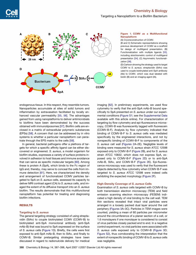
One of the best-characterized protein cages is cowpea
chlorotic mottle virus (CCMV), a 28 nm icosahedral plant
virus that can be produced as a noninfectious protein
cage architecture [50, 51]. The structure of CCMV is
known at the atomic scale [52] (Figure 1A), and targeting
ligands can be added at specific locations on the structure
by using both chemical and genetic methods [50, 53]. In
addition, CCMV has served as a model for implementing
several novel approaches aimed at spatial control of multi-
ligand presentation (Figures 1B and 1C) [38, 54].
Biofilms consist of localized, dense communities of
organisms of a specific cell type growing amidst
387–398, April 2007 ª2007 Elsevier Ltd All rights reserved 387
Chemistry & Biology
Targeting a Nanoplatform to a Biofilm Bacterium
Figure 1. CCMV as a Multifunctional
Nanoplatform
(A) Cryoreconstruction of CCMV.
(B and C) Schematic representations showing
previous development of CCMV as a scaffold
for design of multiligand presentation. (B)
Functionalization with multiple ligands [53],
including stoichiometric control of multiligand
presentation [54]. (C) Asymmetric functionali-
zation [38].
(D) Cartoon showing the strategy used to target
CCMV to S. aureus; streptavidin (StAv) was
used to couple biotinylated anti-SpA antibody
(Ab) to CCMV, which was dual labeled with
biotin (B) and an imaging agent (IA).
endogenous tissue. In this respect, they resemble tumors.
Nanoparticles accumulate at sites of solid tumors and
inflammation by extravasation facilitated by locally en-
hanced vascular permeability [55, 56]. The advantages
gained from using nanoplatforms to deliver antimicrobials
to biofilms have been demonstrated by the success
obtained with immunoliposomes [57]. Biofilm cells are en-
closed in a matrix of extracellular polymeric substances
(EPSs) [58]. A concern that can be addressed by in vitro
systems is whether a particular nanoplatform can pene-
trate through the EPS matrix to the cells [59].
In general, bacterial pathogens offer a plethora of tar-
gets for which a specific affinity ligand can be either dis-
covered or engineered. S. aureus, a model organism for
biofilm studies, expresses a variety of surface proteins in-
volved in adhesion to host tissues and immune avoidance
that can serve as specific molecular targets [60]. Among
these is protein A (SpA), which binds to the Fc region of
IgG and, thereby, may serve to conceal the cells from im-
mune detection [61]. Here, we characterized the density
and arrangement of functionalized CCMV particles tar-
geted to SpA on S. aureus cells, assessed its capacity to
deliver MRI contrast agent (CA) to S. aureus cells, and im-
aged the extent of its diffusive transport into an S. aureus
biofilm. The results demonstrate that this multifunctional
nanoplatform has potential for treating and diagnosing
biofilm infections.
RESULTS
Targeting to S. aureus
The general targeting strategy consisted of using strepta-
vidin (StAv) to couple biotinylated CCMV (CCMV-B) to
biotinylated anti-SpA monoclonal antibody (anti-SpA
mAb-B) that was bound to SpA expressed on the surface
of S. aureus cells (Figure 1D). Briefly, the cells were first
exposed to anti-SpA mAb-B, then to StAv, and finally to
CCMV-B. Similar pretargeting strategies have been
discussed in regard to radionulcide delivery for medical
388 Chemistry & Biology 14, 387–398, April 2007 ª2007 Elsevi
imaging [62]. In preliminary experiments, we used flow
cytometry to verify that the anti-SpA mAb-B bound spe-
cifically to SpA presented on S. aureus under our experi-
mental conditions (Figure S1; see the Supplemental Data
available with this article online). For characterization of
targeting by flow cytometry and epi-fluorescence micros-
copy, CCMV-B was fluorescently tagged with fluorescein
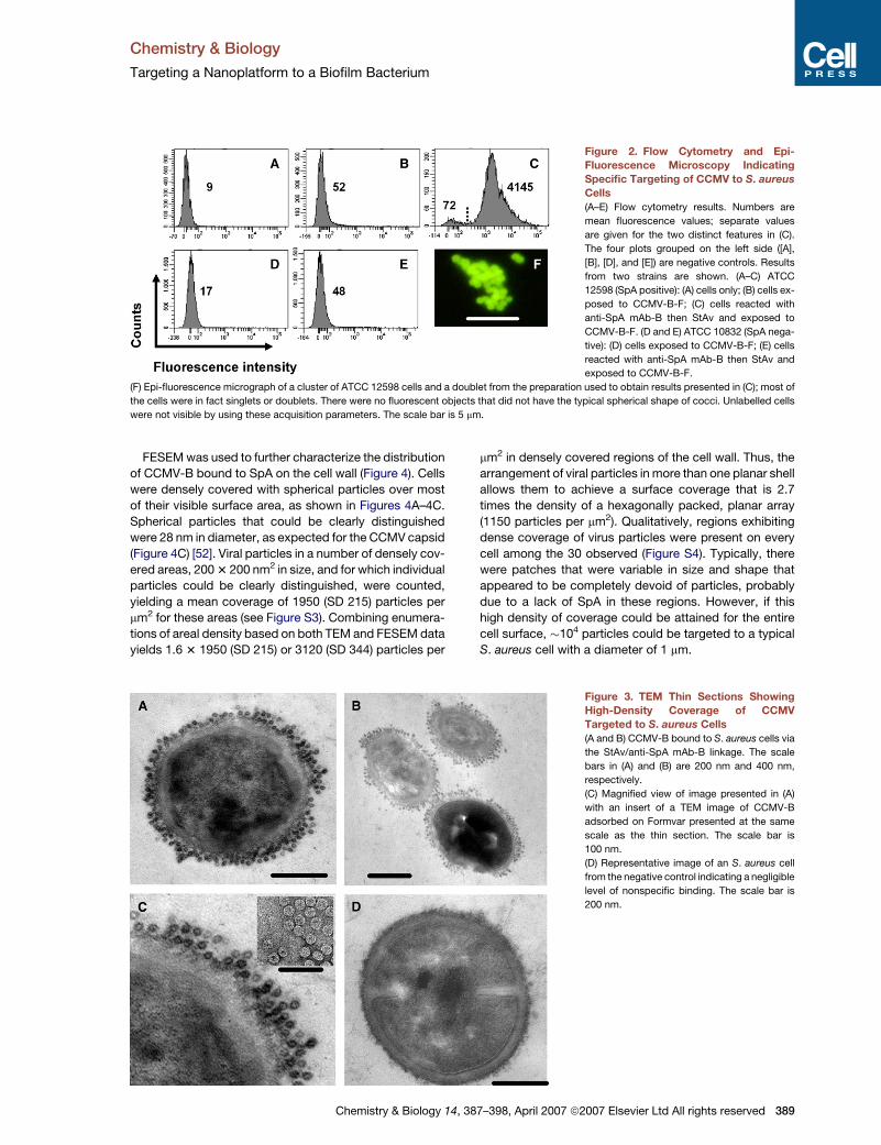
(CCMV-B-F). Analysis by flow cytometry indicated that
binding of CCMV-B-F to S. aureus cells was mediated
specifically by the engineered interactions, and not by
nonspecific binding of CCMV-B-F to components of the
S. aureus cell wall (Figures 2A–2E). Negligible levels of
binding were measured for S. aureus strain ATCC 12598
exposed only to CCMV-B-F (Figure 2B), and for S. aureus
strain ATCC 10832, which does not express SpA, ex-
posed only to CCMV-B-F (Figure 2D) or to anti-SpA
mAb-B, StAv, and CCMV-B-F (Figure 2E). Epi-fluores-
cence microscopy was used to verify that the fluorescent
objects detected by flow cytometry when CCMV-B-F was
targeted to S. aureus ATCC 12598 were indeed cells
exhibiting the expected morphology (Figure 2F).
High-Density Coverage of S. aureus Cells
Examination of S. aureus cells targeted with CCMV-B by
both transmission electron microscopy (TEM) and field
emission scanning electron microscopy (FESEM) indi-
cated a high density of viral binding to the cell wall. TEM
thin sections revealed that intact viral particles were
arranged in a loosely packed dual layer around the cell
periphery (Figures 3A–3C). Particles in TEM images were
counted, yielding a mean of 60 particles per micrometer
around the circumference of a planer section of a cell, or
1.6 monolayers if one monolayer is considered to consist
of virus particles closely packed end to end. In a negative
control experiment, no viral particles were associated with
S. aureus cells exposed only to CCMV-B (Figure 3D;
Figure S2), thus corroborating the interpretation that the
extent of nonspecific binding of CCMV-B to S. aureus cells
was negligible.
er Ltd All rights reserved
Chemistry & Biology
Targeting a Nanoplatform to a Biofilm Bacterium
Figure 2. Flow Cytometry and Epi-
Fluorescence Microscopy Indicating
Specific Targeting of CCMV to S. aureus
Cells
(A–E) Flow cytometry results. Numbers are
mean fluorescence values; separate values
are given for the two distinct features in (C).
The four plots grouped on the left side ([A],
[B], [D], and [E]) are negative controls. Results
from two strains are shown. (A–C) ATCC
12598 (SpA positive): (A) cells only; (B) cells ex-
posed to CCMV-B-F; (C) cells reacted with
anti-SpA mAb-B then StAv and exposed to
CCMV-B-F. (D and E) ATCC 10832 (SpA nega-
tive): (D) cells exposed to CCMV-B-F; (E) cells
reacted with anti-SpA mAb-B then StAv and
exposed to CCMV-B-F.
(F) Epi-fluorescence micrograph of a cluster of ATCC 12598 cells and a doublet from the preparation used to obtain results presented in (C); most of
the cells were in fact singlets or doublets. There were no fluorescent objects that did not have the typical spherical shape of cocci. Unlabelled cells
were not visible by using these acquisition parameters. The scale bar is 5 mm.
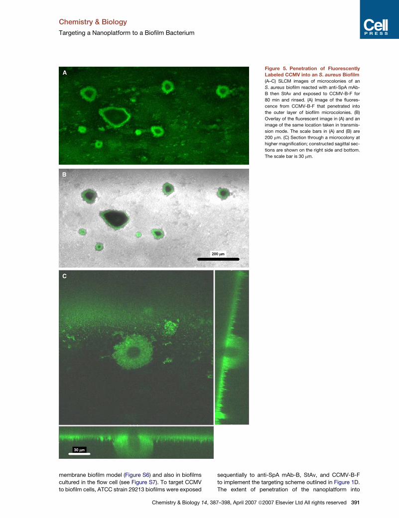
FESEM was used to further characterize the distribution
of CCMV-B bound to SpA on the cell wall (Figure 4). Cells
were densely covered with spherical particles over most
of their visible surface area, as shown in Figures 4A–4C.
Spherical particles that could be clearly distinguished
were 28 nm in diameter, as expected for the CCMV capsid
(Figure 4C) [52]. Viral particles in a number of densely cov-
ered areas, 200 3 200 nm2 in size, and for which individual
particles could be clearly distinguished, were counted,
yielding a mean coverage of 1950 (SD 215) particles per
mm2 for these areas (see Figure S3). Combining enumera-
tions of areal density based on both TEM and FESEM data
yields 1.6 3 1950 (SD 215) or 3120 (SD 344) particles per
Chemistry & Biology 14,
mm2 in densely covered regions of the cell wall. Thus, the
arrangement of viral particles in more than one planar shell
allows them to achieve a surface coverage that is 2.7
times the density of a hexagonally packed, planar array
(1150 particles per mm2). Qualitatively, regions exhibiting
dense coverage of virus particles were present on every
cell among the 30 observed (Figure S4). Typically, there
were patches that were variable in size and shape that
appeared to be completely devoid of particles, probably
due to a lack of SpA in these regions. However, if this
high density of coverage could be attained for the entire
cell surface, �104 particles could be targeted to a typical
S. aureus cell with a diameter of 1 mm.
Figure 3. TEM Thin Sections Showing
High-Density Coverage of CCMV
Targeted to S. aureus Cells
(A and B) CCMV-B bound to S. aureus cells via
the StAv/anti-SpA mAb-B linkage. The scale
bars in (A) and (B) are 200 nm and 400 nm,
respectively.
(C) Magnified view of image presented in (A)
with an insert of a TEM image of CCMV-B
adsorbed on Formvar presented at the same
scale as the thin section. The scale bar is
100 nm.
(D) Representative image of an S. aureus cell
from the negative control indicating a negligible
level of nonspecific binding. The scale bar is
200 nm.
387–398, April 2007 ª2007 Elsevier Ltd All rights reserved 389
Chemistry & Biology
Targeting a Nanoplatform to a Biofilm Bacterium
Figure 4. FESEM Images Showing
High-Density Coverage of CCMV Tar-
geted to S. aureus Cells
(A–D) (C) is a magnified view of the image pre-
sented in (A); (D) is an untargeted cell. The
scale bars are 200 nm in (A), (B), and (D) and
100 nm in (C).
Targeted Delivery of an MRI Contrast Agent
to S. aureus Cells
We used the multifunctional nanoplatform described
above to target MRI CA to S. aureus cells. We first dual
functionalized CCMV with both DOTA, a Gd(III)-chelating
agent, and biotin. (This construct is referred to hence forth
as CCMV-B-Gd.) Characterization by liquid chromatogra-
phy/electrospray mass spectrometry (LC/MS) indicated
that both DOTA and biotin functional groups were cova-
lently linked into the CCMV-B-Gd construct, with some
subunits functionalized with both groups (see Figure S5).
The mean number of Gd ions chelated to the CCMV-
B-Gd construct was estimated to be 166 per virion from
these data (Table 1). The scheme outlined in Figure 1D
was used to target CCMV-B-Gd to S. aureus cells. Analy-
sis with inductively coupled plasma mass spectrometry
(ICP-MS) to determine the concentration of Gd in a cell
sample of known cell density indicated a value of 1.8 3
105 Gd atoms per cell. Assuming that the combined
TEM and FESEM data yield a maximum estimate for
the amount of CCMV that can be specifically targeted to
the S. aureus cell wall by using our method, optimal target-
ing of the CCMV-B-Gd construct to the entire cell pop-
ulation would increase Gd loading by about an order of
magnitude.
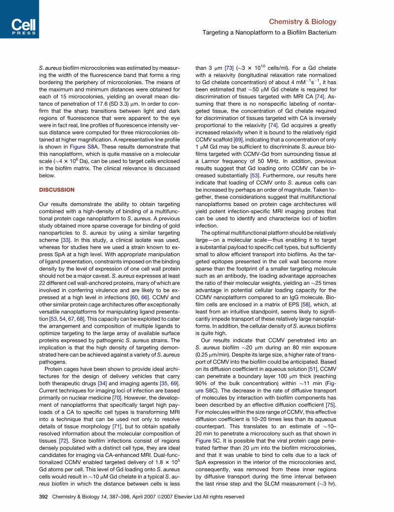
Targeting an S. aureus Biofilm
We used scanning laser confocal microscopy (SLCM) to
image targeting of CCMV-B-F to an S. aureus biofilm (Fig-
ure 5). In preliminary experiments, we confirmed that
390 Chemistry & Biology 14, 387–398, April 2007 ª2007 Elsevi
ATCC strain 29213, known to be SpA positive [63], a bio-
film former [64], and capable of inducing device-related
infections in an animal model [65], expressed SpA in a
Table 1. Functional Group and Gd Abundance onCCMV-B-Gd
FunctionalGroup(s)a
LC/MSb
Group(s)/CCMVc
Gd/Groupd
Gd/Group(per CCMV)e
None 0.24 44 0 0
DOTA 0.05 8 0 0
B 0.06 12 0 0
DOTA-Gd 0.25 45 1 45
2B 0.04 8 0 0
B, DOTA-Gd 0.12 21 1 21
2(DOTA-Gd) 0.10 18 2 36
B, 2(DOTA-Gd) 0.06 11 2 22
3(DOTA-Gd) 0.08 14 3 42
a Functional group(s) on monomer subunit (abbreviations arein text), e.g., B, 2(DOTA-Gd) means a monomer functionalized
with one biotin group and two DOTA groups, each chelating
a Gd atom.b Relative peak height from LC/MS data.c Mean number of functional group(s) of each type (or combi-
nation) per virus particle.d Number of Gd atoms per each functional group(s).e Mean number of Gd atoms per each functional group per
virus particle.
er Ltd All rights reserved
Chemistry & Biology
Targeting a Nanoplatform to a Biofilm Bacterium
Figure 5. Penetration of Fluorescently
Labeled CCMV into an S. aureus Biofilm
(A–C) SLCM images of microcolonies of an
S. aureus biofilm reacted with anti-SpA mAb-
B then StAv and exposed to CCMV-B-F for
80 min and rinsed. (A) Image of the fluores-
cence from CCMV-B-F that penetrated into
the outer layer of biofilm microcolonies. (B)
Overlay of the fluorescent image in (A) and an
image of the same location taken in transmis-
sion mode. The scale bars in (A) and (B) are
200 mm. (C) Section through a microcolony at
higher magnification; constructed sagittal sec-
tions are shown on the right side and bottom.
The scale bar is 30 mm.
membrane biofilm model (Figure S6) and also in biofilms
cultured in the flow cell (see Figure S7). To target CCMV
to biofilm cells, ATCC strain 29213 biofilms were exposed
Chemistry & Biology 14,
sequentially to anti-SpA mAb-B, StAv, and CCMV-B-F
to implement the targeting scheme outlined in Figure 1D.
The extent of penetration of the nanoplatform into
387–398, April 2007 ª2007 Elsevier Ltd All rights reserved 391
Chemistry & Biology
Targeting a Nanoplatform to a Biofilm Bacterium
S. aureus biofilm microcolonies was estimated by measur-
ing the width of the fluorescence band that forms a ring
bordering the periphery of microcolonies. The means of
the maximum and minimum distances were obtained for
each of 15 microcolonies, yielding an overall mean dis-
tance of penetration of 17.6 (SD 3.3) mm. In order to con-
firm that the sharp transitions between light and dark
regions of fluorescence that were apparent to the eye
were in fact real, line profiles of fluorescence intensity ver-
sus distance were computed for three microcolonies ob-
tained at higher magnification. A representative line profile
is shown in Figure S8A. These results demonstrate that
this nanoplatform, which is quite massive on a molecular
scale (�4 3 106 Da), can be used to target cells enclosed
in the biofilm matrix. The clinical relevance is discussed
below.
DISCUSSION
Our results demonstrate the ability to obtain targeting
combined with a high-density of binding of a multifunc-
tional protein cage nanoplatform to S. aureus. A previous
study obtained more sparse coverage for binding of gold
nanoparticles to S. aureus by using a similar targeting
scheme [33]. In this study, a clinical isolate was used,
whereas for studies here we used a strain known to ex-
press SpA at a high level. With appropriate manipulation
of ligand presentation, constraints imposed on the binding
density by the level of expression of one cell wall protein
should not be a major caveat. S. aureus expresses at least
22 different cell wall-anchored proteins, many of which are
involved in conferring virulence and are likely to be ex-
pressed at a high level in infections [60, 66]. CCMV and
other similar protein cage architectures offer exceptionally
versatile nanoplatforms for manipulating ligand presenta-
tion [53, 54, 67, 68]. This capacity can be exploited to cater
the arrangement and composition of multiple ligands to
optimize targeting to the large array of available surface
proteins expressed by pathogenic S. aureus strains. The
implication is that the high density of targeting demon-
strated here can be achieved against a variety of S. aureus
pathogens.
Protein cages have been shown to provide ideal archi-
tectures for the design of delivery vehicles that carry
both therapeutic drugs [34] and imaging agents [35, 69].
Current techniques for imaging loci of infection are based
primarily on nuclear medicine [70]. However, the develop-
ment of nanoplatforms that specifically target high pay-
loads of a CA to specific cell types is transforming MRI
into a technique that can be used not only to resolve
details of tissue morphology [71], but to obtain spatially
resolved information about the molecular composition of
tissues [72]. Since biofilm infections consist of regions
densely populated with a distinct cell type, they are ideal
candidates for imaging via CA-enhanced MRI. Dual-func-
tionalized CCMV enabled targeted delivery of 1.8 3 105
Gd atoms per cell. This level of Gd loading onto S. aureus
cells would result in �10 mM Gd chelate in a typical S. au-
reus biofilm in which the distance between cells is less
392 Chemistry & Biology 14, 387–398, April 2007 ª2007 Elsevi
than 3 mm [73] (�3 3 1010 cells/ml). For a Gd chelate
with a relaxivity (longitudinal relaxation rate normalized
to Gd chelate concentration) of about 4 mM�1s�1, it has
been estimated that �50 mM Gd chelate is required for
discrimination of tissues targeted with MRI CA [74]. As-
suming that there is no nonspecific labeling of nontar-
geted tissue, the concentration of Gd chelate required
for discrimination of tissues targeted with CA is inversely
proportional to the relaxivity [74]. Gd acquires a greatly
increased relaxivity when it is bound to the relatively rigid
CCMV scaffold [69], indicating that a concentration of only
1 mM Gd may be sufficient to discriminate S. aureus bio-
films targeted with CCMV-Gd from surrounding tissue at
a Larmor frequency of 50 MHz. In addition, previous
results suggest that Gd loading onto CCMV can be in-
creased substantially [53]. Furthermore, our results here
indicate that loading of CCMV onto S. aureus cells can
be increased by perhaps an order of magnitude. Taken to-
gether, these considerations suggest that multifunctional
nanoplatforms based on protein cage architectures will
yield potent infection-specific MRI imaging probes that
can be used to identify and characterize loci of biofilm
infection.
The optimal multifunctional platform should be relatively
large—on a molecular scale—thus enabling it to target
a substantial payload to specific cell types, but sufficiently
small to allow efficient transport into biofilms. As the tar-
geted epitopes presented in the cell wall become more
sparse than the footprint of a smaller targeting molecule
such as an antibody, the loading advantage approaches
the ratio of their molecular weights, yielding an �25 times
advantage in potential cellular loading capacity for the
CCMV nanoplatform compared to an IgG molecule. Bio-
film cells are enclosed in a matrix of EPS [58], which, at
least from an intuitive standpoint, seems likely to signifi-
cantly impede transport of these relatively large nanoplat-
forms. In addition, the cellular density of S. aureus biofilms
is quite high.
Our results indicate that CCMV penetrated into an
S. aureus biofilm �20 mm during an 80 min exposure
(0.25 mm/min). Despite its large size, a higher rate of trans-
port of CCMV into the biofilm could be anticipated. Based
on its diffusion coefficient in aqueous solution [51], CCMV
can penetrate a boundary layer 100 mm thick (reaching
90% of the bulk concentration) within �11 min (Fig-
ure S8C). The decrease in the rate of diffusive transport
of molecules by interaction with biofilm components has
been described by an effective diffusion coefficient [75].
For molecules within the size range of CCMV, this effective
diffusion coefficient is 10–20 times less than its aqueous
counterpart. This translates to an estimate of �10–
20 min to penetrate a microcolony such as that shown in
Figure 5C. It is possible that the viral protein cage pene-
trated farther than 20 mm into the biofilm microcolonies,
and that it was unable to bind to cells due to a lack of
SpA expression in the interior of the microcolonies and,
consequently, was removed from these inner regions
by diffusive transport during the time interval between
the last rinse step and the SLCM measurement (�3 hr).
er Ltd All rights reserved
Chemistry & Biology
Targeting a Nanoplatform to a Biofilm Bacterium
SpA expression may have been influenced by a microen-
vironment such as reduced oxygen that is likely to have
developed in central regions of the microcolonies [76]. In
support of this alternative interpretation, the distribution
of fluorescence with respect to distance along the sub-
stratum shows a transition between light and dark regions
that is quite abrupt and thus not compatible with the
description provided by a simple model of diffusive trans-
port (Figures S8A and S8D).
The rate of diffusive transport of a substance through
a biofilm cannot be predicted based exclusively on its
size, which is the primary factor governing its transport
in aqueous medium. In general, the rate of transport of
nanoplatforms into biofilms is expected to be significantly
altered by their surface properties, since this will influence
sorption to biofilm components [75]. Liposomes incorpo-
rating phosphatidylinositol or functionalized with PEG fully
penetrated to the base of a 20 mm thick S. aureus biofilm
during a 2 hr exposure period, while other cationic lipo-
somes adsorbed only to the outer layers [59]. Thus, bind-
ing of targeted CCMV nanoplatforms to cells is likely
to have hindered their transport through the biofilm to
some degree.
Even penetration into a biofilm by a nanoplatform to
a distance of 20 mm may be sufficient to target the majority
of cells in most biofilm infections. With the exception of
two early studies [77, 78], there is a paucity of information
on the structure of in vivo biofilms associated with bioma-
terial-centered infections in humans. S. aureus biofilms in-
volved in a catheter infection in a mouse model colonized
to an areal density of �3 3 107 cells/cm2, which would
yield a nominal thickness of 10 mm for an S. aureus biofilm
with a similar cell density as our in vitro biofilm [79]. An
S. aureus biofilm of similar dimensions was obtained on
an implanted tissue cage in a guinea pig model [80], while
micrographs of osteomyelitis induced in a rat model from
a precolonized implant indicated an S. aureus biofilm of
smaller dimensions [81]. If these studies are representa-
tive of the type of S. aureus biofilms involved in human in-
fections, our results indicate that a nanoplatform approx-
imately the same size as CCMV could penetrate to all of
the cells in a biofilm infection within a 2 hr exposure period.
Our results, together with estimates for transport of very
large molecules through biofilms discussed briefly, sug-
gest that the limiting factor in developing multifunctional
nanoplatforms based on protein cage architectures for di-
agnosis and treatment of biofilm infections is not likely to
be the rate of penetration into the biofilm matrix. A rough
estimate of the plasma half-life that is necessary for effec-
tive penetration to an extravascular site can be obtained
from extensive studies of nanoplatforms used to diagnose
and treat cancer. Liposomes sterically stabilized with PEG
and incorporating doxorubicin (SSL DOX), which are
among the most promising delivery vehicles for anticancer
agents, have plasma half-lives in humans of �50 hr [82].
Ferumoxtran-10, a superparamagnetic iron oxide nano-
particle used to enhance MRI visualization of lymphomas,
has a half-life of between 24 and 30 hr [83]. Placed in the
perspective of residence times in the circulatory system of
Chemistry & Biology 14,
between 24 and 50 hr, the time of about 2 hr required to
penetrate a 30 mm thick biofilm is probably not the factor
that will limit application of this technology.
A fundamental issue that needs to be addressed before
nanoplatforms based on protein cage architectures can
be used for clinical applications is the response of the
immune system. CCMV did not elicit a hypersensitive
response when injected intravenously into mice and was
distributed to a variety of tissues, indicating its ability to
extravasate from the vascular system [84]. However, the
rate of clearance from the circulatory system was rapid
and might preclude passive or active targeting. The clini-
cal application of superparamagnetic iron oxide nano-
particles was enabled primarily by the development of
surface modifications that increased the plasma half-life
[85]. As with liposomal systems, derivatization with PEG
holds promise as a means of modulating the immune
response to viral-based nanoplatforms [86].
SIGNIFICANCE
New approaches are required to effectively diagnose
and treat persistent infections that are recalcitrant to
conventional antimicrobial therapies. Our results indi-
cate that multifunctional nanoplatforms based on tar-
geted protein cage architectures have significant
potential for treating and diagnosing localized infec-
tions. In this respect, two key functional attributes
were demonstrated. First, the density of binding of
targeted CCMV to a surface protein expressed by
S. aureus was exceptionally high. Second, CCMV pen-
etrated into an S. aureus biofilm at a rate that was
rapid enough to make clinical applications feasible.
The multifunctional platform that we used for these
studies is an ideal system for catering ligand presen-
tation to optimize targeting to a variety of surface
proteins expressed by pathogenic bacteria, or to com-
binations of these proteins. In turn, the size range of
these nanoplatforms potentiates targeted delivery of
large payloads of imaging agents or therapeutic drugs
to loci of infections. In short, versatile multifunctional
nanoplatforms such as these offer a template upon
which the chemist and biologist can structure innova-
tive approaches to infection control.
EXPERIMENTAL PROCEDURES
Antibody
Anti-SpA mAb was produced in mouse (clone SPA-27) and was bioti-
nylated by the supplier (P3150 Sigma-Aldrich Co.) It was raised against
the Cowan I strain and binds to an epitope that is distinct from the
Fc-binding region.
Bacterial Strains and Culturing
ATCC strains 12598 (Cowan I, SpA +), 10832 (Wood 46, SpA �), and
29213 (SpA +, biofilm former) were used for these studies. Planktonic
cells were grown in batch cultures in nutrient broth for 8 hr at 37�C.
Biofilms of 29213 were cultured in tryptic soy broth (TSB) diluted
1:10 with nanopure water at 37�C in a square, glass flow cell similar
387–398, April 2007 ª2007 Elsevier Ltd All rights reserved 393
Chemistry & Biology
Targeting a Nanoplatform to a Biofilm Bacterium
to a system used previously to characterize S. aureus biofilms [73].
Biofilms were cultured for between 5 and 7 days, until there was visible
growth in the tube.
Virus Preparation and Functional Group Addition
CCMV was isolated from cowpea plants as previously described [87].
Purity was verified by using size-exclusion chromatography (SEC) and
dynamic light scattering (DLS) [88]. Protein concentration was deter-
mined by using the absorbance at 260 nm [51]. CCMV was biotinylated
by reaction of a 0.5 mM solution of sulfosuccinimidyl-60-(biotinamido)-
6-hexanamido hexanoate (Pierce) with 0.4 mM (1.4 mg ml�1) CCMV in
50 mM HEPES buffer, 150 mM NaCl (pH 7.0) at room temperature with
stirring for 30 min. The reaction was terminated by exchange into 1 mM
sodium acetate buffer (pH 4.8) by using SEC (Superose 6, Amersham
Biosciences, Uppsala, Sweden). Fractions eluting from within the
CCMV peak, the position of which was predetermined by using unla-
beled CCMV, were used for the experiments. These reaction condi-
tions were predetermined to leave some reactive amines on lysine
groups available for fluorescent tagging. Labeled virus was further pu-
rified by dialysis into 100 mM sodium acetate buffer. The integrity of the
labeled virus was confirmed by DLS and TEM as described previously
[88]. Association of biotin with the virus was initially tested by a dot blot
assay by using alkaline phosphatase-conjugated anti-biotin antibody
(A-7064, Sigma), and covalent attachment to protein monomer sub-
units was verified by liquid chromatography/electrospray mass spec-
trometry (LC/MS). Biotin-conjugated CCMV (CCMV-B) was tagged
with fluorescein by reaction of a 0.5 mM solution of 5-(and-6)-carbox-
yfluorescein, succinimidyl ester (Sigma-Aldrich) with 3.0 mM (1.4 mg
ml�1) CCMV in 50 mM HEPES buffer, 150 mM sodium chloride (pH
7.0) at room temperature with stirring for 60 min. The reaction was ter-
minated by dialysis into 1 mM sodium acetate buffer (pH 4.8). CCMV/
K42R, an exceptionally stable genetic construct [89], was dual func-
tionalized with a Gd chelate (DOTA, Macrocyclics, B-280) and biotin
by first reacting the protein cage with DOTA, then chelating the Gd
to the DOTA-functionalized CCMV, and, finally, biotinylating this prod-
uct by following the protocol described above. CCMV/K42R was func-
tionalized with DOTA by reaction with an NHS ester of DOTA in 100 mM
HEPES, 100 mM sodium chloride (pH 7.2) (reaction buffer) by using a
final molar ratio of 20:1 of the DOTA reagent:CCMV/K42R monomer
subunit. The DOTA reagent was added into a 2 mg/ml CCMV/K42R so-
lution in increments of �5 mg, while alternately maintaining the pH at
7.2 by titrating in 0.5 M sodium hydroxide, and was allowed to react un-
til completion, which was determined by using LC/MS to monitor the
progress of the reaction. The reaction was terminated by dialysis
into 100 mM HEPES, 100 mM sodium chloride (pH 6.5) (storage buffer)
overnight. After dialysis, the preparation was centrifuged (17,900 3 g)
for 5 min, and the supernatant was dialyzed against a solution of Gd in
the reaction buffer; Gd concentration was adjusted to be 103 the
DOTA chelate concentration. LC/MS was used to follow the reaction
to completion, and then the reaction was terminated by exchange
into the storage buffer by using SEC (Superose 6, Amersham Biosci-
ences, Uppsala, Sweden). Multiple buffer exchanges were carried
out to rid the sample of unbound Gd, and then the CCMV/K42R func-
tionalized with DOTA-Gd was labeled with biotin as described above.
LC/MS was used to quantify the extent of DOTA-Gd, DOTA, and biotin
conjugation to CCMV/K42R. A functional assay was performed in or-
der to determine the availability of the biotin functional groups on
CCMV-B-Gd for binding to StAv (Figure S9). The construct is quite sta-
ble, probably due to the K42R background. After 1 year of storage, the
construct preparation maintained �80% of its Gd load (ICP analysis)
and was used to target cells with intact virus particles (FESEM analy-
sis) (Figure S10).
Targeting S. aureus Planktonic Cells
We modified protocols previously published to bind antibody to the
cells and then StAv to the antibody-coated cells [90, 91]. Cells were
pelleted at 2500 3 g for 10 min at 4�C and resuspended in 5% bovine
calf serum (VWR), 0.1% intravenous immune globulin (Genesis Bio-
394 Chemistry & Biology 14, 387–398, April 2007 ª2007 Elsevi
Pharmaceuticals), 0.02% sodium azide in PBS (10 mM sodium phos-
phate, 100 mM sodium chloride [pH 7.2]) for 1 hr. The solution was then
incubated for 30 min at 4�C with a 1:1000 dilution of biotinylated anti-
SpA mAb-B. Cells were washed twice in 2% bovine calf serum and
0.02% sodium azide in PBS (wash solution). A portion of the washed
cells was reacted with ExtrAvidin-R-Phycoerythrin (Sigma-Aldrich)
(10 ml of the solution from the supplier in 1 ml wash solution for
30 min at 4�C) to verify expression of SpA for the negative controls
(or lack of SpA expression in the case of the Wood 46 strain). A portion
of the washed cells was reacted with StAv (Sigma-Aldrich) (50 mg/ml in
wash solution) and incubated for 30 min at 4�C. Cells were washed
once in wash solution and then twice in 0.02% sodium azide in PBS.
CCMV-B, CCMV-B-F, or CCMV-B-DOTA-Gd was added at between
130 and 210 mg/ml to the cell pellet in PBS at pH 6.7 and then incu-
bated for 30 min at 4�C. Cells labeled with CCMV-B-F were analyzed
immediately after this last step by using flow cytometry. Before ICP
analysis, cells were washed four times in 1 ml PBS to dilute the
CCMV/K42R-DOTA-Gd-B by a factor of greater than 10�4, leaving
a Gd concentration remaining from the unbound protein cage of less
than 1 picomolar. For TEM preparations and ICP analysis, cells were
fixed in 3% gluteraldehyde. For microscopic analysis, cells were fixed
in 1% paraformaldehyde in PBS. Negative controls consisted of the
same steps, but without addition of the antibody.
Targeting of Biofilm
At the end of the growth period, the capillary tube with the S. aureus
biofilm and silicone leader tubing was removed from the reactor sys-
tem by clamping and cutting the tubing. The biofilm was exposed to
different solutions by using a syringe connected to the effluent tubing
to draw solutions into the capillary via the influent tubing. The biofilm
was rinsed with 1 ml TSB with 0.02% sodium azide (TSB-NaN3) and
then exposed to a 1:100 dilution of anti-SpA mAb-B in TSB-NaN3
for 1 hr. This was followed by another rinse with 1 ml TSB-NaN3 and
exposure to 100 mg/ml StAv (or fluorescein-tagged StAv [StAv-F]
[Sigma-Aldrich]) in TSB-NaN3 for 1 hr to obtain results presented in
Figure S7). After another rinse with 1 ml TSB-NaN3, the biofilm was ex-
posed to CCMV-B-F at 130 mg/ml for 80 min in PBS (pH6.7) and then
rinsed with 1 ml TSB-NaN3. Finally, the biofilm was exposed to 1%
paraformaldehyde in PBS (pH 6.7). (The residual volume of the capil-
lary flow cell and leader tubing was �0.2 ml for each experiment.)
Approximately 3 hr elapsed between the SCLM viewing of the biofilm
and the last rinse step.
Flow Cytometry
Fluorescence from cells was analyzed by using the BD FACSAria cell
sorter (BD BioSciences). Side scatter was used to threshold the signal.
Optical parameters were: excitation, 488 nm; emission, 575/26 for
phycoerythrin and 530/30 for fluorescein.
Transmission Electron Microscopy
Cells were centrifuged at low speed to form a pellet, and a few drops of
2% noble agar were added just before hardening. The pellet was
briefly mixed and allowed to harden, cut into smaller pieces, and fixed
overnight with 3% gluteraldehyde in potassium sodium phosphate
buffer (PSPB [pH 7.2]). Agar pieces were then washed three times
with PSPB for 10 min, postfixed in 2% osmium tetroxide at room tem-
perature for 4 hr, dehydrated in an ethanol series, dissolved in propyl-
ene oxide, gradually infiltrated with Spurr’s resin [92], and baked over-
night at 70�C. Thin sections, 60–90 nm, were cut with a Diatome
diamond knife on a Reichert OM-U2 ultramicrotome. Sections were
floated onto 300 mesh copper grids and stained with uranyl acetate
and Reynold’s lead citrate [93] Negatively stained virus particles
were prepared by placing 5 ml purified virus on a carbon-stabilized,
formvar-coated 300 mesh copper grid and staining it with 2% UA in
water. All grids were viewed with a LEO 912AB transmission electron
microscope.
er Ltd All rights reserved
Chemistry & Biology
Targeting a Nanoplatform to a Biofilm Bacterium
Field Emission Scanning Electron Microscopy
Cells fixed in gluteraldehyde were adsorbed to polylysine-coated Si <
100 > wafers (Virginia Semiconductor Inc., Fredericksburg, VA) by
exposing the coupons to a cell suspension in 3% gluteraldehyde/
PBS for 1 hr. The coupons were rinsed twice in Nanopure water and
dried under a stream of liquid nitrogen. The coupons with adsorbed
cells were coated with a thin film of iridium by exposing the sample
for 15 s at 20 mA in an Emitech sputter coater. Cells were viewed
with a Supra 55VP FESEM (Zeiss) by using the Inlens detector at
1 kV and 3 mm working distance.
Conventional Optical Microscopy
Epi-fluorescence images were acquired through a B2A filter block
(excitation, 450–490 nm; emission, >515 nm) at 10003 (1003 objec-
tive and 103 camera lens) by using a Nikon Eclipse E600 coupled to
an Olympus Camedia camera. Cell counts were made in transmission
mode at 4003 (403 objective and a 103 ocular) by using a Zeiss Axio-
scope microscope to acquire images of the cells suspended in a known
volume (10 ml) of PBS. The mean number of cells in 20 fields was used
for the estimation of cell density.
Scanning Confocal Laser Microscopy
Confocal microscope images were collected on a Leica TCS-SP2-
AOBS confocal with a 633 0.9 NA HCX APO L U-V-I water-immersion
objective. Fluorescein was excited with a 488 nm laser, and fluores-
cence was collected from 504 nm to 619 nm. Images were taken at
0.5 mm intervals throughout the depth of the biofilm, then stacks
were combined in Imaris image analysis software (Bitplane AG, Zurich,
Switzerland) to yield final images. Line profiles were obtained by using
Image-Pro Plus (MediaCybernetics) software.
Liquid Chromatography/Electrospray Mass Spectrometry
LC/MS was performed on a QToF Micro instrument (Waters). CCMV
injected at 50–100 mg ml�1 (1–10 ml) was eluted from a C-8 reverse-
phase column (VYDAC) by using an acetonitrile/H2O gradient in
0.1% formic acid. The virus disassembled in the running buffer into
monomers that were analyzed by the detector. Mass spectra of the
multiply charged ions were deconvoluted by using instrument software
to produce a representation of monomer mass versus intensity. Mono-
mers from labeled virus produced clearly separated peaks in these
processed spectra with masses corresponding to the monomer
mass and the monomer mass plus between one and three reacted
reagent molecules (i.e., reagent molecules minus the sulfo-NHS ester
leaving group). The relative peak height was used to determine the
extent of labeling. This was converted to mean number of biotins,
DOTA, and DOTA-Gd chelates per CCMV particle.
Inductively Coupled Plasma Analysis
An Agilent 7500ce inductively coupled plasma mass spectrometer
(ICP-MS) was used to determine Gd concentration in the S. aureus
cell preparation with bound CCMV/K42R-B-DOTA-Gd. A 1 ml cell
preparation of 6.8 3 107 cells/ml was diluted to a volume of 5 ml in
2% nitric acid for this measurement. Interferences arising from the
plasma and sample matrix were eliminated with an Octopole Reaction
System (ORS), which is positioned between the source and the mass
filter. A buffer solution and a solution containing Gd3+ were both used
to optimize transmission of Gd ions while eliminating interfering spe-
cies. A calibration curve was constructed by using a series of Gd stan-
dards with concentrations spanning the expected range of the sample.
A blank was used in the analysis and was included in the calibration
curve.
Supplemental Data
Supplemental data include flow cytometry results (Figure S1), TEM thin
sections from the negative control (Figure S2), FESEM images of
densely covered regions (Figure S3), SEM images of CCMV-B bound
to S. aureus (Figure S4), an LC/MS spectrum of CCMV/K42R-DOTA-
Gd-B (Figure S5), flow cytometry results (Figure S6), an SCLM image
Chemistry & Biology 14,
of a biofilm of S. aureus reacted with anti-SpA mAb and then StAv-F
(Figure S7), quantitative analysis of confocal images (Figure S8), a func-
tional assay testing the availability of the biotin functional groups on
CCMV-B-Gd for binding to StAv (Figure S9), and FESEM images of
CCMV-B-Gd targeted to cells, demonstrating the stability of this con-
struct after long-term storage (Figure S10). These data are available
online at http://www.chembiol.com/cgi/content/full/14/4/387/DC1/.
ACKNOWLEDGMENTS
This work was funded by grants from the National Institutes of Health
(R01 EB00432), the Office of Naval Research for support of the Center
for BioInspired Nanomaterials (19-00-R0006), and Montana Idea
Network for Biomedical Research Excellence (MT INBRE) (M276-
05W0021).
Received: October 20, 2006
Revised: January 10, 2007
Accepted: February 7, 2007
Published: April 27, 2007
REFERENCES
1. Costerton, J.W., Stewart, P.S., and Greenberg, E.P. (1999). Bacte-
rial biofilms: a common cause of persistent infections. Science
284, 1318–1322.
2. Walsh, C. (2000). Molecular mechanisms that confer antibacterial
drug resistance. Nature 406, 775–781.
3. Davies, J. (1996). Bacteria on the rampage. Nature 383, 219–220.
4. Chen, I., Christie, P.J., and Dubnau, D. (2005). The ins and outs of
DNA transfer in bacteria. Science 310, 1456–1460.
5. Obritsch, M.D., Fish, D.N., MacLaren, R., and Jung, R. (2005).
Nosocomial infections due to multidrug-resistant Pseudomonas
aeruginosa: epidemiology and treatment options. Pharmacother-
apy 25, 1353–1364.
6. DeRyke, C.A., Maglio, D., and Nicolau, D.P. (2005). Defining the
need for new antimicrobials: clinical and economic implications
of resistance in the hospitalised patient. Expert Opin. Pharmac-
other. 6, 873–889.
7. Coimbra, M.V.D., Silva-Carvalho, M.C., Wisplinghoff, H., Hall,
G.O., Tallent, S., Wallace, S., Edmond, M.B., Figueiredo, A.M.S.,
and Wenzel, R.P. (2003). Clonal spread of methicillin-resistant
Staphylococcus aureus in a large geographic area of the United
States. J. Hosp. Infect. 53, 103–110.
8. Gristina, A.G. (1987). Biomaterial-centered infection: microbial
adhesion versus tissue integration. Science 237, 1588–1595.
9. Braxton, E.E., Ehrlich, G.D., Hall-Stoodley, L., Stoodley, P., Veeh,
R., Fux, C., Hu, F.Z., Quigley, M., and Post, J.C. (2005). Role of
biofilms in neurosurgical device-related infections. Neurosurg.
Rev. 28, 249–255.
10. Hall-Stoodley, L., Costerton, J.W., and Stoodley, P. (2004). Bacte-
rial biofilms: from the natural environment to infectious diseases.
Nat. Rev. Microbiol. 2, 95–108.
11. Fux, C.A., Costerton, J.W., Stewart, P.S., and Stoodley, P. (2005).
Survival strategies of infectious biofilms. Trends Microbiol. 13,
34–40.
12. Rice, L.B. (2006). Unmet medical needs in antibacterial therapy.
Biochem. Pharmacol. 71, 991–995.
13. Amaral, M.M., Coelho, L.R., Flores, R.P., Souza, R.R., Silva-
Carvalho, M.C., Teixeira, L.A., Ferrerira-Carvalho, B.T., and
Figueiredo, A.M.S. (2005). The predominant variant of the Brazilian
epidemic clonal complex of methicillin-resistant Staphylococcus
aureus has an enhanced ability to produce biofilm and to adhere
to and invade airway epithelial cells. J. Infect. Dis. 192, 801–810.
387–398, April 2007 ª2007 Elsevier Ltd All rights reserved 395
Chemistry & Biology
Targeting a Nanoplatform to a Biofilm Bacterium
14. Ando, E., Monden, K., Mitsuhata, R., Kariyama, R., and Kumon, H.
(2004). Biofilm formation among methicillin-resistant Staphylococ-
cus aureus isolates from patients with urinary tract infection. Acta
Med. Okayama 58, 207–214.
15. O’Riordan, K., and Lee, J.C. (2004). Staphylococcus aureus
capsular polysaccharides. Clin. Microbiol. Rev. 17, 218–234.
16. Stefani, S., and Varaldo, P.E. (2003). Epidemiology of methicillin-
resistant staphylococci in Europe. Clin. Microbiol. Infect. 9,
1179–1186.
17. Parfitt, T. (2005). Georgia: an unlikely stronghold for bacteriophage
therapy. Lancet 365, 2166–2167.
18. Summers, W.C. (2001). Bacteriophage therapy. Annu. Rev. Micro-
biol. 55, 437–451.
19. Wainwright, M., and Crossley, K.B. (2004). Photosensitising
agents—circumventing resistance and breaking down biofilms:
a review. Int. Biodeterior. Biodegradation 53, 119–126.
20. Demidova, T.N., and Hamblin, M.R. (2004). Photodynamic therapy
targeted to pathogens. Int. J. Immunopathol. Pharmacol. 17, 245–
254.
21. Ahmed, K., and Jones, M.N. (2003). The effect of shear on the de-
sorption of liposomes adsorbed to bacterial biofilms. J. Liposome
Res. 13, 187–197.
22. Catuogno, C., and Jones, M.N. (2003). The antibacterial properties
of solid supported liposomes on Streptococcus oralis biofilms. Int.
J. Pharm. 257, 125–140.
23. Hutchinson, F.J., and Jones, M.N. (1988). Lectin-mediated target-
ing of liposomes to a model surface. An ELISA method. FEBS Lett.
234, 493–496.
24. Jones, M.N., Hill, K.J., Kaszuba, M., and Creeth, J.E. (1998). Anti-
bacterial reactive liposomes encapsulating coupled enzyme sys-
tems. Int. J. Pharm. 162, 107–117.
25. Jones, M.N. (2005). Use of liposomes to deliver bactericides to
bacterial biofilms. Methods Enzymol. 331, 211–228.
26. Kim, H.J., Gias, E.L.M., and Jones, M.N. (1999). The adsorption of
cationic liposomes to Staphylococcus aureus biofilms. Colloids
Surf. A-Physicochem. Eng. Asp. 149, 561–570.
27. Svenson, S., and Tomalia, D.A. (2005). Dendrimers in biomedical
applications—reflections on the field. Adv. Drug Deliv. Rev. 57,
2106–2129.
28. Roy, I., Ohulchanskyy, T.Y., Pudavar, H.E., Bergey, E.J., Oseroff,
A.R., Morgan, J., Dougherty, T.J., and Prasad, P.N. (2003).
Ceramic-based nanoparticles entrapping water-insoluble photo-
sensitizing anticancer drugs: a novel drug-carrier system for
photodynamic therapy. J. Am. Chem. Soc. 125, 7860–7865.
29. Gao, X., Cui, Y., Levenson, R.M., Chung, L.W., and Nie, S. (2004).
In vivo cancer targeting and imaging with semiconductor quantum
dots. Nat. Biotechnol. 22, 969–976.
30. Harisinghani, M.G., Saini, S., Weissleder, R., Hahn, P.F., Yantiss,
R.K., Tempany, C., Wood, B.J., and Mueller, P.R. (1999). MR
lymphangiography using ultrasmall superparamagnetic iron oxide
in patients with primary abdominal and pelvic malignancies:
radiographic-pathologic correlation. AJR Am. J. Roentgenol.
172, 1347–1351.
31. Anderson, S.A., Rader, R.K., Westlin, W.F., Null, C., Jackson, D.,
Lanza, C.M., Wickline, S.A., and Kotyk, J.J. (2000). Magnetic res-
onance contrast enhancement of neovasculature with a(v)b(3)-
targeted nanoparticles. Magn. Reson. Med. 44, 433–439.
32. Zheng, G., Chen, J., Li, H., and Glickson, J.D. (2005). Rerouting
lipoprotein nanoparticles to selected alternate receptors for the
targeted delivery of cancer diagnostic and therapeutic agents.
Proc. Natl. Acad. Sci. USA 102, 17757–17762.
33. Zharov, V.P., Mercer, K.E., Galitovskaya, E.N., and Smeltzer, M.S.
(2006). Photothermal nanotherapeutics and nanodiagnostics for
396 Chemistry & Biology 14, 387–398, April 2007 ª2007 Elsev
selective killing of bacteria targeted with gold nanoparticles. Bio-
phys. J. 90, 619–627.
34. Flenniken, M.L., Liepold, L.O., Crowley, B.E., Willits, D.A., Young,
M.J., and Douglas, T. (2005). Selective attachment and release of
a chemotherapeutic agent from the interior of a protein cage archi-
tecture. Chem. Commun. 447–449.
35. Flenniken, M.L., Willits, D.A., Harmsen, A.L., Liepold, L.O., Harm-
sen, A.G., Young, M.J., and Douglas, T. (2006). Melanoma and
lymphocyte cell-specific targeting incorporated into a heat shock
protein cage architecture. Chem. Biol. 13, 161–170.
36. Douglas, T., and Young, M. (2006). Viruses: making friends with old
foes. Science 312, 873–875.
37. Douglas, T., and Young, M. (1998). Host-guest encapsulation of
materials by assembled virus protein cages. Nature 393, 152–155.
38. Klem, M.T., Willits, D., Young, M., and Douglas, T. (2003). 2-D
array formation of genetically engineered viral cages on Au
surfaces and imaging by atomic force microscopy. J. Am. Chem.
Soc. 125, 10806–10807.
39. Wang, Q., Kaltgrad, E., Lin, T.W., Johnson, J.E., and Finn, M.G.
(2002). Natural supramolecular building blocks: wild-type cowpea
mosaic virus. Chem. Biol. 9, 805–811.
40. Khayat, R., Tang, L., Larson, E.T., Lawrence, C.M., Young, M., and
Johnson, J.E. (2005). Structure of an archaeal virus capsid protein
reveals a common ancestry to eukaryotic and bacterial viruses.
Proc. Natl. Acad. Sci. USA 102, 18944–18949.
41. Kramer, R.M., Li, C., Carter, D.C., Stone, M.O., and Naik, R.R.
(2004). Engineered protein cages for nanomaterial synthesis.
J. Am. Chem. Soc. 126, 13282–13286.
42. Okuda, M., Kobayashi, Y., Suzuki, K., Sonoda, K., Kondoh, T.,
Wagawa, A., Kondo, A., and Yoshimura, H. (2005). Self-organized
inorganic nanoparticle arrays on protein lattices. Nano Lett. 5,
991–993.
43. Scheybani, T., Yoshimura, H., Baumeister, W., and Nagayama, K.
(1996). Stabilization of a fragile two-dimensional protein crystal at
the water-air interface: the square lattice of apoferritin. Langmuir
12, 431–435.
44. Yamashita, I. (2001). Fabrication of a two-dimensional array
of nano-particles using ferritin molecule. Thin Solid Films 393,
12–18.
45. Gilles, C., Bonville, P., Rakoto, H., Broto, J.M., Wong, K.K.W., and
Mann, S. (2002). Magnetic hysteresis and superantiferromagnet-
ism in ferritin nanoparticles. J. Magn. Magn. Mater. 241, 430–440.
46. Flenniken, M.L., Willits, D.A., Brumfield, S., Young, M.J., and
Douglas, T. (2003). The small heat shock protein cage from Meth-
anococcus jannaschii is a versatile nanoscale platform for genetic
and chemical modification. Nano Lett. 3, 1573–1576.
47. McMillan, R.A., Howard, J., Zaluzec, N.J., Kagawa, H.K., Mogul,
R., Li, Y.F., Paavola, C.D., and Trent, J.D. (2005). A self-assem-
bling protein template for constrained synthesis and patterning
of nanoparticle arrays. J. Am. Chem. Soc. 127, 2800–2801.
48. Wiedenheft, B., Mosolf, J., Willits, D., Yeager, M., Dryden, K.A.,
Young, M., and Douglas, T. (2005). An archaeal antioxidant: char-
acterization of a Dps-like protein from Sulfolobus solfataricus.
Proc. Natl. Acad. Sci. USA 102, 10551–10556.
49. Resnick, D.A., Gilmore, K., Idzerda, Y.U., Klem, M.T., Allen, M.,
Douglas, T., Arenholz, E., and Young, M. (2006). Magnetic proper-
ties of Co3O4 nanoparticles mineralized in Listeria innocua Dps.
J. Appl. Phys. 99, 08Q501.
50. Douglas, T., Strable, E., Willits, D., Aitouchen, A., Libera, M., and
Young, M. (2002). Protein engineering of a viral cage for con-
strained nanomaterials synthesis. Adv. Mater. 14, 415–418.
51. Bancroft, J.B., Hiebert, E., Rees, M.W., and Markham, R. (1968).
Properties of cowpea chlorotic mottle virus, its protein and nucleic
acid. Virology 34, 224–239.
ier Ltd All rights reserved
Chemistry & Biology
Targeting a Nanoplatform to a Biofilm Bacterium
52. Speir, J.A., Munshi, S., Wang, G.J., Baker, T.S., and Johnson, J.E.
(1995). Structures of the native and swollen forms of cowpea
chlorotic mottle virus determined by X-ray crystallography and
cryoelectron microscopy. Structure 3, 63–78.
53. Gillitzer, E., Willits, D., Young, M., and Douglas, T. (2002). Chemi-
cal modification of a viral cage for multivalent presentation. Chem.
Commun. 2390–2391.
54. Gillitzer, E., Suci, P., Young, M., and Douglas, T. (2006). Controlled
ligand display on a symmetrical protein-cage architecture through
mixed assembly. Small 2, 962–966.
55. De Schrijver, M. (1989). Scintigraphy of Inflammation with
Nanometer-Sized Colloidal Tracers (London: Kluwer Academic
Publishers).
56. Moghimi, S.M., Hunter, A.C., and Murray, J.C. (2001). Long-
circulating and target-specific nanoparticles: theory to practice.
Pharmacol. Rev. 53, 283–318.
57. Robinson, A.M., Creeth, J.E., and Jones, M.N. (2000). The use of
immunoliposomes for specific delivery of antimicrobial agents to
oral bacteria immobilized on polystyrene. J. Biomater. Sci. Polym.
Ed. 11, 1381–1393.
58. Wimpenny, J., Manz, W., and Szewzyk, U. (2000). Heterogeneity in
biofilms. FEMS Microbiol. Rev. 24, 661–671.
59. Ahmed, K., Gribbon, P., and Jones, M.N. (2002). The application of
confocal microscopy to the study of liposome adsorption onto
bacterial biofilms. J. Liposome Res. 12, 285–300.
60. Marraffini, L.A., DeDent, A.C., and Schneewind, O. (2006).
Sortases and the art of anchoring proteins to the envelopes of
gram-positive bacteria. Microbiol. Mol. Biol. Rev. 70, 192–221.
61. Patel, A.H., Nowlan, P., Weavers, E.D., and Foster, T. (1987).
Virulence of protein-a-deficient and a-toxin-deficient mutants of
Staphylococcus aureus isolated by allele replacement. Infect.
Immun. 55, 3103–3110.
62. Wu, A.M., and Senter, P.D. (2005). Arming antibodies: prospects
and challenges for immunoconjugates. Nat. Biotechnol. 23,
1137–1146.
63. Bernardo, K., Fleer, S., Pakulat, N., Krut, O., Hunger, F., and
Kronke, M. (2002). Identification of Staphylococcus aureus exo-
toxins by combined sodium dodecyl sulfate gel electrophoresis
and matrix-assisted laser desorption/ionization-time of flight
mass spectrometry. Proteomics 2, 740–746.
64. Harrison, J.J., Ceri, H., Stremick, C., and Turner, R.J. (2004).
Differences in biofilm and planktonic cell mediated reduction of
metalloid oxyanions. FEMS Microbiol. Lett. 235, 357–362.
65. Zimmerli, W., Frei, R., Widmer, A.F., and Rajacic, Z. (1994). Micro-
biological tests to predict treatment outcome in experimental
device-related infections due to Staphylococcus aureus. J. Anti-
microb. Chemother. 33, 959–967.
66. Harris, L.G., Foster, S.J., and Richards, R.G. (2002). An introduc-
tion to Staphylococcus aureus, and techniques for identifying
and quantifying S. aureus adhesins in relation to adhesion to bio-
materials. Eur. Cell. Mater. 4, 39–60.
67. Wang, Q., Lin, T.W., Johnson, J.E., and Finn, M.G. (2002). Natural
supramolecular building blocks: cysteine-added mutants of cow-
pea mosaic virus. Chem. Biol. 9, 813–819.
68. Chatterji, A., Ochoa, W.F., Paine, M., Ratna, B.R., Johnson, J.E.,
and Lin, T.W. (2004). New addresses on an addressable virus
nanoblock: uniquely reactive lys residues on cowpea mosaic virus.
Chem. Biol. 11, 855–863.
69. Allen, M., Bulte, J.W.M., Liepold, L., Basu, G., Zywicke, H.A.,
Frank, J.A., Young, M., and Douglas, T. (2005). Paramagnetic viral
nanoparticles as potential high-relaxivity magnetic resonance
contrast agents. Magn. Reson. Med. 54, 807–812.
Chemistry & Biology 14,
70. Rennen, H., Boerman, O.C., Oyen, W.J.G., and Corstens, F.H.M.
(2001). Imaging infection/inflammation in the new millennium.
Eur. J. Nucl. Med. 28, 241–252.
71. Morawski, A.M., Winter, P.M., Crowder, K.C., Caruthers, S.D.,
Fuhrhop, R.W., Scott, M.J., Robertson, J.D., Abendschein, D.R.,
Lanza, G.M., and Wickline, S.A. (2004). Targeted nanoparticles
for quantitative imaging of sparse molecular epitopes with MRI.
Magn. Reson. Med. 51, 480–486.
72. Jaffer, F.A., and Weissleder, R. (2005). Molecular imaging in the
clinical arena. JAMA 293, 855–862.
73. Leid, J.G., Shirtliff, M.E., Costerton, J.W., and Stoodley, P. (2002).
Human leukocytes adhere to, penetrate, and respond to Staphylo-
coccus aureus biofilms. Infect. Immun. 70, 6339–6345.
74. Ahrens, E.T., Rothbacher, U., Jacobs, R.E., and Fraser, S.E.
(1998). A model for MRI contrast enhancement using T-1 agents.
Proc. Natl. Acad. Sci. USA 95, 8443–8448.
75. Stewart, P.S. (1998). A review of experimental measurements of
effective diffusive permeabilities and effective diffusion coeffi-
cients in biofilms. Biotechnol. Bioeng. 59, 261–272.
76. Debeer, D., Stoodley, P., Roe, F., and Lewandowski, Z. (1994).
Effects of biofilm structures on oxygen distribution and mass-
transport. Biotechnol. Bioeng. 43, 1131–1138.
77. Marrie, T.J., Nelligan, J., and Costerton, J.W. (1982). A scanning
and transmission electron-microscopic study of an infected endo-
cardial pacemaker lead. Circulation 66, 1339–1341.
78. Nickel, J.C., Gristina, A.G., and Costerton, J.W. (1985). Electron-
microscopic study of an infected Foley catheter. Can. J. Surg.
28, 50–51, 54.
79. Kadurugamuwa, J.L., Sin, L., Albert, E., Yu, J., Francis, K., DeBoer,
M., Rubin, M., Bellinger-Kawahara, C., Parr, T.R., and Contag,
P.R. (2003). Direct continuous method for monitoring biofilm infec-
tion in a mouse model. Infect. Immun. 71, 882–890.
80. Fluckiger, U., Ulrich, M., Steinhuber, A., Doring, G., Mack, D.,
Landmann, R., Goerke, C., and Wolz, C. (2005). Biofilm formation,
icaADBC transcription, and polysaccharide intercellular adhesin
synthesis by Staphylococci in a device-related infection model.
Infect. Immun. 73, 1811–1819.
81. Monzon, M., Garcia-Alvarez, F., Lacleriga, A., Gracia, E., Leiva, J.,
Oteiza, C., and Amorena, B. (2001). A simple infection model using
pre-colonized implants to reproduce rat chronic Staphylococcus
aureus osteomyelitis and study antibiotic treatment. J. Orthop.
Res. 19, 820–826.
82. Drummond, D.C., Meyer, O., Hong, K.L., Kirpotin, D.B., and
Papahadjopoulos, D. (1999). Optimizing liposomes for delivery of
chemotherapeutic agents to solid tumors. Pharmacol. Rev. 51,
691–743.
83. Harisinghani, M.G., Saksena, M., Ross, R.W., Tabatabaei, S.,
Dahl, D., McDougal, S., and Weissleder, R. (2005). A pilot study
of lymphotrophic nanoparticle-enhanced magnetic resonance
imaging technique in early stage testicular cancer: a new meth-
od for noninvasive lymph node evaluation. Urology 66, 1066–
1071.
84. Kaiser, C.R., Flenniken, M.L., Gillitzer, E., Harmsen, A.G., Harm-
sen, A.L., Jutila, M.A., Douglas, T., and Young, M. (2006). Biodis-
tribution studies of protein cage nanoparticles demonstrate broad
tissue distribution and rapid clearance in vivo. Int. J. Nanomed.
85. Shen, T., Weissleder, R., Papisov, M., Bogdanov, A., and Brady,
T.J. (1993). Monocrystalline iron-oxide nanocompounds (MION):
physicochemical properties. Magn. Reson. Med. 29, 599–604.
86. Raja, K.S., Wang, Q., Gonzalez, M.J., Manchester, M., Johnson,
J.E., and Finn, M.G. (2003). Hybrid virus-polymer materials. 1.
Synthesis and properties of PEG-decorated cowpea mosaic virus.
Biomacromolecules 4, 472–476.
387–398, April 2007 ª2007 Elsevier Ltd All rights reserved 397
Chemistry & Biology
Targeting a Nanoplatform to a Biofilm Bacterium
87. Bancroft, J.B., and Hiebert, E. (1967). Formation of an infectious
nucleoprotein from protein and nucleic acid isolated from a small
spherical virus. Virology 32, 354–356.
88. Basu, G., Allen, M., Willits, D., Young, M., and Douglas, T. (2003).
Metal binding to cowpea chlorotic mottle virus using terbium(III)
fluorescence. J. Biol. Inorg. Chem. 8, 721–725.
89. Speir, J.A., Bothner, B., Qu, C.X., Willits, D.A., Young, M.J., and
Johnson, J.E. (2006). Enhanced local symmetry interactions glob-
ally stabilize a mutant virus capsid that maintains infectivity and
capsid dynamics. J. Virol. 80, 3582–3591.
90. Wann, E.R., Fehringer, A.P., Ezepchuk, Y.V., Schlievert, P.M., Bina,
P., Reiser, R.F., Hook, M.M., and Leung, D.Y.M. (1999). Staphylo-
398 Chemistry & Biology 14, 387–398, April 2007 ª2007 Elsevi
coccus aureus isolates from patients with Kawasaki disease
express high levels of protein A. Infect. Immun. 67, 4737–4743.
91. Yarwood, J.M., McCormick, J.K., and Schlievert, P.M. (2001).
Identification of a novel two-component regulatory system that
acts in global regulation of virulence factors of Staphylococcus
aureus. J. Bacteriol. 183, 1113–1123.
92. Spurr, A.R. (1969). A low-viscosity epoxy resin embedding
medium for electron microscopy. J. Ultrastruct. Res. 26, 31–43.
93. Reynolds, E.S. (1963). The use of lead citrate at high pH as an
electron-opaque stain in electron microscopy. J. Cell Biol. 17,
208–212.
er Ltd All rights reserved