HEV infection in swine from Eastern Brazilian Amazon Evidence of co-infection by different subtypes
10
This article appeared in a journal published by Elsevier. The attached copy is furnished to the author for internal non-commercial research and education use, including for instruction at the authors institution and sharing with colleagues. Other uses, including reproduction and distribution, or selling or licensing copies, or posting to personal, institutional or third party websites are prohibited. In most cases authors are permitted to post their version of the article (e.g. in Word or Tex form) to their personal website or institutional repository. Authors requiring further information regarding Elsevier’s archiving and manuscript policies are encouraged to visit: http://www.elsevier.com/copyright
-
Upload
independent -
Category
Documents
-
view
0 -
download
0
Transcript of HEV infection in swine from Eastern Brazilian Amazon Evidence of co-infection by different subtypes
This article appeared in a journal published by Elsevier. The attached
copy is furnished to the author for internal non-commercial research
and education use, including for instruction at the authors institution
and sharing with colleagues.
Other uses, including reproduction and distribution, or selling or
licensing copies, or posting to personal, institutional or third party
websites are prohibited.
In most cases authors are permitted to post their version of the
article (e.g. in Word or Tex form) to their personal website or
institutional repository. Authors requiring further information
regarding Elsevier’s archiving and manuscript policies are
encouraged to visit:
http://www.elsevier.com/copyright
Author's personal copy
Comparative Immunology, Microbiology and Infectious Diseases 35 (2012) 477– 485
Contents lists available at SciVerse ScienceDirect
Comparative Immunology, Microbiologyand Infectious Diseases
j o ur nal homep age : w ww.elsev ier .com/ locate /c imid
HEV infection in swine from Eastern Brazilian Amazon: Evidence ofco-infection by different subtypes!
Alex Junior Souza de Souzaa,e,!, Michele Soares Gomes-Gouvêab,Manoel do Carmo Pereira Soaresa, João Renato Rebello Pinhob,Andreza Pinheiro Malheirosa, Liliane Almeida Carneiroa,Debora Regina Lopes dos Santosc, Washington Luiz Assunc ão Pereirad
a Sec ão de Hepatologia, Instituto Evandro Chagas, Av. Almirante Barroso, 492, 66093-020 Belém, PA, Brazilb Laboratory of Gastroenterology and Hepatology, São Paulo Institute of Tropical Medicine and Department of Gastroenterology, School of Medicine,University of São Paulo, Brazilc Instituto de Veterinária, Universidade Federal Rural do Rio de Janeiro, Rod. BR 465, km 7, 23890-000 Rio de Janeiro, RJ, Brazild Instituto da Saúde e Produc ão Animal, Universidade Federal Rural da Amazônia, Avenida Presidente Tancredo Neves, 2501, 66077-901 Belém, PA, Brazile Centro de Ciências Biológicas e da Saúde, Faculdades Integradas do Tapajós, Rua Rosa Vermelha, 335, 68010-200 Santarém, PA, Brazil
a r t i c l e i n f o
Article history:Received 2 February 2012Received in revised form 10 April 2012Accepted 12 April 2012
Keywords:Hepatitis E virus (HEV)PigsZoonosisGenotype 3Brazil
a b s t r a c t
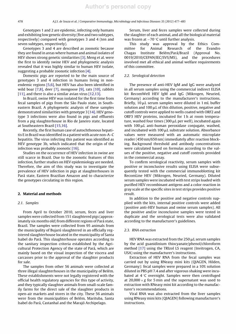
Hepatitis E virus (HEV) is a fecal-orally transmitted member of the genus Hepevirus thatcauses acute hepatitis in humans and is widely distributed throughout the world. Pigshave been reported as the main source of genotypes 3 and 4 infection to humans in non-endemic areas. To investigate HEV infection in pigs from different regions of Pará state(Eastern Brazilian Amazon), we performed serological and molecular analyses of serum,fecal and liver samples from 151 adult pigs slaughtered between April and October 2010 inslaughterhouses in the metropolitan region of Belém, Pará. Among the animals tested, 8.6%(13/151) were positive for anti-HEV IgG but not for anti-HEV IgM. HEV RNA was detectedin 4.8% (22/453) of the samples analyzed and 9.9% (15/151) of the animals had at least onepositive sample. Phylogenetic analysis showed that all sequences belonged to genotype 3that were related to human isolates from other non-endemic regions, suggesting that theisolates had zoonotic potential. Subtypes 3c and 3f were simultaneously detected in somepigs, suggesting co-infection by more than one strain and/or the presence of a recombinantvirus. These results constitute the first molecular and serologic evidence of swine HEVcirculation in the Eastern Brazilian Amazon.
© 2012 Elsevier Ltd. All rights reserved.
1. Introduction
Hepatitis E virus (HEV) is an RNA virus of the genusHepevirus that causes acute hepatitis in humans and is
! The GenBank/EMBL/DDBJ accession numbers for the sequencesdescribed in this study are JN983199–JN983212 (ORF1) andJN983192–JN983198 ORF2.
! Corresponding author at: Instituto Evandro Chagas, Av. AlmiranteBarroso, 492, 66093-020 Belém, PA, Brazil. Tel.: +55 91 3214 2072;fax: +55 91 3214 2139.
E-mail address: [email protected] (A.J.S. de Souza).
transmitted through fecal-oral route [1]. In mammals, HEVstrains have been classified into four genotypes (1–4) basedon their genetic diversity; additionally, subtypes have beenidentified within each genotype group and among themgenotype 3 shows the highest variability [2].
These genotypes show a characteristic geographic dis-tribution: genotype 1 is considered endemic in Asia andnorthern Africa; outbreaks of genotype 2 have beenreported in Mexico and central Africa; genotype 3 is foundin North and South America, parts of Europe and Japan; andgenotype 4 has been reported in China, Japan, Taiwan andVietnam [3].
0147-9571/$ – see front matter © 2012 Elsevier Ltd. All rights reserved.http://dx.doi.org/10.1016/j.cimid.2012.04.004
Author's personal copy
478 A.J.S. de Souza et al. / Comparative Immunology, Microbiology and Infectious Diseases 35 (2012) 477– 485
Genotypes 1 and 2 are epidemic, infecting only humansand exhibiting low genetic diversity (five and two subtypes,respectively) compared with genotypes 3 and 4 (ten andseven subtypes, respectively).
Genotypes 3 and 4 are described as zoonotic becausethey are found in areas where human and animal isolates ofHEV shows strong genetic similarities [3]. Meng et al. werethe first to identify swine HEV and phylogenetic analysisrevealed that it was highly similar to human HEV isolate,suggesting a probable zoonotic infection [4].
Domestic pigs are reported to be the main source ofgenotypes 3 and 4 infection in humans living in non-endemic regions [5,6], but HEV has also been identified inwild boar [7,8], deer [7], mongoose [9], rats [10], rabbits[11], and there is also a similar avian virus [12,13].
In Brazil, swine HEV was isolated for the first time fromfecal samples of pigs from the São Paulo state, in South-eastern Brazil. A phylogenetic analysis of these samplesdemonstrated similarities with genotype 3 [14]. HEV geno-type 3 infections were also found in pigs and effluentsfrom a pig slaughterhouse in Rio de Janeiro state, locatedat Southeastern Brazil [15,16].
Recently, the first human case of autochthonous hepati-tis E in Brazil was identified in a patient with acute non-A–Chepatitis. The virus infecting this patient was identified asHEV genotype 3b, which indicated that the origin of theinfection was probably zoonotic [16].
Studies on the occurrence of HEV infection in swine arestill scarce in Brazil. Due to the zoonotic features of thisinfection, further studies on HEV epidemiology are needed.Therefore, the aim of this study was to investigate theprevalence of HEV infection in pigs at slaughterhouses inPará state, Eastern Brazilian Amazon and to characterizethe genotypes circulating in this region.
2. Material and methods
2.1. Samples
From April to October 2010, serum, feces and liversamples were collected from 151 slaughtered pigs (approx-imately six months old) from different regions of Pará state,Brazil. The samples were collected from 95 animals fromthe municipality of Bujarú slaughtered in an officially reg-istered slaughterhouse located in the municipality of SantaIsabel do Pará. This slaughterhouse operates according tothe sanitary inspection criteria established by the Agri-cultural Protection Agency of the state of Pará, which aremainly based on the visual inspection of the viscera andcarcasses prior to the approval of the slaughter productsfor sale.
The samples from other 56 animals were collected atthree illegal slaughterhouses in the municipality of Belém.These establishments were not legally registered with theofficial health regulatory agencies for this type of activity,and they typically slaughter animals from small-scale fam-ily farms for the direct sale of the slaughter products inopen-air markets and stores in the city. These 56 animalswere from the municipalities of Belém, Marituba, SantaIsabel do Pará, Castanhal and the Marajó Archipelago.
Serum, liver and feces samples were collected duringthe slaughter of each animal, and all the biological materialwas frozen at "70 #C until further analysis.
This study was approval by the Ethics Com-mittee for Animal Research of the EvandroChagas Institute Belém/Pará/Brazil (Approval No.0019/2010/CEPAN/IEC/SVS/MS), and the proceduresinvolved met all ethical and animal welfare requirementsof this committee.
2.2. Serological detection
The presence of anti-HEV IgM and IgG were analyzedin all serum samples using the commercial indirect ELISAkit RecomWell HEV IgM and IgG (Mikrogen, Neuried,Germany) according to the manufacturer’s instructions.Briefly, 10 !L serum samples were diluted in 1 mL buffersolution and 100 !L of this dilution, positive, negative andcutoff controls were applied to wells coated with ORF2 andORF3 HEV proteins, incubated for 1 h at room tempera-ture, washed four times (300 !L per well), incubated againwith 100 !L anti-human peroxidase conjugate, washed,and incubated with 100 !L substrate solution. Absorbancevalues were measured with an automatic microplatereader (450 nm/650 nm) immediately after reaction block-ing. Background threshold and antibody concentrationswere calculated based on formulas according to the val-ues of the positive, negative and cutoff controls providedin the commercial assay.
To confirm serological reactivity, serum samples withpositive or inconclusive results using ELISA were subse-quently tested with the commercial immunoblotting kitRecomLine HEV (Mikrogen, Neuried, Germany). Dilutedserum samples were incubated with test strips loaded withpurified HEV recombinant antigens and a color reaction ingray scale at the specific sites in test strips provides positiveresult.
In addition to the positive and negative controls sup-plied with the kits, internal positive controls were added(positive anti-HEV human and swine serum samples). Allthe positive and/or inconclusive samples were tested induplicate and the serological tests were also validatedaccording to the manufacturer’s guidelines.
2.3. RNA extraction
HEV RNA was extracted from the 250 !L serum samplesby the acid guanidinium thiocyanate/phenol/chloroformmethod [17] using the TRIzol LS reagent (Invitrogen, CA,USA) using the manufacturer’s instructions.
Extraction of HEV RNA from the fecal samples wascarried out by using RNeasy mini kits (QIAGEN, Hilden,Germany): fecal samples were prepared in a 10% solutiondiluted in PBS pH 7.4 and after vigorous shaking were incu-bated at 4 #C overnight. Samples were then centrifugedat 20,000 $ g for 5 min and the supernatant was used toextraction with RNeasy mini kit according to the manufac-turer’s recommendations.
Viral RNA was also extracted from the liver samplesusing RNeasy mini kits (QIAGEN) following manufacturer’sinstructions.
Author's personal copy
A.J.S. de Souza et al. / Comparative Immunology, Microbiology and Infectious Diseases 35 (2012) 477– 485 479
2.4. Nested RT-PCR
Reverse transcription for cDNA synthesis was per-formed using random primers and the Moloney MurineLeukemia Virus Reverse Transcriptase (Invitrogen).
We used nested RT-PCR to amplify three regions of theHEV genome. Initially, two sets of primers that amplifiedfragments of ORF1 and ORF2 genes were used [18].
ORF1 primers were located at nucleotide positions56–79 and 473–451, which corresponded to the methyl-transferase coding region. The external primers (Cons-ORF1-s1: 5%-CTGGCATYACTACTGCYATTGAGC-3% andConsORF1-a1: 5%-CCATCRARRCAGTAAGTGCGGTC-3%) andinternal primers (ConsORF1-s2: 5%-CTGCCYTKGCGAA-TGCTGTGG-3% and ConsORF1-a2: 5%-GGCAGWRTACCA-RCGCTGAACATC-3%) amplified 418 and 287 bp fragments,respectively.
ORF2 primers corresponded to positions6298–6321 and 6494–6470 of the Burmese pro-totype. The external primers (ConsORF2-s1:5%-GACAGAATTRATTTCGTCGGCTGG-3% and ConsORF2-a1: 5%-CTTGTTCRTGYTGGTTRTCATAATC-3%) amplified a197 bp product in the first PCR, and the internal primers(ConsORF2-s2: 5%-GTYGTCTCRGCCAATGGCGAGC-3% andConsORF2-a2: 5%-GTTCRTGYTGGTTRTCATAATCCTG-3%)amplified a 145 bp product in the second PCR.
The samples were also tested using a third set ofprimers that amplified an additional ORF2 region. Theexternal (3156NF: 5%-AATTATGCYCAGTAYCGRGTTG-3% and3157NR: 5%-CCCTTRTCYTGCTGMGCATTCTC-3%) and inter-nal primers (3158NF: 5%-GTWATGCTYTGCATWCATGGCT-3% and 3159NR: 5%-AGCCGACGAAATCAATTCTGTC-3%) usedin the present study were described for the detection ofHEV and amplified 731 and 348 bp products in the first andsecond PCRs, respectively [19,20].
All precautions and procedures suggested to avoid falsepositive results were strictly followed [21]. The extractionof nucleic acids, the PCR setup and especially the additionof first round PCR reaction to second round PCR were per-formed in completely separated rooms, when necessaryinside biohazard cabinets or PCR hoods. Each stage wasperformed with separate micropipettes and all PCR pro-cedures were carried out using aerosol filter tips. Otherprecautions such as frequent changing of gloves, workingwith reagents divided in aliquot, control of daily flow ofpersonnel only from pre- to post-amplification areas andthe use of UV-light and sodium hypochlorite solution todecontaminate surfaces were strictly observed in order toprevent cross-contamination.
Positive control (serum from a pig positive to HEV) toconfirm the effectiveness of the reactions and negative con-trols (water) to check for contaminants were included ineach run.
PCR reactions had a final volume of 50 !L and con-tained the following reagents: 35.3 !L DEPC-treated H2O;5 !L 10$ PCR buffer; 1 !L dNTPs (10 mM) (Invitrogen);1.5 !L MgCl2 (50 mM) (Invitrogen); 20 pmol of each primer(forward and reverse); 1 U of Platinum Taq polymerase(Invitrogen) and 5 !L cDNA sample. In the second PCR,5 !L of the first PCR product was added instead ofcDNA.
PCR amplification cycles were: initial denaturation at94 #C for 2 min, followed by 35 cycles of denaturation (94 #Cfor 30 s), annealing (50 #C for 30 s), extension (72 #C for30 s) and a final extension at 72 #C for 5 min. The anneal-ing temperature differed only for the 348 bp ORF2 primers(42 #C).
The products of the second PCR were labeled withSYBR dye (Invitrogen) and separated by electrophoresison 1% agarose gel. Depending on the primers used, sam-ples that exhibited sharp, intense bands with approximatesizes of 145, 287 or 348 base pairs were considered aspositive.
2.5. Nucleotide sequencing and phylogenetic analysis
PCR products (ORF1 287 bp and ORF2 348 bp) fromthe second round reaction were purified using ExoSAP-IT PCR Clean-up Kit (GE Healthcare) and were thensequenced using the BigDye® Terminator v3.1 CycleSequencing Kit (Applied Biosystems, CA, USA) accordingto the manufacturer’s guidelines. Sequencing was carriedout on an automated ABI 3500 DNA Sequencer (AppliedBiosystems).
The quality of each electropherogram was evaluatedusing the Phred-Phrap software [22,23] and consen-sus sequences of forward and reverse sequences wereobtained using CAP3 software available at the web pagehttp://asparagin.cenargen.embrapa.br/phph/.
Sequences were aligned and edited using BioEdit (v.7.0.8) and the integrated CLUSTAL W program [24]. HEVgenotypes and subtypes were classified by phylogeneticreconstructions using published reference sequences fromGenBank database (http://www.ncbi.nlm.nih.gov/).
Bayesian phylogenetic analyses were conducted usingthe Markov Chain Monte Carlo (MCMC) simulationimplemented in BEAST v.1.6.1 [25] under uncorrelatedexponential relaxed molecular clock using the model ofnucleotide substitution (GTR + G + I). The maximum cladecredibility (MCC) tree was obtained from summarizing the10,000 substitution trees and then it was removed 10% ofburn-in using Tree Annotator v.1.6.1 [25].
Nucleotide sequence divergence was calculated usingthe program MEGA4 (Molecular Evolutionary GeneticsAnalysis) software version 4.0 [26].
3. Results
3.1. Serological detection
Anti-HEV IgG was detected using ELISA (backgroundthreshold = 0.3) in 4.6% (7/151) of the samples (meanabsorbance = 0.643 ± 0.214). In addition, 3.9% (6/151)of the samples showed inconclusive result (meanabsorbance = 0.308 ± 0.024). The average concentra-tions of the anti-HEV IgG antibodies were 42.1 ± 14.6 U/mLfor the seven positive samples and 20.5 ± 1.6 U/mL for thesix inconclusive samples. Anti-HEV IgM was not detectedin any of the analyzed samples.
All thirteen samples (8.6%; 13/151) with reactivity(positive or inconclusive) to anti-HEV IgG showed posi-tive results in the immunoblotting assay. These positive
Author's personal copy
480 A.J.S. de Souza et al. / Comparative Immunology, Microbiology and Infectious Diseases 35 (2012) 477– 485
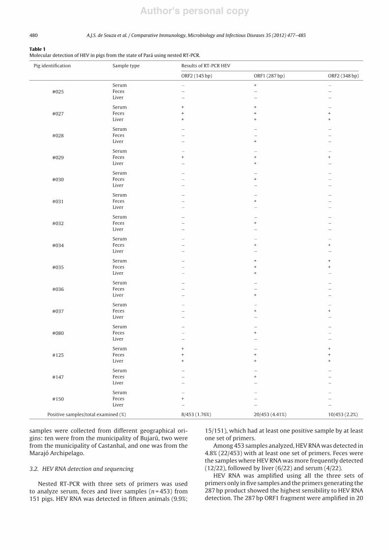
Table 1Molecular detection of HEV in pigs from the state of Pará using nested RT-PCR.
Pig identification Sample type Results of RT-PCR HEV
ORF2 (145 bp) ORF1 (287 bp) ORF2 (348 bp)
#025Serum " + "Feces " " "Liver " " "
#027Serum + + "Feces + + +Liver + + +
#028Serum " " "Feces " " "Liver " + "
#029Serum " " "Feces + + +Liver " + "
#030Serum " " "Feces " + "Liver " " "
#031Serum " " "Feces " + "Liver " " "
#032Serum " " "Feces " + "Liver " " "
#034Serum " " "Feces " + +Liver " " "
#035Serum " + +Feces " + +Liver " + "
#036Serum " " "Feces " " "Liver " + "
#037Serum " " "Feces " + +Liver " " "
#080Serum " " "Feces " + "Liver " " "
#125Serum + " +Feces + + +Liver + + +
#147Serum " " "Feces " + "Liver " " "
#150Serum " " "Feces + " "Liver " " "
Positive samples/total examined (%) 8/453 (1.76%) 20/453 (4.41%) 10/453 (2.2%)
samples were collected from different geographical ori-gins: ten were from the municipality of Bujarú, two werefrom the municipality of Castanhal, and one was from theMarajó Archipelago.
3.2. HEV RNA detection and sequencing
Nested RT-PCR with three sets of primers was usedto analyze serum, feces and liver samples (n = 453) from151 pigs. HEV RNA was detected in fifteen animals (9.9%;
15/151), which had at least one positive sample by at leastone set of primers.
Among 453 samples analyzed, HEV RNA was detected in4.8% (22/453) with at least one set of primers. Feces werethe samples where HEV RNA was more frequently detected(12/22), followed by liver (6/22) and serum (4/22).
HEV RNA was amplified using all the three sets ofprimers only in five samples and the primers generating the287 bp product showed the highest sensibility to HEV RNAdetection. The 287 bp ORF1 fragment were amplified in 20
Author's personal copy
A.J.S. de Souza et al. / Comparative Immunology, Microbiology and Infectious Diseases 35 (2012) 477– 485 481
(4.4%) samples (three serum, eleven feces and six liver); the145 bp ORF2 fragment in eight (1.7%) samples (two serum,four feces and two liver) and the 348 bp ORF2 fragment wasamplified in ten (2.2%) samples (two serum, six feces andtwo liver).
Only three pigs had HEV RNA detectable in all samplesanalyzed (serum, liver and feces); in two pigs HEV RNA wasdetected only in liver samples; viremia was present in fourpigs and twelve were shedding virus in feces (Table 1).
HEV RNA positive pigs were from different origins:twelve were from the municipality of Bujarú, two werefrom Castanhal and one was from Marajó.
Only one pig (#027) with serological evidence of HEVinfection (anti-HEV IgG positive) had HEV RNA detectable(7.7%; 1/13), which was amplified in its three samples.
All amplified samples were sequenced but onlysequences from ORF1 (287 bp) and ORF2 (348 bp) wereused for genotype/subtype classification by phyloge-netic analysis. Due to the short length (145 bp) of theother ORF2 amplified fragment, these sequences werenot used to this analysis, they were only submitted foranalysis in BLAST (Basic Local Alignment Search Tool,http://www.ncbi.nlm.nih.gov/BLAST) to confirm similaritywith HEV.
Sequences with good quality were obtained from fif-teen samples of the ORF1 (287 bp) and from seven of theORF2 (348 bp), that were selected and after used for geno-type/subtype characterization.
3.3. Phylogenetic analysis
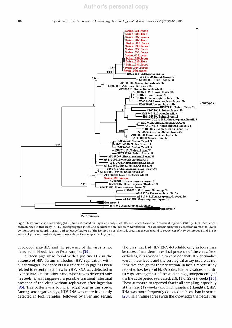
The phylogenetic analyses of fifteen sequences from theORF1 5%-terminal region shows that all isolates groupedwith HEV genotype 3 reference sequences. Among these,fourteen were much more closely related with subtype3c, whereas the other sequence (#035) grouped withsequences of subtype 3f (Fig. 1). A calculation of the diver-gence among the fifteen ORF1 sequences indicated thatthey had a nucleotide identity ranging from 78% to 100%.
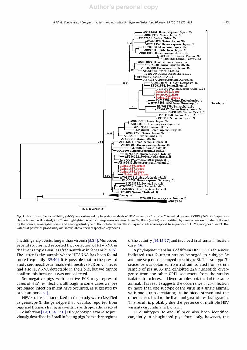
The topology of phylogenetic tree based on the ORF2 5%-terminal sequences also showed that only HEV genotype 3circulated among infected pigs from Pará state (Fig. 2). Inthis analysis, three sequences grouped with subtype 3c andthe other four with subtype 3f. These seven samples had anucleotide identity ranging from 93% to 100%.
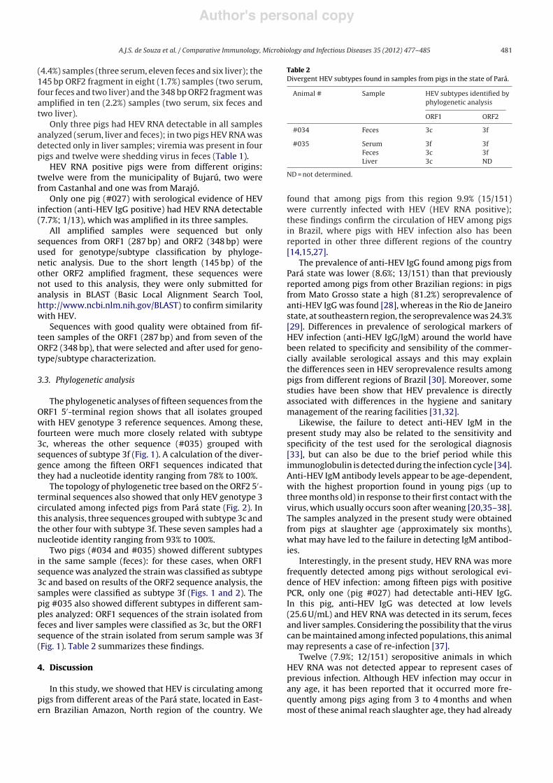
Two pigs (#034 and #035) showed different subtypesin the same sample (feces): for these cases, when ORF1sequence was analyzed the strain was classified as subtype3c and based on results of the ORF2 sequence analysis, thesamples were classified as subtype 3f (Figs. 1 and 2). Thepig #035 also showed different subtypes in different sam-ples analyzed: ORF1 sequences of the strain isolated fromfeces and liver samples were classified as 3c, but the ORF1sequence of the strain isolated from serum sample was 3f(Fig. 1). Table 2 summarizes these findings.
4. Discussion
In this study, we showed that HEV is circulating amongpigs from different areas of the Pará state, located in East-ern Brazilian Amazon, North region of the country. We
Table 2Divergent HEV subtypes found in samples from pigs in the state of Pará.
Animal # Sample HEV subtypes identified byphylogenetic analysis
ORF1 ORF2
#034 Feces 3c 3f
#035 Serum 3f 3fFeces 3c 3fLiver 3c ND
ND = not determined.
found that among pigs from this region 9.9% (15/151)were currently infected with HEV (HEV RNA positive);these findings confirm the circulation of HEV among pigsin Brazil, where pigs with HEV infection also has beenreported in other three different regions of the country[14,15,27].
The prevalence of anti-HEV IgG found among pigs fromPará state was lower (8.6%; 13/151) than that previouslyreported among pigs from other Brazilian regions: in pigsfrom Mato Grosso state a high (81.2%) seroprevalence ofanti-HEV IgG was found [28], whereas in the Rio de Janeirostate, at southeastern region, the seroprevalence was 24.3%[29]. Differences in prevalence of serological markers ofHEV infection (anti-HEV IgG/IgM) around the world havebeen related to specificity and sensibility of the commer-cially available serological assays and this may explainthe differences seen in HEV seroprevalence results amongpigs from different regions of Brazil [30]. Moreover, somestudies have been show that HEV prevalence is directlyassociated with differences in the hygiene and sanitarymanagement of the rearing facilities [31,32].
Likewise, the failure to detect anti-HEV IgM in thepresent study may also be related to the sensitivity andspecificity of the test used for the serological diagnosis[33], but can also be due to the brief period while thisimmunoglobulin is detected during the infection cycle [34].Anti-HEV IgM antibody levels appear to be age-dependent,with the highest proportion found in young pigs (up tothree months old) in response to their first contact with thevirus, which usually occurs soon after weaning [20,35–38].The samples analyzed in the present study were obtainedfrom pigs at slaughter age (approximately six months),what may have led to the failure in detecting IgM antibod-ies.
Interestingly, in the present study, HEV RNA was morefrequently detected among pigs without serological evi-dence of HEV infection: among fifteen pigs with positivePCR, only one (pig #027) had detectable anti-HEV IgG.In this pig, anti-HEV IgG was detected at low levels(25.6 U/mL) and HEV RNA was detected in its serum, fecesand liver samples. Considering the possibility that the viruscan be maintained among infected populations, this animalmay represents a case of re-infection [37].
Twelve (7.9%; 12/151) seropositive animals in whichHEV RNA was not detected appear to represent cases ofprevious infection. Although HEV infection may occur inany age, it has been reported that it occurred more fre-quently among pigs aging from 3 to 4 months and whenmost of these animal reach slaughter age, they had already
Author's personal copy
482 A.J.S. de Souza et al. / Comparative Immunology, Microbiology and Infectious Diseases 35 (2012) 477– 485
Fig. 1. Maximum clade credibility (MCC) tree estimated by Bayesian analysis of HEV sequences from the 5% terminal region of ORF1 (266 nt). Sequencescharacterized in this study (n = 15) are highlighted in red and sequences obtained from GenBank (n = 75) are identified by their accession number followedby the source, geographic origin and genotype/subtype of the isolated virus. The collapsed clades correspond to sequences of HEV genotypes 1 and 3. Thevalues of posterior probability are shown above their respective key nodes.
developed anti-HEV and the presence of the virus is notdetected in blood, liver or fecal samples [39].
Fourteen pigs were found with a positive PCR in theabsence of HEV serum antibodies. HEV replication with-out serological evidence of HEV infection in pigs has beenrelated to recent infection when HEV RNA was detected inliver or bile. On the other hand, when it was detected onlyin stools, it was suggested a possible transient intestinalpresence of the virus without replication after ingestion[35]. This pattern was found in eight pigs in this study.Among seronegative pigs, HEV RNA was more frequentlydetected in fecal samples, followed by liver and serum.
The pigs that had HEV RNA detectable only in feces maybe cases of transient intestinal presence of the virus. Nev-ertheless, it is reasonable to consider that HEV antibodieswere in low levels and the serological assay used was notsensitive enough for their detection. In fact, a recent studyreported low levels of ELISA optical density values for anti-HEV IgG among most of the studied pigs, independently ofthe life cycle period evaluated: 2, 8, 18 or 22–29 weeks [20].These authors also reported that in all sampling, especiallyat the third (18 weeks) and final sampling (slaughter), HEVRNA was more frequently detected in feces than in serum[20]. This finding agrees with the knowledge that fecal virus
Author's personal copy
A.J.S. de Souza et al. / Comparative Immunology, Microbiology and Infectious Diseases 35 (2012) 477– 485 483
Fig. 2. Maximum clade credibility (MCC) tree estimated by Bayesian analysis of HEV sequences from the 5% terminal region of ORF2 (346 nt). Sequencescharacterized in this study (n = 7) are highlighted in red and sequences obtained from GenBank (n = 94) are identified by their accession number followedby the source, geographic origin and genotype/subtype of the isolated virus. The collapsed clades correspond to sequences of HEV genotypes 1 and 3. Thevalues of posterior probability are shown above their respective key nodes.
shedding may persist longer than viremia [5,34]. Moreover,several studies had reported that detection of HEV RNA inthe liver samples was less frequent than in feces or bile [5].The latter is the sample where HEV RNA has been foundmore frequently [35,40]. It is possible that in the presentstudy seronegative animals with positive PCR only in feceshad also HEV RNA detectable in their bile, but we cannotconfirm this because it was not collected.
Seronegative pigs with positive PCR may representcases of HEV re-infection, although in some cases a moreprolonged infection might have occurred, as suggested byother authors [31].
HEV strains characterized in this study were classifiedas genotype 3, the genotype that was also reported frompigs and humans living in countries with sporadic cases ofHEV infection [1,4,18,41–50]. HEV genotype 3 was also pre-viously described in Brazil infecting pigs from other regions
of the country [14,15,27] and involved in a human infectioncase [16].
A phylogenetic analysis of fifteen HEV ORF1 sequencesindicated that fourteen strains belonged to subtype 3cand one sequence belonged to subtype 3f. This subtype 3fsequence was obtained from a strain isolated from serumsample of pig #035 and exhibited 22% nucleotide diver-gence from the other ORF1 sequences from the strainsisolated from feces and liver samples obtained of the sameanimal. This result suggests the occurrence of co-infectionby more than one subtype of the virus in a single animal,with one strain circulating in the blood stream and theother constrained to the liver and gastrointestinal system.This result is probably due the presence of multiple HEVvariants circulating in the farm.
HEV subtypes 3c and 3f have also been identifiedconjointly in slaughtered pigs from Italy, however, the
Author's personal copy
484 A.J.S. de Souza et al. / Comparative Immunology, Microbiology and Infectious Diseases 35 (2012) 477– 485
sequences obtained from multiple samples taken from thesame animal were identical, i.e., they did not group intodifferent subtypes [35], as was found in pig #035 case.Although they did not detect cases of co-infection withmultiple viral subtypes, the aforementioned authors didnot rule out this possibility. Coexistence of more thanone swine HEV subtype from the same genotype circulat-ing in the environment appears to be relatively common[31,39,40]. Co-infection can also occur in humans; a mixedinfection with HEV genotypes 3 and 4 was observed in apatient with acute hepatitis E in Japan [51].
The phylogeny of the seven ORF2 sequences indicatedthat three samples belonged to subtype 3c and four to thesubtype 3f. Lu and collaborators [2] suggested that the clas-sification of a HEV strain tends to remain constant evenwhen different regions of the genome are analyzed. There-fore, the detection of ORF1 and ORF2 sequences belongingto different subtypes in pigs #034 and #035 supports thehypothesis that these pigs were co-infected with multiplesubtypes, but we cannot rule out the existence of a recom-binant virus.
The recombination event may occur as a result ofco-infection or superinfection of the same host withtwo or more strains [52]. The occurrence of inter- andintra-genotype recombination among human and pig HEVstrains have been reported [52–54]. It is possible thatrecombination can lead to the emergence of more viru-lent variants of the virus, especially if it happens withinthe capsid coding gene (ORF2) [53].
The presence of HEV infection in pigs at slaughterage confirms previous evidences that these animals maybe a source of infection not only for pig farm workers,but also to slaughterhouse workers [43,55,56]. Moreover,HEV infected pigs at slaughter age also represent a riskof human infection by consumption of raw or under-cooked meat from these animals [41,57–59]. For thatreason, the detection of HEV in pigs from Pará reinforcesthe need for further studies to evaluate the pathogenic-ity of these isolates and the risk factors for transmissionto humans, especially those that work on pig farms,because the existence of a risk to public health cannot beexcluded.
In conclusion, the present study demonstrated thepresence of HEV infection in animals at slaughter age,suggesting that pigs in the Eastern Brazilian Amazon areexposed to HEV during different stages of their life cycle.The epidemiological HEV dynamics in pig farms at thisregion deserves additional studies. Herein, we reported thefirst identification and genotyping of HEV in Eastern Brazil-ian Amazon. The HEV subtypes circulating in pigs in thePará state were found to be related to human and ani-mal samples from other non-endemic regions of the world,suggesting that these isolates had zoonotic potential. Thepresent investigation also detected the simultaneous pres-ence of HEV subtypes 3c and 3f in some pigs, which suggeststhat mixed infection by multiple and/or recombinant viralstrains occurs in these animals. Further studies shouldbe conducted to clarify the epidemiological significanceof HEV circulating in the state of Pará and its impact onpublic health in the Amazon region and in other Brazilianregions.
Conflict of interest statement
None of the authors has any financial or personal rela-tionships that could inappropriately influence or bias thecontent of the paper.
Acknowledgments
This work was supported by Instituto Evandro Cha-gas/Secretaria de Vigilância em Saúde/Ministério da Saúde,Coordenac ão de aperfeic oamento de pessoal de nível supe-rior – CAPES/Universidade Federal do Pará – UFPA andFundac ão de Amparo à Pesquisa do Estado de São Paulo– FAPESP Nos. 2010/50081-9 and 2009/53946-3.
References
[1] Okamoto H. Genetic variability and evolution of hepatitis E virus.Virus Research 2007;127:216–28.
[2] Lu L, Li C, Hagedorn CH. Phylogenetic analysis of global hepatitis Evirus sequences: genetic diversity, subtypes and zoonosis. Reviewsin Medical Virology 2006;16:5–36.
[3] Purcell RH, Emerson SU. Hepatitis E: an emerging awareness of anold disease. Journal of Hepatology 2008;48:494–503.
[4] Meng XJ, Purcell RH, Halbur PG, Lehman JR, Webb DM, Tsareva TS,et al. A novel virus in swine is closely related to the human hepatitis Evirus. Proceedings of the National Academy of Sciences of the UnitedStates of America 1997;94:9860–5.
[5] Halbur PG, Kasorndorkbua C, Gilbert C, Guenette D, Potters MB, Pur-cell RH, et al. Comparative pathogenesis of infection of pigs withhepatitis E viruses recovered from a pig and a human. Journal ofClinical Microbiology 2001;39:918–23.
[6] Meng XJ. Hepatitis E virus: animal reservoirs and zoonotic risk. Vet-erinary Microbiology 2010;140:256–65.
[7] Takahashi K, Kitajima N, Abe N, Mishiro S. Complete or near-completenucleotide sequences of hepatitis E virus genome recovered froma wild boar, a deer, and four patients who ate the deer. Virology2004;330:501–5.
[8] Kaba M, Davoust B, Marie JL, Colson P. Detection of hepatitis E virusin wild boar (Sus scrofa) livers. Veterinary Journal 2010;186:259–61.
[9] Nakamura M, Takahashi K, Taira K, Taira M, Ohno A, Sakugawa H,et al. Hepatitis E virus infection in wild mongooses of Okinawa Japan:demonstration of anti-HEV antibodies and a full-genome nucleotidesequence. Hepatology Research 2006;34:137–40.
[10] Johne R, Heckel G, Plenge-Bonig A, Kindler E, Maresch C, Reetz J, et al.Novel hepatitis E virus genotype in Norway rats, Germany. EmergingInfectious Diseases 2010;16:1452–5.
[11] Zhao C, Ma Z, Harrison TJ, Feng R, Zhang C, Qiao Z, et al. A novel geno-type of hepatitis E virus prevalent among farmed rabbits in China.Journal of Medical Virology 2009;81:1371–9.
[12] Huang FF, Sun ZF, Emerson SU, Purcell RH, Shivaprasad HL, Pier-son FW, et al. Determination and analysis of the complete genomicsequence of avian hepatitis E virus (avian HEV) and attempts toinfect rhesus monkeys with avian HEV. Journal of General Virology2004;85:1609–18.
[13] Peralta B, Biarnes M, Ordonez G, Porta R, Martin M, Mateu E, et al. Evi-dence of widespread infection of avian hepatitis E virus (avian HEV)in chickens from Spain. Veterinary Microbiology 2009;137:31–6.
[14] Paiva HH, Tzaneva V, Haddad R, Yokosawa J. Molecular characteri-zation of swine hepatitis E virus from Southeastern Brazil. BrazilianJournal of Microbiology 2007;38:693–8.
[15] dos Santos DR, Vitral CL, de Paula VS, Marchevsky RS, Lopes JF, GasparAM, et al. Serological and molecular evidence of hepatitis E virus inswine in Brazil. Veterinary Journal 2009;182:474–80.
[16] Lopes Dos Santos DR, Lewis-Ximenez LL, da Silva MF, de SousaPS, Gaspar AM, Pinto MA. First report of a human autochthonoushepatitis E virus infection in Brazil. Journal of Clinical Virology2010;47:276–9.
[17] Chomczynski P, Sacchi N. Single-step method of RNA isolation by acidguanidinium thiocyanate–phenol–chloroform extraction. AnalyticalBiochemistry 1987;162:156–9.
[18] Wang Y, Ling R, Erker JC, Zhang H, Li H, Desai S, et al. A divergentgenotype of hepatitis E virus in Chinese patients with acute hepatitis.Journal of General Virology 1999;80(Pt 1):169–77.
Author's personal copy
A.J.S. de Souza et al. / Comparative Immunology, Microbiology and Infectious Diseases 35 (2012) 477– 485 485
[19] Huang FF, Haqshenas G, Guenette DK, Halbur PG, Schommer SK, Pier-son FW, et al. Detection by reverse transcription-PCR and geneticcharacterization of field isolates of swine hepatitis E virus from pigsin different geographic regions of the United States. Journal of ClinicalMicrobiology 2002;40:1326–32.
[20] Leblanc D, Ward P, Gagne MJ, Poitras E, Muller P, Trottier YL, et al.Presence of hepatitis E virus in a naturally infected swine herd fromnursery to slaughter. International Journal of Food Microbiology2007;117:160–6.
[21] Kwok S, Higuchi R. Avoiding false positives with PCR. Nature1989;339:237–8.
[22] Ewing B, Green P. Base-calling of automated sequencer traces usingphred. II. Error probabilities. Genome Research 1998;8:186–94.
[23] Ewing B, Hillier L, Wendl MC, Green P. Base-calling of automatedsequencer traces using phred. I. Accuracy assessment. GenomeResearch 1998;8:175–85.
[24] Hall TA. BioEdit: a user-friendly biological sequence alignment editorand analysis program for Windows 95/98/NT. Nucleic Acids Sympo-sium Series 1999;41:95–8.
[25] Drummond AJ, Rambaut A. BEAST: Bayesian evolutionary analysis bysampling trees. BMC Evolutionary Biology 2007;7:214.
[26] Tamura K, Dudley J, Nei M, Kumar S. MEGA4: Molecular EvolutionaryGenetics Analysis (MEGA) software version 4.0. Molecular Biologyand Evolution 2007;24:1596–9.
[27] dos Santos DR, de Paula VS, de Oliveira JM, Marchevsky RS, Pinto MA.Hepatitis E virus in swine and effluent samples from slaughterhousesin Brazil. Veterinary Microbiology 2011;149:236–41.
[28] Guimarães FR, Saddi TM, Vitral CL, Pinto MA, Gaspar AMC, SoutoFJD. Hepatitis e virus antibodies in swine herds of Mato Grosso stateCentral Brazil. Brazilian Journal of Microbiology 2005;36:223–6.
[29] Vitral CL, Pinto MA, Lewis-Ximenez LL, Khudyakov YE, dos SantosDR, Gaspar AM. Serological evidence of hepatitis E virus infection indifferent animal species from the Southeast of Brazil. Memorias doInstituto Oswaldo Cruz 2005;100:117–22.
[30] Khudyakov Y, Kamili S. Serological diagnostics of hepatitis E virusinfection. Virus Research 2011;161:84–92.
[31] Casas M, Cortes R, Pina S, Peralta B, Allepuz A, Cortey M, et al. Longitu-dinal study of hepatitis E virus infection in Spanish farrow-to-finishswine herds. Veterinary Microbiology 2011;148:27–34.
[32] Satou K, Nishiura H. Transmission dynamics of hepatitis E amongswine: potential impact upon human infection. BMC VeterinaryResearch 2007;3:9.
[33] Herremans M, Bakker J, Duizer E, Vennema H, Koopmans MP. Useof serological assays for diagnosis of hepatitis E virus genotype 1and 3 infections in a setting of low endemicity. Clinical and VaccineImmunology 2007;14:562–8.
[34] Meng XJ, Halbur PG, Haynes JS, Tsareva TS, Bruna JD, Royer RL,et al. Experimental infection of pigs with the newly identified swinehepatitis E virus (swine HEV), but not with human strains of HEV.Archives of Virology 1998;143:1405–15.
[35] Di Bartolo I, Ponterio E, Castellini L, Ostanello F, Ruggeri FM. Viraland antibody HEV prevalence in swine at slaughterhouse in Italy.Veterinary Microbiology 2011;149:330–8.
[36] Fernandez-Barredo S, Galiana C, Garcia A, Gomez-Munoz MT, VegaS, Rodriguez-Iglesias MA, et al. Prevalence and genetic character-ization of hepatitis E virus in paired samples of feces and serumfrom naturally infected pigs. Canadian Journal of Veterinary Research2007;71:236–40.
[37] Li Z, Yu S, Dong S, Zhu Y, Si F, Shen S, et al. Reduced prevalenceof genotype 3 HEV in Shanghai pig farms and hypothetical homeo-stasis of porcine HEV reservoir. Veterinary Microbiology 2009;137:184–9.
[38] Seminati C, Mateu E, Peralta B, de Deus N, Martin M. Distribution ofhepatitis E virus infection and its prevalence in pigs on commercialfarms in Spain. Veterinary Journal 2008;175:130–2.
[39] Nakai I, Kato K, Miyazaki A, Yoshii M, Li TC, Takeda N, et al. Differentfecal shedding patterns of two common strains of hepatitis E virus atthree Japanese swine farms. American Journal of Tropical Medicineand Hygiene 2006;75:1171–7.
[40] de Deus N, Seminati C, Pina S, Mateu E, Martin M, Segales J. Detectionof hepatitis E virus in liver, mesenteric lymph node, serum, bile andfaeces of naturally infected pigs affected by different pathologicalconditions. Veterinary Microbiology 2007;119:105–14.
[41] Feagins AR, Opriessnig T, Guenette DK, Halbur PG, Meng XJ. Detectionand characterization of infectious hepatitis E virus from commercialpig livers sold in local grocery stores in the USA. Journal of GeneralVirology 2007;88:912–7.
[42] Pei Y, Yoo D. Genetic characterization and sequence heterogeneityof a canadian isolate of Swine hepatitis E virus. Journal of ClinicalMicrobiology 2002;40:4021–9.
[43] Zheng Y, Ge S, Zhang J, Guo Q, Ng MH, Wang F, et al. Swine as aprincipal reservoir of hepatitis E virus that infects humans in easternChina. Journal of Infectious Diseases 2006;193:1643–9.
[44] Dalton HR, Thurairajah PH, Fellows HJ, Hussaini HS, Mitchell J, Ben-dall R, et al. Autochthonous hepatitis E in southwest England. Journalof Viral Hepatitis 2007;14:304–9.
[45] Di Bartolo I, Martelli F, Inglese N, Pourshaban M, Caprioli A, OstanelloF, et al. Widespread diffusion of genotype 3 hepatitis E virusamong farming swine in Northern Italy. Veterinary Microbiology2008;132:47–55.
[46] Martelli F, Toma S, Di Bartolo I, Caprioli A, Ruggeri FM, Lelli D, et al.Detection of hepatitis E virus (HEV) in Italian pigs displaying differentpathological lesions. Research in Veterinary Science 2010;88:492–6.
[47] Breum SO, Hjulsager CK, de Deus N, Segales J, Larsen LE. HepatitisE virus is highly prevalent in the Danish pig population. VeterinaryMicrobiology 2010;146:144–9.
[48] Legrand-Abravanel F, Mansuy JM, Dubois M, Kamar N, Peron JM, Ros-taing L, et al. Hepatitis E virus genotype 3 diversity, France. EmergingInfectious Diseases 2009;15:110–4.
[49] Munne MS, Vladimirsky S, Otegui L, Castro R, Brajterman L, Soto S,et al. Identification of the first strain of swine hepatitis E virus inSouth America and prevalence of anti-HEV antibodies in swine inArgentina. Journal of Medical Virology 2006;78:1579–83.
[50] Schlauder GG, Frider B, Sookoian S, Castano GC, Mushahwar IK. Iden-tification of 2 novel isolates of hepatitis E virus in Argentina. Journalof Infectious Diseases 2000;182:294–7.
[51] Takahashi M, Nishizawa T, Yoshikawa A, Sato S, Isoda N, Ido K, et al.Identification of two distinct genotypes of hepatitis E virus in aJapanese patient with acute hepatitis who had not travelled abroad.Journal of General Virology 2002;83:1931–40.
[52] van Cuyck H, Fan J, Robertson DL, Roques P. Evidence of recombi-nation between divergent hepatitis E viruses. Journal of Virology2005;79:9306–14.
[53] Wang H, Zhang W, Ni B, Shen H, Song Y, Wang X, et al. Recombinationanalysis reveals a double recombination event in hepatitis E virus.Virology Journal 2010;7:129.
[54] Fan J. Open reading frame structure analysis as a novel genotypingtool for hepatitis E virus and the subsequent discovery of an inter-genotype recombinant. Journal of General Virology 2009;90:1353–8.
[55] Galiana C, Fernandez-Barredo S, Garcia A, Gomez MT, Perez-Gracia MT. Occupational exposure to hepatitis E virus (HEV) inswine workers. American Journal of Tropical Medicine and Hygiene2008;78:1012–5.
[56] Perez-Gracia MT, Mateos ML, Galiana C, Fernandez-Barredo S, GarciaA, Gomez MT, et al. Autochthonous hepatitis E infection in a slaugh-terhouse worker. American Journal of Tropical Medicine and Hygiene2007;77:893–6.
[57] Colson P, Borentain P, Queyriaux B, Kaba M, Moal V, Gallian P, et al. Pigliver sausage as a source of hepatitis E virus transmission to humans.Journal of Infectious Diseases 2010;202:825–34.
[58] Yazaki Y, Mizuo H, Takahashi M, Nishizawa T, Sasaki N, Gotanda Y,et al. Sporadic acute or fulminant hepatitis E in Hokkaido Japan, maybe food-borne, as suggested by the presence of hepatitis E virus inpig liver as food. Journal of General Virology 2003;84:2351–7.
[59] Mizuo H, Yazaki Y, Sugawara K, Tsuda F, Takahashi M, Nishizawa T,et al. Possible risk factors for the transmission of hepatitis E virus andfor the severe form of hepatitis E acquired locally in Hokkaido Japan.Journal of Medical Virology 2005;76:341–9.