Autism Spectrum Disorders in Tanzania - Open Research Online

HERVs Expression in Autism Spectrum DisordersEmanuela Balestrieri1, Carla Arpino2, Claudia Matteucci1, Roberta Sorrentino1, Francesca Pica1,
Riccardo Alessandrelli2, Antonella Coniglio2, Paolo Curatolo2, Giovanni Rezza3, Fabio Macciardi4,5,6,
Enrico Garaci1, Simona Gaudi3, Paola Sinibaldi-Vallebona1*
1 Department of Experimental Medicine and Biochemical Sciences, University of Rome ‘‘Tor Vergata’’, Rome, Italy, 2 Pediatric Neurology Unit, Neuroscience Department,
University of Rome ‘‘Tor Vergata’’, Rome, Italy, 3 Department of Infectious, Parasitic and Immune-Mediated Diseases, Italian National Institute of Health, Rome, Italy,
4 Department of Psychiatry and Human Behavior, University of California Irvine, Irvine, California, United States of America, 5 Department of Psychiatry and the Behavioral
Sciences, Keck School of Medicine at University of Southern California, Los Angeles, California, United States of America, 6 Department of Medicine, Surgery and Dentistry,
University of Milan, Milan, Italy
Abstract
Background: Autistic Spectrum Disorder (ASD) is a heterogeneous neurodevelopmental disorder, resulting from complexinteractions among genetic, genomic and environmental factors. Here we have studied the expression of HumanEndogenous Retroviruses (HERVs), non-coding DNA elements with potential regulatory functions, and have tested theirpossible implication in autism.
Methods: The presence of retroviral mRNAs from four HERV families (E, H, K and W), widely implicated in complex diseases,was evaluated in peripheral blood mononuclear cells (PBMCs) from ASD patients and healthy controls (HCs) by qualitativeRT-PCR. We also analyzed the expression of the env sequence from HERV-H, HERV-W and HERV-K families in PBMCs at thetime of sampling and after stimulation in culture, in both ASD and HC groups, by quantitative Real-time PCR. Differencesbetween groups were evaluated using statistical methods.
Results: The percentage of HERV-H and HERV-W positive samples was higher among ASD patients compared to HCs, whileHERV-K was similarly represented and HERV-E virtually absent in both groups. The quantitative evaluation shows that HERV-H and HERV-W are differentially expressed in the two groups, with HERV-H being more abundantly expressed and,conversely, HERV-W, having lower abundance, in PBMCs from ASDs compared to healthy controls. PMBCs from ASDs alsoshowed an increased potential to up-regulate HERV-H expression upon stimulation in culture, unlike HCs. Furthermore wereport a negative correlation between expression levels of HERV-H and age among ASD patients and a statisticallysignificant higher expression in ASD patients with Severe score in Communication and Motor Psychoeducational Profile-3.
Conclusions: Specific HERV families have a distinctive expression profile in ASD patients compared to HCs. We propose thatHERV-H expression be explored in larger samples of individuals with autism spectrum in order to determine its utility as anovel biological trait of this complex disorder.
Citation: Balestrieri E, Arpino C, Matteucci C, Sorrentino R, Pica F, et al. (2012) HERVs Expression in Autism Spectrum Disorders. PLoS ONE 7(11): e48831.doi:10.1371/journal.pone.0048831
Editor: Grainne M. McAlonan, King’s College London, United Kingdom
Received April 4, 2012; Accepted October 5, 2012; Published November 14, 2012
Copyright: � 2012 Balestrieri et al. This is an open-access article distributed under the terms of the Creative Commons Attribution License, which permitsunrestricted use, distribution, and reproduction in any medium, provided the original author and source are credited.
Funding: This work was supported by a grant from Italia-Usa Project (grant 11US/299). The funders had no role in study design, data collection and analysis,decision to publish, or preparation of the manuscript.
Competing Interests: The authors have declared that no competing interests exist.
* E-mail: [email protected]
Introduction
Autism spectrum disorder (ASDs) is a complex neurodevelop-
mental disorder characterized by varying levels of impairment in
social interaction and communication as well as stereotypes and
rigid patterns of behaviour [1]. The prevalence rates of ASD have
been increasing worldwide, the most recent prevalence studies
indicating that they are present in 6 per 1000 children. The ASD
etiology is still unknown, but data suggest a likely multi-factorial
origin with a strong genetic basis [2]. Twin studies have shown an
inheritance of 92% [3]. While the high heritability of ASD is well
established, the exact underlying causes and relevant mutations
are identified in only a minority of patients. Rare genetic disorders
and chromosomal abnormalities are in fact thought to occur in
only 10% of ASD cases, approximately.
Possible solutions to the many questions regarding the
heritability of complex diseases were eagerly expected after the
completion of the human genome sequence. From these studies,
novel perspectives emerged, suggesting that protein-encoding
genes are not the only actors in the story, that the entire genome
is subjected to plasticity and is intimately tied to disease states, and
that genes and environmental conditions do indeed interact, thus
laying new grounds for ASD etiology [4,5]. In support to this view,
de novo and inherited Copy Number Variants (CNVs), and
inherited point mutations, have been increasingly found to
associate with ASD [6,7]. Moreover, some newly identified
polymorphisms point to non-coding regions and raise the
possibility that regulatory in addition to coding variants may
contribute to the genetics of autism [8].
PLOS ONE | www.plosone.org 1 November 2012 | Volume 7 | Issue 11 | e48831

Mobile retroelements, which make up almost 50% of the
human genome [9], are known to generate extensive structural
variations [10,11] and are regarded as key players in genome
function. Among them, Human Endogenous Retroviruses
(HERVs) constitute about 8–9% of the human genome (9). They
closely resemble infectious retroviruses [12] and are considered to
be remnants from ancient germ line viral infections, integrated as
provirus in chromosomal DNA. Despite the structural similarities
shared with exogenous retroviruses, the vast majority of HERV
sequences are in evolutionary equilibrium with the host genomes
and their mRNAs are variably expressed in a variety of cell types
and tissues [13–15]. During evolution, HERVs were amplified and
spread throughout the genome by repeated events of retrotrans-
position and/or reinfection; their integration in any location of the
genome can alter in consequence the structure and/or function of
other genes [16,17]. Indeed, HERV activity is implicated in many
complex diseases that have multifactorial etiology and genetic
basis, including type 1 diabetes [18], various types of tumors
[19,20], autoimmune diseases (for a review [21]) and neuropatho-
genic diseases such as multiple sclerosis [22].
Variations in specific HERV families have also been detected in
schizophrenia. Elevated levels of HERV-W have been detected in
peripheral blood mononuclear cells (PBMCs) of patients with
recent-onset and chronic schizophrenia [23]. A statistically
significant reduction in the expression of gag protein encoded by
HERV-W in neurons and astroglial cells has been found in brains
from individuals with schizophrenia [24]. Only a subgroup of the
HERV-K family (HERV-K10) was significantly overrepresented
in brains sample of patients with schizophrenia [25].
To the best of our knowledge, HERV elements have not been
assessed in ASD patients as yet. In this study we have investigated
the possible association between the presence and expression levels
of the four major human HERV families and the onset and disease
severity of autism.
Materials and Methods
Patients and healthy controls (Participants)The study included a group of 28 Caucasian children with
either idiopathic primary autism or Pervasive Developmental
Disorder-Not Otherwise Specified (PDD-NOS). Patients (ASD)
were 32 to 113 month-old (median age 58.5 months), of which 22
were males and 6 females. Patients were recruited among those
attending the pedopsychiatry outpatient Unit of ‘‘Tor Vergata’’
University Hospital (Rome). All patients had met the DSM-IV-TR
diagnosis criteria for autistic disorder and were diagnosed
according to Autism Diagnostic Interview-Revised (ADI-R),
Autism Diagnostic Observation Schedule (ADOS) and CARS
(Child Autism Rating Scale) and their developmental level was
assessed by using the Psycho-educational Profile-Third edition
(PEP-3). The skills and behaviors were reported as Adequate, or, if
altered, as Mild, Moderate and Severe according to degree of
impairment in each area of analysis (Table 1).
Patients with known infectious, metabolic or genetic diseases,
chromosomal abnormalities, seizures, identifiable neurological
syndromes or focal signs were excluded from the study. All
children were tested for chromosomal abnormalities and none had
fragile X syndrome. All patients were free of drugs at the time of
blood collection.
The patient group was compared with a control group of
healthy Caucasian children with normal development who
attended the outpatient facilities of the ‘‘Tor Vergata’’ University
Hospital (Rome) for routine visits. The control group included 28
children (HC), who were matched to the patients by age and
gender (Table 2), 32 to 108 month-old (median age 60.0 months),
22 of whom were males and 6 females. None of them had a history
of neurological, psychiatric or infectious disorders.
The University Hospital of ‘‘Tor Vergata’’ Ethics Committee
approved of the study and all examinations were performed after
receiving written informed consent of the parents.
Preparation of samplesPBMCs from heparinized blood samples from both ASD and
HC groups were analyzed immediately after collection (T0) or
after stimulation in culture for 72 hours (T72) with the T-
lymphocyte-specific mitogen phytohemagglutinin (PHA), 2 mg/
ml (Sigma, St Louis, MO) and human recombinant interleukin-2
(IL-2), 20 U/ml (Chiron corporation, Emeryville, CA). PBMCs
were cultured in RPMI 1640 medium (Life Technologies, Paisley,
Scotland, UK) supplemented with 12% fetal calf serum (FCS, Life
Technologies), 2 mM glutamine (Hyclone, Cramlington, UK),
50 U/ml penicillin, 50 U/ml streptomycin (Hyclone) at 37uCunder 5% CO2.
RT-PCRThe presence or absence of retroviral mRNAs of four HERVs
families (HERV-E, HERV-H, HERV-K and HERV-W), selected
on the basis of those more frequently associated with human
diseases, was assessed at T0 in PBMCs of ASDs and HCs by
qualitative RT-PCR.
RNA isolation was performed using a NucleoSpin RNA kit
according to the manufacturer’s instructions (Machenery-Nagel,
Dueren, Germany). Two hundred and fifty nanograms (250 ng) of
DNase-treated RNA from ASD and HC PBMCs were reverse-
transcribed into cDNA using the High Capacity cDNA Reverse
Transcription Kit (Applied Biosystems, Life Technologies, Carls-
bad CA) according to the manufacturer’s protocol. Two hundred
ng cDNA were amplified using specific primers for glyceralde-
hyde-3-phosphate dehydrogenase (GAPDH, forward, 59-
TGGTATCGTGAAAGGACT-39; reverse, 59-ATGCAAGT-
GAGCTTCCCGTTC-39), as an internal control, or using
degenerate primer pairs for HERV-E, HERV-H, HERV-K,
HERV-W, to simultaneously evaluate the presence of different
virus types belonging to an HERV family [26]. No RNA template
control reactions were included in all experiments. The PCR
products were visualized on 1.5% agarose gels containing 10 mg/
ml ethidium bromide (EtBr) in 16 Tris-acetate-EDTA buffer.
Samples in which PCR products could be visualized on EtBr-
stained agarose gels were defined as positive for HERV family
expression, while samples in which no specific band could be
detected for any of the tested HERV families, yet positive for the
GAPDH housekeeping gene, were defined as negative. All PCR
products were sequenced to verify any false positives.
No template controls were included in all experiment.
Real time PCRThe expression of the env of sequence from HERV-H and
HERV-W families in PBMCs from both ASD and HC groups was
quantitatively assessed in PBMC at T0 and T72, both in ASD and
HC by Real-time quantitative PCR., The assays were performed
in a Bio-Rad instrument (CFX96 Real-Time System), using SYBR
Green chemistry (SYBR Real Green PCR Master Mix, Eppen-
dorf). We selected specific pairs primers for env of HERV-H (Gene
Bank accession number AJ289711; env forward, primer 59 –
TTCACTCCATCCTTGGCTAT – 39; reverse, primer 59 –
CGTCGAGTATCTACGAGCAAT – 39), for env of HERV-W
(Gene Bank accession number NM_014590.3; forward, primer 59
– CGTTCCATGTCCCCATTTTAG – 39, reverse, primer 59–
HERVs in Autism Spectrum
PLOS ONE | www.plosone.org 2 November 2012 | Volume 7 | Issue 11 | e48831
TCATATCTAAGCCCCGCAAC – 39) and for env of HERV-K
(Gene Bank accession number AF1646; forward primer 59 –CATGGCAATTCCCAGTAACTGT – 39, reverse primer 59 –
CTCCCTCTTGGGCTCCTTCT – 39). Each sample was
analyzed in triplicate and a negative control, (no template
reaction), was added included in each experiment, to check out
any possible contamination. The house-keeping gene GUSB
(Gene Bank accession number NM_000181; forward primer 59-
CAGTTCCCTCCAGCTTCAATG-39; reverse, primer AC-
CCAGCCGACAAAATGC), was used to normalize the results.
Each experiment was completed with a melting curve analysis to
confirm the specificity of amplification and the lack of non-specific
products and primer dimers. Quantification was performed using the
threshold cycle (Ct) comparative method. The relative expression
was calculated as follows: 22[DCt(sample) 2 DCt(calibrator)] = 22DDCt,
where DCt (sample) = [Ct (HERV-H/W/K env) – Ct (GUSB)], and
DCt (calibrator) was the mean of DCT of all of the controls at T0.
Real time PCR results were represented by box plots, depicting mild
Table 1. Clinical features and assessments obtained using the ADI-R, ADOS, CARS and PEP-3 diagnostic scales.
ADI ADOS CARS PEP-3
Patientscode
Age(months) Gender ASS* (a) (b) (c) (d) (a) (b) (c) (d) C1 M2 MB3
1 51 F 6 15 5 6 4 5 14 4 2 50.5 moderate moderate moderate
2 37 M 4 18 9 10 4 4 9 1 1 45.5 moderate moderate severe
3 49 M 6 21 11 11 3 7 10 4 5 47.5 severe severe severe
4 49 M 4 1 5 7 1 6 7 1 1 41.5 moderate mild moderate
5 56 M 3 8 8 10 4 2 8 2 1 47.5 moderate mild moderate
6 64 M 6 15 13 10 3 5 7 1 0 41.5 mild moderate adequate
7 51 F 7 23 5 11 5 6 14 3 4 49 moderate moderate severe
8 74 M 8 27 12 12 4 8 12 1 2 53 moderate moderate moderate
9 74 M 5 31 22 5 2 3 11 0 0 33.5 moderate moderate moderate
10 32 M 2 19 10 9 3 1 7 3 5 39.5 severe severe severe
11 39 M 7 21 14 12 3 8 14 4 5 51.5 severe severe severe
12 69 F 6 22 14 9 3 4 13 2 2 45 moderate mild moderate
13 69 F 6 20 12 10 3 5 14 2 2 43 moderate mild moderate
14 81 M 7 22 12 5 3 8 12 3 2 45 moderate moderate moderate
15 45 M 6 21 12 11 3 4 13 4 3 45 severe severe severe
16 55 M 6 27 18 12 4 6 7 1 1 28.5 moderate mild mild
17 42 M 7 16 13 6 3 8 14 4 5 31.5 adequate adequate adequate
18 81 M 6 31 15 13 5 6 10 2 6 51 moderate moderate moderate
19 61 M 9 28 13 9 3 8 16 4 5 50 severe severe severe
20 42 M 6 14 9 10 3 6 14 4 2 41.5 moderate mild moderate
21 60 M 3 18 6 12 4 4 4 0 3 45.5 moderate mild moderate
22 112 M 10 27 24 11 1 8 13 2 6 ne adequate adequate adequate
23 113 M 2 ne‘ ne ne ne 0 4 0 1 33 adequate adequate adequate
24 55 F 6 20 12 3 4 6 13 4 5 ne severe mild moderate
25 80 M 4 ne ne ne ne 4 8 4 3 ne severe moderate moderate
26 60 M 6 9 8 8 2 6 11 1 1 25 adequate adequate adequate
27 70 M 3 14 12 6 4 4 4 1 3 ne moderate mild moderate
28 57 F 6 22 11 8 3 7 12 4 3 51 severe severe severe
ne: not evaluated.*ASS: Autism Severity Score.1: Communication;2: Motor;3: Maladaptative behaviors.doi:10.1371/journal.pone.0048831.t001
Table 2. Demographic information for the ASD and controlcohorts.
ASD patients(n = 28)
Healthycontrols(n = 28) p value
Gender Male 22 22
Female 6 6
Ratio (M/F) 3.67 3.67 1
Median age(range)
58.5 (32–113) 60 (32–108) 0.873
doi:10.1371/journal.pone.0048831.t002
HERVs in Autism Spectrum
PLOS ONE | www.plosone.org 3 November 2012 | Volume 7 | Issue 11 | e48831

(black dot) and extreme outliers (asterisk) for each group were
showed.
Statistical analysisFischer exact test was used to compare qualitative expression of
HERV families. The Mann Whitney test was used to compare
quantitative expression of HERVs families between ASD and HC
groups at T0 or T72, and Wilcoxon test was used to compare
stimulation response at T72 in each group. To determine any
correlation between age and HERVs expression, the Spearman’s
rho correlation coefficient was calculated. The ANOVA analysis
of variance and post-hoc Bonferroni tests were used to determine
whether changes in the expression of HERV-H and HERV-W
were associated with clinical parameters. Statistical analyses were
done using the SPSS software (version 17.0). P values are indicated
in the text, and in figures only for statistically significant
comparisons (p values,0.050).
Results
Expression of HERV-H, W, K and E families in PBMCs fromASD patients and healthy controls
We first analysed the expression of four HERV families (H, W,
K and E), selected on the basis of their frequent association with
complex human diseases, in fresh (T0) and in culture stimulated
(T72) PBMCs from both ASD and HC groups by qualitative RT-
PCR. All amplification products were sequenced. Only two false
positives were detected, which were excluded from this study.
Table 3 reports the proportion, within the ASD and the HC
groups, of positive individuals for specific HERV families, either
selectively detected at T0, or selectively detected at T72, or
detected at any one time (T0 or T72).
The percentage of HERV-H-expressing individuals was higher
among ASD cases, compared to controls, at T0 (42.86% vs
21.43%), at T72 (44.44% vs 25%) and at least in one of the two
times analysed (71.43% vs 35.71%). The differences between the
ASD and HC groups, evaluated by Fisher exact test, are significant
only when analyzing the percentage of positive individuals at T0 or
T72 (p = 0.015), but not at T0 (p = 0.152) and at T72 (p = 0.162).
HERV-W was also more commonly detected among ASDs than
in HCs, at both T0 (67.85% vs 57.14%) and T72 (81.48% vs
60.71%) and the percentage of positive samples at at least one of
the analysed times was higher in ASDs (89.29%) compared to HCs
(67.86%), albeit with no significant difference.
HERV-K was almost equally represented in both groups and in
each of the assay conditions (at T0 ASD 42.86%, HC 46.43%; at
T72 ASD 55.56%, HC 46.43%; at T0 or T72 ASD 64.29%, HC
71.43%) and no difference are detected by statistical analysis (ASD
vs HC at T0 p = 1.000; at T72 p = 0.593; at T0 or T72 p = 0.775).
HERV-E was poorly expressed in ASD patients (7.14%) and
absent in HCs, at both times of the analysis.
Analysis of HERV-H, HERV-W and HERV-K expression inPBMCs of ASD patients and healthy controls
We next assessed the expression levels of env sequence from
HERV-H, W and K families (but not HERV-E, due to its low
representation in qualitative RT-PCR assays) in PBMCs from
both ASD and HC groups. Real time assays were performed
immediately after collection (T0) and after in vitro stimulation (T72),
with the intent to investigate HERVs expression in resting versus
proliferating conditions.
At T0, HERV-H expression (Figure 1, panel A), shown as log
(22DDCt), was significantly more elevated in fresh PBMCs from
ASDs (median value 86.35; Interquartile range, IQR = 0.20/
498.39) compared to the expression level in the HC group (median
value 0.65; IQR = 0.35/4.48) (p = 0.044). After in vitro stimulation
(T72), HERV-H expression (Figure 1, panel A) was still more
elevated in ASD (median value 2.55; IQR = 0.25/258.11) than in
HC group (median value 0.89; IQR = 0.57/2.34), though the
statistical differences were not significant. In contrast, HERV-W
was significantly more expressed in PBMCs from HCs compared
to ASD patients (Figure 1, panel B), both at T0 and T72. In
particular, the median value at T0 was 0.56 (IQR = 0.25/0.57) for
ASDs, while being 0.80 (IQR = 0.38/5.15) (p = 0.017) for HCs. At
T72 the median value was 0.56 (IQR = 0.09/0.9) for ASDs and
0.82 (IQR = 0.28/5.00) (p = 0.027) for HCs. Finally, HERV-K
expression levels were similar in ASDs compared to HCs, at both
analysed times (Figure 1, panel C). In fresh PBMCs from ASDs
median value was 1.06 (IQR = 0.021/5.37) compared to 0.54 in
HCs (IQR = 0.06/7.87) (p = 0.077), and after in vitro stimulation it
rose to 3.88 (IQR = 0.01/7.90) in ASDs versus 2.35 (IQR = 0.09/
12.53) in HCs (p = 0.694). Significant differences in HERV-K
expression levels between T0 and T72 were achieved only in HC
group (p = 0.036), but not between patients and controls.
The individual quantitative evaluation of HERV-H expression
showed that 50% of ASD patients (14/28) exhibited very high
levels (22DDCT.10) at T0; in addition, six of the samples showing
low levels at T0 (22DDCT,10) up-regulated HERV-H levels after
Table 3. Percentage of positive samples for expression of HERV families in cultured PBMCs from ASD and HC groups.
HERV-H HERV-W HERV-K HERV-E
ASD HC ASD HC ASD HC ASD HC
T0a 42.86 (12) 21.43 (6) 67.86 (19) 57.14 (16) 42.86 (12) 46.43 (13) 7.14 (2) 0 (0)
p = 0.152 p = 0.582 p = 1.000 p = 0.491
T72b 44.44 (12) 25 (7) 81.48 (22) 60.71 (17) 55.56 (15) 46.43 (13) 0 (0) 0 (0)
p = 0.162 p = 0.138 p = 0.593 ncd
T0 or T72c 71.43 (20) 35.71 (10) 89.29 (25) 67.86 (19) 64.29 (18) 71.43 (20) 7.14 (2) 0 (0)
p = 0.015 p = 0.101 p = 0.775 p = 0.491
aRT-PCR analysis of PBMCs at time 0 after withdrawal.bRT-PCR analysis of PBMCs after 72 hours of stimulation in culture.cRT-PCR analysis of PBMCs at T0 and T72.dNo statistics are computed.In parentheses the number of positive samples over the total (28).doi:10.1371/journal.pone.0048831.t003
HERVs in Autism Spectrum
PLOS ONE | www.plosone.org 4 November 2012 | Volume 7 | Issue 11 | e48831

stimulation in culture. As a result, the overall frequency of HERV-
H highly expressing ASD patients was higher than 70% (20/28)
(Figure 2A). HERV-H expression was instead generally lower in
the HC group: high levels (22DDCT.10) were detected in only a
few individuals at T0 (5/28) and, among low-expressing healthy
controls at T0 (23/28), only 3 showed increased expression after in
vitro stimulation (Figure 2B).
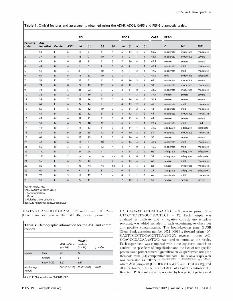
Finally, the individual expression analysis of HERV-W elements
showed that the in vitro stimulation did not significantly modify
their expression levels, in either the ASD or in the HC group
(Figure 3A and 3B).
In summary, therefore, the results pinpoint two distinctive
features of HERV elements in ASD: i.e. a significant overexpres-
sion of HERV-H, paralleled by a significant down-regulation of
HERV-W in PBMCs from ASD patients compared to controls.
Moreover the analysis of individual patients and controls
highlighted an intrinsic potential of PBMCs from ASD patients
to express HERV-H after stimulation in culture, unlike healthy
controls.
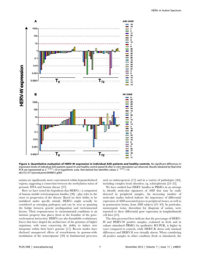
Correlation analysis of HERV-H and HERV-W expressionwith age
We next performed a Spearman correlation analysis between
the expression of HERV-H and HERV-W, both of which showed
a distinctive modulation in the ASD group, with age. Figure 4
shows the HERV-H and HERV-W env gene expression levels,
evaluated by Real time PCR analysis, in PBMCs, plotted against
age, expressed in months. Statistical analysis demonstrated a
significant negative correlation between the expression of HERV-
H at T0 and age in ASD patients (rho = 20.477; p = 0.010) but not
in HCs (rho = 20.117; p = 0.553) (Figure 4, panel A). No
significant correlation emerged between the expression of
HERV-W and age either in the ASD or in the HC group (ASDs:
rho = 0.145, p = 0.460; HCs: rho = 0.013, p = 0.948) (Figure 4,
panel B). Thus, the correlation analysis suggests that higher levels
of HERV-H are associated to lower age of the ASD patients.
High levels of HERV-H expression in PBMCs of autisticpatients with severe score in Communication and MotorPsychoeducational Profile-3
We next evaluated the association of HERV-H and HERV-W
env expression (mean values) in fresh PBMCs with the PEP-3
classification (Psycho-educational Profile-Third edition).
Figure 5 shows the mean values 6 SD of HERV-H expression
in PBMCs at T0 from the 28 ASD patients, grouped in four
developmental levels (Adequate, Mild, Moderate and Severe) and
resulted by analysis of the three areas of analysis Communication
(panel A), Motor (panel B) and Maladaptive Behaviours (panel C).
The relative HERV-H expression mean values were found to be
significantly higher in ASD patients with Severe impairment level
in Communication compared with those with Moderate or
Adequate level (mean values 6 SD: Adequate 55.496106.20,
Mild 106.20, Moderate 143.266254.81, Severe 539.626292.78;
Severe vs Adequate p = 0.028, Severe vs Moderate p = 0.009)
(Figure 5, panel A). When the PEP-3 Motor analysis was
considered, the relative HERV-H expression mean values were
again found to be significantly higher in ASD patients with Severe
compared with those with Moderate or Adequate level (mean
values 6 SD: Adequate 55.496106.20, Mild 231.766335.04,
Moderate 133.786259.37, Severe 570.016187.54; Severe vs
Adequate p = 0.034, Severe vs Moderate p = 0.025) (Figure 5,
panel B). Finally, for the PEP-3 analysis of Maladaptive
Behaviours, HERV-H expression mean values were higher in
ASD patients with Severe impairment level though with no
significant differences (mean values 6 SD: Adequate
93.206124.77, Mild 0.07, Moderate 204.876328.41, Severe
449.706277.47) (Figure 5, panel C). No statistically significant
differences were found in HERV-W env expression when ASD
patients were stratified for the four developmental levels defined by
the PEP-3 analysis (data not shown).
Thus, high levels of HERV-H expression are associated to ASD
patients with Severe impairment of developmental level, as defined
by the Communication and Motor analysis.
Figure 1. Relative HERV-H, W and K expression in ASD patients and healthy controls. Data obtained in fresh PBMCs (T0) and after in vitrostimulation (T72) of ASD patients and healthy controls are represented as box plot. ASD patients show higher levels of HERV-H (panel A) both at T0
and after in vitro stimulation in comparison to healthy controls. Conversely, HERV-W (panel B) expression is higher in healthy controls than in ASDpatients, both at T0 and after in vitro stimulation. Significant differences between groups are shown. HERV-K (panel C) expression levels were similar inASDs compared to HCs in both conditions, and significant differences were achieved only inside control group. Relative env gene expression levelswere analyzed by Real-time PCR and represented by 22DDCt in logarithmic scale.doi:10.1371/journal.pone.0048831.g001
HERVs in Autism Spectrum
PLOS ONE | www.plosone.org 5 November 2012 | Volume 7 | Issue 11 | e48831

Discussion
Despite extensive research efforts, the etiopathogenesis of ASD
thus far remains elusive. To date ASD remains a behaviorally
defined spectrum with no known biological markers suitable to
support diagnosis or subgroup categorization [27].
The genetic architecture of ASD is highly heterogeneous [28],
and only about 10–20% of individuals with ASD have an
identified genetic etiology [29]. The transmission pattern is
complex in most families and is not compatible with simple
Mendelian inheritance [30,31], suggesting that protein-coding
genes are responsible for only part of the ASD etiology. Growing
evidence supports the involvement of epigenetic regulatory
mechanisms in the pathogenesis of ASD [32,33], with a
contribution of DNA methylation, genomic imprinting, chromatin
modifications and non coding RNA [34].
As for other complex diseases, the full etiology most probably
relies on a complex interplay between genes, the genome
organization and the environment. A well-understood example
of such interplay comes from studies of the Rett syndrome. It is
worth recalling that LINE-1 elements, a retrotransposon family
accounting for 17% of the human genome, play fundamental roles
in neurogenesis by altering the expression of neuronal genes,
which, in turn, influence neuronal cell fate [35]. Rett syndrome
patients have recently been found to display an increased
susceptibility to LINE-1 retrotransposition, dependent on the
abnormal methylation status of the overall genome due to
mutation of the DNA methyl-binding protein, MeCP2 [36],
which is regarded as the causative alteration of the Rett disease.
Importantly, growing evidence links germline hypomethylation
and genomic instability. Structural mutations in individuals with
schizophrenia, bipolar disorder, developmental retardation and
Figure 2. Quantitative evaluation of HERV-H expression in individual ASD patients and healthy controls. Comparative analysis ofexpression levels in individual ASD patients (panel A) shows that in vitro stimulation of PBMCs induces HERV-H expression increase in the patientswith low levels at T0. Conversely, in vitro stimulation does not modify HERV-H levels in individual HC (panel B). Results obtained by Real time PCR arerepresented as 22DDCT in logarithmic scale. Red dashed line identifies values 22DDCT.10.doi:10.1371/journal.pone.0048831.g002
HERVs in Autism Spectrum
PLOS ONE | www.plosone.org 6 November 2012 | Volume 7 | Issue 11 | e48831
autism are significantly more concentrated within hypomethylated
regions, suggesting a connection between the methylation status of
genomic DNA and human disease [37].
Here we have tested the hypothesis that HERVs - a component
of human mobile retrotransposon families [38] - play roles in the
onset or progression of the disease. Based on their ability to be
mobilized under specific stimuli, HERVs might actually be
considered as emerging pathogens and can be seen as spanning
the bridge between genetic predisposition and environmental
factors. Their responsiveness to environmental conditions is an
intrinsic property that places them at the frontline of the gene-
environment interaction. HERVs are also formidable evolutionary
forces that have shaped the architecture of the genomes of higher
organisms, with some conserving the ability to induce new
integrants within their host’s genome [17]. Recent studies have
disclosed unsuspected effects of retroelements in genome-wide
modulation of the transcriptome [39] in fundamental processes
such as embryogenesis [17] and in a variety of pathologies [40],
including complex brain disorders, eg, schizophrenia [23–25].
We have studied four HERV families in PBMCs in an attempt
to identify molecular signatures of ASD that may be easily
detected in peripheral samples. An increasing number of
molecular studies indeed indicate the importance of differential
expression of ASD-associated genes in peripheral tissues, as well as
in postmortem brains, from ASD subjects [41–43]. In particular,
monozygotic twins, discordant for diagnosis of autism, were
reported to show differential gene expression in lymphoblastoid
cell lines [41].
The data presented here indicate that the percentage of HERV-
H and HERV-W positive samples, evaluated in fresh and in
culture stimulated PBMCs by qualitative RT-PCR, is higher in
cases compared to controls, while HERV-K shows only minimal
differences and HERV-E was virtually absent. When considering
all positive samples in either condition (fresh or stimulated), the
Figure 3. Quantitative evaluation of HERV-W expression in individual ASD patients and healthy controls. No significant differences inexpression levels of individual ASD patients (panel A) and healthy control (panel B) after in vitro stimulation are observed. Results obtained by Real timePCR are represented as 22DDCT.10 in logarithmic scale. Red dashed line identifies values 22DDCT.10.doi:10.1371/journal.pone.0048831.g003
HERVs in Autism Spectrum
PLOS ONE | www.plosone.org 7 November 2012 | Volume 7 | Issue 11 | e48831
Figure 4. Association of HERV-H and W expression levels with the age of ASD patients and healthy controls. HERV-H (panel A) andHERV-W (panel B) levels at T0 are plotted as a function of corresponding age expressed in months. Spearman analysis shows a significant negativecorrelation between HERV-H expression levels and age only in ASD patients (rho = 20.477; p = 0.010). No significant correlation was found betweenHERV-W expression and age of both ASD and HC subjects. Relative env gene expression levels were analyzed by Real-time PCR and represented by22DDCt in logarithmic scale.doi:10.1371/journal.pone.0048831.g004
Figure 5. HERV-H expression and Psychoeducational Profile-3 in ASD patients. The skills and behaviours of individual cases were classifiedas Adequate, Mild, Moderate or Severe according to the degree of impairment for Communication PEP-3 (panel A), Motor PEP-3 (panel B) andMaladaptive Behavior PEP-3 (panel C) analysis. Mean values 6 SD of HERV-H env expression in PBMCs at T0 from 28 ASD patients were analyzed. Inparenthesis the number of patients classified for each analysed domain. Significant differences between groups are shown. Relative env geneexpression levels were analyzed by Real-time PCR and represented by 22DDCt in linear scale.doi:10.1371/journal.pone.0048831.g005
HERVs in Autism Spectrum
PLOS ONE | www.plosone.org 8 November 2012 | Volume 7 | Issue 11 | e48831
differences were significant for HERV-H, but not for the other
HERV families analyzed. Quantitative determination of HERVs
in PBMCs showed that HERV-H expression, indeed, was
statistically significantly higher in ASD, and, conversely, HERV-
W was higher in healthy controls.
Furthermore, HERV-H expression negatively correlated with
age only in ASD patients. Based on the evidence that HERV-H is
expressed in high levels selectively in ASDs, the correlation with
age might be viewed as a disease-dependent feature not present in
HCs.
Interestingly, high expression of HERV-H was also associated
with ‘‘severe’’ score in Communication and Motor Psychoeduca-
tional Profile-3.
To the best of our knowledge, this is the first evidence linking
retrotransposon activity and ASD. Notwithstanding the relatively
small size of the samples tested in this work, the statistical
significance of the present findings supports the hypothesis that
HERV-H overexpression might be regarded as a potential early
marker detectable in ASD patients. The analysis of individual
patients and controls also suggests an increased intrinsic predis-
position of the PBMCs from ASD patients to express HERV-H in
response to mitogenic stimulation in culture. HERV-H overex-
pression might be of help to differentiate young ASD children
from age-matched controls. Because detecting autism at the
earliest possible age is of outmost importance to optimize
outcomes for children with the disorder, identifying the presence
of HERV-H in PBMCs of young children could be useful for this
purpose. Furthermore, because autism remains a behaviorally
defined disorder, the identification of a biological marker could
also be of support for a confident diagnosis. The identification of a
reliable biomarker for ASD could supplement and validate existing
clinical methods; in particular, a biomarker that is expressed at, or
even before, the onset of symptoms might obviate the need to wait
for behavioral criteria to be met before beginning treatment.
Larger number of ASD patients and follow-up data will be
needed to further substantiate the present results. Yet, as the first
comparative analysis of ASD patients and controls focusing on
HERV families, we believe that the present findings are well worth
pursuing in future research. More generally, the quantitative
differences in HERV-H and HERV-W env expression between
ASD patients and controls suggest a contribution of a ‘‘non-
coding’’ fraction of the genome to ASD.
Conclusions
Our results demonstrate that the expression of two particular
HERV families is distinctive of ASD and may represent a possible
molecular marker for ASD patients. HERVs may be thought of as
components of the genome that interact with environmental
factors and/or infectious agents, potentially capable to interplay
with different molecular pathways in determining individual
genetic differences in ASD.
Acknowledgments
We wish to dedicate this study to the memory of our dear friend and
colleague Carla Arpino, who was a founding member in this study.
The authors gratefully acknowledge all the children and families who
participated in this study. They thank Dr. Gianpiero Conflitti for help with
experiments and expert technical assistance, Dr. Martino Tony Miele for
assistance with manuscript preparation, Dr. Mario Castorina for providing
some control samples and Dr. Orietta Casaglia for technical assistance
They thank also Dr. Patrizia Lavia and Dr. Corrado Spadafora for careful
critical revision of the manuscript.
Author Contributions
Conceived and designed the experiments: CA PSV EB SG. Performed the
experiments: EB CM RS. Analyzed the data: EB CM FM PSV SG.
Contributed reagents/materials/analysis tools: PC RA AC. Wrote the
paper: PSV EB CM FP SG GR PC. Critical evaluation of the study: EG.
References
1. American Psychiatric Association. Diagnostic and statistical manual of mental
disorder 4th edn, (2004) Washington DC: American Psychiatric Press.
2. Aldinger KA, Plummer JT, Qiu S, Levitt P (2011) SnapShot: genetics of Autism.
Neuron 72: 418–8.e1.
3. Benvenuto A, Moavero R, Alessandrelli R, Manzi B, Curatolo P (2009)
Syndromic autism: causes and pathogenetic pathways. World J Pediatr 5: 169–
176.
4. Lupski JR, Belmont JW, Boerwinkle E, Gibbs RA (2011) Clan genomics and the
complex architecture of human disease. Cell 147: 32–43.
5. Baillie JK, Barnett MW, Upton KR, Gerhardt DJ, Richmond TA, et al. (2011)
Somatic retrotransposition alters the genetic landscape of human brain. Nature
10.1038/nature10531.
6. Ben-David E, Granot-Hershkovitz E, Monderer-Rothkoff G, Lerer E, Levi S, et
al. (2011) Identification of a functional rare variant in autism using genome-wide
screen for monoallelic expression. Hum Mol Genet 20: 3632–3641.
7. Salyakina D, Cukier HN, Lee JM, Sacharow S, Nations LD, et al. (2011) Copy
number variants in extended autism spectrum disorder families reveal candidates
potentially involved in autism risk. PLoS One 6: e26049.
8. Gunther T, Schmitt AO, Bortfeldt RH, Hinney A, Hebebrand J, et al. (2011)
Where in the genome are significant nucleotide polymorphisms from genome-
wide association studies located? OMICS 15: 507–512.
9. International Human Genome Consortium (2001) Initial sequencing and
analysis of the human genome. Nature 409: 860–921.
10. Huang CR, Schneider AM, LuY, Niranjan T, Shen P, et al. (2010) Mobile
interspersed repeats are major structural variants in the human genome. Cell
141: 1171–1182.
11. Iskow RC, McCabe MT, Mills RE, Torene S, Pittard WS, et al. (2010) Natural
mutagenesis of human genomes by endogenous retrotransposons. Cell 141:
1253–1261.
12. Boeke JD, Stoye JP (1997) Retrotrasposons, endogenous retroviruses, and the
evolution of retroelements. In: Retroviruses, Coffin JM, Hughes SH and Varmus
HE editors. New York: Cold Spring Harbor Laboratory Press. pp. 343–346.
13. Forsman A, Yun Z, Hu L, Uzhameckis D, Jern P, et al. (2005) Development of
broadly targeted human endogenous gammaretroviral pol-based real time PCRs
Quantitation of RNA expression in human tissues. J Virol Methods 129: 16–30.
14. Seifarth W, Frank O, Zeilfelder U, Spiess B, Greenwood AD, et al. (2005)
Comprehensive analysis of human endogenous retrovirus transcriptional activity
in human tissues with a retrovirus-specific microarray. J Virol 79: 341–352.
15. Yi JM, Kim HM, Kim HS (2006) Human endogenous retrovirus HERV-H
family in human tissues and cancer cells: expression, identification, and
phylogeny. Cancer Lett 231: 228–239.
16. Bannert N, Kurth R (2006) The evolutionary dynamics of human endogenous
retroviral families. Annu Rev Genomics Hum Genet 7: 149–173.
17. Rowe HM, Trono D (2011) Dynamic control of endogenous retroviruses during
development. Virology 411: 273–287.
18. Marguerat S, Wang WY, Todd JA, Conrad B (2004) Association of human
endogenous retrovirus K-18 polymorphisms with type 1 diabetes. Diabetes 53:
852–854.
19. Ryan FP (2009) An alternative approach to medical genetics based on modern
evolutionary biology. Part 4: HERVs in cancer. J R Soc Med 102: 474–480.
20. Serafino A, Balestrieri E, Pierimarchi P, Matteucci C, Moroni G, et al. (2009)
The activation of human endogenous retrovirus K (HERV-K) is implicated in
melanoma cell malignant transformation. Exp Cell Res 315: 849–862.
21. Balada E, Ordi-Ros J, Vilardell-Tarres M (2009) Molecular mechanisms
mediated by human endogenous retroviruses (HERVs) in autoimmunity. Rev
Med Virol 19: 273–286.
22. Christensen T (2005) Association of human endogenous retroviruses with
multiple sclerosis and possible interactions with herpes viruses. Rev Med Virol
15: 179–211.
23. Yao Y, Schroder J, Nellaker C, Bottmer C, Bachmann S, et al. (2008) Elevated
levels of human endogenous retrovirus-W transcripts in blood cells from patients
with first episode schizophrenia. Genes Brain Behav 7: 103–112.
24. Weis S, Llenos IC, Sabunciyan S, Dulay JR, Isler L, et al. (2007) Reduced
expression of human endogenous retrovirus (HERV)-W GAG protein in the
cingulate gyrus and hippocampus in schizophrenia, bipolar disorder, and
depression. J Neural Transm 114: 645–655.
25. Frank O, Giehl M, Zheng C, Hehlmann R, Leib-Mosch C, et al. (2005) Human
endogenous retrovirus expression profiles in samples from brains of patients with
schizophrenia and bipolar disorders. J Virol 79: 10890–10901.
HERVs in Autism Spectrum
PLOS ONE | www.plosone.org 9 November 2012 | Volume 7 | Issue 11 | e48831

26. Johnston JB, Silva C, Holden J, Warren KG, Clark AW, et al. (2001) Monocyte
activation and differentiation augment human endogenous retrovirus expression:implications for inflammatory brain diseases. Ann Neurol 50: 434–442.
27. Anagnostou E, Taylor MJ (2011) Review of neuroimaging in autism spectrum
disorders: what have we learned and where we go from here. Mol Autism 2: 4.28. Abrahams BS, Geschwind DH (2008) Advances in autism genetics: on the
threshold of a new neurobiology. Nat Rev Genet 9: 341–355.29. Bill BR, Geschwind DH (2009) Genetic advances in autism: heterogeneity and
convergence on shared pathways. Curr Opin Genet Dev 19: 271–278.
30. Jorde LB, Hasstedt SJ, Ritvo ER, Mason-Brothers A, Freeman BJ, et al. (1991)Complex segregation analysis of autism. Am J Hum Genet 49: 932–938.
31. Autism Genome Project Consortium (2007) Mapping autism risk loci usinggenetic linkage and chromosomal rearrangements. Nat Genet 39: 319–328.
32. Schanen NC (2006) Epigenetics of autism spectrum disorders. Hum Mol Genet15 Spec No 2: R138–150.
33. Beaudet AL (2007) Autism: highly heritable but not inherited. Nat Med 13: 534–
536.34. LaSalle JM (2011) A genomic point-of-view on environmental factors influencing
the human brain methylome. Epigenetics 6: 862–869.35. Muotri AR, Chu VT, Marchetto MC, Deng W, Moran JV, et al. (2005) Somatic
mosaicism in neuronal precursor cells mediated by L1 retrotransposition. Nature
435: 903–910.36. Muotri AR, Marchetto MC, Coufal NG, Oefner R, Yeo G, et al. (2010) L1
retrotransposition in neurons is modulated by MeCP2. Nature 468: 443–446.
37. Li J, Harris RA., Cheung SW, Coarfa C, Jeong M, et al. (2012) Genomic
hypometilation in the human germline associates with selective structural
mutability in the human genome. Plos genetics 8(5).
38. Kurth R, Bannert N (2010) Beneficial and detrimental effects of human
endogenous retroviruses. Int J Cancer 126: 306–314.
39. Faulkner GJ, Kimura Y, Daub CO, Wani S, Plessy C, et al. (2009) The
regulated retrotransposon transcriptome of mammalian cells. Nat Genet 41:
563–571.
40. Perron H, Lang A (2010) The human endogenous retrovirus link between genes
and environment in multiple sclerosis and in multifactorial diseases associating
neuroinflammation. Clin Rev Allergy Immunol 39: 51–61.
41. Hu VW, Frank BC, Heine S, Lee NH, Quackenbush J (2006) Gene expression
profiling of lymphoblastoid cell lines from monozygotic twins discordant in
severity of autism reveals differential regulation of neurologically relevant genes.
BMC Genomics 7: 118.
42. Baron CA, Liu SY, Hicks C, Gregg JP (2006) Utilization of lymphoblastoid cell
lines as a system for the molecular modeling of autism. J Autism Dev Disord 36:
973–982.
43. Nishimura Y, Martin CL, Vazquez-Lopez A, Spence SJ, Alvarez-Retuerto AI, et
al. (2007) Genome-wide expression profiling of lymphoblastoid cell lines
distinguishes different forms of autism and reveals shared pathways. Hum Mol
Genet 16: 1682–1698.
HERVs in Autism Spectrum
PLOS ONE | www.plosone.org 10 November 2012 | Volume 7 | Issue 11 | e48831
Copyright © 2022 FDOKUMEN