Relationship between Bile Acid Transplacental Gradients and ...
Upload
independentCategory
view
3download
0
Vieira et al. BMC Microbiology 2014, 14:315http://www.biomedcentral.com/1471-2180/14/315
RESEARCH ARTICLE Open Access
Hepadnavirus detected in bile and liver samplesfrom domestic pigs of commercial abattoirsYasmine Rangel Vieira1*, Debora Regina Lopes dos Santos2, Moyra Machado Portilho3, Carlos Eduardo Pereira Velloso1,Marcia Arissawa4, Livia Melo Villar3, Marcelo Alves Pinto1 and Vanessa Salete de Paula1
Abstract
Background: Preliminary studies showed the prevalence of a virus similar to human hepatitis B virus (HBV-like) inswine from farms in China and the molecular evidence of Hepadnavirus infection in domestic pigs herds in Brazil.In this study, we genetically characterize the swine Hepadnavirus strains in swine from slaughterhouses located incertified abattoirs from Rio de Janeiro State, Brazil and evaluate its hepatotropic potential.
Results: Bile and liver samples from swine were positive for partial genome amplification (ORF S and ORF C), directsequencing and viral load quantification. Sequencing of the gene encoding the surface antigen allowed classificationof Hepadnavirus into genotypes, similar to HBV genotype classification. Indirect immunofluorescence confirmed thepresence of HBsAg antigen in liver tissue sections.
Conclusions: So far our data suggest that commercial swine house an HBV-like virus and this relevant finding shouldbe considered in studies on the origin and viral evolution.
Keywords: Hepadnavirus, Brazil, Commercial swine, Bile, Liver, Genotyping, Immunofluorescence
BackgroundHepatitis B virus (HBV) is a dual polarity and partiallydouble-stranded enveloped DNA virus of Hepadnaviridaefamily [1]. The agent can be transmitted by sexual, peri-natal and percutaneous means [2], and is considered amajor cause of acute and chronic liver disease, that mayprogress to cirrhosis and hepatocellular carcinoma [3].Beyond the prototype member that infects humans andnon-human primates (chimpanzees, gibbons, gorillas,orangutans and woolly monkeys), Hepadnaviridae familyalso houses HBV-related viruses circulating in mammalianhosts, like woodchuck (WHV) and squirrels (GSHV/ASHV), and avian hosts, like ducks (DHBV), geese(GHBV), herons (HHBV), and storks (STHBV) [4].A virus similar to HBV has been diagnosed by serology
(HBsAg, anti-HBs, anti-HBc) in swine herds [5] and chick-ens flocks [6] from China, and in domestic pig herds inBrazil [7]. Moreover, positive molecular diagnosis wasdemonstrated for the first time in swine from Brazil [7]
* Correspondence: [email protected]ório de Desenvolvimento Tecnológico em Virologia, Pavilhão Hélio ePeggy Pereira – 2° andar - sala B220, Instituto Oswaldo Cruz, FIOCRUZ, Av.Brasil, n° 4365, Manguinhos - RJ, Cep: 21045-900 Rio de Janeiro, RJ, BrasilFull list of author information is available at the end of the article
© 2014 Vieira et al.; licensee BioMed Central LCommons Attribution License (http://creativecreproduction in any medium, provided the orDedication waiver (http://creativecommons.orunless otherwise stated.
and in chickens flocks from China [6]. Similarity withhuman HBV (90.8-96.3%) was confirmed for swine strainsby phylogenetic analysis and by cross reactivity in non-host specific commercial serological assays. However, thepartial nucleotide sequencing (360 bp) was equivalent toabout 11.2% of full-length HBV genome. And even forchickens, despite the high percentage of similarity withHBV (92.2-97.9%), the short length of amplified productin both cases limits a conclusion.Therefore, we performed the current study to improve
the molecular characterization of Hepadnavirus circulat-ing in swine from abattoirs in Brazil, revealing if there isevidence that pigs destined for human consumption mightact as a potential new reservoir or host for a virus HBV-similar.
MethodsThis study was approved by the Institutional Committeefor Ethics in the Use of Research Animals (CEUA-Fiocruz:PO 0132/01). On December 2008, a total of 36 bile andliver samples were collected from domestic pigs Sus scrofa(aged > 5 months), breed Large white, from three slaugh-terhouses located in Petrópolis (SPET), Itaocara (SITC)and Itaperuna (SITP) (North and Hill region of Rio de
td. This is an Open Access article distributed under the terms of the Creativeommons.org/licenses/by/4.0), which permits unrestricted use, distribution, andiginal work is properly credited. The Creative Commons Public Domaing/publicdomain/zero/1.0/) applies to the data made available in this article,
Vieira et al. BMC Microbiology 2014, 14:315 Page 2 of 6http://www.biomedcentral.com/1471-2180/14/315
Janeiro State, Brazil). Commercial establishments are sub-mitted to controlled inspection by an official agency of Riode Janeiro State. All animals from the three abattoirs wereclassified healthy and approved for slaughter and furthercommercialization according to inspect evaluation criteria.Swine from each abattoir were acquired from distinct pigfarms suppliers. The samples were collected during theevisceration process under sanitary requisites determinedby regulations of Animal Sanitary Protection Agency inRio de Janeiro State (ASPA).Five milliliters of bile were collected by vaccum-punction
through the gallbladder wall with sterile syringe; and500 mg of liver samples were collected with Medblade®bisturi blades. All samples were stored in Nalgene® cryo-genic vials and immediately frozen in dry ice. At the labora-tory, bile samples were stored at -80°C and liver samples inliquid nitrogen until analysis. Viral DNA was extractedfrom bile and liver samples using DNA Purification Kit(QIAamp DNA Mini Kit, Qiagen®) according to Moriczet al, 2010 [8] and was concentrated to a final volumeof 25 uL. Extracted DNA was analyzed by semi-nestedPCR (PS1-S2 and PS1-SR) specific for open reading frame(ORF S) of HBV - as previously established for the firstround of amplification [9], and for the second round [10] -and by PCR specific for core gene of HBV – as previously
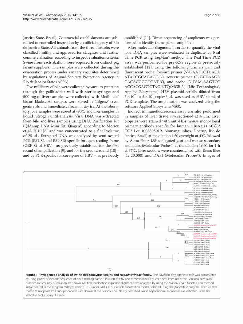
Figure 1 Phylogenetic analysis of swine Hepadnavirus strains and Heby using partial nucleotide sequence of open reading frame S (566 nt) of HBVnumber and country of isolation are shown. Multiple nucleotide sequence aliimplemented in the program MrBayes version 3.1.2 under GTR + G nucleotiderooted at midpoint. Posterior probabilities are shown at the branch label. Newindicates evolutionary distance.
established [11]. Direct sequencing of amplicons was per-formed to identify the sequence amplified.After molecular diagnosis, in order to quantify the viral
load DNA samples were evaluated in duplicate by RealTime PCR using TaqMan® method. The Real Time PCRassay was performed for pre-S2/S region as previouslyestablished [12], using the following primers pair andfluorescent probe: forward primer (5’-GAATCCTCACAATACCGCAGAGT-3’), reverse primer (5’-GCCAAGACACACGGGTGAT-3’), and probe (5’-FAM-AAGTCCACCACGAGTCTAG-NFQ/MGB-3’) (Life Technologies®,Applied Biosystems). HBV plasmid serially diluted from5 × 107 to 5 × 101 copies/ μL was used as HBV standardPCR template. The amplification was analyzed using thesoftware Applied Biosystems 7500.Indirect immunofluorescence assay was also performed
in samples of liver tissue cryosectioned at 4 μm. Liverbiopsies were stained with anti-HBs mouse monoclonalprimary antibody specific for human HBsAg (19-CC6/CG2 Lot 100830S019, Biomanguinhos, Fiocruz, Rio deJaneiro, Brazil) at the dilution 1:50 overnight at 4°C, followedby Alexa Fluor 488 conjugated goat anti-mouse secondaryantibodies (Molecular Probes®) at the dilution 1:400 for 1 hat 37°C. Liver sections were counterstained with Evans Blue(1: 20,000) and DAPI (Molecular Probes®). Images of
padnaviridae family. The Bayesian phylogenetic tree was constructedand related viruses. For each sequence used, the GenBank accession
gnment was analyzed by using the Markov Chain Monte Carlo methodsubstitution model, selected using the jModeltest program. The tree wasly described swine hepadnavirus sequences are indicated. Scale bar

Vieira et al. BMC Microbiology 2014, 14:315 Page 3 of 6http://www.biomedcentral.com/1471-2180/14/315
the green fluorescent HBsAg-positive liver cells wereobserved and photographed using a Confocal ScanningLaser Microscope equipped with camera (Nikon® Instru-ments, Model C2, Inc., New York, USA).
ResultsHepadnavirus-DNA was detected in bile and liver samplesfrom about 11.11% pigs (4/36). All samples from Petrópolis(0/9) were tested negative. Hepadnavirus-DNA was de-tected in both samples types from 1 animal in Itaocara(1/10) and from 3 animals in Itaperuna (3/17). Thesesamples showed a positive result for the semi-nestedPCR specific for HBV ORF S (1,100 bp) [GenBank:KC832935, GenBank:KC832936, GenBank:KC832937,GenBank:KC832938], and 3 of them also for the PCRspecific for core gene (431 bp) [GenBank:KF859967,GenBank:KF859968, GenBank:KF859969]. The sequencesfound in bile and liver of the same animal matched. Phylo-genetic reconstruction using partial nucleotide sequences
Table 1 Nucleotide identity between Hepadnavirus strains fro
Species AccessionNumberGenBank
Swine Hep
KC832935
Chimpanzee (HBV) FJ798099.1 0.950
AF222322.1 0.937
Gibbon (HBV) AF477494.2 0.939
AF274499.2 0.918
Orangutan (HBV) Y17562.1 0.916
Y17559.1 0.911
Gorilla (HBV) AJ131567.1 0.946
JQ664502.1 0.942
Woolly Monkey (WMHBV) AF046996.1 0.850
AY226578.1 0.850
Bat (Hepadnavirus) KC790378.1 0.689
KC790376.1 0.755
Woodchuck (WHV) FM212013.1 0.707
FM212009.1 0.716
Squirrel (ASHV/GSHV) AGU29144 0.709
K02715.1 0.709
Heron (HHBV) AY552597.1 0.474
AY552595.1 0.483
Stork (STHBV) AJ251937.1 0.464
AJ251935.1 0.464
Snow Goose (GHBV) AF110999.1 0.458
AF110997.1 0.461
Duck (DHBV) AY494851.1 0.467
M21953.1 0.473
Legend: The nucleotide identity matrix was constructed by using partial nucleotideeach sequence used, the GenBank accession number and specie infected are show
of ORF S (566 nt) showed a close relationship ofHepadnavirus strains from pigs and Hepadnavirus nucleo-tide sequences from non-human primates (from 84.8 to96.1%), bats (from 68.9 to 75.6%), rodents (from 70.5 to71.8%) and birds (from 45.8 to 48.6%) (Figure 1, Table 1).Genetic distances calculated with nucleotide sequences
of ORF S (972 nt) showed filogenetic distance betweenthe virus detected in Brazilian domestic pigs and hu-man samples from significant alignment listed by NCBIBlast [GenBank:JF784228, GenBank:JF784230, GenBank:JF784231, GenBank:JF784234, GenBank:EF662027-49,GenBank:DQ060826, GenBank:DQ060829] ranging from98.9-99.7%. These data also revealed that distinct strainsmight be responsible for the infection in domestic pigs(Figure 2). Comparing to human HBV strains previouslygenotyped, swine samples were assigned to two differentgenotypes groups. Three of them were close to samplesfrom genotype A, the most prevalent genotype in Brazil[13]. And, another was similar to samples that belong
m pigs and other species
adnavirus
KC832936 KC832937 KC832938
0.950 0.951 0.961
0.937 0.939 0.948
0.939 0.940 0.953
0.918 0.920 0.933
0.918 0.918 0.929
0.913 0.913 0.926
0.946 0.948 0.951
0.942 0.944 0.948
0.848 0.851 0.848
0.848 0.851 0.848
0.689 0.691 0.695
0.753 0.756 0.755
0.705 0.709 0.707
0.714 0.718 0.714
0.709 0.710 0.712
0.709 0.710 0.716
0.477 0.476 0.481
0.484 0.484 0.486
0.466 0.466 0.471
0.466 0.466 0.468
0.461 0.460 0.461
0.465 0.463 0.465
0.470 0.468 0.461
0.476 0.475 0.476
sequence of open reading frame S (566 nt) of HBV and related viruses. Forn. Newly described swine hepadnaviruses sequences are indicated.
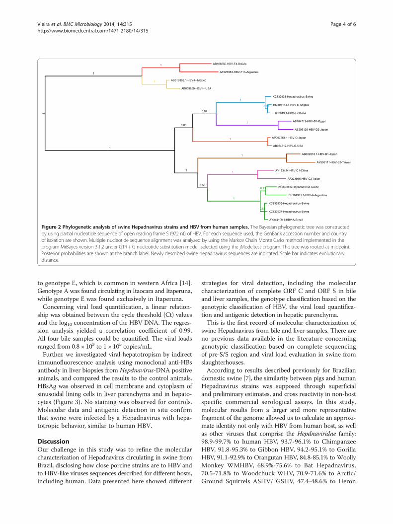
Figure 2 Phylogenetic analysis of swine Hepadnavirus strains and HBV from human samples. The Bayesian phylogenetic tree was constructedby using partial nucleotide sequence of open reading frame S (972 nt) of HBV. For each sequence used, the GenBank accession number and countryof isolation are shown. Multiple nucleotide sequence alignment was analyzed by using the Markov Chain Monte Carlo method implemented in theprogram MrBayes version 3.1.2 under GTR + G nucleotide substitution model, selected using the jModeltest program. The tree was rooted at midpoint.Posterior probabilities are shown at the branch label. Newly described swine hepadnavirus sequences are indicated. Scale bar indicates evolutionarydistance.
Vieira et al. BMC Microbiology 2014, 14:315 Page 4 of 6http://www.biomedcentral.com/1471-2180/14/315
to genotype E, which is common in western Africa [14].Genotype A was found circulating in Itaocara and Itaperuna,while genotype E was found exclusively in Itaperuna.Concerning viral load quantification, a linear relation-
ship was obtained between the cycle threshold (Ct) valuesand the log10 concentration of the HBV DNA. The regres-sion analysis yielded a correlation coefficient of 0.99.All four bile samples could be quantified. The viral loadsranged from 0.8 × 10³ to 1 × 10⁵ copies/mL.Further, we investigated viral hepatotropism by indirect
immunofluorescence analysis using monoclonal anti-HBsantibody in liver biopsies from Hepdnavirus-DNA positiveanimals, and compared the results to the control animals.HBsAg was observed in cell membrane and cytoplasm ofsinusoidal lining cells in liver parenchyma and in hepato-cytes (Figure 3). No staining was observed for controls.Molecular data and antigenic detection in situ confirmthat swine were infected by a Hepadnavirus with hepa-totropic behavior, similar to human HBV.
DiscussionOur challenge in this study was to refine the molecularcharacterization of Hepadnavirus circulating in swine fromBrazil, disclosing how close porcine strains are to HBV andto HBV-like viruses sequences described for different hosts,including human. Data presented here showed different
strategies for viral detection, including the molecularcharacterization of complete ORF C and ORF S in bileand liver samples, the genotype classification based on thegenotypic classification of HBV, the viral load quantifica-tion and antigenic detection in hepatic parenchyma.This is the first record of molecular characterization of
swine Hepadnavirus from bile and liver samples. There areno previous data available in the literature concerninggenotypic classification based on complete sequencingof pre-S/S region and viral load evaluation in swine fromslaughterhouses.According to results described previously for Brazilian
domestic swine [7], the similarity between pigs and humanHepadnavirus strains was supposed through superficialand preliminary estimates, and cross reactivity in non-hostspecific commercial serological assays. In this study,molecular results from a larger and more representativefragment of the genome allowed us to calculate an approxi-mate identity not only with HBV from human host, as wellas other viruses that comprise the Hepdnaviridae family:98.9-99.7% to human HBV, 93.7-96.1% to ChimpanzeeHBV, 91.8-95.3% to Gibbon HBV, 94.2-95.1% to GorillaHBV, 91.1-92.9% to Orangutan HBV, 84.8-85.1% to WoollyMonkey WMHBV, 68.9%-75.6% to Bat Hepadnavirus,70.5-71.8% to Woodchuck WHV, 70.9-71.6% to Arctic/Ground Squirrels ASHV/ GSHV, 47.4-48.6% to Heron
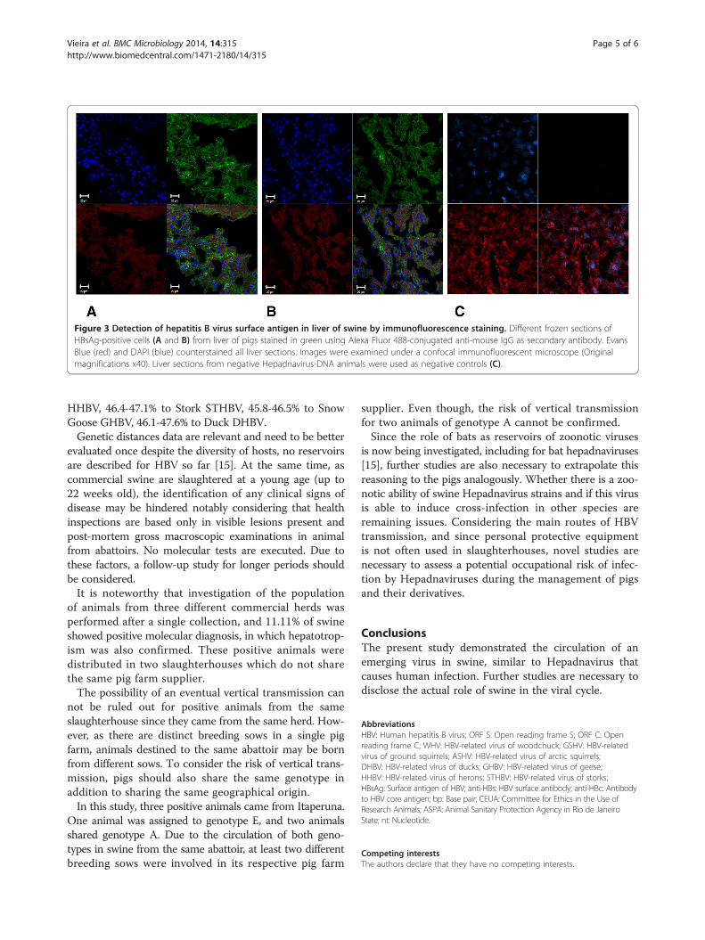
Figure 3 Detection of hepatitis B virus surface antigen in liver of swine by immunofluorescence staining. Different frozen sections ofHBsAg-positive cells (A and B) from liver of pigs stained in green using Alexa Fluor 488-conjugated anti-mouse IgG as secondary antibody. EvansBlue (red) and DAPI (blue) counterstained all liver sections. Images were examined under a confocal immunofluorescent microscope (Originalmagnifications x40). Liver sections from negative Hepadnavirus-DNA animals were used as negative controls (C).
Vieira et al. BMC Microbiology 2014, 14:315 Page 5 of 6http://www.biomedcentral.com/1471-2180/14/315
HHBV, 46.4-47.1% to Stork STHBV, 45.8-46.5% to SnowGoose GHBV, 46.1-47.6% to Duck DHBV.Genetic distances data are relevant and need to be better
evaluated once despite the diversity of hosts, no reservoirsare described for HBV so far [15]. At the same time, ascommercial swine are slaughtered at a young age (up to22 weeks old), the identification of any clinical signs ofdisease may be hindered notably considering that healthinspections are based only in visible lesions present andpost-mortem gross macroscopic examinations in animalfrom abattoirs. No molecular tests are executed. Due tothese factors, a follow-up study for longer periods shouldbe considered.It is noteworthy that investigation of the population
of animals from three different commercial herds wasperformed after a single collection, and 11.11% of swineshowed positive molecular diagnosis, in which hepatotrop-ism was also confirmed. These positive animals weredistributed in two slaughterhouses which do not sharethe same pig farm supplier.The possibility of an eventual vertical transmission can
not be ruled out for positive animals from the sameslaughterhouse since they came from the same herd. How-ever, as there are distinct breeding sows in a single pigfarm, animals destined to the same abattoir may be bornfrom different sows. To consider the risk of vertical trans-mission, pigs should also share the same genotype inaddition to sharing the same geographical origin.In this study, three positive animals came from Itaperuna.
One animal was assigned to genotype E, and two animalsshared genotype A. Due to the circulation of both geno-types in swine from the same abattoir, at least two differentbreeding sows were involved in its respective pig farm
supplier. Even though, the risk of vertical transmissionfor two animals of genotype A cannot be confirmed.Since the role of bats as reservoirs of zoonotic viruses
is now being investigated, including for bat hepadnaviruses[15], further studies are also necessary to extrapolate thisreasoning to the pigs analogously. Whether there is a zoo-notic ability of swine Hepadnavirus strains and if this virusis able to induce cross-infection in other species areremaining issues. Considering the main routes of HBVtransmission, and since personal protective equipmentis not often used in slaughterhouses, novel studies arenecessary to assess a potential occupational risk of infec-tion by Hepadnaviruses during the management of pigsand their derivatives.
ConclusionsThe present study demonstrated the circulation of anemerging virus in swine, similar to Hepadnavirus thatcauses human infection. Further studies are necessary todisclose the actual role of swine in the viral cycle.
AbbreviationsHBV: Human hepatitis B virus; ORF S: Open reading frame S; ORF C: Openreading frame C; WHV: HBV-related virus of woodchuck; GSHV: HBV-relatedvirus of ground squirrels; ASHV: HBV-related virus of arctic squirrels;DHBV: HBV-related virus of ducks; GHBV: HBV-related virus of geese;HHBV: HBV-related virus of herons; STHBV: HBV-related virus of storks;HBsAg: Surface antigen of HBV; anti-HBs: HBV surface antibody; anti-HBc: Antibodyto HBV core antigen; bp: Base pair; CEUA: Committee for Ethics in the Use ofResearch Animals; ASPA: Animal Sanitary Protection Agency in Rio de JaneiroState; nt: Nucleotide.
Competing interestsThe authors declare that they have no competing interests.
Vieira et al. BMC Microbiology 2014, 14:315 Page 6 of 6http://www.biomedcentral.com/1471-2180/14/315
Authors’ contributionsThis study was designed by YRV, DRLS, MAP and VSP. YRV performedqualitative molecular assays, sequence analysis, interpreted the data,collaborated to quantitative molecular assays, to immunofluorescence assayand wrote the manuscript. DRLS contributed to the sample collection andhelped to write the manuscript. MMP performed quantitative molecularassays. CEPV contributed to the immunofluorescence assay. MA contributedwith immunological reagents. LMV contributed with molecular reagents,materials and analysis tools. MAP contributed to the sample collection,coordinated the study and revised the manuscript. VSP coordinated thestudy and revised the manuscript. All authors read, corrected and approvedthe final manuscript.
AcknowledgmentsThis study was supported by Oswaldo Cruz Institute (FIOCRUZ), Coordenaçãode Aperfeiçoamento de Pessoal de Nível Superior (CAPES) and Fundação deAmparo à Pesquisa do Estado do Rio de Janeiro (FAPERJ/ E-26/110.607/2014).The authors appreciated the help from Gilberto Vaughan (Centers for DiseaseControl and Prevention/ CDC) and Leandro Layter Xavier (Universidade doEstado do Rio de Janeiro/ UERJ).
Author details1Laboratório de Desenvolvimento Tecnológico em Virologia, Pavilhão Hélio ePeggy Pereira – 2° andar - sala B220, Instituto Oswaldo Cruz, FIOCRUZ, Av.Brasil, n° 4365, Manguinhos - RJ, Cep: 21045-900 Rio de Janeiro, RJ, Brasil.2Departamento de Mircobiologia Veterinária e Imunologia, UniversidadeFederal Rural do Rio de Janeiro, UFRRJ, Seropédica, RJ, Brasil. 3Laboratório deHepatites Virais, Instituto Oswaldo Cruz, FIOCRUZ, Rio de Janeiro, RJ, Brasil.4Laboratório de Tecnologia de Anticorpos Monoclonais, Bio-Manguinhos,FIOCRUZ, Rio de Janeiro, RJ, Brasil.
Received: 21 June 2014 Accepted: 26 November 2014
References1. Wei Y, Neuveut C, Tiollais P, Buendia MA: Molecular biology of the hepatitis B
virus and role of the X gene. Pathol Biol (Paris) 2010, 58:267–272.2. Dienstag JL: Hepatitis B virus infection. N Engl J Med 2008, 359:1486–1500.3. Prange R: Host factors involved in hepatitis B virus maturation, assembly,
and egress. Med Microbiol Immunol 2012, 201:449–461.4. Yang J, Xi Q, Deng R, Wang J, Hou J, Wang X: Identification of interspecies
recombination among hepadnaviruses infecting cross-species hosts.J Med Virol 2007, 79:1741–1750.
5. Li W, She R, Liu L, You H, Yin J: Prevalence of a virus similar to humanhepatitis B virus in swine. Virol J 2010, 7:60.
6. Tian J, Xia K, She R, Li W, Ding Y, Wang J, Chen M, Yin J: Detection ofhepatitis B virus in serum and liver of chickens. Virol J 2012, 9:2.
7. Vieira YR, Vieira AA, Ciacci-Zanella JR, Barquero G, do Lago BV, Gomes SA,Silva MFM, Santos DRL, Pinto MA, de Paula VS: Serological and molecularevidence of Hepadnavirus infection in swine. Ann Agric Environ Med, in press.
8. Moricz A, Melo M, Castro AM, Campos T, Silva RA, Pacheco AM Jr:Prevalence of Helicobacter spp in chronic cholecystitis and correlationwith changes on the histological pattern of the gallbladder. Acta Cir Bras2010, 25:218–224.
9. Niel C, Moraes MT, Gaspar AM, Yoshida CF, Gomes SA: Genetic diversity ofhepatitis B virus strains isolated in Rio de Janeiro, Brazil. J Med Virol 1994,44:180–186.
10. Motta-Castro AR, Martins RM, Yoshida CF, Teles SA, Paniago AM, Lima KM,Gomes SA: Hepatitis B virus infection in isolated Afro-Brazilian communities.J Med Virol 2005, 77:188–193.
11. Lanford RE, Chavez D, Brasky KM, Burns RB 3rd, Rico-Hesse R: Isolation of ahepadnavirus from the woolly monkey, a New World primate. Proc NatlAcad Sci U S A 1998, 95:5757–5761.
12. Portilho MM: Desenvolvimento de testes de detecção e quantificação dovírus da hepatite B em amostras de soro e fluido oral. MSc thesis. InstitutoOswaldo Cruz, Departamento de Virologia. Rio de Janeiro, Brazil; 2013.
13. Becker CE, Mattos AA, Bogo MR, Branco F, Sitnik R, Kretzmann NA:Genotyping of hepatitis B virus in a cohort of patients evaluated in ahospital of Porto Alegre, South of Brazil. Arq Gastroenterol 2010, 47:13–17.
14. Kramvis A, Kew MC: Epidemiology of hepatitis B virus in Africa, itsgenotypes and clinical associations of genotypes. Hepatol Res 2007,37:S9–S19.
15. Drexler JF, Geipel A, Konig A, Corman VM, Van Riel D, Leijten LM, Bremer CM,Rasche A, Cottontail VM, Maganga GD, Schlegel M, Müller MA, Adam A,Klose SM, Carneiro AJ, Stöcker A, Franke CR, Gloza-Rausch F, Geyer J, Annan A,Adu-Sarkodie Y, Oppong S, Binger T, Vallo P, Tschapka M, Ulrich RG, Gerlich WH,Leroy E, Kuiken T, Glebe D, Drosten C: Bats carry pathogenic hepadnavirusesantigenically related to hepatitis B virus and capable of infectinghuman hepatocytes. Proc Natl Acad Sci U S A 2013, 110:16151–16156.
doi:10.1186/s12866-014-0315-2Cite this article as: Vieira et al.: Hepadnavirus detected in bile and liversamples from domestic pigs of commercial abattoirs. BMC Microbiology2014 14:315.
Submit your next manuscript to BioMed Centraland take full advantage of:
• Convenient online submission
• Thorough peer review
• No space constraints or color figure charges
• Immediate publication on acceptance
• Inclusion in PubMed, CAS, Scopus and Google Scholar
• Research which is freely available for redistribution
Submit your manuscript at www.biomedcentral.com/submit
Copyright © 2022 FDOKUMEN