guidelines-hiv-diagnosis-and-monitoring-antiretroviral-therapy ...
85
SEA-HLM-382 (Rev. 1) Distribution: General Guidelines for HIV DIAGNOSIS and Monitoring of ANTIRETROVIRAL THERAPY WHO Project: ICP BCT 001
-
Upload
khangminh22 -
Category
Documents
-
view
0 -
download
0
Transcript of guidelines-hiv-diagnosis-and-monitoring-antiretroviral-therapy ...

SEA-HLM-382 (Rev. 1) Distribution: General
Guidelines for HIV DIAGNOSIS and Monitoring of
ANTIRETROVIRAL THERAPY
WHO Project: ICP BCT 001

© World Health Organization
This document is not a formal publication of the World Health Organization (WHO), and all rights are reserved by the Organization. The document may, however, be freely reviewed, abstracted, reproduced or translated, in part or in whole, but not for sale or for use in conjunction with commercial purposes.
The views expressed in documents by named authors are solely the responsibility of those authors.
The mention of specific companies or of certain manufacturers’ products does not imply that they are endorsed or recommended by the World Health Organization in preference to others of a similar nature that are not mentioned.
December 2005
The technical material included in these guidelines has been compiled by Dr Rajesh Bhatia and Dr Jai P Narain of World Health Organization, Regional Office for South-East Asia. Various experts provided material for these guidelines which were reviewed by eminent scientists in this field. WHO acknowledges their contributions. Any communication regarding this document may be addressed to [email protected].
Note on revised version
The revised version incorporates advances that have taken place during the past one year in technologies for CD4 enumeration and viral load assays as well as the mechanism for packing, storage and transportation of clinical material.

Page iii
CONTENTS
Page
Acronyms and Abbreviations .............................................................................................. v
Preface............................................................................................................................vii
1. HIV and Laboratories – An Overview ....................................................................... 1
2. Diagnosis of HIV Infection....................................................................................... 7
3. Immunological Monitoring of Antiretroviral Therapy: CD4 Counts............................ 17
4. Virological Monitoring of ART................................................................................ 27
5. Microbiological Monitoring of ART......................................................................... 31
6. Laboratory Monitoring of Side-effects of ART.......................................................... 35
7. Tuberculosis in HIV/AIDS...................................................................................... 39
8. New Technologies in HIV Diagnosis and ART Monitoring ........................................ 43
9. Laboratory Infrastructure and Quality System.......................................................... 47
10. Collection and Shipping of Biospecimens ............................................................... 61
Suggested Further Reading ............................................................................................... 65
Annexes
1. Suggested Sources of Availability of Laboratory Equipment and Supplies ................... 67
2. Collection and Storage of Clinical Specimens.......................................................... 68
3. Procedures Carrying Potential Risks of HIV, HBV and other Bloodborne Agents......... 70
4. Summary of CD4 T-cell Enumeration Technologies: Flow Cytometry ........................ 71
5. Summary of Cd4 T–Cell Enumeration Technologies: Dedicated and Manual Assays ............................................................................... 72
6. Summary of Main Characteristics of Viral Load Technologies based on Nucleic Acid Testing (NAT)...................................................................... 74
7. Summary of Main Characteristics of Viral Load Technologies not Based on Nucleic Acid (Non-NAT) ................................................................... 75
8. Contributors and Reviewers................................................................................... 76


Page v
Acronyms and Abbreviations
AIDS Acquired Immunodeficiency Syndrome
ALT Alanine Aminotransferase
ART Antiretroviral Therapy
ARV Antiretroviral
CD4 T-lymphocyte CD4+
CMV Cytomegalovirus
CNS Central Nervous System
DOT Directly Observed Therapy
FBC Full Blood Count
Hb Haemoglobin
HIV Human Immunodeficiency Virus
IDU Injecting Drug Users
IFT Immunofluorescence Test
LA Latex Agglutination
MTCT Mother-to-child Transmission (of HIV)
NGO Nongovernmental Organization
NNRTI Non-nucleoside Reverse Transcriptase Inhibitor
NsRTI Neucleoside Analogue Reverse Transcriptase Inhibitor
NtRTI Nuncleotide Analogue Reverse Transcriptase Inhibitor
PCR Polymerase Chain Reaction
PI Protease Inhibitor
RT Reverse Transcriptase
RTV-OU Ritonavir-Boosted Protease Inhibitor
TB Tuberculosis
TLC Total Lymphocyte Count
UN United Nations

Page vi
UNAIDS Joint United Nations Programme on HIV/AIDS
WBC White Blood Cells
WHO World Health Organization
ELISA Enzyme Linked Immunosorbent Assay
EQAS External Quality Assessment Scheme
GCLP Good Clinical Laboratory Practice
QA Quality Assurance
QC Quality Control
VCT Voluntary Counselling and Testing
OIs Opportunistic Infections
SOP Standard Operating Procedures

Page vii
Preface
HIV/AIDS is among the greatest health crisis ever faced by humanity. Already this pandemic has killed 20 million people. Today, 40 million people are living with HIV. Each year, 3 million are dying of HIV/AIDS. However, most of these deaths could be prevented if they had access to antiretroviral therapy (ART).
In September 2003, WHO declared that failure to provide antiretroviral therapy to patients in developing countries a global public health emergency. Accordingly, WHO with UNAIDS and partners set a target of providing 3 million people in developing countries with antiretroviral treatment by the end of 2005 (the “3 by 5” initiative). While this is an interim target, long-term goal is of universal access to ART for all those who need it.
The primary objective of the antiretroviral therapy is to prolong the survival as well as improve the quality of life of the people living with HIV/AIDS. By bringing down the HIV viral load to sustained undetectable level, it is expected that ART will contribute also to HIV prevention.
Laboratory support is critical in all the areas of HIV diagnosis and management. Diagnosis of HIV infection cannot be established by any means other than blood tests by the laboratory. CD4 lymphocyte count is a prerequisite for the initiation of antiretroviral therapy and for monitoring treatment outcome. Both immunological and microbiological monitoring of antiretroviral therapy is therefore exclusively dependent on an efficient laboratory service.
While laboratory support to AIDS programmes is very important, the infrastructure, expertise and networking require strengthening in most countries of our Region.
In order to assist countries in building laboratory capacity, WHO has developed Regional Guidelines on HIV Diagnosis and Monitoring of Antiretroviral Therapy. WHO is committed to provide all possible technical support to the countries including for strengthening laboratory support. I sincerely hope that these Guidelines will be helpful to Member Countries in scaling up ART and responding to the rapidly evolving HIV/AIDS epidemic.
Samlee Plianbangchang, M.D., Dr.P.H. Regional Director


Page 1
1. HIV and Laboratories – An Overview
The human immunodeficiency virus (HIV) has changed the social, moral, economic and health fabric of the world in a short span. Today HIV/AIDS is the greatest health crisis faced by the global community. Till date, this pandemic has killed nearly 30 million people. More than 40 million are living with HIV, and to this pool, an additional 14 000 are added everyday. It is expected that, if not treated, 3 million people will die every year of HIV/AIDS. It is estimated that of the millions of people living with HIV/AIDS (PLWHA) in developing countries, 6 million people require antiretroviral therapy (ART). Most of these are in 34 high burden countries of Africa and Asia.
HIV/AIDS in the South-East Asia Region
It is estimated that more than 6.7 million people are living with HIV in the countries of the South-East Asia Region. India ranks as the country with the
Figure 1.1 People estimated to be living with HIV/AIDS, December 2005

Guidelines for HIV Diagnosis and Monitoring of Antiretroviral Therapy
Page 2
second largest number (5.1 million) of such people in the world and is next only to South Africa. Four countries (India, Thailand, Myanmar and Indonesia) are considered as high burden countries. Currently, only 100 000 people with HIV in the countries of the Region are receiving ART, which is 12% of the number of people who need it.
The understanding of the transmission of HIV has given rise to various interventions which can prevent the occurrence of new cases. Reduction of viral load by efficient antiretroviral therapy is also a powerful tool in the overall interventions against HIV. To accelerate global efforts in augmenting antiretroviral therapy WHO has launched an ambitious initiative called the “3 by 5” initiative with the long-term goal of universal access to all those who need it. The basic principles of this initiative are: expanding access to ART and ensuring quality and adherence.
Role of Laboratory
Areas in which laboratories will play a critical role in implementing the “3 by 5” initiative at the country level include detection of anti-HIV antibody as well as monitoring of ART.
Detection of HIV
The presence of HIV infection in individuals can be ascertained only through the use of laboratory tests on body fluids such as blood, plasma etc. WHO-UNAIDS have established an algorithm for the use of various tests for screening, surveillance and diagnostic purposes. These are being widely followed and their successful utilization has shown their utility.
Monitoring of ARV
Monitoring of patients on chemotherapy is essential in all infectious diseases. It is of greater importance in HIV because of the severity of illness, the potential of the virus to mutate and become resistant to drugs and the cost of treatment. Hence, laboratories are bound to play a critical role in the successful implementation of any ART programme. Various areas which need to be monitored are shown in Table 1.1: ART aims at reducing the viral load and augmenting the immune potential of the person.

Guidelines for HIV Diagnosis and Monitoring of Antiretroviral Therapy
Page 3
Table 1.1 Monitoring of ART
Monitoring Laboratory area
Virological Viral load Resistance to antiretroviral drugs
Immunological and Haematological
CD4 count Total lymphocyte counts
Microbiological Occurrence of new opportunistic infections Recurrence of treated opportunistic infections Antimicrobial susceptibility of bacterial pathogens Reactivation of TB
Adverse drug reaction Liver and kidney function tests Haematological parameters
HIV-1 viral load measurement has also been found to be useful in monitoring treatment. It requires the establishment of a baseline plasma viral load before starting ART. The viral load in the case of successful ART becomes undetectable in 4 to 6 months of therapy. It can be measured using a variety of commercial kits. A number of homebrew assays for viral load are in use. However, these need rigorous standardization and continued quality assurance which often is not possible in a developing country setting. The assessment of viral load, though possible, is a very expensive, complex and sophisticated procedure and hence not recommended by WHO under the “3 by 5” initiative.
Functions of laboratories
The laboratory techniques for supporting ART can be effectively applied for diagnosis and monitoring if a functional network of laboratories is created. Since many of the techniques are new, a network of laboratories with suggested functions at different levels is given in Table 1.2.
Table 1.2 Recommended tiered laboratory capabilities for diagnosis and treatment of HIV disease in resource-limited settings
Laboratory tests : Diagnosis and monitoring Peripheral Intermediate Central
HIV Antibody testing (Rapid and/or ELISA
Yes Yes Yes
Heamoglobin Desirable Yes Yes
Total Lymphocyte count(TLC) Desirable
Pregnancy Testing Desirable Yes Yes
Guidelines for HIV Diagnosis and Monitoring of Antiretroviral Therapy
Page 4
Laboratory tests : Diagnosis and monitoring Peripheral Intermediate Central
Basic microscopy for TB and malaria (sputum smear for TB and blood film for malaria diagnosis)
Desirable Yes Yes
Full blood count(FBC) No Yes Yes
CD4+ T-cell count No Yes (with Flow cytometry or Non-Flow cytometry method)
Yes (with Flow cytometry)
Liver and Renal functions tests No Yes Yes
Full cerebro-spinal fluid (CSF) microscopy (including India Ink for cryptococal meningitis), syphilis and other sexually transmitted infections diagnostic tests.
No Yes Yes Diagnostic tests for treatable HIV coinfections and major AIDS-related opportunistic diseases
Diagnostic tests for other major treatable HIV coinfections and AIDS-related opportunistic diseases (Hepatitis B virus, Hepatitis C virus serology, bacterial cultures and diagnostic procedures for cryptococcosis Toxoplasmosis and other major OIs.
No Desirable Yes
Full chemistry (including but not restricted to liver enzymes, renal function, glucose, lipids, amylase, lipase and serum electrolytesd).
No No Yes
HIV viral load measurement and HIV Drug Resistance testing
No No Desirablee
Other Activities − − Evaluation of diagnostic kits and technology, EQAS program.

Guidelines for HIV Diagnosis and Monitoring of Antiretroviral Therapy
Page 5
Support by WHO
“3 by 5” initiative is being implemented by WHO with globally coordinated activities. A strategic framework has been developed with the following five pillars:
Ø Global leadership, strong partnership and advocacy
Ø Urgent, sustained country support
Ø Simplified, standardized tools for delivering ART
Ø Effective, reliable supply of medicines and diagnostics
Ø Rapid identification and reapplication of new knowledge and successes
One of the pillars of this initiative is to ensure reliable supply of medicines and diagnostics. Realizing the inadequacies in the mechanisms at the country level to procure these, WHO has established AIDS Medicines and Diagnostic Services (AMDS) which will provide continuous technical support in procuring drugs and diagnostics.


Page 7
2. Diagnosis of HIV Infection In primary infection with human immunodeficiency virus, the virus in the blood can be demonstrated by nucleic acid-based test (PCR for pro-viral DNA and RT-PCR for viral RNA), p24 antigen testing or culture. Antibodies to HIV are detectable within four to six weeks of infection by commonly employed tests and in virtually all infected individuals within six months. Once antibodies appear in the bood, they persist for the lifetime (Fig 2.1).
The level of viraemia immediately after the acute primary HIV infection, or the viral load set point, is a predictive marker of HIV disease progression.
Figure 2.1 Laboratory markers during HIV-1 disease progression
Diagnosis of HIV infection can be carried out by detecting any of the following:
Ø Antibodies to HIV
Ø P24 HIV antigen

Guidelines for HIV Diagnosis and Monitoring of Antiretroviral Therapy
Page 8
Ø HIV nucleic acid (RNA/DNA )
Ø Human Immunodeficiency Virus in clinical samples
The most commonly used test for the diagnosis of HIV infection is by serological tests detecting anti-HIV antibodies. It is economical, rapid and can be performed easily in most laboratories. HIV antibody assays are now commercially available in various formats.
Detection of Antibody to HIV
ELISA is the most widely used technique for the detection of antibody to HIV. HIV antibody tests have been classified as first to fourth generation tests based on the principle used in the assays as well as the type of antigens used. The first generation tests used viral lysates as antigens and used to give numerous false positive reactions. The second generation tests used recombinant HIV proteins and/or synthetic peptides as antigens. The third generation kits used recombinant proteins/peptides since antigens were used in third generation assays. The fourth generation kits are based on simultaneous detection of HIV antibodies and immune complexes and have very high sensitivity and specificity.
ELISA techniques require an ELISA Washer and Reader and are suitable for use in laboratories where more than 30 samples are tested each time. Using antigens employed in the third generation ELISA systems, several rapid tests have been developed and are widely used. The commonly employed rapid anti-HIV tests are based on the principle of immunochromatography, dot immunoassay, or particle agglutination (e.g. gelatin or latex). Rapid tests are visual tests that do not require the ELISA Reader. These tests are available in smaller test packs and each test has independent controls. Therefore, these are suitable for a laboratory that tests smaller sample numbers as well as for stand alone sample. They are technically simple to perform and most of them have sensitivity and specificity comparable to ELISA.
The cost of HIV antibody tests varies depending on the type of test used. ELISA-based tests cost between US$1−3 per test while rapid tests range from US$ 2−5 per test. The specimen of choice for anti-HIV antibody testing is serum or plasma. Assays for the detection of anti-HIV antibodies in whole blood, saliva/oral fluid, urine and dried blood spot have also been developed. However, these specimens (other than plasma or serum) should be subjected to testing only when their utility has been thoroughly validated.

Guidelines for HIV Diagnosis and Monitoring of Antiretroviral Therapy
Page 9
Diagnosis of HIV infection in babies born to HIV-infected mothers cannot be established by conventional antibody tests. The presence of anti-HIV antibody in the newborn may not necessarily indicate primary infection. It may be due to passive transmission of anti-HIV antibody from mother to uninfected child. These maternal antibodies may persist even up to 18 months. Hence, diagnosis in children less than 18 months of age is possible only by the detection of HIV nucleic acids, viral culture, or detection of the p24 antigen.
Specimen collection
Optimal time of specimen collection
Blood specimens can be collected at any convenient time.
Correct specimen type and method of collection
Whole blood or anticoagulated blood. Dried blood spots may also be collected. However, it is advisable to collect multiple spots for repeat assay or quality control.
Adequate quantity and appropriate number of specimens
Approximately 3−5 ml. An additional blood sample is required subsequently if the first sample gives a positive result.
Specimen transport and storage
Time between specimen collection and processing
Specimens should be transported and processed as soon as possible or within 24−48 hours.
Special considerations to minimize deterioration
If serum/plasma has been separated, it can be stored in a refrigerator for a week or in a freezer at –20 °C or lower temperature for a longer period.
Criteria for rejection of sample
If specimen is haemolysed, turbid or has not been stored and transported properly, does not carry appropriate label, the container has leaked
Serum samples should be clear and straw coloured and sent in appropriate containers to the laboratory under cold conditions
Samples that are old/ aged Samples that are collected more than a week before and not stored in appropriate conditions should not be tested.

Guidelines for HIV Diagnosis and Monitoring of Antiretroviral Therapy
Page 10
Table 2.1: UNAIDS and WHO recommendations for HIV testing strategies according to test objective and prevalence of infection
in the sample population
Objective of testing Prevalence of infection in the category to which the patient belongs (percentage)
Testing strategy applicable
Screening of blood and blood products
- I
Surveillance >10 <10
I II
Diagnosis: With clinical signs/symptoms
>30 <30
I II
Asymptomatic >10 <10
II III
Figure 2.2 Schematic representation of the UNAIDS and WHO HIV testing strategies
Strategy I: Strategy II: Strategy III:
Transfusion/transplant safety Surveillance
Surveillance Diagnosis
Diagnosis
1 Assay A1, A2 and A3 represent three different assays. 2 Such a result is not adequate for diagnostic
purposes: use strategy II or III. Whatever the diagnosis, donations which were earlier reactive should not be used for transfusion or transplant.
3 Report: result may be reported. 4 For newly diagnosed individuals a positive result
should be confirmed on a second specimen. 5 Testing should be repeated on a second specimen
taken after 14 days. 6 Result is considered negative in the absence of any
risk of HIV infection.
A1
A1+ Consider2 positive
A1- Report
negative
A1 A1
A1- Report3 negative
A1+A2+ Report
positive4
A1+A2-
Repeat A1 and A2
A1+
A2
A1- Report
negative
A1+A2+ A1+A2-
RepeatA1 and A2
A1+
A2
A1+A2+ A1+A2- A1-A2- Report
negative A1+A2+
Report positive4
A1+A2+- Consider
indeterminate5
A1-A2- Report
negative A3
A1+A2+A3+ A1+A2+A3- A1+A2-A3+
Report positive4
Consider indeterminate5
Consider indeterminate5
Consider negative6
A1+A2-A3-
High risk
Low risk

Guidelines for HIV Diagnosis and Monitoring of Antiretroviral Therapy
Page 11
The follow-up sample from patients with indeterminate result should be collected two weeks after the first sample collection. If the second sample also shows indeterminate result, it should be tested by a confirmatory assay (e.g. Western Blot). However, if the confirmatory test fails to resolve the serodiagnosis, follow up testing should be undertaken at four weeks, three months, six months, and 12 months. After 12 months, such indeterminate results should be considered negative.
Reporting Procedure
Report Negative – if initial/screening test shows non-reactive result.
Positive – if the sample shows reactive results concordantly by the three screening tests.
Indeterminate (equivocal) – if the sample shows discordant results by the three screening tests, the follow up samples are required to retest at two weeks and at three, six and 12 months before the final status of the test results should be conveyed. If the status remains indeterminate after one year, the person is considered to be HIV antibody negative.
Special considerations
Pre-test, post-test counselling service should be provided and confidentiality should be maintained.
Selection of test kits
Selection of assays/reagents is a complex process that needs to be planned carefully. The overall performance of an assay/reagent depends upon a number of local factors. Quite often the manufacturer’s quoted sensitivity and specificity figures may not reflect the actual working figures. Therefore, selection of an assay/reagent needs to consider the testing needs of a centre and the resources available to meet those needs. Procurement systems may have a significant impact on the selection of kits. Stock control is vital, especially where continuity of supply cannot be guaranteed. Ongoing monitoring systems are essential to identify problems either with the assay/reagent or the laboratory.
A wide variety of test kits are now available for HIV diagnosis. The selection of appropriate test kits/assays/reagents is critical to ensure quality in laboratory services. Every country or laboratory must therefore define a policy

Guidelines for HIV Diagnosis and Monitoring of Antiretroviral Therapy
Page 12
for selection of the most appropriate kit. The steps that are involved in the selection of a test kit/reagent for a testing laboratory are:
Ø Careful consideration of various factors that influence the utilization of kits
Ø Evaluation and then selection of any kit Ø Procurement and development of an appropriate distribution
mechanism Ø Ensuring stock control Ø Monitoring of performance at users level
Factors that influence utilization and selection of kits
Ø Sensitivity and specificity of the system Ø Intended use of assay/reagent Ø Experience, qualification and competence of users and
infrastructure available in the laboratory Ø Ease of testing Ø Type of sample and available volume Ø Type of test controls provided Ø Number of tests per kit Ø Shelf life of kit Ø Stability during transportation Ø Storage and handling requirements during transport and their
feasibility Ø Resource availability Ø Time scale of results Ø Facilities and infrastructure for evaluation of kits
Sensitivity and specificity are undoubtedly key factors in the selection of a kit. Sensitivity is the ability of an assay to identify all infected individuals. The specificity of an assay, on the other hand, is the degree of false reactivity associated with an assay. Kits/assays with high sensitivity (>99% for Rapid test and 100% for ELISA) and specificity (>98%) are desirable for use in HIV testing laboratories. A test with maximum sensitivity is desirable when it is to be used as a screening test, while high specificity is desirable for a test that is to be used as a confirmatory test.

Guidelines for HIV Diagnosis and Monitoring of Antiretroviral Therapy
Page 13
Although sensitivity and specificity are crucial factors to be considered in the selection of a test kit/assay, other factors listed above are also equally important while choosing a kit. The purpose for which a test kit is being purchased is also a key factor that determines the choice. In the context of HIV this means whether the kit is being used for screening of blood, surveillance or diagnosis. In addition to this, competence of the staff who will perform the test as well as the infrastructure available in the laboratory are also important factors that need to be considered while choosing a kit. The ease of performance of an assay is also an important criteria used in the selection of kits. A number of rapid, simple and ELISA-based methods are available for the detection of HIV antibodies. If HIV results are required to be obtained within a short time and only a few samples need to be tested in a laboratory, rapid and simple HIV kits may be preferred over other assays. On the contrary, if a large number of samples are to be tested, assays such as ELISA may be preferred. The type of sample, volume of sample and whether dilution of sample is required are also factors that need to be considered while selecting the kits. These would depend on the location of the laboratory, the number of tests that are performed, need for trained personnel and the facilities that are available.
Reliable kit systems that provide internal controls are preferred. Similarly, for rapid tests, kits that provide a sample addition check are generally preferred. Kits with a long expiry date are preferred over those that have short expiry. Finally, the choice of a test kit/reagent would depend a great deal on the availability of financial resources, existing systems in place in the laboratory and the time scale in which results are expected to be delivered.
Evaluation and final selection of kit
Define specific requirements for assays/reagents for the country
Prepare a protocol for laboratory assessment
Collect all available relevant data pertaining to kits/reagents
Assess on paper each assay/reagent against specific requirements and list most suitable assays
Perform laboratory assessment of most suitable candidate kits
Evaluate results
Select assay/reagent

Guidelines for HIV Diagnosis and Monitoring of Antiretroviral Therapy
Page 14
Procurement
Procurement of test kits/reagents depends on several factors which need to be considered. These factors in turn would depend on the logistics and practices that are prevalent in the laboratory such as mechanism of procurement of kits, specific requirements of laboratory and time frame for procurement.
Monitoring subsequent performance
Monitoring the performance of a reagent/kit is a continuous process which begins from the time of procurement until all the kits are used or reach expiry date. Each country should draw up a plan for periodic monitoring of reagents/kits at various testing levels in the country, much akin to those that are already existing for vaccines in many countries. It may be advisable to have periodic “post marketing surveillance” of the kits carried out in the central laboratory which gets these kits for evaluation from various peripheral testing laboratories.
Technical support from WHO
Realizing the inadequacies in the mechanisms at the country level to procure reagents/kits, WHO has established a mechanism called AIDS Medicines and Diagnostic Services (AMDS) with the following features:
Ø AMDS is created to expand access to quality, effective anti-retroviral therapy (ART) by facilitating increased supply of drugs and diagnostics in developing countries.
Ø AMDS will provide to manufacturers information and forecasting about global demand/market.
Ø AMDS will provide to buyers sources, process and patents on drugs and diagnostics and assist them in obtaining the best prices for individual or pooled demand.
Ø AMDS will provide technical support to countries in improving their procurement mechanisms.
Ø AMDS will assist countries, NGOs and other Non-profit organizations.
AMDS has also created a pre-qualification project (PQP) which aims at ensuring quality, safety and efficacy of HIV/AIDS medicines and diagnostics. It assesses products voluntarily submitted by manufacturers and certifies their

Guidelines for HIV Diagnosis and Monitoring of Antiretroviral Therapy
Page 15
conformation to WHO standards. The approved drugs and diagnostics are shown in the public domain. Twenty-three HIV kits are available at present. CD4 and viral load kits are also being monitored. These services are managed by WHO in collaboration with UNICEF, UNAIDS, UNFPA and the World Bank. AMDS, however, will not provide free drugs or diagnostics to the countries. All the information pertaining to AMDS can be accessed from the WHO website.


Page 17
3. Immunological Monitoring of Antiretroviral Therapy: CD4 Counts
What are CD4 T Cells?
Cellular components of blood comprise red blood cells and white blood cells. Two populations of leucocytes constitute the latter - the granulocytes and non-granulocytes including the lymphocytes. Surface receptors of the lymphocytes provide identity to sub-populations of lymphocytes which differentiate into unique clusters. This property gives the subtypes of lymphocytes a nomenclature of clusters of differentiation followed by the number of the unique subtype (CD1, CD2, CD3, CD4…). CD stands for cluster of differentiation; CD numbers are now used to identify cell surface antigens that can be distinguished by monoclonal antibodies. CD4 T cells are also known as helper cells and play a vital role in maintaining the integrity of the human immune system.
Importance of CD4 T Cells
A primary target of HIV is CD4 T cells which are preferentially depleted during the course of the disease. The utility of CD4 T cell measurements involves clinical considerations for HIV disease classification and AIDS definition, assessment of prognosis, and the design of clinical trials. It is well recognized now that accurate and reliable enumeration of CD4 T cell counts is very crucial for monitoring the rate of progression to AIDS, both for initiating prophylaxis for opportunistic infections as well as monitoring the impact of antiretroviral therapy (ART).
Methods of Enumeration of CD4 T cells
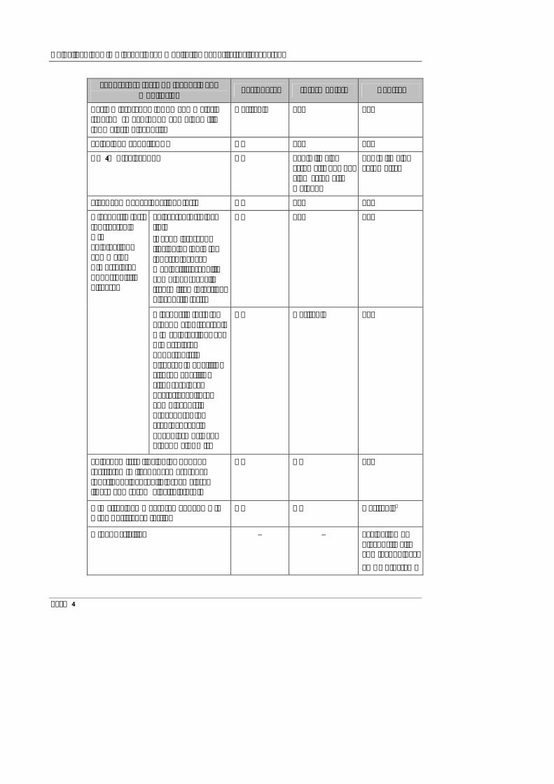
Immunofluorescence analysis by flow cytometry (FCM) is the gold standard for CD4 T cell measurements and also the first choice, if higher specimen volume is expected. To obtain an absolute CD4 T cell count, two concepts (Annex 4 and 5) are utilized:
Guidelines for HIV Diagnosis and Monitoring of Antiretroviral Therapy
Page 18
Dual-platform (DP) Approach
The DP approach uses two instruments to generate absolute CD4 T cell counts: a FCM for generating a percentage CD4 T cells among lymphocytes and a haematological analyzer to enumerate the absolute lymphocyte counts. An absolute CD4 T cell count is derived by multiplying %CD4 T cells by the absolute lymphocyte count. Examples of these DP FCM instruments include BD FACSVantage, FACSCalibur/FASCScan/FACSort or Beckman-Coulter Epics XL/XL-MCL.
Single-platform (SP) Approach
This technique enables absolute CD T cell counts to be derived directly without the need for a haematological analyzer, i.e., the use of volumetric counting (Partec CyFlow), microfluorometry (Guava) and, most commonly, the addition of a known density of reference fluorescent beads to the sample (FACSCount).
Other Alternative Methods
Flow cytometry is a widely used method for estimation of CD4 counts. Flow cytometer and reagents are expensive and hence cause concern in the developing countries. For those countries and settings where infrastructure is not available or difficult to setup for such FCM technologies, a number of accepted alternative assays have been developed and most of them are commercially available and sufficiently validated against gold standard method:
Total lymphocyte count
In case where CD4 testing cannot be assessed, the presence of a total lymphocyte count of 1200 cells/ul or below may be used as a substitute indication for ARV treatment in symptomatic HIV-infected patients. While the total lymphocyte count correlates poorly with the CD4 T cell count in asymptomatic patients, in combination with clinical staging it is still a useful marker of prognosis and survival.
Microscope-based CD4 counting systems
These alternative assays, are however fairly labour intensive and thus less appropriate for a large number of samples. Moreover, qualified personnel are required for making accurate measurements.

Guidelines for HIV Diagnosis and Monitoring of Antiretroviral Therapy
Page 19
Ø Microbead system (Dynal Biotech, Oslo, Norway) uses two types of beads. The first bead removes monocytes from the sample and the second (CD4) estimates CD4 cells that get stained with acridine orange to make the cell nuclei visible for counting under a fluorescent microscope. The initial cost of equipment for a fluorescence microscope is low (US$ 10,000) with a running cost of US$ 3 to 5 per test. A modified DynaBeads system with an alternate stain for the cells can be used with a light microscope.
Ø The Cytosphere system (Beckman Coulter, USA) has the advantage of ease of use by those haematology technicians, who are familiar with the shapes of cells. Here the monocytes are not removed but appear different under the microscope, so the bead-covered CD4+ T cells can be counted.
Evaluations of these two assays were highly comparable with the standard FCM and found to be more accurate and reproducible. However, these methods are manual and labour intensive. To scale these up to match expanding access to ART may prove a challenge. The system could be cheaper than other alternatives and will be useful in small settings, if it is backed up with flow cytometry for quality assurance.
ELISA-based assays Ø Capciella CD4/CD8 Test kit (Biorad Laboratories, France) is a one-
step immunoenzymatic assay based on capture of T-cells by CD2 antibodies bound to the wells of a microtitre plate, followed by detection with anti-CD4 or CD8 peroxidase conjugate in an ELISA format. The CD4 and CD8 number is obtained by conversion from pmol/L values on a standard curve.
Ø TRAx CD4 Test kit (T Cell Diagnostics, USA) is a test based on solubilization of CD4 cells from whole blood by a lysis solution to release its antigen(CD4 molecules) followed by its detection in an ELISA format.
Ø Zymmune CD4/CD8 Test kit (Intracel, USA) uses a mixture of antibody coated magnetic and flourescent beads. The magnetic beads isolate the cells of choice and the fluorescent beads provide the signal to count the cells.
These assays have proven either too cumbersome or have an insufficient concordance with standard FCM. In addition, these assays need a well-trained technician for getting accurate results. The initial cost is approximately US$ 5000. These systems have shown some promise but still need to be validated on larger scale.

Guidelines for HIV Diagnosis and Monitoring of Antiretroviral Therapy
Page 20
Economic flow cytometry
Combined use of CD4 and CD45 conjugated antibodies (panleucogating methodology) has been found to be workable and cost-effective. The commercial kit is available from Beckman Coulter, USA.
Modified flow cytometry
Cyflow from Partec
The system works on volumetric method i.e. known amount of blood with a single antibody reagent. It can also be run on solar panel and car batteries and hence may be used in remote areas. The methodology is simple to carry out based on a single platform method with single antibody reagent and ten minutes incubation. The system showed good correlation with the CD4 counts obtained by conventional flow cytometry. However, an experienced technician is required for accurate measurement. The CyFlow capital equipment cost is approximately US$20 000 and the cost per test US$2.
Guava technology
A single platform system that uses CD3 antibodies to measure T lymphocytes and CD4 antibodies to estimate absolute T cells expressing CD4. The system showed good correlation with conventional flow cytometry and is easy to operate. It uses smaller blood volume. It requires very minimal infrastructure facilities and it is easy to train the technologists to perform the test. The cost per test is around US$ 2. However, the equipment costs about US$ 26 000.
The recent evaluation studies with these two modified flowcytometry system have shown the good correlation with the standard FCM.
Selection of Alternative Methodology for CD4 Count
The following specifications should be considered for the selection of the better technology for CD4 count:
Ø The equipment should be simple to operate, easy to maintain and require minimal training.
Ø Methodology needs minimal infrastructure laboratory facilities. Ø Methodology should be simple to train the person and involves
minimal steps.

Guidelines for HIV Diagnosis and Monitoring of Antiretroviral Therapy
Page 21
Ø Methodology should have the internal QC procedures.
Ø Test kits should be cost-effective and available anytime. Ø Equipment and test procedures should have been validated against
the standard assay. Ø Easy access to the technical specialist/service engineer support.
Ø Supplying company should be in a position to supply the critical parts of the equipment with short Turn Around Time.
For HIV Monitoring all CD4 estimations for the same patient must be done using the same technology to ensure comparability.
Utility of CD4 count in monitoring
According to the WHO recommendation, HIV-infected adults should start ARV therapy when infection has been confirmed and one of the following conditions is present (Table 3.1).
Table 3.1 WHO recommendations for initiating ARV therapy in HIV-infected adults and adolescents according to CD4 T cell counts and total lymphocyte counts*
WHO Stage CD4 T Cell Counts Total Lymphocyte Counts
IV Irrespective Irrespective
III <350 cells/uL Irrespective
II <200 cells/uL <1200 cells/uL
I <200 cells/uL Not applicable
* It should be noted that in HIV-related symptoms (Stage II, III), a total lymphocyte count of 1200 cells/uL can be substituted for the CD4 T cell count when the latter is not available. However, asymptomatic HIV-infected patients (Stage I) need not be treated because the total lymphocyte count correlates poorly with the CD4 T cell count in asymptomatic patients.
Sample Guidelines
Blood samples are collected by venipuncture into K3EDTA-containing tubes, mixed well and processed within the time-frame (the sample stability varies depending upon the equipment and the type of kit) for example, the samples should be processed within 48 hrs (stored between 20−25 °C) after the

Guidelines for HIV Diagnosis and Monitoring of Antiretroviral Therapy
Page 22
collection for the FACSCount. Blood samples should preferably be collected within a fixed time in the day (morning or evening) so as to avoid diurnal variations. Blood sample that is not suitable should be rejected (for example, blood sample that cannot be performed within the time-frame, or if blood sample is hemolyzed or frozen or clotted or without proper labeling).
Suggestions for Testing at Different Levels
Samples can be processed at different clinical laboratory levels and different volume of samples/day. The choice could also depend on the availability of FCM it, if not, simple total lymphocyte count or one of the alternative light microscope staining and counting assays could be used (Table 3.2).
Table 3.2 Guidelines on appropriate assay for different clinical laboratory levels
Laboratory Level Number of Samples/day Assay
Peripheral 1−5 Total lymphocyte count
1−5 Dynabeads or Cytospheres
1−5 FCM CD4 testing*
Intermediate 5−20 FCM CD4 testing*
5-20 Transfix**/Central lab
Central >20 FCM CD4 testing
* Modified or economic flowcytometry methodologies could be considered after the required validation. ** Transfix is a whole blood stabilizing reagent. When Transfix is added to fresh blood, the sample retains its integrity for almost one week. The use of Transfix is therefore suitable to increase the catchment area of central laboratories.
Gold Standard for CD4 Count: Flow Cytometry
Principle
Flow cytometer operates by introducing cells stained with fluorescence conjugated antibody or absorption dyes in a fluid stream under a slight pressure to pass through a nozzle into the beam of light, usually generated by a laser. Light that is scattered and emitted by cells is then separated into constituent wavelengths by a series of optical filters and mirrors. This

Guidelines for HIV Diagnosis and Monitoring of Antiretroviral Therapy
Page 23
separated optical light falls on individual photodetectors and then is translated into electrical pulse, or analog signals, proportional to the amount of incident light detected by the detectors. Each analog signal is finally converted into a digital signal. The magnitude of digital signals is then processed by the data processing and analysis unit. The numbers are proportional to the amount of light emitted from, or scattered by, individual stained cells.
Samples
Cells to be measured must be suspended in a liquid. So, ideally, this is simple for whole blood. Anticoagulant blood is stained with appropriate monoclonal antibody that binds to the specific antigens, i.e. CD4 that are to be measured. Normally, the monoclonal antibody is directly conjugated with fluorochrome.
Advantages and Disadvantages
Table 3.3 Advantages and disadvantages of using flow cytometer for CD4 testing
Advantages Disadvantages
Reference method Expensive instrument
High reliability Expensive reagents
High accuracy, precision and reproducibility
Expensive maintenance
Handling large number of samples Need well-trained technician
Cost
At present, the cost of CD4 testing varies from country to country and ranges between US$ 12−US$ 30. However, the recent development of a simplified panleucogating method together with the use of generic monoclonal antibody reagents drastically cut the cost of CD4 test by more than one-fourth.
Quality system
Several sets of guidelines addressing quality control of CD4 T cell enumeration have been developed. It is important that accurate daily internal quality control and proficiency test or external quality assurance programmes be employed to

Guidelines for HIV Diagnosis and Monitoring of Antiretroviral Therapy
Page 24
ensure the reliability and value of CD4 T cell data. However, participation in most of the current international external quality assurance programme requires a substantial amount of funding to cover the cost of participation fees and carriages. There are free external quality assurance programmes, in spite of their irregular schedules, they are useful and cost saving.
Reporting result
All data are reported in terms of percentages and absolute counts of CD4 T cells, or %CD4 T. It should be noted that the absolute counts are used in adult HIV-infected patients, whereas only the percentages are used in paediatric HIV-infected patients. In the report, the mean and reference of both percentage and absolute CD4 T cells (normal ranges) should also be given. Each clinical laboratory should validate normal ranges.
Total Lymphocyte Counts
Principle
The total lymphocyte counts are easily performed by automated haematology cell counters on aspirated blood samples appropriately diluted with a solution (e.g., acid or detergent) that lyses the red blood cells but preserves leucocyte integrity. A typical automated haematology cell counter performs white blood cell counts by either impedance or light-scattering technology or both. Cells in suspension are made to flow through a small orifice across which an electric current is flowing as in impedance technology. As a cell enters the orifice, the flow current is reduced. Electronic circuits detect the decrease in current and thus the presence of the cell. In a light-scattering haematology cell counter, cells flow through a light beam rather than through an orifice. Different cell types intercepted by light will show different patterns based on the size and shape of the cells. In this way, the device can count the number of cells per second of flowing cells through the orifice or light beam, and because the volume flow rate can be measured one can thus determine the number of white blood cells per uL and or total lymphocyte count per uL of blood sample.
Samples
Collect blood samples by venipuncture into tubes containing K3EDTA anticoagulant. Mix the blood well to prevent clotting. Aliquot appropriate amount of blood into tube containing lysing buffer (e.g. an acid buffer or a detergent) to lyse red blood cells. The lysed whole blood samples are

Guidelines for HIV Diagnosis and Monitoring of Antiretroviral Therapy
Page 25
processed and analysed for their white blood cell count and total lymphocyte count by using haematology cell counter.
Advantages and disadvantages
Table 3.4 Advantages and disadvantages of using total lymphocyte count for ARV therapy
Advantages Disadvantages
Inexpensive unit cost High variation (CV up to 15%)
Simple and commonly available test No EQAS
Less complicated instruments compared to FCM
Fault results due to nucleated red blood cells, platelet aggregation and nonlysed red blood cells.
Cost
The cost of total lymphocyte counts is cheap compared to CD4 testing, and it is varies from country to country but is around US$ 1.0−US$ 2.0
Quality system
At present no external quality assurance on white blood cell counting is available. Only internal quality control supplied from each manufacturer is recommended. It is important that accurate daily internal quality control and proficiency test, if applicable, be employed to ensure reliability and value of total lymphocyte count data.
Reporting result
In haematology cell counter the total lymphocyte count is calculated from direct measurement of lymphocyte count, and expressed as cells x 103/ul. Normal value for total lymphocyte count is 2.6 x 10³/ul (range 1.0-4.9 x 10³/ul). Normal ranges should be validated by each clinical laboratory.


Page 27
4. Virological Monitoring of ART
Viral Load Testing
HIV1 viral load measurement is useful for monitoring treatment. A baseline plasma viral load is established before starting ART. Periodic monitoring is essential. It is predicted that with successful therapy a fall of 1.5 to 2 log in plasma viral load occurs within 4-6 weeks. With successful ART, it should become undetectable in 4 to 6 months of therapy.
Viral load is measured using a variety of commercial kits based on Nucleic Acid Testing (NAT) and non-NAT (Annex 6 and 7). The Amplicor HIV-1 Monitor test, version 1.5 is used widely. The assay uses gag specific primers for the highly conserved region. The lower limit of detection with the standard assay kit is 400 RNA copies/ml and the upper limit is 750 000 RNA copies/ml. The Amplicor ultra sensitive kit detects down to 50 copies/ml of plasma.
A real time PCR is being tested in some laboratories. The cheaper cost of the test and the advantage of avoiding batch testing are some of the plus points. The test uses primers and probe set specific for the LTR region of the HIV genome, which is conserved across subtypes. The primers need to be selected carefully as some subtypes may be estimated in relatively lower copy numbers with some of the primers. The lower limit of detection is in the range 50 IU/mL. The virological assays are useful tools in monitoring for the emergence of resistance in HIV against antiretroviral drugs.
HIV Drug Resistance Assays
The increase in the use of ART is expected to lead to the emergence of drug-resistant mutants of HIV1. This has been the experience from other parts of the world. The inherent mutability of the reverse transcriptase (RT) gene of HIV allows for drug resistance to emerge under selection pressure. The protease gene also undergoes mutations, which manifest as failure to respond to protease inhibitors.
A survey of drug resistance of indigenous HIV strains is essential to ascertain the usefulness of the antiretrovirals especially, in public health programmes. Drug resistance should be suspected if the plasma viral load does not show a greater than 1 log fall within 8 weeks of therapy.

Guidelines for HIV Diagnosis and Monitoring of Antiretroviral Therapy
Page 28
WHO had prioritized the prevention, surveillance and monitoring of HIV drug resistance within its planning for the 3 x 5 initiative and had developed an approach to prevent the emergence and transmission of HIV drug resistant virus. HIV drug resistance surveys are targeting untreated individuals, particularly the patients, who had recently become HIV- infected. Public health action would be essential if such a survey indicated HIV drug resistance to be >15%.
The examination of plasma RNA for resistant HIV strains indicates circulating mutants, if any, including emergent polymorphisms. The proviral DNA testing reveals mutants that have emerged in the individuals about a year ahead and has slowly replaced the native wild type derived provirus.
Two types of antiretroviral drug resistance assays exist today are: Phenotypic Assays, and Genotypic Assays.
Phenotypic assays
The phenotypic assays amplify the RT and protease genes from the predominant quasispecies in the patient’s plasma virus RNA or proviral DNA. These amplicons are then inserted into the laboratory virus lacking the genes. The hybrid virus is then propagated in cell cultures and its ability to propagate in the presence of varying concentrations of drug is measured. Results are expressed as the concentrations of drug required to inhibit 50% of growth (IC50) are relative to a wild type control strain. This work can be carried out only in laboratories with recommended bio-safety measures in place.
Genotypic Assays
The genotypic assays detect changes in the sequence of the relevant HIV1 gene. This measures resistance by detecting mutations in the HIV1 genome that leads to one or more specific amino acid substitutions in the HIV1 reverse transcriptase or protease enzymes. These specific changes cause drug resistance. Viral RNA from plasma or proviral DNA can be used for testing. The amplified products are then sequenced. The sequences are analysed using the special software such as Stanford University Online software(http://hivdb.stanford.edu/), Los Alamos National Laboratories HIV Sequence Database (hiv-web.lanl.gov), http://www.hivresistanceweb.com/ request/pda.shtml, etc.

Guidelines for HIV Diagnosis and Monitoring of Antiretroviral Therapy
Page 29
Table 4.1 Advantages and disadvantages of genotypic assays over phenotypic assays
Category Genotypic assay Phenotypic assay
Advantages • Less expensive than phenotypic assays
• Short turn around time (< 1 week)
• May detect presence of resistance mutations before they have resulted in phenotypic resistance
• Can be standardized in a BSL-2 facility laboratory
• Interpretation similar to susceptibility testing of bacteria
• Assesses the total effect of mutational changes
• Good reproducibility • The threshold to define
susceptibility is arbitrary
Disadvantages • Interpretation requires knowledge of mutational changes
• May show discrepancy with phenotypic assay
• Very expensive to set up and run
• Turn around time is more (3 weeks) than that of genotypic assays.
• Slower to show resistance relative to genotypic assays
• To be handled only in a BSL-3 level laboratory
A third approach to resistance testing is the "virtual" phenotype. This assay is really a genotype resistance that is interpreted with the aid of a large database of samples with paired genotypic and phenotypic data. Viruses with genotypes that are similar to the patient's virus are identified by searching the database, and the average IC50 of these matching viruses is calculated. This information is then used to estimate the likely phenotype of the patient's virus. The major advantage of this approach is that it reduces complex genotypic data to simple phenotypic categories based on a rational, data-driven analysis of similar genotypes. The major disadvantage of this approach is that the confidence placed in the result depends on the number of matches, and on picking the right codons to incorporate into the database search. Correlation between actual and virtual phenotype will be weaker for newer drugs or in cases where there are fewer matches due to unusual genotypes. However, it needs more validation studies.


Page 31
5. Microbiological Monitoring of ART Morbidity and mortality in HIV disease is due to the occurrence of life-threatening opportunistic infections (OIs) during the natural course of the disease. These are the direct consequence of a decline in CD4 count. A wide variety of opportunistic infections (Table 5.1) are encountered in patients with AIDS which are caused by various microorganisms. Very often these represent reactivation of organisms that have been dormant in the host for several years. The incidence of these diseases increases as the patient's CD4 count declines. The pattern/repertoire of opportunistic infections may vary in different geographic areas. The knowledge of important OIs specific for particular areas/countries is useful for correct diagnosis and management of OIs.
Table 5.1 Common opportunistic infections in HIV/AIDS
Disease Pathogen Infection type
Bacterial
Tuberculosis and non-tuberculous infections
Mycobacterium tuberculosis Mycobacterium avium complex
Pulmonary or meningeal or other extra-pulmonary or systemic
Salmonellosis Salmonella sp. Typhoid fever or diarrhoea
Bacterial pneumonia
Streptococcus pneumoniae, Nocardia sp., Pseudomonas aeruginosa
Pulmonary infection
Fungal
Candidiasis Candida albicans Oral thrush or vulvovaginitis
Cryptococcosis Cryptococcus neoformans Meningoencephalitis, or pulmonary or systemic disease
Aspergillosis Aspergillus sp. Pulmonary disease or sinusitis
Histoplasmosis Histoplasma capsulatum Pulmonary disease or disseminated

Guidelines for HIV Diagnosis and Monitoring of Antiretroviral Therapy
Page 32
Disease Pathogen Infection type
Penicilliosis Penicilium marneffei Pneumonitis or disseminated disease
Parasitic
Pneumocystis carinii pneumonia
Pneumocystis carinii Pneumonia
Toxoplasmosis Toxoplasma gondii Encephalitis
Cryptosporidiosis Cryptosporidium parvum Diarrhoea
Isosporosis Isospora belli Diarrhoea
Viral
CMV infection Cytomegalovirus(CMV) Retinitis or encephalitis or esophagitis or colitis
Herpes Herpes Simplex virus(HSV) Chronic ulcer or bronchitis or pneumonitis, or esophagitis
Utility of Microbiological Monitoring
Natural history studies from India by shows that persons with a CD4 lymphocyte count of <200 cells/uL are 19 times more likely to die than those with a CD4 cell count of >350 cells/uL. Most of the diagnosed individuals have more than one opportunistic infections. Patients who had more than 1 opportunistic infections were 2.6 times more likely to die than those who did not have an opportunistic infection.
Diagnosis of Opportunistic Infections
Infrastructure facilities should be established based on the level of set-up required (Table 5.2). Although intermediate laboratories have sufficient facilities, further identifications and confirmations may be done by central laboratory. The flow of specimens should be worked out from low to higher level and the flow of technical, scientific information and QA/QC procedures should be higher to lower levels.

Guidelines for HIV Diagnosis and Monitoring of Antiretroviral Therapy
Page 33
Table 5.2 Suggested laboratory diagnostic procedures to be performed at different levels
Diagnosis at different level Disease
Peripheral Intermediate Central
Tuberculosis and non-tuberculous infections
AFB smear AFB smear, Culture
AFB smear, Culture and sensitivity, PCR
Salmonellosis - Culture and sensitivity
Culture and sensitivity
Bacterial Pneumonia - Culture and sensitivity
Culture and sensitivity
Candidiasis Gram’s staining Gram’s staining Culture and sensitivity
Cryptococcosis Negative staining, LA Test
Culture Culture
Aspergillosis 10% KOH mount
Culture Culture
Histoplasmosis 10% KOH mount
Culture Culture
Penicilliosis 10% KOH mount
Culture Culture
PCP - - IFT
Toxoplasmosis LA Test LA Test IFT
Cryptosporidiosis - Wet mount, Modified Acid-Fast staining
Modified acid-fast staining, IFT
Isosporosis - Wet mount, Modified Acid-Fast staining
Modified Acid-Fast staining, IFT
CMV infection - - IFT, Cell culture, PCR
Herpes - - Cell culture, PCR, IFT
PML - - PCR for JC virus
IFT: Immunoflourescent technique, LA: Latex agglutination

Guidelines for HIV Diagnosis and Monitoring of Antiretroviral Therapy
Page 34
Role of Central Laboratory in Sharing Data
The microbiological monitoring of opportunistic infections will require complementary activities of various partners in the laboratory network. The central laboratory (or National Reference Laboratories for the particular organism) shall act as the apical laboratory and collate data from other laboratories to feed them into the national programme. The central laboratory, in addition, shall train laboratory staff and assure appropriate management including safe collection and processing of bio-specimens; act as country focal point for national coordination if there is a problem in identification and confirmation; act as technical resource centre; assure and/or assess quality in subordinate laboratories; prepare relevant guidelines and their distribution to other laboratories; maintain a repository of the isolates and reference strains and monitor the resistance in isolates against the specific antimicrobial agents.

Page 35
6. Laboratory Monitoring of Side-effects of ART Toxicity is related to the inability to tolerate the side-effects of medications and to significant organ dysfunction that may result because of toxicity. All the available anti-retroviral agents have potential toxicities (Table 6.1). Careful monitoring of patients by laboratory investigations play a major role in better clinical management. Toxicity should be monitored clinically based on patient reports and physical examination, supplemented by a limited number of laboratory tests depending on the symptoms that arise and the specific combination regimen that is used.
Table 6.1 Adverse drug reactions of individual antiretroviral agents
Agent Affects quality of life Serious, may require intervention
Serious and life-threatening
NRTI (Nucleoside Reverse Transcriptase Inhibitor)
Abacavir (ABC)
Headache Systemic hypersensitivity reaction
Didanosine (DDI)
Diarrhoea, peripheral neuropathy
Pancreatitis Fatal pancreatitis with lactic acidosis
Lamivudine (3TC)
Nausea, vomiting Pancreatitis in children; increased anemia
Stavudine (D4T)
Lipoatrophy, peripheral neuropathy
Hepatotoxicity Rapidly ascending neuromuscular weakness associated with lactic acidosis
Tenofovir (TDF)
Diarrhoea, headache, asthenia
Zalcitabine (DDC)
Aphthous ulcer, peripheral neuropathy
Zidovudine (AZT)
Headache; nausea, vomiting; myalgia
Neutropenia; megaloblastic anemia
Severe anemia

Guidelines for HIV Diagnosis and Monitoring of Antiretroviral Therapy
Page 36
Agent Affects quality of life Serious, may require intervention
Serious and life-threatening
NNRTI (Non-Nucleoside Reverse Transcriptase Inhibitor)
Delavirdine (DLV)
Nausea, vomiting Skin rash, liver enzyme elevation
Efavirenz (EFV)
Insomnia, somnolence, abnormal dreams, dizziness, decreased concentration, drowsiness
Depression, suicidal ideation, hallucination, psychosis, skin rash, liver enzyme elevation
Nevirapine (NVP)
Skin rash, liver enzyme elevation
Stevens-Johnson syndrome, fulminant hepatic necrosis
PI (Protease inhibitor)
Amprenavir (APV)
Nausea, vomiting, abdominal pain
Skin rash
Indinavir (IDV)
Requires adequate daily fluid intake to prevent crystalluria; paronychia; hair thinningloss
Nephrolithiasis, renal failure
Rarely hemolytic anemia
Lopinavirritonavir Diarrhoea, nausea, vomiting; perioral paresthesia, taste perversion
Nelfinavir (NFV)
Diarrhoea Diarrhea may lead to severe dehydration
Ritonavir (RTV)
Nausea, vomiting; diarrhea,abdominal pain,anorexia, perioral paresthesia, taste perversion
Saquinavir (SQV)
Nausea, vomiting, abdominal pain
Monitoring of Adverse Reactions
Monitoring toxicities of the drug can be done clinically based on patient reporting and physical examination. However, inclusion of limited laboratory

Guidelines for HIV Diagnosis and Monitoring of Antiretroviral Therapy
Page 37
investigations in the ARV monitoring will determine the severity of the toxicity and this will help physician to change the dose and specific drug combination in regimen. When the toxicity is related to an identifiable drug in the regimen, the offending drug can be replaced with another drug that does not have the same side effect.
Monitoring of ART
The different level testing capabilities and type of specimen to be collected for monitoring of ART toxicity are described in Table 6.2.
Table 6.2 Suggested testing capabilities at different levels
Lab tests Level 1: Peripheral Level 2: Intermediate Level 3: Central
Haemoglobin Hemoglobinometer1 Haematology Analyser2 Haematology Analyser
Total and differential cell count
Microscopic - Manual
Haematology Analyser Haematology Analyser
Complete blood count
As above Haematology Analyser Haematology Analyser
Liver and renal function markers
- Medium throughput autoanalyser3
High Throughput Autoanalyser5
Complete clinical chemistry markers including serum electrolytes.
- Medium throughput autoanalyser and Ion selective electrode4
High throughput autoanalyser and Ion selective electrode
Pregnancy testing Rapid test Rapid test Rapid test
Approximate cost of the instruments:100$1, 10000$2, 24000$3, 40000$4, 80000$5 ,
Ø Patients with Hepatitis B (HBV) and C (HCV) viruses should be monitored more closely for liver toxicity. HBV and HCV screening testing may be undertaken in the particular community, where the prevalence is high.
Ø The critical values (abnormal values) of the laboratory results should be reported only after consultation with the treating physician.

Guidelines for HIV Diagnosis and Monitoring of Antiretroviral Therapy
Page 38
Role of Central Laboratories
The laboratory-based monitoring of adverse drug reactions will require complementary activities of various partners in the laboratory network. The central laboratory (or National Reference Laboratories for the particular parameter) shall act as the apical laboratory and collate data from other laboratories to feed them into the national programme. The central laboratory, in addition, shall train laboratory staff and assure appropriate management including safe collection and processing of bio-specimens; act as country focal point for national coordination; act as a technical resource centre; assure and/or assess quality in subordinate laboratories; prepare relevant guidelines and their distribution to other laboratories; help other laboratories by providing the information regarding the selection of reagent make and methods of the test; and establish and maintain normal ranges for Haematology and Chemistries.

Page 39
7. Tuberculosis in HIV/AIDS The South-East Asia Region bears 40% of the global TB burden and ranks second after sub-Saharan Africa in the estimated number of people living with HIV/AIDS. Each year, nearly 3 million cases of TB and 750 000 TB deaths are estimated to occur in the Region. Of the estimated 6 million adults living with HIV in the Region, about half are likely to be infected with TB. The extent to which HIV will contribute to the TB epidemic depends on the degree of overlap between the population groups infected with TB and those with HIV.
Pulmonary tuberculosis accounts for more than 90% of total tuberculosis manifestation in HIV patients. Before the AIDS pandemic, non-tuberculosis mycobacteria rarely caused serious illness, even in the immunocompromised individuals. The prolonged immunosuppresion of the cell-medicated immune system caused by HIV provided the opportunity for these relatively avirulent organisms (non-tubercular mycobacteria) to cause disease.
Laboratory Diagnosis
Tuberculosis in HIV-infected persons may occur with different manifestations. All these forms of tuberculosis, except when cavitations occur in pulmonary tuberculosis, are paucibacillary in nature. Depending upon the form of disease manifestation, several specimens such as sputum and/or gastric lavage, bronchoalveolar lavage (BAL), lymph nodes and other biopsy specimens, pus, ascetic fluid, pleural and cerebrospinal fluid should be examined. If delay is anticipated, biopsy specimens may be collected in a suitable transport medium for sending them to the laboratory.
Collection of Sputum
For diagnosis of tuberculosis, three specimens of sputum are to be examined (spot morning-spot) over a period of two days. Specimens are to be collected in sterile universal containers, and should have a fixed label for noting patient’s details on the side of the container.
Specimen collection • Aerosol free container (Sputum cup with a lid or Mc Cartney bottle)
Adequate quantity and appropriate number of specimens
• Ideally, a minimum volume of 5 ml • Three consecutive sputum specimens should be
collected

Guidelines for HIV Diagnosis and Monitoring of Antiretroviral Therapy
Page 40
Specimen transport and storage
• Sterile leak-proof container in a sealed plastic bag. • To be transported within three days for culture; if
delay is anticipated, add equal volume of 1% CPC
Specimen staining • Ziehl-Neelsen staining Note: heat-fixing may not kill all Mycobacterium species, Slides should be handled carefully.
Culture and investigation • Specimen processing • Sputum: Petroff’s method for digestion and
decontamination. • Other samples: Direct inoculation for aseptic
specimens. • Mild acid or other standard pretreatment
procedure • Inoculation on Lowenstein – Jensen media or
Middlebrook 7 H9 or Kirchner’s medium. • Incubation at 37 °C for M tuberculosis and 25 °C,
37 °C and 45 °C for NTM . • Examination for eight consecutive week or until it
becomes positive or contaminated.
Sputum Smear Microscopy
Microscopy of sputum is of great value in the detection of open or infectious cases of tuberculosis. The establishment of a good sputum microscopy service is of prime importance in developing countries for the detection and treatment of open cases.
Smears are stained by the Ziehl-Neelsen (ZN) method, or by one of its various modifications. Grading of the positive smears gives a broad indication of the severity of the disease and the response to therapy.
The laboratory number is written, with a diamond marker, on one edge of a new, clean, grease-free and unscratched slide’s using a broomstick or a wire loop (5mm diameter), a purulent portion of the sputum is placed on the slide and spread evenly to give a smear of approximately 3 x 2 cm. The smear is allowed to air-dry and fixed by pressing over a flame 3-5 times for 3−4 seconds each time. It is then stained with carbol fuchsin decolorized with 25% sulphuric acid and counter-stained with methylene blue and dried using standard staining protocol. A drop of cedarwood oil or liquid paraffin is placed on the slide without touching the smear and examined under an oil-immersion objective for at least 10 minutes.

Guidelines for HIV Diagnosis and Monitoring of Antiretroviral Therapy
Page 41
Sputum Culture
Sputum culture for mycobacteria requires more sophisticated laboratory facilities with biosafety level 2 or 3. Bio-safety hoods and arrangements for safe disposal of culture media and sputum samples need to be available. The standard culture technique for mycobacteria is to decontaminate by modified Petroff’s method, where sputum is decontaminated with 4% Na0H and then inoculated on to Lowenstein-Jensen slopes. The incubated cultures are examined once a week for 8 consecutive weeks or until they become positive or contaminated.
Identification
The identification of strains isolated as belonging to M. tuberculosis complex or non-tuberculous mycobacteria (NTM) can be ascertained by performing a few simple tests, i.e. susceptibility to p-nitrobenzoic acid (PNB), niacin production test, catalase activity requirement and the morphological appearance. Important differences between Mycobacterium tuberculosis and NTM are shown in Table 7.1.
Table 7.1 Differentiation between mycobacterium tuberculosis and NTM
Characteristic M.tuberculosis NTM
Growth rate Slow grower Slow/Rapid grower
Temperature 37 °C 25 –45 °C
Colony morphology Dry rough Dry
Colony on solid media Eugonic Dysgonic
Colour of colony Buff Yellow, orange or
Emulsification Difficult Creamy
Cord formation + Easy
Niacin test + -
Nitrate reduction test + -
Growth on p-nitrobenzoic acid (PNB) 500 µg/ml
- +


Page 43
8. New Technologies in HIV Diagnosis and ART Monitoring
High costs of assays used in diagnosis and monitoring and resource limitations are the major concerns for HIV control and implementing treatment programmes. Newer technologies providing economical assays are very important for such programmes. The specific areas where such assays will be needed are:
Ø HIV diagnosis
Ø CD4 count estimation
Ø Plasma viral load determination
Ø HIV incidence testing
The new technologies must be properly validated before applying them in resource poor settings and should follow strict quality control regimen in the programme.
HIV Diagnosis
Conventional HIV diagnostic methods have disadvantages because of the need for cold chain for sample transport and storage as well as for conducting invasive procedure. Some of the newer approaches obviate these shortcomings.
Tests on samples other than blood (saliva and urine): Commercial tests are available for the detection of anti-HIV antibodies in urine (Calypte Biomedical Corp, USA) and saliva specimens (OraSure Technologies, USA and BioMerieux Inc, France) have shown promising results. Now approved confirmatory tests are also available for urine and saliva samples (Epitope Inc, USA and Calypte Biomedical Corp, USA). The tests that use saliva samples are more expensive than blood tests.

Guidelines for HIV Diagnosis and Monitoring of Antiretroviral Therapy
Page 44
Tests using dried blood spot: Antibody detection on dried blood spots (DBS) has been employed in field surveys in infections other than HIV. The advantages include stability at room temperature obviating the need for cold chain, suitability for community-based surveys and adaptability for collection of large number of samples. It can be adopted for confirmation in Western Blot. However, No data on borderline samples/recently infected individuals with probable low titer antibody are available. The system has still to undergo vigorous quality assessment. Some of the commercially available kits for antibody detection may not be suitable for this strategy. However, there are some specific kits, which are approved for the use of DBS (BioMerieux Inc, France, Abbott Laboratories, UK and Bio-Rad Laboratories, USA).
CD4 Count Estimation
Consistency in the CD4 counts for adults and CD4% monitoring for children (aged up to 6 years) is the essence in monitoring disease progression in HIV infection, to decide when to start treatments and to monitor response to ARV treatment (in the absence of viral load) and decide on when it is safe to stop prophylaxis for opportunistic infections. However, to date, only high-end flow cytometry instrument could produce reliable CD4%. The BD FACSCount system (single platform) had not been configured for paediatric use.
Research is ongoing for the development of simple, inexpensive and point of care methodologies. The development of such technologies is of the utmost importance.
Flow cytometry is a widely used method for estimation of CD4 counts. Flow cytometer and reagents are expensive and hence cause concern in the developing countries. Cheaper non-flow based assays for the field level use are likely to be available very soon.
Newer Technological Approach CD4 Counts
There are several assays being developed to provide immediate CD4 cell readings for the point of care. The attempts are being made to design such technologies for the rapid assay to provide accurate results. Some of these are:
Ø Lateral flow assay
Ø Dipstick assay

Guidelines for HIV Diagnosis and Monitoring of Antiretroviral Therapy
Page 45
Ø Capillary tube assay
Ø LabNow Microchip Technologies Ø SemiBio Microscopic Slide Test
CD4 count estimation from dried blood spots
The technique uses an antibody “sandwich” to capture and detect CD4 proteins in the sample. As it stands, it does not give a close match to CD4 T-lymphocytes measured by flow cytometry, and the discrepancy (an overestimation of CD4 T-cells) increases as the actual CD4 count declines.
Viral Load Measurement
Measurement of plasma virus load is currently being used for monitoring the progression of HIV infection as well as to monitor the efficacy of anti-retroviral treatment. Current methods of determining of plasma virus load include quantitative RT-PCR, branched DNA technology and NASBA. These technologies are expensive and may not be affordable for programmes in developing countries. A few alternative approaches that are under investigation are mentioned below.
Ø Real time PCR: The technique can be used as a modified cost-effective approach for viral load measurement with around 30 to 50% cost reduction. However, the methodology is still under validation. Few commercial kits are available for real time PCR application for determining plasma virus load. On validation, they may provide cheaper option.
Ø Viral load measurement by Flow cytometry: The quantifications of PCR products by dedicated flow cytometry is of significant and under development (Partec, (Germany).
Ø Viral load measurement on dried blood spot: Dried blood spots and dried plasma spots have been evaluated for viral load measurements in Zambia on 51 HIV infected individuals. The viral load estimation was performed by nucleic acid amplification (NASBA) technique with good correlation standard assay. However, continuing research on DBS is needed to better understand the effects of variables such as drying time, humidity and temperature on RNA stability.

Guidelines for HIV Diagnosis and Monitoring of Antiretroviral Therapy
Page 46
HIV Incidence Testing
The ability to discriminate between long-standing and recently acquired or recent HIV infection was important, providing insight into current transmission trends. It aids the tracking of outbreaks and possibly assisting in the monitoring of treatment programmes. Several techniques have been described, including STARHS (serologic testing algorithm to detect recent seroconversion or detuned assays), IgG capture assay (BED Assay) (Calypte Biomedical Corporation, Rockville, USA) and antibody avidity assays.

Page 47
9. Laboratory Infrastructure and Quality System The organization of laboratories in any country is usually a three - or four - tier system with various possible functional linkages between them. There must also be transport and communication facilities between peripheral and intermediate laboratories for referral of samples and patients, procurement of supplies and personal discussion. Laboratory investigations can be established based on the level of the laboratory set-up required for that region, as follows:
Level 1: Peripheral Laboratories
Functions: These laboratories provide technical support for the diagnosis and clinical management of HIV-infected individuals (monitoring of HIV and ART). This level of laboratory will perform some simple testing, such as HIV rapid testing, haemoglobin estimation, TB smear examination, cell count (TC and DC), pregnancy testing and basic biochemistry tests which do not require much skills.
Staffing: The staff in peripheral laboratories should include at least one microbiologist/pathologist, one technician and one laboratory assistant/ attendant.
Space: Space available in peripheral laboratories should include at least one laboratory-cum-office/record room (approx 5 metres x 3 metres ) and one store-room which can be used for other services also (approx 5 metres x 3 metres).
Equipment: Light microscope (binocular) – 1; small refrigerator (165 litre capacity) – 1 Bio-safety centrifuge (maximum RPM 5000) – 1; autoclave– 2; safety spectacles; micropipettes (variable volume 40 – 200 uL) – 2; micropipettes (variable volume 5 – 40 uL) – 2; Bunsen burner with LPG; sample racks; computer with printer – 1; needle destroyer – 1.
Other facilities: Other facilities include prompt laboratory supplies (glass slide, coverslip, staining solutions, microtips, transport media, sterile swabs, vacutainer or syringe, test tubes, sample containers, serological pipettes, Pasteur pipettes, diagnostic kits etc.), work bench (at least 0.5 x 6

Guidelines for HIV Diagnosis and Monitoring of Antiretroviral Therapy
Page 48
metres length of concrete), chairs and cupboards, supply of safe water, stabilized power supply (power cut should not be for more than 2 hours), telephone, waste disposal facilities, PEP availability, Separate sinks for bio-waste and clean in each laboratory area, eye wash stations, fire extinguishers etc.
Level 2: Intermediate Laboratories
Functions: The functions expected to be performed by the intermediate laboratories include support to clinical diagnosis/public health, quality assurance, logistic and technical support and training of staff for peripheral laboratories. These laboratories also serve as links between peripheral laboratories and the state/central laboratory for the following: collection, storage and analysis of data, distribution of reagents, media, preparation of laboratory manuals and standard operative procedures for the functions to be carried out at peripheral as well as intermediate laboratories, purchase of equipment, supervision of peripheral laboratories, to conduct EQA for peripheral laboratories, to take part in EQA organized by state/central laboratories and to send samples to higher/reference laboratories for characterization of isolate/confirmation of diagnosis.
Staffing: One qualified laboratory supervisor/manager (master’s degree in pathology/medical microbiology), three Technical Assistants (undergraduate in biochemistry or microbiology plus DMLT, three Technicians (cum Phlebotomist), one Laboratory attendant-cum-Housekeeper and one Clerk-cum-storekeeper.
Space: Space available in level 2 laboratories should include one room (approx 5 x 5 meters) for microbiology, one room for sterilization, media preparation (approx 3 meters x 3 meters), one room (approx 5 x 5 meters) for serology and biochemistry, one room (approx. 5 x 5 meters) for haematology and immunology, store-room (approx 3 meters x 3 meters), office (approx 3 meters x 5 meters) and one room for staff resting/dining (approx 3 meters x 3 meters).
Equipment: Light microscope (binocular) – 2, biosafety cabinet (class II) – 2, autoclave – 2, incubator – 2, hot air oven – 1, water bath – 2, VDRL shaker – 1, autoanalyser – 1 and at least one semi-autoanalyser as a back-up, computer with printer – 3, refrigerators (>200 litre capacity) – 3, bio-safety centrifuge (the maximum RPM 5000) – 2, weighing balance – 1, pH meter – 1, ELISA washer –1, ELISA reader – 1, flow cytometer (single or dual platform) – 1, Western blot rocker – 1, hematolology analyser (3 part) – 1, deep freezer (-20 °C or colder) – 1, inspissator – 1, distilled water apparatus – 1, safety

Guidelines for HIV Diagnosis and Monitoring of Antiretroviral Therapy
Page 49
spectacles/protective glass shields, micropipettes (variable volume 200 uL – 1000 uL), micropipettes (variable volume 40–200 uL), micropipettes (variable volume 5–40 uL), needle destroyer, Bunsen burners with LPG, laminar flow hood (biosafety cabinet class II) and sample racks.
Other facilities: Other facilities include prompt laboratory supplies including plastic wares, glasswares and diagnostic kits, work bench (at least 0.5 x 6 meters length of concrete with granite table top) in each laboratory area, Air-conditioned laboratory rooms, sufficient chairs and cupboards, supply of safe water, stabilized power supply with battery back-up for at least 4 hours (power cut should not be for more than 2 hours), telephone, internet, waste disposal facilities, PEP availability, readily available technical supports for all the equipment, separate sinks for bio-waste and clean in each laboratory area, eye wash stations, fire extinguishers, etc.
Level 3: Central or National Laboratories
Functions: The National laboratory shall provide technical support tointermediate and peripheral laboratories by training, maintenance of laboratory QA/QC, purchase of equipment, validation and selection of diagnostic kits, preparation of testing algorithms, supervision, preparation of laboratory manuals and standard operative procedures for the functions to be carried out at all three levels of laboratories, dissemination of newsletter, to conduct EQA and characterize of isolate/confirm the diagnosis.
These laboratories should have more facilities than those for the Intermediate level plus additional facilities such as cell culture facility, facilities to carry out techniques like PCR, viral load estimation. The laboratory should also have a flow cytometer to run a QA program for intermediate laboratory where an alternate system for CD4 count estimation might be in use. This level of laboratories should be participating in international EQAS programme, conducting continuing education, workshops and hands-on training programme.
The functions of the laboratories at various levels have been described in Chapter 1.
Health services are utilizing laboratories more extensively now than ever before. Laboratories play an important role in the diagnosis of HIV infection as well as monitoring of anti-retroviral treatment. The demand for quality results from laboratories has been echoed by all the health care professionals caring for HIV-positive individuals. Unreliable laboratory results are known to have serious consequences for the health of the individual as well as the

Guidelines for HIV Diagnosis and Monitoring of Antiretroviral Therapy
Page 50
community. It is important to have a system of networking peripheral with regional and central laboratories of a country in order to facilitate quick and accurate processing and testing of specimens collected from patients, quick dissemination of information to the peripheral levels as well as to run a quality assurance programme. One possible way of networking of laboratories is shown in Figure 9.1.
Figure 9.1 Possible networking of laboratories
Quality System in the Laboratory
In any laboratory dealing with HIV, quality is an essential element to ensure consistency, reproducibility, traceability and efficacy of products or services. In the context of HIV-related laboratory services also, quality assumes great importance since the physician utilizes the report generated in the laboratory for the benefit of the patient and the community. A quality laboratory report thus helps the physician to establish proper diagnosis rapidly and supports better health care for the patient. Apart from providing accurate results, maintaining quality in laboratory services also helps in generating confidence among patients in the health care system, creating a good reputation for the laboratory, reducing costs by avoiding repetition of tests, sustains motivation in laboratory staff, helps in the accreditation of the laboratory and above all prevents legal suits and associated complications. In recent times, quality has been considered to be synonymous with consistency which denotes providing the same product or service time after time thus making the outcome more predictable.
Intermediate laboratory
Intermediate laboratory
Intermediate laboratory
Peripheral laboratory
Central laboratory
Peripheral laboratory
Peripheral laboratory

Guidelines for HIV Diagnosis and Monitoring of Antiretroviral Therapy
Page 51
The external quality assessment scheme compares the performance of different testing sites and is a challenge to other components of the quality assurance system (internal quality control). This assessment is achieved through processing of specimens of undisclosed but known contents, and it measures the accuracy of the results. Quality is considered the degree of congruence between expectation and realization. In simpler words, it is the matching of what you want with what you get i.e. expectations versus fulfilment. In recent times quality has been considered to be synonymous with consistency which denotes providing the same product or service time after time thus making the outcome more predictable. In the context of health laboratory services, the product denotes the material
Key Elements of a Quality System
A quality system has the following key elements:
Ø Organizational management and structure Ø Training Ø Referential (quality) standards Ø Documentation Ø Monitoring and evaluation Ø Assessment of quality system
Organizational Management and Structure
The overall responsibility for the design, implementation, maintenance and improvements in a quality system rests with the laboratory management.
Quality assurance is the responsibility of all the staff members of the organization. However, the top management needs to make a firm commitment to ensure quality and allocate adequate resources. A quality policy reflects the intention and commitment of the organization to attain quality. The policy can be translated into implementation through a quality plan, which, along with the policy, needs to be documented in the form of a quality manual.
Laboratory management delegates responsibility and authority to appropriate individuals who are directly responsible for implementing the quality policy and quality system. It makes available to them adequate resources to efficiently discharge their duties. The inter-relationship between various staff members and their job descriptions are decided by the management.

Guidelines for HIV Diagnosis and Monitoring of Antiretroviral Therapy
Page 52
Training
The quality system is only as good as the staff that actually works with it. No matter how good the quality system is on paper, if the theory cannot be translated into practice, quality cannot be achieved. Staff may need to be trained and this training must also include an understanding of why quality is important. Training should be competency based and must be followed by post-training support to enable staff to maintain high standards.
Referential (Quality) Standards
The referential standards are an integral part of the quality system. These aim at ensuring safety and consistency of the procedures. These need to be followed to meet the regulatory requirements as well as to monitor the functioning of the laboratory.
Both management and technical standards need to be followed to ensure quality. These must also conform to the local laws.
Documentation
Documentation is a record of any information or instructions including policy statements, quality manuals, procedures, specifications, calibration tables, standard operative procedures for maintenance and calibration of equipment, reports, job descriptions, documents of external origin such as regulations, standards and examination procedures etc. These would include information on various media, including the electronic media.
The quality system of a laboratory shall define, document and maintain procedures to control all documents and information (from internal and external resources). The current version of relevant documents shall be available at all locations where operations needed for effective functioning of the quality system are performed.
Monitoring and Evaluation
The laboratory management must develop and implement quality indicators to systematically monitor and evaluate the laboratory’s contribution to patient care. When the programme identifies opportunities for improvements within the system, the laboratory management needs to take appropriate steps to address them. Error management must be vigorously implemented.

Guidelines for HIV Diagnosis and Monitoring of Antiretroviral Therapy
Page 53
Besides, quality assessment through internal and external audits and participation in external quality assessments schemes should also serve to improve the quality.
Assessment of Quality System
A quality system can be assessed either through an onsite inspection (audit) or by sending known but undisclosed material to the laboratories for testing (quality assessment scheme). The latter can be done within an institute by internal staff (internal quality assessment scheme – IQAS) or through an external agency (external quality assessment scheme – EQAS).
Good Laboratory Practice
Good laboratory practice (GLP) indicates the performance of all the activities of the laboratory in the best possible way so that the results obtained are of the highest possible accuracy and reliable. GLP encompasses various factors as follows:
Laboratory infrastructure
Laboratory should be of suitable size and location and meet biosafety requirements. The facilities should be spacious enough to avoid problems such as overcrowding, cross contamination and cramped working conditions. Utilities (water, electricity, etc.) must be adequate and stable.
Human resource
The laboratory should be manned by qualified and trained staff. Specific training records regarding the procedures to be carried out by the specific technicians should be available. The responsibilities of all personnel should be defined and recorded in job descriptions. The number of personnel must be sufficient to perform the tasks required in a timely manner.
Equipment and reagents
Ø All equipment must be in working condition. A strict programme of validation, calibration and maintenance should be in place.
Ø All equipment should be suitable for its intended use, and it should be properly calibrated and maintained to ensure accurate

Guidelines for HIV Diagnosis and Monitoring of Antiretroviral Therapy
Page 54
performance. Records of repairs and routine maintenance are essential.
Ø Back-up for vital equipment should be available whenever possible as well as back-up in the event of services failures, such as power cuts.
Documentation
Ø Routine procedures must be described in written Standard Operating Procedures (SOPs). They must be reviewed regularly and modified, if necessary. The modified versions should be signed and dated by the laboratory director. The recent most version of SOPs should be available directly at the work place. The old versions should be achieved in the laboratory and should be archived if required.
Ø The records must be stored for long periods of time but be available for prompt retrieval, safe archiving of all records must be ensured. Archiving of the source documents and other essential documents must be such that data are kept in an integer state and can neither be lost nor altered to achieve this goal.
Ø Records of usage, maintenance and calibration should be kept in the laboratory and should be routinely monitored. Reports of the tests should be released only after proper scrutiny and documentation of the scrutiny with the signature and date by the lab-supervisor.
General practices
Ø Proper collection and transportation of bio-specimens in appropriate condition to testing laboratory.
Ø Collection and storage under conditions which prevent deterioration of the sample before the performance of test.
Ø Accurate performance of test. Ø Maintaining internal quality control and quality assurance system. Ø Participating in the External Quality Assessment Scheme (EQAS). Ø Delivery of reports to the correct destination with shortest turn
around time (TAT). Ø Availability of SOP for all the procedures carried out in the laboratory,
monitoring and documentation of strict adherence to SOP.

Guidelines for HIV Diagnosis and Monitoring of Antiretroviral Therapy
Page 55
Bio-safety practices
Safety procedures include laboratory protection of the material to be tested, environment and the staff. Personal and laboratory safety can be achieved only by informed, trained responsible individuals through the application of standard precautions.
Standard precautions are simple infection control measures that reduce the risk of transmission of pathogens through exposure to blood or body fluids. The laboratory personnel should be trained in safe handling of the clinical specimen and disposal of bio-waste materials. Documentation of biosafety training for laboratory staff is essential including records on vaccinations such as hepatitis B vaccination. SOP for biosafety procedures should be in place. The following are the recommendations for the safe practice in the laboratory and for better bio-waste management:
Ø Never eat, drink or apply cosmetics in a laboratory. Ø Vaccinate laboratory personnel against hepatitis B. Ø Use gloves as a barrier protection during phlebotomy. Ø Use barrier protection with gloves, gowns/aprons, face shield,
deflector mask, goggles whenever splash of infectious fluid are expected.
Ø Use biosafety centrifuges for sample processing. Ø Use biosafety cabinet while working on specimens which may
produce aerosols. Ø Use puncture-proof containers for disposing of sharps and these
must be as close to the point of use as possible Ø Dispose of sharps yourself immediately. Ø Do not recap, bend or break used needles – but recapping may be
done with single- handed method. Ø Carefully place used plastic syringes, needles and sharps in the
containers, then disinfect by chemical or physical methods i.e. boiling or autoclaving before disposing into Incinerator.
Ø Replace the sharps disposal container by a new one when it is three quarters full.
Ø Never pass used sharps directly from one person to another. Ø Keep all sharps and sharp disposal containers out of the reach of
children.

Guidelines for HIV Diagnosis and Monitoring of Antiretroviral Therapy
Page 56
Ø Do not touch blood spills with your bare hands; cover blood with a 0.5−1% sodium hypochlorite solution (household bleach); leave it for at least 15−30 minutes; cover with gauze, cotton or sponge and finally wipe away with gloved hands.
Ø If spills involve any broken glassware, it must be picked up using a mechanical means, such as a brush and dustpan, or forceps. In cases where the absorbent becomes saturated with blood and bleach, the spill clean up materials should be autoclaved prior to being disposed of in the normal trash.
Ø Label the bio-waste materials container with the “Biohazard Label” (Figure 9.2).
Figure 9.2 Biohazard label
Ø Segregate waste into dry solid waste and liquid waste.
Ø Package all waste to prevent spills, leaks or breaks during transportation.
Ø Before disposing, pre-treat the waste. Ø Liquid waste such as human blood and blood components such as
serum, plasma, etc., and human body fluids should be considered as potentially infectious. Before disposing, this liquid waste should be decontaminated by adding a sufficient volume of sodium hypochlorite to obtain a final concentration of at least 1%. A 30 minute exposure to 1% sodium hypochlorite may be sufficient to ensure effective decontamination. Commercial liquid household bleach typically contains a concentration of 5.25% sodium

Guidelines for HIV Diagnosis and Monitoring of Antiretroviral Therapy
Page 57
hypochlorite. A 1:5 dilution of household bleach will produce a 1% sodium hypochlorite solution. After decontamination, the waste could be disposed in the sewer.
Ø Solid biohazardous waste must be pretreated by decontamination (such as autoclaving) before disposal. All waste must be collected in biohazard bags or closed, leak-proof, labeled containers, to prevent spillage or protrusion of contents during handling or transport.
Ø Color-coded containers (red) marked with the universal biohazard symbol should be used. Plastic liner should be used inside bio-waste container.
Ø Disinfect (with 0.5% sodium hypochlorite) work places when procedures are completed or after any spill of blood or other potentially infectious material in the work place.
Figure 9.3 Classification of laboratory waste
Health care workers and laboratory staff are normally at very low risk of
acquiring HIV infection during management of HIV-infected patients. In prospective studies, the average risk of HIV transmission after a percutaneous
Non-infectious (90%) Paper, Cardboard, Thermocol box, Plastic/PVC, Discarded
reagents
Infectious (10%) (Bio-hazardous)
Sharps Needles, Syringes, Blades,
Scalpels, Glassware
Non--sharps Human waste, blood/ Blood products,
body fluids, Animal waste, Microbiological/ biotechnological waste, Solid linen/gauze/cotton
Solids waste (Treat and dispose)
Liquid waste (Treat and dispose)

Guidelines for HIV Diagnosis and Monitoring of Antiretroviral Therapy
Page 58
exposure to HIV-infected blood has been estimated to be approximately 0.3% and after a mucous membrane exposure, approximately 0.09%. The risk of infection varies with
Ø type of exposure (superficial or deep injury) Ø the amount of blood involved in the exposure Ø amount of virus in patient’s blood at the time of exposure
Ø Whether post-exposure prophylaxis (PEP) was taken within the recommended time (not later than 36 hours after exposure) as shown in Table 10.1.
It is necessary to have a comprehensive programme in place to deal with anticipated accidental exposure. Prevention is the mainstay of strategy to avoid occupational exposure to blood/body fluids. Adequate infrastructure and training should be provided in the settings.
What to do after a Needle Stick Injury?
Ø Wash the injured site thoroughly with soap and water (antiseptics may be used).
Ø Administer post-exposure prophylaxis (PEP) for HIV, based on institutional policy.
Ø If as a result of a laboratory accident the skin is broken the wound should be cleaned and irrigated with a mild disinfectant such as chlorhexidine with cetrimide (e.g. Savlon).
The person should be provided with pretest counselling and anti-retroviral drug prophylaxis started as per the physician’s recommendation. Before starting prophylaxis, a sample should be collected for baseline anti-HIV antibody estimation. Sample should be tested for anti-HIV antibodies immediately after the exposure to know the HIV status. In case the sample tests positive, the individual is referred to the clinician for management, as a case of HIV infection. In case the sample tests negative, a second sample is collected at 6 weeks and third at 12 weeks after the exposure and tested for HIV antibodies. Post-test counselling should be done in all cases. During the follow-up period, especially the first 6−12 weeks, when most infected persons are expected to show signs of infection, the recommendations for preventing transmission of HIV are to be followed by healthcare workers. These include refraining from blood, semen, organ donation and abstaining from sexual

Guidelines for HIV Diagnosis and Monitoring of Antiretroviral Therapy
Page 59
intercourse. In case sexual intercourse is undertaken, a latex condom must be used correctly and consistently. In addition, women should not breast-feed their infants during the follow-up period after exposure to prevent exposing their infants to HIV in breast milk.
The technical personnel should be familiar with the safety precautionary measures.
Table 9.1. Recommendations procedures for post-exposure management
Infectious agent Postexposure prophylaxis
HIV Antiretroviral therapy
HBV Hepatitis B immune globulin (HBIG) and/or hepatitis B vaccine
HCV Immune globulin and antiviral agents (e.g., Interferon with or without Ribavirin)
Drugs Recommended for HIV Post-exposure Prophylaxis
It is recommended that Zidovudine (ZDV) 300 mg bid and Lamivudine (3TC) 150 mg bid be used in combination for a period of four weeks. In selected cases, Indinavir (or one of the other protease inhibitors) can be used as per PEP guidelines. The drugs are usually well tolerated except for transient nausea, vomiting, diarrhoea, tiredness or headache. An emergency supply of these drugs should be available in all hospitals dealing with potentially infected material.


Page 61
10. Collection and Shipping of Biospecimens Biospecimens may be sent to the testing laboratory for HIV diagnosis (serum or plasma), CD4 count (whole blood with EDTA), Viral load testing (plasma) and HIV drug resistance testing and viral isolation (plasma).
Blood specimens represent the most common analytical sample requirement. Certain techniques and care must be exercised to ensure that an acceptable specimen is collected and to that minimize any adverse affect to the patient is minimized. In general, the phlebotomy requires the use of a 20 to 22 gauge needle to minimize mechanical haemolysis during aspiration and either a syringe or evacuated tube collection system.
The collection of blood is described as follows:
Ø Gloves should be worn and sterilized/disposable syringes and needles should be used.
Ø For avoiding soiling, a piece or simply a big piece of absorbent cotton may be placed below the forearm before commencing venepuncture.
Ø After collecting 5ml of blood aseptically, it should be carefully transferred from the syringe in to a sterile, plastic leak-proof specimen container, preferably screwcapped.
Ø The containers should be labeled before commencement of venepuncture.
Ø If the vial has anticoagulants, then a second person wearing gloves should help in shaking the vial for mixing the blood well with the anticoagulants. The cap should be tightly screwed after the blood has been transferred to the vial.
Ø After the blood is collected, the tourniquet is removed and the needle is withdrawn. The patient is given a dry sterile cotton swab to press over the site of venepuncture. Elbow may be flexed to keep the cotton swab in place till the blood stops. Any blood spill should be carefully wiped with 70% ethanol.
Ø All the swabs and cotton pieces are placed in plastic bags for disposal. If the outside of the vial is visibly contaminated with

Guidelines for HIV Diagnosis and Monitoring of Antiretroviral Therapy
Page 62
blood, it should be cleaned with 10% freshly prepared sodium hypochlorite solution.
Ø The blood is allowed to clot for 30 minutes (not more than 2 hours) at room temperature. The clot may be gently broken if necessary using sterile Pasteur pipettes.
Separation of Serum
Ø The collection tube is centrifuged at 1200 g (3000 rpm) for 10 to 15 minutes to separate serum to avoid haemolysis. If no centrifuge is available, the blood with clot may be left in the refrigerator at +4 °C for overnight. The clot will retract and get separated from serum.
Ø Allow the blood to clot for at least 30 minutes, before centrifugation. Ø The specimen vial is un-stoppered, the serum is drawn off by
sterile Pasteur pipette and transferred to a sterile plastic screwcapped leak-proof tube.
Storage of Serum Specimens
Ø The sera samples are placed in leak-proof plastic containers in the refrigerator at +4 °C, for temporary storage (up to 5 days).
Ø The outside of the container is checked for visible contamination with blood which should be cleaned.
Ø For storage for a long time, deep-freezing at –70 °C is advised and cryovial should be used for deep-freezing purposes.
For plasma or whole blood specimen, K3 EDTA (1.5 ± 0.15 mg/ml blood) tube could be used and after the collection, the tube should be gently inverted at least five times to mix blood and additive. The separation of plasma can be done as in the case that of serum. For viral load and HIV drug resistance testing the whole blood specimen should be processed within 6 hours.
Transport of Serum/Plasma Specimens
Ø The specimen tube, in which serum/plasma is to be transported, should not have a crack or leakage. Preferably, it should be made of plastic and should be screwcapped. The outside of the container should be checked for any visible contamination with blood which should be disinfected.

Guidelines for HIV Diagnosis and Monitoring of Antiretroviral Therapy
Page 63
Ø The tube should be labeled and then placed in a second tightly-capped unbreakable container (Figure 2) surrounded by adequate packing materials (like tissue paper, absorbent cotton etc.) to absorb liquid, if leakage occurs accidentally.
Ø The secondary container should also have a label. This is placed in a thermocol box with having ice packs or dry-ice to maintain proper cold chain system during transit.
Ø A proforma with details i.e., name, age, sex, risk factors, history of previous testing etc. should accompany the specimen.
Ø A biohazard symbol (Figure 3) must be fixed outside the thermocol box. This box can now be sent to a distant laboratory.
Figure 10.1 Packing infectious substances for the shipment

Guidelines for HIV Diagnosis and Monitoring of Antiretroviral Therapy
Page 64
Figure 10.2 Infectious substance label
Transport of Whole Blood for CD4/CD8
Ø The tube containing the specimen should be placed in a leakproof container (e.g. a sealed plastic bag). This container should be packed inside a cardboard canister containing sufficient material to absorb all the contents should the tube break or leak. Cap the canister tightly. Fasten the request slip securely to the outside of this canister with a rubber band. For mailing, this canister should be placed inside another canister containing the mailing label.
Ø The specimen should be transported as early as possible after the collection and the test should be performed within the time frame allowed by instrument manufacturer (e.g. For FACSCount within 48 hours).
Ø During the transport of specimens, room temperature (18–22 °C) should be maintained and specimens should not be exposed to extreme temperatures that could allow them to freeze or become too hot.
Ø Temperatures >37 °C might cause cellular destruction and affect flow cytometry measurements. In hot weather, pack the specimen in an insulated container. If necessary, place this container inside another, containing an ice pack and absorbent material. This method helps retain the specimen at ambient temperature.

Page 65
Suggested Further Reading
(1) Balakrishnan P, Mandy Dunne, Kumarasamy N, Suzanne Crowe, et al. An inexpensive, simple and manual method of CD4 T- cell quantitation in HIV infected individuals for use in developing countries. J Acquir Immune Defic Syndr 2004, 36, 1006-1010.
(2) Dunne AL, Mitchell FM, Coberly SK, Hellmann NS, et al. Comparison of genotyping and phenotyping methods for determining susceptibility of HIV1 to antiretroviral drugs. AIDS 2001;15 :1471-
(3) Guidelines: CDC Guidelines for performing single-platform absolute CD4+ T-cell determinations with CD45 gating for persons infected with human immunodeficiency virus. MMWR, 2003; 52(No. RR 02).
(4) Guidelines: CDC Revised guidelines for performing CD4+ T-cell determinations in persons infected with human immunodeficiency virus (HIV). MMWR 1997; 46 (No. RR-2).
(5) Guidelines: CDC Guidelines for National Human Immunodeficiency Virus Case Surveillance, Including Monitoring for Human Immunodeficiency Virus Infection and Acquired Immunodeficiency Syndrome. MMWR. December 10, 1999 / 48 (RR 13); 1-28.
(6) Guidelines: Guidelines for preventing HIV, HBV and other Infections in the Health care setting. WHO-SEARO Publication, New Delhi, India 1999.
(7) Guidelines: Guidelines on Prevention and control of Hospital associated Infections. WHO, New Delhi, (SEA-HLM-343), 2002.
(8) Guidelines: CDC Updated U.S. Public Health Service Guidelines for the Management of Occupational Exposures to HBV, HCV, and HIV and Recommendations for Postexposure Prophylaxis. MMWR.2001.
(9) Guidelines. Guidelines on Standard Operating Procedures for Microbiology. WHO New Delhi, India. 2001.
(10) Guidelines: Guidelines on Standard Operating Procedures for Laboratory Diagnosis of HIV-Opportunistic Infections. WHO – New Delhi. 2001.
(11) Harrigan PR, Montaner JS, Wegner SA, Verbiest W, et al. World-wide variation in HIV1 phenotypic susceptibility in untreated individuals: biologically relevant values for resistance testing. AIDS 2001; 15: 1671-7.
(12) Kumari S, Bhatia R. Guidelines for Peripheral and Intermediate Laboratories in Quality Assurance in Bacteriology and Immunology. WHO Regional Publication, South-East Asia Series No. 28, New Delhi, India. 2003.
(13) Kumarasamy N, Suniti Solomon, Timothy P. Flanigan, Hemalatha R, et al. Natural History of Human Immunodeficiency Virus Disease in Southern India. Clinical Infectious Diseases 2003;36:79-85

Guidelines for HIV Diagnosis and Monitoring of Antiretroviral Therapy
Page 66
(14) Kumarasamy N, Solomon S, Chaguturu SK, Mahajan AP, et al. The safety, tolerability and effectiveness of generic antiretroviral drug regimens for HIV-infected patients in south India. AIDS2003; 17: 2267-9.
(15) Manual: Guidelines on Standard Operating Procedures for Laboratory Diagnosis of HIV-Opportunistic Infections. WHO–New Delhi, India. 2001.
(16) Report: Role of Health Laboratories in the Implementation of the “3 by 5” Initiative. SEARO Newsletter on Quality Assurance in Blood Safety and Health Laboratory Services, New Delhi, India. January 2004. Vol. 4, No.1.
(17) Suzanne Crowe, Shannon Turnbull, Robert Oelrichs, Amanda Dunne. Monitoring of Human Immunodeficiency Virus Infection in Resource-Constrained Countries. Clin Infect Dis 2003; 37: 25–35.
(18) WHO consultation on technical and operational recommendations for scale-up of laboratory services and monitoring HIV antiretroviral therapy in resource –limited settings. Geneva, 13-15 December 2004.
(19) Balakrishnan P, Kumarasamy N, Kantor R, Solomon S, Vidya S, Mayer KH, Newstein M, Thyagarajan SP, Katzenstein D, Ramratnam B. HIV type 1 genotypic variation in an antiretroviral treatment-naive population in southern India. AIDS Res Hum Retroviruses. 2005 Apr; 21(4): 301-5.
(20) Syed IH, Balakrishnan P, Solomon SS, Murugavel KG, Kumarasamy N, Vidya S, Martin SP, Thyagarajan SP, Mayer KH, Solomon S. HIV-1 western blot assay: What determines an indeterminate status? Indian J Med Sci. 2005 Oct; 59(10): 443-50.
(21) Balakrishnan P, Solomon S, Kumarasamy N, Mayer KH. Low-cost monitoring of HIV infected individuals on highly active antiretroviral therapy (HAART) in developing countries. Indian J Med Res. 2005; 121: 345-55.
(22) WHO Technical Information: CD4+ T-cell enumeration technologies (available at www.who.int/eht/).

Guidelines for HIV Diagnosis and Monitoring of Antiretroviral Therapy
Page 67
Annex 1
SUGGESTED SOURCES OF AVAILABILITY OF LABORATORY EQUIPMENT AND SUPPLIES
The following web-sites are suggested only for information and not recommended by WHO. Programme managers are encouraged to get more information from other sources also.
www.axygen.com; www.bio-rad.com;www.bd.com; www.beckmancoulter.com www.abbottdiagnostics.com; www.biomerieux.com; www.tranasia.co.in; www.sysmex.com www.ambalayellowpages.com/5.htm ;www.kendro.com; www.dmoz.org/Science/Instruments_and_Supplies/Laboratory_Equipment; www.wheecon.com; www.roche.com; www.guavatechnologies.com; www.span.co.in; www.orthoclinical.com; www.labindia.com; www.catalogs.indiamart.com/category/scientific-instruments.html; www.ifcc.org/ejifcc/vol15no1/150102200401n.htm; www.trade-india.com/dyn/gdh/catalog/hp/1919/catalog.htm; www.trade-india.com/dyn/gdh/eyp/Industrial_Supplies/Laboratory_ Glassware_and_Equipment/; www.noor-scientific.com/bio/thermo.htm ; www.indiayellowpagesonline.com/a-z/l/2.htm; www.biowire.com; www.labsafety.com;www.labfurnishers.com; www.remelinc.com/products/clinical/ laboratory_supplies.cfm; www.who.amds.com
Sources of EQAS
www.cms.hhs.gov/clia/ptlist.pdf; www.phppo.cdc.gov/mlp/EQA_qlist.asp; www.who.int/eht/Main_areas_of_work/DIL/Lab_Tech/External_Quality_Assessment.htm www.aab.org, www.nrl.gov.au/, www.cap.org
Bio-specimen Shipping Training (IATA)
www.worldcourier.com; www.saftpak.com

Guidelines for HIV Diagnosis and Monitoring of Antiretroviral Therapy
Page 68
Annex 2
COLLECTION AND STORAGE OF CLINICAL SPECIMENS
Lab. test Specimen Recommended activities
Specimen integrity at
RT
Short-term storage
Long term
storage
Factors associated with erroneous
results
HIV testing and other serology
Serum - - Serum should be separated within 4 hours if stored at RT and 24 hours if stored at 2-80C
- 200C or colder
Hemolysis, hyperlipemic, contamination
CD4 Whole blood
* *K3 EDTA or heparin
Preferably <25±20C
- Clot hemolysis, storage more than recommended contamination
Viral load Plasma K3 EDTA Preferably <25±20C
Plasma should be separated within 6 hours
- 700C or colder
Use of unsterile storage vials, fluctuation in freezer temperature. More than 3 freezethaw cycle
Hemoglobin complete blood count
Whole blood
K3 EDTA Preferably <25±20C
- Clot, Hemolysis
Blood chemistry, sugar, lactate
Plasma Sodium flouride Preferably <25±20C
Plasma should be separated within 30 min
At 2-80C for same day testing
- 200C or colder
Clot, delayed specimen processing. For lactate fasting specimen is recommended
Blood chemistry Serum electrolytes
Serum - Preferably <25±20C
Serum should be separated within 30 min
At 2-80C for same day testing
- 200C or colder
Delayed specimen processing

Guidelines for HIV Diagnosis and Monitoring of Antiretroviral Therapy
Page 69
Lab. test Specimen Recommended activities
Specimen integrity at
RT
Short-term storage
Long term
storage
Factors associated with erroneous
results
Blood chemistry LFT, RFT, etc.,
Serum - Preferably <25±20C
At 2-80C for same day testing
- 200C or colder
Haemolysis
Pregnancy Testing
Urine or Serum
- Preferably <25±20C
At 2-80C for same day testing
- Early morning urine is recommended
OI diagnosis Sputum CSF, serum, swabs, stool etc.
- Preferably <25±20C
At 2-80C Stool should be processed and tested immediately for parasites
- Improperly collected, transported specimen

Guidelines for HIV Diagnosis and Monitoring of Antiretroviral Therapy
Page 70
Annex 3
PROCEDURES CARRYING POTENTIAL RISKS OF HIV, HBV AND OTHER BLOODBORNE AGENTS
Procedure Person at risk Mode of transmission
Collection of blood sample Patient Contaminated needle Contaminated hand or gloves of health worker Skin puncture by needle or broken specimen container Contamination of hands by blood
Transport of specimens (within laboratory)
Laboratory personnel Transport worker
Contaminated exterior of specimens container Broken container Spill or splash of specimen
Skin puncture or contamination of skin or Mucous membrane
HIV serology and virology Laboratory personnel
Contaminated exterior of specimen container Contaminated work surface Spill or splash of specimen Broken specimen container Perforated gloves
Cleaning and maintenance Laboratory personnel Support staff
Skin puncture or skin contamination Spills or splashes Contaminated work surface
Waste disposal Laboratory personnel Support staff Transport worker
Contact with contaminated waste Puncture wounds and cuts
Shipment of specimens (to other centers)
Public transport worker Postal worker Public
Broken or leaking specimen containers and packages

Guidelines for HIV Diagnosis and Monitoring of Antiretroviral Therapy
Page 71
Annex 4
SUMMARY OF CD4 T-CELL ENUMERATION TECHNOLOGIES: FLOW CYTOMETRY
Single Platform Parameter Double platforma
Volumetricb Bead-basedc
Instruments, manufacturers • Partec GmbH (Munster, Germany)d
• Becton Dickinson (California, USA)e
• Coulter Corporation (Florida, USA)e
• Partec GmbH (Munster, Germany)d
• Guava Technologies (California, USA)
• Becton Dickinson (California, USA)e
• Coulter Corporation (Florida, USA)e
Cost of instrument (US$) 20,000−95,000 20,000−70,000 20,000−95,000
Cost of reagents/test (US$) 3−11 2−10 8−25
Specimen Whole blood Whole blood Whole blood
Results • Absolute CD4 and CD8 count
• CD4% and CD8% • CD4/ CD8 ratio
• Absolute CD4 and CD8 count
• CD4% and CD8% • CD4/ CD8 ratio
• Absolute CD4 and CD8 count
• CD4% and CD8% • CD4/ CD8 ratio
Throughput (samples/day) Up to 200 Up to 50 Up to 200 Advantages • Accurate pipetting
less crucial • One tube assay
possible without QC problems
• EQAS available
• No need for extra beads or haematology analyzer
• Protocols for aged Samples available
• EQAS available
• No need for haematology analyzer
• Protocols for aged samples available
• EQAS available
Disadvantages • Requires the use of a haematology analyser
• Fresh samples needed in order to obtain absolute counts
• Requires accurate pipetting technique.
• Internal QC for pipetting requires two tubes assay.
• Limited validation data available
• Requires accurate pipetting technique
• Some equipments are not suitable for paediatric use (e.g. BD FACSCount)
• Internal QC for pipetting requires two tubes assay
• Beads are expensive and require careful handling
a) Any flow cytometer from any of the three manufacturers can operate with this method to provide absolute counts. The results of flow cytometry are combined with those from haematology in order to calculate absolute counts.
b) Volumetric instruments have the inherent hardware property of measuring the volume of the sample, providing direct absolute counts without the use of haematology analysers or beads.
c) Any flow cytometer from any of the three manufacturers can operate with this method to provide absolute counts. d) Instruments from this manufacturer, including the cyflow, remain to be validated as volumetric absolute CD4 T –cell counters by
independent investigators in multicentre studies. e) Economic flowcytometry approach (Pan Leukogating –PLG) is possible with these instruments, either with beads (single platform) or
without beads (double platform).

Guidelines for HIV Diagnosis and Monitoring of Antiretroviral Therapy
Page 72
Annex 5
SUMMARY OF CD4 T–CELL ENUMERATION TECHNOLOGIES: DEDICATED AND MANUAL ASSAYS
Dedicated technology Manual assays
Parameter FACS Count Cyflow
counter PointCARE Cytospheres Dynabeads
Manufacturer Becton Dickinson (California, USA)
Partec GmbH (Munster, Germany)
Beckman Coulter Inc.,(CA, USA)
Coulter Corporation (Florida, USA)
Dyna AS (Oslo, Norway)
Instrumentation Dedicated CD4 counter
Dedicated CD4 counter
Dedicated CD4 and CD4% counter
Haemocytometer Light microscope
Magnet Haemocytometer Light or fluorescence microscope
Assay principle Flow cytometry Flow cytometry Flow cytometry Direct observation of bead-rosetted cells
Direct observation of immunocaptured cells
Detection system
Fluorochrome-labelled anti-CD3,CD4 and CD8 Mab
Fluorochrome – labeled anti-CD4, (CD45,CD3, CD8) MAb
Anti-CD4 MAb conjugated with colloidal gold particles.
Latex beads conjugated to anti-CD-4 MAb
Magnetic beads conjugated to anti anti-CD4 and CD8 MAb
Specimen Whole blood Whole blood Whole blood Whole blood Whole blood
Results • Absolute CD4 and CD8 counts
• CD4/CD8 ratio
• CD4%and CD8%
• Absolute CD4 count
• Absolute and % CD4 count
• Absolute WBC count
• Absolute and % lymphocyte count
• Absolute CD4 count
• Absolute CD4 count
• Absolute CD8 count
• CD4/CD8 ratio
Correlation with flow cytometry a
(r value)
0.93−0.98 (several international studies)
0.94−0.98 (some international studies)
0.95 and 0.97 0.67−0.93 (several international studies)
0.94 and 0.96 (several international studies)
Cost of instrument (US$)
28,000 20,000 17,000 1,000 1,000−10,000 b
Cost of reagents/test (US$) c
6−20 2 5−8 4−8 3−5

Guidelines for HIV Diagnosis and Monitoring of Antiretroviral Therapy
Page 73
Dedicated technology Manual assays
Parameter FACS Count Cyflow
counter PointCARE Cytospheres Dynabeads
Advantages • Automated • Fewer steps,
less human error and low biohazard risk
• Absolute CD4 and CD8 counts
• Quick results • EQAS
available
• Reagents available at low cost
• Quick Results
• EQAS available
• Simple and no manual gating
• No manual sample preparation
• Pointcare system
• Simple • Rapid
• Simple • Rapid • Absolute CD4
and CD8 counts
Disadvantages • Expensive reagents
• CD4% not reported
• CD4% not reported
• Limited validation data available
• Each sample processing takes 17 minutes
• No EQAS available
• No published data available yet
• Low-throughput
• Subjectivity in visual counting
• CD4% or CD8 counts not reported
• No EQAS available
• Low-throughput
• Subjectivity in visual counting
• CD4% not reported
• No EQAS available
Licensed by USA-FDA
Approved − Approved Approved −
a) The analysis of correlation using linear regression is not appropriate for comparison of methods. Instead, analysis of agreement should be performed. Unfortunately most of the published studies has used this analysis to compare these methods with flow cytometry. The r values are therefore reported there.
b) Depending on whether a light microscope or a fluorescence microscope is used.
c) Equipment cost may vary and reagent cost may decrease substantially in the near future.

Guidelines for HIV Diagnosis and Monitoring of Antiretroviral Therapy
Page 74
Annex 6
SUMMARY OF MAIN CHARACTERISTICS OF VIRAL LOAD TECHNOLOGIES BASED ON NUCLEIC ACID TESTING (NAT)
Company Abbott Roche Bayer Bio Merieux Primagen
Assay Name
LCx HIV RNA Quantitative
Amplicor HIV-1
Monitor Test Versant HIV-1
RNA 3.0 Assay NucliSens EasyQ
HIV-1 Retina
Rainbow
Type of assay
RT-PCR RT-PCR bDNA RT-NASBA RT-NASBA
Dynamic range (copies/ml)
50−1,000,000 50−750,000 75−500,000 50−3,000,000 500−50,000,000
Specimen type
Plasma Plasma, Dried blood spots
Plasma Plasma, Serum, Dried blood spots
Plasma, Serum, Whole blood, Dried blood spots
Specimen Volume
200−1000 µl 100−500 µl 1000−200 µl 10−2000 µl 200 µl
Area of HIV genome amplified
Pol Gag Pol Gag LTR
HIV-1
subtypes amplified
Group M (subtypes A to G) and Group O
All, plus some HIV-2 Group M (subtypes A to G)
All All
Time for result
5 Hours 6−7 Hours 22 Hours 2.5−3 Hours 1.5 Hours
Cost/testa $ 20−70 $ 28−90 $ 125 $ 38−76 $ 17−23
Number of samples/run
21(+3 controls) 9−48 12−168 8−48 96
Equipment requiredb
Vaccum Pump , Centrifuge (x2), Heat block
LCX analyzer Thermal Cycler
COBAS Ampliprep, Dead-air box, Computer/ printer
Safety hood, Heat block (x 2), Centri –fuge (x2)
Bayer System 340 (bDNA analyzer, data manage-ment and computer system)
Centrifuge, Heat block, Water bath, Vaccum system
Nucli Sens miniMAG System / Nucli Sens east MAG system
Nucli Sens EasyQ analyzer , Strip centrifuge
Retin Alyser, Heat block
Computer, Centrifuge
Equipment cost(US$)
8,500 + LCX Analyzer 25,000
10,000 + COBAS Ampliprep 30,000
10,000 + Bayer System Analyser
23,000
Licensed by USA-FDA
− Approved Approved Approved −

Guidelines for HIV Diagnosis and Monitoring of Antiretroviral Therapy
Page 75
Annex 7
SUMMARY OF MAIN CHARACTERISTICS OF VIRAL LOAD TECHNOLOGIES NOT BASED ON NUCLEIC ACID (NON-NAT)
Company Cavidia Perkin Elmera
Assay Name ExaVir Load Quantitative HIV-RT Load kit HIV-1 p24 Ultra ELISA amplification system
Type of Assay Enzyme immunoassay for quantitation of RT activity
Enzyme immunoassay for quantitation of p24 antigen
Dynamic range (copies/ml)
750 to over 50,000 copies/ml Pg/ml 400 copies /ml
Specimen type Plasma Plasma, Serum or Cell culture supernatant
Specimen Volume 1000 µl 100 µl
Area of HIV genome amplified
RT activity P24 antigen
HIV-1 subtypes amplified
All plus HIV-2 HIV-1
Time for result 24 Hours 6 Hours
Cost/test $13−115 $10
Number of samples/run
30 96
Equipment required b Incubator (33°C), freezer, ELISA reader, computer Incubator, ELISA reader, refrigerator
Equipment cost (US$) 9,000−10,000 (start-up pack includes other necessary equipment and three kits)
7,000−9,000
a These assays need extensive validation with the standard assay. b Both assays require piptettes and vortex mixers.

Guidelines for HIV Diagnosis and Monitoring of Antiretroviral Therapy
Page 76
Annex 8
CONTRIBUTORS AND REVIEWERS
Dr Suniti Solomon, MD. Director, YRG Centre for AIDS Research and Education(YRG CARE) Voluntary Health Services Hospital Campus, Taramani Chennai – 600 113 India E-mail : [email protected]
Dr Kovit Pattanapanyasat, Ph.D. Center of Excellence for Flow Cytometry Faculty of Medicine Siriraj Hospital Mahidol University Bangkok Thailand E-mail: [email protected]
Dr Ramesh Paranjape, Ph.D. Officer-In-Charge National AIDS Research Institute(NARI) G-73, M.I.D.C. Bhosari Pune-411 026, India E-mail: [email protected]
Dr V. Ravi, MD. Professor and Head Department of Neuroviroloy National Institute of Mental Health and Neurosciences (NIMHANS) Bangalore – 560029, India E-mail: [email protected]
Dr G. Sridharan,Ph.D. Professor and Head Department of Clinical Virology Christian Medical College Hospital Vellore - 632 004, India E-mail: [email protected]
Dr Madhuri Thakar, Ph.D. Senior Research Officer Department of Immunology National AIDS Research Institute (NARI)
G-73, M.I.D.C.,Bhosari Pune- 411 026, India Email: [email protected]
Dr Soumya Saminathan, MD. Director Division of AIDS Tuberculosis Research Centre Chetpet, Chennai, India E-mail : [email protected]
Dr P.Balakrishnan,Ph.D. Laboratory Manager for NIH funded Projects Associate Professor of Microbiology YRG Centre for AIDS Research & Education Voluntary Health Services Hospital Campus Taramani, Chennai - 600 113, India E-mail : [email protected]
Dr N.Kumarasamy, MBBS, Ph.D. Chief Medical Officer YRG Centre for AIDS Research and Education(YRG CARE) Voluntary Health Services Hospital Campus Taramani, Chennai-600 113, India E-mail: [email protected]
Dr Rajesh Bhatia, MD. Regional Adviser, Blood safety and Clinical Technology WHO/South-East Asia Regional Office IP Estate, Ring Road New Delhi 110 002, India 00-91-11-23370804 Extn 26504 E-mail: [email protected]
Reviewers Dr Bharat Ramratnam, MD Laboratory of Retrovirology Division of Infectious Diseases Brown Medical School Providence, RI 02903, USA E-mail: [email protected]

Guidelines for HIV Diagnosis and Monitoring of Antiretroviral Therapy
Page 77
Dr Sirirat Likanonsakul Chief, Immunology and Virology Laboratory Bamrasnaradura Institute 126 Tiwanon Road Nonthaburi 11000, Thailand E-mail: [email protected]
Ms Monthasawat Rattanasrithong Chief of Medical Technology Section Bamrasnaradura Institute 126 Tiwanon Road Nonthaburi 11000, Thailand
Dr Renu Garg STP-HIV WHO/SEARO New Delhi 110002, India [email protected]