Green synthesis of nano-propolis and nanoparticles (Se and ...
15
Review Article Bindiya Barsola and Priyanka Kumari* Green synthesis of nano-propolis and nanoparticles (Se and Ag) from ethanolic extract of propolis, their biochemical characterization: A review https://doi.org/10.1515/gps-2022-0059 received January 27, 2022; accepted May 02, 2022 Abstract: Propolis is a natural bee product with phenolic components and flavonoid content. As propolis is hydro- phobic, it is poorly absorbed by the body, querying the use of other technologies. This review focuses on the biosynthesis, characterization, and evaluation of some biological activities of nanoparticles (AgNPs and SeNPs). The nanoparticles were generated utilizing bee propolis extract, taking into account the benefits of green nanopar- ticle synthesis. Due to the smaller size, nano-propolis is more easily absorbed by the body. Nano-propolis has the potential to improve efficacy in the realms of medicine and biology. Green chemistry approach to nanoparticle synth- esis offers several advantages, including process scaling, economic feasibility, and a safe technique to make nano- particles. Bioreduced AgNPs can be employed as a thera- peutic agent to treat a variety of human ailments. After deeply studying and reviewing different research studies, it was evaluated that the natural nanoparticles have the potential to be effective in the treatment of bacterial and fungal infections. Keywords: nanopropolis, nanoparticles, propolis, biolo- gical activities, nanotechnology 1 Introduction Nanotechnology is the science and technology of small, precise objects that are between 1 and 100 nm in size [1]. The term “nano” is derived from a Greek word that means “dwarf,” and it is frequently used in conjunction with other words such as “nanometer,”“nanobots,” and “nano- technology” [1,2]. Nanomaterials may undergo novel che- mical and physical changes in their structure as a result of their smaller size, indicating increased reactivity and solu- bility [1,3]. When compared to transition metal nanoparti- cles such as iron, nickel, and cobalt, noble metals such as ruthenium, rhodium, palladium, silver, osmium, iridium, platinum, and gold are the most widely used in nanotech- nology because of their relatively low cytotoxicity for biolog- ical or environmental conditions [4–6]. The risk for environmental contamination is heigh- tened with the development of new chemical or physical techniques, as the chemical procedures involved in the production of nanoparticles generate a substantial number of harmful byproducts. As a result, green chemistry is required, which comprises a clean, non-toxic, and envir- onmentally acceptable approach of nanoparticle synthesis [7,8]. Green synthesis refers to the production of nanopar- ticles via a process involving a natural molecule as a redu- cing agent and metal salts [9]. Because of the natural coating of the organic molecules, biosynthesis of selenium nanoparticles (SeNPs) is safe, affordable, eco-friendly, more stable, and do not aggregate over time [10–14]. Due to the presence of reducing, stabilizing properties, and eco-friendly characteristics, the use of propolis is pre- ferred for the biosynthesis of nanoparticles [4]. Chemical and physical methods of producing nanoparticles are not commercially feasible and culminate in the generation of unsafe nanoparticles and environmental pollution. The use of green nanotechnology is an impeccable solution for combating these adverse affects. Selenium, an essential Bindiya Barsola: Department of Zoology, School of Biological and Environmental Sciences, Shoolini University of Biotechnology and Management Sciences, Solan 173229, India, e-mail: [email protected] * Corresponding author: Priyanka Kumari, Department of Zoology, School of Biological and Environmental Sciences, Shoolini University of Biotechnology and Management Sciences, Solan 173229, India, e-mail: [email protected] Green Processing and Synthesis 2022; 11: 659–673 Open Access. © 2022 Bindiya Barsola and Priyanka Kumari, published by De Gruyter. This work is licensed under the Creative Commons Attribution 4.0 International License.
-
Upload
khangminh22 -
Category
Documents
-
view
5 -
download
0
Transcript of Green synthesis of nano-propolis and nanoparticles (Se and ...

Review Article
Bindiya Barsola and Priyanka Kumari*
Green synthesis of nano-propolis andnanoparticles (Se and Ag) from ethanolic extractof propolis, their biochemical characterization:A review
https://doi.org/10.1515/gps-2022-0059received January 27, 2022; accepted May 02, 2022
Abstract: Propolis is a natural bee product with phenoliccomponents and flavonoid content. As propolis is hydro-phobic, it is poorly absorbed by the body, querying theuse of other technologies. This review focuses on thebiosynthesis, characterization, and evaluation of somebiological activities of nanoparticles (AgNPs and SeNPs).The nanoparticles were generated utilizing bee propolisextract, taking into account the benefits of green nanopar-ticle synthesis. Due to the smaller size, nano-propolis ismore easily absorbed by the body. Nano-propolis has thepotential to improve efficacy in the realms of medicine andbiology. Green chemistry approach to nanoparticle synth-esis offers several advantages, including process scaling,economic feasibility, and a safe technique to make nano-particles. Bioreduced AgNPs can be employed as a thera-peutic agent to treat a variety of human ailments. Afterdeeply studying and reviewing different research studies,it was evaluated that the natural nanoparticles have thepotential to be effective in the treatment of bacterial andfungal infections.
Keywords: nanopropolis, nanoparticles, propolis, biolo-gical activities, nanotechnology
1 Introduction
Nanotechnology is the science and technology of small,precise objects that are between 1 and 100 nm in size [1].The term “nano” is derived from a Greek word that means“dwarf,” and it is frequently used in conjunction withother words such as “nanometer,” “nanobots,” and “nano-technology” [1,2]. Nanomaterials may undergo novel che-mical and physical changes in their structure as a result oftheir smaller size, indicating increased reactivity and solu-bility [1,3]. When compared to transition metal nanoparti-cles such as iron, nickel, and cobalt, noble metals such asruthenium, rhodium, palladium, silver, osmium, iridium,platinum, and gold are the most widely used in nanotech-nology because of their relatively low cytotoxicity for biolog-ical or environmental conditions [4–6].
The risk for environmental contamination is heigh-tened with the development of new chemical or physicaltechniques, as the chemical procedures involved in theproduction of nanoparticles generate a substantial numberof harmful byproducts. As a result, green chemistry isrequired, which comprises a clean, non-toxic, and envir-onmentally acceptable approach of nanoparticle synthesis[7,8]. Green synthesis refers to the production of nanopar-ticles via a process involving a natural molecule as a redu-cing agent and metal salts [9]. Because of the naturalcoating of the organic molecules, biosynthesis of seleniumnanoparticles (SeNPs) is safe, affordable, eco-friendly,more stable, and do not aggregate over time [10–14].Due to the presence of reducing, stabilizing properties,and eco-friendly characteristics, the use of propolis is pre-ferred for the biosynthesis of nanoparticles [4]. Chemicaland physical methods of producing nanoparticles are notcommercially feasible and culminate in the generation ofunsafe nanoparticles and environmental pollution. Theuse of green nanotechnology is an impeccable solutionfor combating these adverse affects. Selenium, an essential
Bindiya Barsola: Department of Zoology, School of Biological andEnvironmental Sciences, Shoolini University of Biotechnology andManagement Sciences, Solan 173229, India,e-mail: [email protected]
* Corresponding author: Priyanka Kumari, Department of Zoology,School of Biological and Environmental Sciences, ShooliniUniversity of Biotechnology and Management Sciences, Solan173229, India, e-mail: [email protected]
Green Processing and Synthesis 2022; 11: 659–673
Open Access. © 2022 Bindiya Barsola and Priyanka Kumari, published by De Gruyter. This work is licensed under the Creative CommonsAttribution 4.0 International License.

trace element [10,15–19] is mostly preferred for the bio-synthesis of nanoparticles as it evinces different propertiesas given in Table 1. The presence of free selenium exertsharmful effects on cells, despite its physiological rele-vance. As a result, researchers in this sector are focusingon Se synthesis in nano molecules. Selenium in nano formhas demonstrated remarkable biological activity whilecausing no toxicity [10,16].
Silver nanoparticles (AgNPs) are now the most widelyutilized nanomaterial in consumer products [3]. AgNPsare the most common and important of all types becausethey have been shown to have different properties likeantibacterial properties against a broad variety of humanpathogens [20,21] antifungal, antiviral, catalytic [20,22],antioxidant, anticancer, anti-inflammatory, hepatoprotec-tive, and larvicidal effects [23], as given in Table 2. AgNPsare well-known among noble metal NPs for their extensivemedical applications [21]. Drug delivery, food processing,agriculture, textile manufacturing, water treatment, redoxcatalysis, green house construction, and medicine have allbenefited from AgNPs [24–32].
This article focuses on the green synthesis of nano-particles using bee product propolis, as well as their mostsignificant applications in medical sciences. Propolis is ahoneybee product, a resinous mixture with a complexchemical profile that has been utilized in folk medicinesince antiquity [20,33], and it is influenced by variousfactors like climate zones, seasons, vegetation, and envir-onmental circumstances [20,34,35]. Propolis have manyadvantages such as antibacterial, antifungal, hepatopro-tective, antiproliferative, antimicrobial, antioxidative, anti-inflam-matory, antiviral, immunomodulatory, and regenerative activities[1,10,35–42] as given in Figure 1.
Polyphenols and flavonoids have been identified asactive components of propolis so far. These chemicals haveanti-atherosclerotic, anti-inflammatory, and anti-angiogenicproperties, as well as cardioprotective, vasoprotective, anti-oxidant, anti-atherosclerotic, anti-inflammatory, and anti-
angiogenic properties [35]. But low bioavailability, low solu-bility, and low absorption of free form of propolis restrict allits advantages [1]. To reap all of the benefits of free-formpropolis, nanopropolis can be produced using a variety ofapproaches [1]. Nano-propolis is a nano-sized (1–100 nm indiameter) propolis particles that have been linked togetherto improve its effectiveness without altering its characteris-tics. To procure nano-propolis, various nanoencapsulationtechnologies can be employed [1,43]. Because of the smallersize, nano-propolis can be readily absorbed by body [1]. Thereduction power of propolis is dependent on the existence ofseveral active groups with reducing activity, and this is oneof the modern strategies for producing nanoparticles [10].When compared to propolis, nano-propolis is expected toprovide better antibacterial action [1]. Various properties ofnanopropolis have been given in Table 3.
The hypoglycaemic impact of nano powder propoliswas studied in ref. [44]. Streptozotocin-induced diabeticrats were categorized into two groups: the diabetic controlgroup and the group that received nano powder propolis(0.9mL). After the rats had been fed with nano-propolisfor 4 weeks, an oral glucose tolerance test was performed,and blood sugar, blood lipid levels, and body weights weremeasured after a 16 h fast. Author of ref. [44] discoveredthat nano-propolis was efficacious in the treatment of dia-betes by lowering blood sugar levels and regeneratingdamaged β-cells in streptozotocin-induced diabetic mice.
2 Methods used in various studiesfor the preparation ofnanopropolis and nanoparticles(Ag and Se)
2.1 Preparation of nanopropolis
To improve the handling properties of nano- and micro-propolis, author of ref. [43] used encapsulation methodsto create nano- and micropropolis via casein micelles.
Table 1: Different biological activities of selenium which makes itmost preferable for the biosynthesis of nanoparticles
No. Biological activities References
1 Antioxidant [10,45]2 Anti-inflammatory [10,46]3 Antimutagenic [10,47]4 Anticarcinogenic [10,48,49]5 Chemopreventive [10,50]6 Antiviral [10,51]7 Antibacterial [10,52]8 Antifungal [10,53,54]
Table 2: Properties exhibited by AgNPs
No. Biological activities References
1 Antimicrobial [5,55–59]2 Antibacterial [5,22,60–63]3 DNA sequencing [5,64]4 Antifungal [22,65,66]5 Antiviral [22,67–69]6 Catalytic [22,70–72]
660 Bindiya Barsola and Priyanka Kumari
Encapsulation is a process that traps a substance or mix-ture of coated components in a system. A coated material isknown as the active or core material, and the coating mate-rial is known as the shell, wall material, carrier, or encapsu-lant. Thus, nanopropolis coated with casein micelle wouldbe produced using a high-pressure ball mill homogenizerto produce nanosized particles [73,74]. Encapsulation viacasein micelle with a homogenizer was done for the Indo-nesian Propolis following sonication and then separationby micro and ultrafiltration system, synthesizing microand nano-particles [1,43].
2.1.1 Encapsulation via casein micelle with ahomogenizer
The pH of cow milk obtained from a market store isadjusted to 6.4 by incubating it at 30°C for 1 h with
hydrochloride acid of 1 N. After adding rennet and agi-tating it for 15 min at 30°C, the casein was aggregated.To increase the particle size, the aliquot was incubatedat the same temperature for 15 min. Filtration was usedto separate the casein and other proteins. After inacti-vating the rennet with hot water (70°C) for 5min, thecasein and water were separated by filtration. Using 12%SDS PAGE, the molecular weight of casein was exam-ined [43].
2.1.2 Synthesis of nanopropolis
5 g casein was taken and diluted in 50mL of 10 mM phos-phate buffer with pH = 10. In ethanol, 5 mL propolis wasadded while stirring. Every 5 min, 1 mL of 10% CaCl2 wasadded to the mixture. The pH of the mixture was adjustedto 7 using 0.1 N HCl or 0.1 N NaOH. For 5 min, the mixturewas also sonicated. Microfiltration (Whatmann paperNo. 42) was used to separate the mixture. Cut off thepermate at 10 kDa and ultrafiltrate it. Phosphate bufferwas used to dilute the retentante of micro and ultra fil-tration. The total of polyphenols and total flavonoidswere assessed in the ultrafiltration permeate, which con-tained unencapsulated propolis [43].
Figure 1: Biological properties of propolis.
Table 3: Biological activities exhibited by nanopropolis
No. Biological activity References
1 Antibacterial [1,3,43]2 Antifungal [3]3 Anti-diabetic [44]
Green synthesis of nano-propolis and its biochemical characterization: A review 661
2.2 Methods used in experiments forpreparation of polymeric nanoparticles
2.2.1 Preparation of NIPAAM/VP/PEG-A copolymericnanoparticles
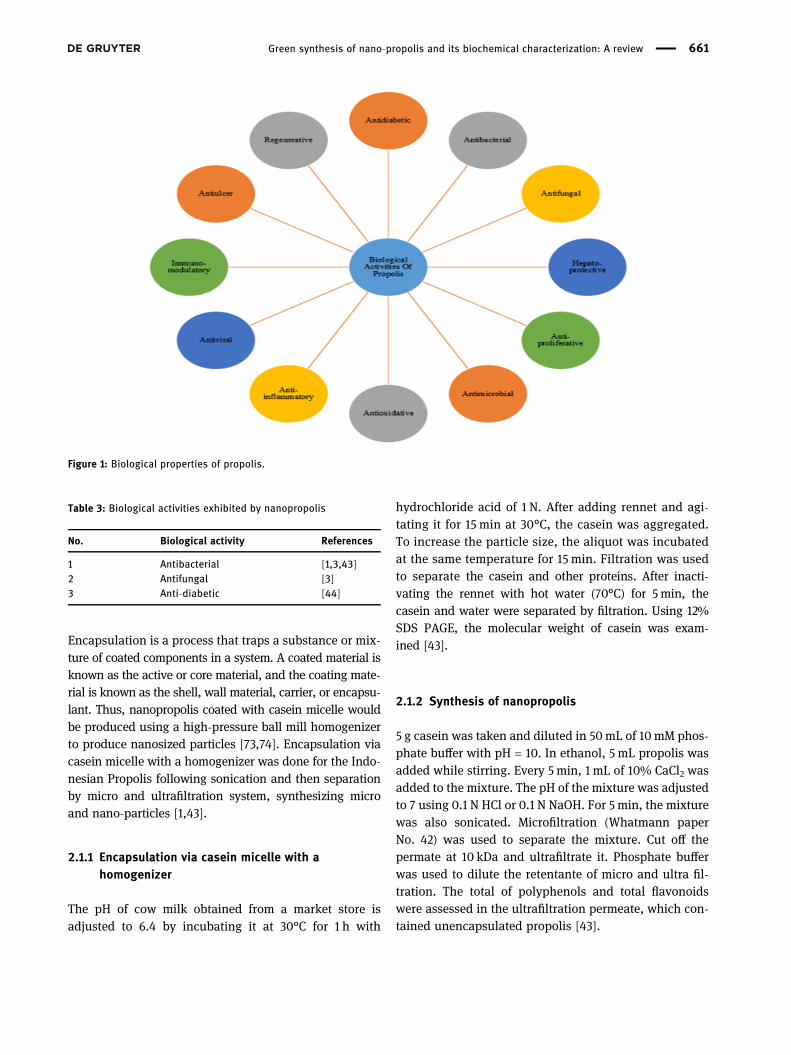
NIPAAM + VP + PEG-A + MBA were dissolved in water inmolar ratios of 90:5:5, they took 90mg NIPAAM, 5 μL offreshly distilled VP, 500 μL of PEG-A (1% w/v), 10 mL ofwater. To cross link polymer chains, 30 μL of MBA wasused. Polymerization was done using 30 μL of APS, 20 μLof TEMED, 20 μL of FAS (0.5% w/v) in N2 atmosphere at30°C. After that dialysis for 2–3 min was done in aqueousmedium for co-polymeric nanoparticles containing unreactedmonomers as given in Figure 2. Pure co-polymeric nanopar-ticles in aqueousmediumwere employed for lyophilization toobtain dry powder of co-polymeric nanoparticles. To removeany inhibitors, NIPAAM was recrystallized using hexane, VPwas freshly distilled before use, and PEG-Awas washed threetimes with n-hexane [76].
2.2.2 Preparation of propolis loaded polymericnanoparticles with polycaprolactone-pluronicpolymeric matrix
Nanoprecipitation with poly-caprolactone and pluroniccombination is another approach for the synthesis ofpropolis loaded polymeric nanoparticles. The preformedpolymer interfacial deposition method, also known asnanoprecipitation preformed polymer, was used to createcolloidal suspensions of nanoparticles. The organic andaqueous phase components were weighed and placed inseparate containers, then subjected to sonication until fin-ished evaporation. The organic phase (500 μL) was thenadded into the aqueous phase (50mL) with vortexingfor 1 min following the freeze drying of nanoparticles.Polymeric nanoparticle suspensions with loaded propoliswere submitted to the assays for characterization [75].
2.2.3 Extraction of propolis
Various authors prepared propolis extract by dissolvingdry propolis. 35 g of dried propolis powder was dissolvedin 250mL of deionized water. The extraction was carriedout by placing the flask on an orbital shaker at roomtemperature for 72 h and agitating it at 100 rpm. The aqu-eous solution was filtered with Whatman filter paperNo. 1 after extraction. Crude extract was prepared by con-centrating the extract under reduced pressure [9].
2.2.4 Loading of propolis
Loading of the propolis in the polymeric nanoparticles wasdone using a post-polymerization method. For this, in 10mLof distilledwater, 100mg of the lyophilized powderwas addedand stirred to reconstitute the micelles. In Chloroform, freepropolis was dissolved (CHCl3; 10mg‧mL−1). With constantvortexing and mild sonication, the propolis solution inCHCl3 was slowly added to the polymeric solution. By phy-sical entrapment, propolis was directly loaded into thehydrophobic core of the nanoparticles. Then, these pro-polis-loaded nanoparticles were lyophilized to dry powderfor subsequent use [75].
2.3 Biosynthesis of SeNPs
Propolis extract has potential application in green synth-esis of Se NPs due to the presence of natural reducing and
Propolis loaded dry powder of co -polymeric nanoparticles
Lyophilization
Loading (Propolis loaded in co-polymeric nanoparticles)
Dry Powder of co-polymeric nanoparticles
Lyophilization
Pure Co-polymeric nanoparticles in aqueous medium
Dialysis
Figure 2: Procedure studied in the preparation of co-polymeric nano-particles. NIPAAM–N-isopropylacrylamide; VP–N-vinyl-2-pyrrolidone;PEG-A–poly(ethyleneglycol)monoacrylate; MBA–N,N′-methylene bis-acrylamide; APS– ammonium persulfate; TEMED – tetramethyl ethy-lene diamine; FAS– ferrous ammonium sulfate [76].
662 Bindiya Barsola and Priyanka Kumari
stabilizing biocompounds in its composition, such aschalcones, flavones, phosphoric acid, acetic acid, butanol,butanoic acid, butyl ester, hydroxyl and keto waxes, ketones,terpenoids, steroids, and sugars. The reduction of seleniumion to was mostly mediated by metabolites from propolisextracts, specifically alcohol and phenolic compounds [77].
2.3.1 From ethanol extract of propolis
For the synthesis of SeNPs, three main methodologies canbe applied, including physical methods which involvespyrolysis, physical vapor deposition, crushing, lithography,grinding, attrition, and ball milling [10,78,79], chemicalmethods including solvothermal, sol gel, thermal decom-position, microwave-assisted synthesis, ultrasonic-assisted,and electrochemical methods [80–83] and biosynthetic pro-cesses [10,18]. The most prevalent method for preparingSeNPs is reduction,which includeschemical reduction [18,84],γ-radiolytic reduction [85], and bacterial reduction [86].
Main drawback of these methods is that they havelong synthesis time, requires high temperature, havehigh production cost, low yield, and also involves somehazardous chemicals affecting human health and the envir-onment as they might be genotoxic, cytotoxic, or carcino-genic. As a result green chemistry methods have beendeveloped and have become popularized which involvethe use of biological systems such as microorganisms (bac-teria, fungi, algae, and yeast) and plant extracts [87]. Use ofbiogenic synthesis of SeNPs is more advantageous as it iseconomical, non-toxic, uses non-hazardous materials, pro-duces stable nanomaterials, also have natural organic cov-ering to avoid the nanoparticles aggregation. SeNPs haveexceptional applications such as significant biocidal role,capability to provide protection against free radicals, leasttoxicological effects, and prominent biological activities.
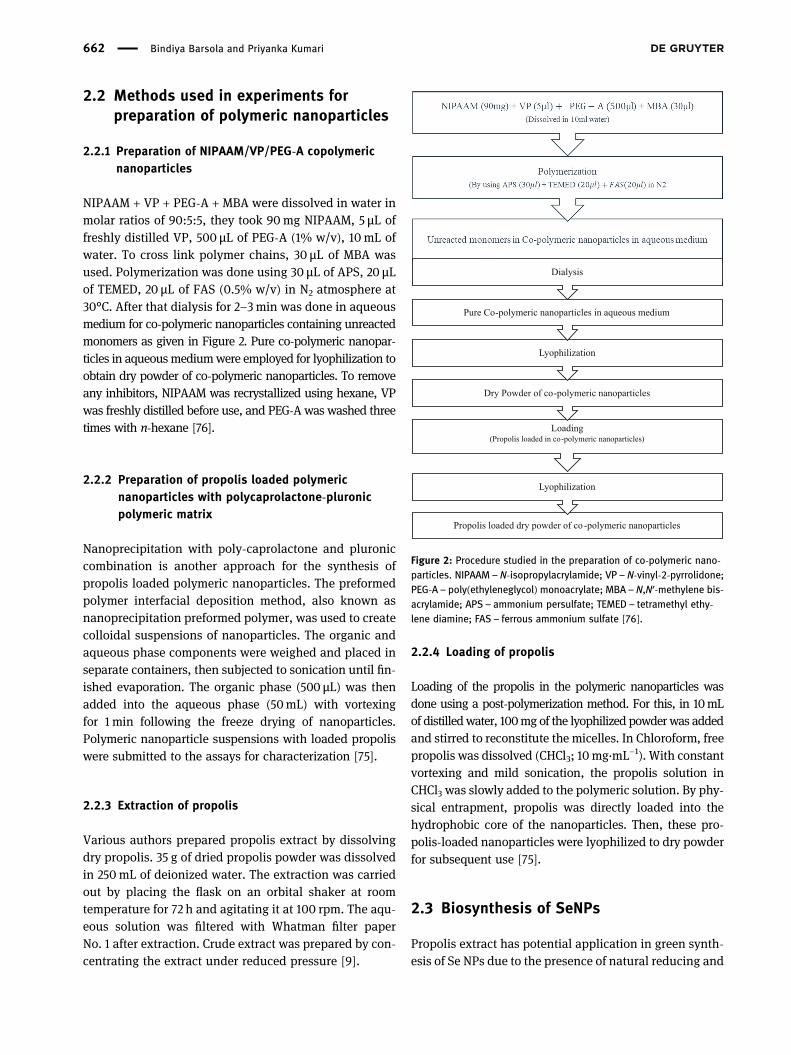
To 30mM sodium selenite solution, 10mL of freshlyprepared ethanol extract was added along with 40mMascorbic acid. Mixture was placed at room temperaturefor 24 h until the light brown color changed to orangecolor. Centrifugation was done for newly biosynthesizedSeNPs at 10,000 rpm for 30min at 4°C. Washing wasdone for the separated nanoparticles thrice with doubledistilled water and then washed by using ethanol. Afterthat SeNPs were dried overnight and their powdered formwas used for further characterization and analysis [88].Cycloheximide, a common fungicide, was employed as apositive control, and ethanol was utilized as a negativecontrol. Schematic representation is given in Figure 3.
2.3.2 Other techniques for fabrication of SeNPs withpropolis
It has been revealed in many studies that there are alsosome other techniques such as microwave irradiation,UV radiation, hydrothermal, ultrasonication, self-assem-bling, and conventional heating for the fabrication ofSeNPs with propolis extract [77]. Initially propolis extractwas added to the prepared selenium salt solution andthis combination was used as sample for the differentmethods. In hydrothermal technique, colloidal solutionwas autoclaved at 121°C for 15 min with 1.5 atm. By using800Wmicrowave device, the mixture solution was heatedfor 30 s in microwave radiation technique. In the ultraso-nication method, the probe of the equipment wasengrossed in the colloidal solution and further by usinga laboratory ultrasound device it was treated with a fre-quency and power of 20 kHz and 300W, respectively.From a 6W UV lamp, UV light of 365 nm was used forthe exposure of mixture solution at room temperature fora maximum of 1 h. Colloidal solution was kept overnightat room temperature (33°C) in the self-assembling tech-nique [77].
Figure 3: Schematic representation for the synthesis of seleniumnanoparticles from ethanolic extract of propolis.
Green synthesis of nano-propolis and its biochemical characterization: A review 663

2.3.3 From watery extract of propolis
150mL of sodium selenite solution was poured gentlydrop by drop to a conical flask containing 300mL ofpropolis watery extract solution. To avoid the effect oflight on the nanoparticle composition process, a sheetof aluminum foil was placed over the flask. By addingdrops of 1 N NaOH to the mixture, the pH was adjustedto 8, and it was agitated for 6 h at 37–40°C. Centrifugationat 12,000 rpm for 30min in 20°C separated the producednanoparticles [10].
2.4 Methods used in various studies for thepreparation of AgNPs
Due to the formation of AgNPs in the reaction mixture,the color changed from light yellow to dark brown duringthe incubation period. This color change was due to theirspecific properties (surface plasmon resonance). UV-Visspectroscopy, X-ray diffraction (XRD) pattern, transmis-sion electron microscopy (TEM), scanning electron micro-scope (SEM) with energy dispersive X-ray (EDX), atomicforce microscopy, dynamic light scattering, and thermalgravity-differential thermal analysis were all used to vali-date the synthesis of AgNPs [7]. AgNPs can be prepared byusing the green synthesis method as shown in Figure 4and by using silver nitrate and sodium hydroxide withpropolis extract.
Green synthesis method was used for the preparationof propolis AgNPs [9]. 1 mL of 10% propolis extract was
mixed with 9mL of 5 mM silver nitrate (AgNO3) at roomtemperature using a magnetic stirrer to make AgNPs[9,20,89]. Color change had been used to visually assessthe reaction, and the UV-Vis spectrumwas regularly eval-uated. For 30min, the colloidal solution of the producednanoparticles was centrifuged at 10,000 rpm. The finalAgNPs were separated and dried after the process wasrepeated three times [20].
Propolis extract, AgNO3 (Dynamics, Brazil), and sodiumhydroxide (Vetec, Brazil) were employed in the productionof AgNPs containing propolis (AgNP-P). Propolis extractswere resuspended in water and stirred on the Quimis®
Q261M23 plate, Brazil. This solution’s pH (0.1M NaOH) wasadjusted to 10.6, which is required for flavonoids to haveelectrons accessible for the silver reduction process 14. Afteradding AgNO3 and waiting for 30min, the colloidal suspen-sion was stored away from light. The experiments were car-ried out at a temperature of 32 ± 2°C [4].
3 Characterization of nanopropolisand nanoparticles (Ag and Se)
3.1 Characterization of nanopropolis
3.1.1 Determination of total encapsulated polyphenolsor flavonoids
A BA
The efficiency of encapsulation 100=
−
× (1)
Figure 4: Schematic procedure for the biosynthesis of silver nanoparticles with propolis.
664 Bindiya Barsola and Priyanka Kumari

where A– total polyphenols or flavonoids added initially,and B– total unencapsulated polyphenols or flavonoids [43].
3.1.2 Particle size analysis
The diameters of the nano/micro particles were measuredusing a laser light scattering granularity analyzer (DelsaTMNano, Germany) Beckman Coulter. A sample was taken andtriplicate analyses were performed [43].
3.1.3 TEM analysis
TEMwas used to study themicrostructure of particles whichwas operated at 80 kV. The sample was obtained by placingone preparation drop on a collodion support on grids.
3.2 Characterization of SeNPs
Nanoparticles property was determined by evaluating thecharacteristics of nanoparticles such as size, structure, andshape [3,5,18,21,57,88,90]. Different methods to characterizethe biosynthesized nanoparticles are given in Figure 5.
3.2.1 UV-Vis spectrum analysis
For the identification and characterization of propolis,best used technique was UV-Vis spectrum. By usingGenesys 10S UV-Vis spectrophotometer, the wavelengthof biosynthesized SeNPs from 200–800 nm was recordedand analyzed for the maximum absorption peaks [48].
Shimadzu UV-1600, Japan, was used by authors of ref. [10]to study the spectrumwithwave range of 190–1,100nm, scanspeed of sampling interval was 0.5min.
3.2.2 Fourier transform infrared spectroscopy analysis(FT-IR)
Shimadzu – 00463 model spectrophotometer was usedto obtain FT-IR spectrum. It was obtained in the spec-tral region of 4,000–600 cm−1 with a resolution of4 cm−1 and 64 co-added scans [88]. FT-IR spectroscopy(ABB/spectrolab/MB3000/UK) was used to examine theproduced SeNP. The FTIR characterization was doneusing the conventional KBr pellet technique, which mea-sures infrared intensity vs wavelength (wave number) oflight from 400 to 4,000 cm−1 [10]. Organic functionalgroup responsible for stabilization of nanoparticles, natureof associated functional groups, structural features of bio-logical extracts with nanoparticles and surface chemistrywas confirmed by using FT-IR spectrum [10,88].
3.2.3 XRD analysis
XRD was used to investigate the composition of thesynthesized particles [10], and also for the structureof nanoparticles and phase identification [88]. BrukerD8 Advanced Eco X-ray diffractometer having Bragg-Brentano focusing geometry was used to take the XRDreadings and then this XRD data were analyzed for theidentification of the crystalline phases by using X’perthigh score software [88].
3.2.4 SEM analysis
To illustrate the morphological characteristics and sizeof biosynthesized SeNPs, scanning electron microscope(Hitachi, Model SU3500) with ultrahigh resolution fieldemission was used [88]. To analyze the morphology andform of the particles, samples were mildly pressed intopellets at 0.5 ton-load to achieve the best picture underSEM [10,91].
3.2.5 Antimicrobial assay
3.2.5.1 Antibacterial assay – resazurin microtiter platemethod
To check the minimum inhibition concentration resa-zurin microtiter plate method was used. By this method,Figure 5: Characterization of biosynthesized nanoparticles.
Green synthesis of nano-propolis and its biochemical characterization: A review 665
antibacterial activity of the biosynthesized SeNP’s againstpathogenic gram-positive bacterial strains (Staphylococcusaureus, Bacillus cereus, and Streptococcus mutans) andgram-negative bacterial strains (Escherichia coli, Salmonellatyphi, and Pseudomonas aeruginosa) was studied. In sterilewater, resazurin solution (10%)was prepared and was storedas a stock solution at 20°C. When required it was used bydiluting to 1:10 with sterile water. 100 μL of sterilized LuriaBertani (LB) broth, along with 30 µL of 0.1% resazurin solu-tion, 100 μL of serially diluted biosynthesized SeNPs (1,000,500, 250, 125, 62.5, 31.25, 15.25, and 7.81 µg‧mL−1)were addedin eachwell of themicrotiter plate. Consequently, inoculationwas done for 100 μL of each bacterial culture, and to preventthe sample from drying, 200 μL of deionized water wasadded. The control wells were prepared with culture media,resazurin dye, and bacterial suspension. Broth and dye wereadded in color blank wells. Sealed plates were incubated for24 h at 37°C. It was observed that after incubation, the colorchanged from blue to pink, whereas no growth of organismswas noticed in blue colored wells, only pink and orangecolored wells exhibited positive results with growth of organ-isms [88].
3.2.5.2 Antifungal assay
Antifungal assay of biosynthesized SeNPs from propolisextract was studied by using well diffusion method.Pathogenic fungi such as Aspergillus niger, Aspergillusflavon, and Candida albicans were used. With solidifiedpotato dextrose agar media, fungal inoculums of Aspergillusniger, Aspergillus flavon, and Candida albicans were thor-oughly disseminated on sterilized Petri plates. Using sterilecork borer, 5 wells with 5.5 diameter were drilled out oneach agar plate. Various sample concentrations of 50 μLwere filled in these wells. Incubation of culture plate wasdone at 25°C for 72 h. For all the three pathogenic fungi, thezone of inhibition was measured in diameter (mm) sur-rounding the wells [88].
3.3 Characterization of AgNPs
The synthesis of AgNPs was monitored using a Jasco V530doublebeamUV-Visspectrophotometer to record theUV-Visspectra. The scanning speed was 1,000 nm‧min−1 [20].
Bruker Vertex 70 (USA) was used for the FTIR mea-surements with a scanning range of 4,000–310 cm−1, andthe materials were placed in KBr discs for spectral acqui-sition [20].
Delsa nano submicron particle size analyzer (BeckmanCoulter, USA)was used for the determination of particle sizeand zeta potential of the synthesized AgNPs [20].
To determine the presence of elemental silver in AgNPsQuanta 200 environmental scanning electron microscope(ESEM) with EDX silicon-drift detector was used [20]. Forthe determination of surface morphology and size, TEM(Hitachi High-Tech HT7700 transmission electron micro-scope, Japan) was used [20].
3.3.1 Analysis of photocatalytic activity of synthesizedAgNPs
Under sunlight irradiation, the degradation of malachitegreen was observed to determine the photocatalytic activityof the synthesized AgNPs. In 50mL of malachite green solu-tion (10mg‧L−1), 10mg AgNPs were added, and the mixturewas magnetically swirled for 45min in the dark. After that,the colloidal suspension was exposed to sunlight at roomtemperature with steady stirring under constant irradia-tion. The color shift was tracked over time, and the sam-ples were obtained and centrifuged every 30min. In theregion of 400–800 nm, the supernatant was scanned.Meanwhile, a control containing only the dye was testedat the same conditions, as was a sample comprising AgNPssuspended in dye solution, constantly swirled in thedark [20].
3.3.2 Phagocytosis activity assay
Phagocytosis activity assay was performed to check theeffectiveness of nanoparticles in stimulation of phago-cytic cells. Suliman’s protocol (with some modifications)was used to assess macrophage phagocytic activity. Freshhuman blood sample was isolated in tubes containingEDTA. 200 μL of nanoparticles with concentrations of10 g‧mL−1 were added to 200 μL of blood samples. For30min, the samples were incubated in a water bath at37°C. After incubation, each sample was given 20 μLof S. aureus bacterial suspension and again incubatedfor 30min at 37°C. The blood samples and bacterial sus-pensions were mixed and utilized as a positive control.Then, slide smear was prepared by taking one drop fromeach blood sample with control. Slides were dried atroom temperature. Staining was done with the help ofLeishman stain. Five drops of Leishman stain were used,and the slides were left for 10 min before observing themunder light microscope (100×) after washing with dis-tilled water [9].
666 Bindiya Barsola and Priyanka Kumari
3.3.3 Biochemical assay
3.3.3.1 In-vitro antimicrobial assay
Agar diffusion method was used to evaluate the antimi-crobial activity, for fungi, Mueller-Hintonmedium (HiMedia,Germany) was used, and for bacteria, Mueller-Hinton agar(Oxoid, UK) was used [20]. Microorganisms used for antimi-crobial assay were Staphylococcus aureus (S. aureus) ATCC25923, Pseudomonas aeruginosa (P. aeruginosa) ATCC 27853,and Candida parapsilosis (C. parapsilosis) ATCC 22019 [20].Inhibition zones in mm were measured after the samples(extract and AgNPs) were applied and incubated at 24°Cfor fungi and 37°C for bacteria for 24 h [20,92]. Ciprofloxacin(5 µg‧disc−1) and nystatin (100 µg‧disc−1) were used as posi-tive controls. All experiments were carried out in triplicate,and the findings were expressed as mean ± standard devia-tion [20].
3.3.3.2 Determination of antioxidant activity
DPPH free radical scavenging assay and lipoxygenaseinhibition assay were used to assess the antioxidantactivity of the synthesized AgNPs. IC50 values were cal-culated for samples [20].
3.3.3.3 Determination of antibacterial activity
Author of ref. [4] studied the antibacterial activity againstStaphylococcus aureus (CCCD-S007), Staphylococcus epi-dermidis (CCCD-S010), Escherichia coli (ATCC-25922), andPseudomonas aeruginosa (ATCC-27853). M-11 Protocolof Clinical and Laboratory Standards Institute 30 wasemployed to determine the antibacterial activity. Minimuminhibitory concentration (MIC) was studied by broth micro-dilution. A 50% (v/v) Tween® 20 (Dynamics, Brazil) wasused to dissolve the samples, which had an initial concen-tration of 33.3mg‧mL−1. The microorganisms were added toall samples using the McFarland 0.5% scale, at a concen-tration of 1.5 × 10⁶ CFU. The microplates were thenconditioned in a bacteriological oven at 36°C for 24 h.2,3,5-Triphenyltetrazolium chloride (1.0%) solution wasadded and allowed to react for 3 h to allow visualization.The growth control consisted of culture medium and bac-teria, the negative control was Tween® 20 at the concen-tration used, the sterility control consisted only of culturemedium, and the positive control was chlorhexidine. ForMIC, culture medium used was brain heart infusion [4].
Aliquots from the MIC plate and above were ali-quoted, seeded in Petri dishes, and transferred to a bac-teriological oven under the same conditions as before toobtain the minimum bactericidal concentration (MBC).The microbe was still alive and well, as evidenced bycolony expansion. The culture medium used for MBCwas Mueller-Hinton (Both KSVI, Brazil) [4].
4 Analysis of results and discussionsof various experiments for thecharacterization of nanopropolisand nanoparticles (Ag and Se)
4.1 Characterization of nanopropolis
4.1.1 Result for the determination of total encapsulatedpolyphenols and flavonoids
High flavonoid and moderate polyphenol capacities wereexhibited by nanopropolis [1,43]. It was reported byauthors of ref. [7] that encapsulation efficiency of totalpolyphenols and total flavonoids was 67% and 94%,respectively.
4.1.2 Particle size analysis
Particle size analyzer was used to determine the size ofparticles, variation was reported in nanoparticle sizebetween 252 and 530 nm [43].
4.2 Results for the characterization ofSeNPs
4.2.1 UV-vis spectrum analysis
In the range of 200–800 nm, the absorption spectrum wasrecorded for biosynthesized SeNPs. For selenium in thesamples, a high absorption peak was recorded between250 and 280 nm [88].
4.2.2 FT-IR analysis
FT-IR analysis confirmed the presence of different func-tional groups like alcohol and polyphenols. It was
Green synthesis of nano-propolis and its biochemical characterization: A review 667
reported that the presence of these functional groups wasmainly involved in the reduction of selenium ion toSeNPs. Peak values were observed between 2,950 and2,850 cm−1 which were due to the presence of organicpolymer lignin. It was evident from different researchpapers that absorption peak was present between 1,750and 1,470 cm−1 which corresponded to the carboxyl grouppeak, also at 1,030 and 730 cm−1, another peak wasobserved which corresponded to phenolic group [88].
4.2.3 XRD analysis
The diffraction intensity was observed from 2θ anglesvarying from 10° to 100° when XRD pattern of SeNP’swas studied. It was reported from different papersthat crystalline nature of SeNPs was determined bythe distinct peaks at 23.51°, 29.50°, 41.30°, 45.35°,and 48.10°. With lattice constants a = 4.3662 Å andc = 4.9536 Å, all the diffraction peaks recorded in the2θ range related to the hexagonal structure of Se. Thesample’s amorphous/hexagonal nano-crystalline naturewas revealed by the sharpness and broad diffraction ofthe peaks at low angle [10,88].
4.2.4 SEM analysis
For the assessment of the physical dimensions, mor-phology, and shape of the nanoparticles, widely usedtechnique is SEM. Researchers revealed that the structureand size of biosynthesized nanoparticles get influencedby the concentration of polyphenol in propolis extract.Biosynthesized SeNPs by propolis extract were found tohave spherical shape with a smooth surface and sizerange between 30 and 36 nm [10].
4.3 Results for the characterization ofAgNPs from different researchapproaches
4.3.1 Determination of MIC and MBC
It was noticed that S. aureus and P. aeruginosawere amen-able to AgNPs effect. Gram-positive bacteria (S. aureus)exhibited highest antibacterial activity that was mostprobably due to the differences in molecular makeupof cell walls [29,93]. AgNPs could be a source of reactiveoxygen species (ROS), which can damage bacteria
proteins and DNA, causing cell membrane permeabilityto change and bacterial membrane destruction [20,90].MIC values for antibacterial activity of propolis wasreported to be 4,162, 8,325, and 16,650 μg‧mL−1 forS. aureus, S. epidermidis, and P. aeruginosa, respectively.Different results had been reported in the literature: someshow that AgNPs are more effective against Gram-positivebacteria than Gram-negative bacteria [20,56], while otherslead to the opposite conclusion [20,24] given in Table 4.Furthermore, Gram-negative bacteria had an exteriorlipidic membrane with a negative surface charge. TheAgNPs linked with propolis (AgNP-P) share the samesurface charge, which can cause electrostatic repulsionbetween them, making it difficult to fix and penetrateAgNPs into bacterial cells and necessitating a greaterconcentration to kill this type of bacteria.
4.3.2 Visual and UV-Vis analysis
AgNPs’ biosynthesis was confirmed by observing thechange in color of the reaction mixture which is due tothe excitation of surface plasmon vibrations in AgNPs[7,20,94]. The color of the reaction mixture changedfrom white-yellowish to brown [20,94]. After continuousstirring, the UV-Vis spectra of the mixture extract, silvernitrate, was recorded in the 350–600 nm wavelengthregion, revealing the surface plasmon resonance [20].Due to the formation of small spherical nanoparticles,an absorption band at 480 nm was recorded. AgNPsderived from aqueous propolis extract were active at ashorter wavelength [20]. Many other studies revealedthe presence of peak at 420 nm [7], and in the propolissamples from Tamilnadu, absorption spectra was observedbetween 260 and 290 nm [95].
4.3.3 FT-IR analysis
FT-IR analysis was performed to determine the biomole-cules identified in the propolis extract that are respon-sible for the reduction, capping, and stabilization of
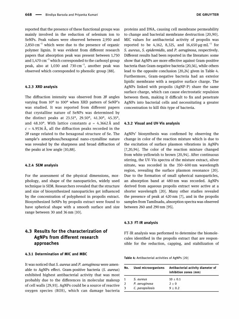
Table 4: Antibacterial activities of AgNPs [20]
No. Used microorganisms Antibacterial activity diameter ofinhibition zones (mm)
1 S. aureus 10 ± 0.12 P. aeruginosa 2 ± 03 C. parapsilosis 9 ± 0.2
668 Bindiya Barsola and Priyanka Kumari

AgNPs. From the propolis extract the functional groupswere recorded at different wavelengths [20]. The car-boxylic group was detected in the FT-IR spectrum of thepropolis aqueous extract without AgNO3 with an intensepeak at 1,716 and 1,100 cm−1 [7]. The stretching vibrationsof OH fromphenolic compoundswere observed at 3,403 cm−1
and stretching vibration of C–H were recorded at 2,919 and2,849 cm−1. 1,635 cm−1 mainly corresponded to stretchingvibration of C]C and C]O groups from flavonoids andasymmetric bending vibration of N–H from amino acids.Due to stretching C]C of aromatic ring, bending vibrationof C–H from CH3, CH2, and the stretching vibration of aro-matics from flavonoids and aromatic rings’ peak wererecorded at 1,514 and 1,449 cm−1, respectively. 1,264 cm−1
corresponded to C–O group of polyols (hydroxyflavo-noids), 1,082 cm−1 due to C–O stretching ester group,815 cm−1 due to aromatic ring vibration, and 698 cm−1
corresponded to phenyl group [20,96]. The majority ofthe FT-IR bands in propolis aqueous extract were due totriterpenoids, flavonoids, furanoids, sugars, coumarins,quinines, tannins, phenols, and acids [7]. Some peaks inthe FT-IR spectra of AgNPs shifted or vanished as aresult, indicating that particular polyphenols, flavo-noids, or amino acids are involved in the formation ofnanoparticles [20].
4.3.4 Particle size and TEM analysis
The majority of AgNPs were spherical in shape [7,20] andranged in size from 10 to 50 nm, with an average size ofabout 15 nm [20], 9–30 nm [7]. The TEM pictures revealeda weak aggregation tendency, which could be due to theextract’s contribution to AgNPs’ stabilization [20]. In astudy, it was reported that the size of nanoparticleswith ethanolic extract of propolis at pH = 10.62 was50 nm and at same pH for water extract of propolis, itwas 20 nm [97]. It was revealed in the field emissionSEM analysis that the size of synthesized AgNPs was62, 41, and 82 nm for the samples from different regionsof Tamilnadu [95].
4.3.5 Determination of zeta potential
AgNPs coated with polyphenolic chemicals found in pro-polis extract had a negative zeta potential of −21.36 mV.This suggested that the suspension of colloidal nanopar-ticles was stable [20,24]. The AgNPs’ negatively chargedsurface reduces particle agglomeration by preventingparticle rejection [20,57].
4.3.6 Determination of elemental silver in AgNPs
The existence of elemental silver was confirmed by theEDX spectra of AgNPs, which showed a peak pattern at3 keV in the silver region [7,20,93]. Apart from silver, themost prominent elements in the chemical composition ofAgNPs were carbon, which made up 53.26%, oxygen –14.06%, and nitrogen –which made up 03.12%, showingthe presence of compounds connected to nanoparticles[20]. After 4 h, a spectroscopic signal indicating the pro-duction of AgNPs was seen around 480 nm. In the extractand silver nitrate spectra, this peak does not appear.AgNPs are polydispersed, as seen by the spreading con-tour of the peak [5,295]. Atomic % reported via EDX spec-trum of AgNPs was 91.23, 1.71, 0.66, 0.53, 0.96, and 4.91for elements C-K, Na-K, Mg-K, Si-K, Ca-K, and Ag-L,respectively [95].
4.3.7 Analysis of photocatalytic activity
Malachite green (colorant) degradation was analyzed todetermine the photocatalytic activity of the synthesizedAgNPs. Malachite green, as well as its reduced form, canbe found in both aquatic and terrestrial systems, posing arisk to human health [20,98]. Initially, the photocatalyticdegradation was analyzed by color change, a color shiftfrom dark blue to light blue was noticed. Thereafter, thediminution in color was measured spectrophotometri-cally by recording the absorbance at 617 nm. It could bepossible to get a significant decline in absorbance overtime, indicating a decrease in dye concentration owing todegradation. The control containing merely the dye didnot change color when exposed to sunshine, while thecolor and absorbance of the sample mixed in the darkdropped.
Dye degradation was calculated by the formula:
C CC
Dye degradation % 100to
o=
−
× (2)
where Co is the initial concentration of the malachitegreen, and Ct is the concentration of the malachite greenafter each period of exposure time to sunlight [20,81]. Itwas illustrated that the obtained AgNPs were highlyphotocatalytically active under light radiation as whenthe sample was exposed to sunlight, dye degradationobtained was 90%, for the control degradation it was5%, and 30% for the sample mixed in dark. Photonsof visible light hitting AgNPs are absorbed via thesurface plasmon resonance phenomenon, which causes
Green synthesis of nano-propolis and its biochemical characterization: A review 669

electrons to be excited to a higher energy state duringsunlight exposure.
4.3.8 Determination of phagocytosis activity assay
Phagocytosis is a process in which phagocytic cells engulf,destroy, and digest bacteria or foreign substances [9,99].Significant increase was noticed in the activity of phago-cytic cells. This increased activity of phagocytic cells toengulf bacteria could be due to certain chemical compo-nents present in the AgNPs of propolis which acted asimmune modulators [9].
5 Conclusion
It was observed that due to the presence of natural redu-cing and stabilizing compounds in propolis extract, theyhad potential application in green synthesis of nanopar-ticles. A significant number of chemical componentsfound in propolis have a synergistic influence in the for-mation of AgNPs. Nanopropolis exhibited high flavonoidcontent. UV-Vis spectra, XRD, FT-IR, and SEM images allverified the production of nanoparticles and their func-tionalization with ethanol extract of bee propolis. TheseSeNPs had shown substantial antioxidant action againstbacterial and fungal strains. So they could be utilized todestroy pathogens in order to prevent diseases. In thisreview, applicability of propolis for biosynthesis of nano-particles and its biomedical applications has been explored.Moreover, the role of propolis extract as stabilizer and reducerwas confirmed by the biogenic synthesis of AgNPs. The find-ings of this review represents a promising nanoproduct withits pharmaceutical and biomedical applications.
6 Research possibilities
It was evident from different studies that alcohol andphenolic compounds found in propolis extracts were pri-marily responsible for the reduction of selenium ion toSeNPs [88]. The biosynthesized SeNPs from propolisextract could be a powerful antioxidant and antibacterialagent used to cure diseases caused by bacterial andfungal strains [3,88]. Still more research is needed toenhance propolis-based nanoparticles synthesis, their effi-ciency, particle size control, and use in medicine and health-care [88]. Further, biosynthesized nanoparticles from propolis
extract can also be checked for antitumoral, regenerativeproperties and its therapeutic efficacy.
Acknowledgment: Throughout the writing of this reviewarticle I have received a great deal of support and assis-tance. I would like to thank my supervisor, Dr. PriyankaKumari, whose expertise was invaluable in formulatingthis review article.
Funding information: Authors state no funding involved.
Author contributions: Priyanka Kumari and Bindiya Barsolahave made substantial contributions to conception anddesign, acquisition of data, analysis and interpretation ofdata from research papers. Priyanka Kumari has given finalapproval of the version to be published. Bindiya Barsolahave been involved in drafting the manuscript or revisingit critically for important intellectual content.
Conflict of interest: Authors state no conflict of interest.
References
[1] Tatli Seven P, Seven I, Gul Baykalir B, Iflazoglu Mutlu S,Salem AZ. Nanotechnology and nano-propolis in animalproduction and health: An overview. Ital J Anim Sci.2018;17(4):921–30. doi: 10.1080/1828051X.2018.1448726.
[2] Chakravarthi V, Balaji S. Applications of nanotechnology inveterinary medicine. Vet World. 2010;3(10):477–80.doi: 10.5455/vetworld.2010.
[3] Afrouzan H, Amirinia C, Mirhadi SA, Ebadollahi A, Vaseji N,Tahmasbi G. Evaluation of antimicrobial activity of propolisand nanopropolis against Staphylococcus aureus andCandida albicans. Afr J Microbiol Res. 2012;6(2):421–5.doi: 10.5897/AJMR.
[4] Barbosa VT, Souza JK, Alvino V, Meneghetti MR, Florez‐Rodriguez PP, Moreira RE, et al. Biogenic synthesis of silvernanoparticles using Brazilian propolis. Biotechnol Prog.2019;35(6):e2888. doi: 10.1002/btpr.2888.
[5] Kagithoju S, Godishala V, Nanna RS. Eco-friendly and greensynthesis of silver nanoparticles using leaf extract ofStrychnos potatorum Linn. F. and their bactericidal activities. 3Biotech. 2015;5(5):709–14. doi: 10.1007/s13205-014-0272-3.
[6] Leela A, Vivekanandan M. Tapping the unexploited plantresources for the synthesis of silver nanoparticles. Afr JBiotechnol. 2008;7(17):3162–5. http://www.academicjournals.org/AJB
[7] Priyadarshini JF, Sivakumari K, Selvaraj R, Ashok K,Jayaprakash P, Rajesh S. Green synthesis of silver nanoparti-cles from propolis. Res J Life Sci Bioinform Pharm Chem Sci.2018;4:23–36. doi: 10.26479/2018.0404.02.
[8] De Marco S, Piccioni M, Pagiotti R, Pietrella D. Antibiofilm andantioxidant activity of propolis and bud poplar resins versus
670 Bindiya Barsola and Priyanka Kumari
Pseudomonas aeruginosa. eCAM. 2017;2017:1–11.doi: 10.1155/2017/5163575.
[9] Taqi ZJ, Abdul-Wahed HE, AL-Saadi HK, Jabir MS. Potentialactivity of silver nanoparticles synthesized by Iraqi propolis onphagocytosis. AIP Conf. Proc. 2020;2213(1):020104.doi: 10.1063/5.0000155.
[10] Wali AT. Biosynthesis, characterization and bioactivity ofselenium nanoparticles synthesized by propolis. Iraqi J VetMed. 2019;43(1):197–209. doi: 10.30539/iraqijvm.v43i1.490.
[11] Nancharaiah YV, Lens PN. Selenium biomineralization forbiotechnological applications. Trends biotechnol.2015;33(6):323–30. doi: 10.1016/j.tibtech.2015.03.004.
[12] Sondi I, Salopek-Sondi B. Silver nanoparticles as antimicro-bial agent: a case study on E. coli as a model for Gram-nega-tive bacteria. J Colloid Interface Sci. 2004;275(1):177–82.doi: 10.1016/j.jcis.2004.02.012.
[13] Dubey SP, Lahtinen M, Sillanpää M. Green synthesis andcharacterizations of silver and gold nanoparticles using leafextract of Rosa rugosa. Colloids Surf A Physicochem Eng Asp.2010;364(1–3):34–41. doi: 10.1016/j.colsurfa.2010.04.023.
[14] Ndwandwe BK, Malinga SP, Kayitesi E, Dlamini BC. Advancesin green synthesis of selenium nanoparticles and their appli-cation in food packaging. Int J Food Sci. 2020;56(6):2640–50.doi: 10.1111/ijfs.14916.
[15] Lu J, Holmgren A. Selenoproteins. J Biol Chem.2009;284(2):723–7. doi: 10.1074/jbc.R800045200.
[16] Hadrup N, Loeschner K, Mandrup K, Ravn-Haren G,Frandsen HL, Larsen EH, et al. Subacute oral toxicity investi-gation of selenium nanoparticles and selenite in rats. DrugChem Toxicol. 2019;42(1):76–83. doi: 10.1080/01480545.2018.1491589.
[17] Levander OA. Clinical consequences of low selenium intakeand its relationship to vitamin E. Ann N Y Acad Sci.1982;393(1):70–82. doi: 10.1111/j.1749-6632.1982.tb31233.x.
[18] Dwivedi C, Shah CP, Singh K, Kumar M, Bajaj PN. An organicacid-induced synthesis and characterization of seleniumnanoparticles. J Nanotechnol. 2011;2011:1–6. doi: 10.1155/2011/651971.
[19] Cao XB, Xie Y, Zhang SY, Li FQ. Ultra‐Thin Trigonal SeleniumNanoribbons Developed from Series‐Wound Beads. Adv Mater.2004;16(7):649–53. doi: 10.1002/adma.200306317.
[20] Corciova A, Mircea C, Burlec AF, Cioanca O, Tuchilus C, Fifere A,et al. Antioxidant, antimicrobial and photocatalytic activitiesof silver nanoparticles obtained by bee propolis extractassisted biosynthesis. Farmacia. 2019;67(3):482–9.doi: 10.31925/farmacia.2019.3.16.
[21] Kumar DA, Palanichamy V, Roopan SM. Green synthesis ofsilver nanoparticles using Alternanthera dentata leaf extract atroom temperature and their antimicrobial activity.Spectrochim Acta A Mol Biomol Spectrosc. 2014;127:168–71.doi: 10.1016/j.saa.2014.02.058.
[22] Khatoon N, Mazumder JA, Sardar M. Biotechnological appli-cations of green synthesized silver nanoparticles. J NanosciCurr Res. 2017;2(1):107.
[23] Heydari R. Biological applications of biosynthesized silvernanoparticles through the utilization of plant extracts. HerbMed J. 2017;2(2):87–95. doi: 10.22087/hmj.v2i2.618.
[24] Patil S, Chaudhari G, Paradeshi J, Mahajan R, Chaudhari BL.Instant green synthesis of silver-based herbo-metallic col-loidal nanosuspension in Terminalia bellirica fruit aqueous
extract for catalytic and antibacterial applications. 3 Biotech.2017;7(1):36. doi: 10.1007/s13205-016-0589-1.
[25] Jagtap UB, Bapat VA. Green synthesis of silver nanoparticlesusing Artocarpus heterophyllus Lam. seed extract and itsantibacterial activity. Ind Crop Prod. 2013;46:132–7.doi: 10.1016/j.indcrop.2013.01.019.
[26] Chaloupka K, Malam Y, Seifalian AM. Nanosilver as a newgeneration of nanoproduct in biomedical applications. TrendsBiotechnol. 2010;28(11):580–8. doi: 10.1016/j.tibtech.2010.07.006.
[27] Prow TW, Grice JE, Lin LL, Faye R, Butler M, Becker W, et al.Nanoparticles and microparticles for skin drug delivery. AdvDrug Deliv Rev. 2011;63(6):470–91. doi: 10.1016/j.addr.2011.01.012.
[28] Chaudhry Q, Castle L. Food applications of nanotechnologies:an overview of opportunities and challenges for developingcountries. Trends Food Sci Technol. 2011;22(11):595–603.doi: 10.1016/j.tifs.2011.01.001.
[29] Nair R, Varghese SH, Nair BG, Maekawa T, Yoshida Y,Kumar DS. Nanoparticulate material delivery to plants. PlantSci. 2010;179(3):154–63. doi: 10.1016/j.plantsci.2010.04.012.
[30] Kelly FM, Johnston JH. Colored and functional silver nanopar-ticle−wool fiber composites. ACS Appl Mater Interfaces.2011;3(4):1083–92. doi: 10.1021/am101224v.
[31] Dankovich TA, Gray DG. Bactericidal paper impregnated withsilver nanoparticles for point-of-use water treatment. Env SciTechnol. 2011;45(5):1992–8. doi: 10.1021/es103302t.
[32] Küünal S, Kutti S, Rauwel P, Wragg D, Hussainova I, Rauwel E.New methodology for the antifungal testing of surfactant-freesilver metal nanoparticles for applications in green housing.Key Eng Mater. 2016;674:133–8. doi: 10.4028/www.scienti-fic.net/KEM.674.133.
[33] Kim DM, Lee GD, Aum SH, Kim HJ. Preparation of propolisnanofood and application to human cancer. Biol Pharm Bull.2008;31(9):1704–10. doi: 10.1248/bpb.31.1704.
[34] Silva-Carvalho R, Baltazar F, Almeida-Aguiar C. Propolis: acomplex natural product with a plethora of biological activitiesthat can be explored for drug development. eCAM.2015;2015:1–29. doi: 10.1155/2015/206439.
[35] Daleprane JB, Abdalla DS. Emerging roles of propolis: anti-oxidant, cardioprotective, and antiangiogenic actions. eCAM.2013;2013:1–8. doi: 10.1155/2013/175135.
[36] Ghisalberti EL. Propolis: a review. Bee world.1979;60(2):59–84. doi: 10.1080/0005772X.1979.11097738.
[37] Bankova V. Chemical diversity of propolis and the problem ofstandardization. J Ethnopharmacol. 2005;100(1–2):114–7.doi: 10.1016/j.jep.2005.05.004.
[38] Banskota AH, Nagaoka T, Sumioka LY, Tezuka Y, Awale S,Midorikawa K, et al. Antiproliferative activity of theNetherlands propolis and its active principles in cancer celllines. Ethnopharmacol. 2002;80(1):67–73. doi: 10.1016/s0378-8741(02)00022-3.
[39] Marcucci MC. Propolis: chemical composition, biologicalproperties and therapeutic activity. Apidologie.1995;26(2):83–99. doi: 10.1051/apido:19950202.
[40] Athikomkulchai S, Awale S, Ruangrungsi N, Ruchirawat S,Kadota S. Chemical constituents of Thai propolis. Fitoterapia.2013;88:96–100. doi: 10.1016/j.fitote.2013.04.008.
[41] Bueno-Silva B, Alencar SM, Koo H, Ikegaki M, Silva GV,Napimoga MH, et al. Anti-inflammatory and antimicrobial
Green synthesis of nano-propolis and its biochemical characterization: A review 671
evaluation of neovestitol and vestitol isolated from Brazilianred propolis. J Agric Food Chem. 2013;61(19):4546–50.doi: 10.1021/jf305468f.
[42] Sforcin JM. Propolis and the immune system: a review.J Ethnopharmacol. 2007;113(1):1–14. doi: 10.1016/j.jep.2007.05.012.
[43] Sahlan M, Supardi T. Encapsulation of Indonesian propolis bycasein micelle. Int J Pharma Bio Sci. 2013;4(1):297–305.
[44] Chung NK, Cho YC, Ha CS, Kim HS. Hypoglycemic effects ofnano powder propolis on streptozotocin-induced diabetic rats.Korean J Vet Serv. 2010;33(2):199–206.
[45] Dkhil MA, Zrieq R, Al-Quraishy S, Abdel Moneim AE. Seleniumnanoparticles attenuate oxidative stress and testiculardamage in streptozotocin-induced diabetic rats. Molecules.2016;21(11):1517. doi: 10.3390/molecules21111517.
[46] Aaseth J, Alexander J, Bjørklund G, Hestad K, Dusek P,Roos PM, et al. Treatment strategies in Alzheimer’s disease: areview with focus on selenium supplementation. Biometals.2016;29(5):827–39. doi: 10.1007/s10534-016-9959-8.
[47] Peng F, Guo X, Li Z, Li C, Wang C, Lv W, et al. Antimutageniceffects of selenium-enriched polysaccharides from pyracanthafortuneana through suppression of cytochrome P450 1A sub-family in the mouse liver. Molecules. 2016;21(12):1731.doi: 10.3390/molecules21121731.
[48] Hassan CE, Webster TJ. The effect of red-allotrope seleniumnanoparticles on head and neck squamous cell viability andgrowth. Int J Nanomed. 2016;11:3641–54. doi: 10.2147/IJN.S105173.
[49] Stolzoff M, Webster TJ. Reducing bone cancer cell functionsusing selenium nanocomposites. J Biomed Mater Res.2016;104(2):476–82. doi: 10.1002/jbm.a.35583.
[50] Maiyo F, Singh M. Selenium nanoparticles: Potential in cancergene and drug delivery. Nanomedicine. 2017;12(9):1075–89.doi: 10.2217/nnm-2017-0024.
[51] Rayman MP. The importance of selenium to human health.lancet. 2000;356(9225):233–41. doi: 10.1016/S0140-6736(00)02490-9.
[52] Wang Q, Larese-Casanova P, Webster TJ. Inhibition of variousgram-positive and gram-negative bacteria growth on seleniumnanoparticle coated paper towels. Int J Nanomed.2015;10:2885–94. doi: 10.2147/IJN.S78466.
[53] Shakibaie M, Mohazab NS, Mousavi SAA. Antifungal activity ofselenium nanoparticles synthesized by Bacillus species Msh-1against Aspergillus fumigatus and Candida albicans.Jundishapur J Microbiol. 2015;8(9):e26381. doi: 10.5812/jjm.26381.
[54] Guisbiers G, Wang Q, Khachatryan E, Mimun LC, Mendoza-Cruz R, Larese-Casanova P, et al. Inhibition of E. coli and S.aureus with selenium nanoparticles synthesized by pulsedlaser ablation in deionized water. Int J Nanomed.2016;11:3731–6. doi: 10.2147/IJN.S106289.
[55] Wijnhoven SW, Peijnenburg WJ, Herberts CA, Hagens WI,Oomen AG, Heugens EH, et al. Nano-silver–a review of avail-able data and knowledge gaps in human and environmentalrisk assessment. Nanotoxicology. 2009;3(2):109–38.doi: 10.1080/17435390902725914.
[56] Khalil MM, Ismail EH, El-Baghdady KZ, Mohamed D. Greensynthesis of silver nanoparticles using olive leaf extract and itsantibacterial activity. Arab J Chem. 2014;7(6):1131–9.doi: 10.1016/j.arabjc.2013.04.007.
[57] Padalia H, Moteriya P, Chanda S. Green synthesis of silvernanoparticles from marigold flower and its synergistic anti-microbial potential. Arab J Chem. 2015;8(5):732–41.doi: 10.1016/j.arabjc.2014.11.015.
[58] Pal S, Tak YK, Song JM. Does the antibacterial activity of silvernanoparticles depend on the shape of the nanoparticle? Astudy of the gram-negative bacterium Escherichia coli. ApplEnv Microbiol. 2007;73(6):1712–20. doi: 10.1128/AEM.02218-06.
[59] Kim JS, Kuk E, Yu KN, Kim JH, Park SJ, Lee HJ, et al.Antimicrobial effects of silver nanoparticles. NanomedNanomed – Nanotechnol. 2007;3(1):95–101. doi: 10.1016/j.nano.2006.12.001.
[60] Baker C, Pradhan A, Pakstis L, Pochan DJ, Shah SI. Synthesisand antibacterial properties of silver nanoparticles.J Nanosci Nanotechnol. 2015;5(2):244–9. doi: 10.1166/jnn.2005.034.
[61] Shahverdi AR, Minaeian S, Shahverdi HR, Jamalifar H, Nohi AA.Rapid synthesis of silver nanoparticles using culture super-natants of Enterobacteria: a novel biological approach.Process Biochem. 2007;42(5):919–23. doi: 10.1016/j.procbio.2007.02.005.
[62] Sharma VK, Yngard RA, Lin Y. Silver nanoparticles: greensynthesis and their antimicrobial activities. Adv ColloidInterface Sci. 2009;145(1–2):83–96. doi: 10.1016/j.cis.2008.09.002.
[63] Stoimenov PK, Klinger RL, Marchin GL, Klabunde KJ. Metaloxide nanoparticles as bactericidal agents. Langmuir.2002;18(17):6679–86. doi: 10.1021/la0202374.
[64] Cao Y, Jin R, Mirkin CA. DNA-modified core− shell Ag/Aunanoparticles. J Am Chem Soc. 2001;123(32):7961–2.doi: 10.1021/ja011342n.
[65] Kim JS, Kuk E, Yu KN, Kim JH, Park SJ, Lee HJ, et al.Antimicrobial effects of silver nanoparticles. NanomedNanomed-Nanotechnol. 2007;3(1):95–101. doi: 10.1016/j.nano.2006.12.001.
[66] Panáček A, Kolář M, Večeřová R, Prucek R, Soukupova J,Kryštof V, et al. Antifungal activity of silver nanoparticlesagainst Candida spp. Biomaterials. 2009;30(31):6333–40.doi: 10.1016/j.biomaterials.2009.07.065.
[67] Sun RWY, Chen R, Chung NPY, Ho CM, Lin CLS, Che CM. Silvernanoparticles fabricated in Hepes buffer exhibit cytoprotectiveactivities toward HIV-1 infected cells. ChemComm.2005;40:5059–61. doi: 10.1039/B510984A.
[68] Elechiguerra JL, Burt JL, Morones JR, Bragado AC, Gao X,Lara HH, et al. Interaction of silver nanoparticles with HIV-1.J Nanobiotechnol. 2005;3(1):6. doi: 10.1186/1477-3155-3-6.
[69] Lara HH, Ayala-Nuñez NV, Ixtepan-Turrent L, Rodriguez-Padilla C. Mode of antiviral action of silver nanoparticlesagainst HIV-1. J Nanobiotechnol. 2010;8(1):1–10. doi: 10.1186/1477-3155-8-1.
[70] Suvith VS, Philip D. Catalytic degradation of methylene blueusing biosynthesized gold and silver nanoparticles.Spectrochim Acta A Mol Biomol Spectrosc. 2014;118:526–32.doi: 10.1016/j.saa.2013.09.016.
[71] Kumar P, Govindaraju M, Senthamilselvi S, Premkumar K.Photocatalytic degradation of methyl orange dye using silver(Ag) nanoparticles synthesized from Ulva lactuca.Colloids Surf B. 2013;103:658–61. doi: 10.1016/j.colsurfb.2012.11.022.
672 Bindiya Barsola and Priyanka Kumari
[72] Jiang ZJ, Liu CY, Sun LW. Catalytic properties of silver nano-particles supported on silica spheres. J Phys Chem.2005;109(5):1730–5. doi: 10.1021/jp046032g.
[73] Madene A, Jacquot M, Scher J, Desobry S. Flavour encapsula-tion and controlled release–a review. Int J Food Sci.2006;41(1):1–21. doi: 10.1111/j.1365-2621.2005.00980.x.
[74] Hamdi D, Wijanarko A, Hermansyah H, Asih SC, Sahlan M.Production of nanopropolis using high pressure ball millhomogenizer. IOP Conf Ser Earth Env Sci. 2019;217(1):012014.doi: 10.1088/1755-1315/217/1/012014.
[75] do Nascimento TG, da Silva PF, Azevedo LF, da Rocha LG, deMoraes Porto IC, Lima E, et al. Polymeric nanoparticles ofBrazilian red propolis extract: preparation, characterization,antioxidant and leishmanicidal activity. Nanoscale res lett.2016;11(1):301. doi: 10.1186/s11671-016-1517-3.
[76] Bisht S, Feldmann G, Soni S, Ravi R, Karikar C, Maitra A, et al.Polymeric nanoparticle-encapsulated curcumin (“nanocurcumin”):a novel strategy for human cancer therapy. J Nanobiotechnology.2007;5(1):1–18. doi: 10.1186/1477-3155-5-3.
[77] Hatami R, Javadi A, Jafarizadeh-Malmiri H. Effectiveness of sixdifferent methods in green synthesis of selenium nanoparti-cles using propolis extract: Screening and characterization.Green Process Synth. 2020;9(1):685–92. doi: 10.1515/gps-2020-0065.
[78] Zhang J, Wang H, Yan X, Zhang L. Comparison of short-termtoxicity between Nano-Se and selenite in mice. Life Sci.2005;76(10):1099–109. doi: 10.1016/j.lfs.2004.08.015.
[79] Noah NM, Ndangili PM. Green synthesis of nanomaterials fromsustainable materials for biosensors and drug delivery. SensInt. 2022;3:100166. doi: 10.1016/j.sintl.2022.100166.
[80] Iranifam M, Fathinia M, Rad TS, Hanifehpour Y, Khataee AR,Joo SW. A novel selenium nanoparticles-enhanced chemilu-minescence system for determination of dinitrobutylphenol.Talanta. 2013;107:263–9. doi: 10.1016/j.talanta.2012.12.043.
[81] Latha D, Arulvasu C, Prabu P, Narayanan V. Photocatalyticactivity of biosynthesized silver nanoparticle from leaf extractof Justicia adhatoda. Mech Mater Sci Eng J. 2017;9(1)):1–6.doi: 10.2412/mmse.81.72.41.
[82] Tan Y, Wang Y, Jiang L, Zhu D. Thiosalicylic acid-functionalizedsilver nanoparticles synthesized in one-phase system.J Colloid Interf Sci. 2002;249(2):336–45. doi: 10.1006/jcis.2001.8166.
[83] Petit C, Lixon P, Pileni MP. In situ synthesis of silvernanocluster in AOT reverse micelles. J Phys Chem.1993;97(49):12974–83. doi: 10.1021/j100151a054.
[84] Wing-WaháYam V. High-yield synthesis of selenium nanowiresin water at room temperature. ChemComm. 2006;9:1006–8.doi: 10.1039/b515025f.
[85] Zhu Y, Qian Y, Huang H, Zhang M. Preparation of nanometer-size selenium powders of uniform particle size by γ-irradia-tion. Mater Lett. 1996;28(1–3):119–22. doi: 10.1016/0167-577X(96)00046-8.
[86] Oremland RS, Herbel MJ, Blum JS, Langley S, Beveridge TJ,Ajayan PM, et al. Structural and spectral features of seleniumnanospheres produced by Se-respiring bacteria. Appl EnvMicrobiol. 2004;70(1):52–60. doi: 10.1128/AEM.70.1.52-60.2004.
[87] Hosnedlova B, Kepinska M, Skalickova S, Fernandez C,Ruttkay-Nedecky B, Peng Q, et al. Nano-selenium and itsnanomedicine applications: a critical review. Int J Nanomed.2018;13:2107–28. doi: 10.2147/IJN.S157541.
[88] Shubharani R, Mahesh M, Murthy V. Biosynthesis andCharacterization, Antioxidant and Antimicrobial Activities ofSelenium Nanoparticles from Ethanol Extract of Bee Propolis.J Nanomed Nanotechnol. 2019;10(1):1–7. doi: 10.4172/2157-7439.1000522.
[89] Alsaedi II, Taqi ZJ, Hussien AMA, Sulaiman GM, Jabir MS.Graphene nanoparticles induces apoptosis in MCF-7 cellsthrough mitochondrial damage and NF-KB pathway. Mater ResExp. 2019;6(9):095413. doi: 10.1088/2053-1591/ab33af.
[90] Nayagam V, Gabriel M, Palanisamy K. Green synthesis of silvernanoparticles mediated by Coccinia grandis and Phyllanthusemblica: a comparative comprehension. Appl Nanosci.2018;8(3):205–19. doi: 10.1007/s13204-018-0739-3.
[91] Wang Z, Fang C, Megharaj M. Characterization of iron–poly-phenol nanoparticles synthesized by three plant extracts andtheir fenton oxidation of azo dye. ACS Sustain. Chem Eng.2014;2(4):1022–5. doi: 10.1021/sc500021n.
[92] Ioannou E, Poiata A, Hancianu M, Tzakou O. Chemical com-position and in vitro antimicrobial activity of the essential oilsof flower heads and leaves of Santolina rosmarinifolia L. fromRomania. Nat Prod Res. 2007;21(1):18–23. doi: 10.1080/14786410600921706.
[93] Kumar TVR, Murthy JSR, Rao MN, Bhargava Y. Evaluation ofsilver nanoparticles synthetic potential of Couroupita guia-nensis Aubl, flower buds extract and their synergistic anti-bacterial activity. 3 Biotech. 2016;6(1):92. doi: 10.1007/s13205-016-0407-9.
[94] Shetty P, Supraja N, Garud M, Prasad TNVKV. Synthesis,characterization and antimicrobial activity of Alstonia scho-laris bark-extract-mediated silver nanoparticles.J Nanostructure Chem. 2014;4(4):161–70. doi: 10.1007/s40097-014-0132-z.
[95] Jayanthi B. Biosynthesis of Silver Nanoparticles using sting-less bee propolis and their biomedical applications. PhD, PostGraduate and Research Department of Chemistry EthirajCollege for Women (Autonomous) Chennai; 2017.
[96] Oliveira RN, Mancini MC, Oliveira FCSD, Passos TM, Quilty B,Thiré RMDSM, et al. FTIR analysis and quantification of phe-nols and flavonoids of five commercially available plantextracts used in wound healing. Rev Mater. 2016;21:767–79.doi: 10.1590/S1517-707620160003.0072.
[97] Roy N, Mondal S, Laskar RA, Basu S, Mandal D, Begum NA.Biogenic synthesis of Au and Ag nanoparticles by Indian pro-polis and its constituents. Colloids Surf B. 2010;76(1):317–25.doi: 10.1016/j.colsurfb.2009.11.011.
[98] Cha CJ, Doerge DR, Cerniglia CE. Biotransformation of mala-chite green by the fungus Cunninghamella elegans. Appl.Environ. Microbio. 2001;67(9):4358–60. doi: 10.1128/AEM.67.9.4358-4360.2001.
[99] Jabir MS, Sulaiman GM, Taqi ZJ, Li D. Iraqi propolis increasesdegradation of IL-1β and NLRC4 by autophagy followingPseudomonas aeruginosa infection. Microbes Infect.2018;20(2):89–100. doi: 10.1016/j.micinf.2017.10.007.
Green synthesis of nano-propolis and its biochemical characterization: A review 673