Modeling of flexural behavior of RC beams strengthened with mechanically fastened FRP strips
Upload
khangminh22Category
view
0download
0
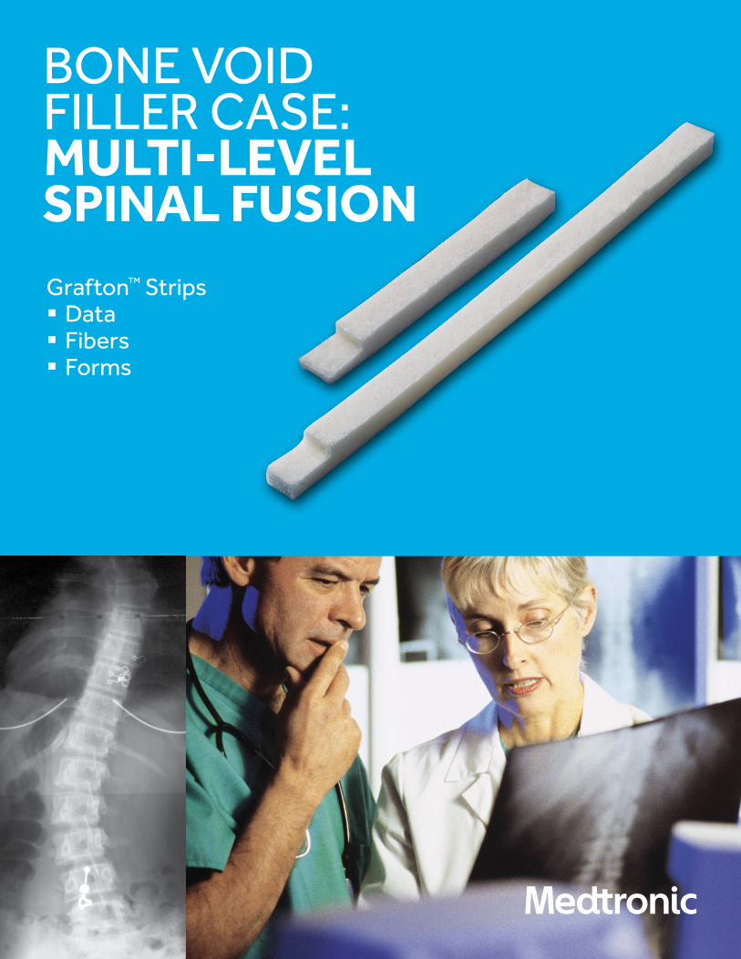
Grafton™ Strips§ Data§ Fibers§ Forms
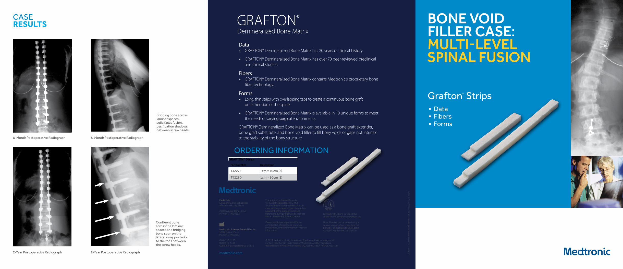
Data
§ Grafton™ Demineralized Bone Matrix has 20 years of clinical history.
§ Grafton™ Demineralized Bone Matrix has over 70 peer-reviewed preclinical and clinical studies.
Fibers § Grafton™ Demineralized Bone Matrix contains Medtronic’s proprietary bone fiber technology.
Forms § Long, thin strips with overlapping tabs to create a continuous bone graft on either side of the spine.
§ Grafton™ Demineralized Bone Matrix is available in 10 unique forms to meet the needs of varying surgical environments.
§ Grafton™ Demineralized Bone Matrix can be used as a bone graft extender, bone graft substitute, and bone void filler to fill bony voids or gaps not intrinsic to the stability of the bony structure.
Grafton™ Strips
Part Number Description
T42275 1cm × 10cm (2)
T42280 1cm × 20cm (2)
ORDERING INFORMATION
GRAFTON™ DEMINERALIZED BONE MATRIX BONE VOID
FILLER CASE: MULTI-LEVEL SPINAL FUSION
CASE RESULTS
2-Year Postoperative Radiograph 2-Year Postoperative Radiograph
8-Month Postoperative Radiograph 8-Month Postoperative Radiograph
Bridging bone across laminar spaces, solid facet fusion, ossification shadows between screw heads.
Confluent bone across the laminar spaces and bridging bone seen on the lateral x-ray posterior to the rods between the screw heads.
© 2017 Medtronic. All rights reserved. Medtronic, Medtronic logo andFurther, Together are trademarks of Medtronic. All other brands aretrademarks of a Medtronic company. UC201804831 EN PMD006113-3.0
The surgical technique shown is for illustrative purposes only. The technique(s) actually employed in each case will always depend upon the medical judgment of the surgeon exercised before and during surgery as to the best mode of treatment for each patient.
Please see the package insert for the complete list of indications, warnings, precautions, and other important medical information.
Consult instructions for use at this website www.medtronic.com/manuals.
Note: Manuals can be viewed using a current version of any major internet browser. For best results, use Adobe Acrobat® Reader with the browser.Medtronic Sofamor Danek USA, Inc.
1800 Pyramid PlaceMemphis, TN 38132
(901) 396-3133(800) 876-3133Customer Service: (800) 933-2635
Medtronic Spinal and Biologics Business Worldwide Headquarters
2600 Sofamor Danek DriveMemphis, TN 38132
medtronic.com
Step 1Prepared Fusion Bed
Step 2Decortication
Step 3Placed Grafton™ Strips
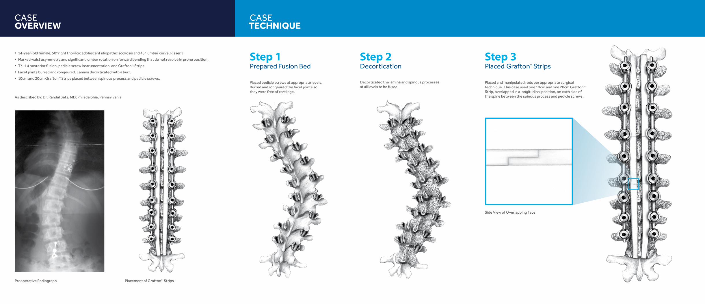
Placed pedicle screws at appropriate levels. Burred and rongeured the facet joints so they were free of cartilage.
Decorticated the lamina and spinous processes at all levels to be fused.
As described by: Dr. Randal Betz, MD; Philadelphia, Pennsylvania
CASE OVERVIEW
CASE TECHNIQUE
§ 14-year-old female, 50° right thoracic adolescent idiopathic scoliosis and 45° lumbar curve, Risser 2.
§ Marked waist asymmetry and significant lumbar rotation on forward bending that do not resolve in prone position.
§ T3–L4 posterior fusion, pedicle screw instrumentation, and Grafton™ Strips.
§ Facet joints burred and rongeured. Lamina decorticated with a burr.
§ 10cm and 20cm Grafton™ Strips placed between spinous process and pedicle screws.
Preoperative Radiograph Placement of Grafton™ Strips
Side View of Overlapping Tabs
Placed and manipulated rods per appropriate surgical technique. This case used one 10cm and one 20cm Grafton™ Strip, overlapped in a longitudinal position, on each side of the spine between the spinous process and pedicle screws.
Grafton™ Strips§ Data§ Fibers§ Forms
BONE VOID FILLER CASE: MULTI-LEVEL SPINAL FUSION

Grafton™ Strips§ Data§ Fibers§ Forms
BONE VOID FILLER CASE: MULTI-LEVEL SPINAL FUSION
Grafton™ Strips§ Data§ Fibers§ Forms
Data » GRAFTON® Demineralized Bone Matrix has 20 years of clinical history.
» GRAFTON® Demineralized Bone Matrix has over 70 peer-reviewed preclinical and clinical studies.
Fibers » GRAFTON® Demineralized Bone Matrix contains Medtronic’s proprietary bone
fiber technology.
Forms » Long, thin strips with overlapping tabs to create a continuous bone graft
on either side of the spine.
» GRAFTON® Demineralized Bone Matrix is available in 10 unique forms to meet the needs of varying surgical environments.
GRAFTON® Demineralized Bone Matrix can be used as a bone graft extender, bone graft substitute, and bone void filler to fill bony voids or gaps not intrinsic to the stability of the bony structure.
GRAFTON® Strips
Part Number Description
T42275 1cm × 10cm (2)
T42280 1cm × 20cm (2)
ORDERING INFORMATION
GRAFTON® Demineralized Bone Matrix
BONE VOID FILLER CASE: MULTI-LEVEL SPINAL FUSION
CASE RESULTS
2-Year Postoperative Radiograph 2-Year Postoperative Radiograph
8-Month Postoperative Radiograph 8-Month Postoperative Radiograph
Bridging bone across laminar spaces, solid facet fusion, ossification shadows between screw heads.
Confluent bone across the laminar spaces and bridging bone seen on the lateral x-ray posterior to the rods between the screw heads.
LITG
RSBR
13 ©
2013
Med
tron
ic, I
nc. A
ll Ri
ghts
Res
erve
d. P
MD
0061
13-2
.0 2
8843
© 2018 Medtronic. All rights reserved. Medtronic, Medtronic logo andFurther, Together are trademarks of Medtronic. All other brands aretrademarks of a Medtronic company. UC201804013 EN PMD017053-1.0
The surgical technique shown is for illustrative purposes only. The technique(s) actually employed in each case will always depend upon the medical judgment of the surgeon exercised before and during surgery as to the best mode of treatment for each patient.
Please see the package insert for the complete list of indications, warnings, precautions, and other important medical information.
Consult instructions for use at this website www.medtronic.com/manuals.
Note: Manuals can be viewed using a current version of any major internet browser. For best results, use Adobe Acrobat® Reader with the browser.Medtronic Sofamor Danek USA, Inc.
1800 Pyramid PlaceMemphis, TN 38132
(901) 396-3133(800) 876-3133Customer Service: (800) 933-2635
Medtronic Spinal and Biologics Business Worldwide Headquarters
2600 Sofamor Danek DriveMemphis, TN 38132
medtronic.com

Step 1Prepared Fusion Bed
Step 2Decortication
Step 3Placed Grafton™ Strips
Placed pedicle screws at appropriate levels. Burred and rongeured the facet joints so they were free of cartilage.
Decorticated the lamina and spinous processes at all levels to be fused.
As described by: Dr. Randal Betz, MD; Philadelphia, Pennsylvania
CASE OVERVIEW
CASE TECHNIQUE
» 14-year-old female, 50° right thoracic adolescent idiopathic scoliosis and 45° lumbar curve, Risser 2.
» Marked waist asymmetry and significant lumbar rotation on forward bending that do not resolve in prone position.
» T3–L4 posterior fusion, pedicle screw instrumentation, and GRAFTON® Strips.
» Facet joints burred and rongeured. Lamina decorticated with a burr.
» 10cm and 20cm GRAFTON® Strips placed between spinous process and pedicle screws.
Preoperative Radiograph Placement of GRAFTON® Strips
Side View of Overlapping Tabs
Placed and manipulated rods per appropriate surgical technique.This case used one 10cm and one 20cm GRAFTON® Strip, overlapped in a longitudinal position, on each side of the spine between the spinous process and pedicle screws.

Grafton™ Strips§ Data§ Fibers§ Forms
Data
§ Grafton™ Demineralized Bone Matrix has 20 years of clinical history.
§ Grafton™ Demineralized Bone Matrix has over 70 peer-reviewed preclinical and clinical studies.
Fibers § Grafton™ Demineralized Bone Matrix contains Medtronic’s proprietary bone fiber technology.
Forms § Long, thin strips with overlapping tabs to create a continuous bone graft
on either side of the spine.
§ Grafton™ Demineralized Bone Matrix is available in 10 unique forms to meet the needs of varying surgical environments.
§ Grafton™ Demineralized Bone Matrix can be used as a bone graft extender, bone graft substitute, and bone void filler to fill bony voids or gaps not intrinsic to the stability of the bony structure.
Grafton™ Strips
Part Number Description
T42275 1cm × 10cm (2)
T42280 1cm × 20cm (2)
ORDERING INFORMATION
GRAFTON™ DEMINERALIZED BONE MATRIX BONE VOID
FILLER CASE: MULTI-LEVEL SPINAL FUSION
CASE RESULTS
2-Year Postoperative Radiograph 2-Year Postoperative Radiograph
8-Month Postoperative Radiograph 8-Month Postoperative Radiograph
Bridging bone across laminar spaces, solid facet fusion, ossification shadows between screw heads.
Confluent bone across the laminar spaces and bridging bone seen on the lateral x-ray posterior to the rods between the screw heads.
© 2018 Medtronic. All rights reserved. Medtronic, Medtronic logo andFurther, Together are trademarks of Medtronic. All other brands aretrademarks of a Medtronic company. UC201804013 EN PMD017053-1.0
The surgical technique shown is for illustrative purposes only. The technique(s) actually employed in each case will always depend upon the medical judgment of the surgeon exercised before and during surgery as to the best mode of treatment for each patient.
Please see the package insert for the complete list of indications, warnings, precautions, and other important medical information.
Consult instructions for use at this website www.medtronic.com/manuals.
Note: Manuals can be viewed using a current version of any major internet browser. For best results, use Adobe Acrobat® Reader with the browser.Medtronic Sofamor Danek USA, Inc.
1800 Pyramid PlaceMemphis, TN 38132
(901) 396-3133(800) 876-3133Customer Service: (800) 933-2635
Medtronic Spinal and Biologics Business Worldwide Headquarters
2600 Sofamor Danek DriveMemphis, TN 38132
medtronic.com

Step 1Prepared Fusion Bed
Step 2Decortication
Step 3Placed Grafton™ Strips
Placed pedicle screws at appropriate levels. Burred and rongeured the facet joints so they were free of cartilage.
Decorticated the lamina and spinous processes at all levels to be fused.
As described by: Dr. Randal Betz, MD; Philadelphia, Pennsylvania
CASE OVERVIEW
CASE TECHNIQUE
§ 14-year-old female, 50° right thoracic adolescent idiopathic scoliosis and 45° lumbar curve, Risser 2.
§ Marked waist asymmetry and significant lumbar rotation on forward bending that do not resolve in prone position.
§ T3–L4 posterior fusion, pedicle screw instrumentation, and Grafton® Strips.
§ Facet joints burred and rongeured. Lamina decorticated with a burr.
§ 10cm and 20cm Grafton™ Strips placed between spinous process and pedicle screws.
Preoperative Radiograph Placement of Grafton™ Strips
Side View of Overlapping Tabs
Placed and manipulated rods per appropriate surgical technique. This case used one 10cm and one 20cm Grafton™ Strip, overlapped in a longitudinal position, on each side of the spine between the spinous process and pedicle screws.
Copyright © 2022 FDOKUMEN