l l l l l l l l l l l l l l l l l l l l l l l l l l l l l l l l l l l l - ISS
Upload
khangminh22Category
view
0download
0
Studies on Rhizosphere Mycoflora
and AM Fungi associated with
Gossypium herbaceum L.
Thesis submitted to
THE MAHARAJA SAYAJIRAO UNIVERSITY OF BARODA
For the degree of
Doctor of Philosophy in Botany
By
Ms. Hiral J. Buch M.Sc.
Under the Guidance of
Prof. Arun Arya
Department of Botany
Faculty of Science
The Maharaja Sayajirao University of Baroda
Vadodara – 390002
2014

Acknowledgments
I am immensely grateful to Prof. Arun Arya, Research Guide, Dept. of Botany for providing
me opportunity to explore the fascinating world of microorganisms.
I am thankful to Dr. Chitra Arya, for her guidance during my research, Dr. P.S. Nagar and
Prof. Vinay Raole for their support and timely help.
My sincere thanks to the Head, Dept. of Botany for all her constructive criticism.
My special thanks to my parents, my husband Dr. Vijay Mane and my in-laws for their
constant encouragement and support.
Thanks to Ms. Poonam Mangalorkar , Mr. Deepak Tadvi, Mr. Shailesh Patel, Mr. Maroti
More, Mr. Paresh Patil and entire Botany office and non –teaching staff.
I would also like to thank my lab collegues Dr. Shipra Chaudhary, Dr. Shraddha Olpadkar, Dr.
Praveen Nagadesi, Mr. Pradyut Dhar.
Hiral Buch

Abbreviations
mg Milligram
mg/g Milligram/ gram
cm Centimeter
PDA Potato Dextrose Agar Medium
pH Potential of Hydrogen Ion
AM Arbuscular Mycorrhizae
VAM Vesicular Arbuscular Mycorrhiza
0C Degree in Celcius
mm Millimeters
ml Millilitre
PVLG Polyvinyl Lactoglycerol
nm Nanometre
ha Hectare
mMT Million Metric Tonnes
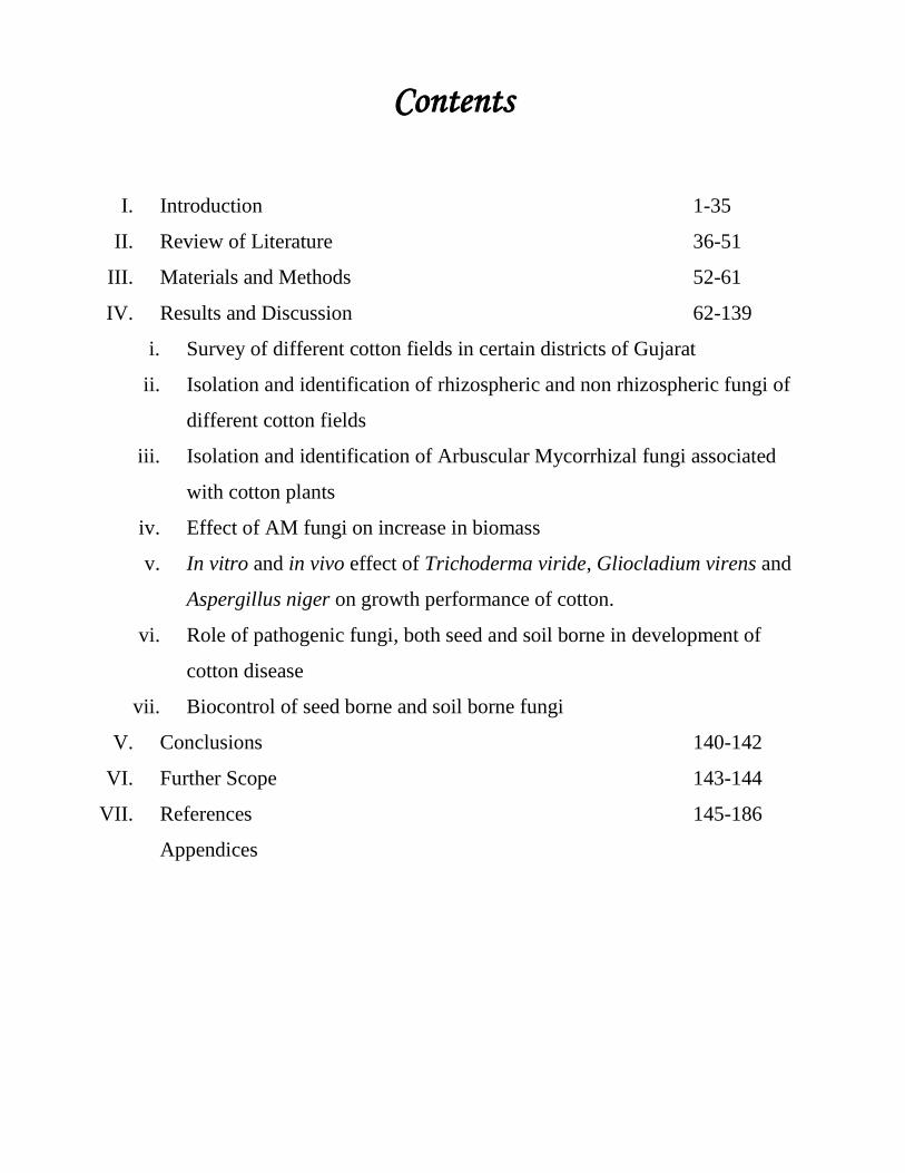
Contents
I. Introduction 1-35
II. Review of Literature 36-51
III. Materials and Methods 52-61
IV. Results and Discussion 62-139
i. Survey of different cotton fields in certain districts of Gujarat
ii. Isolation and identification of rhizospheric and non rhizospheric fungi of
different cotton fields
iii. Isolation and identification of Arbuscular Mycorrhizal fungi associated
with cotton plants
iv. Effect of AM fungi on increase in biomass
v. In vitro and in vivo effect of Trichoderma viride, Gliocladium virens and
Aspergillus niger on growth performance of cotton.
vi. Role of pathogenic fungi, both seed and soil borne in development of
cotton disease
vii. Biocontrol of seed borne and soil borne fungi
V. Conclusions 140-142
VI. Further Scope 143-144
VII. References 145-186
Appendices
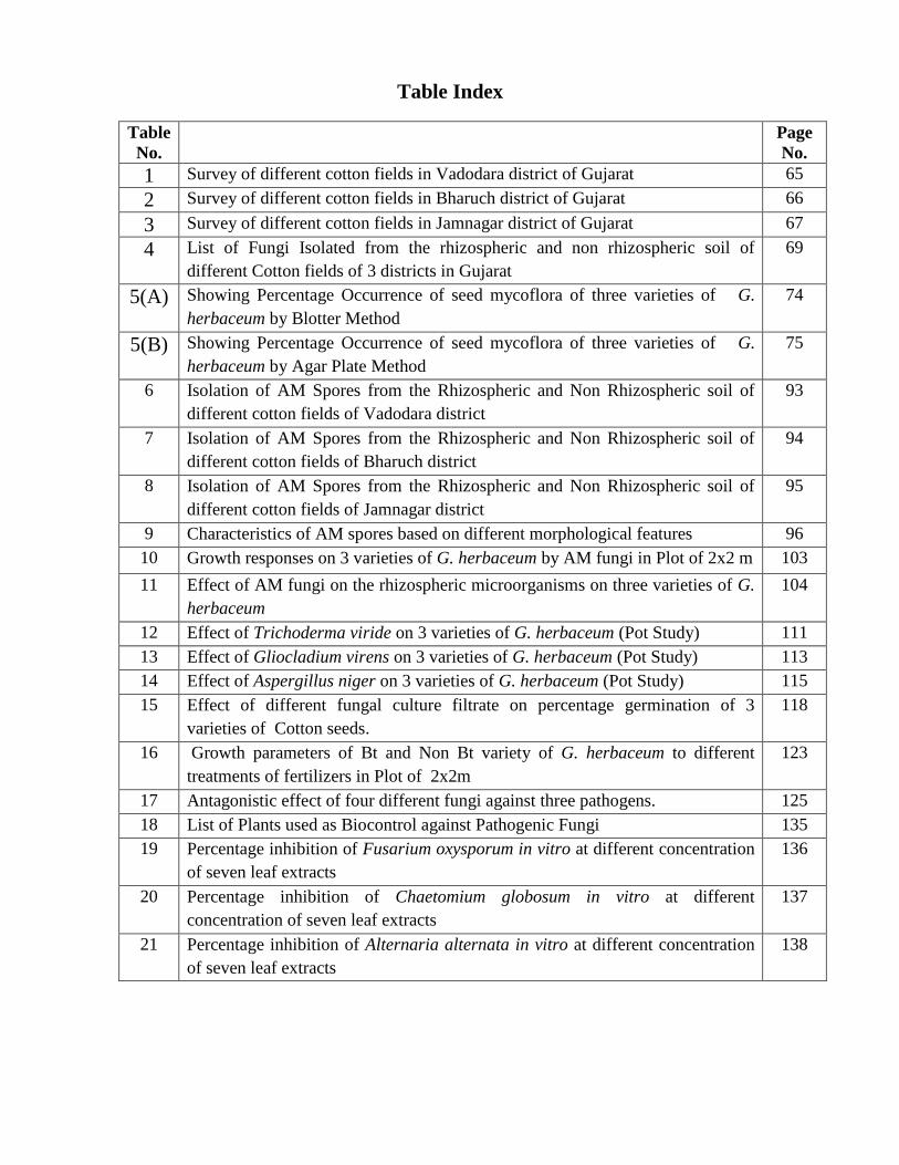
Table Index
Table
No.
Page
No.
1 Survey of different cotton fields in Vadodara district of Gujarat 65
2 Survey of different cotton fields in Bharuch district of Gujarat 66
3 Survey of different cotton fields in Jamnagar district of Gujarat 67
4 List of Fungi Isolated from the rhizospheric and non rhizospheric soil of
different Cotton fields of 3 districts in Gujarat
69
5(A) Showing Percentage Occurrence of seed mycoflora of three varieties of G.
herbaceum by Blotter Method
74
5(B) Showing Percentage Occurrence of seed mycoflora of three varieties of G.
herbaceum by Agar Plate Method
75
6 Isolation of AM Spores from the Rhizospheric and Non Rhizospheric soil of
different cotton fields of Vadodara district
93
7 Isolation of AM Spores from the Rhizospheric and Non Rhizospheric soil of
different cotton fields of Bharuch district
94
8 Isolation of AM Spores from the Rhizospheric and Non Rhizospheric soil of
different cotton fields of Jamnagar district
95
9 Characteristics of AM spores based on different morphological features 96
10 Growth responses on 3 varieties of G. herbaceum by AM fungi in Plot of 2x2 m 103
11 Effect of AM fungi on the rhizospheric microorganisms on three varieties of G.
herbaceum
104
12 Effect of Trichoderma viride on 3 varieties of G. herbaceum (Pot Study) 111
13 Effect of Gliocladium virens on 3 varieties of G. herbaceum (Pot Study) 113
14 Effect of Aspergillus niger on 3 varieties of G. herbaceum (Pot Study) 115
15 Effect of different fungal culture filtrate on percentage germination of 3
varieties of Cotton seeds.
118
16 Growth parameters of Bt and Non Bt variety of G. herbaceum to different
treatments of fertilizers in Plot of 2x2m
123
17 Antagonistic effect of four different fungi against three pathogens. 125
18 List of Plants used as Biocontrol against Pathogenic Fungi 135
19 Percentage inhibition of Fusarium oxysporum in vitro at different concentration
of seven leaf extracts
136
20 Percentage inhibition of Chaetomium globosum in vitro at different
concentration of seven leaf extracts
137
21 Percentage inhibition of Alternaria alternata in vitro at different concentration
of seven leaf extracts
138
Cotton (Gossypium herbaceum L.) belonging to the family Malvacae is one of the important
fibre crop of global significance, which is cultivated in tropical and sub-tropical regions of more
than seventy countries of the world. It is known as ‗King of Fibres‘. The major producers of
cotton are China, India, USA, Pakistan, Uzbekistan, Argentina, Australia, Greece, Brazil,
Mexico, and Turkey. These countries contribute about 85% to the global cotton production. India
has the largest acreage (10.33 m. ha) under cotton at global level and has the productivity of 486
kg Lint /ha and ranks second in production 295 lakh bales (5.02 m MT) after China during 2009-
10. Cotton plays a key role in the National economy in terms of generation of direct and indirect
employment in the Agricultural and Industrial sectors (http:/www.cicr.org).
Cotton is a natural vegetable fibre of great economic importance as a raw material for
cloth. Its widespread use is largely due to the ease with which its fibres are spun into yarns.
Cotton's strength, absorbency, and capacity to be washed and dyed also make it adaptable to a
considerable variety of textile products. Besides being a major natural fibre crop, cotton also
provides edible oil and seed by-products for livestock food. Cottonseed oil is a vegetable oil
ranking fifth in world use among edible oils (accounting for about 4% of world consumption of
vegetable oil). The cotton seed meal is usually used as roughage in the diet of cattle for its high
protein and energetic value.
About fifty species of cotton plants is known within the world of these only four are
domestically cultivated for their fibres. The most commonly cultivated species of cotton in the
world include Gossypium hirsutum and G. barbadense (also referred to as "New World"
species), G. herbaceum, G. hirsutum originated in Mexico. It is the most important agricultural
cotton, accounting for more than 90% of world fibre production. Gossypium barbadense, of
Peruvian origin, accounts for about 5% of world fibre. It includes cotton fibres of the highest
quality, such as the Jumel variety (from the Barbados), among the finest cotton in terms of
quality and fibre length.

History
The genus Gossypium has a long history of taxonomic and evolutionary study. Our
taxonomic understanding of the cotton tribe developed from more than a century of study
involving traditional taxonomic methods as well as modern tools such as comparative analysis of
DNA sequences. Speculation regarding the time and place of origin of Gossypium has a long
history (Hutchinson et al., 1947; Saunders, 1961; Fryxell, 1965; Edwards et al., 1974; Valiček,
1978).
The place of origin of the genus Gossypium is not known, however the primary centers of
diversity are west-central and southern Mexico (18 species), north-east Africa and Arabia (14
species) and Australia (17 species). DNA sequence data from the existing Gossypium species
suggests that the genus arose about 10-20 million years ago (Wendel and Albert 1992; Seelanan
et al., 1999). The antiquity of cotton in the Indian subcontinent has been traced to the 4th
millennium BC (Santhanam and Sundaran, 1997). The first reference to cotton is found in Rig
Veda hymn (Khadi and Kulkarni, 2001). Most commercially cultivated cotton is derived from
two species, G. hirsutum (Upland cotton, 90% of world plantings) and G. barbadense (Pima, or
Long-staple cotton). Two other species, G. arboreum and G. herbaceum, are indigenous to Asia
and Africa and are popularly referred as desi cottons in India.
India is the only country in the world where all the four cultivated species of cotton, viz.
G.hirsutum, G.arboreum, G.herbaceum and G.barbadense, are cultivated on commercial scale,
besides their hybrid combinations. The diversity of cotton cultivars and cotton agroclimatic
zones in India is considerably larger as compared to other major cotton growing countries in the
world.
Asiatic Cotton (Gossypium arboreum, Gossypium herbaceum)
India, China and the near east are the places which are the growers of this kind of cotton. It has
coarse and harsh fibres and thus, is suitable for manufacturing products like blankets, filters,
coarse clothes, padding materials and the like.
Under the rain fed growing conditions rainfall ranges from <400 to > 900 mm coupled
with aberrant precipitation patterns over the years leading to large-scale fluctuations in
production. In the irrigated tract canal and well irrigation is practiced including the use of micro-
irrigation system.
The cultivated G. herbaceum was derived from the truly wild form of the diploid, G.
herbaceum race africanum which has distribution in South Africa. It has been assumed that
traders sailing between Mozambique and Western India introduced this wild form of G.
herbaceum into Southern Arabia, where the first domestication in the Old World cotton took
place. From here, the spread of the species led to the development of new races (Biology of
cotton: www.dbtbiosafety.nic.in).
G. herbaceum is known primarily as a crop plant (grown from Ethiopia to Western India),
with the exception of an endemic form from southern Africa, G. herbaceum sp. africanum. This
morphologically distinct entity, which occurs in regions far removed from historical or present
diploid cotton cultivation, has a unique ecological status in that it is fully established in natural
vegetation in open forests and grasslands. Its small fruit, thick, impervious seed coats, sparse lint,
and absence of sympatric cultivated G. herbaceum suggest that G. herbaceum sp. africanum is a
wild plant. Consistent with the expectation that the site of original domestication lies within the
range of the wild progenitors, this is generally accepted as the source of the original G.
herbaceum cultigens (Hutchinson, 1954). The most agronomically primitive G. herbaceum
cultivars, the perennial race acerifolium forms, are distributed along the coasts boarding the

Indian Ocean trade routes. This suggests that the primary dispersion involved the diffusion of G.
herbaceum northward into northern Africa, Arabia and Persia. Hutchinson (1954) suggests that
secondary agronomic development and diffusion led to expansion into western Africa and the
development of annualized forms in more northerly temperate climates. The agronomic success
of the annualized G. herbaceum races fostered a later dispersal into peninsular India that
replaced perennial G. arboreum cultigens (Wendel et al., 2010).
Cotton Cultivation in India
There has been a significant enhancement in production from 2004-05 onwards as compared to
the earlier years (from 17.7 m bales in 2003-04 to nearly 29.5 m bales in 2009-10). Adoption of
improved technologies IPM, IRM, new chemistry (including Bt cotton) coupled with favourable
weather and low insect pest pressure in major cotton growing tracts has enabled this
transformation in production and productivity. Punjab and Gujarat states recorded much higher
productivity than national average and contributed to a large measure in enhancing productivity
and production at the national level. The average national productivity showed a remarkable spurt
from nearly 309 kg lint/ha (2001-02) to 560 kg lint per ha in 2007-08 and 486 kg lint/ha in 2009-
10. A trend of continuous improvement is quite clear from 2002-03 onwards(Biology of cotton:
www.dbtbiosafety.nic.in).
Bt cotton
Advancement of biotechnological tools and genetic engineering paved the way for
development of transgenic cotton (Boll guard), which offers great promise in the control of
bollworms. The commercial cultivation of such transgenic cotton conferring pest resistance
began during 1996.

Bt cotton, which confers resistance to Lepidopteron pests of cotton, was first adopted in
India as hybrid in 2002 after stringent assessment for bio-safety and profitability. In India, after
extensive testing of Bt cotton hybrids (with Cry1 Ac gene) in All India Coordinated Cotton
Improvement Project (AICCIP) and farmers field, Government of India has approved
commercial cultivation of Bt cotton hybrid with effect from 2002 crop season. The transgenic
hybrids released in the country can be categorized in different ways on the basis of transgene
involved. They can be categorized into two groups viz., (i) Bollgard I (single gene) and (ii)
Bollgard II (double gene).
Bt Cotton is a genetically engineered form of natural cotton. The main advantage of
utilizing biotechnology in agriculture are the possibilities of increase in productivity through the
use of newer varieties that possess properties such as resistance to pests, diseases, and other
stressful conditions like drought, salinity, or water logging. Of these measures, imparting the
property of insect (specific) resistance through the transfer of a gene from Bacillus thuringiensis
Berliner (Bt) into target plants by modern biotech methods is presently considered to be one of
the most advanced applications of biotechnology.
Bt or Bacillus thuringiensis is a gram positive, ubiquitous soil bacterium first discovered
in 1901 by Ishiwata, a Japanese Microbiologist (Kumar et al., 1996). Later it was found that
some Bt strains (Cry+) were highly toxic to larvae of certain insect species which are also plant
pests. Bt was first sold as a spray formulation in 1938 in France for the management of European
corn borer. Subsequent research has revealed that Bt carries proteinaceous crystals that cause
mortality in those insects which carry receptor proteins in gut membranes that bind to Bt
proteins. Other organisms that do not contain receptors to Bt proteins are not affected by the
toxin. The advent of genetic transformation technology made it possible to incorporate cry genes
and thus the ability to produce Bt proteins in plant cells so that target insect larvae infesting the

crop plants are effectively killed. The first Bt crops viz., Bt cotton, Bt corn and Bt potato were
commercialized in USA in 1996. Bt crops are currently cultivated in 23 countries over an area of
46 mha (James, 2008).
Agro climatic conditions
Cotton requires a daily minimum temperature of 16°C for germination and 21°C to 27°C for
proper crop growth. During the fruiting phase, the day temperature ranging from 27°C to 32°C
and cool nights are needed. The sowing season of cotton varies considerably from tract to tract
and is generally early (April-May) in northern India where it is mostly irrigated. It is delayed on
proceeding to down south. It is cultivated largely under rainfed or dryland conditions. An annual
rainfall of atleast 50 cm distributed through-out the growing season is required for good yield. It
is mainly raised during tropical monsoon season, although in southern India it is cultivated
during late-monsoon season in winter.
Cotton is successfully grown on all soils except sandy, saline or water logged types. It is
grown in well drained deep alluvial soils in the north to black clayey soils of varying depth in
central zone and in the black and mixed black and red soils in south zone. It is moderately
tolerant to salinity and is sensitive to water logging as well as frost and chilling temperature in
winter.
Zonal Distribution
Cotton is grown in India in three distinct agro-ecological zones viz., north zone (Punjab,
Haryana, Rajasthan and Western Uttar Pradesh), central zone (Gujarat, Madhya Pradesh,
Maharashtra and Orissa) and south zone (Karnataka, Andhra Pradesh and Tamil Nadu). The
northern zone is totally irrigated, while the percentage of irrigated area in the central and
southern zones is much lower, the lowest being in the central zone which has nearly 60% of
cotton area. It is also grown in small area in the eastern region in Sundarbans of West Bengal and
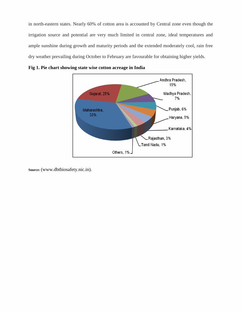
in north-eastern states. Nearly 60% of cotton area is accounted by Central zone even though the
irrigation source and potential are very much limited in central zone, ideal temperatures and
ample sunshine during growth and maturity periods and the extended moderately cool, rain free
dry weather prevailing during October to February are favourable for obtaining higher yields.
Fig 1. Pie chart showing state wise cotton acreage in India
Source: (www.dbtbiosafety.nic.in).

Table showing list of Bt cotton varieties grown in Gujarat
Sr.
No.
Variety Company Name
1 MECH 12 Bt* M/s Mahyco
2 MECH 162 Bt* M/s Mahyco
3 MECH 184 Bt* M/s Mahyco
4 RCH 2 Bt M/s Rasi Seeds Ltd
5 NCS –207 Mallika Bt M/s Nuziveedu Seeds Ltd
6 NCS –145 Bunny Bt M/s Nuziveedu Seeds Ltd
7 RCH –144 Bt M/s Rasi Seeds Ltd
8 RCH –118 Bt M/s Rasi Seeds Ltd
9 RCH -138 Bt M/s Rasi Seeds Ltd
10 RCH –20 Bt M/s Rasi Seeds Ltd
11 Ankur –651 Bt M/s Ankur Seeds Ltd
12 Ankur – 09 M/s Ankur Seeds Ltd
13 RCH 377 Bt M/s Rasi Seeds Ltd
14 GK 205 Bt M/s Ganga Kaveri Seeds PvtLtd
15 GK 205 Bt M/s Ganga Kaveri Seeds PvtLtd
16 KDCHH 9632 Bt M/s Krishidhan Seeds Pvt Ltd
17 KDCHH 9821 Bt M/s Krishidhan Seeds Pvt Ltd
18 ACH-33-1 Bt M/s Ajeet Seeds Ltd
19 ACH-155-1 M/s Ajeet Seeds Ltd
20 Tulasi 4 Bt M/s Tulasi Seeds Pvt Ltd
21 ACH-11-2 BG II M/s Ajeet Seeds Ltd
22 JK Varun Bt M/s JKAgri Genetics Seeds Ltd
23 Ankur 2226 BG M/s Ankur Seeds Ltd
24 Sigma Bt M/s Vibha Agrotech Ltd
25 VBCH-1010 Bt M/s Vibha Seeds (P) Ltd
26 SP 504BI (Dhanno) Bt M/s Proagro Seeds Co (P) Ltd
27 VICH-15 Bt cotton M/s Vikram Seeds Ltd
28 322 Bt cotton M/s Bioseeds Research India Pvt Ltd
29 NCHB-992 M/s Nuziveedu Seeds Pvt Ltd
30 Ajeet 155 BG II M/s Ajeet Seeds Ltd
31 RCH-515 BG II M/s Rasi Seeds (P) Ltd
32 JK Durga Bt M/s J K Agri Genetics Ltd
33 Atal BG II M/s Monsanto Genetics Pvt Ltd
34 Paras Lakshmi BG II M/s Monsanto Genetics Pvt Ltd
35 Sarju BG M/s Solar Agrotech Pvt Ltd
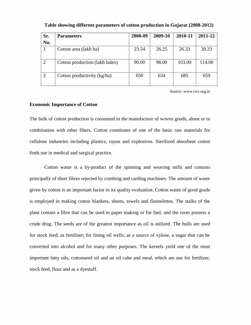
Table showing different parameters of cotton production in Gujarat (2008-2012)
Sr.
No.
Parameters
2008-09 2009-10 2010-11 2011-12
1 Cotton area (lakh ha)
23.54 26.25 26.33 30.23
2 Cotton production (lakh bales)
90.00 98.00 103.00 114.00
3 Cotton productivity (kg/ha)
650 634 685 659
Source: www.cicr.org.in
Economic Importance of Cotton
The bulk of cotton production is consumed in the manufacture of woven goods, alone or in
combination with other fibers. Cotton constitutes of one of the basic raw materials for
cellulose industries including plastics, rayon and explosives. Sterilized absorbent cotton
finds use in medical and surgical practice.
Cotton waste is a by-product of the spinning and weaving mills and consists
principally of short fibres rejected by combing and carding machines. The amount of waste
given by cotton is an important factor in its quality evaluation. Cotton waste of good grade
is employed in making cotton blankets, sheets, towels and flannelettes. The stalks of the
plant contain a fibre that can be used in paper making or for fuel, and the roots possess a
crude drug. The seeds are of the greatest importance as oil is utilized. The hulls are used
for stock feed; as fertilizer; for lining oil wells; as a source of xylose, a sugar that can be
converted into alcohol and for many other purposes. The kernels yield one of the most
important fatty oils, cottonseed oil and an oil cake and meal, which are use for fertilizer,
stock feed, flour and as a dyestuff.
Cotton Pests and Diseases
Cotton pests
Cotton insects are the principal cause of yield losses. Estimates indicate that the yield losses due
to insect infections would amount to almost 15% of world annual production.
More than 1300 different species of insect pests attack the crop. Among the most common and
endogenous species found in cotton fields are:
• The pink bollworm (Pectinophora gossypiella Saunders) was first described in 1843 by W.W.
Saunders as Depressaria gossypiella, from specimens found to be damaging cotton in India in
1842. The pink worm withdraws nutrients from the inside of the cottonseed and may cause
serious yield losses. Although the most sever infestations have occurred in Africa and India, the
pink bollworm has been recorded in nearly all cotton-producing countries and is a key pest in
many of these areas. Infestations may be reduced by the heating of cottonseeds at about 55°C, as
well as by other management tactics, including plantation treatment and destruction of the
infested crop.
• The boll weevil (Anthonomus grandis Boheman), also known as bollworm, is most common in
American cotton plantations.
• The Egyptian (spiny) bollworm (Earias insulana Boisduval) and the red bollworm (Diparopsis
castanea Hmp.) feed on the developing cotton bolls.
• Cotton stainers (Dysdercus superstitious Fabr.) attack maturing cotton bolls and seeds. They
may cause the staining of the lint. In addition, feeding wounds may allow the entry to the boll of
saprophytic fungi (organisms which draw nutrients from the host, but do not harm it, contrary to
parasites).

• Other insect pests of cotton, such as the white flies (Bemisia gossypiella Saund.), may
adversely affect lint quality and yield potential. They suck sap from leaves and pose the most
serious threat in India and Africa.
• The cotton aphid (Aphis gossypii Glover), also known as the melon aphid, infests the cotton
seedlings. Cotton aphids are among the most injuring insects found in cotton. They suck sap
from leaves and secret honeydew on the undersides of leaves. Honeydew secretions may burn
the leaves and interfere with photosynthesis. In addition, aphid is a vector of viruses and a carrier
of other insects. In Africa, aphid infestations are among the most injuring insect pests in terms of
economic yield lost.
• Nematodes: Nematodes or round worms are a diverse group of animanls belonging to the
phylum Nematoda inhibiting a very broad range of environments. They are found in almost all
habitats. There are approximately 128 species of nematodes associated with cotton. Five parasitic
forms pose the most serious threat to the crop, including the Meloidogyne incognita Goldi (or
root knot nematode) and the Rotylenchulus reniformis Lindford and Oliveria (or reniform
nematode). These two species can become serious pests (in the United States, particularly in the
State of Virginia, they accounted for 99% of the damage caused by cotton parasitic nematodes).
These parasites live in the soil (the root knot nematode favours rough and arenaceous soil) and
withdraw nutrients from the plant roots. Symptom patterns associated with nematodes include
stunting, potassic deficiency or early maturity. Nematodes can reduce yields (in Alabama, United
States, yield losses are estimated to average 10% or 20%, but can peak to 50% in arenaceous dry
soil). In addition, depending upon the stage of development of the infested crop, they can hamper
the quality of cotton. Root knot nematodes do produce plant damage symptoms that are rather
easy to recognise, such as the yellowing or whitening of normally green plant tissue because of a
decreased amount of chlorophyll. Damage symptoms caused by other kinds of nematodes (for

example, the reniform nematode) are more difficult to detect, since they are generally small and
sparse. Besides the direct damage, nematodes are also an important factor in the incidence of
Fusarium and other wilts of cotton. Nematodes may be controlled by cultural practices, such as
crop rotations, soil tilling, and use of resistant varieties, or by chemical treatment through
nematicides. The two types of nematodes seldom coexist in the same fields.
Bacterial and Fungal diseases in the cotton plant
Bacterial blight of cotton
Also called angular leaf spot (Xanthomonas malvacearum (E.F. Smith) Dowson) is favoured by
wet weather conditions (temperature above 25°C and relative humidity exceeding 85%). Disease
incidence is higher in plants with injured tissues (due to insect pests or cold temperatures). The
disease causes stunting and yellowing of the leaves (mainly lower leaves). As diseases progresses,
it may result in defoliation. Affected bolls are smaller than normal and exhibit small black spots
on their surface. Bolls may fail to open or produce bad quality lint.
Use of copper oxychloride and streptomycin sulphate has been suggested against the disease
Singh et al., (2010) suggested use of fungicides against foliar pathogens.
Boll rots (Diplodia gossypina Berk and M.A. Curtis, Colletotrichum spp., Fusarium spp.)
Attacks lower bolls near maturity. Warm, humid conditions favour the disease. Affected bolls are
dark brown, with a white to salmon-pink overgrowth. The fungus is capable of giving a brownish
tint to the lint. This disease is a stress-related one, in the sense that it infects plants that have been
previously damaged by insect pests.
Verticillium wilt of cotton
The Verticillium dahlia Kleb, a common soil inhabitant, penetrates though roots and grows up
along the stem tissue. Cooler temperatures, excessive soil moisture and excessive soil nitrogen
levels favour the fungus. Symptoms first appear on the lower leaves, which turn yellow. Larger
plants are stunted (as diseases progresses, defoliation may occur), whereas younger seedlings may
die.
Management strategies include proper management of irrigation and the selection of resistant
varieties. Under conditions favorable to the development of the disease, yield reductions of up to
30% are possible. Seedling diseases (fungi Rhizoctonia solani, Pythium spp.) may result in to seed
and root rott. In the case of Rhizoctonia solani, girdling of the stem at ground level is
observed. Pythium spp. is characterized by the similar symptom patterns, with a water soaked
lesion at the soil line.
Fusarium wilts (Fusarium oxysporum f. sp. . vasinfectum)
Wet weather conditions (temperature above 23°C and relative humidity exceeding 85%)
are particularly conducive for disease development. Disease incidence can be higher in
plants with injured tissues (for example, plants damaged by nematodes). The disease can
affect plants at any stage during the season. The vascular tissue of infected plants exhibits a
brown/chocolate discoloration through the main stem. Infected water-conducting stem
tissues become inactive, causing wilted foliage. Plant death, wilting, yellowing and
defoliation are typical of disease symptoms. Leaves turn yellow between veins and
eventually shed to leave bare stems. Once the fungus has colonized the plant (diagnosis is
confirmed by splitting the stem to reveal dark brown), it most likely causes the death of the
host. There is no commercially viable way to eradicate the disease once established (apart
from soil fumigation, which is excessively expensive). The impact of the disease may
nonetheless be reduced by the use of varieties with high levels of resistance to Fusarium
wilt, or by avoiding crop stresses such as over-irrigation and over-application of nitrogen.
Fusarium wilt is now an important constraint to sustainable cotton production, especially
in Australia.
Fusarium wilt of cotton, caused by F. oxysporum f sp.vasinfectum, was first recognized in
Australia in 1993. It is a soil-inhabiting fungus that invades cotton plants via the roots and
causes a blockage of the water conducting tissues resulting in wilting and eventual death of
affected plants. The pathogen can also be seed borne.
SYMPTOMS
External: Growth is stunted and leaves initially appear dull and wilted, before yellowing or
browning progresses to eventual death from the top of the plant. Some affected plants may re-
shoot from the base of the stem. External symptoms can appear in the crop at any stage but most
commonly become apparent in the seedling phase when the plants begin to develop true leaves
and after flowering when the bolls are filling.
Internal: Lengthwise cutting of the stem of an affected plant will reveal continuous brown
discolouration of the stem running from the main root up into the stem. The internal
discolouration is similar to that of Verticillium wilt but usually appears as continuous browning
rather than flecking in the stem tissue. The severity of external symptoms does not always reflect
the degree of internal discolouration that might be seen when the plant is cut open. Often the
discolouration might only be visible up one side of the plant. Symptoms can appear as only a few
individual plants or as a small patch, often but not always in the tail drain or low-lying
(waterlogged) areas of a field.

Other Fungal Diseases
Of all diseases known to occur in cotton, cotton root rot (Phymatotrichum omnivorum
(Duggar) Hennebert) is one of the most destructive and difficult to control. The fungus
lives in alkaline soils low in organic matter. It occurs only at elevations below 1500m. The
fungus has unique biological characteristics that contribute to management difficulties.
Fungus P. omnivorum has a remarkably wide host range, although it attacks only mature
plants and does not easily spread from field to field. Second, the fungus survives for long
periods in the soil (much of the fungus is found as deep as 60cm to 2m in soils). This
explains why fungicides are not effective treatment. The fungus is only active when air and
soil temperatures are high (respectively above 40°C and 27°C). When environmental
conditions are conducive to its development, the fungus invades the plants through the root
system. Infected plants can die in two weeks. The first disease symptom is slight yellowing
of the leaves, which then quickly turn to a bronze colour and begin to wilt.
List of Fungi already reported from G. herbaceum includes (Bilgrami et al., 1981)
Chaetomium spiralotrichum Lodha.
Colletotrichum sp.
Helminthosporium gossypii Tucker
Myrothecium roridum Tode.
Sclerotium rolfsii Sacc.
Trichothecium roseum (Pers.)Link
Verticillium alboatrum var dahliae Reinke and Berthold.
Soil forms a rich and dynamic medium for all microorganisms. Soil being a complex
ecosystem, is composed of multiple, minute habitat and harbours almost all major taxonomic
groups of fungi. Considering all living forms, the diversity of soil microorganisms in general is

more extensive than any other environment in the world. It has been found that more number of
genera and species of fungi exists in soil than any other environment, as soil is exposed to
various conditions and basically receives all microorganisms present on this plane (Stotzky,
1997). Along with the bacteria, actinomycetes and algae , fungi are primary decomposers ;
agents of biogeochemical transforms and recyclers of stored energy and nutrients of the organic
matter already degraded by invertebrates and other microbes for plant growth. Fungi occur in
soil either in mycelia state or reproductive stage (Nagamani et al., 2006).
Rhizosphere Mycoflora
The rhizosphere may be defined as that portion of the soil which is adjacent to the root
system of a plant and is influenced by the root exudates. The area of this zone depends on the
soil type and host plant under study and soil environment conditions. The roots exert influences
on various type of microorganisms. The stimulatory effect on microorganisms is known as the
―Rhizosphere effect‖ as indicated by the interaction of soil and rhizosphere microbes and their
ratio. The chemical and physical nature of the root zone is quite different from the soil away
from the root zone and the biology of this complex zone has been studied extensively. The term
‗Rhizosphere‘ was proposed by Hiltner (1904). The phenomenon of accumulation of
microorganisms around the root zone was reported by a number of earlier workers
(Agnihothrudu 1955; Starkey 1958; Rouatt 1959; Katznelson 1946). Various compounds such as
amino acids, vitamins, sugars, tannins etc. are exuded by the roots. Some root exudates are also
known to affect certain microbial species adversely leading to their decrease in the root zone and,
in return, microorganisms are known to exert profound influence on the plant itself by
decomposition, affecting nutrient uptake, antagonistic effect on other microbes and by
parasitism.

Interestingly different types of microbes like fungi, bacteria, nematodes and viruses may
interact with the same plant simultaneously either independently, synergistically or
antagonistically. Factors such as soil type, soil moisture, pH, temperature, plant age, relative
humidity and several other factors are known to influence the rhizosphere effect.
According to Pinton et al. (2001), rhizosphere represents a poorly defined zone of soil
with a microbiological gradient in which maximum changes in the population of microflora in
soil is evident adjacent to root and decline with distance away from it (Newman 1978; Bowen
and Rovira 1991). Root exudates stimulate microbial activity selectively in rhizosphere and
rhizoplane regions (Bansal and Mukerji 1994). There is an intense competitive activity by the
obligate saprobes, unspecialized root parasites and root inhabiting fungi depending on their
behaviour towards exudates. In case of root diseases the pathogen has to react with the
rhizosphere and rhizoplane fungi before entering the root tissues. These may show antagonism
and check its advancement. Plant microbe interaction is a regular and continuous feature of the
biological world. The beneficial fallouts of such interactions have been extensively exploited for
economic gain in recent years.
The term ‗rhizoplane‘ was proposed by Clark (1949) to refer to the immediate surface of
plant roots together with any closely adhering particles of soil or debris. Using different isolation
techniques microorganisms have been isolated and identified by a number of Mycologists.
Fungi are very large and diverse group of organisms which have a unique lifestyle. They
have worldwide distribution and successfully exploit many different habitats. They are extremely
variable in form and versatile in the ways they solve the problems posed by the environments
they inhabit (Susan, 1992). Fungi are ubiquitous; some having beneficial effects on plants, while
others may be detrimental (Anderson and Cairney, 2004; Ipsilantis and Sylvia, 2007).
Micro organisms are beneficial in increasing the soil fertility and plant growth as they
are involved in several biochemical transformation and mineralization activities in soil.
Type of cultivation and crop management practices found to have greater influence on the
activity of soil microflora (Mc Gill et al., 1980).
The relationship between biodiversity of soil fungi and ecosystem function is an issue of
paramount importance; particularly in the face of global climate change and human alteration of
ecosystem processes. Fungi are the important component of the soil micro biota typically
constituting more of the soil biomass than bacteria, depending on soil depth and nutrient
conditions (Ainsworth and Bisby, 1995, Saravanakumar and Kaviyarasan 2010). Soil is an
important panorama of interactions between microbes and plants (Shekh et al., 2012). It is one of
the most important habitats for filamentous fungi are major contributors to soil biomass (Pandey
et al., 2013).
The term rhizosphere was first introduced by a German microbiologist, L. Hiltner (1904).
It is describe the zone of metabolically active soil which contains higher microbial community
that surrounds and is influenced by the roots of plants (Mishra, 1967; Chamle et al., 2011).
Microbial population size and community structure are sensitive to changes in chemical
properties of the surrounding soil (Pansombat et al., 1997; Tokuda and Hayatsu, 2002).
Microbial communities, particularly bacteria and fungi constitute an essential component
of biological characteristics in soil ecosystems. It has been estimated that 1.5 million fungal
species are present in natural ecosystems, but only 5 –10% have been described formally
(Hawksworth 2001). Schmit and Mueller (2007) estimated that there is a minimum of 7, 12,000
fungal species worldwide. The actual number of fungi is still unknown; however, only 5-13 % of
the total estimated global fungal species have been described (Wang et al. 2008). Research on
fungal diversity provides a basis for estimating the functional role of fungi in ecosystems.
One of the most important factors responsible for the growth of microorganisms is
organic substances exuded by roots i.e. root exudates (Liljeroth and Baath, 1988). The exudates
include simple sugars, amino acids, organic acids, vitamins and many other compounds

(Singleton and Sainsbury, 1991; Klein, 1992). The influence of exudates upon rhizosphere
microorganisms varies with plant age as well as plant type (Abdel-Rahim et al., 1983; Oyeyiola
2009). However soil factors, such as moisture influences the amount of exudation and hence
colonization of the roots (Whipps and Lynch, 1986). The organisms inhabiting soil includes
microalgae, fungi, bacteria, actinomycetes, protozoa etc (Garrett, 1981). They carry out
numerous transformations as a part of their normal activities like addition of organic matter,
nitrogen fixation, solubilization and immobilization of several nutrients (Katayama et al., 1998;
Lal, 1998; Muller et al., 1998; Brady and Weil, 1999).
The fungi responsible primarily for the decomposition of organic compounds (Paul and
Clark, 1989) actively participate in processes related to biodeterioration and biodegradation
(Allsop and Seal, 1986; Eggins and Allsopp, 1975; Molin and Molin, 1997; Trevors, 1998; Wall
and Virginia, 1999) and also influence above- ground ecosystem by contributing to soil fertility
(Yao et. al., 2000; O‘Donnell, 2001; Van der Heijden, 1998; Cairney,2000; Klironomos et.al.,
2000; Ovreas, 2000).
Besides this soil type, macro and micronutrients may also adversely affect the mycoflora
(Rama Rao, 1957). The plant type, age and soil type have a significant influence the nature and
number of mycoflora. (Wahegaonkar 2009; Namdas and Bhosale, 2009; Abdul-Hafez, 1982).
Most rhizosphere fungi are highly dependent on association with plants that are regulated by
root exudates (Bais, 2004).
The rate of biodegradation depends on environmental factors, numbers and types of
microorganisms present and the enzymatic processes leading to the disappearance of the
parent molecular structure and the formation of smaller organic species (Sharma and Raju
2013).
Soil micro-organism has the capacity to detoxify and inactivate pesticide present in the
soil (Hill et al., 1995). The micro-organisms present in soil depend on many
environmental factors such as the amount and type of nutrients, moisture, degree of aeration pH
and temperature etc. The main focus of the study is to isolate mycoflora from different
agricultural fields and to observe the percentage contribution of different fungal species
Soil bore plant pathogenic fungi a major economic loss, which is a major problem among
the agricultural community. Nowadays the diseases are managed with the application of
chemical pesticides. Use of chemical pesticides causes environmental problem, as they don‘t
undergo biodegradation. So minimizing the application of pesticides has become order of the
day. To achieve this goal the biological control methods can be effectively used along with other
methods of disease control. Antagonistic interactions and cell free culture filtrate have been used
to demonstrate the role of antibiotics in biological control.
Knowledge on the modes of survival of pathogens and the ways by which they could be
suppressed are important especially in the control of plant diseases. The pathogens, in the
absence of their hosts, survive either as dormant propagules or actively as saprophytes on dead
organic substrates of the host in the soil. The survival structures of the pathogens in the soil are
suppressed either due to manipulation of the soil environment. The pathogen suppression in the
soil is considered as important step in the control of diseases as it involves the direct
disinfestations of the soil.
A decrease in crop yield as a result of a plant disease caused by a pathogen is a negative
effect. Some fungi are the main pathogens responsible for plant diseases and they may cause
high yield losses. There are many ways to reduce yield losses caused by fungal diseases, with the
application of chemical fungicides, presently being the most common method. Chemical
fungicides however, have a negative effect on human health and on the environment. The
application of such fungicides over a long period may result in plant pathogenic fungi developing
resistance. When this happens the chemical fungicides become ineffective and other fungicides
must be used for effective disease control. The use of microorganisms as biological control
agents to control plant disease is a potentially powerful alternative method (Kulkarni et al.,
2007). Over the past 30 years, microorganisms have been described, characterized, and tested for
their use as biocontrol agents against diseases caused by soil borne plant pathogens. A wide
range of biological control agents have been developed as commercial mycofungicide products
in past few years (Benítez et al., 2004; Kim and Hwang, 2004; Fravel, 2005).
An alternative way to increase the crop yield besides using chemical fertilizers is
biofertilizers. Biofertilizers promote increased absorption of nutrients in plants (Vessey, 2003;
Hart and Trevors, 2005; Chen, 2006). Biofertilizers include materials derived from living
organisms and microbial sources (Rola, 2000; Chen, 2006). Biofertilizers have various benefits,
such as increased access to nutrients, providing growth-promoting factors for plants, and
composting and effective recycling of solid wastes (Gaur and Adholeya, 2004; Das et al., 2007).
Biofertilizers, commonly known as microbial inoculants are produced from cultures of certain
soil organisms that can improve soil fertility and crop productivity such as mycorrhizae (Malik et
al., 2005; Marin, 2006).
Use ofBiofertilizer and Mycorrhiza to increase the yield of cotton
Complex interactions take place in the volume of soil around roots, which traditionally
has been termed the ‗‗rhizosphere.‘‘ More appropriately, that soil volume constitutes a
mycorhizosphere (Rambelli 1973) because of the dramatic influence an abundance of fungal
external hyphae has on root and soil associated microorganisms, as well as the effects of those
microorganisms, on the establishment and spread of mycorrhizae (Bagyaraj 1984). Mycorrhizal
roots also have altered root exudation patterns (Marschner 1998). The Rhizobium -
Bradyrhizobium association with legumes most notably is affected by mycorrhizal fungi, largely
as a result of increased availability of phosphorus in host roots, which drives nitrogenase activity
in nodules (Aźcon 1994). Other indirect interactions affect both pathogens and beneficial
organisms, either through effects of mycorrhizal formation on root exudates or through
competition (Linderman 1988; Garbaye 1994).
The influence of fungal hyphae in the mycorrhizosphere is much greater than previously
thought (Tisdall et al., 1997) with the discovery of ‗glomalin,‘ a heat stable glycoprotein that
coats hyphae and spore surfaces and accumulates in soil (Wright and Upadhyaya 1996).
Evidence indicates a strong involvement of glomalin in soil aggregate stability; researches have
revealed found a highly significant correlation between glomalin concentration and soil
aggregate stability (Wright and Upadhyaya 1996).
Perhaps the largest obstacle to understanding the biology and ecology of Arbuscular
Mycorrhiza (AM) fungi is our inability to culture them apart from their plant hosts. The plant
provides carbon to the fungus largely via an arbuscule – plant cell plasmalemma interface. It also
provides a protected site in root cells where the fungus can live. The external fungal hyphae
improve phosphorus acquisition by the plant in soils with low levels of phosphorus (Safir 1987;
Smith and Gianinazzi-Pearson 1988; Smith and Read 1997). In soils in which phosphorus levels
exceed requirements of the host, however the AM symbiosis often is inhibited. Under those
conditions for certain host-soil interactions, mycorrhizal development can reduce plant growth
and thus become pathogenic (Modjo and Hendrix 1986).
In nature, fungal communities are taxonomically complex and rarely, if ever, consist of
only one species (Morton 1988). When individual AM fungi are cultured on plants, host
specificity appears to be minimal or absent (Smith and Gianinazzi-Pearson 1988; Brundrett
1991; Smith and Read 1997). Investigators at the International Culture collection of (vesicular)
Arbuscular Mycorrhizal Fungi (IN VAM) in West Virginia showed that more than 1000 isolates
of 98 species of fungi in all genera were able to grow and sporulate on one plant host, Sorghum

sudanese, or Sudan grass (Morton et al., 1993). Roots of Sudan grass accommodate colonization
by as many as 10 species of fungi at one time in pot culture using field soil as inoculum. And
those fungi can be members of any genus (Morton 1988). Lack of host specialization may be the
result of mutualistic co evolution of the plants and their fungal partners over the 400 million
years since their origin (Simon et al., 1993; Taylor et al., 1995).
A combination of host and environmental factors can differentially influence rates and
degrees of colonization and/or sporulation by different AM fungi in a community (Johnson et al.,
1992; Bever et al., 1996), which are manifested as changes in species richness and relative
abundance in sporulation. In general, those responses represent compatibility adjustments rather
than specificity, although Mc Gonigle and Fitter (1990) and Brundett (1991) explained the
‗ecological specificity,‘ respectively. Divergence in physiological and life –history traits within a
fungus species and between species is expected in these asexual organisms as a natural response
to local and regional selection pressures (Morton et al.,1990).
Members of a small number of plants do not form AM associations (Tester et al.,1987).
Many other orders, however, include both mycorrhizal and non-mycorrhizal families and genera.
Non mycorrhizal taxa are assumed to have evolved away from the symbiosis, based on evidence
from their distributions, relative to those of their mycorrhizal relatives (Trappe 1987), and from
the presence of activated defense mechanisms in chemically induced mycorrhizal-resistant
mutants (Peterson and Bradbury 1999). A few genera, however, support both arbuscular fungi
and ecto mycorrhizaal fungi. One of the most widely studied plant being Eucalyptus (Lapeyrie
and Chilvers 1985).
Mycorrhiza is a mutualistic beneficial association between fungi and plant roots. It is
more or less a universal phenomenon throughout the plant kingdom (Mosse 1981). More than
80% of all land plant families are thought to have a symbiotic relationship with AM Fungi that

belong to Glomeromycota. This interaction of AM symbiosis is the evolutionary precursor of
most of the mutualistic root-microbe associations (House and Fester, 2005).
Arbuscular Mycorrhozal association with plants is an ancient (>460 million years BC)
and widespread terrestrial symbiotic association formed between fungi of the phylum
Glomeromycota and the roots of vascular plants (Schussler et al., 2001, Redecker et al., 2000 a,
b, Toljander, 2006) and they, therefore, represent an ancient phylogenetic clade within the fungi.
It is estimated that about 250,000 species of plants, are capable of forming the symbiosis with
AM fungi worldwide (Smith and Read 1997). The colonization of terrestrial ecosystems by the
ancestors of modern vascular plants was facilitated by symbiotic fungi similar to modern
endomycorrhizae. AM comprise of over 150 species that are not host specific and form
symbiotic associations with a wide range of host species. AM bestow a selective advantage on
their host over competing non-host species by making available nutrients, providing defence
against several pathogenic organisms and by influencing the composition of the microflora of the
rhizosphere (Kothamasi et al., 2001).
The symbiotic relationship benefits both- the individual plant and the fungus (Francis
and Read, 1995). Exchange of nutrients-mineral nutrients supplied by the fungal microsymbiont
versus carbohydrates provided by the plant-is considered to be the main benefit for the symbiotic
partners (Smith and Read 1997). According to the phylogenetic position of these partners and
according to the symbiotic structures, several types of mycorrhiza have been defined such as
arbuscular mycorrhiza (AM), ectomycorrhiza, ericoid mycorrhiza, and orchid mycorrhiza. The
efficiency of each AM Fungi for increasing plant growth, nutrient contents, water stress
tolerance (Vazquez et al., 2001) and providing defense against several pathogenic organisms
(Kothamasi et al., 2001) is well documented. In endomycorrhiza, the fungus grows inter- and/or
intra cellularly. Specific fungal structures of endomycorriza are produced within the cortical cells
by non septate fungi which are commonly known as vesicles and arbuscules, hence it was called

earlier as vesicular- arbuscular mycorrhiza (VAM). As many of the endomycorrhiza fungi do not
necessarily form internal vesicles, the abbreviated term ‗VAM‘ was suggested to be replaced by
‗AM‘ (Strack et al., 2003).
Growth in plant communities is often governed by the availability of nutrients such as P
and N. In contrast, C is growth-limiting element in fungal communities. It was but obvious for
natural selection to have favoured the development of symbiotic associations between plants and
fungi. Plants provide C to fungal symbionts and the fungi transfer nutrients from the soil to the
host (Kumar et al. 1999; Pierzynski et al. 2000; Read 1990; Sen 2000). Mycorrhizae have been
associated with vascular plants since the Palaeozoic era (Taylor 1990). The colonization of land
by the ancestors of modern vascular plants seems to have been hastened by the origin of
symbiotic associations between these plants and some phycomycetous fungi similar to those of
modern endomycorrhizae (Malloch et al. 1980; Phipps and Taylor 1996; Simon et al. 1993).
AM, the most prevalent plant-fungus association, comprise about 150 species, belonging to the
order Glomales of Zygomycotina (Morton and Bentivenga 1994; Myrold 2000; Perry et al. 1989;
Schenck 1981; Simon 1996). AM are one of the few plant-fungus associations with a fossil
record (Taylor 1990) and are believed to have assisted vascular plants in their growth and
survival (Simon et al. 1993). AM are present in most soils and are generally not considered to be
host specific. However, population sizes and species composition are highly variable and
influenced by plant characteristics and a number of environmental factors such as temperature,
soil pH, soil moisture, P and N levels, heavy metal concentration (Boddington and Dodd 1999),
the presence of other microorganisms, application of fertilizers and soil salinity (Barea and
Azcon-Aguilar 1983; Bationo et al., 2000). Species and strains of AM differ in their ability of
tolerance to physical and chemical properties of soil (Abbot and Robson 1991), as a result they
also differ in their effectiveness in improving plant growth.

AM forms the connecting link between the biotic and geochemical portions of the
ecosystem (Miller and Jastrow 1994). Mycorrhizae aid the plant in better growth by assisting it
in absorbing useful nutrients from the soil, in the competition between plants and in increasing
the diversity of a given area. A number of reviews have appeared recently on AM, particularly
dealing with the application of AM in agriculture. Information on the role of AM in plant
adaptations has been scattered and the present review deals with the critical appraisal of the role
of AM in plant community dynamics, nutrient mobilization and overcoming both abiotic and
biotic stresses.
AM fungi are known to infect a wide range of host species. They have a large
geographical distribution (Malloch et al., 1980), being found even in the Arctic tundras and the
Antarctic region (DeMars and Boerner 1995b; Gardes and Dahlberg 1996). Unlike most
ectomycorrhizal species, AM are not host specific. This enables them to form associations with a
large number of plant species. Mycorrhizae owing to their role in nutrient cycling, keep more
nutrients in the biomass and in doing so increase the productivity of the ecosystem (Newman
1988). AM fungi regulate plant communities by affecting competition, composition and
succession (Allen and Allen 1984; Kumar et al,. 1999).
Classification of Mycorrhizae
Mycorrhizal fungal association widely varies in structure and function, but the AM fungi exhibit
most common associations (Harrier, 2001). Six genera of AM fungi have been recognized based
on the morphological characteristics of sexual spores and also based on various biochemical
studies as well as molecular methods (Peterson et al., 2004). Further, various criterion has been
used for the identification of AMGF like hyphal charaters, auxillary cells subtending hyphae,
spore or sporocarp ontogeny, morphology, germination, shield spore wall etc. (Mukherji et al.,
2002). AMF are zygomycetous belonging to the genera Glomus, Gigaspora, Sclerocystis,
Acaulospora, Enterophospora and Scutellospora (Garbaye, 1994).

The classification of AMF is based on the structure of their soil borne resting spore,
biochemical properties and molecular studies (Morton and Benny, 1990). The latest
classification of AMF contains 4 orders and 9 families (Siverding and Ohel, 2006). Plant species
belonging to the Cruciferae, Chenopodiaceae and Cactaceae are not known to form AMF
symbiosis (Smith and Read, 1997). AMF reproduce asexually by spore production. There is no
evidence that AMF reproduce sexually (Kuhn et al., 2001).
Table showing Recent Classification of Arbuscular Mycorrhizal Fungi (Siverding and
Ohel, 2006)
___________________________________________________________________________
Phylum Glomeromycota
Class Glomeromycetes
Orders Families Genera
___________________________________________________________________________ 1 – Glomerales Glomeraceae Glomus
___________________________________________________________________________
2 – Diversisiporales Gigaspraceae Gigaspora, Scutellospora
Acaulospraceae Acaulospora, Kuklospora
Entrophosporaceae Entrophospora
Pascisporaceae Pacispora
Diversisporaceae Diversispora
___________________________________________________________________________
3 - Paralomerales Paraglomeraceae Paraglomus
Geosiphonaceae Geosiphon
___________________________________________________________________________
4 - Archaeosporales Arthaeosporaceae Archaeospora,
Inraospora
Types of Mycorrhizae:
Several types of mycorrhizal fungi have been recognized and the most important types are
mentioned below:
Endomycorrhizae:
Endomycorrhizae represents a group of fungi that are associated with most of the agricultural
crops and provide biochemical protection against soil borne diseases (Smith and Read, 2008).
They occur in most of the ecosystems of the world and are found in many important crop species

i.e. (cotton, wheat, maize, rice and soyabean) and horticultural species like grapes, roses and
petunias etc. (Peterson et al., 2004). AMF are obligate biotrophs feeding on the products of their
live host and those fungi are not specialized to their potential hosts. The host plants receive
mineral nutrients from outside the root depletion zone via the extraradical mycelium, while the
AMF obtains photo-synthetically produced carbon compounds from the host (Smith and Read,
1997).
Many endomycorrhizal fungi form terminal or intercalary vescicles in the root cortex.
When the vesicles are expanded the thin walled structures, contain large quantity of lipids (Tahat
et al., 2010). They may be oval, spherical or lobed in shapes and may become thick walled and
resting spores (Pirozynski and Dalphe, 1989).
Ectomycorrhizae (ECM):
ECM fungi form a thick mantle like structure and within the intracellular spaces of root cortex
form network. These fungi do not penetrate living cells in the host roots, but can only surround
them. They are most common in ornamental and forest tree species in the family Pinaceae,
Myrtaceae, Salicaceae, Dipterocarpaceae, Fagaceae and Gnetum plants (Shalini et al., 2000).
Ectomycorrhizas are distinguished by the presence of mantle and harting net. Harting net
develops in cortical cells or epidermal cells. Harting net consists of branch system which can
provide a large surface contact between cells of the two symbionts (Peterson et al., 2004). Other
type of mycorrhizal fungi includes Ecto-endo Mycorrhizae, Ericoid Mycorrhizae, Monotropoind,
Arbutoid and Orchid mycorrhizae (Smith and Read, 2008).
Uptake of the nutrient by AM Fungi:
When soil resources, such as P or N, limit photosynthesis, C is in excess, mycorrhizal
fungal hyphae explore the soil volume for P and N, and transport the nutrient (over distances of
cm to m) in exchange for excess plant C. Mycorrhizal hyphae are more efficient at exploring the
soil volume then even fine roots. As long as P or N are limiting plants will support mycorrhizal

fungi. Even as the availability of the limiting resources shifts through time, mycorrhizal fungi
similarly shift resource provisioning (Molina et al., 1992). Linking space and time is important
because of exploring a neighboring patch (Aikkio and Ruotsalainen, 2002 ). Since a complex
network of fungal mycelia and plant roots are distributed horizontally across a landscape and
extend vertically into the soil and rock substrate (Egerton-Warburton et al., 2003), resource
extraction becomes dynamic.
Energy, in the form of C compounds is the currency for exchange of soil resources. This
connection occur at the (fungus) membrane: interspace: (plant) membrane interface in the form
of simple sugars or amino acids. Photosynthetic rates depend on the concentrations of N (as
RuBP carboxylase and other enzymes), P (for ATP, ADP). Fe and Mg (for cholorophyll),
internal CO2 , and water ( to keep stomata open to fix CO2). These interactions create several
important and well known linear and curvilinear relationships that form that basis of
stoichiometeric ratios between elements. Mycorrhizae, by increasing P and N uptake, create a C
sink and enhance the photosynthetic machinery. Mycorrhizae also increase water release through
transpiration by, opening the stomata. Together these increase rates of total carbon gain by 10%
to 40% (Allen et al., 1981). In the field, this increased CO2 fixation is associated with
environmental change, such as drought, or as a function of particular fungal – plant species
combination.
Effects and processes involved in the growth enhancement by Vesicular Arbuscular
Mycorrhizas (Tinker et al., 1994)
1. Growth increase occurs by improved supply of elements of low mobility in growth
medium, predominantly phosphate.
2. This arises by increased uptake rate per unit amount of root length (inflow).
3. This is caused by proliferation of a considerable length of external hyphae.

4. Hyphae absorb, translocate and transfer P to host, from soil outside the root depletion
zone.
5. Uptake is normally from the isotopically labile pool of nutrient, from which the root also
absorbs.
6. There is a feedback effect by absorbed Phosphorus on the percentage of infected root.
7. Infection of phosphate-deficient plants is accompanied by a rapid but temporary increase
in internal P concentration.
8. Much of phosphorus in the fungal partners is in the form of phosphate.
9. The fungus is maintained by carbon supplies from its host, and infection results in a large
proportion of total fixed carbon being allocated below ground.
10. The uptake efficiency should be large for this mechanism than for uptake by the
uninfected root system.
Plants used for Biocontrol
Plants are the richest source of renewable bioactive organic chemicals. The total number of plant
based chemicals may exceed 4,00,000 of these 10,000 are secondary metabolites, whose major
role in the plants is reportedly defensive (Swain, 1977).
The screening of plant extracts for antimicrobial activity has shown that a great number
of these plants contain active compounds. The presence of antibacterial, antifungal, and other
biological activities has been demonstrated in extracts of different plant species used in
traditional medicine practices (Hashem, 2011).
Basic researches for over more than forty years in the fields of biological and
biochemical have made it possible to envisaged not only how new pesticides may be synthesized
but also a completely new approach for the protection of plants using secondary plant products,
which may be toxic to a specific pest yet harmless to man. There has been a renewed interest in

botanical pesticides because of several distinct advantages (1) Pesticidal plants are generally
much safer than conventionally used synthetic pesticides. These pesticides will not cause harm in
nature. (2) Plant based pesticides will be renewal in nature and would be economical. (3) Some
plants have more than one chemical as an active principle responsible for their biological
properties. These may either be selected for one particular biological effect or may have diverse
biological effects (Singh, 1993).
Efforts are being made these days to shift from the conventional use of chemicals to the
use of eco-friendly botanicals for the management of plant parasitic nematodes. Organic
amendments are not only safe to use but also have the capacity to improve soil structure and
fertility (Trivedi, 2002).
Leaves of following plants were used to study the biocontrol activity:
1. Annona reticulata L.
Family: Annonaceae
It is a small deciduous or semi-evergreen tree reaching 8 metres to 10 metres tall with an open,
irregular crown. The slender leaves are not hairy, straight and pointed at the apex 10 cm to 20 cm
long and 2 cm to 7 cm wide. The yellow-green flowers are generally produced in clusters of
three or four 2 cm to 3 cm diameter, with three long outer petals and three very small inner ones.
The fruits are variable in shape: heart-shaped, spherical, oblong or irregular.
2. Sapindus emarginatus Vahl.
Family: Sapindaceae
Sapindus emarginatus is a deciduous tree. Commonly called as Soapnut tree and is the south
Indian species of genus Sapindus. It is an economically significant tropical tree species meagerly
distributed in diverse geographical provinces like Gangetic Plains, Western Ghats, and Deccan
Plateau in India. The trunk of the tree is straight and cylindrical, approximately 4-5 m in height.

5-10 pairs of leaves, solitary alternate, 15–40 cm long, pinnate, with 14-30 leaflets, the terminal
leaflet often absent. The flowers form in large panicles and each flower is small and creamy
white in colour. It flowers during summer. The fruit is small leathery-skinned drupe which is 1–2
cm in diameter, which is yellow and turn blackish when ripen, containing one to three seeds.
The members of genus Sapindus are well known for their medicinal values. Traditionally it is
used as anti-inflammatory and antipyretic. Its fruits are natural substitute for chemical soaps and
hair dyes.
3. Cochlospermum religiosum (L.) Alston
Family: Bixaceae
It is a flowering plant from the tropical region of Southeast Asia and the Indian Subcontinent. In
India it is commonly found in Andhra Pradesh, Maharashtra, Madhya Pradesh, Uttar Pradesh and
Bihar. It is a small tree growing to a height of 7.5 m usually found in dry deciduous forests. Also
known as Silk-Cotton Tree because the capsules containing the seeds have a fluffy cotton-like
substance similar to kapok. Plant can be identified by deeply furrowed bark, palmately 5-lobed
leaves and bright golden yellow bisexual flowers. Quick growing tree yield a gum known as gum
katira from a juice orange in colour exudes from the bark. The dried leafves and flowers are used
as stimulants, antipyretic, laxative and sedative. Root powder mixed with water when applied to
face reduce wrinkles.
4. Gliricidia sepium (Jacq.) Kunth ex Walp.
Family: Fabaceae
It is a medium-sized tree, semi deciduous tree that grows from 10 to 12 meters high. The bark is
smooth and its color can range from a whitish gray to deep red-brown. It has compound leaves
that can be 30 cm long. Flowers have a bright pink to lilac color that is tinged with white. Leaves
are rich in protein and highly digestible for ruminants like goat and cattle, as they are low in fibre

and tannin. There is evidence of improved animal production (both milk and meat) in large and
small ruminants when Gliricidia is used as a supplement to fodder. However, non-ruminants fed
on Gliricidia sepium have shown clear signs of poisoning. The flowers attract honeybees (Apis
spp.), hence it is an important species for honey production. Good for firewood and charcoal
production. The wood burns slowly without sparking and with little smoke. Very durable and
termite resistant; used for railway sleepers, farm implements, furniture, house construction and
as mother posts in live-fence establishment. A traditional remedy for hair loss, boils, bruises,
burns, cold, cough, debility, eruptions, erysipelas, fever, fractures, gangrene, headache, itch,
prickly heat, rheumatism, skin tumours, ulcers and wounds
5. Feronia accidissima (L.) Swingle
Family: Rutaceae
The tree is native and common in India, Sri Lanka, China and Indonesia and widely
distributed in most tropical and subtropical countries. Commonly known in India as wood-apple.
It has economic as well as medicinal value. It contains important medicinal compounds like
umbelliferol, dictamnine, xanthotoxol, scoparone etc. those could be used in the pharmaceuticals
industries. The fruit is used in India as a liver and cardiac tonic. and when unripe, as an
astringent means of halting diarrhoea and dysentery and effective treatment for hiccough, sore
throat and diseases of the gums. Juice of young leaves is mixed with milk and sugar candy and
given as a remedy for biliousness and intestinal troubles of children. Oil derived from the
crushed leaves is applied on itch and the leaf decoction is given to children as an aid to digestion.
6. Balanites roxburghii Planch.
Family: Zygophyllaceae
It is a spiny, evergreen tree. It is common in open sandy plains. Commonly called as Hingoli/
Hingoru. Bark, Friuts, Seeds and Leaves are used. Fruits are used in treatment of whooping
cough, skin diseases and in antifertility. Leaves are used for the treatment of jaundice. In case

of pain and swelling, the bark of plants is used as traditional healers. The paste of bark is
prepared and applied externally on the affected part of the body to treat snake-bite and dog
bite.
7. Tephrosia jamnagarensis Sant.
Family: Fabaceae
It ia an annual herb of 1mt with simple linear leaved covered by hairs. Flowers purplish blue
in colour. Pods densely hairy and oblique at both the ends. Seeds reniform, brownish. Leaves
contain glycosides and favanoides. The leaves could be used as insecticide, pesticide and is
having hepatoprotective properties.
Objectives
1. Survey of different cotton fields in certain districts of Gujarat, to assess the varieties of
cotton (Normal / Bt) under cultivation, yield obtained, and associated seed and soil borne
diseases.
2. Isolation and identification of different fungi from the rhizospheric and non- rhizospheric
soils.
3. Effect of certain fungi like Trichoderma sp., Gliocladium sp. and Aspergillus niger will
be observed on percentage germination and growth performance of cotton.
4. Isolation and identification of different AM Fungi from soil associated with roots of Bt
and Non Bt cotton plants.
5. Effect of AM fungi on increase in plant biomass/yield.
6. Role of pathogenic fungi both seed and soil borne in cotton and their control in vitro.
7. Effect of compost and vermicompost on increase in plant biomass.

Soil is a complex and dynamic environment in which the biological activities are mostly
governed by microorganisms. The beneficial effects of soil microorganisms are manifold and
range from nitrogen fixining and organic matter deposition to the breakdown of metabolic
byproducts and enhancing the availability of sulphates, phosphates, nitrates and essential metals
(Bridge and Spooner, 2001).
The root system is important for plant fitness because it provides anchorage, contributes
to water use efficiency, and facilitates the acquisition of mineral nutrients from the soil (Lopez-
Bucio et al., 2005a)
Rhizosphere microbial communities can significantly influence phytopathogens
development (Nehl et. al., 1997; Glick, 1995), nutrient acquisition (Lynch, 1990), heavy metal
resistance (Bradly et.al., 1981) and ecological fitness of the plant. Qualitative as well as
quantitative distribution of fungi in the rhizosphere and non-rhizosphere soil has been discussed
in detail (Harley and Waid, 1955; Parkinson and Waid, 1960; Burges and Raw, 1976).
Fungi represent a very important component of the ecosystem, along with the other
microbes of the biomass (Harrison et al., 1994). Fungi are important component of the soil
microbiota typically constituting more of the soil biomass than bacteria, depending on soil depth
and nutrient conditions (Ainsworth and Bisby, 1995). They perform ecological services that
strongly impact the quality of human life and have enormous potential for providing economic
benefits (Diana, 1994). It is estimated that there are 1.5 million fungal species on earth of which
only about 70,000 have been described up to now (Hawksworth and Rossman, 1997).
A mycorrhiza is a symbiotic association between fungus and the roots of a plant (Kirk et
al., 2001), where in this association the fungus may colonize the roots of host plant either
intracellularly or extracellularly.
Azcon et al., (1995) reported that inoculation of VAM fungi greatly increased shoot and
root biomass and leaf area in micropropagated Annona plants. Bagyaraj (1984) generalized that
modern high input agricultural practices are detrimental to AM fungi, while the low input
sustainable agriculture methods enhance the population of AM fungi in the soil.
Hayman (1981) reported that the mycorrhizal association in groundnut improved growth
and nutrition of plants. More than 100% increase in the dry matter yield and P uptake of
groundnut plants inoculated with mycorrhizal fungi compared to non-inoculated control.
Arbuscular mycorrizal fungi are the most frequent in plants growing on mineral soils.
The populations of AM fungi is greatest in plant communities with high diversity such as
tropical rainforests and temperate grasslands, where they have many potential host plants and can
take advantage of their ability to colonize a broad host range. There is a lower incidence of
mycorrhizal colonization in very arid or nutrient rich soils. Mycorrhizas have not been observed
in aquatic habitats; however, waterlogged soils have been shown to have colonization in some
species (Smith and Read, 2002). In costal soils infection of AM has been influenced by soil
temperature and moisture content of the soil (Mohankumar et al., 1988a).
Literature survey showed that the work has also been carried out on colonization of AMF
and its effects on growth performance of some tree species by various workers such as Tectona
grandis L.f. by Rajan et al., (2009) on Eucalyptus spp. and Acacia sp. by Malajczuk et al.,
(1981) on Acacia leucocephala and Moringa concanensis by Pawar and Vyas (2002) ; on
Carica papaya L. by Kennedy and Rangarajan (2001); on Casuarina equisetifolia J.R. and G.
Forst, (Rajeshwari et al., 2001).
Jain and Gupta (2002) reported effect of rhizosphere fungi on nodule number, shoot and
root length of Vigna mungo. Inoculation of AM fungi and Rhizobium increases the percentage of
chlorophyll content in the leaves of Arachis hypogea L (Charitha and Reddy, 2004). AM fungi
have the effect on control of disease infection in plants and it has been successfully shown in
plants viz. tomato for nematode infection (Suresh et al., 1985).
Nematode infection has also been reduced in arbuscular mycorrhiza inoculated plants of
Piper nigrum L. (Sivaprasad et al., 1990). Fusarium wilt disease severity in Albizia procera
Benth. and Dalbergia sissoo Roxb. was significantly reduced when inoculated with mycorrhizal
fungi (Chakravarty and Mishra, 1986). In case of papaya plants having arbuscular mycorrhizae
showed drought resistance (Shivaputra et al, 2004).
In the experiments carried out by Edwards et al., in 1998, plants grown in the presence of
Glomus mossae had a significantly higher shoot dry weight than those grown in the absence of
G. mossae. Colonization and the activity of G. mossae was unaltered in the presence of
Pseudomonas fluroscence Migula isolated and presence of G. mossae increased the population of
P. fluroscence in the rhizosphere.
Dodd et al., (1996) in their investigations aimed at using morphological and molecular
characters to study inter- and intraspecific variation within isolates of G. mossae and Glomus
coronatum from different parts of the world. Morphological evaluations of various possible
taxonomic characters including spore colour, size sporocarp architecture and hyphal attactment
morphology, showed that only spore colour discriminate the two groups.
In 1982, Scheneck and Smith studied that there was considerable variation in the plant
response to various combinations of temperature and fungus species, with both growth
stimulatory and growth repressive and effects occurring. The use of AM fungi in ecological
restoration projects have been shown by Jeffries et al., (2003) to enable host plant establishment
on degraded soil and improve soil quality and health (phytoremediation).

With the establishment of the facts that AM fungi has the role in phosphate solubilisation
in the rhizosphere and thereby providing the host plant the solubilised form of the phosphorus
has led to think regarding the means and ways to mass multiply the different strains of AM fungi.
Mass multiplication methods at bioreactor (Jolicoeur et al., 1999) and invitro (Gryndler et al.,
1998; Pawlowska et al., 1999; Gadkar and Adholeya, 2000) level apart from conventional way at
pot level have been standardized (Gaur and Adholeya, 2000) along with the selection of suitable
host (Singh and Pandya, 1995). The post harvest storage management of these AM fungi cultures
has also been worked out (Mohankumar and Mahadevan, 1988b; Sreenivasa and Kulkarni,
1993).
Jakobsen et al., (1992) indicated that the efficiency of phosphorus uptake by VAM
fungus is strongly affected by its spatial distribution of hypahe in the soil and possibly also by
differences in capacity for uptake by unit length of hyphae. Pearson and Jakobsen (1993)
strongly indicated that the P uptake by root cells is influenced by the presence of mycorrhizal
fungi and this effect varies from species to species.
According to Jensen (1982), AM fungi are universally associated with the crop plants.
They helps the host in the uptake of Phosphorus and other minerals. The growth increase may be
controlled more by available soil P than by endophytic species (Clarke and Mosse, 1981). Grant
et al., (2005) indicated that as the phosphorus availability levels in the soil increases, the amount
of phosphorus also increases in the plant tissues and carbon drain on the plant by the AMF
symbiosis become non-beneficial to the plants.
In 2002, Panwar and Vyas reported the use of AM fungi as a biological approach
towards conservation of endangered plants in Thar desert of India. The efficiency of eight AM
species, Acaulospora mellea, Gigaspora margarita, Gigaspora gigantean, Glomus deserticola,
Glomus fasciculatum, Sclerocystis rubiformis, Scutellospora calospora collected from the
rhizosphereic soils of Moringa concanensis, was evaluated for nutrient uptake and enhancement
of acid phosphatase, nitrate reductase, peroxidase and polyphenol oxidase activities in this
endangered tree of the Thar desert.
George et al., (1995) showed that AM symbiosis increases the phosphorus and
micronutrient uptake and growth of their plant host. Uptake of micronutrients is enhanced by the
same mechanisms that operate for P, Cu and Zn have consistently been shown to be increased by
VAM fungal colonization was indicated by Menge et al., (1978).
Wani and Lee (1995) reported for studying AM fungi colonization the best sampling time
is flowering stage up to maturity and in field conditions inclusion of appropriate crop in crop
rotations or intercropping can increase the native population of AM fungi in soil which is often
objective of the artificial inoculation. Tian et al., (2009) studied different AM species showing
different distributions among the three plant communities and the results indicate that both biotic
and abiotic factors were important in determining the AMF communities, with biotic factors
possibly the more important.
Braunberger et al., (1996) indicated that the development and function of mycorrhizas
after late summer and early autumn rains may be limited by the occurrence and predominance of
propagules of different AM fungi. In 2008, Mikkelsen et al., showed ability of AM fungal
mycelia to anastomose in soil for the formation of large plant-interlinking functional networks,
long distance nutrient transport and retention of nutrients in readily plant –available pools.
In nature the Mycorrhizal condition is the rule and non- mycorrhizal condition is the
exception according to Gerdemann (1969). The association of AM fungi is found with most of
the vascular plant communities examined so far except members belonging to families
Brassicaceae, Chenopodiaceae, Caryophyllaceae, Cyperaceae, Araceae, Asteraceae, Poaceae,

Onagraceae, Polygonaceae and Portulacaceae (Hirrel et al., 1978; Berch, 1988). In recent times it
has been reported that few members of Chenopodiaceae (Kruchkelmann, 1973; Ross and Harper,
1973; Williams et.al., 1974), Cyperaceae (Mejstrik, 1972) and Cruciferae (Kruchkelmann, 1973;
Ross and Harper, 1973) may have AM association.
Rhizosphere soil and roots were sampled for root colonization, AMF identification and
spore counts Moreira et al., (2006). They found that percent root colonization and spore numbers
were inversely related to each other in all ecosystems. Guar and Adholeya (2000) reported that
plants with sand particle sizes of 0.50-0.78mm has higher root fresh weights, spore production
and percentage mycorrhizal colonization than with other particle sizes.
Dalal and Hippalgaonkar (1995) reported that the spore density was found more in rainy
season as compared to winter and was high in soils collected under water stress in summer.
Allelochemicals induced in mycorrhizal plants play an important role in disease resistance
(Inderjit and Mukherji, 2006). Graham et al.,(1981) found that reduced root exudation limited
the growth of pathogen in the rhizosphere and there by, its ability to cause disease.
Vyas (1990) on the basis of his observation reported that distribution of endomycorrhizal
fungi in soybean and chick pea such as Glomus monosporum, G. fasciculatum, G. epigaeum, G.
constrictum and Acaulospora morrowae were associated with soybean and chick pea. Glomus
monosporum, G. fasciculatum occurred in 40% and 25% of samples and 50% and 25% of
samples in soybean and chickpea respectively. G. epigaeum occurred in 25% of samples in
soybean and 30% samples in chickpea. Glomus mossae and G. constrictum were found to be
associated with all cultivars of soybean.
Singh and Mishra (1995) reported that in two paddy varieties plant biomass is increased
when these are associated with AM fungi, the mean shoot and root dry weights of was

significantly higher than those of the non-mycorrhizal plants. They observed that the increasing
soil phosphate levels reduced the percentage of root infection in both the varieties at all the host
stages examined.
Ammani and Rao (1996) reported effect of eight varieties of upland rice in sterilized soil
in pot culture with inocula of Acaulospora spinosa and A. scrobiculata. All the eight varieties
showed a positive response to inoculation with the AM fungi in terms colonization of roots,
formation of vesicles and spores, plant biomass and grain yield. Three of the eight varieties
showed a greater response than the others. The traditional variety, Mettasannalu gave the least
response. A. spinosa was found to be more effective than A. scrobiculata on all the varieties.
In 1988, Khan et. al., studied mycorrhizal status of some Bangladesh soils and the effect
of indigenous AM fungi on the growth of rice plants. Mycorrhizal infection greatly improved the
growth and nutrient (nitrogen, phosphorus and zinc) content of rice.
Microorganisms growing on the plant roots can influence plant growth positively or negatively
(Liljeroth and Baath, 1988). In this respect, the soil microflora can be manipulated and protected
to improve the bio-physico-chemical characteristics and regulates decomposition process in the
soil (Rezacova et al., 2007). The rhizosphere mycoflora affect the health of the plants in many
ways hence, it becomes necessary that there is adequate information on the rhizosphere
mycoflora of crops (Odunfa, 1979).
Fungi are fundamental for soil ecosystem functioning (Warcup, 1950). Especially in
forest and agricultural soils; they play a key role in many essential processes such as
organic matter decomposition and elemental release by mineralization (Christensen et al., 1989).
The enhancing effect of AM fungi on other crop plants has been studied by different
workers from time to time. The effect of AM fungi in acid soils (Potty and Indira, 1990), soils
with low available phosphate (Haymann and Mossae, 1972; Ross and William, 1973; Khan
1975; Mosse et al., 1976; Swaminathan and Verma, 1977; Bagyaraj and Manujunath, 1980;
Koide and Roger, 1985; Manjunath and Bagyaraj, 1980), rice crops (Khan et al., 1988),
microelements absorption (Arines et al., 1989), enhancing biomass (Saif and Khan, 1977; Gaunt,
1978; Hall, 1978; Kough et al., 1986; Sasa et al., 1993; Wang et al., 1989: Hooker et al., 1922a;
Sheoran et al., 1992; Rocha et.al., 1993; Azcon et al., 1995; Clark and Zeto, 1996; Sitaramaiah
and Khanna, 1977), enhancing productivity (Khan, 1974; Mosse, 1976; Riazi et al., 1977;
Sukahda, 1978; Bagyaraj, 1989; Bryla et al., 1990; Neeraj et al., 1991; Singh and Tilak, 1990;
Sulochana and Manoharachary, 1990; Allsopp and Stock, 1992; Edathil et al., 1996; Selvaraj et
al., 1996; Clark et al., 1999 ), minimal usage of fertilizers (Roncadori, 1971; Babu et al., 1988;
Gisela and Honrubia, 1993; Bagyaraj, 1989; Chandraghatgi and Sreenivasa, 1955).
Saif and Khan (1977) observed effect of AM fungi on barley plant. They investigated that
mycorrizal plants removed more phosphate from the soil and had greater dry matter. Yield was
increased four fold by the fungus, but weight of individual grain was not affected. Number of
tillers and ears per plant was more i.e. double in mycorrhizal plants as compared to non-
mycorrhizal plants.
Swaminathan and Verma (1977) reported symbiotic effect of AM fungi on the
phosphorus nutrition in potatoes. In an unfertile soil, mycorrhizal plants observed 8 times more
phosphorus and showed greater growth than non-mycorrhizal ones. Gaunt (1978) compared
growth of onion and tomato plants inoculated with the mycorrhizal fungus through seed and soil.
Plants treated with the fungus grew better than the uninoculated ones. There were no significant
differences between the two methods of mycorrhizal inoculation. Sukhada (1978) reported
response of Lycopersicon esculantum Mill. to inoculation with the AM fungus Glomus
fasciculatum and bacterium Azotobacter vinelandii singly and in combination in the field. G.
fasciculatum as well as A. vinelandii significantly increased leaf area, shoot dry weight, nitrogen
content, phosphorus content and yield in respect to uninoculated control. While AM fungal
treatment alone brought about substantial increase in growth, nitrogen content, phosphorus
content and yield.
Bagyraj and Manjunath (1980) reported that the three crop plants i.e. cotton, cow pea
and finger millet inoculated with AM fungus (Glomus fasciculatum) in an unsterile Indian soil
low in available phosphorus significantly increased their biomass.
Arshi and Roy (2008) studied the effect of vermicompost and endomycorrhizae on
growth performance of Gliricidia sepium and observed maximum growth in shoot height,
number of branches and number of leaves.
Bhatia et al., (1998) showed biomass production and changes in soil productivity during
long term, cultivation of Prosopis julifera (Sw.) DC. inoculated with vesicular arbuscular
mycorrhiza and Rhizobium species.
Manjunath and Bagyaraj (1986) observed response of black gram (Vigna mungo), chick
pea (Cicer arietinum) and mung bean (Vigna radiata) to inoculation with mycorrhizal fungus
Glomus fasciculatum with and without added phosphate (22kg per hectare) in a phosphate
deficient unsterilized soil. Inoculation with a G. fasciculatum increased the dry weight and
phosphorus content of the shoot and root significantly.
Sulochana and Manoharachary (1990) reported in sesame out of eight AM fungi used,
Gigaspora margarita followed by Glomus fasciculatum were found to be the most beneficial in
relation to plant height, number of leaves and dry matter production.
Neeraj e .al., (1990) reported that Glomus sp. and Gigaspora sp., used as a source of
inocula on Cyamposis tetragonoloba var. Pusa Navbahar, for shoot production the best response
was obtained with Glomus fasciculatum followed by G. mossae. The total dry weight of leaves

increased with G. fasciculatum and thus they concluded that G. fasciculatum seems to be most
suitable AM fungal species for C. tetragonoloba.
Champawat and Pathak (1993) reported shoot and root dry weight, phosphorus and
nitrogen uptake was significantly greater in mycorrhizal plants than non-mycorrhizal control.
The experiments performed in phosphorus deficient soil suggested that inoculation of pearl
millet with AM fungi could be extremely useful in plants growth and nutrient uptake.
Druege and Schoenbeck (1993) studied the effect of arbuscular mycorrhizal fungi
infection on transpiration, photosynthesis and growth of Linum usitatissimum L. (Flax) in
relation to cytokinin levels. Their results lead to the conclusion that enhanced internal cyokinin
levels were responsible for improved photosynthesis and growth of mycorrhizal roots in flax.
Gisela and Honrubia (1993) investigated that Lygum spartum L. inoculated with G.
fasciculatum at several fertilization rates of phosphate as 0, 30, 60 and 90 mg of phosphate per
kilogram of soil produced a significant growth improvement, especially at low fertility levels.
The maximum yield was obtained in mycorrhizal plants growing in soil with 60 mg per kg of
added phosphate.
Bethlenfalvay and Barea (1994) concluded that AM fungi effects on plants and soil
determine if AM fungi mediate a relationship between changes in seed yield and soil
aggregation. Their result suggested carbon allocation between the plant (measured as seed yield)
and the soil (measured as the formation of water soluble aggregates) were influenced by AM
fungus. The soil appeared to gain carbon at the expense of carbon lost by the plant. Mycorrhizal
fungi thus seems to affect to biologically controlled aspects of sustainable agriculture, plant
production and soil quality.

Champawat (1988) observed that plants treated with three mycorrhizal fungi viz. Glomus
fasciculatum, G. constrictum and Gigaspora calospora in Arachis hypogea enhanced root and
shoot dry weight and significantly, over non-mycorrhizal plants. G. constrictum was significantly
superior over G. fasciculatum and G. calospora in enhancing growth, nutrient uptake and root
colonization. Increased root and shoot growth was recorded in mycorrhizal plants and the same
was absent in non-mycorrhizal controls.
Investigations by Van der Heijden et al., (2006) showed that AMF play a key role in
grassland by improving plant nutrition and soil structure and by regulating the make-up of the
plant community.
Ojha et al., (2008) showed that different growth parameters like height of the plant, fresh
and dry weight of the roots and shoots were observed to be significantly high in G. fasciculatum
treated plants compared to the respective controls. Kandasamy et al., (1985) noticed 23,22 and
87% higher plant height, dry weight and shoot P content of the chilli seedlings respectively,
inoculated with a mixture of Glomus fasciculatum and Glomus mossae over un inoculated chilli
seedlings. Mohandas (1987) recorded significantly higher leaf area, shoot dry weight, nitrogen
and phosphorus contents and yield of ― Pusa Ruby‖ variety of tomato with the inoculation of
Glomus fasciculatum than the control.
Rasal et. al., (1988) observed significant increase in the shoot dry weight and P-uptake
when the Cicer arietinum var, ―Vishwas‖ plants were inoculated with VA mycorrhizae. AM
inoculated plants recorded higher shoot dry weight which was comparable with the un-inoculated
plants supplied with recommended level of phosphorus and had significantly higher P-uptake
than the fertilized un-inoculated plants.

Baqual et al., (2005) carried out the analysis of Chlorophyll a, b and total chlorophyll
content of the leaves of mulberry. The results revealed a significant variation due to the different
treatments. Both chlorophyll-a, chlorophyll-b and the total chlorophyll of the leaves were highly
influenced due to co-inoculation of mulberry with different microorganisms.
Rajasekaran Nagarajan (2005) concluded that dual inoculation with mycorrhizal fungi
and Rhizobium species is effective in increasing the chlorophyll content, leading to enhanced
growth in legumes.
AMF are particularly important in tropical and sub tropical regions, where the soils are
usually of low fertility and mycorrhizae is thought to play a crucial role for the growth, survival
and development of plant species thus influencing plant secondary succession and community
structure (Janos, 1996). Their benefits may involve better access to soil resources and
enhancement of soil aggregation, stability (Rillig and Mummey, 2006) and protection against
phytopathogens (Newsham et al., 1995). In addition to their individual plant effects at plant
community level, AMF can be mediators of competition influencing plant biodiversity (Van der
Heijden et al., 1998) and sustainability of terrestrial ecosystems. The ubiquitous presence of
AMF and their taxonomic, genetic and functional diversity are directly related to plant and soil
processes (Oehl et al., 2003).Hence, there is an increasing interest in the assessment of the
biodiversity and functions of AMF communities (Lovelock and Ewel, 2005).
The recent developments in the AM taxonomy with help of molecular techniques along
with morpho-taxonomy, Arbuscular mycorrhizal fungi are placed in the Class Glomeromycetes
of the phylum Glomeromycota consisting four orders namely Archaesporales, Diversisporales,
Glomerales, and Paraglomerales with 14 families comprises of about 23 genera namely
Acaulospora, Entrophospora, Gigaspora, Scutellospora, Sclerocystis, Glomus, Paraglomus,
Claroideoglomus, Ambispora, Archaeospora, Geosiphon, Pacispora, Kuklospora, Racocetra,

Centraspora, Diversispora, Fuscutata, Dentiscutata, Quatunica, Redeckera, Octospora,
Rhizophagus, and Funneliformis.
Root characters, either morphological or physiological, affect plant uptake of nutrients
from soil (Muthukumar et al., 1999). The influence of root morphology on development of
―Magnolioid root hypothesis‖ which predicts that plants with coarse root and with no or few
short root hairs develop intense mycorrhizal colonization in natural soil compared to those with
fine roots and abundant long root hairs. Peat and Fitter (1993) have indicated significant
differences in root characters between mycorrhizal and non-mycorrhizal plant species.
Mycorrhizal associations in cotton (Gossypium hirsutum) have been studied by various
researchers and several studies have shown that AMF can promote cotton nutrient uptake and
growth (Rich and Bird 1984; Smith and Roncadori 1986). Gossypium herbaceum showed heavy
mycorrhizal colonization with almost every cortical cell of the white roots included an arbuscule
(Jeffries et al., 1988).
In agriculture seeds of many crops are known to carry various types of pathogenic and
non-pathogenic fungi which are commonly known as seed mycoflora or seed-borne fungi. It is
observed from the literature on seed pathology and seed biodeteioration that due to association of
seed borne fungi several abnormalities occurred on the seeds are toxic and poor in quality for
consumption as well as for seed industry (Rathod 2012). The most vital input in a crop
production programme is seed, it should be of high quality and pathogen free. Pathogen free
sound seeds are preferred for sowing to have desired germination, emergence, health seedlings
and plant population (Hanuman et al., 2005; Morel et. al., 2005; Basm et.al., 2000). Fungi form
the largest group among such microorganisms causing seed damage, seed rot diseases at later
stages of crop growth till maturity. Seed borne fungi may be present in the form of hyphae,
conidia, oospores, chlamydospores, sclerotia, microspores, hyalospores and phaeospores (Behura
et al., 2000; Bhamaeapravati et. al., 2006). Seed-borne diseases have been found to affect the

growth and productivity of crop plants (Kubiak and Korbas, 1999; Weber et al., 2001; Dawson
and Bateman, 2001).
A seedborne pathogen present externally or internally or associated with the seed as
contaminant, may cause seed abortion, seed rot, seed necrosis, reduction or elimination of
germination capacity as well as seedling damage resulting in development of disease at later
stages of plant growth by systemic or local infection (Khanzada et al., 2002; Bateman and
Kwasna, 1999). Seeds provide natural substrate for the growth of associated fungi, they get
associated with seed externally on the seed surface, seed coat and internally with the endosperm,
cotyledons, plumule, radical, embryo. Some are on the seed surface as contaminant this
influences the seed to plant transmission of the pathogen. Seed borne pathogens result in heavy
losses in crop yield and seed quality (Sangvikar 2012).
Fungi form a major group of pathogens that can be seed-borne or transmitted through
seeds. Fungi are multicellular plants without roots, leaves or chlorophyll. Therefore they must
live off other materials including grains. The vegetative parts of fungi produce enzymes that
interact with seeds to extract nutrients need for the growth. Fungi reproduce primarily by means
of small, light airborne spores that are easily distributes by the wind (Ramesh and Marihal,
2012).
A large number of fungal pathogens are transmitted through seeds and vegetative
propagating parts. Some of the fungal pathogens transmitted through seeds and vegetative
propagating parts. Metabolic products of seed-borne microorganisms may affect the seed itself or
sometimes may have other serious consequences such as toxicity to animals and human beings.
Poor storage facilities add substantially to this loss in different parts of the country.
The antagonistic activities of Trichoderma harzianum against several pathogenic fungi
have been reported by many workers [Henis and Chet, (1975); Backman and Rodrigues-Kabana,
(1974); Hadar et al., (1979) and Elad et al., (1981)]. Kakde and Chavan (2011) studied the
antagonistic activity of Trichoderma viride and Trichoderma harzianum against storage fungi
and found that growth of Curvularia lunata, Rhizopus stolonifer, Fusarium oxysporum
Macrophomina phaseolina and Penicillium chrysogenum was retarded due to Trichoderma
species.
The studies on antagonism between F. oxysporum f. sp. ciceri and the fungal antagonists
viz., T. viride, T. harzinaum, T. hamatum were carried out by applying ‗Direct Bit Placement
Method‘ (Brodbent et al. 1971).
The increased growth parameter in crop plants by the application of Trichoderma might
be due to biological control of minor plant pathogen or by the production of growth regulatory
metabolites by Trichoderma (Widham and Baker, 1986). Sinaga (1986) reported that
Gliocladium sp. constitute another group of soil fungi investigated for their potential as
biocontrol agents against Rhizoctonia solani, Sclerotium rolfsi and F. oxysporum. Antifungal
response of G. virens isolate most likely is due to the production of gliotoxin and / or an
antibiotic and also the hyper parasitic and antibiotic activity of G. virens with eight
phytopathogenic fungi including F. solani and R. solani were confirmed. The action of bioagent
on soil borne pathogen has been discussed by Benhamou et al. (2000) who stated that in addition
to mycoparasitism, antagonistic process might rely on the dual action of the antibiotics and
hydrolytic enzymes.
2.1 SURVEY
A survey was carried out in certain districts of Central Gujarat and Saurshtra namely 1)
Vadodara 2) Bharuch and 3) Jamnagar to find out the different varieties of cotton (Normal / Bt)
under cultivation, yield obtained, and associated seed and soil borne diseases.
2.2 ISOLATION OF FUNGI
3 samples from each field were collected from 30 cm depth in clean polythene bags from
different locations and at a distance of 50 m and brought to the laboratory for further studies.
Both rhizospheric and non rhizospheric soil of the cotton was used for the isolation.
Following method was incorporated for the isolation of different soil fungi:
Serial Dilution Technique (Waksman, 1961)
To study soil mycoflora, serial dilution method is commonly used. The method is based upon the
principle that when material containing microorganisms are cultured each viable propagule viz.
spore, hyphae, sclerotia etc. will develop into a colony. Hence, the number of colonies appearing
on the plates represents the number of living colony forming units (CFU) present in the sample.
10 g of soil sample was dissolved in 100 ml of sterile distilled water. After settling of the
soil, 1ml of soil suspension was transferred into tubes containing 9ml of sterile distil water.
Similar procedure was repeated 4 times to get the desired dilution.
0.5 ml of soil suspension from each tubes having 10-3
and 10
-4 dilution was spread into
the plates containing PDA medium. These plates were incubated at 25±2°C for 7days. Once
fungal colonies were formed in PDA plates, each colony was transferred to a new agar slant to
obtain a pure culture.
The following culture medium was employed:

Potato Dextrose medium: (pH- 6.0-7.0)
200g of potato were peeled and sliced into small pieces. It was boiled in an autoclave for 40 min
in 500 ml of distilled water and then filtered through a cloth. Twenty grams of dextrose was
added and total volume was raised to 1000ml.
Borosil glassware and pure reagents supplied by Qualigens or SRL were used throughout the
present investigation.
For cultural studies Petri dishes of (100 mm diameter, containing 20 ml agar) were
inoculated with a piece of mycelium at the edge kept in diffused daylight at room temperature
(20-25oC) and examined at 7 days intervals. Separate slides were prepared and mounted in
lactophenol.
LACTOPHENOL – COTTON BLUE STAIN
Phenol : 20 g
Lactic acid : 20 ml
Glycerol : 40 ml
Distilled water: 20 ml
The above mentioned chemicals were mixed and heated at 70 0C and then 5 ml of 1 % aqueous
cotton blue was added.
2.2.1. Identification of fungi based on morphological characters
During field survey the Materials were collected in clean polythene bags from different locations
and brought to the laboratory. The identification of fungal cultures was done based on
morphological characters of conidia/ spore and final confirmation was done from IARI, New
Delhi and Agharkar Research Institute, Pune.

2.3 Isolation of Seed Mycoflora
Before the isolation of the seed mycoflora, cotton seeds were tested for their viability. For
seed viability test, four replications of 100 seeds were fully immersed in distilled water for 18
hours to start activity of dehydrogenase enzyme and for the facilitation of the penetration of
tetrazolium solution (1% of TTC was used) for three hours at room temperature i.e. 25 ± 2 ºC in
dark. After three hours the solution was decanted and excess reagent was removed, the seeds
were then rinsed with water and evaluated. Individually the seeds were assessed for their
viability. On the basis of staining of embryo, staining of cotyledon assessment on basis of
necrosis, assessment on the basis of necrosis, and on the basis of color intensity was done,
(Khare and Bhale 2000).
Isolation of seed mycoflora was done using-
(i) Blotter Method and (ii) Agar Plate Method
Blotter Method
In this method the Petri dish was lined with 3 layers of filter paper and then moistened with
distilled water, the seeds were then placed and the dishes are kept at 25 ± 2 o
C temperature and
the germination of seeds was recorded on 4th
and 7th
day. Altogether 300 seeds were used (3
replicates of 100 seeds each) for this experiment. However, according to ISTA a sample size of
400 seeds is recommended. If any fungal mycelium was seen associated with seeds it was
isolated under sterile conditions in to PDA slants.
Agar Plate Method
In this method, hundred seeds in four replictes were placed on potato dextrose agar. Before
placing in Petri plate, seeds were surface sterilized with 0.1% mercuric chloride solution for one
or two minutes and washed thrice with sterile distilled water to avoid surface contaminants and
for the isolation of internal mycoflora, which were placed at equidistance in Petri plates. These

plates were then incubated for seven days. Different fungal colonies were examined under
microscope for further identification.
2.4 Isolation of Arbuscular Mycorrhizal Fungi
Soil samples were collected from different locations of cotton growing area (Plate I ) to study the
natural distribution of AM Fungi. Soil was dug out with a trowel to a depth of 30 cm. Three
representative samples were taken in clean polythene bags, labelled and stored until they were
processed further. The number of AM spores found in rhizospheric and non rhizospheric soil are
depicted in Tables 6-9. Isolation of AM Fungal spores from the rhizospheric and non
rhizospheric soil of cotton was done.
Wet Sieving and Decanting Method (Gerdemann and Nicolson, 1963 )
The method includes mixing of 100g soil with 1litre of tap water and then decanting it in a stack
of sieves. In the present case 5 sieves ranging from 63 to 500 µm were used. The AM spores
were separated on different sieves, based on their size. They were then picked up by hypodermic
syringe using a stereo microscope. Permanent slides were prepared in polyvinyl alcohol glycerol.
Similar types of 5 spores were placed in a vial containing 0.05% streptomycin sulphate for
further use.
Mounting of AM spores
AM spores were mounted with polyvinyl-lacto-glycerol (PVLG) and were examined for their
various characteristics and fungi were identified using the standard keys (Schenck and Perez,
1990; Mehrotra and Baijal, 1994)
Ingredients Quantity
Lactic acid 100 ml
Glycerol 10 ml
Polyvinly alcohol (PVA) 16.6 g
Distilled Water 100 ml

PVLG is used to mount the spores semi -permanently on glass slides. For the best results,
mounted specimens should not be studied for 2-3 days after they were mounted to give time for
spore contents to clear. Whole spores will change color, generally darkening to varying degrees,
and shrink or collapse with plasmolysis of spore contents. Broken spores are also needed to
mounted as discrete layers of the spore wall or flexible inner walls of broken spores will swell to
varying degrees and appear fused after long storage in some instances.
It is most important to mix all ingredients in a dark bottle before adding the polyvinyl
alcohol. The PVA should have the following properties: 99-100% hydrolyzed, and a viscosity of
24 - 32 centipoise in a 4% aqueous solution at 20oC. The PVA is added as a powder to the other
liquid ingredients. The PVA dissolves slowly, and then only when placed in a hot water bath (70
- 80oC). The solution will be clear in 4-6 hours. The solution was prepared by the mixture in the
evening and letting it incubated in the water bath overnight. PVLG stores well in dark bottles for
approximately one year. According to Koske in 2013 (http://invam.wvu.edu/methods/recipes),
PVA powder can be added to the water, followed by autoclaving for 15 minutes. The lactic acid
and glycerin are added, and the solution then is stored at room temperature for at least 24 h
before using.
Storage of AM spores
Isolated spores were collected in 0.1% Streptomycin sulphate solution and were preserved in
small injection vials at low temperature, in a refrigerator (10-150C).
2.4.1. Identification of AM spores
Spores are the main structures used for the Identification of AM fungi. AM fungi were identified
by using following morphological structures i.e. color, size, shape, wall structure, presence or
absence of hyphae, surface ornamentations, nature and size of subtending hyphae and bulbous
suspensor attachment. For identification Manual of Scheneck and Perez (1990) and Mehrotra and
Baijal (1994) and identification key of http://invam.caf.wvu.edu was used. Spores were
photographed using Leica DME Research Microscope. The details are presented in Table 10.
2.5. Percentage Root Colonization by Root Clearing and Staining
Technique (Phillips and Hayman, 1970)
The roots were washed and rinse in several changes of tap water. Then add 10% KOH and
autoclave at 120o C for 15 min. Decant KOH and rinse with water to remove KOH. Acidify
roots by adding 1% HCl for 5 min. Decant HCl, do not rinse with water because the
specimens must be acidified for proper staining. Add 0.05% trypan blue in lacto glycerol and
simmer for 10 min. Decant stain and add lacto glycerol. Examine under microscope for
mycorrhizal colonization.
2.6 Mass Multipication of AM fungi
50: 50 sand + soil mixture was sterilized for 1 hr. After cooling down fill the pots (30 cm dia)
upto 3/4th
with this mixture. AM spores were sterilized by keeping in 200 ppm Streptomycin
sulphate solution for 5 min. After disinfection, the spores were thoroughly washed in sterilized
distilled water and were used as inoculant for initiating mycorrhization in maize plant. Add AM
spores (20 spores in each pot) in center and spread one layer of soil (3cm) over to it. Sow the five
maize seeds and again spread one layer of soil (2cm) to it and add water to it. The pots were kept
in green house under constant observation. After 90 days, soil containing AM spores was used to
inoculate nursery seedlings. Further, after 90 days, the maize roots were cut into pieces of 1cm
and were mixed with the soil for preparing the AM inoculum. This incoulum was also used in
plots containing garden soil without any fertilizer for mass multiplication of AM spores.
2.7 Mass culture of Fungi

For mass culture, the fungi were inoculated in to maize meal sand medium (Singh 1977). In
250ml conical flask, 150 g of sand and 4.5 g of crushed maize grains. 20ml of distilled water was
added. The 150 g sand maize meal mixture gives 65% saturation. Then the flasks were
autoclaved for 30 min at 15 p.s.i and were then inoculated with agar inoculum discs (Garrett,
1936) with 10 day old cultures flasks were incubated for 21 days at room temperature and in
between they were shaken so that the fungus gets distributed equally inside the flask. When the
fungal mycelium was well grown then it was ready to be incorporated in the soil. 5 g of upper
10cm inoculum was used per pot and mixed with the pot soil. Then seeds of Cotton varieties
were sown. The pots were then watered once in a day to maintain appropriate moisture and then
the effect of fungus on plant biomass was observed.
2.8 .Biomass Study:
Plant Analysis:
Morphological and Biochemical Analysis of the Plants.
The plants were harvested on 15th
, 30th
, 45th
, 60th
, 75th
and 90th
day and morphological and
biochemical analyses of G. herbaceum L. were considered in three replicates. The parameters
that were taken into considerations during the plant analysis were as follows:
(A) Morphological Parameters:
I. Shoot Length (cm): At every harvest shoot length of all three varieties of G.
herbaceum were noted.
II. Root Length (cm): At every harvest the root length of the plants was noted.
III. Number of leaves per plant: At every harvest number of leaves per plant was
counted.
(B) Biochemical Parameters:
I.Fresh weight /plant (g):
The whole plant after harvesting was washed with water properly and air dried till the water
evaporates from the plant surface. This plant was then cut to separate root and shoot and was
weighed on the accurate digital balance. This procedure was repeated with 3 plants and the
average of all the plants was calculated.
II.Dry weight /plant (g):
After noting the fresh weight of these plants , they were kept in oven for drying at 600 C for 48 hr
separately and then the plant was weighed again and the average dry weight was calculated.
III. Estimation of Total Chlorophylls (Sadasivam and Manickam, 1996)
Weigh 1g of fresh leaf tissue and grind in the mortar. Add a pinch of CaCO3 and the tissue was
grinded to make fine pulp with the addition of 20 ml of 80% Acetone. The acetone extract was
centrifuged at (5000 rpm for 5 min) and transfer the supernatant to a 100ml volumetric flask. The
same procedure was repeated till the residue became colourless. The volume of volumetric flask
was make up to 100ml with 80% acetone. The absorbance of the solution was read at 645 and
663 nm against the solvent (80% acetone) as blank in the spectrophotometer.
Calculations:
The amount of chlorophyll present in the extract mg chlorophyll per gram tissue was calculated
using the following equations:-
Chlorophyll-a (mg/g tissue) : [ 12.7(A663) – 2.69(A645)] x __ _ V __
1000 x W
Chlorophyll-b (mg/g tissue) : [ 22.9(A645) – 4.68(A663)] x __ _ V __
1000 x W
Total Chlorophyll (mg/g tissue) : [ 20.2(A645) + 8.02 (A663)] x __ _ V __
1000 x W
Where,
A= Absorbance at specific wavelength
V= Final volume of chlorophyll extract in acetone
W= Fresh weight of tissue extracted
2.9 Effect of Fungal Cultural Filtrates on cotton seed germination
Agar blocks of equal size (5 mm) was cut from the actively growing margin of the individual
species of soil fungi namely Aspergillus niger, Fusarium oxysporum, Gliocladium virens, T.
viride and T. harzianum, and inoculated separately into the 250 ml conical flasks containing 100
ml sterile potato dextrose broth. The flasks were incubated at 25 ± 20C for 15 days. After
incubation, the fungal cultures were filtered through Whatman filter paper No.1. The filtrates
were transferred to conical flasks and stored at 4°C for further use.
2.10 Antagonistic Effect
To study the antagonistic effect, an experiment was laid out in petriplates poured with sterilized
PDA. Solidified medium in the petri plates was inoculated by placing the discs (5 mm diameter)
of bioagents culture. It was exactly opposite to this disc of test fungus (7 days old culture) were
placed in such a manner that both organisms would get equal opportunity for their growth. The
experiment was conducted with four replications and one control plate containing only test
fungus. The growth rate of both test fungus and antagonistic fungi were recorded at 24 h
intervals. Assessment was made when the fungi had achieved an equilibrium after which there
was no further alternation in the growth. These plates were then incubated at 25 ± 20C.
Observations were recorded after seven days of inoculation on area covered by the antagonist
fungi and pathogen and percent growth inhibition was calculated as per formula (Skidmore and
Dickinson,1976).
The percentage inhibition of growth was calculated as follows.
Percent inhibition = r - r1 x 100
r
where, r = colony diameter of pathogenic fungi in control set
r1 = colony diameter of pathogenic fungi in treated set
2.11. Bio – control of Pathogenic fungi (Poisoned Food Technique)
Effect of aqueous leaf and Methanolic extracts were obtained by Soxhlet Extraction method of 7
plants was observed on test organisms. It was tested on 3 different pathogenic fungi. The healthy
leaves of Annona reticulata, Balanites roxburghii, Cochlospermum religiosum, Gliricidia
sepium, Ferronia limonia, Sapindus emarginatus and Tephrosia jamagerensis were collected and
washed well and dried in oven at 60 0C for 48h. The dried leaves were powdered and stored in
plastic bags. Twenty grams of leaf powder was extracted in a Soxhlet extractor with 200 ml
methanol for 8 hours. The extract was concentrated then the residue was treated with 20 % of
methanol. It was added to dry residue and water soluble compounds were filtered out. The leaf
extracts were mixed with appropriate volume of medium (PDA) to obtain concentrations ranging
from 5 to 25 % in the final volume of 100 ml of medium. This 100 ml medium was dispensed
into 100 mm Petri plates with triplicates (Nene and Thapliyal, 1979).
Fungal isolates of was placed in the centre of each plate. Control sets were also prepared
without plant extract. The plates were incubated at 25 °±2°C and growth of colony was measured
after 7 days of inoculation. The radial growth of mycelium was measured at two points along the
diameter of the plate and the mean of these two readings was taken as the diameter of the colony.
The growth of the colony in control sets was compared with that of various treatments and the
difference was converted into percent inhibition by following formula
Percent inhibition = Diameter of control set - diameter of treated set X 100
Diameter of control set

4.1. Survey
Gujarat is a prosperous state in western India. It was part of Bombay before 1st May 1960, when
the two states Gujarat and Maharashtra were formed. It has an area of 196,077 km² with a
coastline of 1,600 km, most of which lays on the Kathiawar peninsula, and a population in
excess of 50 million (2010 census). The state is bordered by Rajasthan to the north, Maharashtra
to the south, Madhya Pradesh to the east and the Arabian Sea as well as the Pakistani province of
Sindh on the west. Gujarat played an important role in the economic history of India throughout
the history of India. Gujarat has some of the largest businesses in India. Gujarat is the main
producer of tobacco, cotton and groundnuts in India. Groundnut research station is situated in
Junagadh. Other major food crops produced are rice, wheat, jowar, bajra, maize, pigeonpea, and
gram. Gujarat has an agricultural economy; the total crop area amounts to more than one-half of
the total land area. In India 31% of cotton production is contributed by Gujarat.
To find out the different varieties of cotton (Non Bt/ Bt) under cultivation, yield obtained
and associated seed and soil borne diseases, a survey was conducted in different fields of 1)
Vadodara 2) Bharuch and 3) Jamnagar districts of Gujarat during 2011 and 2012. The location of
these districts is depicted in Fig. 2. All the fields surveyed in different districts of Gujarat 95%
fields were cultivated with the Bt cotton and results are recorded in Tables 1, 2 and 3
respectively.
About 3-4 fields cultivated with the non Bt cotton showed the presence of the wilted plants
in Jambusar area of Bharuch district. Wilting was also observed in the fields of Jamnagar where
Bt cotton was cultivated.

1. Vadodara district:
Vadodara District is a district in the eastern part of the state of Gujarat in western India.
Vadodara District covers an area of 7,794 km².The district is bounded by Panchmahal and Dahod
districts to the north, Anand and Kheda districts to the west, Bharuch and Narmada districts to
the south, and the state of Madhya Pradesh to the east. The tallest point in the region is the hill of
Pavagadh. The Mahi River passes through the district. Extensive surveys were conducted in the
following areas.
Kayavarohan village
Kayavarohan is a village in the Vadodara district of the state of Gujarat, India. Kayavarohan is
popularly known as Karvan and is situated on the National Highway 8 at a distance of 30 km
from Vadodara. It is famous for its Shiva temple.
Por
Por is a Village in Vadodara Taluka in Vadodara District of Gujarat State, India. It is located at
22008‘25.32‖ N, 73
011‘08.66‖ E . Por is surrounded by Padra Taluka towards west , Vadodara
Taluka towards North , Dabhoi Taluka towards East , Waghodia Taluka towards East . Gujarati
is the Local Language here. Crops grown in this area includes mainly of Jowar, Bajra and cator.
Padra
Padra is a city in Vadodara district in the Indian state of Gujarat. Padra is located about 16 km
from Vadodara city. Padra is located at 22.23°N 73.08°E. It has an average elevation of
79 metres (259 feet). Padra is also known for producing cotton and tobacco like cash crops.
Dabhasa village
Dabhasa is one of the Village in Padra Taluka, Vadodara District, Gujarat State . Dabhasa is 17
km distance from its district headquarter Vadodara. It is situated on baroda-jambusar highway
located at 22°14'42"N 73°2'13"E.
Dabka Village
It is a village in Padra taluka of Vadodara district. Dabka is 60.45 km from the maincity
vadodara.
Dabhoi
Dabhoi also called as Darbhavati is a city in Vadodara district in the state of Gujarat, India. It
was originally known as Darbhavati. It is located at 22.18°N 73.43°E. It has an average elevation
of 99 metres (324 feet). It is famous for its Jain temple of Shri Lodhan Parshvanath. Major crops
cultivated in this include Cotton, Castor, Maize, Bajra and Jowar.
Kunvarpura Village
It is a village in Dabhoi Taluka in Vadodara district of Gujarat State, India. It is located at
22005‘45.44‖ N 73
015‘21.08‖ E and is 38 km towards East from district head quarters
Vadodara.
2. Bharuch district
Bharuch (formerly commonly known as Broach) in India, is a district in the southern part of the
Gujarat peninsula on the west coast of state of Gujarat with a size and population comparable to
that of Greater Boston. The Narmada River outlets into the Gulf of Khambat through its lands
and that shipping artery gave inland access to the kingdoms and empires located in the central
and northern parts of the sub-continent of India. Administratively, it contains the talukas
(administrative subdistricts similar to a borough or township) of Ankleshwar, Hansot, Jambusar,
Jhagadia, Amod, Valia and Vagra. Bharuch district is the fourth major cotton producing district
in Gujarat.
Jambusar
Jambusar is a city and a municipality in Bharuch district in the Indian state of Gujarat. It is
located at 22.05°N 72.8°E. It has an average elevation of 4 metres (13 feet). The villages in

Jambusar Taluka are Degam , Hamadpor Kanthariya , Islampore , Jafarpara , Jantran , Kahanava
, Kaliari , Kansagar , Kapuria , Kareli , Karmad, Kavi.
Kavi village
Kavi is one of the costal village in Jambusar Taluka of Bharuch District. Kavi is situated at a
distance of 63.30 km from the main city Bharuch. Place kavi is situated on the banks of Arabian
Sea. It is famous for the Stambheshwar Mahadev Temple.
3. Jamnagar district
Jamnagar district is located in state of Gujarat, India on the southern coast of the Gulf of Kutch.
This district has 10 sub districts and 600 villages.
Table 1: Survey of different cotton fields in Vadodara district of Gujarat.
Village Name of Farmer
interviewed
Land owned
(Acres)
Variety
Sown
Yield
(quintal)
Por
Sunil Patel 1 Rashi 5
Surendra Bhai Patel 0.5 Mallika 3
Kayavarohan
Shanker Bhai 2 Ajeet -11 11
Praful Bhai 3 RCH 17
Ashok Bhai 3.2 Ajeet-155 14
Padra Jayesh Bhai 20 Bollgard 40-50
Ghanshyambhai Patel 3.5 Ajeet-11 43
Dabhasha JayantiBhai Patel 2.5 Bt- Sigma 18
Kalubhai 5 RCH 19
Dabhoi Lalji Bhai Patel 3.5 Bt- Gabbar 12
Mohan bhai 5 Ajeet-11 37
Kunvarpura Ramji bhai Patel 2 Bt- Mallika 10
Labhu Bhai 4 Vikram-5 28
Muval Bachu Bhai Patel 1.5 Bt- Drona 9

Table 2: Survey of different cotton fields in Bharuch district of Gujarat.
Village Name of Farmer
interviewed
Land owned
(Acres)
Variety
Sown
Yield
(quintal)
Simor Shivabhai Vasava 2 Bt- Sigma 6.4
Amdada Chiman Vasava 2 Rasi-2 7
Matar Mohan Bhatt 10 Rasi 100
Jambusar Badri bhai Joshi 4 Desi kapas 12
Bakabhai Patel 16 Desi 48
Dasrath Sinh 2.27 Desi kapas 27
Kavi Damji Bhai 3 Vikram-5 30
Chandu bhai Solanki 2.5 Ajeet-155 19
Umber Fatehsinh Solanki 30 Ajeet-11 184
Hasmukh Patel 4.21 Ajeet-11 54.2
Dabhali Khushal bhai 3.42 Bt- Pratik 20
Chandrashanker Rawal 3.42 Ajeet-11 30
Asanvad Ramanbhai Makwana 0.38 Ajeet-155 13
Anastu Amit Bhai Bhatt 2.8 Ajeet-155 23
Karjan Hamjibhai Raval 4.21 Vikram-5 54. 2
Ajinbhai 1.23 Obama 18
Chhapra Bhagwan Bhai Pawar 3.38 Vikram-5 400
Haripura Magan Bhai Patel 2.37 Obama 36
Intola ParagbhaiVankar 2.3 Desi 12
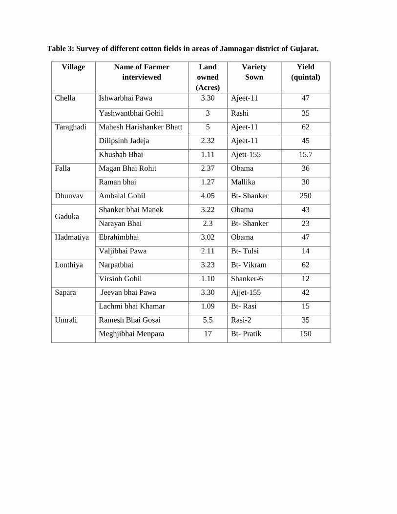
Table 3: Survey of different cotton fields in areas of Jamnagar district of Gujarat.
Village Name of Farmer
interviewed
Land
owned
(Acres)
Variety
Sown
Yield
(quintal)
Chella Ishwarbhai Pawa 3.30 Ajeet-11 47
Yashwantbhai Gohil 3 Rashi 35
Taraghadi Mahesh Harishanker Bhatt 5 Ajeet-11 62
Dilipsinh Jadeja 2.32 Ajeet-11 45
Khushab Bhai 1.11 Ajett-155 15.7
Falla Magan Bhai Rohit 2.37 Obama 36
Raman bhai 1.27 Mallika 30
Dhunvav Ambalal Gohil 4.05 Bt- Shanker 250
Gaduka Shanker bhai Manek 3.22 Obama 43
Narayan Bhai 2.3 Bt- Shanker 23
Hadmatiya Ebrahimbhai 3.02 Obama 47
Valjibhai Pawa 2.11 Bt- Tulsi 14
Lonthiya Narpatbhai 3.23 Bt- Vikram 62
Virsinh Gohil 1.10 Shanker-6 12
Sapara Jeevan bhai Pawa 3.30 Ajjet-155 42
Lachmi bhai Khamar 1.09 Bt- Rasi 15
Umrali Ramesh Bhai Gosai 5.5 Rasi-2 35
Meghjibhai Menpara 17 Bt- Pratik 150

4.2. Studies on Soil borne fungi associated with cotton:
Soil is a complex system. It is the most precious natural resource and contains the most
diverse assemblages of living organisms. Soil microflora plays a pivotal role in evaluation of soil
conditions and in stimulating plant growth (Singh et al., 1999). It is an important panorama of
interactions between microbes and plants (Shekh et al., 2012). Many biological processes take
place in soil and determine functions that provide various services within ecosystems:
turn-over of organic matter, symbiotic and non-symbiotic atmospheric nitrogen fixation,
denitrification, aggregation, etc (Chenu and Stotzky, 2002). It regulates global biogeochemical
cycles, filters and remediates anthropogenic pollutants, and enables food production (Kennedy
and Smith, 1995; Richards, 1987). One significant component of soil is occurrence of
microorganisms. Soil is a medium with solids, liquids and gases in which the mineral
and organic particles form differently-sized aggregates that delimit pores (Tisdall and
Oades, 1982; Feller and Beare, 1997). This organization creates micro-environments that are
suited to microbial activity to varying extents (Chotte et al., 1997).
Indigenous microbial populations in soil are of fundamental importance for ecosystem
functioning in both natural and managed agricultural soils (O‘Donnell et al. 1994) because of
their involvement in such key processes as soil structure formation, organic matter
decomposition, nutrient cycling and toxic removal (Van Elsas, 1997; Doran and Zeiss 2000).
Rhizosphere and non Rhizosphere soil samples were collected from cotton fields of study area.
The soil samples were collected with the help of a soil augur upto a depth of 30 cm. To compare
the fungal population present in the rhizosphere with that of the non rhizosphere regions, soil
around the plant roots and for non rhizosphere fungal studies one meter away from the root zone
is selected. Three soil samples for each (around the root and away from the root zone) were
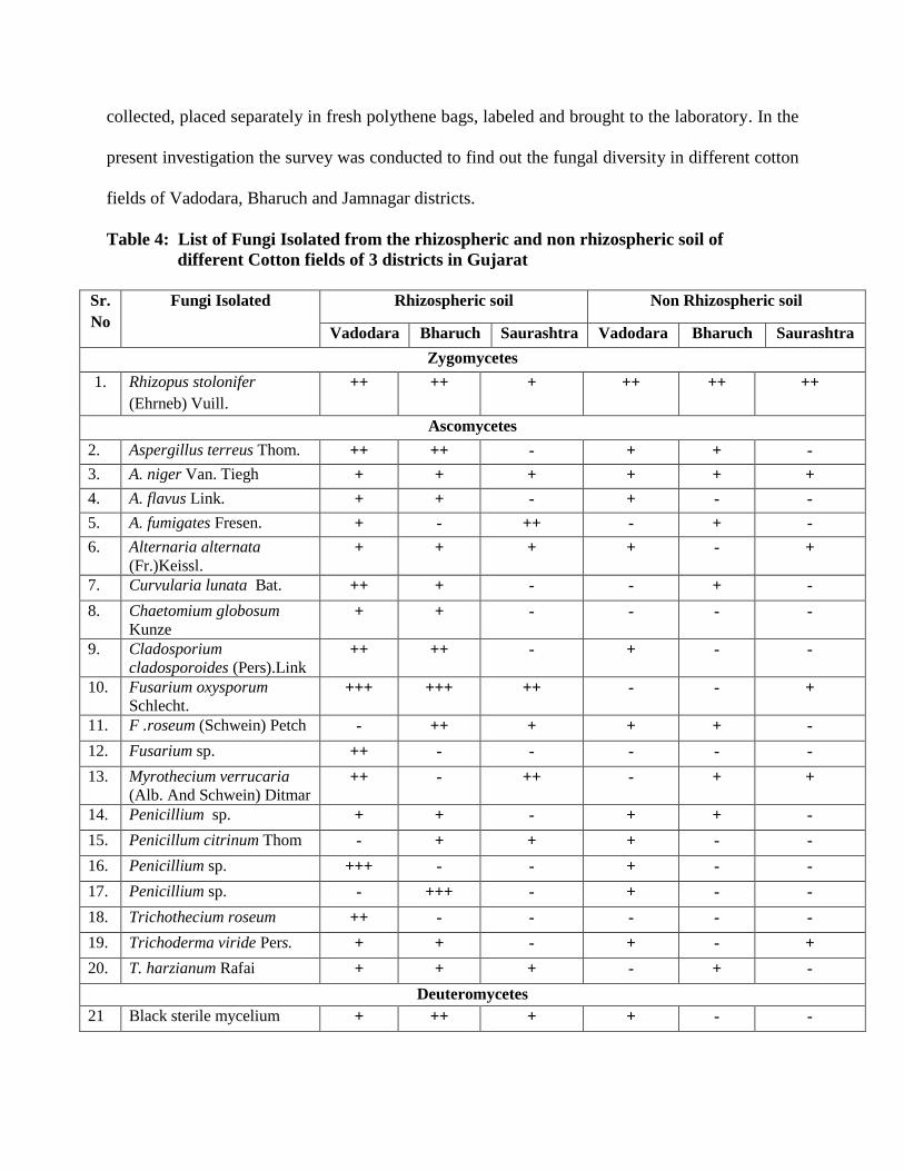
collected, placed separately in fresh polythene bags, labeled and brought to the laboratory. In the
present investigation the survey was conducted to find out the fungal diversity in different cotton
fields of Vadodara, Bharuch and Jamnagar districts.
Table 4: List of Fungi Isolated from the rhizospheric and non rhizospheric soil of
different Cotton fields of 3 districts in Gujarat
Sr.
No
Fungi Isolated Rhizospheric soil Non Rhizospheric soil
Vadodara Bharuch Saurashtra Vadodara Bharuch Saurashtra
Zygomycetes
1. Rhizopus stolonifer
(Ehrneb) Vuill.
++ ++ + ++ ++ ++
Ascomycetes
2. Aspergillus terreus Thom. ++ ++ - + + -
3. A. niger Van. Tiegh + + + + + +
4. A. flavus Link. + + - + - -
5. A. fumigates Fresen. + - ++ - + -
6. Alternaria alternata
(Fr.)Keissl. + + + + - +
7. Curvularia lunata Bat. ++ + - - + -
8. Chaetomium globosum
Kunze + + - - - -
9. Cladosporium
cladosporoides (Pers).Link ++ ++ - + - -
10. Fusarium oxysporum
Schlecht. +++ +++ ++ - - +
11. F .roseum (Schwein) Petch - ++ + + + -
12. Fusarium sp. ++ - - - - -
13. Myrothecium verrucaria
(Alb. And Schwein) Ditmar ++ - ++ - + +
14. Penicillium sp. + + - + + -
15. Penicillum citrinum Thom - + + + - -
16. Penicillium sp. +++ - - + - -
17. Penicillium sp. - +++ - + - -
18. Trichothecium roseum ++ - - - - -
19. Trichoderma viride Pers. + + - + - +
20. T. harzianum Rafai + + + - + -
Deuteromycetes
21 Black sterile mycelium + ++ + + - -

*Readings based on 3 replicates
+++: Dominant ++: Abundant +: Present -: Absent
Serial dilution method (Waksman, 1916) for rhizospheric and non rhizospheric soil
revealed the presence of total 21 species belonging to 10 genera. The results depicted that
maximum number of fungal strains were isolated from the rhizospheric soils of Vadodara district
(18) followed by Bharuch district (16) and areas of Saurashtra viz Rajkot and Jamnagar (10).
Various Ascomycetes fungal members like Aspergillus niger, A. terreus, A. flavus, A. fumigatus,
Alternaria alternata, Curvularia lunata, Chaetomium globosum, Cladosporium cladosporaoides,
Fusarium oxysporum, F. roseum, Myrothecium verrucaria, P. citrinum, Tricothecium roseum, T.
harzianium, T. viride and Rhizopus stolonifer- Zygomycetes member were isolated which are
depicted in Table 4. The occurrence of these fungi was found to more in the rhizospheric soil as
compared to non rhizospheric soil.
The most dominant species F. oxysporum known to cause wilting was found in cotton
fields of all three districts. Other than F. oxysporum , Penicillium sp . was found dominating in
Vadodara district. A. terreus, Curvularia lunata, Cladosporium herabceum, Fusarium sp., M.
verrucaria, R. stolonifer, Trichothecium roseum were found abundantly, whereas, fungi like A.
niger, A. flavus, A. fumigatus, Alternaria alternata, C. globosum, Penicillum sp., Trichoderma
viride, T. harzianum and Black sterile mycelium was found but in lesser amount, whereas,
Fusarium roseum and Penicillium sp. were not found from the rhizospheric soils of Vadodara
district.
The non rhizospheric soil of Vadodara district showed the presence of 13 fungal genera
which included four species of Penicillium followed by three species of Aspergillus viz. A.
terreus, A. niger, A. flavus and other single species of Alternaria alternata, Cladosporium
herbaceum, Fusarium roseum, Rhizopus stolonifer, Trichoderma viride and black sterile

mycelium. Among the non rhizospheric soils of Vadodara, Rhizopus stolonifer was recorded
abundantly.
Aspergillus fumigates, Curvularia lunata, Chaetomium globosum, Fusarium oxysporum,
Fusarium roseum, Fusarium sp., Myrothecium verrucaria, T. roseum and T. viride and sterile
mycelium was not found in the non rhizospheric soil as compared to the rhizospheric soils.
In Bharuch district, rhizospheric soil showed presence of 16 fungal strains of which Fusarium
oxysporum was found dominant. As depicted in Table 4, Rhizopus stolonifer was found
abundantly, whereas A. terreus, A.niger, A. fumigatus, C. lunata, F. roseum, M. verrucaria,
Penicillium sp. and Trichoderma harzianum was found from the non rhizospheric soils. Fungi
like A. flavus, A. alternata, C. globosum, C. herbaceum, F. oxysporum, Fusarium sp., P.
citrinum, Trichothecium roseum, T. viride and sterile mycelium were not found in non
rhizosphereic soil of Bharuch.
Rhizospheric soil of Saurashtra region viz . Jamnagar showed presence of 10 different
fungi of which 3 fungal genera namely A. fumigatus, F. oxysporum and Myrothecium verrucaria
were dominantly found. Whereas other fungi like A. niger, A. alternaria, P.citrinum, R.
stolonifer, T. harizanum and Sterile mycelium were also recorded in less amount.
Similar results reported by Shekh et. al., (2012) from the rhizospheric and non
rhizospheric soil samples of cotton fields in Nanded district and also found that total number of
fungal species were higher in rhizosphere soil as compared to non rhizosphere soil. The results
obtained in the present investigation are similar to the investigations made by Oyeyiola (2009) in
case of Hibiscus esculentus The results obtained in the study for Sorghum bicolor (Odunfa,
1979), cowpea (Odunfa and Oso, 1979), Sugarcane (Abdel- Rahim et al., 1983) and Amaranthus
hybridus (Oyeyiola, 2002) were similar to present results obtained for cotton.
Kalich (1988) investigated soil fungi of some low-altitude desert cotton fields and found
forty two taxa from which genera like Aspergillus, Penicillium and Fusarium were found
predominantly. These three genera were present in all the soil samples of three districts in
Gujarat.
4.2.1. Studies on Fungi Associated with seeds of cotton varieties
The significance of sustainable agricultural production is hidden in the use of quality of
seed. It is the most crucial and vital input for enhancing crop productivity. Since, seed is the
custodian of the genetic potential of the cultivar thus, the quality of the seed determines the
limits of productivity realized in a given system. The shortage of quality of seed, absence of
techno-infrastructure facilities needed for processing, storage and distribution of seed are
amongst the major considerable attributes for such outbreaks.
Seeds are the basic input for crop production and are of great economic interest and also
constitutes a major part of diet, they play a vital role in associating microorganisms which prove
hazardous for seed or the new plant created from it. So any infectious agent (bacteria, fungi,
nematode, etc), which is associated with seeds having potential agent of causing a disease of a
seedling or plant, is termed as seed borne pathogen (Agarwal and Sinclair, 1987). The
associating microorganisms may be pathogenic, weak parasites or saprophytes.
Seeds are regarded as highly effective means for transporting plant pathogens over long
distances. Numerous examples exist in agriculture literature for the international spread of plant
diseases as a result of the importation of seeds that were infected or contaminated with pathogens
(Agarwal and Sinclair, 1996).
Neergaard (1973) reported several types of abnormalities occurred to the seeds, which
mainly include seed discoloration, necrosis, seed abortion, seed toxification, seed rotting, etc. He

further reported that these types of abnormalities occur due to dominate fungi like Aspergillus,
Curvularia, Drechslera, Fusarium, Penicillium, Rhizoctonia, Verticilastum and Alternaria.
Seed-borne mycoflora of wheat reported recently included Alternaria alternata,
Drechslera sorokiniana, Fusarium moniliforme, F. avenaceum, F. graminearum, F. nivale, F.
culmorum, F. equiseti, F. sporotirchioides, Cladosporium herbarum, Stemphylium botryosum
(Nirenberg et al., 1994; Glazek, 1997; Mirza and Qureshi,1978). Seed-borne mycoflora of
sorghum reported from different parts of the world include Alternaria alternata, Aspergillus
flavus, A. fumigatus, A. niger, Cladosporium sp., Fusarium moniliforme, F.oxysporum, F.
pallidoroseum, Drechslera tetramera, Nigrospora sp., Phoma sp., and Rhizopus sp., (Abdullah
and Kadhum, 1987; Ahmed et al., 1992).
Cotton seeds are also used as oil seeds. The economic value of cotton seed is greatly
influenced by the presence of fungi in the seed. Fungi or associated metabolites may reduce the
vigor of planting seed (Davis, 1982; Halloin and Bourland, 1981) increase the amount of free
fatty acid in the seed thereby reducing the quality of the oil (Ashworth et al., 1971; Roncadori et
al., 1971), or produce mycotoxins that render the seed unsuitable for consumption (Diener et al.,
1976). An understanding of the distribution and frequency of the mycoflora could lead to
practical measures for control of the fungi which devalue cotton seed. Concomitant with the need
to assess the mycoflora of cotton seed, an understanding of the conditions conducive to their
presence is important for both predictive purposes and to develop methods of reducing seed
infection (Klich, 1986).
Cotton seedling diseases are a worldwide problem; they are caused by a complex of
microorganisms. Fungi are the widest pathogens that affect cotton crop especially at the seedling
disease stage causing pre or post emergence damping off (Aly et al., 2008). Cotton seedling

diseases may lead to stand losses when the disease is not managed or environmental conditions
are highly conducive for disease occurrence and development (Blasingame and Mukund, 2001;
Rothrock et al., 2007).
Cotton seedlings are vulnerable to disease injuries upto one month after sowing. Of these,
fungal diseases are the most widespread and devastating diseases that affect crop yield
quantitatively and qualitatively (Aly et al., 2000; Nehl et al., 2004).
Cotton seedlings damping off, the serious problem in most of the cotton producing
regions often attributes to Rhizoctonia, Pythium, Fusarium (Disfani and Zangi, 2006; Omar et
al., 2007). Alternaria, Fusarium, Macrophomina, Rhizoctonia and several other fungi were
frequently isolated from cotton seeds and seedlings (Colyer and Vernon, 2005; Asran-Amal,
2007; Mikhail et al., 2009; Fard and Mojeni, 2011). Palmateer et al., (2004) isolated fifty eight
species of fungi belonging to 37 genera, including 9 species of Fusarium. Fusarium oxysporum,
F. solani and F. equiseti were the most common members of this genus occurring at seedling
stage.
Table 5(A): Showing Percentage Occurrence of seed mycoflora of three varieties of
G. herbaceum by Blotter Method.
Sr.
No.
Fungi Isolated Non- Bt Ajeet-11 Vikram-5
Control Treated Control Treated Control Treated
1. Aspergillus niger 28 16 19 9 11 5
2. A. flavus 32 16 23 12 24 11
3. Alternaria alternata 10 8 6 4 - -
4. Chaetomium globosum - - 3 - 1 1
5. Curvularia lunata - - 2 - 1 1
6. Fusarium oxysporum 19 11 13 9 10 7
7. F. roseum 9 2 11 7 14 10
8. Mucor hemalis 12 - 12 - 9 -
9. Rhizospus stolonoifer 10 8 17 7 13 6
10. Trichoderma harzianum 3 1 1 1 - -

*Readings based on 3 replicates of 100 seeds each -: Absence of colony
Table 5 (B): Showing Percentage Occurrence of seed mycoflora of three varieties of
G. herbaceum by Agar Plate Method
Sr.
No.
Fungi Isolated Non- Bt Ajeet-11 Vikram-5
Control Treated Control Treated Control Treated
1. Aspergillus niger 30 18 26 10 22 9
2. A. flavus 35 20 24 18 20 12
3. Alternaria alternata 22 8 11 5 14 9
4. Chaetomium
globosum
- - - - 3 1
5. Curvularia lunata 13 7 14 9 10 2
6. Fusarium
oxysporum
33 20 29 16 30 18
7. F. solani 4 12 5 10 7 8
8. Mucor hemalis 18 - 12 - 15 -
9. Rhizospus
stolonoifer
17 8 19 9 15 10
10. Trichoderma
harzianum
18 5 16 3 13 5
11. Penicillium citrinum - - - - 4 2
12. Sterile Mycelium 10 - 6 - 4 -
*Readings based on 3 replicates of 100 seeds each -: Absence of colony
Fungi were isolated from seeds using the standard blotter method as recommended by ISTA
(2003). Three replications of 100 seeds each from each variety were placed on three layers of
well moistened filter paper r and agar plate methodusing Potato Dextrose Agar (PDA) from
unsterilized and sterilized seeds.
It is evident from the Tables 5A and 5B that total 12 different fungal organisms were
obtained from the untreated and treated three varieties of cotton of which 2 fungi belonged to
Zycomycetes, 8 fungal organisms belonged to Ascomycetes and Black non spourlating

unidentified fungi was also recorded. . In Blotter method less number of fungal organisms were
isolated and observed in comparison to agar plate method.
In blotter method as depicted in Table 5A, in-total 10 fungal isolates were observed.
Aspergillus flavus, A. niger, Fusarium oxysporum and Rhizopus stolonifer were found dominant
in all the three varieties of cotton. Alternaria alternata was from the seeds of two varieties
namely Non-Bt and Ajeet-11, it was not recorded from Vikram-5 variety. Chaetomium globosum
was not recorded from Non Bt variety. It was isolated from both Bt varieties Vikram-5 variety
and treated seeds of Ajeet-11.
Curvularia lunata was isolated from the Vikram-5 variety and untreated seeds of Ajeet-11,
whereas it was totally absent in the Non Bt seeds. Fusarium roseum was observed from all three
varieties of cotton.
Presence of Mucor hemalis was recorded in all the control sets. Seeds of all three
varieties after treated with 1% NaOCl did not show the presence of Mucor. Contrary to it
Rhizopus stonlonifer was recorded in both treated and untreated seeds and was found to be one of
the dominant fungal species. Trichoderma harzianum was found to present in Non Bt and Ajeet-
11 variety.
From Table 5(B) it is depicted that 13 different fungal isolates were recorded with
maximum colonies of Aspergillus flavus and A. niger followed by F. oxysporum which was
recorded in both treated and untreated seeds of all three varieties of cotton.
Alternaria alternata was isolated from Non Bt and Ajjet-11 variety of cotton where as it
was not at all recorded from the Bt variety Vikram-5. Chaetomium globosum was recorded only
in Vikram-5 variety in comparison to other two cotton varieties.Curvularia lunata was isolated
from all hybrid and Bt variety.

F. solani was recorded only in Agar plate method where it was isolated maximum from
the treated seeds of all three varieties. It was found more in Non Bt variety followed by Bt
varieties Ajeet-11 and Vikram-5.
Mucor hemalis was found present in untreated seeds of all three varieties as compared to
the seeds treated with 1% NaOCl. R. stolonifer and T. harzianum was recorded in all three
varieties of seeds, both treated and untreated seeds of Ajeet -11 showed maximum presence
followed by Non Bt and Vikaram-5 respectively.
Penicillium citrinum was recorded only in the agar plate method and it was found only in
the seeds of variety Vikram-5. In both Non Bt and Ajeet-11 varieties it was not found. Black non
sporulating mycelium was also recorded from the untreated seeds of all three varieties.
The results obtained in the present investigation are similar to the results of Tomar et al.,
(2012), where they have investigated the presence of eleven fungal flora viz., Aspergillus niger,
A. flavus, Penicillium spp, Altemaria alternata, Chaetomium spp, Rhizopus niger, Fusarium
solani, Macrophomina phaseolina, Myrothecium roridum, Trichothecium roseum and
Curvularia lunata from JK 4 cotton cultivar.
Crawford (1923) isolated eight taxa, predominantly Colletotrichum sp. and Fusarium
spp. from the interior of cotton seed in Arkansas. Colletotrichum gossypii , a seed-borne
pathogen causing leaf anthracnose have been reported to produce myco toxins (Diener et al.,
1976; Suzuki et al., 1980).
Templeton et al., (1967) reported Alternaria alternata from seed coat of cotton. In 1979
Padaganur found Alternaria macrospora on cotton seeds. Similarly (Gawade et al,. 2006)
reported Alternaria macrospora from cotton seeds.
Davis (1977) reported Alternaria as a dominant member of the mycoflora of cotton seed
in Mississippi. However, Alternaria was listed as an infrequent fungus by Roncadori et al.
(1971) and was present in more than 10% of the seeds from only one location and Aspergillus
niger was a dominant fungus at several locations infecting up to 23% of the seed in the study by
Simpson et al., (1973).
Fulton and Bollenbacher (1959) isolated Rhizoctonia solani, Pythium ultimum, Fusarium
oxysporum, F. moniliforme, F. semitectum and several other fungi from cotton seeds and
seedlings and found that the most isolated fungi were pathogenic to cotton seedlings. Also,
Alfred (1963) indicated that fungi belonging to Alternaria, Aspergillus, Diploidia, Fusarium and
Rhizoctonia were associated with the seed hairs and the actual seed during boll development.
Kuch (1986) isolated Fusarium equiseti and Fusarium semitectum for more than 10% of the seed
at any sampling of delinted surface sterilized cotton seeds in the southern USA.
Mansoori and Hamdolahzadeh (1995) isolated Alternaria alternata, Aspergillus niger,
Fusarium acuminatum, Fusarium solani, Pythium ultimum, Rhizopus arrhizus and Rhizoctonia
solani from cotton seeds.

4.2.2. Identification of fungus based on morphological characters
i. Alternaria alternata (Fr.) Keissl. (Plate-III, Fig.- A)
Colonies growing rapidly reaching 6cm in diameter in one week olive black without aerial
mycelium, conidiophores short, simple, straight, 3 –septate, branched or unbranced, upto 50µm
long, 3-6 µm wide; conidia forming often in branched chains of 2-10 or more, muriform with 3-
8 transverse septa, walls rough in lower part with longitudinal or oblique septa, obclavate,
ovoid, ellipsoidal, often with a short or cylindrical beak, smooth or verruculose, medium
golden brown, pale, beak pale and upto 1/3 the length of conidium, 2-5 µm wide.
ii. Apergillus flavus Link. (Plate- IV , Fig.-B )
Synonym: Aspergillus fasciculatus Bat.and Maia
Colonies growing rapidly, 6-7 cm in 10 days, conidial heads yellow when young, becomes dark
yellow in age, in older culture deep green, conidial heads radiate, splitting into poorly defined
column, conidiophores arising separately from the substratum, 0.5-1.5 mm long, metuale

present predominantly, only phialides arising on metuale, conidia globose to subglobose,
echinulate, yellowish green 4.5-14x 3-5.5µm.
iii. Aspergillus fumigatus Fresen.
Colonies white at first becoming dull blue green, velvety to floccose, conidial heads columnar,
compact, often densely crowded upto 400x 50 µm, conidiophores short, smooth, light green
upto 300 µm in length, septate, gradually enlarging into flask shaped vesicle, phialides closely
packed, 6-8x2-3 µm, conidia globose to subglobose, green inmass, echinulate, 2.5-3 µm,
cleistothecia are absent.
iv. Aspergillus niger Van. Tiegh. (Plate- IV, Fig.- E)
Synonym: Sterigmatocystis nigra Tiegh.
Colonies growing moderately 3.5-4.5 cm in 10 days on PDA, with abundant submerged
mycelium, conidial heads carbon black; conidial heads large and black, at first globose then
radiate or splitting in well defined columns; conidiophores arising directly from the substratum,
smooth, non septate, thick walled; vesicles globose, walls thick, occasionally longer, bearing
two series of fully packed phialides; conidia globose, spinulose with colouring substance, black;
globose to subglobose sclerotia are produced .
v. Aspergillus terreus Thom. (Plate- III , Fig.- D)
Synonym: Aspergillus boedijnii Blochwitz.

Colonies attaining 3.5-5cm in 10 days, plain or with radial furrows, velvety, floccose in some
strains, cinnamon-buff to wood brown; conidial heads long columnar, cinnamon
brown;conidiophores smooth, colourless; vesicles hemispherical, dome like, phialides biseriate;
metuale crowded, parallel; phialides closely packed; conidia globose to subglobose, smooth,
relatively heavy walled hyaline cells globose to ovate or even truncate, produced singly or in
small clusters on submerged vegetative mycelium, sclerotia like mass absent.
vi. Chaetomium globosum Kunze (Plate- III, Fig.-E)
Synonym: Chaetomium coarctatum Sergeeva,
Chaetomium olivaceum Cooke and Ellis
Colonies well growing, aerial mycelium pale brown, producing perithecia after 5 days,
perithecia scattered or gregarious, globose to subglobose to broadly oval, olive green to grayish
green, 160-390x 230-300µm, thickly clothed with hairs, terminal hairs abundant, light coloured,
finely roughened, obscurely septate, lateral hairs light coloured, straight to slightly flexed or
undulate, asci oblong, clavate, ascospores dark, lemon shaped, broadly ovoid, apiculate at both
ends.
vii. Cladosporium cladosporoides (Pers.) Link (Plate- III , Fig.-F )
Colonies velvety, oliviaceous green to oliviaceous brown, with a greenish black reverse, ca. 2.5
cm diam when 5-day old and 5.5 cm diam when 10-day old and grown on potato dextrose
agar (PDA) at a room temperature.
Conidiophores macronematous, semimacronematous, straight or flexuous, simple or branched,
intercalary or terminal, smooth, sometimes verruculose, septate, up to 360 µm long and 3.3-4.1
µm wide, not geniculate nor nodose, apically truncate with 1-3 denticles, or sympodially
denticulate, with scars at conidiogenous.

Conidia in heads of densely crowded, profusely branched chains, oblong, limoniform, ellipsoid
or fusiform with truncate ends, light olive, 0-1 septate, smooth, 5.0-6.8 x 2.3-3.5 µm in the
broadest part with prominent, protuberant, dark scar at each end.
viii. Curvularia lunata Bat., J.A. Lima and and C.T. Vasconc.
Synonym: Curvularia aeria (Bat., J.A. Lima and and C.T. Vasconc.) Tsuda.
Colonies on PDA dark gray, usually zonate; stroma regularly and abundantly formed in culture;
mycelium branched, septate, conidiophores long, conidia elliptic, curved, septa 2-3, middle cells
broad and darker than other cells, middle septum not median, smooth.
ix. Fusarium oxysporum Schlecht. (Plate- III , Fig.-B )
Colonies reaching 4.5 cm diameter in 4 days at 250C, aerial mycelium sparse to floccose, white
or peach, but usually purple or violet tinge, conidiophores unbranched or sparsely branched,
monophialidic, stroma white, smooth, effuse, microconidia usually abundant, mostly 0-septate,
oval, ellipsoidal, kidney shaped or straight, 5-12 x2.5-3.5 µm; macroconidia 2-5 septate, spindle
to fusiform, curved or almost straight, pointed at both ends, definitely or weakly pedicellate, 27-
60x3-5 µm; chlamydospores mostly terminal, globose, smooth or roughened.
x. Fusarium roseum
Synonym: Gibberella zeae (Schwein.) Petch
Colonies growing very fast, reaching 9 cm diameter in four days, grayish rose to livid red
crimson, often becoming vinaceous with a brown tinge, aerial mycelium floccose, somewhat
lighter cfoloured and becoming brown, sporulation often scarce, conidiphores densely branched
along with solitary phialides, conidia slender, falcate, moderately curved, with pointed curved
apical cell, basal cell pedicillated, mostly septate, chlamydospores scarce, often completely
absent, perithecia dark blue, tuberculate, ascospores 3-septate.
xi. Fusarium pallidoroseum
Colonies growing very fast, aerial mycelium sparse to floccose, white, conidiophores unbranched
or sparsely branched, monophialidic, stroma white, smooth, effuse, microconidia usually
abundant.
xii. Myrotehcium verrucaria (Alb. and Schwein.) Ditmar
Synonym: Gliocladium fimbriatum Gilman and Abbott
Colonies reaching 4-5 cm diam on PDA at 25 0C after 14 days, conidiomata sporodochial or
rarely synnematous, spore mass wet, black, convex, surrounded by white, floccose margin;
mycelium absent ort floccose, white to rosy buff; sporulation diffuse or coalesced into pale
olivaceous to black sporodochia with a white margin, hyphae hyaline , smooth, thin walled,
rarely branched, septate, conidiophores branched repeatedly, usually forming 2-4 branches at
each level, phialides 3-6 in a whorl, closely packed in a dense parallel layer, conidia broadly
fusiform, one end pointed, other protruding and truncate.
xiii. Penicillium citrinum Thom. (Plate- IV, Fig.- A)
Synonym: Penicillium baradicum Biourge.
Colonies growing 25-30 mm in 7 days at 250C, sometimes smaller, radially sulcate, marginal
areas ventunous, centrally floccose, mycelium white in peripheral areas, at the centres white to
grayish orange or apricot, conidiation moderate, turquoise grey to greyish turquoise or glaucous
sky blue, exudates clear, pale yellow to reddish brown, conidiophores borne from surface to
subsurface hyphae, phialides in compact verticils, conidia spheroidal to subspheroidal, smooth
or finely roughened, typically borne in long well defined columns, one per metula, arranged in a
characteristic whorl on each conidia.
xiv. Rhizopus stolonifer (Ehrenb.) Vuill. (Plate- IV, Fig.-C)
Synonym: Mucor mucedo L.
Colonies growing profusely, white at first, turning brownish black, stolons spreading,
internodes brown, with well branched rhizoides at each node; spornagiophores in clusters of 3-
10 unbranced, white, becoming pale to dark at maturity; sporangia globose, hemispherical,
granular, olivaceous, black; columella hemispherical, very often becoming pilate;
spornagiophores irregular, round to oval, angular, straight.
xv. Trichoderma harzianum Rafai.
Synonym: Sporotrichum narcissi Tochinai andShimada
Colonies growing rapidly upto 9cm in 4 days, smooth surfaced, watery white, with sparse
mycelia mat but soon developed aerial hypahe on their surface; conidiation predominantly
effuse, appearing granular or powdery with formation of conidia, rapidly turning to yellowish
green to dark green, producing tufts fringed by sterile white mycelium, hypahe septate,
branched, smooth walled, hyaline, chlamydospores fairly abundant, intercalary or terminal on
short branches, conidiophores hyaline, smooth walled, straight or flexuous, much branched,
phialides in false verticils, conidia produce singly and successively, accumulate at the tip of
each phialide and form into globose conidial head, subglobose or short obovoid, often with
truncate base, pale green singly, darker in mass, 2.8-3.2x2.5-2.8 µm.
xvi. Trichoderma viride Pers. (Plate- IV, Fig.- F)
Synonym: Trichoderma lignorum (Tode) Harz,
Colonies growing rapidly upto 9 cm, watery white becoming hairy from the formation of loose
scanty aerial mycelium, floccose, somewhat whitish; conidiation effuse or in compact tuffs,
galucous to dark bluish green; reverse uncoloured, conidiophores much branched, arise in
compact or loose tufts, main conidiophores 4-5 µm wide, producing smaller branches ,
ultimately a conifer- like branching system is formed, all the branches stand at wide angles to

the bearer, tip terminated by phialides, conidia globose or short obovoid, or broadl;y
ellipsoidal, blueish green to dark green.
xvii. Trichothecium roseum (Pers.) Link (Plate- III, Fig.- C)
Colonies growing fast, reaching 9 cm diameter in ten days, pinkish, powdery due to conidial
formation; reverse colourless to light pink; exudate lacking; conidiophores erect, produced
singly or in groups, mostly simple, hyaline, septate, smooth, upto 2 mm long (base difficult to
trace), 4-5 µm wide often with three septa in the lower part; conidia ellipsoidal to pyreform,
pear shaped, with an obliquely prominent truncate basal scar, 2 celled, upper cell slightly larger,
smooth, thick walled, hyaline.
4.3. Studies on Arbuscular Mycorrhizal fungi associated with cotton
For millennia, diverse microorganisms have yielded important biological materials useful to
human beings such as antibiotics, drugs, enzymes, herbicides and growth promoters. Microbial
diversity is the key to human survival and economic well being and provides a huge reservoir to
resources which can utilize for our benefit. Diverse microorganisms are essential to a sustainable
biosphere. Microbes are able to recycle nutrients, produce and consume gases that affect global
climate, destroy pollutants, treat our wastes and they can be used for biological control of plant
and animal pests (P. Jones Nirmalnath, 2010).
Soil microbiota plays a fundamental role for the productivity and stability of horticulture
and agroecosystems (Castillo et. al., 2006 b, 2009). Within this microbiota AM fungi stand out
because they are important for the phosphate (P) nutrition of the plants (Borie et al., 2010;
Castillo et al., 2010). Among the soil beneficial microorganism, Arbuscular mycorrhizal fungi
(AMF) are one of the most important components of the soil biota in natural and agricultural
systems. They are obligate symbionts currently placed within the division Glomeromycota
(Schüßler et al., 2001), that establish endomycorrhizal associations with up to 90% of plant
families (Smith and Read, 1997). AMF are recognized as an important, widespread component
of terrestrial ecosystems, benefiting plant establishment by enhancing plant nutrient acquisition,
improving soil quality and increasing resistance to environmental stresses (Smith and Read,
1997), and also playing an important role in plant biodiversity, ecosystem variability and
productivity (Wang and Zhao, 2008) .
The diversity of AMF species is measured mainly by identifying the characters of asexual
spores, which are the fungal propagules that possess morphological characters that help to define
species in this group of organisms (Morton et al., 1995). Molecular techniques serve as an useful

tool for characterization and identification of AMF (Kowalchuk et al., 2002). To the date,
although only fewer than 200 species of AM fungi have been described (Redecker and Philipp,
2006), numerous studies on AM fungal diversity in different ecosystems worldwide have shown
that AM fungi distribute globally (Treseder and Cross, 2006).
In order to study AM fungal diversity, representative soil samples were collected from various
cotton fields of Vadodara, Bharuch and Jamnagar districts of Gujarat (Fig. 4).
The present study represents 4 genera and 28 species were observed in the soils
associated with cotton in the area studied (Table 9).
In the present study maximum number of AM fungal spores was isolated from the
rhizospheric soil of Kayavarohan area of Vadodara district and minimum number of spores was
found in the soil of Padra agricultural fields (Table- 6). Three AM fungal genera namely
Acaulospora, Glomus and Gigaspora were isolated from the soils of Vadodara district of which
Glomus fasciculatum was found in maximum numbers, followed by G. aggregatum and G.
mossae. Acaulospora laevis was recorded only from the soils of Kayavarohan and Muval village
of Padra in the Vadodara District (Table-6, Fig. 5).
As depicted in table 7 and Fig. 6, from the field soils of Bharuch district, three AM fungal
genera were isolated of which species of Glomus were found maximally. Fields of Karjan
village showed highest number of AM fungal spores which belonged to the Glomus genera. G.
fasciculatum, G. fugienaum and G. melanosporum were found in this area. Minimum AM fungal
spores were observed in the Kavi area of Bharuch district. In the entire district survey, Glomus
mossae was found frequently in the fields. In total 15 species of Glomus, 4 species of Gigaspora
and 1 species of Acalulospora was found in Bharuch district. Acaluospora laevis was found only
in the soils of Asanvad village in the sites surveyed of this district. As depicted in table 8, from
the field soils of Jamnagar district, species of only Glomus genera were found.
4.3.1. Morphological Characters of AM FSpores
Isolation studies showed presence of 3 genera in rhizospheric and 3 genera in non rhizosperic
soil of different places. The details of various species are described below.
Acaulospora
Acaulospora Gerd. and Trappe emend. Berch, Mycotaxon 23: 409, 1985.
Kuklospora Oehl and Sieverd., J. Applied Bot. Food Quality 80: 74, 2006.
Azygospores produced singly in soil, large generally globose, or sub globose, with oily contents,
borne laterally on the stalk of a large, terminal thin walled vesicle. Vesicles about the same size
as the spores, with vesicle contents transferred to spore at maturity. Spore walls continuous
except for a small occluded pore. Germ tube produced directly through walls near spore base.
Forming endo mycorrhizae with lobed vesicles and arbuscules.
1. Acaulospora laevis Gerd. and Trappe. (Plate- VIII , Fig.- B )
Hyphae hyaline, thin walled, 6-8 μm broad, fertile hyphae terminate with the vesicles. Vesicles
globose, up to 320 μm diam., 30-40 μm broad at the base, contents flown into the spore and
collapsed as the spore matures. Spores formed singly and laterally on the hyphae just below the
base of the vesicles, smooth, globose to subglobose, ellipsoid, occasionally reniform to irregular,
initially dull yellow, turn deep yellowish-brown, red-brown to dark olive green at maturity, 119-
300 x 119-520μm; spore wall three layered, continuous to the hyphae except for the occluded
opening, outer wall 3-4 μm thick, rigid, yellowish-brown to reddish brown, inner layers hyaline,
the inner most layer sometimes minutely roughened. In older spores, wall minutely perforated
and the outer surface sloughing away. Spore contents globular to polygonal in appearance.

The family Gigasporaceae
Gigasporaceae J.B. Morton and Benny emend. Sieverd, Souza and Oehl, Mycotaxon 106: 328, 2008.
Gigasporaceae J.B. Morton and Benny, Mycotaxon 37: 471, 1990.
Gigaspora Gerd. and Trappe emend. C. Walker and F.E. Sanders Mycotaxon 27:179, 1986.
Sporocarps unknown. Spores formed singly in soil or rarely in roots. Spores formed on bulbous
suspensor cell arising from subtending hyphae (sporophore). Spores with one wall consist of
three layers: a unit, semi-persistent to persistent outer layer, a laminate middle layer and a thin
inner germinal layer. The germinal layer has multiple, irregularly arranged germ pores. Most of
the germ pores produce germ tubes, which penetrate the spore wall and branch profusely in the
soil. Axillary cells round, spiny, with nodulous elevations. Form arbuscular mycorrhiza, vesicles
unknown.
1. Gigaspora ramisporophora Spain, Sieverd. and N.C. Schenck. (Plate- VIII , Fig.-D )
Hyphae sub hyaline to pale yellow, up to 10μm broad, subtending hyphae septate, simple,
branched, with 1-3 suspensor cells, light brown, 9-14 μm broad, wall up to 3 μm thick. Spores
produced singly on the apex of bulbous suspensor cells, predominantly globose, often
subglobose, smooth, golden yellow to yellowish brown, 150-400 x 200-450 μm. Spore wall three
layered, 9-20 μm thick. Outer layer hyaline to subhyaline, brittle, up to 4 μm thick, continuous
with outer layer of suspensor cell and usually adherent to middle layer. Middle layer yellow to
yellowish brown, 4-25 μm thick, adherent to inner layer; inner layer yellow to yellowish brown,
up to 3 μm thick. Suspensor cells globose to ovate, 60-80 x 40-60 μm in diam., usually with
three walls, 6-10 μm thick; outer and middle walls hyaline; innermost wall brown. Auxiliary
cells round to clavate.
2. Gigaspora albida Scenck and Smith (Plate- VIII , Fig.- C )

Spores globose to subglobose, 200 – 280 µm in size, Cream with pale green tint in colour, spore
wall consists of three layers, the first two layers adherent and of equal thickness, hyaline to pale
yellow in colour.
3. Gigaspora candida Bhattacharjee, Mukherjee (Plate- VIII , Fig.-E )
Azygospores found singly in soil, white, globose, 200-300 µm diameter, spore wall smooth,
2-layered, the two layers distinctly visisble in fractured azygospores, suspensor like cell
attached to the azygospore, white globose to subglobose, usually detached during wet
sieving. Soil borne vesicles not isolated.
The genus Glomus
Glomus Tulasne and Tulasne emend. C. Walker and Schüßler
Spores glomoid, produced terminally on undifferentiated, non-gamitangial hyphae, solitary, in
clusters or produced in Sporocarps. Peridium complete or imcomplete. Spore contents at
maturity separated from attached hyphae by a septum or occluded by spore wall thickening.
1. Glomus aggregatum N.C. Schenck and Smith emend. Koske. (Plate- V , Fig.-A )
Hyphae hyaline to subhyaline, up to 8 μm broad, 8-12 μm wide at the point of attachment of the
spore. Spores formed in loose clusters or in Sporocarps without peridium; Sporocarps hyaline to
light yellow with a greenish tint, becoming yellow with age. 660-1500 x 330-1000 μm. Spores
globose, subglobose, obovate, cylindrical to irregular, hyaline to yellow, 73-105 x 60-85 μm in
diam; wall yellow to yellowish brown, 1-3 μm thick, outer walls slightly thicker and lighter than
the inner wall; walls separable with slight pressure and most apparent in stained preparations.

2. Glomus formosanum Wu and Chen. (Plate- VIII , Fig.-F )
Hyphae pale yellow to honey yellow, up to 9 μm broad; subtending hyphae 1-4 in numbers, 7-18
μm broad, with opening at the attachment, occluded by spore wall thickening, two nearby
attached hyphae fused together or closely separated at the attachment. Spores produced in
Sporocarps or in aggregates without peridium. Sporocarps yellowish brown to reddish brown,
globose, subglobose to irregular, 360-500 x 450-500 μm, peridium composed of septate, thin
walled, loosely interwoven hyphae, up to 10 μm broad. Spores globose to sub globose, 82-125 x
95-135 μm diam., yellowish brown to reddish brown; Spore wall single, yellowish brown to
reddish brown, 5- 12 μm thick, thickest at attachment, up to 20 μm broad, surface smooth.
3. Glomus glomerulatum Sieverd., (Plate- V, Fig.- D)
Hyphae up to 6 μm broad. Sporocarps dark brown, without peridium, become compact with age,
globose, subglobose, rectangular, flattened or some times irregular in shape, surface knobby, 330
x 460 μm diam., formed by the interwoven hyaline hyphae, 2-6 μm in diam., walls up to 0.5 μm
thick. Spores in the sporocarps are clustered in the mycelium and embedded in an unordered
gleba, globose to subglobose, 40-70 μm diam., yellow to brown; wall consists of two walls in
one group; yellow to brown, laminated and 4-9 μm thick, the spore surface smooth; second wall
hyaline, membranous, up to 0.5 μm thick and adherent to first wall. Chlamydospores formed in
sporocarps, have two hyphal attachment at irregular distance along the hypha; hyphal
attachments yellow to brown, 5-7 μm broad, straight to recurved; hyphal attachment pore 1-2 μm
in diam. and are closed by the second wall or by a septum.
4. Glomus macrocarpum Tulasne and Tulasne. (Plate- V, Fig.- F)

Hyphae yellow to light brown, up to 12 μm broad, subtending hyphae up to 15 μm broad.
Sporocarps globose, subglobose, elongate to irregular, 10x10 mm in diam. Spores sub globose,
globose to irregular, 70-150 x 75-120 μm in diam.; spore wall composed of two distinct layers:
outer layer thin, hyaline, 1-2 μm thick in water or glycerol mount, swell enormously in lactic
acid mount. Inner wall layer yellow, 6-12 μm thick, laminated, rarely seen as two layers, swell
slightly in lactic acid. Spores taper towards the attachment, attachment hyphae single and
persistent, wall thickening continues into the subtending hyphae, subtending hyphae up to 90 μm
long from the spore proper. The pore closed by a thinner septum.
5. Glomus rubiformis Gerd. and Trappe. (Plate- VI, Fig.-F )
Hyphae hyaline to light yellow, up to 9 μm broad. Sporocarps dark brown, subglobose to
ellipsoid, 150-175 x 190-410 μm, consisting of a single layer of chlamydospores surrounding a
central plexus of hyphae. Peridium absent, individual spores at times partially enclosed in a thin
network of tightly appressed hyphae. Chlamydospores dark brown, obovoid, ellipsoid to
subglobose, 30-40 x 80-100 μm, with a small pore opening into the thick walled subtending
hyphae. Spore wall laminate, 3-8 μm thick, up to 12 μm thick at spore base, often perforated
projections on the inner surface. A variable stalk-like projection produced near the base of some
spores. Hyphal attachment simple, thickening of wall extended along subtending hyphae. Pore
occluded at maturity.
6. Glomus fasciculatum (Thaxt.) Gerd and Trappe (Plate- V , Fig.- C)
Zygospores found in loosely coherent spongy mass, pale yellow to pale yellow-brown in colour,
globose to subglobose. 60-110 μm in diameter. Spore wall consisting of three layers, subtending
hypae is cylindrical to slightly flared.
7. Glomus intraradices N.C.Schenek andG.S. Smith (Plate- VIII , Fig.- A)
Spores pale cream to yellowish brown sometimes with green tint, globose to subglobose with
some elliptical spores. Spore size ranges from 40-400 μm. Spore wall consists of 3 layers, in
juvenile spores, initial sublayer is thin and become thick due to formation of additional
sublayers. Thickness of the wall layers varies from 3.2-12 μm in mature spores, subtneding
hyphae is cylindrical, occasionally slightly constricted.
8. Glomus mosseae (T.H. Nicolson and Gerd.) Gerd. and Trappe (Plate- V, Fig.-B)
Syn: Funneliformis mosseae
Spores are surrounded by tight peridium to form sporocarps, spores found singly in soil, spores
straw to dark orange borwn in colour, majority are yellow- brown, globose to subglobose in
shape, 100-260 μm in diameter, spore wall three layered, subtending hyphae funnel shaped with
14-32 μm thickness.
9. Glomus hoi Berch and Trappe
Spores borne singly in soil, globose, subglobose, ellipsoidal or irregular, (50)-80-120 (-155) µm,
yellow- brown. Wall of spore composed of two distinct, seperable layers,subtending hypahe
cylindrical or slightly flared toward the point of attachment to the spore where it is (5-)8-11(-13)
µm wide.
10. Glomus geosporum (Nicol.and Gerd.) Walker
Spores borne singly in soil, yellow- orange, globose to subglobose (120-)175(-260) µm diameter,
sometimes ovoid, with single subtending hypahe.

11. Glomus etunicatum Becker and Gerd.
Spores borne singly, pale yellow to yellow, globose to subglobose, (75-)95(-135) µm diam;
occasionally ovoid, with one subtending hypha.
12. Glomus caledonium (Nicol. and Gerd.) Trappe and Gerdemann
Spores single in the soil; pale yellow to golden yellow; globose to subglobose; (90- )224(-370)
µm diam; with a single subtending hypha. Spore wall consist of three layers.

Table 6: Isolation of AM Spores from the Rhizospheric and Non Rhizospheric soil of
different cotton fields of Vadodara district.
Location
Sample No. of AM Spores/ 100g soil Major AM fungi isolated
Rhizospheric
soil
Non Rhizospheric
Soil
Por 1 135 53 G. aggregatum, G. mossae,
G. fasciculatum 2 147 68
3 180 60
Kayavarohan 1 243 98 G. aggregatum, G. intraradices,
Acoulospora laevis,
G. fasciculatum 2 197 78
3 210 89
Kunvarpura 1 159 71 G.maculosum,G.fugienaum,
Gigaspora albida
2 130 65
3 165 83
Dabhoi 1 180 88 G.melanosporum,G.fugienaum,
G.fasciculatum 2 172 79
3 183 85
Varnama 1 192 102 G.aggregatum,
G.mosseae,
G.geosporum 2 189 99
3 183 89
Sundarpura 1 218 119 G. geosporum, G.convolutum,
G. fasciculatum, G.
aggreagatum, G.etunicatum 2 201 109
3 189 99
Padra 1 102 67 G.glomerulatum, G.clarum,
G.fuegianum,, G.hoi 2 130 78
3 172 85
Dabhasa 1 210 98 G. fecundisporum, G. mossae,
G. melanosporum, G.
etunnicatum, G. intraradices,
G. microcarpa
2 198 93
3 190 81
Muval 1 155 79 G. fasciculatum, G. tenerum,
Acaluospora laevis 2 168 86
3 184 92
Dabka 1 166 72 G.fasciculatum,
G.mosseae 2 172 79
3 181 82
* Data based on average of three samples
Table 7: Isolation of AM Spores from Rhizospheric and Non Rhizospheric soil of
different cotton fields of Bharuch district.
Locations Sample No. of AM Spores/ 100g soil Major AM Fungi isolated
Rhizospheric soil Non Rhizospheric Soil
Jambusar 1 102 86 G. clarum, Gigaspora
ramisporophora, G.etunicatum 2 91 74
3 88 69
Kavi 1 82 49 G. mossae, Gigspora sp.,
G. microcarpa 2 96 45
3 76 52
Umber 1 102 67 G. aggregatum, G. rubiformis
2 93 71
3 110 82
Simor 1 97 58 G. fasciculatum, G. tenerum, G.
segmentum 2 106 62
3 103 77
Andada 1 84 59 G. mossae, Gigaspora candida
2 93 57
3 101 69
Matar 1 91 53 G. fasciculatum, G.
glomerulatum 2 104 61
3 94 50
Karjan 1 180 88 G.melanosporum,G.fugienaum,
G.fasciculatum 2 172 79
3 183 85
Anastu 1 113 90 G. aggregatum, Gigaspora
albida, G. tenerum 2 129 72
3 120 69
Asanvad 1 145 60 G. melanosporum, G.
etunnicatum, G. intraradices,
Acaulospora laevis 2 122 53
3 133 57
Chhapra 1 107 59 G. aggregatum
2 104 85
3 103 77
Haripura 1 89 31 G. mossae
2 97 64
3 98 58
Intola 1 92 40 Gigaspora albida, Glomus

* Data based on average of three samples
2 102 89 clarum
3 76 56
Table 8: Isolation of AM Spores from the Rhizospheric and Non Rhizospheric soil of
different cotton fields of Jamnagar district
* Data based on average of three samples
Location Sample No. of AM Spores/ 100g soil Major AM Fungi isolated
Rhizospheric
soil
Non Rhizospheric
Soil
Chella 1 71 47 G. mossae, G. intraradies,
G. aggregatum 2 62 39
3 58 51
Taraghadi 1 140 96 G. fasciculatum, G. caledonium
G. aggregatum, 2 43 23
3 58 27
Falla 1 102 89 G. intraradices, Acaulospora
laevis, G. fasciculatum,
G. glomerulatum 2 118 92
3 103 86
Dhunvav 1 97 64 G. melanosporum G.
etunnicatum 2 71 45
3 83 56
Gaduka 1 112 87 G. fecundisporum, G. mossae,
G. melanosporum,
G. etunnicatum, G.
intraradices,
2 105 79
3 127 100
Hadmatiya 1 111 90 G. fasciculatum, G.
aggregatum
,G. microcarpa 2 97 60
3 91 54
Lonthiya 1 100 77 G. etunnicatum
G. fasciculatum, G.
aggregatum 2 108 86
3 127 103
Sapara 1 113 98 G. microcarpa,
G. mossae, G. melanosporum
2 121 102
3 98 79
Umrali 1 98 58 G. aggregatum, G. rubiformis
G. fasciculatum, G.
aggregatum 2 111 89
3 108 95
Table 9: Characteristics of AM spores based on different morphological features
Sr.
No.
Name of AM Fungi Colour of the Spore Size
(µm)
Wall
layers
Thickness
of wall /Hyphae
(µm)
1 Glomus agrregatum (Schenck
Smith) emend. Koske
Yellow-Brown 131.2 2 12.8 - W
--- - H
2 Glomus mosseae Nicol.& Gerd. Yellow- Brown 127.3 2 9.6 – W
15.2 – H
3 G. fasciculatum (Thaxter) Gerde.&
Trappe emend. Walker & Koske
Yellow-Brown 92.8 2 9.6 – W
9.99 - H
4 Glomus glomerulatum Sieverding Yellow-Brown 64 2 6.4 – W
--- - H
5 G. hoi Berch. & Trappe Yellow- Brown 92.8 3 12.8 –W
--- - H
6 G. macrocarpum (Tul. & Tul.) Berch
& Fortin
Hyaline- Brown 121.6 2 9.6 –W
--- - H
7 G. geosporum (Nicol. & Gerd.)
walker
Yellow-Brown 123.2 2 9.6 – W
--- - H
8 G. etunicatum Becker & Gerd. Yellow 83.2 2 6.4 –W
--- - H
9 G. claroides Schenck& Smith Yellow-Brown 76.8 2 12.8 – W
--- - H
10 G. fuegianum (Spegazzii) Yellow- Brown 66.6 3 9.99 – W
--- - H
11 Glomus microcarpa Iqbal & Bushra Yellow - Brown 112 1 12.8 –W
--- - H
12 Glomus rubiformis Gerdemann &
Trappe
Brown 169.6 1 6 – W
--- - H
13 Glomus convolutum Gerde.& Trappe. Yellow to brown 83.2 2 9.6 – W
--- - H
14 Glomus monosporum Gerdemann &
Trappe
Yellow to brown 169.7 2 12.1 – W
--- - H
15 G. caledonium (Nicol. & Gerd)
Trappe & Gerdemann
Brown 139.3 2 9.0 – W
--- - H
16 G. segmentum Trappe, Spooner and
Ivory
Yellow 80 2 6.4 – W
--- H
17 G.tenerum (Tandy) Mcgee Orange 109.1 2 9.0 – W
W: Width , H- Hyphae , --- : Absent
--- - H
18 G. formosanum Wu & Chen Yellow to brown 73.6 2 6.4 – W
--- - H
19 G. intraradies Schenek & Smith Yellow to Brown 109.1 2 9.0 –W
--- - H
20 Acaulospora laevis Gerde & Trappe Hyaline to yellow 102.4 2 9.6 – W
--- - H
21 Gigaspora albida Scenck & Smith Yellow to brown 92.8 2 6.6 – W
--- - H
22 Gigaspora albida Scenck & Smith Brown 262.4 2 16 – W
12.8 - H
23 Gigaspora ramisporophora Spain,
Siverding & Schenck
Brown 284.8 2 12.8 – W
12.8 - H
24 Gigaspora candida
Bhattacharjee,Mukherji, Tiwari &
Skoropad
Brown 204.8 2 16 – W
16 - H
25 Glomus fecundisporum Schenck &
Smith
Hyaline-Yellow 92.8 2 9.6 – W
--- -H
26 G. claroides Schenck& Smith Yellow-Brown 70.4 2 6.4 – W
--- -H
27 G. melanosporum Gerde. &
Nicolson
Yellow-Brown 166.5 2 13.32 –W
--- -H
28. Glomus fecundisporum
Schenck & Smith
Yellow-Brown 83.2 2 6.4 –W
--- - H
4.4. Effect of Arbscular Mycorrhizal Fungi on Growth of Cotton Varieties
The importance of mycorrhizal fungi in sustainable agriculture is based on their role as a
link between plant and soil (Bethlenfalvay 1992). The symbiotic fungi that predominate in the
roots of agricultural, native and weed plants are of the Vesicular-arbuscular mycorrhizal type,
recently.
AM fungi have gained much importance in the field of agriculture as this play an
important role in the capture of nutrients from the soil of all ecosystems. AM has beneficial role
in providing soil nutrients for the host-plantpartner, mycorrhizal fungi are an important, and
much overlooked, contributor to this process. AMF are natural plant growth regulators and
stimulants (Wood and Cummings 1992). The main advantage of mycorrhiza is its greater soil
exploration and increasing uptake of P, N, K, Zn, Cu, S, Fe, Mg, Ca and Mn and the supply of
these nutrients to the host roots (Javot et al., 2007; Sundar et al., 2010). The AM symbiosis
confers resistance to the plant against abiotic stresses such as drought, salinity, metal toxicity and
environmental stresses.
AM fungal colonization of plant roots has also been suggested to increase plants
tolerance to pathogens thereby acting as a biocontrol agent (Azcon-Aguilar and Barea, 1996;
Chhabra et al., 1992).
AMF mycelium in soil results in greater efficiency of nutrient absorption particularly for
slowly diffusing mineral ions, especially phosphorous as observed by Smith et al., (2003). In
addition to phosphorous, AMF mycelium also enhances the uptake of nitrogen in the form of
NO3 (Frey and Schuepp, 1993; Morte et al., 2001) and also increase the potassium content in
plants (Azcon and Barea, 1992; Maksoud et al., 1994). AM fungi helps in water regulation of
plants by extending their hyphae towards the available moisture zone for continuous water
absorption and translocating them to plants. AM association can affect the host plants in terms of
stomatal movement and photosynthesis of leaves and has been shown to increase the rate of
transpiration, photosynthesis and chlorophyll content (Panwar, 1991; Bethlenfalvay et al., 1992).
Cotton is a monotrophic plant in which growth and nutrient uptake is usually increased
by mycorrhizal colonization (Belgard and Williams, 2002; Nehl et al., 2004). Generally, AMF
show little specificity and the factors that determine mycorrhization appear to depend on the
genotype of the host plant (Damodaran et al., 2012; Koide and Schreiner 1992; Klironomos
2002). Host preference may be under the genetic control of the host, the fungus or a complex of
interactive effect of both symbiotic partners with edaphic factors (Sylvia and Chellemi 2001).
Host dependant sporulation among common lawn plants is also well demonstrated (Bever et al.,
1996). Evidence for this is provided by the existence of non-host plant species (Giovannetti and
Sbrana 1998) and Myc- mutant Pisum sativum plants unable to form AM symbiosis (Gollotte et
al., 1993). Mycorrhizal colonization patterns in inbred lines of Zea mays selected for resistance
to fungal pathogens indicate that they had significantly low levels of mycorrhizal colonization
and larger root systems (Toth et al., 1990).
Earlier studies on vesicular-arbuscular mycorrhiza (VAM) reported the beneficial effect
of inoculation on plant growth in sterilized soil with low available phosphorus (Gerdemann,
1964; Mosse and Haymann, 1976). Since most of the natural soils usually harbour AM it was felt
that plants might not respond to mycorrhizal inoculation in unsterile soils. But later
investigations indicated that even in unsterile soils plants do respond to inoculation with efficient
strains of AM (Mosse and Hayman, 1976; Khan, 1974). Rich and Bird (1974) reported that
early-season root and shoot growth of cotton was increased in the presence of mycorrhizal fungi
and that these plants flowered and matured bolls earlier. Zak et al., (1998) suggested that the

fungus forms a hyphal network in the soil that can serve as an extension of the plant root system.
Thus a seedling that is colonized early can explore a much greater soil volume than is possible
with an uncolonized newly developing root system. Inorganic ions such as Phosphorus (P) and
Zinc (Zn) are absorbed by the fungus and transferred to the plant (Kumar et al., 2001; Qureshi et
al., 2012). This improvement of P nutrition is a critical factor in soils with low P content. In turn,
this can lead to reduced fertilizer requirements and more efficient use of soil nutrients
(Marschner and Dell, 1994). Such seedlings are likely to be more persistent in adverse conditions
than non mycorrhizal associated seedlings counterparts.
The growth of cotton plant will be the better, when the seeds will be grown in the
presence of AM along with bioinoculants (Vazquez et al., 2000). This is because seeds with AM
and bioinoculant have better adaptability to critical sites since they have better tolerance to harsh
conditions. In mycorrhizal associations, the hyphae of the fungal species invade plant roots and
form arbuscles, which facilitate ready exchange of nutrients between the host and the fungus,
resulting in the association known as AM (Arbuscular Mycorrhizae) (Linderman, 1998). This
association may be parasitic, benign or beneficial (Siqueira, 1986), but it is commonly
mutualistic with the fungus receiving energy from the plant. The plant in turn, may receive
several benefits from the association (Sutton, 1973).
VAM fungi significantly increase the net photosynthesis by increasing total chlorophyll
and carotenoid contents ultimately increasing carbohydrate accumulation. The VAM fungi have
also increased stomatal resistance, thereby reducing the rate of transpiration (Mathur and Vyas,
1995). Shrestha et al., (1995) have shown that photosynthesis and transpiration rates of
mycorrhizal Satsuma mandarian trees are higher than non-mycorrhizal trees. Mycorrhizal turf,
creeping bent grass has maintained significantly higher chlorophyll concentration than non-

mycorrhizal turf during the drought period. Wright et al. (1998) also showed that mycorrhizal
Trifolium repens L. exhibited a higher specific leaf area and increased rate of photosynthesis
compared with nonmycorrhizal plants.
Mycorrhizal plants very often show higher rate of photosynthesis than non mycorrhizal
plants Huixing Song (2005). Arbuscular mycorrhizal symbiosis increased the rate of
photosynthesis, and so as to increase the rates of photosynthetic storage and export at the same
time . It has been proved that the amount of chlorophyll in mycorrhizal plants was higher than
non mycorrhizal plants Gemma et al., (1997); Davies et al., (1993); Mathur and Vyas (1995) and
higher concentration of chlorophyll is associated with higher photosynthesis rate Davies et al.,
(1993).
AM pure cultures are single spore cultures isolated from rhizosphere soil of cotton
surveyed cotton fields surrounding Vadodara district. These pure Glomus cultures are always
maintained in active stage by sowing Maize seeds at regular time intervals. A thin layer of the
mycorrhizal consortium of infected maize roots and rhizosphere soil of pot was mixed in top soil
upto 2 cm.
The present study reflects on effect of AM fungi on the growth of cotton plant seedlings
The experiment was conducted in Arboretum of Department of Botany in campus premises of
The Maharaja Sayajirao University of Baroda. Two different hybrid varieties (Ajeet-11, Vikram-
5) of cotton seeds were procured and sown in plots of 2x2 m size containing nursery soil under
natural condition without providing any chemical or biological fertilizers. These plants were
routinely watered and all the routine nursery precautions were taken.
Till 90 days the seedlings were carefully extricated and different growth parameters were
recorded. Philips and Hayman (1970) procedure was employed for clearing and staining the

roots. Percentage of infection was calculated by the formula of Gioventti and Mosse (1980).
Among all the three variety of seedlings treatment of AM with Vikram-5 showed maximum
shoot length and fresh weight and its 90% roots had AM colonization. The results are presented
in table-10.
Measurement of growth parameters:
Growth measurement of seedlings of cotton varieties were recorded after every 15 days. The
length of the shoot was adjusted by taking the physical count of the length of the shoot from
colour region to apical bud. The length of the root was adjusted by taking the physical count of
the length of root from collar region to the tip of the tap root. The fresh weight of shoot increased
to almost double in treatment with AM fungi. Root colonization was 68% in non Bt cotton after
90 days while in inoculated saplings it became 87%. This increase was more in Vikaram-5 (90%)
and Ajeet-11 (88%). Thus we can conclude that inoculation of AM fungi will increase the shoot
biomass and thus yield of cotton.
It is evident from table -11 that bacterial count was 131 x 104 after 90 days and fungal cfu
was 14 x 104 which increased to 160 x 10
4 and 20 x 10
4 after 90 days in non Bt+ AM treatment.
Maximum fungal colonies were recorded in rhizosphereic soil of Vikram-5 + AM fungi.

Table10: Growth responses on 3 varieties of G. herbaceum by AM fungi in Plot of 2x2 m
Variety
Days
Fresh wt(g) Dry wt (g) Shoot
length
(cm)
Root
length
(cm)
No. of
leaves
Total
Chlrophyl
Content
(mg/g)
Root
colonizati
on
(%)
Shoot Root Shoot Root
Non Bt
(Control)
15 0.71 0.038 0.10 0.018 12.1 3.23 4 0.30 0
30 1.195 0.114 0.162 0.020 16.2 6.1 6 0.61 0
45 6.732 0.874 1.703 0.201 19.33 7.0 9 0.91 20
60 8.964 1.165 2.271 0.269 25.77 8.6 11 1.12 38
75 11.205 1.456 2.839 0.336 32.21 11.0 14 1.53 56
90 13.447 1.748 3.407 0.404 38.66 13.0 17 1.83 68
Non Bt+
AM
15 0.72 0.122 0.018 0.019 13.2 4.19 4 0.40 0
30 1.2 0.24 0.025 0.025 19.6 7.0 6 0.62 0
45 18.90 1.95 10.5 0.820 27.40 13.0 8 1.0 36
60 26 2.59 14 1.109 36.54 18.32 11 1.39 58
75 31.80 3.19 17.21 1.39 45.80 22.11 15 1.75 79
90 37.6 3.72 20.82 1.66 54.62 28.0 19 2.10 87
Ajeet- 11
15 0.77 0.082 0.11 0.027 12.0 3.0 4 0.31 0
30 1.29 0.145 0.133 0.022 18.3 6.4 6 0.72 0
45 9.985 1.254 2.135 0.366 21.0 8.5 9 0.93 26
60 13.314 1.672 2.874 0.488 28.0 11.33 12 1.24 44
75 16.642 2.090 3.559 0.610 35.00 14.166 15 1.55 64
90 19.971 2.509 4.271 0.733 42.0 17.0 17 1.87 76
Ajeet-11 +
AM
15 1.271 0.123 0.17 0.029 14.0 4.16 5 0.36 0
30 1.49 0.23 0.183 0.032 19.2 7.0 7 0.62 0
45 18.810 1.821 4.525 0.553 25.16 10.0 10 1.08 30
60 25.080 2.482 6.03 0.738 33.55 13.33 14 1.44 50
75 31.350 3.035 7.541 0.922 41.941 16.66 17 1.81 72
90 37.621 3.642 9.05 1.107 50.33 20.0 21 2.17 88
Vikram-5
15 0.610 0.06 0.072 0.014 10.3 3.0 3 0.35 0
30 1.31 0.081 0.165 0.015 19.3 5.93 6 0.70 0
45 17.585 1.683 5.626 0.511 22.16 9.0 10 1.05 28
60 23.447 2.244 7.501 0.681 29.55 12.0 13 1.40 40
75 29.309 2.805 9.376 0.851 36.94 15.0 16 1.75 54
90 35.171 3.366 11.252 1.022 44.33 18.0 19 2.10 70
Vikram-5 +
AM
15 1.065 0.153 0.095 0.018 13.4 4.15 4 0.47 0
30 1.35 0.160 0.100 0.025 19.9 7.1 6 0.95 0
45 34.671 2.489 10.41 0.832 27.33 15.16 11 1.43 34
60 46.22 3.318 13.88 1.109 36.44 20.22 14 1.91 56
75 57.785 4.418 17.350 1.368 46.0 25.27 17 2.38 76
90 69.342 4.978 20.82 1.664 54.66 30.33 20 2.87 90
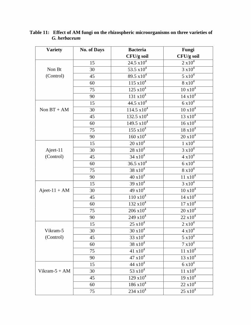
Table 11: Effect of AM fungi on the rhizospheric microorganisms on three varieties of
G. herbaceum
Variety No. of Days Bacteria
CFU/g soil
Fungi
CFU/g soil
Non Bt
(Control)
15 24.5 x10⁴ 2 x10
4
30 53.5 x10⁴ 3 x10
4
45 89.5 x10⁴ 5 x10
4
60 115 x104 8 x10
4
75 125 x104 10 x10
4
90 131 x104 14 x10
4
Non BT + AM
15 44.5 x10⁴ 6 x10
4
30 114.5 x10⁴ 10 x10
4
45 132.5 x10⁴ 13 x10
4
60 149.5 x10⁴ 16 x10
4
75 155 x104 18 x10
4
90 160 x104 20 x10
4
Ajeet-11
(Control)
15 20 x104 1 x10
4
30 28 x104 3 x10
4
45 34 x104 4 x10
4
60 36.5 x10⁴ 6 x10
4
75 38 x104 8 x10
4
90 40 x104 11 x10
4
Ajeet-11 + AM
15 39 x104 3 x10
4
30 49 x104 10 x10
4
45 110 x104 14 x10
4
60 132 x104 17 x10
4
75 206 x104 20 x10
4
90 249 x104 22 x10
4
Vikram-5
(Control)
15 25 x104 2 x10
4
30 30 x104 4 x10
4
45 33 x104 5 x10
4
60 38 x104 7 x10
4
75 41 x104 11 x10
4
90 47 x104 13 x10
4
Vikram-5 + AM
15 44 x104 6 x10
4
30 53 x104 11 x10
4
45 129 x104 19 x10
4
60 186 x104 22 x10
4
75 234 x104 25 x10
4

90 261 x104 27 x10
4
Mycorrhizal colonization, height of the plant, fresh and dry weight of the plant was
studied and data are presented inTable-10.
Vikram-5 seedling with Glomus sp. colonization is 90%, showed maximum shoot length
54.66 cm, highest root length 30.33 cm, highest dry weight of shoot i.e. 20.82 g and highest dry
weight of root i.e. 1.66 g is recorded. The effect of inoculations of AM pure culture on plant
height, root length and chlorophyll content of cotton was studied and data are presented in Table-
10.
In single combination of AM+ hybrid seedling the data clearly indicated that Vikram -5
seedling which was provided pure culture of Glomus sp. showed maximum chlorophyll content
of 2.87 mg/g compare to 2.10 mg/g for Vikram -5 without AM.
In non Bt seedling after 90 days of growth the colonization in combination of Non Bt +
AM is 79% compare to only 56 % colonization in control set of plants.
There is a vast difference recorded in fresh and dry weight of shoot and root of non Bt seedlings.
Shoot length after 90 days in AM inoculated plants is 45.80 cm compare to 38.66 cm in control
sets.
Similarly length of root is 28.0 cm higher as compare to 13 cm in control (non Bt) seedlings.
Even in Ajeet -11 + AM combination colonization is 88% with plant shoot length 50.3 cm, root
length is 20 cm, shoot fresh weight is 37.62 g, root fresh weight is 3.6, shoot dry weight is 9.05
g, root dry weight is 1.1 g, chlorophyll content is 2.10 (mg/g) is recorded. The readings are
higher compare to Ajeet -11 seedling growth without any Glomus association.
Non mycorrhizal plants showed significantly less biomass than the mycorrhizal plants.
Roots inoculated with AM fungi showed maximum colonization. Differences in host genome can

control the degree of mycorrhizal colonization as per (Toth et al., 1990) and bean (Sutton 1973)
on maize.
The growth of cotton plant will be the better, when the seeds will be grown in the
presence of AM along with bioinoculants (Vazquez et al., 2000). This is because seeds with AM
and bioinoculant have better adaptability to critical sites since they have better tolerance to harsh
conditions. The plants inoculated with AM fungi showed increase in the chlorophyll content in
comparison to the control variety. Plants inoculated with AM fungi brought about significant
changes in chlorophyll a, b and total chlorophyll content. As reported by Gemma et al., (1997)
mycorrhizal Agrostis plastids had higher chlorophyll concentration compared with non
mycorrhizal.
The increase in root length and root biomass directly indicates improvement of the health
of the plant. AM inoculated plants were having better root and shoot growth. Plant dry weight is
influenced by AMF as a result of enhanced efficiency of resource acquisition by mycorrhizal
plants. The data in Table.10 clearly indicate that among all cotton seed varieties with AM
inoculated hybrid grown best in all parameters. Similar results were reported by Damodaran et
al., 2012) for certain Indian cotton cultivars.

4.5. Studies on in vivo effect of three fungi on growth performance of cotton
Cotton is the third largest important economic crop in India produced for cloth and other kind of
things of human need serving many other important uses (Hutchinson et al., 1947). Cotton is the
fast turning out to be a cash-crop in this region. So it is thought to find out growth promoter from
rhizosphere. Studies on the same line have been carried out by Hande (2000) on Cajanus cajan
and Subhedar et. al., (2006) successfully. Recently, efficient and exploitive agriculture
throughout the world is practiced at great cost to the environment. After decades of warning, the
inappropriate usage of pesticides has led to development of more than 500 resistant pathogens
(Georghiou, 1990).
Seed germination and seedling establishment are determined by several factors including
quality of seeds and environmental factors. Within the environment of the seed and seedling are
physical, chemical and biological factors that influence growth (Okoth et. al., 2011). The
rhizosphere, is relatively rich in nutrients, because as much as 40% of plant photosynthetic
products are exudates from roots (Bais et al., 2006). Consequently the rhizosphere supports large
microbial populations capable of exerting beneficial, neutral, or detrimental effects on plant
growth.
Some bacteria and fungi prevent diseases and enhance plant growth. Beneficial microbes
associate with plants in several ways. Some may inhabit the rhizosphere, taking advantage of
root exudates; others may live on root or leaf surfaces and some may colonize intracellular
spaces and vascular tissues inside the plant (Preston, 2004). Plant-associated microbial diversity
encompasses symbionts, protecting their host against various aggressions and to determine the
ecological success of plants. They drastically modify plant communities and to improve the

inventory of diversity and functions of in situ plant-associated microorganisms (Selosse et al.,
2004).
Plant-associated microorganisms fulfil important functions for plant growth and health (Gabriele,
2009) such as enhancement of plant growth and protection of plants from various plant
pathogens in several crops such as cucumber, radish, tomato, sugar cane, and rice as reported by
Viswanathan and Samiyappan (1999), Ongena et al. (2000) and Ramamoorthy et al. (2001).
Many resistance-inducing fungi and bacteria increase both shoot and root growth, some
non-pathogenic root-colonizing fungi also have similar effect (Harman et al., 2004). The
increased growth response induced by Trichoderma sp. has been reported for many crops such as
beans (Phaseolus vulgaris) cucumber (Cucumis sativus), pepper (Capsicum annum), carnation
(Dianthus carophyllus), maize (Zea mays), and wheat (Tritichum aestivum) (Lo and Lin, 2002).
The effectiveness of the use of microorganisms as biofertilizers and biocontrols however,
is determined by a myriad of factors including virulence of the isolate, environmental factors,
time of application, ability to survive in the environments other than their origin and colonize
plants roots during certain period of time to control plant pathogens (Kredrics et al., 2003;
Nemec et al., 1996; Stephan et al., 2005; Vinale et al., 2008 ) suggesting that augmenting of a
local virulent strain would be more successful.
Fungi are ubiquitous; some having beneficial effects on plants, while others may be
detrimental (Anderson and Cairney, 2004; Ipsilantis and Sylvia, 2007). Chemical fungicides
however, have a negative effect on human health and on the environment (Voorrips et al., 2004;
Soytong et al., 2005; Gavrilescua and Chisti, 2005; Calhelha et al., 2006; Haggag and Mohamed,
2007). The application of chemical fungicides over a long period may result in plant pathogenic
fungi developing resistance (Benítez et al., 2004, Agrios, 2005; Kim and Hwang, 2007). An

alternative way to increase the crop yield besides using chemical fertilizers is biofertilizers.
Biofertilizers promote increased absorption of nutrients in plants (Vessey, 2003; Hart and
Trevors, 2005; Chen, 2006). Biofertilizers include materials derived from living organisms and
microbial sources (Rola, 2000; Chen, 2006). Biofertilizers have various benefits, such as
increased access to nutrients, providing growth-promoting factors for plants, and composting and
effective recycling of solid wastes (Gaur and Adholeya, 2004; Das et al., 2007). Biofertilizers,
commonly known as microbial inoculants are produced from cultures of certain soil organisms
that can improve soil fertility and crop productivity (Malik et al., 2005; Marin, 2006).
Trichoderma species are common in soil and root ecosystems and are ubiquitous
saprobes (Harman et al., 2004; Thormann and Rice, 2007; Vinale et al., 2008; Kodsueb et al.,
2008) and they are easily isolated from soil, decaying wood, and other organic material (Howell,
2003; Zeilinger and Omann, 2007).
Trichoderma species can not only reduce the occurrence of disease and inhibit pathogen
growth when used as mycofungicides, but they also increase the growth and yield of plants (Elad
et al., 1981; Harman et al., 2004; Vinale et al., 2008). They are also increase the survival of
seedlings, plant height, leaf area and dry weight (Kleifeld and Chet, 1992). Trichoderma species
improve mineral uptake, release minerals from soil and organic matter, enhance plant hormone
production, induce systematic resistance mechanisms, and induced root systems in hydroponics
(Yedidia et al., 1999). For these reasons Trichoderma species are known as plant growth
promoting fungi (Hyakumachi and Kubota, 2004; Herrera- Estrella and Chet, 2004) or are
increasing plant growth (biofertilization) (Benítez et al., 2004).
Trichoderma also has various applications and important sources of antibiotics, enzymes,
decomposers and plant growth promoters (Daniel and Filho, 2007).
Trichoderma spp. produce a growth regulating factor that increases the rate of seed
germination and dry weight of shoot and stems (Windham et al., 1986).
Trichoderma has been widely studied for their capacity to enhance plant growth, produce
antibiotics, parasitize other fungi and compete with deleterious plant microorganisms (Adams et
al., 2007; Chang et al., 1986; Harman et al., 2004 a; Yedidia et al., 2001).

Table 12: Effect of Trichoderma viride on 3 varieties of G. herbaceum (Pot Study)
Variety
Days
Fresh wt(g) Dry wt (g) Shoot
length
(cm)
Root
length
(cm)
No. of
leaves
Total
Chlorophyll
Content
(mg/g)
Shoot Root Shoot Root
Non Bt
(Control)
15 0.71 0.038 0.10 0.018 12.1 3.23 4 0.20
30 1.195 0.114 0.162 0.020 16.2 6.1 6 0.41
45 6.732 0.874 1.703 0.201 19.33 7.0 9 0.71
60 8.964 1.165 2.271 0.269 25.77 8.6 11 1.02
75 11.205 1.456 2.839 0.336 32.21 11.0 14 1.23
90 13.447 1.748 3.407 0.404 38.66 13.0 17 1.53
Non Bt+
T.viride
15 0.72 0.122 0.018 0.019 13.2 4.19 4 0.30
30 1.2 0.24 0.025 0.025 19.6 7.0 6 0.52
45 18.90 1.95 10.5 0.820 27.40 13.0 8 1.0
60 26 2.59 14 1.109 36.54 18.32 11 1.29
75 31.80 3.19 17.21 1.39 45.80 22.11 15 1.45
90 37.6 3.72 20.82 1.66 54.62 28.0 19 1.90
Ajeet- 11
15 0.77 0.082 0.11 0.027 12.0 3.0 4 0.31
30 1.29 0.145 0.133 0.022 18.3 6.4 6 0.52
45 9.985 1.254 2.135 0.366 21.0 8.5 9 0.83
60 13.314 1.672 2.874 0.488 28.0 11.33 12 1.14
75 16.642 2.090 3.559 0.610 35.00 14.166 15 1.35
90 19.971 2.509 4.271 0.733 42.0 17.0 17 1.67
Ajeet-11 +
T. viride
15 1.271 0.123 0.17 0.029 14.0 4.16 5 0.36
30 1.49 0.23 0.183 0.032 19.2 7.0 7 0.62
45 18.810 1.821 4.525 0.553 25.16 10.0 10 1.18
60 25.080 2.482 6.03 0.738 33.55 13.33 14 1.44
75 31.350 3.035 7.541 0.922 41.941 16.66 17 1.71
90 37.621 3.642 9.05 1.107 50.33 20.0 21 2.01
Vikram-5
15 0.610 0.06 0.072 0.014 10.3 3.0 3 0.35
30 1.31 0.081 0.165 0.015 19.3 5.93 6 0.70
45 17.585 1.683 5.626 0.511 22.16 9.0 10 1.05
60 23.447 2.244 7.501 0.681 29.55 12.0 13 1.40
75 29.309 2.805 9.376 0.851 36.94 15.0 16 1.75
90 35.171 3.366 11.252 1.022 44.33 18.0 19 2.10
Vikram-5
+ T.viride
15 1.065 0.153 0.095 0.018 13.4 4.15 4 0.47
30 1.35 0.160 0.100 0.025 19.9 7.1 6 0.95
45 34.671 2.489 10.41 0.832 27.33 15.16 11 1.43
60 46.22 3.318 13.88 1.109 36.44 20.22 14 1.71
75 57.785 4.418 17.350 1.368 46.0 25.27 17 2.28
90 69.342 4.978 20.82 1.664 54.66 30.33 20 2.67

As depicted in table 12 and Fig.10,11 growth parameters of cotton varieties i.e.
Conventional and Hybrid both showed the maximum growth reponse to Trichoderma viride. All
the growth parameters were recorded till 90 days of the plant growth.
Seedlings of Vikram-5 cotton variety showed maximum shoot length (30.33 cm), root
length (20 cm), highest dry weight of shoot (20.82g) and dry weight of root (1.66 g). Chlorophyll
content of the plants treated with T. viride was found maximum (2.67 mg/g) in comparison to
Ajeet-11 and Non Bt variety. Trichoderma exerted beneficial effects on the plant growth and
development. Increased root size resulted into increased shoot size which translates into shoot
biomass production indicating beneficial effect of inoculation on plant growth and developemt.
The positive influence of Trichoderma on root system architecture would therefore relate to
increased yield of plants.
Shanmugaiah et al., (2009) showed similar results where single application of T. viride
showed the growth promotion of cotton plants.
Okoth et al., 2011 reported similar results for maize and beans where Trichodrma
increased the rate of germination and seedling growth of maize and beans. The increased root
and shoot length illustrated the direct effect of the fungi on the plant.
The increased growth response induced by Trichoderma sp. has been reported for many
crops such as beans (Phaseolus vulgaris) cucumber (Cucumis sativus), pepper (Capsicum
annum), carnation (Dianthus carophyllus), maize (Zea mays), and wheat (Tritichum aestivum)
(Lo and Lin, 2002).A number of mechanisms for plant growth promotion by Trichodema have
been proposed by Harman et al. (2004) and Jaleed et al., (1988).
We conclude that the Trichoderma spp. isolate tested against cotton varieties increased
the rate of seed germination and shoot and root growth.


Table 13: Effect of Gliocladium virens on 3 varieties of G. herbaceum (Pot Study)
Variety
Days
Fresh wt(g) Dry wt (g) Shoot
length
(cm)
Root
length
(cm)
No. of
leaves
Total
Chlorophyll
Content
(mg/g)
Shoot Root Shoot Root
Non Bt
(Control)
15 0.51 0.028 0.10 0.018 12.5 3.23 4 0.20
30 1.175 0.104 0.152 0.020 16.7 6.1 5 0.31
45 6.832 0.864 1.603 0.201 19.81 7.0 7 0.61
60 8.978 1.155 2.171 0.269 25.57 8.6 9 1.12
75 11.211 1.446 2.739 0.336 32.21 11.0 13 1.33
90 13.450 1.738 3.307 0.404 38.66 13.0 16 1.73
Non Bt+
G.virens
15 0.72 0.112 0.28 0.019 13.8 4.19 5 0.30
30 1.26 0.28 0.163 0.025 19.6 7.0 6 0.42
45 18.95 2.03 2.456 0.820 27.40 13.0 9 1.0
60 26.52 2.63 8.667 1.109 36.54 18.32 10 1.29
75 32.80 3.25 11.21 1.39 45.80 22.11 14 1.65
90 38.6 3.81 13.82 1.66 54.62 28.0 18 2.12
Ajeet- 11
15 0.67 0.062 0.11 0.027 12.0 3.0 4 0.21
30 1.39 0.135 0.133 0.022 18.3 6.4 6 0.42
45 9.98 1.264 2.135 0.366 21.0 8.5 8 0.73
60 13.35 1.662 2.874 0.488 28.0 11.33 11 1.14
75 16.68 2.19 3.559 0.610 35.00 14.166 14 1.35
90 19.98 2.53 4.271 0.733 42.0 17.0 17 1.77
Ajeet-11 +
G.virens
15 1.274 0.129 0.17 0.029 14.0 4.16 5 0.36
30 1.53 0.27 0.183 0.032 19.2 7.0 7 0.62
45 18.86 1.831 4.525 0.553 25.16 10.0 9 1.05
60 25.15 2.49 6.03 0.738 33.55 13.33 11 1.24
75 31.38 3.025 7.541 0.922 41.941 16.66 15 1.61
90 37.67 3.62 9.05 1.107 50.33 20.0 19 1.97
Vikram-5
15 0.62 0.09 0.062 0.014 10.3 3.0 4 0.35
30 1.37 0.081 0.155 0.015 19.3 5.93 6 0.60
45 17.59 1.683 3.626 0.511 22.16 9.0 7 1.09
60 23.46 2.244 7.521 0.681 29.55 12.0 10 1.46
75 29.32 2.805 9.366 0.851 36.94 15.0 13 1.78
90 35.19 3.366 11.262 1.022 44.33 18.0 16 2.17
Vikram-5 +
G.virens
15 1.073 0.153 0.095 0.018 13.4 4.15 5 0.47
30 1.42 0.160 0.178 0.025 19.9 7.1 7 0.85
45 34.682 2.489 3.898 0.832 27.33 15.16 9 1.33
60 46.38 3.318 8.013 1.109 36.44 20.22 11 1.71
75 57.79 4.418 12.343 1.368 46.0 25.27 14 2.18
90 69.41 5.978 14.421 1.664 54.66 30.33 19 2.27

As depicted in table 13, growth parameters of cotton varieties i.e. Conventional and
Hybrid both showed the good growth reponse to Gliocladium virens. All the growth parameters
were recorded till 90 days of the plant growth.
Seedlings of Vikram-5 cotton variety showed maximum shoot length (54.66 cm), root
length (30.33 cm), highest dry weight of shoot ( 20.82 g) and dry weight of root (1.66 g).
Chlorophyll content of the plants treated with G.virens was found maximum (2.27 mg/g) in
comparison to Ajeet-11 and Non Bt variety.

Table 14: Effect of Aspergillus niger on 3 varieties of G. herbaceum (Pot Study)
Variety
Days
Fresh wt(g) Dry wt (g) Shoot
length
(cm)
Root
length
(cm)
No. of
leaves
Total
Chlorophyll
Content
(mg/g)
Shoot Root Shoot Root
Non Bt
(Control)
15 0.71 0.038 0.10 0.018 12.1 3.23 4 0.10
30 1.195 0.114 0.162 0.020 16.2 6.1 6 0.31
45 6.732 0.874 1.703 0.201 19.33 7.0 9 0.51
60 8.964 1.165 2.271 0.269 25.77 8.6 11 0.73
75 11.205 1.456 2.839 0.336 32.21 11.0 14 0.96
90 13.447 1.748 3.407 0.404 38.66 13.0 17 1.06
Non Bt+
A.niger
15 0.72 0.122 0.018 0.019 13.2 4.19 4 0.30
30 1.2 0.24 0.025 0.025 19.6 7.0 6 0.52
45 18.90 1.95 10.5 0.820 27.40 13.0 8 0.78
60 26 2.59 14 1.109 36.54 18.32 11 1.03
75 31.80 3.19 17.21 1.39 45.80 22.11 15 1.19
90 37.6 3.72 19.82 1.66 54.62 28.0 19 1.31
Ajeet- 11
15 0.77 0.082 0.11 0.027 12.0 3.0 4 0.21
30 1.29 0.145 0.133 0.022 18.3 6.4 6 0.62
45 9.985 1.254 2.135 0.366 21.0 8.5 9 0.93
60 13.314 1.672 2.874 0.488 28.0 11.33 12 1.14
75 16.642 2.090 3.559 0.610 35.00 14.166 15 1.45
90 19.971 2.509 4.271 0.733 42.0 17.0 17 1.77
Ajeet-11 +
A.niger
15 1.271 0.123 0.17 0.029 14.0 4.16 5 0.36
30 1.49 0.23 0.183 0.032 19.2 7.0 7 0.72
45 18.810 1.821 4.525 0.553 25.16 10.0 10 1.24
60 25.080 2.482 6.03 0.738 33.55 13.33 14 1.56
75 31.350 3.035 7.541 0.922 41.941 16.66 17 1.84
90 37.621 3.642 9.05 1.107 50.33 20.0 21 2.17
Vikram-5
15 0.610 0.06 0.072 0.014 10.3 3.0 3 0.35
30 1.31 0.081 0.165 0.015 19.3 5.93 6 0.70
45 17.585 1.683 5.626 0.511 22.16 9.0 10 1.15
60 23.447 2.244 7.501 0.681 29.55 12.0 13 1.49
75 29.309 2.805 9.376 0.851 36.94 15.0 16 1.76
90 35.171 3.366 11.25 1.022 44.33 18.0 19 2.13
Vikram-5 +
A.niger
15 1.065 0.153 0.095 0.018 13.4 4.15 4 0.47
30 1.35 0.160 0.100 0.025 19.9 7.1 6 0.98
45 34.671 2.489 10.41 0.832 27.33 15.16 11 1.49
60 46.22 3.318 13.88 1.109 36.44 20.22 14 1.83
75 57.785 4.418 17.35 1.368 46.0 25.27 17 2.09
90 69.342 4.978 21.82 1.664 54.66 30.33 20 2.25

A.niger was found to be most common in the soils of cotton fields. The mass cultres of A.
niger was grown on Maize Sand Meal medium for the further experiment. It was observed that
cotton seeds gown in the pots treated with A. niger germinated faster in comparision to control
pots. As depicted in table 14, growth parameters of cotton varieties i.e. Conventional and
Hybrid both showed the maximum growth reponse to Aspergillus niger. All the growth
parameters were recorded till 90 days of the plant growth.
Seedlings of Ajeet-11 cotton variety showed maximum shoot length (50.33 cm), root
length (22 cm), highest dry weight of shoot (20.09 g) and dry weight of root (1.117 g).
Chlorophyll content of the plants treated with A.niger was found maximum (2.27 mg/g) in
comparison to Vikram-5 and Non Bt variety.
Hande D.V. (2010) showed similar results in cotton palnts where A. niger was found to
enhance the growth of cotton.

4.5.1. Studies on in vitro effect of fungal metabolites on cotton seeds.
A large number of microorganisms are known to produce toxic metabolites when cultivated on
synthetic media. Fungal metabolites are substances discharged by fungi in their metabolic
processes. The metabolites are products of some amino acids, cyclic peptides, aromatic, phenols,
terpenoids and plant growth regulators (Graffin, 1981; Madhosing, 1995; Nema, 1992). These
metabolites are many and diverse and they are known to cause diseases in plants, animals and
humans who eat infected food (Jalander and Gachande, 2012).
Fungi of the genera Aspergillus, Fusarium, Penicillium and Rhizoctonia are commonly
known to produce toxic substances, such as aflatoxin B1 and B2, Aspergellic acid, cyclopiczonic
acid, kojic acid, naphthoquinones, fumonizin and fusaric acid (Singh et al., 1991) that threaten
the health of our plants and animals. The role of toxic metabolites of pathogenic fungi in plant
disease development has been reported by several workers. Anaso et al., (1981) found out that
toxic metabolites of Drechslera rostrata and Fusarium equiseti retarded root growth of wheat.
Reduction in percentage seed germination of soybean seeds was observed in seeds soaked in
filtrates of Phomopsis phaseoli (Hilty et al., 1988). Soybean seeds soaked in cultures filtrates of
Fusarium solani, F. oxysporum, Aspergillus flavus, A. niger, Alternaria tenuis and A. alternata
for 24 hours showed reduction in percentage seed germination (Ibraheem et. al., 1987). Filtrate
from mycelial cultures of Verticillum alboatrum was found to inhibit cell growth and reduced the
viability of alfalfa (Medicago sativo) seeds (Frame et al., 1991) reported that the culture filtrate
of seed-borne strains of Alternaria alternata (Fr.) Keissler inhibited the germination and vigour
of sunflower seeds and maize kernels. Culture filtrates of Fusaruim moniliforme, F. semitectum
and F. oxysporum gave very high percentage reduction in seed germination and also inhibited
root and shoot growth of Sorghum.

The fungi have been found to cause quantitative and qualitative changes in chemical
composition of the seeds. This is called Biodeterioration of seeds. These fungi degrade the
chemicals present in the seeds, which are rich in protein, carbohydrate and fats by producing
enzymes and toxins (Patil et al., 2012).
Table 15: Effect of different fungal culture filtrate on percentage germination of 3
varieties of Cotton seeds.
Name of Fungus
Percentage germination
Non Bt Ajeet-11 Vikram-5
30 min 1 hr 30 min 1 hr 30 min 1 hr
Fusarium
oxysporum
70 20 90 30 90 23
Alternaria
alternata
60 30 70 40 67 42
Chaetomium
globosum
50 25 55 23 52 21
Trichoderma
viride
90 85 100 93 97 91
T. harzianum 70 66 90 85 80 77
Gliocladium
virens
60 60 74 70 80 70
Aspergillus niger 63 50 93 73 80 75
Control 100 100 100 100 100 100
In the present study, the seed samples of three cotton varieties were treated with culture filtrates
of Aspergillus niger, Alternaria alternata, Chaetomium globosum, Fusarium oxysporum,
Gliocaldium virens, Trichoderma harzianum and T. viride and their effect on percentage of seed
germination and seedling growth was studied.

The effect of presoaking seeds in undiluted filtrates of seed-borne fungi for time period of
30 min and 1 hour on seed germination. The percentage of germination varied with presoaking
period as depicted in Table 15.
It was found that F. oxysporum was the most pathogenic fungi which inhibited seed
germination in all three cotton varieties. 30% germination was observed in both Non Bt variety
and Bt variety Ajeet-11 after 1hour of seed soaking whereas in Vikram -5 the germination
percentage was recorded 40%. Alternaria alternata and Chaetomium globosum was found to
inhibit the seed germination in all three varieties of cotton seeds after 1hour of presoaking of the
seeds. Rate of seed germination observed was 30 % in Non Bt , 40% and 42% in Ajeet-11 and
Vikram-5 respectively when the seeds were presoaked in the filterate of A. alternata.whereas,in
case of C.globosum it was recorded 25%, 23% and 21% in Non Bt, Ajeet-11 and Vikram-5
respectively.
Fungal filtrates of T. harzianum, T.viride and Aspergillus niger were not found to
inhibit much of the seed germination in all three varities of cotton seeds.

4.5.2. Effect of different organic composts on the growth of cotton
In developing countries like India, the situation is comparatively grimmer as it has limited
resources to feed the burgeoning population (Saini et al., 2004). The maximum yield of crops
can be achieved by introducing high yielding varieties with the application of the suitable
fertilizers and growth promoters. Use of chemical fertilizers has been the kingpin of modern
agriculture over the past 100 years (Mathivanan et al., 2012). In today‘s era, heavy doses of
chemical fertilizers and pesticides are being used by the farmers to get a better yield of various
field crops ( Joshi and Vig, 2010). Enviornmental degradation – a major threat conforting the
world and the rampant use of chemical fertilizers contribute largely to the deterioration of the
environment through depletion of fossils fuels, generation of carbon dioxide and contanmination
of water. These chemical fertilizers and pesticides decrease the soil fertility that has adversely
impacted agricultural productivity and caused soil degradation.
The long term use of inorganic fertilizers without organic supplements damages the soil
physical, chemical and biological properties and causes the environmental pollution (Albiach et
al., 2000). Due to the adverse effects of chemical fertilizers, interest has been stimulated for the
use of organic manures (Follet et al., 1981). Organic manures are one more important factor
which influences soil microflora (Chamle et al., 2011). They not only act as source of nutrients
and organic matter but also increase the size, biodiversity and activity of the microbial
population in the soil, influence structure, nutrients get turnover and many other change related
to physical, chemical and biological parameters of the soil (Albiach et al., 2000). Organic
fertilizers like plant residues, manures and composts play a vital role in changing the soil
ecosystem, physico-chemical properties and soil mycoflora are of great importance in soil
microbiology (Anastasi et al., 2005). Microbial community composition can be more sensitive to
soil amendment with plant residues than microbial biomass. The different kinds of soil
amendments such as compost, vermicompost, farm yard manure (FYM) etc., stimulate soil
microbial growth and activity with successive mineralization of soil nutrients (Randhawa et al.,
2005). In the previous studies application of FYM (Toyota et al.,1999) and spent mushroom
compost (Piqueres et al., 2006) significantly affected soil microflora.
Vermicompost is the microbial composting of organic wastes formed through the
earthworm activity to organic fertilizer which contains higher level of organic matter, organic
carbon, total and available N,P,K and micronutrients, microbial and enzyme activities (Edwards
and Bohlen, 1996; Ranganathan ,2006; Parthasarthi et al., 2007; Orozco et al., 1996; Parthasarthi
,2004).
Decomposition of leaf litter is an integral and significant part of biochemical (i.e.,
intrasystem) nutrient cycling and food webs of floodplain forests. Decomposition refers to both
the physical and chemical breakdown of litter and the mineralization of nutrients (Boulton and
Boon, 1991). Through decomposition the nutrients within leaf litter are converted into a form
available for uptake by vegetation, thereby exercising a critical control on vegetation
productivity (Mitsch and Gossselink, 1993 and Groffman et al., 1996). Litter plays a
fundamental role in the nutrient turnover and in the transfer of energy between plants and soil,
the source of the nutrient being accumulated in the upper most layers of the soil (Singh, 1971).
In the present study two organic manures namely vermicompost and dried leaf litter had
been selected for conducting the experiments and observing their effects on conventional and
hybrid variety of cotton. A plot study was conducted in the Arboretum, of The M.S. University
of Baroda. The 1x1 m plots were used for the experiments and to study the effect plot soil was
mixed with 2.5 kg vermicompost and leaf litter both against the control plot, where no compost
was mixed.
Different plant growth parameters such as height of the plant, fresh and dry weight of the
plant and chlorophyll estimation was studied and data are presented inTable-16. Convention as
well as hybrid variety showed maximum response in the soil mixed with vermicompost.
In hybrid variety maximum shoot length of 45.3 cm, highest root length 20 cm, highest
dry weight of shoot (3.38 g) and dry weight of root (0.48 g) is recorded.
As depicted in Table 16 chlorophyll content of both conventional and hybrid cotton
variety was recorded highest in the plants treated with vermicompost as compared to dried leaf
litter and control plants. Chlorophyll content of 5.146 mg/g was recorded in the hybrid variety
with vermicompost where as the conventional variety (Non Bt) 2.680 mg/g cholorphyll content.

Table 16: Growth parameters of Bt and Non Bt variety of G. herbaceum to different
treatments of fertilizers in Plot of 2x2m
Variety
Days
Fresh wt(g) Dry wt (g) Shoot
length
(cm)
Root
length
(cm)
No. of
leaves
Total
Chlorophyll
Content
(mg/g)
Shoot Root Shoot Root
Non Bt
(Control)
15 0.801 0.056 0.107 0.011 13.73 4.1 4 1.828
30 1.134 0.091 0.113 0.021 15.98 5.36 5 1.942
45 1.471 0.123 0.127 0.034 18.33 6.66 7 2.096
60 2.265 0.124 0.495 0.041 27.0 9.0 9 2.889
75 2.344 0.504 0.798 0.111 31.0 10.5 13 3.109
90 2.378 0.561 0.877 0.149 34.0 12.5 16 3.670
Bt (Control)
15 0.952 0.074 0.14 0.011 16.0 4.766 5 1.896
30 2.096 0.158 0.433 0.042 22.04 6.49 6 2.311
45 3.239 0.239 0.722 0.074 26.66 8.0 9 2.707
60 4.879 0.688 1.727 0.226 29.33 9.5 10 3.056
75 5.549 0.746 1.814 0.27 34.5 17 14 3.829
90 5.827 1.385 1.822 0.415 38.0 19.0 18 5.143
Non Bt +
Vermicompost
15 0.836 0.076 0.125 0.017 13.46 4.5 4 1.917
30 2.010 0.150 0.413 0.043 18.01 5.0 6 2.172
45 3.166 0.222 0.707 0.068 23 6.5 8 2.237
60 4.237 0.455 1.124 0.119 29 9.5 11 2.498
75 4.275 0.600 1.282 0.247 33.5 13.5 14 2.541
90 4.554 0.661 1.447 0.277 39.5 17 17 2.662
Bt+
Vermicompost
15 0.693 0.056 0.186 0.016 13.26 7.1 5 2.486
30 1.621 0.121 0.302 0.045 20.17 7.6 7 2.504
45 2.569 0.190 0.614 0.055 27.3 8 9 2.543
60 3.546 0.303 0.762 0.094 29 9.16 11 3.563
75 7.635 0.911 2.279 0.372 37.5 17.3 15 3.580
90 12.543 1.255 3.381 0.481 45.3 20 19 5.146
Non Bt + Leaf
litter
15 0.457 0.134 0.095 0.008 10.5 3.93 4 1.455
30 1.635 0.145 0.445 0.031 17.01 5.85 6 1.643
45 2.808 0.157 0.654 0.053 22.66 7.66 7 1.825
60 2.359 0.304 0.919 0.097 30 10 10 2.120
75 3.77 0.613 0.863 0.291 30.5 12.5 13 2.315
90 4.246 0.725 2.061 0.332 35 16 16 2.680
Bt + Leaf
litter
15 0.908 0.07 0.136 0.029 14.13 6.5 5 2.779
30 1.865 0.164 0.435 0.057 19.17 8.10 7 2.962
45 2.818 0.246 0.631 0.066 24 9.66 9 3.157
60 4.538 0.353 1.114 0.114 32 11.33 11 3.264
75 4.686 0.725 1.561 0.285 34 17.5 14 3.474
90 14.14 1.16 4.303 0.396 40.66 21.33 19 3.892
4.7. Studies on in vitro antagonistic activity of pathogenic fungi
In recent years, large number of synthetic fungicides have been banned in the world
because of their harmful toxicity (Jat and Agalave, 2013). Many pathogenic microorganisms
have developed resistance against chemical fungicides (Gaigole et al., 2011). This seriously
hinders the management of diseases of plants and agricultural crops. Worldwide traditional
agricultural practices are increasingly being affected by various problems such as diseases, pests,
droughts, decreased soil fertility due to use of hazardous pesticides, pollution and global
warming. There is a need for some eco-friendly biocontrol agent that may help to resolve some
of these problems, Biological control, the use of specific microorganisms that interfere with plant
pathogens and pests, is a nature-friendly, ecological approach to overcome the problems caused
by standard chemical methods of plant protection (Harman et al., 2004; Saba et al., 2012).
The word antagonism means hostility that results in active resistance, opposition, or
contentiousness. In terms of phytopathology it is the action of any microbe that suppresses the
activity of a plant pathogen due to an opposition in physiological action. Antagonistic
microorganisms play an active role in micro environment. If a plant surface harbours few
microbes these can compete with the pathogens for nutrients, inhibit pathogen multiplication by
secreting antibiotics or toxins, or reduce pathogen population through hyperparasitism. This kind
of interaction between microorganisms leads to effective control of several diseases. The
intensive use of fungicides has resulted in the accumulation of toxic compounds potentially
hazardous to humans and the environment, and also in the build-up of resistance of the
pathogens. In view of this, investigation and the application of biological control agents (BCAs)
seems to be one of the promising approaches (Cook, 1985). Biocontrol involves the use of

naturally occurring nonpathogenic microorganisms that are able to reduce the activity of plant
pathogens and thereby suppress plant diseases. In this direction fungal endophytes may prove
one of the most appropriate biological control agents (BCA).
Antagonists have been frequently used as biocontrol agents. Important constraints being
adverse environmental conditions such as extreme dryness, heat and cold limited shelf life and
inability to control latent infections. For suitability of an antagonists to be used as a commercial
product, Hofstein et al., (1994) has outlined following criteria-
1. It should be genetically stable.
2. Effective at low concentration.
3. Effective against wild rang of pathogens.
4. Does not produce metabolites that are deleterious or harmful to human health.
5. Compatible with commercial processing procedures.
In the present study the antagonistic activity of three pathogenic fungi viz. F. oxysporum,
C. globosum and A. alternata isolated from the field soil and seeds of cotton as examined using
the dual culture method.
Table 17 : Antagonistic effect of four different fungi against three pathogens.
Sr.
No.
Fungi Percentage Inhibition
Fusarium
oxysporum
Chaetomium
globosum
Alternaria
alternata
1. Trichoderma viride 50.98 64.66 70.33
2. T. harzianum 47.66 41.66 61.33
3. Gliocadium virens 23.07 35.33 57.66
4. Aspergillus niger 51.33 59.33 63.66
The observation based on the comparative analysis revealed that T. viride hampered the
growth of pathogenic fungi and showed the maximum percentage of inhibition against all three
pathogens. It reduced the growth of F. oxysporum by 50.98%, in case of C. globosum 64.66%
and in case of A. alternata 70%. Two other fungi T. harzianum and A. niger also inhibited the
growth of three pathogenic fungi and least inhibition was recorded by antagonist Gliocladium
virens as depicted in the above table-17.
Similar results were obtained by Tapwel et al., (2011) for T. viride against five
phytopathogens isolated from the nursery seedlings; Dhar et al., (2006) reported similar results
for Trichoderma and Gliocladium against Fusarium udum.
Three Trichoderma spp. viz., T. viride, T. harzianum, T. hamatum were tested
against the F. oxysporium f. sp. ciceri in laboratory applying direct bit placement method. The
results indicated that all Trichoderma spp. significantly inhibited the growth of F. oxysporum f.
sp. ciceri as against 90 mm radial growth in control treatment. The combined effect of three
Trichoderma spp. (T. viride + T. harzianum + T. hamatum) was found to be most effective in
checking the growth (11 mm) of F. oxysporum f. sp. ciceri over control (90 mm). It was also
revealed all Trichoderma spp. when used individually as biocontrol agents also exhibited
antagonistic effect against F. oxysporum f. sp. ciceri leading to reduced radial growth of the
fungus.
The conidia of Fusarium oxysporum were found to be inhibited by all the three
antagonistic microorganisms. Among them, highest percent inhibition of conidial germination
was brought out by Trichoderma viride [89.4%] followed by Trichoderma harzianum [85.7%].
This inhibition is due to the volatile and non volatile metabolites and cell wall degrading
enzymes produced by Trichoderma spp. (Rajeshwari et al., 2011). Inhibition of colony growth of
F. oxysporium was earlier reported (Brasier 1975). Fakhrunnisa et al. (2006) confirmed that T.
harzianum inhibited radial growth of F. oxysporum to the extent of 79.97%. Pawar (2011)

investigated antifungal property of leaf extracts from 18 plants against 5 seed borne pathogenic
fungi viz. Alternaria alternata, Aspergillus niger, Curvularia lunata, Fusarium moniliforme and
Trichoderma viride. Deepa et al., (2012) investigate antifungal activity of methanolic and
aqueous extracts of Sapindus emarginatus leaves against Aspergillus niger, they reported that the
methanolic extract showed inhibitory effect on A. niger in comparision to aqueous extract.
0
10
20
30
40
50
60
70
80
Trichoderma viride
T. harzianum Gliocadium virens Aspergillus niger
Per
cen
tage I
nh
ibit
ion
(%
)
Different Antagonist Fungi
Fig. 17: Histogram showing in vitro antagonistic activity
Fusarium oxysporum
Chaetomium globosum
Alternaria alternata

4.7.1. Biocontrol Studies of Seed borne and soil borne fungi
India is the largest consumer of pesticides in the world. Pesticides which include insecticides,
fungicides, herbicides, rodenticides and fumigants, are undoubtedly the largest group of toxic
chemicals that are introduced profusely into the environment. They are defined as any substance
or mixture of substances used for preventing, destroying, repelling or mitigating the pest. Most
of the chemicals products fall within four main categories viz. organochloride insecticides,
organophosphate insecticides, carbamate insecticides and pyrethroid. Pesticides have an inmate
capacity to cause damage to the biological system, which may involve human health or
environment. The most dramatic of such effects on human are accidental acute poisoning (Sinha
and Choudhary, 2008).
Synthetic fungicides are currently used as the primary means for the control of plant
diseases. However, the alternative control methods are needed because of the negative public
perceptions about the use of synthetic chemicals, resistance to fungicides among fungal
pathogens, and high development cost of new chemicals.
Some fungicides are not readily biodegradable and tend to persist for years in the
environment. This leads to third problem, the detrimental effects of chemicals on organisms
other than target fungi. Because of these problems associated with the use of chemicals,
researches are now trying to use environmentally safe alternative methods of fungal control.
The commonly used synthetic fungicides have been found to display side effects in form
of carcinogenicity, teratogenecity and pollutive effects. Uses of less harmful and true eco –
friendly products of plant origin are replacing the routine fungicides (Fawcett and Spencer 1970,
Khanna and Chandra 1972, Dixit et al., 1983, Arya and Mathew 1990, Arya et al., 1995). Efforts
are on to find out substitutes for chlorine containing, pentachlorophenol, ethylene dioxide,
Gammexane and Dieldrin like pesticides. Use of synthetic pesticide is increasing day by day to
meet the challenges of agriculture sector. Modern scientific developments are in no way less
than concern with the health of common man.
Plants produce diverse range of pre-infectional metabolites including alkaloids,
chalcones, flavanones, organic acids, saponins, sesquiterpene lactones, steroids, sulphur containg
amides, and terpenoids, many of which display a broad spectrum antifungal activity (Ebel,
1986). These secondary metabolites with no direct effect on the growth and development of
plants in which they have produced, have a potential bioactivity (Nychas, 1995) and are
attempted as natural fungicides (Benner, 1993). Wilkins and Board (1989) reported
approximately 1400 plants as potential sources of microbial agents with different classes of
compounds and several other metabolites from new plant species being identifies every year
(Aqil and Ahmad, 2003; Eksteen et al., 2001; Qasem and Abu-Blan, 1996; Ushiki et al., 1996).
A detailed description of the plant derived antifungal metabolites representing different
classes of compounds has been described by numerous workers earlier (Grayer and Harbone,
1994; Kishore and Pande, 2004). Majority of the identified natural fungicides are terpenes,
phenolic compounds or nitrogen containing secondary products such as alkaloids. Medicinal
plants represent a rich source of antimicrobial agents (Mahesh and Satish, 2008). Many of the
plant materials used in traditional medicine are readily available in rural areas at relatively
cheaper than modern medicine (Bobbarala et al., 2009; Mann et al., 2008).
Plants generally produce many secondary metabolites which constitute an important
source of microbicides, pesticides and many pharmaceutical drugs. The secondary metabolites of

the plants are a vast repository of biologically active compounds. Plant products still remain the
principal source of pharmaceutical agents used in traditional medicine (Ibrahim, 1997; Ogundipe
et al., 1998). The effects of plant extracts on bacteria have been studied by a very large number
of researchers in different parts of the world (Reddy et al., 2001; Ateb and ErdoUrul, 2003).
Much work has been done on ethno medicinal plants in India (Maheshwari et al., 1986; Negi et
al., 1993). Interest in a large number of traditional natural products has increased (Taylor et al.,
1996). Plants are the sources of natural pesticides that make excellent leads for new pesticide
development (Arokiyaraj et al., 2008; Gangadevi et al., 2008; Satish et al., 2008; Brinda et al.,
2009; Jagadish et al., 2009; Pande et al., 2009; Shanmugavalli et al., 2009; Swarna Latha and
Neelakanta Reddy, 2009; Rajan et al., 2009).
The Botanical pesticides like pyrethrum, rotenone, ryania and nicotine, but thereafter,
these botanicals were relegated to insignificant position in pest control. Pyrehthrum is extracted
from flowers of Chrysenthemum cinerieolium and rotenone is derived from rhizomes of Derris
and Lonchorpus. It has been promising source of biopesticide. Neem owes its toxic attributes
azadirachtin, nimbin, salannin, meliantriol etc. Neem seed kernels are richest source of meliacins
and contain 0.2 – 0.3 % azadirachtin and 30 – 40% oil. Though neem leaves and seeds contain
azadirachtin, bark also contains this yet in smaller quantities. George (1999) reported Swallow
root (Decalepis hamiltonii) of family Asclepediaceae causing protection of food grains against
insect infestation. Rice borer (Sitophilius oryzae) and Red rust of beetle (Tribolium) were
controlled by the application of Swallowrot. Inhibition of growth was observed on garlic extract
(Tansy and Appleton 1975). Electron microscopic studies revealed thickening in cell wall in
Rhizoctonia solani, wheras, Colletotrichum lindemuthianum revealed a singular accumulation of

osmiophill bodies immediately under the cell membrane when subjected to suspension of
micronized garlic powder in distilled water (Bianchi et al., 1997).
Extracts of Azadiracthta indica, Lantana camara, Lawsonia inermis, Datura spp., Acacia
spp. Trachyspermum ammi etc are commonly available in India and other tropical countries, are
widely used as natural fungicides. There are several increasing reports on the potent antagonistic
activity of extracts from many other several plant spp. (Afolayan et al., 2002; Dhaliwal et al.,
2002; Letessier et al., 2001; Pinto et al., 1998; Singh and Tripathi, 1999).
Antifungal spectrum and stability of natural fungicides is dependent on the chemical
nature of their constituents. Antifungal activity of aqueous extracts of Padus aviam, Populus
tremata and Chelidonium majus against Puccinia tritica can be correlated with the high phenolic
content and peroxidise activity. Fungicide potential of extracts from different parts of Heraceum
sibiricum was in correlation with the phenolic compounds (Karavaev et al., 2002). Leaf extract
was evaluated, owing to its high content of phenols and flavonoids (Parimelazhagan, 2001).
Essential oils, the complex mixture of volatile compounds, mainly monoterpenes (C10)
and sesquiterpenes (C15), and their oxygenated derivatives such as alcohols, aldehydes, ketones,
acids and esters (Wijesekara et al., 1997), are a major group of natural fungicides. Multiple
components rather than a single component, were responsible for fungicidal activity of essential
oils. Majority of the essential oils were broad spectrum antifungal. However, the composition of
these active components is affected by the genotype, geographical location, environment and
agronomic conditions and even with diurnal rythm.
To control fungal pathogens of fruit crops Arya (2010) suggested use of natural
fungicides like plant extracts, essential oils, gel and latex etc. Arya et al., (2005) found fruit

peelings (at 25 % conc. for Myrothecium roridum and Chaetomium ganglegarum) and seeds (at
25% against Phoma multirostrata and Eurotium chevalieri) of bitter gourd (Momordica
charantia L. and Cucurbitaceae) effective against 4 fungi. The effect may be due to presence of
alkaloid momordicine (0.038%) and some saponines in the fruit (Sabnis and Daniel, 1990) and
Elaterin a (Cucurbitacin) present in seeds and fruit wall.
Mode of action of Natural fungicides
Though the chemical nature of several natural fungicides is available, very few attempts have
been made to determine the mechanisms operating to control the fungal pathogens. Based on the
available findings we can conclude that any one or more than one of the following mechanisms
are responsible to restrict (fungistatic) or kill fungicidal) the phytopathogeneic fungal agents.
A) Inhibition of fungal Metabolic pathways
Chemical fistulosin (Octadecyl 3 – hydroxyvindole) isolated from the roots of Allium fistulosum,
inhibits the protein synthesis of Fusarium oxysporum. Eugenol (4 – allyl – 2 methoxy phenol), a
major component of several medicinal and aromatic plants, inhibits the involved in free radical
scavenging, lipid peroxidation and maintenance of redox potential, which together reduce the
aflatoxigenecity of the fungus (Jayshree and Subrmanyam, 1999).
B) Alteration in cell wall composition and structure
The cell wall protects the fungi against external agents including antifungal metabolites. Many
antifungal concentration target at cell wall composition and affects the integrity of cells resulting
in fungal death.
C) Changes in Membrane Permeability

Membranes act as barrier between the cell and its external environment and also separate various
organelles of the cell. Natural fungicides, particularly essential oils and their monoterpenoid
components affect the structure and function (Knobloch et al., 1989). This happens due to
inhibition of membrane enzymatic reactions such as respiratory electron transport, proton
transport and coupled phosphorylation steps (Knobloch et al., 1986). Essential oils can
degenerate hyphal tips and promote cytoplasmic retraction (de Bilerbeck et al., 2001)
D) Alterations in the Hyphal structure
Treatments with natural fungicides result in microscopically detectable and often
macroscopically visible changes in the hyphal structure. The hyphal deformations are mainly due
to altered or lysed cell wall, and vacuolization or evacuation of the cytoplasm. Trypsin and
chmotrypsin inhibitors from cabbage foliage cause leakage of intracellular contents of Botrytis
cinera and Fusarium solani. Kaemoferol – 3 – O-b-D- apiofuranosyl -12) –b-D-
glucopyranoside, a flavonol diglycoside from the leaves of Phytolacca americana, lyse the cell
walls diverse pathogenic fungi such as B. cinera, Magnaporthe grisea, Penicillium italicum,
Diaporthe actinidiae, Botryosphaeria dothidea and Colletotrichum gloeosporiodes (Bae et al.,
1997).
E) Inhibition of Fungal Cell Wall degrading enzymes
Pathogenic fungi produce cell wall degrading fungi produce cell wall degrading enzymes that
degrade the plant cell wall polymers and facilitate the pathogen penetration and further
colonization. Production of (CWDE) cell wall degrading enzymes is of significance in the
pathogenisis of necrotrophic fungal pathogens, and is of minor significance in case of biotrophic
pathogens. Important CWDE involved in the pathogenesis of necrotrohic fungi is

polygalacturonases, pectinlyase, pectimethylesterase,β – 1,4 – glucanase and celulase. The
virulence of several necrotrohioc is often related to the differences in their production of CWDE
(Carder et al., 1987).
Extracts of Allium cepa and A. porrum inhibits the production of polygalacturonase by
Sclerotinia scleroternum, B. cinerea, Fusarium moniliforme, Phoma terrastris, P. lycopersici, D.
Bryoniae, Sclerotium cepivorum and Rhizoctonia bataticola mediated by the heat labile and
protease inhibitor in sensitive factors (Flavaron et al., 1993). Aqueous extracts of Ocimum
sanctum inhibits the production of pectinolytic and cellulolytic enzymes of Rhizopus arrhizus
and Botryodiplodia theobromae (Patil et al., 1992). Putrescine reverses the inhibitory effect of O.
sanctum extract suggesting its effect on fungal fungal ornithin decarboxylases pathway. Fruit and
flower extracts of Datura innoxia inhibits the in vitro production of endo and exo pectinolytic
and cellulotytic enzyme of Colletotrichum capsici. (Chitra et al., 2001). Purified chestnut
cystatin strongly affects the protease activity of B. cinerea. However unlike biocontrol agents
(Elad and Kapt, 1999, Kapat et al., 1998) the inhibitory action of natural fungicides on fungal
CWDE in the infection courts has not studied and needs further investigation.
In the present study 7 types of leaf extracts were used against three pathogenic fungi viz.
Alternaria alternata, Chaetomium globosum and Fusarium oxysporum. Table 18 shows the list
of plants used for the antifungal study.

Table 18: List of Plants used as Biocontrol against Pathogenic Fungi
Sr.
No.
Plants Used Family Chemical Constituents
1 Annona reticultata L. Annonaceae Alkaloids, Flavanoids, Glycosides,
Triterpenoides
2 Balanites roxburghii Planchon. Zygophyllaceae Alkaloids, Glycosides, Saponins,
Flavones and Phenolic compounds.
3 Cochlospermum religiosa (L.)
Alst.
Cochlospemaceae Flavanoids, Phytosterols, Saponins
and Tanins
4 Gliricida sepium (Jacq.) Kunth
ex Walp.
Papilionaceae Tanins, Isoflavanoids like
Aformosin, Formentin, Glyricidin
A and B and Medicarpin.
5 Limonia acidissima L. Rutaceae Essential oil containing estragol,
Flavone, 3‘OMe-quercitin nad
Phenolic acids like p-hydroxy
benzoic, vanillic, syringic, p-
coumaric and ferulic acids.
6 Sapindus emarginatus Vahl. Sapindaceae Alkaloids, Phenols, Flavonoids,
Saponins.
7 Tephrosia jamnagarensis Sant. Fabaceae Alkaloids, Phenolic acids,
Flavonoids and mucilage.
Methanolic fractions exhibited more promising results than aqueous fractions in
suppressing the fungal growth. The periodic data regarding fungal growth, exposed to various
concentrations of plant extracts of Annona reticultata, Balanites roxburghii Cochlospermum
religiosa, Gliricida sepium, Limonia acidissima, Sapindus emarginatus and Tephrosia
jamnagarensis are present in below table.
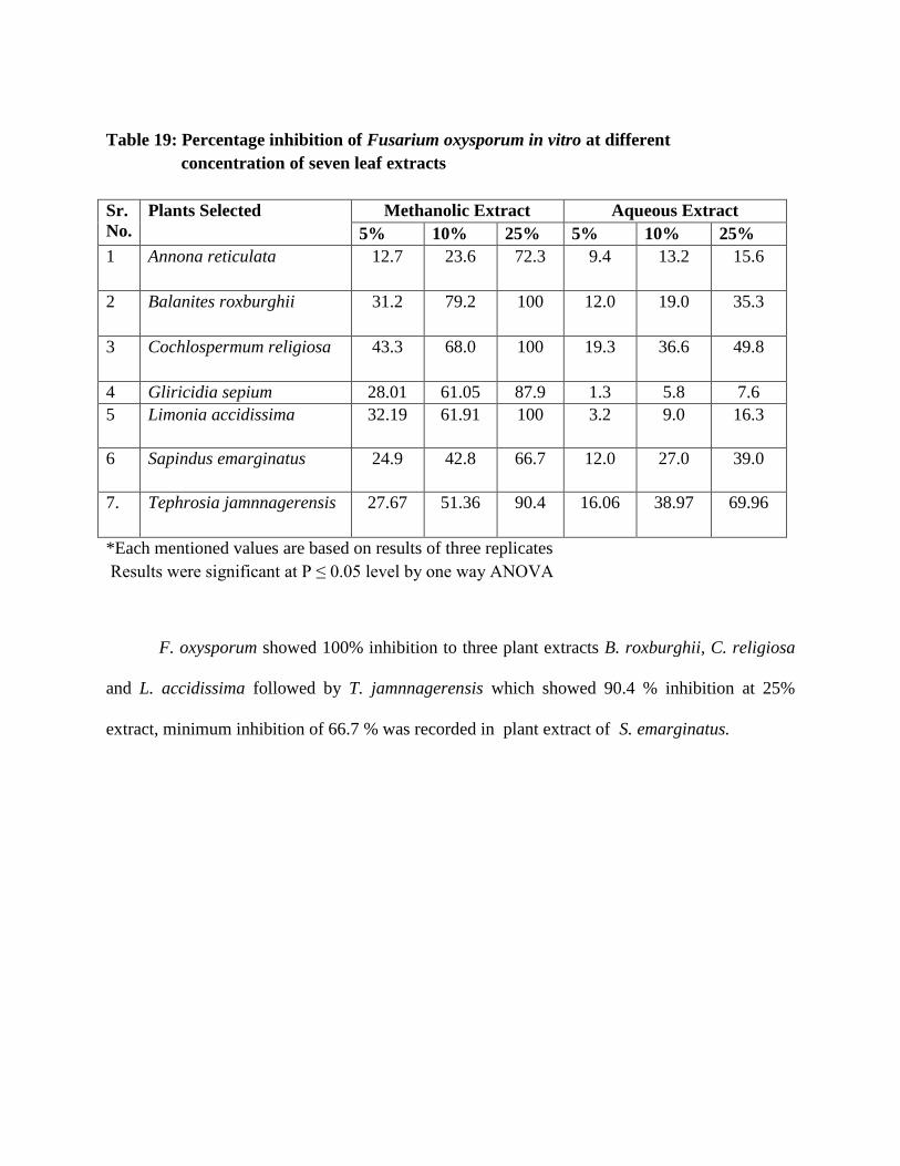
Table 19: Percentage inhibition of Fusarium oxysporum in vitro at different
concentration of seven leaf extracts
Sr.
No.
Plants Selected Methanolic Extract Aqueous Extract
5% 10% 25% 5% 10% 25%
1 Annona reticulata 12.7 23.6 72.3 9.4 13.2 15.6
2 Balanites roxburghii 31.2 79.2 100 12.0 19.0 35.3
3 Cochlospermum religiosa 43.3 68.0 100 19.3 36.6 49.8
4 Gliricidia sepium 28.01 61.05 87.9 1.3 5.8 7.6
5 Limonia accidissima 32.19 61.91 100 3.2 9.0 16.3
6 Sapindus emarginatus 24.9 42.8 66.7 12.0 27.0 39.0
7. Tephrosia jamnnagerensis 27.67 51.36 90.4 16.06 38.97 69.96
*Each mentioned values are based on results of three replicates
Results were significant at P ≤ 0.05 level by one way ANOVA
F. oxysporum showed 100% inhibition to three plant extracts B. roxburghii, C. religiosa
and L. accidissima followed by T. jamnnagerensis which showed 90.4 % inhibition at 25%
extract, minimum inhibition of 66.7 % was recorded in plant extract of S. emarginatus.

Table 20: Percentage inhibition of Chaetomium globosum in vitro at different
concentration of seven leaf extracts
Sr.
No.
Plant Selected Methanolic Extract Aqueous Extract
5% 10% 25% 5% 10% 25%
1 Annona reticulata 27.2 23.6 72.3 6.17 16.7 62.0
2 Balanites roxburghii 100 100 100 18.0 30.16 62.7
3 Cochlospermum religiosa 100 100 100 56.7 78.6 100
4 Gliricidia sepium 39.18 63.48 100 28.2 41.5 50.3
5. Limonia accidissima 82.27 100 100 9.1 52.0 65.9
6 Sapindus emarginatus 17.5 37.4 100 18.0 30.16 62.7
7. Tephrosia jamnagarensis 65.2 86.5 100 37.4 46.5 70.9
*Each mentioned values are based on results of three replicates
Results were significant at P ≤ 0.05 level by one way ANOVA
C. globosum showed 100% inhibition to two plant extracts B. roxburghii, C. religiosa in all
concentrations viz 5%, 10% and 25% , Gliricidia sepium L. accidissima, S. emarginatus and T.
jamnnagerensis showed 100 % inhibition at 25% extract, maximum inhibition (72.3%) was
recorded in plant extract of A. reticulata in 25% concentration of methanolic extract.

Table 21: Percentage inhibition of Alternaria alternata in vitro at different
concentration of seven leaf extracts
Sr.
No.
Plant Selected Methanolic Extract Aqueous Extract
5% 10% 25% 5% 10% 25%
1 Annona reticulata 20.4 48.5 75.2 5.9 14.5 32.0
2 Balanites roxburghii 8.3 38.8 100 27.3 26.0 32.8
3 Cochlospermum religiosa 1.3 25 100 18.7 33.2 60.2
4 Gliricidia sepium 29.77 40.81 100 5.2 9.7 13.5
5. Ferronia accidissima 2.0 37.5 100 8 12 43
6 Sapindus emarginatus 11.3 33.1 65.0 7.8 12.2 15.3
7. Tephrosia jamnnagerensis 5.0 21.0 100 5.0 19.6 52.5
*Each compound values are based on results of three replicates
Results were significant at P ≤ 0.05 level by one way ANOVA
A. alternata depicted 100% inhibition to five plant extracts B. roxburghii, C. religiosa, G.
sepium, L. accidissima and T. jamnnagerensis, whereas S. emarginatus showed minimum
inhibition of 65 % . It is evident from Table 17 that leaf extracts of 7 different plants were tested
against three pathogenic fungi in vitro. In most of the cases 25% methanolic extract was more
effective than 5% and 10%. From the above results it is depicted that the extracts of B.
roxburghii, C. religiosa and L. accidissima were most effective in the against all three
pathogenic fungi as compared to the other plants.
Earlier Jamuna Bai et.al, (2011) showed the antimicrobial activity of Cochlospermum
religiosum. Sivia et al., (2002) showed the antifungal activity of A. reticulata leaf and stem
extract against Colletotrichum gleospoeoides.

The presence of antibacterial substances in the higher plants is well established
(Srinivasan, 2001). Plants have provided a source of inspiration for novel drug compounds as
plants derived medicines have made significant contribution towards human health.
Phytomedicine can be used for the treatment of diseases as is done in case of Unani and
Ayurvedic system of medicines or it can be the base for the development of a medicine, a natural
blueprint for the development of a drug (Didry et al., 1998). Successive isolation of botanical
compounds from plant material is largely dependent on the type of solvent used in the extraction
procedure.
The variation in antifungal activity of the extracts in different solvents may be attributed
to the different chemical nature of the solvents. It is likely that different types of chemical were
dissolved in different solvent that resulted in variable activity of the extracts of same part of the
plant in different solvents.
Survey was conducted in three districts viz. Vadodara, Bharuch and Jamnagar of Gujarat
to find out different cotton varieties (Bt/non Bt/ hybrid) under cultivation. Data of their yield
obtained and associated seed and soil borne fungi. The results depicted that most of the farmers
preferred to grow Bt cotton variety in all the present three districts and maximum yield was
obtained from the seeds belonging to Ajeet-11 variety.
For the study Bt cotton varieties Ajeet-11 and Vikram -5 were used. Comparison was
made with non Bt cotton.
Soil and Seed Borne fungi associated with cotton
Isolation was done from the rhizospheric and non rhizospheric soils to find out the
associated fungi. Twenty one different types of fungi were isolated belonging to twelve genera.
Various Ascomycetes fungal members like Aspergillus niger, A. terreus, A. flavus, A. fumigatus
Chaetomium globosum and P. citrinum were present in the rhizospheric soil. Members belonging
to Deuteromycetes were Alternaria alternata, Curvularia lunata, Cladosporium
cladosporaoides, Fusarium oxysporum, F. roseum, Myrothecium verrucaria, Tricothecium
roseum, T. harzianium and T. viride. Presence of Mucorale member Rhizopus stolonifer of
Zygomycetes group was interesting feature.
The seed borne fungi were isolated by using Blotter method and Agar Plate method as
recommended by ISTA. In blotter method ten types of fungi and in case of Agar plate method
thirteen types of fungi were isolated.
Occurrence of Arbuscular Mycorrhizal fungi in rhizosphere soil of cotton

To find out the occurence of mycorrhizal fungi with rhizospheric and non rhizospheric
soils of different cotton fields. Method suggested by Gerdemann and Nicolson was followed.
Arbuscular Mycorrhizal fungi belonging to 4 genera and 28 species were observed in the soils of
different cotton fields of Gujarat.
Isolated different arbuscular mycorrhizal fungi were identified on the basis of
morphological characteristics of the fungal spores and their hyphal attachment.
Effect of Arbscular Mycorrhizal Fungi on Growth of Cotton Varieties
The effect of inoculations of AM fungi on plant height, root length, mycorrhizal
colonization and chlorophyll content of cotton was studied. Mycorrhizal colonization was found
to be superior in hybrid seedling inoculated with consortium of Glomus sp. in comparison to
hybrid cotton plants without AM inoculation. The data clearly show that the plants which were
inoculated with Glomus consortium, showed maximum mycorrhizal colonization, better shoot
and root length and more dry weight. The chlorophyll content in leaves also increased as
compared to non mycorrhizal plants.
Studies on in vivo effect of three fungi on growth performance of cotton
Effect of three selected fungi viz. A. niger, T. viride and G. virens was observed on
growth performance of cotton plants. All the three varieties of cotton viz. Non Bt and Hybrid
varieties Ajeet-11 and Vikram-5 showed maximum shoot length, root length of both fresh and
dry weight and highest chlorophyll content in plants treated with T. viride followed by G. virens
and A. niger as compared control plants. Thus we can conclude that the Trichoderma viride
increased the rate of seed germination and shoot and root growth.
Effect of fungal metabolites on germination and growth of cotton

Effect of fungal metabolites on seed samples of three cotton varieties and their effect on
percentage of seed germination and seedling growth was observed. F. oxysporum was found to
be the most pathogenic and inhibited the seed germination in all three cotton varieties. Alternaria
alternata and Chaetomium globosum were also found to inhibit the seed germination in all three
varieties of cotton seeds after 1hour of presoaking of the seeds. Fungal filtrates of T. harzianum,
T. viride and Aspergillus niger were not found to inhibit the seed germination of all three
varieties of cotton.
Effect of two different organic composts on the growth of cotton
Effect of organic composts like vermicompost and dried leaves was observed on plant
height, root length and chlorophyll content of cotton plants. The data evidently indicated that the
plant treated with vermicompost showed maximum, shoot length, root length of both fresh and
dry weight. The chlorophyll content was more in leaves of the plants grown with vermicompost
as compared to plants supplemented with dried leaf powder and control plants.
Studies on in vitro antagonistic activity of pathogenic fungi
Study on in vitro antagonistic activity of three pathogenic fungi viz. F. oxysporum, C.
globosum and A. alternata isolated from soil and seeds of cotton were examined using the dual
culture method. The observation based on the comparative analysis of the radial growth three
pathogens revealed that T. viride hampered the growth of pathogenic fungi and showed the
maximum percentage of inhibition against all three pathogens. It reduced the growth of F.
oxysporum by 50.98%, in case of C. globosum 64.66% and in case of A. alternata 70% of growth
inhibition. Fungi T. harzianum and A. niger also inhibited the growth of three pathogens and
least inhibition of pathogens was recorded by Gliocladium virens.
Biocontrol Studies of Seed borne and Soil borne fungi

Botanical pesticides obtained from leaf extracts aqueous and methanolic using Soxhlet
method were used. The activity was tested by Poisoned Food technique. Best results were
obtained in the methanolic extract of Balanites roxburghii, Cochlospermum religiosa and
Limmonia accidissima. These extracts were highly effective against all three test fungi used.

Bio-fertilizers are being viewed as the future of fertilizers, as they have the ability to
solve the problems of salinity of the soil, chemical-run offs from the fields. They ensure the well
being of the nutrients present in the soil, therefore making the soil more fertile with time. These
are more in demand for organic farming. During the past two decades advances in biological
sciences have generated a new set of tools that allows us to use our biological resources. One of
the biological tools that is now being integrated into biotechnology is the development of
commercial Mycorrhizal inoculants for the use in agricultural and frest plants. The impetus for
commercialization comes largely from the scientific literature and reports showing that AM
associations benefit the plant growth, development and vigor. Benefits derived by AM fungi
inoculums include the increase in root surface area for water and nutrient uptake, increased
growth rate and yield of the plants etc. AM fungi also contribute to soil stabilization, mechanical
aggregation and soil fertility. AM fungi are inoculated into wastelands, degraded sites, polluted
soils and mine soils so as to convert them into green pastures. Therefore, mycorrhizae can be
used as biofertilizers in the fields so as to decrease the use of chemical fertilizers, pesticides and
fungicides. AM fungi can be multiplied in the form of soil pot culture and root organ culture.The
soil and the root pieces containing the mycorrhizal infection can be supplied to the farmers for
implications in their fields. Another method for supplying the mycorrhiza is by mixing the soil
inoculum with several carriers like vermiculite, sawdust and clay etc. These carriers can be
directly applied to the field before sowing the seeds.
Other than AM fungi use of Trichoderma sp., which is an eco-friendly fertilizer and also
acts as a biocontrol agent and is hyper parasitic against different pathogens in the field should be
applied to the fields for the better growth and yield of the plants. It can be supplied to the farmers
in form of powder so that at the time of sowing seeds it can be directly apply in the soils.

Aspergillus and Gliocladium can also be used as mineral solubilizing agents. Present study is
indicative of use of othr fungi than Trichoderma. Also they can be mixed with the seeds that
farmers store so that seeds can be protected from the storage fungi and thus, they can be sotred
for a long period of time.
Natural products or eco-friendly pesticides are an excellent alternative to synthetic
pesticides as a means to reduce negative impacts to human health and the environment. The
concept of “Green Pesticides‖ refers to all types of nature-oriented and beneficial pest control
materials that can contribute to reduce the pest population and increase food production.
Pesticides based on essential oils obtained from plants or their constituents have demonstrated
efficacy against some plant pathogenic fungi responsible for pre and post-harvest diseases. They
may be applied as fumigants, granular formulations or as direct sprays.
In the present study extracts from the plants like Balanites roxburghii, Cochlospermum
religiosum and Limmonia accidissima were found to be effective against the pathogenic fungi.
Farmers may be encouraged to use these plants with the proper information. Cultivation of these
plants should be ensured and the extracts should be used as foliar spray to achieve integrated
control against the pathogenic fungi.
Developing protocol for mass production of PSB, mycorrhizal consortium for effective
field application should be tried. New research advances may be linked to call centre services
for farmers by KVK (Krishi Vigyan Kendra) a unit of ICAR in Central Gujarat region.
The use of biopesticide will reduce the diseases in cotton and prevent the farmers from
harmful effect of pesticides

References:
Abbot L.K. and Robson A.D. (1991) Factors influencing the occurrence of vesicular arbuscular
mycorrhizae. Agriculture, Ecosystem and Environment, 35: 120-150.
Abdel-Rahim, A.M., Baghadami A.M. and Abdalla M.H. (1983) Studies on the fungus flora in
the rhizosphere of sugar cane plants. Mycopathologia, 81: 183-186.
Abdul-Hafez S.I.I. (1982) Rhizosphere and rhizoplane fungi of Triticum vulgare cultivated in
Saudi Arabia., Mycopathologia 78: 79-86.
Abdullah S.K. and Kadhum S.A. (1987). Seed mycoflora of Sorghum bicolor in Iraq. Art Gulf J.
Sci. Res., 5(3): 401-410.
Adams P, De-Leij FA, Lynch JM.(2007) Trichoderma harzianum Rifai 1295-22 mediates growth
promotion of crack willow (Salix fragilis) saplings in both clean and metal-contaminated soil,
Microb Ecol. 4(2):306-13.
Afolayan A.J., Grierson D.S., Kambizi L., Madamombe I. and Masika P.J. (2002) In vitro
antifungal activity of some South African medicinal plants, South African J. Bot. 68:72-76.
Agarwal, V.K. and Sinclair J.B. (1996) Principles of Pathology. 2nd
edi, CRC Press, Inc., Boca
Raton, Fl. 539 pp.
Agnihothrudu V. (1995) State in which fungi occur in the rhizosphere, Naturwissenschefen,
42:515-516.
Agrawal V.K. and Sinclair J.B. (1987) Principles of seed pathology Vol I. CRC Press. New
Delhi pp.1-2.
Agrios G.N. (2005). Plant Pathology. 5th edn. Academic Press, San Diego: 922.
Ahmed, I., Iftikhar S. and Bhutta A.R. (1992) Seed-borne microorganism in Pakistan Checklist
1991. PARC, Islamabad.
Aikio, S. and Ruotsalainen A. L. (2002) The modeled growth of mycorrhizal and non-
mycorrhizal plants under constant versus variable soil nutrient concentration. Mycorrhiza. 12:
257- 261.
Ainsworth J and Bisby H. (1995) Dictionary of the Fungi. 8th edn. Wallingford. CABI
International, UK. 616 pp.
Albiach, R., Canet R., Pomares F. and Ingelmo F. (2000) Microbial biomass content and
enzymatic activities after the application of organic amendments to a horticultural soil.
Bioresour. Technol., 75: 43-48.
Alfred B. W. (1963) Relation of seed–borne fungi to boll rots of cotton. Phytopathology, 53:
984.
Allen M. F., Smith W.K., Moore T.S. and Christensen M. (1981) Comparative water relations
and photosynthesis of mycorrhizal Bouteloua gracilis HBK lag ex steud, New Phytologist, 88
:683-693.
Allen E.B. and Allen M.C. (1984) Competition between plants of different successional stages;
mycorrhizaeas regulators. Canadian Journal of Botany, 62 :2625-2629.
Allsop D. and Seal K.J. (1986) Introdution to biodeterioration. Edward Arnold (Publishers) Ltd.
London, 132
Allsopp, N. and Stock, W.D. (1992). Density dependent interactions between VA mycorrhizal
fungi and even-aged seedlings of two perennial Fabaceae species. Oecologia. 91(2): 281-287.
Aly A.A., Hussien E.M., Allam A.D.A., Amein A.M. and El-Samawaty A.M.A. (2000)
Pathological studies on fungi involved in damping off of cotton seedlings and root rot of adult
plants in upper Egypt governorates. J. Agric. Sci., 25: 4015-4034.
Aly A.A., Omar M.R., El- Abbasi I.H., El-Samawaty A.M.A. and Amal A.A. (2008) Effect of
seed mycoflora on incidence of Fusarium wilt disease in cotton genotypes. J. Agric. Sci., 33:
7243-7251.
Ammani, K. and Rao, A.S. (1996). Effect of two arbuscular mycorrhizal fungi Acaulospora
spinosa and A. scrobiculata on upland rice. Microbiological Research, 151(3): 235-237.
Anaso, A.B., Tyagi P.D and Emechebe A.M. (1981) Preliminary studies on toxic metabolites
produced by Drechslera rostrata and Fusarium equiseti and their effect on root growth in wheat.
Nigerian J. Plant Protec., 5: 102-105.
Anastasi, A., Varese G. C. and Marchisio V. F. (2005) Isolation and identification of fungal
communities in compost and vermicompost. Mycologia, 97: 33 – 44.
Anderson C.I. and Cairney W.G.J. (2004) Diversity and ecology of soil fungal communities:
increased understanding through the application of molecular techniques. Environmental
Microbiology 6: 769-779.
Aqil F and Ahmad I (2003) Broad spectrum antibacterial and antifungal properties of certain
traditionally used Indian medicinal plants, World J. Microbiol. Biotechnol. 19:653-657.
Arines, J.A., Vilarino and Sainz, M. (1989). Effect of different inocula of VAM fungi on
manganese content and concentration in red clover (Trofolium pretense L.) plants. New Phytol.
112(2): 215-220.
Arokiyaraj S, Martin S, Perinbam K, Marie Arockianathan P and Beatrice V (2008) Free radical
scavenging activity and HPTLC finger print of Pterocarpus santalinus L. – an in vitro study.
Indian J. Sci. Technol. 1 (7), 1-7.
Arshi, Anfal and Royy, A.K. (2008). Effect of vermicompost and endomycorrhizae on growth
performance of Gliricidia sepium (JACO. KUNTH.) on overburden dump soil of coal field area.
J. Indian bot. Soc. 87(3-4): 178-181.
Arya A. (2010) Recent Advances in the management of fungal pathogens of fruit crops In:
Management of Fungal Pathogens pub: Cabi International U.K. pp:3-13.
Arya A. and D .Mathew (1990) Control of Chiku fruit rot by leaf extracts of certain medicinal
plants, Res.J. Pl. Environ. 6(1):31-33.
Arya A., Bedi S.J. Jasrai Y.T., Patel V.S. (2005) Ecofriendly approach to control fungal
bioderiogens.In: Urban Pollution issues and solutions pub. By Nidhi pub. New Delhi.pp. 50 –
56.
Arya, A., Chauhan, R. and Arya, C. (1995). Inhibition of growth of 200 pathogenic fungi by
garlic extract. Mycologia, 67: 882 - 885.
Ashworth, L. J., McMeans J. L, Houston B. R., Whitten J. S. and. Brown C. M. 1971. Mycoflora
aflatoxins and free fatty acids in California cottonseed during 1967-1968. J. Amer. Oil Chem.
Soc. 48: 129-133.
Asran-Amal A.M. (2007) Effect of Trichoderma isolates, delivery systems and host genotype on
biological control of seedlings diseases. J. Plant Prot. Res. 112:339-356.
Ateb D.A. and ErdoUrul O.T. (2003) Antimicrobial activities of various medicinal and
commercial plant extracts. Turk. J. Biol. 27, 157-162.
Azcon, R., Andrade, G. and Bethlenfalvay, G.J. (1995). A rhizobacterium modifies plant and soil
responses to the mycorrhizal fungus Glomus mosseae. Applied Soil Ecology. 2(3): 195-202.
Azcón, R. (1994) The role of arbuscular mycorrhizalfungi on nitrogen nutrition and metabolism
byplants. Mycorrhiza News , 6 : 1-5.
Azcón-Aguilar C. and Barea J. M.(1996) Arbuscular mycorrhizas and biological control of soil
borne pathogens—an overview of the mechanisms involved. Mycorrhiza, 6:457–464.

Babu, R., Lokeshwar, S.H., Rao, N.S. and Rao, B.R.B. (1988). The response of chilli (Capsicum
annum L.) plants to early inoculation with mycorrhizal fungi at different levels of phosphorus. J.
Hortic. Sci. 63(2): 315-320.
Backman PA and Rodriguez-kabana R, (1974) A system for the growth and delivery of
biological control to the soil. Phytopathology, 65:819-821.
Bae E.Y. Shin E.J., Lee D.H. Hoh Y.J., Kin J,H. (1997) Antifungal Kaempferol-3-O-β-D-
apiofuranosy 1-(1,2)-β-D-glucopyranoside from leaves of Phytolacca americana L. Korean J.
Plant Pathol. 13: 371-376.
Bagyaraj DJ. (1984) Biological interactions with VA mycorrhizal fungi. pp. 131-153 In :VA
Mycorrhiza. L. L. Conway Powell and D. J. Bagyaraj. CRC Press, Boca Raton, FL.
Bagyaraj, D.J. (1989). Role of VA mycorrhiza in red soils. Mycorrhiza News. 1: 67.
Bagyaraj, D.J. and Manjunath, A. (1980). Response of crop plants to VAM (G. fasculata)
inoculation in an unsterile Indian soil. New Phytol. 85(1): 141-145.
Bais H. P, Weir T. L, Perry L, Gilroy S, and Vivanco J.M. (2006) The role of root exudates in
rhizosphere interactions with plants and other organisms. Annu Rev Plant Biol .57: 233-266.
Bais H.P. (2004) How plants communicate using the underground information superhighway?
Trends in plant science. 9: 26-32.
Bais H.P., (20040 How plants communicate using the underground information superhighway?
Trends in plant science. 9: 26-32
.
Balasubramanian N (2003) Strain improvement of Trichoderma spp. by protoplast fusion for
enhanced lytic enzyme and biocontrol potential. Ph.D thesis, University of Madras, Chennai,
India.
Bansal M. and Mukerji K.G. (1994) Positive correlation between VAM induced changes in root
exudation and mycorrhizosphere mycoflora. Mycorrhiza 5:39–44.
Baqual, M.F., Das, P.K. and Katiyar, R.S. (2005). Effect of arbuscular mycorrhizal fungi by
using expanded clay as carrier material for mycorrhiza. Z. Pflanzenkr. Pflanzenschutz. 94: 419-
430.
Barea, J.M. and Azcón-Aguilar C. (1983) Mycorrhizae and their significance in nodulating
nitrogen fixing plants. Advances in Agronomy, 36 : 1-54.
Basm H., Yegen O. and Zeller W. (2000) Antimicrobial effect of essential oil of Thymbra
spiculata var spicata on some plant pathogenic bacteria. Zeitschrift fur planzenkranheitenand
pflanzenschutz., 107(3): 279-28.
Bateman, G.L. and Kwasna H. (1999) Effects of number of winter wheat crops grown
successively on fungal communities on wheat roots. Applied Soil Ecology, 13: 271-282.
Bationo, A., Wani S.P., Bielders C.L., Vlek P.L.G and A.U.Mokwanye. (2000) Crop residue and
fertilizer management to improve soil organic carbon content, soil quality and productivity in the
desert margins of West Africa. pp. 117-145. In : R. Lal, J.M. Kimble and B.A. Steward (eds.)
Advances in Soil Science.Global Climate Change and Tropical Ecosystems.CRC Press, LLC
Washington DC, USA.
Behura C, Ray P., Rath C.C., Mishra R.K., Ramchandraiah O.S. and Charyulu J,K, (2000)
Antifungal activity of essential oils of Curcuma longa against five rice pathogens in vitro. J.
essential oil Bearing plants, 3(2):79-84.
Benítez, T., Rincón, M.A., Limón, M.C. and Codón, C.A. (2004). Biocontrol mechanisms of
Trichoderma strains. International microbiology 7: 249-260.
Benítez, T., Rincón, M.A., Limón, M.C. and Codón, C.A. (2004) Biocontrol mechanisms of
Trichoderma strains. International microbiology 7: 249-260.
Benner A.Z. (1993) Pesticidal compounds from higher plants. Pestic. Sci. 39:95-102.
Benhamou N., Gagne S., Quere D.L. and Dehbi L. (2000) Bacterial mediated induced resistance
in cucumber: beneficial effect of the endophytic bacterium, Serrantia plymuthica on the
protection against infection by Pythium ultimum, Phtopathology, 90:45-56.
Bellgard S.E. and Willam S.E. (2002) Beneficial mycorrhizas associated with commercial cotton
and native Hibiscus species growing in the monsoonal tropics of Northern Ausstralia., Scientific
Report, cotton CRC, Scientific Exchange Programme, pp:1-7.
Berch, S.M., Gamiet, S. and Deom, E. (1988). Mycorrhizal status of some plants of southwestern
British Columbia. Can. J. Bot. 66: 1924-1928.
Bethlenfalvay G.J. (1992) Mycorrhizae and crop productivity. IK GJ. Bethlenfalvay and R.G.
Linderman (eda) Mycorrhizae in sustainable agriculture,. Am Soc. Agron. Special Publication
No. 54. American Society of Agronomy, Madison, WI.
Bethlenfalvay, G.UJ. and Barea, J.M. (1994). Mycorrhizae in sustainable agriculture. I. Effects
on seed yield and soil aggregation. Am. J. Alternative Agric. 9(4): 157-161.
Bever J.D., Morton J.B., Antonovics J, Schultz P.A. (1996) Host dependent sporulation and
species diversity of Arbuscular mycorrhixal fungi in a mown grassland, J. Ecol, 84:71-82.
Bhamaeapravati S., Juthajpruth S., Mahacahi W. and Mahady G. (2006) Antimicrobial activity
of Boesenbergia rotunda (L.) Mansf. and Mlyristica fragrans Houtt. Against Helicobacter
pylori. Songklankarian , J. Sci. Technol. 28 (1):157-163.

Bhatia, N.P., Adholeya, A. and Sharma, A. (1998). Biomass production and changes in soil
productivity during long term, cultivation of Prosopis Juliflora (Awartz)DC, inoculated with
VAM and Rhizobium sps. In a semi arid waste land. Biol Fertil Soil. 26: 208-214.
Bianchi A, Zambonelli A, D‘Aulerio A.Z., Bellesia F (1997) Ultrastructural studies of the effects
of Allium sativum on phytopathogenic fungi in vitro. Plant Disease. 81: 1241-1246.
Bilgrami K.S., Jamaluddin and Rizwi M.A. (1981) Fungi of India Part-II, Host Index and
Aggenda, Pub. Today and Tomorrow‘s Printer and Publishers, New Delhi, pp: 1-140.
Blasingame D. and Mukund V.P. (2001) Cotton disease loss estimate committee report.
Proceedings of the Beltwide Cotton Conference, National Cotton Council (BCCNCC‘01),
Nashville, pp: 102-103.
Bobbarala V, Katikala P, Naidu K. C and Penumajji S (2009) Antifungal activity of selected
plant extracts against phytopathogenic fungi Aspergillus niger F2723, Indian Journal of Science
and Technology, 2(4):87-90.
Boddington C.L. and Dodd J.C. (1999) Evidence that differences in phosphate metabolism in
mycorrhizae formed by species of Glomus and Gigaspora might be related to their life cycle
strategies. New Phytologist, 142 : 531-538.
Borie F., Rubio R., Morales A. and Cornejo P. (2010) Arbuscular Mycorrhizae in agricultural
and forest ecosystems in Chile, J. Soil Sci. Plant Nutr., 10: 204-223.
Boulton A.J. and Boon P.I. (1991) A review of methodology used to measure leaf litter
decomposition in lotic environments: time to turn over an old leaf? Australian, Journal of
Marine and Freshwater Research, 42: 1-43.
Bowen GD, Rovira AD. (1999) The rhizosphere and its management to improve plant growth.
Advances in Agronomy, 66: 1–102.
Bradley R, Burt A.J. Read D. J. (1981) Mycorrhizal infection and resistance to heavy metal
toxicity in Calluna vulgaris, Nature, 292: 335–337.
Brady, N. C. and R. P. Weil (Eds.). Prentice Hall Inc., Upper Saddle River, New Jersy, pp; 446 –
490.
Brady, N. C. and Weil R. P. (1999) Soil Organic Matter. In: The Nature and Properties of Soils,
Braunberger, P. G., Abbott, L. K. and Robson A. D. (1996) Infectivity of arbuscular mycorrhizal
fungi after wetting and drying. New Phytol. 134: 673–684.
Brasier C.M (1975) Stimulation of sex organ formation of Phtopthora by antagonistic species of
Trichoderma.I. The effect in vitro, New Phytologist, 74:183-194.

Bridge, P. and Spooner, B. (2001) Soil fungi: diversity and detection. Plant and Soil, 232:147–
154.
Brindha V, Saravanan A and Manimekalai R (2009) Drug designing for ring finger protein 110
involved in adenocarcinoma (human breast cancer) using casuarinin extracted from Terminalia
arjuna. Indian J. Sci. Technol. 2 (2), 22-26.
Brodbent P. Baker K.F. and Waterworth Y. (1971) Bacteria and actinomycetes antagonistic to
fungal root pathogens in Australian soils. Austr. J. Biol. Sci., 24: 925-944.
Brundrett, M.C. (1991) Mycorrhizas in natural ecosystems p. 171-133. In A. MacFayden, M.
Begon and A.H. Fitter (eds). Advances in Ecological Research. Academic Press, London.
Bryla, D.R. and Koide, R.T. (1990). Regulation of reproduction in wild and cultivated
Lycopersicon esculentum Mill. by VAM infection. Oecologia cheidelb. 84(1): 74-81.
Burges A and Raw F (1976) Soil Biology. Academic Press, New York, USA.
Cairney J.W.G (2000) Evolution of mycorrhiza systems, Naturwissenschaften, 87:467–475.
Calhelha, R.C., Andrade, J.V., Ferreira, I.C. and Estevinho L.M. (2006). Toxicity effects of
fungicide residues on the wine-producing process. Food Microbiology, 23: 393-398.
Campbell R., (1989) Biological Control of Microbial Plant Pathogens. 1st Edn., Cambridge
University Press, Cambridge, ISBN: 0 521 34900 1.
Carder J.H., Hignett R.C. and Swinbrune T.R. (1987) Relationship between the virulence of hop
isolates of Verticillium alboatrum and their in vitro secretion of cell wall- degrading enzymes.
Physiol. Mol. Plant Pathol. 31: 441-452.
Castillo C, Rubio R., Borie F. and Sieverding E. (2010) Diversity of Arbuscular Mycorrhizal
Fungi in Horticultural production systems of Southern Chile, J. Soil Sci. Plant Nutr., 10(4):407-
413.
Castillo C.G., Rubio R., Rouanet R. and Borie F (2006 b) Early effects of tillage and crop
rotation on arbuscular mycorrhizal fungi propagules in an Ultisol. Biol. Fertil. Soils, 43: 83-92.
Castillo C.G., Sotomayor L., Ortiz C., Leonelli G., Borie F. and Rubio R. (2009) Effect of
Arbuscular Mycorrhizal fungi on ecological crop of chili peppers (Capsicum annum L.), Chilean
J. Agric. Res., 69: 79-87.
Chakravarty, P. and Mishra, R.R. (1986). The influence of VA mycorrhizae on the wilting of
Albizia procera and Dalbergia sissoo. European Journal of Forest Pathology. 16(2): 91-97.
Chamle D.R., Dhale D.A. and Mogle U.P. (2011) Effect of Parthenium weed manures on
rhizosphere mycoflora of Maize, Current Botany 2(4): 31-33.

Champawat, R.S. (1998). Influence of three VAM fungi on nutrient uptake and growth in
groundnut (Arachis hypogea) Mycorrhizae for Green Asia. First Asian Conference on
Mycorrhizae, January 29-31, 1988, Madras, India, Edited by Mahadevan, N. Raman and K.
Natarajan. pp. 132-133.
Champawat, R.S. and Pathak, V.N. (1993). Effect of vesicular arbuscular mycorrhizal fungi on
growth and nutrition uptake of pearl millet. Indian Journal on Mycology and Plant Pathology.
23(1): 30-34.
Chandraghatgi, P.S. and Sreenivasa, M.N. (1995). Possible synergistic interactions between
Glomus macrocarpum and Bacillus polymyxa in chilli. In. Mycorrhizae: Biofertilizers for the
Future Proc. (eds.) Adholeya, A. and Singh, S., TERI, Delhi. pp. 180-183.
Chang, Y.C., Baker R., Kleifeld O. and Chet I.. (1986). Increased growth of plants in the
presence of the biological control agent Trichoderma harzianum. Plant Dis., 70:145-8.
Charita Devi, M. and Reddy, M.N. (2004). Effect of arbuscular mycorrhizal fungi and
Rhizobium association on chlorophyll content of ground nut (Arachis hypogea). Mycorrhiza
News. 16(1): 15-17.
Chen, J-H. (2006). The combined use of chemical andorganic fertilizers and/or biofertilizer for
crop growth and soil fertility. International Workshop on Sustained Management of the Soil-
Rhizosphere System for Efficient Crop Production and Fertilizer Use. 16 – 20 October 2006
Land Development Department, Bangkok 10900 Thailand: 1-11.
Chenu, C. and Stotzky, G., (2002), Interactions between microorganisms and soil particles:
an overview. In: Interactions between soil particles and microorganisms (P.M. Huang,
J.M. Bollag and N. Senesi), pp 3-40. Wiley, New York.
Chitra H., Gomathi V. and Kannabiran B. (2001) Inhibitory effect of Datura innoxia on the
enzymatic activities of the anthracnose fungus Collectotrichum capsici in vitro. Indian
Phytopath. 54: 253-255.
Chhabra ML, Bhatnagar MK, Sharma MP (1992). Influence of vesicular arbuscular (VA)
mycorrhizal fungus on important diseases of maize. Indian Phytopathol., 45: 235-236.
Chotte, J.L., Ladd, J.N. and Amato, M., (1997), Sites of microbial assimilation, and turnover of
soluble and particulate 14C-labelled substrates decomposing in a clay soil, Soil biology and
biochemistry, 30: 205-218.
Christensen, M. (1989) A view of fungal ecology, Mycologia, 81: 1-19.
Clark F.E (1949) Soil microorganisms and plant roots. Adv Agron 1:241–288.

Clark, R.B. and Zeto, S.K. (1996). Growth and root colonization of mycorrhizal maize grown on
acid and alkaline soil. Soil Biol. Biochem. 28(10-11): 1505-1511.
Clark, R.B., Zeto, S.K. and Zobel, R.W. (1999). Arbuscular mycorrhizal fungal isolate
effectiveness on growth and root colonization of Panicum virgatum in acidic soil. Soil Biol.
Biochem. 31(13): 1757-1763.
Clarke C. and Mosse B., (1981) Plant growth responses to vesicular- arbuscular mycorrhiza. Part
XII: Field inoculation responses of barley at two soil P levels. New Phytologist 87, 695-703.
Colyer P.D. and Vernon P.R. (2005) Impact of stale seedbed production on seedling diseases in
cotton. Plant Dis., 89: 744-748.
Cook R.J. (1985) Biological control of Plant pathogens: Theory to application. Phytopathology,
75: 25-29.
Crawford R. F. (1923) Fungi isolated from the interior of cotton seed. Phytopathology 13: 501-
503.
Dalal, S. and Hippalgaonkar, K.V. (1995). The occurrence of vesicular-arbuscular mycorrhizal
fungi in arable soils of Konkan and Solapur. In- Mycorrhizae: Biofertilizers for the Future. Proc.
of the Third Nat. Conf. on Mycorrhizae (eds.) Adholeya, A. and Singh, S., 13-15, March, pp: 3-7.
Damodaran P.N., Udaiyan K., and Roh K.S. (2012) Mycorrhizal Dependency in certain Indian
Cotton Cultivars. Research in Plant Biology, 2(4): 55-66.
Daniel, F. De S.R. and Filho, R.E. (2007). Peptaibols of Trichoderma. Natural Product Reports
24: 1128- 1141.
Das, A., Prasad, R., Srivastava, A., Giang, H.P., Bhatnagar, K. and Varma, A. (2007). Fungal
siderophores: structure, functions and regulation. In: Soil Biology Volume 12 Microbial
Siderophores (eds. A. Varma and S.B. Chincholkar). Springer-Verlag Berlin Heidelberg: 1-42.
Davis R. G. (1982) Relationships between seedborne microorganisms and cotton seedling
emergence. Mississippi Agric. and Forest. Exp. Sta. Res. Rep. No.7. p- 3 .
Davies F.T., Potter J.R. and Lindermann R.G. (1993) Drought resistance of mycorrhizal pepper
plants independent of leaf P-concentration response in gas exchange and water relations,
Physiologia Plantarum, 87:45-53.
Davis, R. G. (1977) Fusarium species in the internal microflora of Mississippi cottonseed. Seed
Sci. and Technol. 5: 587-591.
Dawson, W.A. and Bateman G.L. (2001) Fungal communities on roots of wheat and barley and
effects of seed treatments containing fluquinconazole applied to control take-all.,Plant
Pathology, 50: 5-82.
deBillerbeck V.G., Roques C.G., Bessiere J.M., Fonvieille J.L. and Dargent R. (2001) Effect of
Cymbopogon nardus (L.) W. Watson essential oil on the growth and morphogenesis of
Aspergillus niger. Can, J. Microbiol. 47:9-17.
Deepa T, Elamathi R, Kavitha R, Kamalakannan, Sridhar S. and Suresh Kumar J (2012)
Screening for Physical, Phytochemical and Antimicrobial Activities of Leaf Extracts of Sapindus
emarginatus Vahl., International Journal of PharmTech Research, 4(1): 392-397.
DeMars, B.G. and Boerner R.E.J. (1995b). Mycorrhizal status of Deschampsia Antarctica in the
Palmer station area, Antarctica. Mycologia, 87: 451-453.
Dhaliwal H.S., Thind T.S., Mohan C. and Chhabra B.R. (2002) Activity of some essential oils
against Uncinula necator causing powdery mildew of grapevine. Indian Phytopath. 55:529-531.
Dhar V., Mishra S. and Chaudhary R.G. (2006) Differential efficacy of bioagents against
Fusarium udum isolates, Indian Phytopath, 59(3): 290-293.
Diana W.F. (1994) Soil biodiversity: its importance to ecosystem processes. Report of a
workshop held at the natural history museum, London, England.
Diener, U. L., Wagener R. E., Morgan-Jones G. and Davis N. D. (1976) Toxigenic fungi from
cotton. Phytopathology, 66: 514-516.
Disfani F.A. and Zangi M.R. (2006) Pre and post emergencies damping off in cotton. Plant
Pathol. J., 5: 51-53.
Dixit S.N, Dubey N.K. and Tripathi N.N. (1983) Fungitoxic essential oils vis-à-vis disease
control In: Recent Advances in Plant Pathology (Eds. Husain A., Singh K., Singh B.P. and
Agnihotri V.P.) Print house, Lucknow, pp:521.
Dodd J.C., Rosendahl S., Giovannetti M., Broom E A., Lanfranco L. and Walker C. (1996):
Inter- and intraspecific variation within the morphologically-similar arbuscular mycorrhizal
fungi Glomus mosseae and Glomus coronatum. New Phytol. 133: 113–122.
Doran J.W. and Zeiss, M.R. (2000) Soil health and sustainability: managing the biotic
component of soil quality. Applied Soil Ecology, 15: 3-11.
Druege, U. and Schoenbeck, F. (1993). Effect of vesicular-arbuscular mycorrhizal infection on
transpiration, photosynthesis and growth of flax (Linum usitatissimum L.) in relation to cytokinin
levels. J. Plant Physiol. 141(1): 40-48.
Ebel J. (1986) Phytoalexin synthesis: The biochemical analysis of the induction process. Annu.
Rev. Phytopathol. 24:235-264.
Edathil, T.T., Manian, S. and Udaiyan, K. (1996). Interaction of multiple VAM fungal species on
root colonization, plant growth and nutrient status of tomato seedlings (Lycopersicon esculentum
Mill.) Agriculture, Ecosystem and Environment. 59(1-2): 63-68.
Edwards G.A., Endrizzi J.E., Stein R. (1974) Genomic DNA content and chromosome
organization in Gossypium. Chromosoma 47: 309–326.
Edwards, C.A. and Bohlen P.J. (1996) Biology and Ecology of Earthworms. 3rdEdn., Chapm,an
and Hall, London.
Edwards, S.G., Young, J., Peter, W. and Fitter, A.H. (1998). Interactions between Pseudomonas
fluorescens biocontrol agents and Glomus mosseae, an arbuscular mycorrhizal fungus, within the
rhizosphere. FEMS Microbiology Letters. 166(2):
Egerton-Warburton, Graham L.M., R.C., and Hubbert K.R. (2003) Spatial variability in
mycorrhizal hyphae and nutrient and water availability in a soil–weathered bedrock profile. Plant
Soil, 249:331–342.
Eggins H.O. and Allsopp D. (1975) Biodeterioration and biodegradation by fungi. In:Smith,
J.E.; Berry, D.R. (eds.). Industrial Micology. The Filamentous Fungi. EdwardArnold, London,
pp.301-319.
Eggins H.O.W. and Allsopp D. (1975) ― Biodeterioration by fungi‖ In: The Filamentous Fungi,
Vol. I Industrial Mycology, J.E. Smith and D.R. Berry (Eds.), Edward Arnold (Publishers)Ltd.
25 Hill Street, London, W1X8LL, UK.
Eksteen D, Pretorius J.C., Nieusoudt T.D., Zeitsman P.C. (2001) Mycelial growth inhibition of
plant pathogenic fungi by extracts of South African plant species. Ann. Appl. Biol.139: 243-249.
Elad Y. and Kapat A. (1999) The role of Trichoderma harzianum protease in the biocontrol of
Botrytis cinerea. Eur. J. Plant Pathol. 105: 177-189.
Elad, Y., Chet, I. and Henis, Y. (1981). Biologicalcontrol of Rhizoctonia solani in strawberry
fieldsby Trichoderma harzianum. Plant and Soil ,60:245-254.
Elad Y., Chet I. and Katan J (1980) Trichoderma harzianum: A biocontrol agent of Sclerotium
rolfsii and Rhizoctonia solani, Phytopathology, 70: 119-121.
Fakhrunnisa, Hashmi M.H. and Ghaffar (2006) In vitro interaction of Fusarium spp., with their
fungi, Pak. J. Bot., 38(4):1317-1322.
Fard M.H. and Mojeni D.T. (2011) Effects of two systematic insecticides on damping off
pathogens of cotton. J. Agric. Sci. Technol., 13:27-33.

Fawcett CH, Spencer DM (1970) Plant Chemotherapy with natural product; Annual Rev.
Phytopathol. 8: 403-419.
Feller C. and Beare M.H., (1997) Physical control of soil organic matter dynamics in the tropics,
Geoderma, 79: 69-116.
Follet R., Donahue R and Murphy (1981) Soil and Soil Amendments . Prentice hall, Inc. New
Jersey.
Flavaron F., Castiglioni C. and diLenna P. (1993) Inhibition of some rot fungi
polygalacturonases by Allium cepa L. and Allium porrum L. extracts. J. Phytopathol. 139: 201-
206.
Frame B., Kang-Fu Y., Christie B.R. and Pauis K.P. (1991) In vitro selection for resistance to
Verticilium wilt in alfalfa (Medicago sativa L.) using a fungal culture filtrate. Physiol. Mol. Plant
Pathol., 39: 325-348.
Francis R. and read D.J. (1995) Mutalism and antagonism in the mycorrhizal symbiosis, with
special reference to impacts on plant community structure, Candian Journal of Botany, 73:1301-
1309.
Fravel, R.D. (2005) Commercialization and implementation of biocontrol. Annual Review
Phytopathology, 43:337-359.
Frey, B. and Schiiepp, H. (1993) Acquisilion of nilrogen by external hyphae of arbuscular
mycorrhizal fungi associated wilh Zea mays L. New Phytol. 124; 221-230.
Fryxell P. A. (1965). The genus Gossypium in Australia. Austral. J. Bot. 13: 71-102.
Fulton, N.D. and Bollenbacher, K. (1959): Pathogenicity of fungi isolated from diseased cotton
seedling in Arkansas. Phytopathology, 49: 684–689.
Gabriele B (2009) Plant–microbe interactions promoting plant growth and health: perspectives
for controlled use of microorganisms in agriculture. Appl. Microbiol. Biotechnol. 84:11–18.
Gadkar V and Adholeya A. (2000) Intraradical sporulation of AM Gigaspora margarita in long-
term axenic cultivation in Ri-T- DNA carrot root, Mycol.Res.,104(6): 716-721.
Gaigole A.H., Wagh G.N. and Khadse A.C. (2011) Antifungal activity of Trichoderma species
against soil borne pathogens., Asiatic Journal of Biotechnology Resources, 2(4):461-465.
Gangadevi V, Yogeswari S, Kamalraj S, Rani G and Muthumary J (2008) The antibacterial
activity of Acalypha indica L. Indian J. Sci. Technol. 1 (6), 1-5.
Garbaye J.(1994) Helper bacteria: A new dimension to the mycorrhizal symbiosis. New Phytol.,
128: 197-210.
Gardes, M. and Dahlberg A. (1996) Mycorrhizal diversity in arctic and alpine tundra: an open
question. New Phytologist, 133: 147-157.
Garrett S. D. (1981) “Soil Fungi and Soil Fertility‖. Pergamon Press, MacMillan Company, New
York.
Gaunt, R.B. (1978). Inoculation of VA mycorrhizal fungus on onion and tomato seeds. N.Z. J.
Bot. 16: 69-71.
Gaur A. and Adholeya A. (2000). Response of three vegetable crops to VAM fungal inoculation
in nutrient deficient soils amended with organic matter. Symbiosis 29:19 - 31.
Gaur, A. and Adholeya, A. (2004). Prospects of arbuscular mycorrhizal fungi in
phytoremediation of heavy metal contaminated soils. Current Science 86: 528-534.
Gavrilescu M. and Chisti Y. (2005) Biotechnology-a sustainable alternative for chemical
industry. Biotechnology Advances 23: 471-499.
Gawade S.B., Padule D.N. Game B.C. and Dumbre A.D.(2006) Detection of pathogenic fungi
and bacteria in cotton seeds and their impact on seed quality. Nat. semi on New Frontiers in Plt.
Path. 28-30.
George E, Marschner H and Jakobsen I. (1995) Role of arbuscular mycorrhizal fungi in uptake
of phosphorus and nitrogen from soil. Critical Reviews in Biotechnology, 15(3-4), 257-270.
George J., Ravishankar G.A., Keshava N and Udayashanker K. (1999) Antibacterial activity of
supercritical extract from Dcalepis hamiltonii roots, Fitoterapia.70:17.
Georghiou G.P. (1990). Overview of insecticide resistance. In Green, M.B., Le Baron, H.M. and
Moberg, W.K. Managing resistance to agrochemicals: from fundamentals research to practical
strategies. pp. 18-41. Am. Chemical Society: Washington, D.C.
Gemma J.N., Koske R.E. and Roberts E.M. (1997) Mycorrhizal fungi improve drought
resistance in creeping bentgrass, Journal of Turfgrass Science, 75: 15-29.
Gerdemann, J.W. (1969). Fungi that form the vesicular-arbuscular type of endomycorrhizas. In:
Proc. 1st N. Amer. Conf. Mycorrhizae (ed.). Hacskaylo, E. pp. 9-17.
Gerdemann, J.W. and T.H. Nicolson. (1963) Spores of mycorrhizal Endogone extracted from
soil by wet sieving and decanting. Trans. Br. Mycol. Soc., 46: 235-244.
Giovannetti, M . and Mosse B. (1980). An evaluation of techniques for measuring vesicular-
arbuscular mycorrhizal infection in roots. New Phytologist, 84: 489-500.
Gerdemann, J.W. (1975). Vesicular arbuscular Mycorrhizae. In: The development and function
of roots. (Eds.), J.G. Torrey and D.T. Clarkson, Academic press, London, 575-591.
Giovannetti M and Sbrana C (1998) Meeting a non host: the behavior of AM fungi, Mycorrhiza,
8:123-130.
Gisela and Honrubia, M. (1986). Citrus mycorrhizae: Potential benefits and interactions with
pathogens. Hort. Science. 21:1302-1306.
Glazek M. (1997) Mycoflora of winter wheat seeds harvested from flooded commercial fields in
South-Western Poland In: Plant Protection Institute in Poznaniu, Sosnicowice Branch, Gliwicka
St. 29:44-153 Sosnicowice, Poland.
Glick, B.R. (1995) The enhancement of plant growth by free-living bacteria. Can. J. Microbiol.
41: 109-117.
Gollotte A., Gianinazzi- Pearson V., Giovanetti M, Sbrana C, Avio Gianinazzi S (1993) Cellular
localization and cytochemical probing of resistance reactions to Arbuscular mycorrhizal fungi in
a ‗Locus a‘ myc-mutant of Pisum sativum L. , Planta, 191:12-22.
Grant, C., Bittman S., Montreal M., Plenchette C. and. Morel C. (2005) Soil and fertilizer
phosphorus: Effects on plant P supply and mycorrhizal development. Can. J. Plant Sci., 85: 3-
14.
Grayer R.J. and Harborne J.B. (1994) A survey of antifungal compounds from higher plants,
1982-1993. Phytochemistry 37: 19-42.
Graham J., Leonard R. and Menge J. A. (1981) Membrane mediated decrease in root exudation
responsible for ohosohorus-inhibition of vesicular-arbuscular mycorrhiza formation. Plant
Physiology 68: 548- 552.
Griffin DH. (1981) Fungal Physiology. John Wiley and Sons, New york, pp. 383.
Groffman P. M., Eagan P. , Sullivan W. M., and LEMUNYON J. L. (1996) Grass species and
soil type effects on microbial biomass and activity. Plant and Soil, Dordrecht, 183(1): 61-67.
Gryndler M, Hrselova H, Chvatalova I, Jansa J (1998) The effect of selected plant hormones on
in vitro proliferation of hyphae of Glomus fistulosm, Biol. Plant, 41:255-263.
Haggag W.M. and Mohamed H.A.A. (2007). Biotechnological aspects of microorganisms used
in plant biological control. American-Eurasian Journal of Sustainable Agriculture 1: 7-12.

Hall, I.R. (1978). Effect of VAM on two varieties of Maize and one of the sweet corn. N.Z. J.
Agric. Res. 21(3): 517-520.
Halloin, J. M., and Bourland F. M. (1981) Deterioration of planting seed. In: Compendium of
cotton diseases. Ed., G. M. Watkins. The American Phytopathological Society, St. Paul,
Minnesot, Pp. 11-13.
Hande D.V. (2000) Effect of Rhizo-sphere fungi on Cajanus cajan abstract in national
conference in Biotechnology, Department of Biotechnology Amravati University, Amravati pp.
62.
Hande D.V. (2010) Effect of Rhizosphere fungi, Asperigillux niger on the germination and
growth of cotton plant, Biosci. Biotech. Res. (2): 211-212.
Hanuman A, Mukhtar I, Riaz T. and Khan S.N. (2005) Effect if Plant extracts on black point
infection of wheat. Mycopath., 3(1-2): 53-55.
Harley, J. L. and Waid, J. S. (1955) A method for studying active mycelia on living roots and
other surfaces in the soils, Trans. Brit. Mycol. Soc., 38: 104-118.
Harman G.E., Howell Viterbo C.R., Chet I. and Lorito M. (2004) Trichoderma species-
opportunistic, avirulent plant symbionts. Nat. Rev. 2: 43–56.
Harder Y., Chet I and Henis Y. (1979) Biocontrol of Rhizoctonia solani damping off with wheat
bran culture of Trichoderma harzianum, Phytopathology, 69:64-68.
Hashem, M. (2011) Antifungal properties of crude extracts of five Egyptian medicinal plants
against dermatophytes and emerging fungi. Mycopathologia 172: 37–46.
Harrier L.A. (2001) The arbuscular mycorrhizal symbiosis: A molecular review of the fungal
dimension. J. Exp. Bot., 52: 469-478.
Harrison P.J., Snedaker S.C., Ahmed S.I. and Azam F. (1994) Primary Procedures of the Arid
Climate Mangrove Ecosystem of the River Indus Delta. Tropical Ecology, 35: 155-184.
Hart, M.M. and Trevors, J.T. (2005). Microbe management: application of mycorrhyzal fungi in
sustainable agriculture. Frontiers in Ecology and the Environment, 3: 533-539.
Hause B, Fester T. (2005) Molecular and cell biology of arbuscular mycorrhizal symbiosis.
Planta 221: 184–196.
Hawksworth D.L. (2001) The magnitude of fungal diversity: the 1.5 million species estimate
revisited. Mycological Research, 105: 1422-1432.
Hawksworth D.L. and Rossman A.Y. (1997) Where are all the undescribed fungi?
Phytopathology, 87: 888–891.

Haymann, D.S. (1981). Mycorrhizae and production of crops. Nature. 287: 487-488.
Haymann, D.S. and Mosse, B. (1972). Plant growth responses to vesicular arbuscular mycorrhiza
III. Increased uptake of labeled P from soil. New Phytol. 71: 41-47.
Henis Y nd Chet I (1975) Microbial control of plant pathogens. Adv. Appl. Microbiol. 198: 85-
111.
Herrera-Estrella, A. and Chet, I. (2004). The biological control agent Trichoderma from
fundamentals toapplication. In: Fungal Biotechnology in Agricultural, Food, and Environmental
Applications (ed. K.A. Dilip). New York Basel: 147-156.
Hexon A.C, Lourdes M.R, Carlos C.P. and Jose L.B. (2009) Trichoderma virens, a plant
beneficial fungus, enhances biomass production and promotes lateral root growth through an
auxin-dependent mechanism in Arabidopsis. Plant Physiology ,149: 1579-1592.
Hill G.D., and Gahen M.C., (1995) Effect of moisture and micro-organisms on the persistence
and metabolism of some organo phosphorus insecticide in soils. Proc. S. weed. Conf., 8: 284-
293.
Hiltner L (1904) Über neuere Erfahrungen und Probleme auf dem Gebiete der
Bodenbakteriologie unter besonderer Berücksichtigung der Gründüngung und Brache. Arbeiten
der Deutschen Landwirtschaftlichen Gesellschaft 98:59–78.
Hilty, T.C. and Lee H.L. (1988) Hot acidified zinc sulphate as seed soaking agent for the control
of crucifer black rot. Plant Protec. Bull., 30: 245-248.
Hirrel M. C., Mehravasan H. and Gerdemann, J. W. (1978) Vesicular-Arbuscular Mycorrhizae
on the chenopodiaceae and cruciferae: do they occur?. Canadian J. Bot., 56: 2868- 2817.
Hofstein R., Fridlender B, Chautz E, Wisnewski M, and Wilson C.L. (1994) Large scale
production and pilot testing of biological control against postharvest diseases. In: Wilson C. and
Winiewsji M, (eds.) Biological control of post harvest diseases of fruits and vegetables-theory
and practice.CRC press Boca Raton, Florida.pp>89-100.
Hooker, J.E., Munro, M. and Atkinson, D. (1992a). Effects of VA mycorrhizas on nutritionally
independent carbon partitioning in tree root systems. Journal of Experimental Botany. 43: 12.
Howell, R.C. (2003). Mechanisms employed by Trichoderma species in the biological control of
plant diseases: the history and evolution of current concepts. Plant Disease ,87: 4-10.
Hutchinson J. B. (1954) New evidence on the origin of the Old World cottons. Heredity, 8: 225-
241.

http://www.dbtbiosafety.nic.in
http://www.cicr.org
Hutchinson J.B., Silow R.A., Stephens S.G. (1947) The evolution of Gossypium and the
differentiation of the cultivated cottons. Oxford University Press, London p.160.
Huixing S. (2005) Effects of VAM on host plant in the condition of drought stress and its
mechanisms, Electronic Journal of Biology, 1(3): 44-48.
Hyakumachi, M. and Kubota, M. (2004). Fungi as plant growth promoter and disease suppressor.
In: Fungal Biotechnology in Agricultural, Food, and Environmental Applications (ed. K.A.
Dilip). New York Basel: 101-110.
Hyde K.D (1990). Intertidal mycota of five mangrove tree species. Asian Marine Biology, 7:93-
107.
Hyde KD (1992). Fungi From decaying Intertidal fronds of Nypa Fructicans, Including Three
New Genera and Four New Species. Bot J Linn Soc,110: 95-110.
Ibraheem, S.A., Okesha A.M. and Mlhathem K.T. (1987) Interrelationship between protein and
oil content of soybean seed with some associated fungi. J. Agric. Water Resources Res. Plant
Prod.,6: 53-66.
Ibrahim M.B (1997) Anti-microbial effects of extract leaf, stem and root bark of Anogeissus
leiocarpus on Staphylococcus aureaus, Streptococcus pyogenes, Escherichia coli and Proteus
vulgaris. J. Pharma. Devpt. 2:20-30.
Inderjit and Mukerji, K.G. (2006). Allelochemicals: Biological Control of Plant Pathogens and
Diseases, pp. 181-192.
Ipsilantis I. and Sylvia D.M. (2007) Abundance of fungi and bacteria in a nutrient-impacted
Florida wetland Applied Soil Ecology 35: 272-280.
ISTA (2003) International rules for seed testing, 2003 (Draper, SR Eds.) Zurich, Switezerland,
ISTA, pp 1-121.
Jamuna Bai A, Ravishankar RV, Pradeepa VS (2011). Evaluation of the antimicrobial activity of
three medicinal plants of South India. Malaysian J. Microbiol. 7:14-18.
Jat J.G. and Agalave H.R. (2013) Antagonistic properties of Trichoderma species against oilseed
borne fungi, Sciecne Research Reporter, 3(2): 171-174.
Jagadish L, Anand Kumar V.K and Kaviyarasan V (2009) Effect of Triphala on dental bio-film.
Indian J. Sci. Technol. 2 (1), 30-33.
Jain V. and Gupta V. K. (2002) Effect of rhizosphere on nodule number, shoot and root length
of Vigna mungo. Indian Phytopath., 55: 323-324.
Jakobsen I, Abbott L.K. and Robson A.D (1992) External hyphae of vesicular–arbuscular
mycorrhizal fungi associated with Trifolium subterraneum L. 1. Spread of hyphae and
phosphorus inflow into roots. New Phytol. 120: 371–380.
Jalander V. and Gachande B.D. (2012) Effect of Fungal metabolites of some rhizosphere soil
fungi on seed germination and seedling growth of some pulses and cereals, Science Research
Reporter 2(3): 265-267.
Jaleed S. Ahmad and Ralph Baker.(1988) Trichoderma enhances plant growth and controls
damping off of seedlings caused by Pythium ultimum. Canadian Journal Microbiology. 34:
229– 234.
James C (2008) Global status of commercialized biotech/GM crops: 2008, ISAAA Brief No. 39,
ISAAA, Ithaca, NY
Janos, D. P. (1996) Mycorrhizas, succession and rehabilitation of deforested lands in the humid
tropics. In : Frankland, J. C., Gadd, G. M. (eds.). Fungi and Environmental Change. Cambridge
University Press, Cambridge, UK, p. 1-18.
Javot H., Pumplin N. AND Harrison M.J., (2007) Phosphate in the arbuscular mycorrhizal
symbiosis: transport properties and regulatory roles. Plant Cell and Environmen,t 30: 310-322.
Jayshree T and Subramanyam C. (1999) Antiaflatoxicogenic activity of eugenol is due to the
inhibition of lipid peroxidation. Lett. Appl. Microbiol. 28:179-183.
Jeffries P., Spyropoulous T., Vardavrkis E. (1988) Vesicular –arbuscular mycorrhizal status of
various crops in different agricultural soils of northern Greece, Biol. Fertil. Soils, 5:333-337.
Jeffries P., Gianinazzi S., Perotto S., Turnau K., Barea J.M. (2003) The contribution of
arbuscular mycorrhizal fungi in sustainable maintenance of plant health and soil fertility. Biol.
Fertil. Soils 37: 1–16.
Jensen A. (1982) Infuence of four vesicular±arbuscular mycorrhizal fungi on nutrient uptake and
growth in barley (Hordeum vul gare). New Phytologist 90, 45-50.
Johnson, N. C., Tilman, D . and Wedin, D. 1992. Plant and soil controls on mycorrhizal fungal
communities. Ecology 73: 2034 – 2042.
Joshi R. and vig A.P (2010) Effect of Vermicompost on growth, yield and quality of Tomato
(Lycopersicon esculentum L.), African Journal of Basic and Applied Science, 2(3-4): 117-123.

Jolicoeur, M., William, R.D., Chavarie, C., Fortin, J.A. and Archambault J. (1999) Production of
endomycorrhizae propagules in bioreactors. Biotechnol. Bioeng. 64, 224–232.
Jones E.V.G., Uyenco F.R. and Follosco M. (1988). Fungi on Drift Wood Collected in the
Intertidal Zone from the Philippines. Asian Marine Biology, 5:103-106.
Khare, D. and Bhale, M. S. (2000) Seed Technology. Scientific Publishers (India) P.O. Box. 91.
Jodhpur, pp. 260.
Kalich M.A. (1988) Soil fungi of some low-altitude desert cotton fields and ability of their
extracts to inhibit Aspergillus flavus, Mycopathologia,:142(2):97-100.
Kakde R.B. and Chavan A.M. (2011) Antagonistic properties of Trichoderma viride and
Trichoderma harziannum against storage fungi, Elixir Appl. Botany, 41:5774-5778.
Kandasamy, D., Mohanraj, S.G. and Oblisami, G. (1985). Influence of VA mycorrhizae and
phosphobacteria on growth of brinjal and chillies in nursery. South Indian Hort., 33: 172-176.
Kapat A., Zmand G. and Elad Y. (1998) Effect of two isolates of Trichoderma harzianum on the
activity of hydrolytic enzymes produced by Botrytis cinerea. Physiol. Mol. Plant Pathol. 52:
127-137.
Karavaev V.A., Solntsev M.K., Kuznestov A.M., Polyakova I.B., Frantsev V.V., Yurina E.V.,
Yurina T.V. (2002) Plant extracts as the source of physiologically active compounds suppressing
the development of pathogenic fungi. Plant Prot Sci. 38: 200-204.
Katayama, A., Hu H. Y., Nozawa M., Yamakawa H. and Fujie K., (1998) Longterm changes in
microbial community structure in soils subjected to different fertilizing practices revealed by
quinine profile analysis. Soil. Sci. Plant Nutri., 44: 559 – 569.
Katznelson H. (1946) The ―Rhizosphere effect‖ on mangles on certain groups of soil
microorganisms, Soil Sci., 62: 343-354.
Kennedy A.C. And Smith K.L. (1995) Soil microbial diversity and the sustainability of
agricultural soils, Plant and soil, 170: 75-86.
Kennedy, J.Z. and Rangarajan, M. (2001). Biomass production, root colonization and
phosphatase activity by six VA-mycorrhizal fungi in papaya. Indian Phytopath. 54(1): 72-77.
Khadi B.M.,and Kulkarni V.N. (2001) Cotton. In: Chopra VL (ed.) Breeding Field Crops.
Theory and Practice Oxford and IBH Publishing Co. Pvt. Ltd. Delhi and Calcutta. pp :531-575.

Khan, A.G. (1974). The occurrence of mycorrhizas in halophytes, hydrophytes and xerophytes
and Endogone spores in adjacent soils. J. Gen. Microbiol. 81: 7-14.
Khan, A.H., Islam, A., Islam, R., Begum, S. and Imamul H.S.M. (1988). Mycorrhizal status of
some Bangladesh soils and the effect of indigenous VA mycorrhizal fungi on the growth of rice
plants. Bangladesh J. Bot. 17(1): 49-56.
Khan A.G. (1972) The effect of vesicular-arbuscular mycorrhizal associations on growth of
cereals. I. Effects on maize growth. New Phytol, 71:613–619
Khanna, K.K. and Chandra, S. (1972) Antifungal activity in some plant extracts. Proc. Nat.
Acad. Sci. India., 42: 111.
Khanzada, K.A., Rajput M.A., Shah G.S., Lodhi A.M. and Mehboob F. (2002) Effect of seed
dressing fungicides for the control of seed borne mycoflora of Wheat, Asian Journal of Plant
Sciences, 1(4):441-444.
Kim, B.S. and Hwang, B.K. (2004). Biolofungicides. In: Fungal Biotechnology in Agricultural,
Food, and Environmental Applications (ed. K.A. Dilip). New York Basel: 123-133.
Kirk P.M, Cannon P.F., David J.C. and Stalpers J.A. (2001) (eds) Ainsworth and Bisby‘s
dictionary of the fungi. 9th
edition. CABI Publishing, Wallingford.
Kishore, G. K. and Pande, S. (2004). Natural fungicides for management of phytopathogenic
fungi. Annu. Rev. Plant Pathol. 3:331 -356.
Kleifeld, O. and Chet, I. (1992). Trichoderma harzianum- interaction with plants and effect on
growth Response. Plant and Soil 144: 267-272.
Klein, D. A. (1992) Rhizosphere. In: Encyclopedia of Microbiology, Ledeberg, J. (Ed.). Vol. 3,
Academic Press, Inc., San Diego,ISBN: 0-12-226893-8, pp: 565-565.
Klich, M.A. (1986) Mycoflora of cottonseed from the southern United States: A three-year
study of distribution and frequency. Mycologia, 78: 706–712.
Klironomos J (2002) Host specifity and functional diversity among Arbuscular mycorrhizal
fungi. http://plato.acadiau.ca/isme/symposium26/Klironomos .pdf.
Knobloch, K., Pauli, P., Iberl, B., Weigand, H. and Weiss, N. (1989) Antibacterial and antifungal
properties of essential oil components. J. Essent Oil Res, 1:119–128,
Knobloch, K., Weigand, H., Weiss, N., Scharm H.M. and Vigenschow H (1986) Action of
terpinoids on energy metabolism. In: Progress in essential oil research. 249-445, Walter de
Gruyter & Co., Berlin

Kodsueb, R., McKenzie, E.H.C., Lumyong, S. and Hyde, K.D. (2008). Diversity of saprobic
fungi on Magnoliaceae. Fungal Diversity, 30: 37-53.
Koide, R. (1985). The nature of growth depressions in sunflower caused by vesicular arbuscular
mycorrhizal infection. New Phytol. 99(3): 449-462.
Koide R.T and Schreiner R.P. (1992) regulation of the vesicular –arbuscular mycorrhizal
symbiosis, Ann. Rev. Plant Physiol and Plant Mol., 43:557-581.
Kothamasi, D., Kuhad, R.C and Babu, C.R. (2001). Arbuscular mycorrhizae in plant survival
strategies. International Society for Tropical Ecology 42(1):1-13.
Kough, J.L., Gianinazzi-Pearson, V. and Gianinazzi, S. (1986). Depressed metabolic activity of
vesicular-arbuscular mycorrhizal fungi after fungicides application. New Phytol. 106(4): 707-
715.
Kowalchuk, G. A., De souza, F. A. and Van veen, J. A., (2002) Community analysis of
arbuscular mycorrhizal fungi associated with Ammophila arenaria in Dutch coastal sand dunes.
Mol. Ecol, 11 : 571–581.
Kredrics L., Antal Z, Manczinger L. SzekeresA., Kevei F., and Nagy E. (2003) Influence of
Environmental Parameters on Trichoderma Strains with Biocontrol Potential. Food Technology
Biotechnology. 41: 37-42.
Krishna, K.R., Balakrishna, A.N. and Bagyaraj, D.J. (1982) Interacüons between a vesicular-
arbuscular mycorrhizal fungus and Slreptomyceles cinnamomeous and their effect on finger
millet. New Phytol. 92: 401-405.
Kruchkelmann, H.W. (1973). Die vesicular-arbuscular mycorrhiza and ihre beeingflussung in
landwirtschaftlichen Kulturen. Diss. Naturwiss. Fakultat Tech. Universitat, Carolo-Wilhelmina,
Braunschweigh. pp. 1-56.
Kubiak K. and M. Korbas (1999) Occurrence of fungal diseases on selected winter wheat
cultivars. Postepy w Ochronie Roslin 39 (2): 801-804.
Kubicek CP, Mach RL, Peterbauer CK, Lorito M (2001). Trichoderma: From genes to
biocontrol. J. Plant Pathol. 83: 11–23.
Kuch, M.A. (1986): Mycoflora of cotton seed from the Southern United States: a Three year
study of distribution and frequency. Mycology, 78: 796–712 .
Kuhn, G., Hijri M. and Sanders I.R. (2001) Evidence for the evolution of multiple genomes in
arbuscular mycorrhizal fungi. Nature, 414: 745-748.

Kulkarni, M., Chaudhari, R. and Chaudhari, A. (2007). Novel tensio-active microbial
compounds for biocontrol applicatins. In: General Concepts in Integrated Pest and Disease
Management (eds. A. Ciancio and K.G. Mukerji). Springer: 295-304.
Kumar, A., Nivedita and Upadhyaya R.S..(1999) VA my-corrhizae and revegetation of coal
mine spoils: a re view. Tropical Ecology, 40: 1-10.
Kumar R. Gupta P.P. and Jalali B.L. (2001) Impact of VA-mycorrhiza, Azotobacter and
Rhizobium on the growth and nutrition of cowpea., J.Mycol.Plant. Pathol., 31:38-41.
Kumar, P. A., Sharma, R. P. and Malik, V. S. (1996) Insecticidal proteins of Bacillus
thuringiensis. Advances in Applied Microbiology, 42:1-43.
Lal R. (1998) Soil Quality and Agricultural Sustainability. In: Soil Quality and Agricultural
Sustainability, Lal, R. (Ed.). Ann Arbor Press, Chelsea, pp: 3 – 12.
Lapeyrie FF, Chilvers GA (1985) An endomycorrhiza–ectomycorrhiza succession associated
with enhanced growth of Eucalyptus dumosa seedlings planted in a calcareous soil. New Phytol
,100: 93–104
Letessier M.P., Svoboda K.P. and Walters D.R. (2001) Antifungal activity of the essential oil of
hyssop (Hyssopus officinalis). J. Phytopath. 149: 673-678.
Liljeroth, E. and Baath E. (1988) Bacteria and Fungi on roots of different barley varieties
(Hordeum vulgare L.). Biol. Fert. Soils., 7: 53-57.
Linderman RG. (1988) Mycorrhizal interactions with the rhizosphere microflora: The
mycorrhizosphere effect. Phytopathology, 78:366-371.
Lo C.T. and Lin C.Y (2002). Screening strains of Trichoderma spp. for plant growth
enhancement in Taiwan. Plant pathology Bull. 11: 215–220.
Lopez-Bucio J, Cruz-Ramirez A, Perez-Torres A, Ramirez-Pimentel J.G, Sanchez-Calderon L.
and Herrera-Estrella L (2005a) Root architecture In: C Turnbull, ed, Plant Architecture and its
Manipulation. Blackwell Annual Review Series. Blackwell Scientific, Oxford, pp 181-206.
Lovelock, C. E. and Ewel, J. J. (2005) Links between tree species, symbiotic fungal diversity and
ecosystem functioning in simplified tropical ecosystems. New Phytologist, 167: 219-228.
Lynch J.M (1990) The Rhizosphere, Wiley Interscience, John Wiley & Sons Ltd., Chichester.
581 p.
Madhosing C (1995) Relative wilt-inducing capacity of the culture filtrates of isolates of
Fusarium oxysporum f.sp.radicis-lycopersici, the tomato crown and root-rot pathogen. J.
Phytopathol.4: 193-198.
Mahesh B and Satish S (2008) Antimicrobial activity of some important medicinal plant against
plant and human pathogens. World J. Agri. Sci. 4 (2): 839-843.
Maheshwari J.K, Singh K.K and Saha S. (1986) Ethnobotany of tribals of Mirzapur District,
Uttar pradesh, Economic Botany Information Service, NBRI, Lucknow.
Maksoud M.A, Haggag L.F, Azzazy M.A and Saad R.N (1994). Effect of VAM inoculation and
phosphorus application on growth and nutrient content (P and K) of Tamarindus indica L.
seedlings. Ann. Agric. Sci., Caito. 39: 355-363.
Malajczuk N., Linderman R. G., Kough J and Trappe J. M. (1981) Presence of vesicular-
arbuscular mycorrhizae in Eucalyptus spp. and Acacia sp., and their absence in Banksia sp.
after inoculation with Glomus fasciculatus, New Phytol. 87: 567-572.
Malik, K.A., Hafeez, F.Y., Mirza, M.S., Hameed, S., Rasul, G. and Bilal, R. (2005).
Rhizospheric plant – microbe interactions for sustainable agriculture. In: Biological Nitrogen
Fixation, Sustainable Agriculture and the Environment (eds. Y-P. Wang, M. Lin, Z.-X. Tian, C.
Elmerich and W.E.Newton), Springer. The Netherlands: 257-260.
Malloch, D.W., Pirozynsky K.A. and Raven P.H. (1980) Ecological and evolutionary
significance of my-corrhizal symbiosis in vascular plants. Proceedingsof the National Academy
of Sciences USA77: 2113-2118.
Manjunath, A. and Bagyaraj, D.J. (1986). Response of black gram, chickpea and mung bean to
VAM inoculation in an unsterile soil. Tropical Agriculture. 63(1): 33-35.
Mann A., Banso A. and Clifford L.C. (2008) An antifungal property of crude plant extracts from
Anogeissus leiocarpus and Terminalia avicennioides. Tanzania J. Health Res. 10 (1) 34-38.
Mansoori B and Hamdolahzadeh A. (1995) Seed test and seedling disease of cotton in Gorgon
and Gonbad. Applied Entomology and Phytopathology, 62: 1-2 17.
Marin, M. (2006). Arbuscular mycorrhizal inoculation in nursery practice. In: Handbook of
Microbial Biofertilizers (ed. M.K. Rai), Food products press: 289-324.
Marschner H. (1998) Role of root growth, arbuscular mycorrhiza, and root exudates for the
efficiency in nutrient acquisition. Field Crop Research 56: 203–207.
Mathivanan S., Chidambaram A.L. A. Sundaramoorthy P. and Kalaikandhan R. (2012) Effect
of vermicompost on Germination and Biochemical constitutes of Groundnut (Arachis hypogea)
seedling, International Journal of Research in Biological Sciences, 2 (2): 54-59.
Marschner H. and Dell B. (1994) Nutrient uptake in mycorrhizal symbiosis, Plant and Soil,
159:89-102.

Mathur N. and Vyas A. (1995) Influence of VA mycorrhizae on net photosynthesis and
transpiration of Zizipus mauritiania, Journal of Plant Physiology, 147 (3/4): 328-330.
Mc. Gill, W.B., Cannon, K.R., Robertson, J.A and Coock, G.D. (1980),Dynamics of soil
microbial biomass and water stable organic carbon in Breton.L after fifty years of cropping to
two rotations, Canadian journal of soil science, 66: 1-19.
McGonigle, T. P. and Fitter, A. H. (1990). Ecological specificity of vesicular-arbuscular
mycorrhizal associations. Mycol. Res. 94:120-122.
Mehrotra V.S., Baijal U. (1994) Advances in the taxonomy of vesicular – arbuscular
mycorrhizal fungi. In: B.K. Dwivedi and G. Pandey (eds.) Biotechnology in India. Bioved
Research Society, Allahabad, India pp. 227 – 286.
Mejstrik, V. K. (1972) Vesicular-arbuscular mycorrhizas of the species of a Molinietum
coeruleae L. I. association: the ecology. New Phytologist 71: 883–890.
Menge J. A, Johnson, E. L. V. and Platt, R. G. (1978). Mycorrhizal dependency of several citrus
cultivars under three nutrient regimes. New Phytologist, 81, 553-559.
Mikhail M.S., Sabet K.K., Omar M.R., Hussein E.M. and Kasem K.K. (2009) Differntiation
among cotton Rhizoctonia solani isolates by pathogenicity and isozymes electrophoresis In:
Egypt. Proceedings of the 4th
Conference on Recent Technologies in Agriculture, Cairo, Egypt,
pp: 110-118.
Mikkelsen B L, Rosendahl S. and Jakobsen I. (2008) Underground resource allocation between
individual networks of mycorrhizal fungi. New Phytologist, 180(4): 890–898.
Milind Pande, Sanjay Ingale and Suryaprakash Gupta (2009) The Pharmacognostic and
phytochemical studies on the leaves of Murraya koenigii (L) Spreng. Indian J. Sci. Technol. 2
(3), 53-54.
Miller R.M. and Jastrow J.D (1994) Vesicular arbuscularmycorrhizae and biogeochemical
cycling, In: F.L. Pfleger & R.G. Linderman (eds.) My-corrhizae and Plant Health . APS Press
pp. 189-212.
Mirza, J.H. and Qureshi M.S.A. (1978) Fungi of Pakistan. University of Agric., Faisalabad.
Mishra R.R. (1967) Nature of Rhizosphere fungal flora of certain plants. Plant and soil XXVII,
2:162-166.
Mitsch W. J. and Gosselink J. G. ( 1993) Wetlands, 2nd ed. Van Nostrand Reinhold.
Modjo H.S. and Hendrix J.W. (1986) The mycorrhizal fungus Glomus macrocarpum as a cause
of tobacco stunt disease. Phytopathology, 76:688-691.
Mohandas, S. (1992). Effect of VAM inoculation on plant growth, nutrient level and root
phosphatase activity in papaya (Carica papaya cv. COORG Honey Dew). Fertilizer Research.
31: 263-267.
Mohandas S. (1987) Field response of tomato (Lycopersicon esculentum Mill.) cv. Pusa Ruby to
inoculation with VAM fungus Glomus fasciculatum and Azotobacter venetandii. Plant Soil,
98:295-297.
Mohankumar, V. and Mahadevan, A. (1988b). Viability of VAm spores in a tropical forest soils.
In- First Asian Conference on Mycorrhiaze, C.A.S. in Botany, Madras, (ed.) A. Mahadevan, N.
Raman & K. Natrajan, 29-341, January. pp. 80-81.
Mohankumar, V., Ragupathy, S., Nirmala, C.B. and Mahadevan, A. (1988a). Distribution of
vesicular arbuscular mycorrhizae (VAM) in the sandy beach soils of Madras University, Madras
Coast. Current Science. 57(7): 367-368.
Molin J. and Molin S. (1997) CASE: complex adaptive systems ecology. In: Jones, J.G.(Ed.),
Advances in Microbial Ecology, vol. 15. Plenum, New York, pp. 27– 79.
Molina, R., Massicotte, H. and Trappe, J.M. (1992) Specific phenomena in mycorrhizal
symbiosis; Community ecological consequences and practical implications. In: Mycorrhizal
functioning an integrative plant fungi process. Ed. My Allen. Pp. 357-423. Chapman and
Hall, New York.
Moreira, M., Baretta, D., Tsai, S.M. and Cardoso, E.J.B.N. (2006). Spore density and root
colonization by arbuscular mycorrhizal fungi preserved or disturbed Araucaria augustifolia
(Bert.) O. Ktze. ecosystems. Sci. agric. (Piracicaba, Braz.). 63(4): 380-385.
Morel A.F., Maldaner G., Ilha V., Missau F., Silva U. F. and Dalcol I. (2005) Cyclopeptide
alkaloids from Scutia buxifolia Reiss and their antimicrobial activity. Phytochem.,66: 2571-2576.
Morte A., Díaz G., Rodríguez P., Alarcón J.J. and Sánchez-Blanco M.J. (2001) Growth and
water relations in mycorrhizal and nonmycorrhizal Pinus halepensis plants in response to
drought. Biol. Plant. 44:263-267.
Morton J.B. and Benny. G.L. (1990). Revised classification of arbuscular mycorrhizal fungi
(zygomycetes), a new order Glomales, two new suborders Glomineae and Gigasporinae and two
new families Acaulosporaceae and Gigasporaceae with an emendation of Glomaceae.
Mycotaxon 37: 471-479.

Morton J. B. and Bentivenga S. P. (1994) Levels of diversity in endomycorrhizal fungi
(Glomales, Zygomycetes) and their role in defining taxonomic and nontaxonomic groups. Plant
and Soil 159: 47-59.
Morton JB (1998) Taxonomy of VA mycorrhizal fungi: classification, nomenclature and
identification. Mycotaxon 32: 267– 324.
Morton JB, Bentivenga SP and Bever JD. (1995) Discovery, measurement and interpretation of
diversity in symbiotic endomycorrhizal fungi, Canadian Journal of Botany, 73S: 25-32.
Morton JB. (1988) Taxonomy of VA mycorrhizal fungi: classification, nomenclature and
identification. Mycotaxon 32:267–324.
Morton J. B., Bentivenga S. P. and Wheeler W. W. (1993) Germplasm in the international
collection of arbuscular and vesicular-arbuscular mycorrhizal fungi (INVAM) and procedures for
culture development, documentation and storage. Mycotaxon 48: 491–528.
Morton, J. B., Bentivenga, S. P. and Bever, J. D. (1995) Discovery, measurement, and
interpretation of diversity in arbuscular endomycorrhizal fungi (Glomales zygomycetes).
Canadian J. Bot., 73 : 825-832.
Mosse B (1981) Vesicular-arbuscular Mycorrhizal Research for Tropical Agriculture. Research
Bull. 194. College of Tropical Agriculture and Human Resources. University of Hawaii.
Honolulu. USA. 82 p
Mosse, B., Powell, C.L. and Haymann, D.S. (1976). Plant growth responses to vesicular
arbuscular mycorrhiza. IX. Interaction between VA mycorrhiza, rock phosphate and symbiotic
nitrogen fixation. New Phytol. 76: 331-342.
Mosse B, Powell CL, Hayman DS (1976) Plant growth response to vesicular-arbuscular
mycorrhiza. IX. Interactions between VA mycorrhiza, rock phosphate and symbiotic nitrogen
fixation.New Phytol,76: 331–342.
Mukerji, K.G., Manoharachary C. and Chamola B.P. ( 2002) Techniques in Mycorrhizal
Studies. 1st Edn., Kluwer Academic Publishers., London-Netherlands, ISBN-10: 1402005326,
pp: 285-296.
Muller, M. M., Sundmam V., Saoinvara O. and Meriloinoe A., (1988) Effect of chemical
composition on the release of nitrogen from agricultural plant materials decomposing in soil
under field condition. Biol. Fertil.Soils, 6: 78 – 83.
Muthukumar T, Udaiyan K, Vasantha K., Kliener D. and Manian S (1999) Mycorrhizae in
sedges as related to root character and its ecological significance, Pertanika J trop Agric Sci,
22(1): 9-17.

Myrold, D.D. (2000) Microorganisms In: D.E. Alexander & R.W. Fairbridge (eds.)
Encyclopedia of Environmental Science. Kluwer Academic Publish-ers, The Netherlands. . pp.
409.
Nagamani A., Kunwar I.K. and Manoharachary C. (2006) .Handbook of Soil Fungi, I.K
International Pvt. Ltd, New Delhi, pp 461.
Namdas D, Bhasale A and Khillare C. (2009) Rhizosphere and soil mycoflora of Sorghum and
Tomato growing at Ahemadnagar. Bioinfolet. 6(3): 244-245.
Neergaard Paul (1973). Detection of seed borne pathogen by culture tests. Seed Sci. and technol.
1: 217-254.
Negi K.S, Tiwari J.K and Gaur R.D (1993) Notes on ethnobotany of five districts of Garhwal
Himalaya, Uttar pradesh, India. Ethno Botany. 5,73-81.
Nehl D.B., Allen S.J., Mondal A.H. and Lonergan P.A. (2004) Black root rot: A pandemic in
Australian cotton. Australas. Plant. Pathol., 33:87-95.
Nehl, D.B., Allen, S.J., and Brown, J.F. (1997) Deleterious rhizosphere bacteria:an integrating
perspective. Appl. Soil Ecol. 5: 1-20.
Nehl D.B., McGee P.A., Torrisi V, Pattison G.S. and Allen S.J. (1999) Patterns od Arbuscular
Mycorrhiza down the profile of a heavy textured soil do not reflect associated colonization
potential, New Phytologist, 142: 495-503.
Neeraj, Shankar A, Mathew J, Varma AK (1991). Occurrence of VA mycorrhizae within Indian
semi-arid soil. Biol. Fert. Soils, 11:140-144.
Nema A.G. (1992) Studies on pectinolytic and cellulolytic enzymes produced by Fusarium udum
causing wilt of Pigeonpea. Indian j. Forest., 15:353-355.
Nemec, S., Datnoff, L. and Strandberg, J (1996) Efficacy of biocontrl agents in planting mixes to
colonize plant roots and control root disease of vegetables and citrus. Crop Protection 15: 735 –
742.
Nene Y.L. and Thapliyal P.N. (1979) Fungicides in plant disease control, Oxford IBH Pub.,
New Delhi, pp.570.
Newman, E.I. (1988) Mycorrhizal links between plants: their functioning and ecological
significance. Advances in Ecological Research, 18: 243-270.
Newman,E. I. (1978) Root microorganisms: their significance in the eco-system. Biol. Rev. 53:
511-554.

Newsham, K. K., Fitter, A. H. and Watkinson, A. R. (1995) Arbuscular mycorrhiza protect an
annual grass from root pathogenic fungi in the field. J. Ecol., 83 : 991-1000.
Nirenberg, H., H. Schmitz-Elsherif, C.I. Kling. (1994) Occurrence of Fusaria and some
―blackening moulds‖ on durum wheat in Germany. 1. Incidence of Fusarium species.
Pflanzenkrankheiten und Pflanzenschuntz, 101: 449-459.
Nirenberg H (1976) Untersuchungen uber die morphologische und biologischi Differenzierung
in der Fusarium Sektion Liseeola. Mitteilungen aus der Biologischen Bundesant Salt purlandund
Forstwirtschaft, Berlin-dehlena, 169: 1-117.
Nirenberg, H. (1976) Untersuchungen uber die morphologische und Differenzierung in der
Nychas G.J.E (1995) Natural antimicrobials from plants. In: New Methods of Food Preservation.
ed.Gould GW, 58-59. Blackie Academic, London, UK.
O‘Donnell, A.G., Seasman, M., Macrae, A., Waite, I., Davies, J.T., (2001) Plants and fertilisers
as drivers of change in microbial community structure and function in soils. Plant Soil 232: 135
– 145.
O‘Donnell A.G, Goodfellow M. and Hawksworth D.L. (1994) Theoretical practical aspects of
the quantification of biodiversity among microorganism. Philosophical Transactions of the
Royal Society of London. Series B, Biological Sciences. 345: 65-73.
Odunfa, V.S.A. (1979) The rhizosphere mycoflora of sorghum (Sorghum bicolor L. Moench).
Nig. J. Sci., 13: 363-370.
Odunfa, V.S.A. (1980) Fusaria associated with the roots of cowpea. Nig. J. Agric. Sci., 2: 53-58.
Odunfa, V.S.A. and Oso B.A. (1979). Fusaria associated with the Roots of Cowpea in Nigeria. J.
of agric Sci. 2, 53-58.
Oehl, F., Sieverding, E., Ineichen, K., Mader, P., Boller, T. and Wiemken, A. (2003) Impact of
land use intensity on the species diversity of arbuscular mycorrhizal fungi in agroecosystems of
central Europe. Appl. Environ. Microbiol. 69 : 2816–2824.
Ogundipe O, Akinbiyi O and Moody J.O. (1998) Antibacterial activities of essential ornamental
plants. Nigeria J. Natural Products & Medicine 2, 46-47.
Ojha, S., Chakraborty, M.R., Dutta, S. and Chatterjee, N.C. (2008). Influence of VAM on
Nutrient Uptake and Growth of Custard apple. Asian J. Exp. Sci., 22(3): 221-224.
Okoth, S. A., Jane A. Otadoh and James O. Ochanda (2011). Improved seedling emergence and
growth of maize and beans by Trichoderma harziunum. Tropical and Subtropical
Agroecosystems, 13: 65 – 71.

Omar M.R., El-Samawaty A.M.A. and El-Wakil D.A. (2007) Suppression of Pythium ultimum
involved in cotton seedling damping off by Trichoderma spp. Egypt .J. Phytopathol., 35:111-
124.
Ongena M, Daayf F, Jacques P, Thonart P, Benhamou N, Paulitz T. and Belanger R.R (2000).
Systemic induction of phytoalexins in cucumber in response to treatments with fluorescent
Pseudomonas. Plant Pathol. 49: 523–530.
Orozco,F.H., Cegrra H. and Trujillo M.A.R. (1996) Vemicomposting of coffee bulb using
earthworm Eisenia fetida on C and N contents and the availability of nutrients. Bio fertile soil.,
22: 162-166.
Ovreas L (2000) Population and community level approaches for analysing microbial diversity
in natural environments. Ecology Letters 3: 236-251.
Oyeyiola G. P. (2009) Rhizosphere Mycoflora of Okro (Hibiscus esculentus), Research Journal
of Soil Biology, 1(1): 31-36.
Oyeyiola G.P. (2002) Fungi present in the root zone of Amaranthus hybridus. Bios. Res.
Commun., 14: 301-306.
P. Jones Nirmalnath (2010) Molecular diversity of Arbuscular Mycorrhizal fungi and Pink
Pigmented Facultative Methylotrophic bacteria and their influence on Grapevine (Vitis vinifera),
Thesis submitted to the University of Agricultural Sciences, Dharwad, pp:148
Padaganu, G.M. (1979) The seed-borne nature of Alternaria macrospora Zimm. In cotton,
Madras Agric. J. 66: 325.
Palmateer A., McLean K., Morgan-Jones G and Santen E (2004) Frequency and diversity of
fungi colonizing tissues of upland cotton. Mycopathologia, 157: 303-316. (c. f. Rev .Pl. Pathol.,
83(10): 1234.
Pandey A.K., Jamaluddin, Dubey R., Awasthi A.K. and Pandey A. (2013) Mycoflora Inhabiting
In Soil of Sugar Cane Industries of Madhya Pradesh, JECET, 2(1): 13-18.
Pansombat K, Kanazawa S. and Horiguchi T, (1997) Microbial ecology in tea soils I. Soils
properties and microbial populations. Soil Science and Plant Nutrition, 43: 317-327.
Panwar J and Vyas A. (2002) Occurrence of arbuscular mycorrhizal fungi in rhizosphere of an
endangered tree species of Indian Thar desert, Biofertilizer Newsletter, 8(2): 14–16.
Panwar, J. and Vyas, A. (2002). AM fungi: A biological approach towards conversation of
endangered plants in Thar desert, India. Current Science, 82(5): 576-578.
Panwar J.D.S (1991). Effect of VAM and Azospirillum brasilense on photosynthesis, nitrogen
metabolism and grain yield in wheat. Indian Journal of Plant Physiology, 34: 357-361.
Panwar, J. and Vyas A. (2002) AM fungi: A biological approach towards conservation of
endangered plants in Thar Desert, India. Curr. Sci., 82: 576-578.
Panwar, J. and Vyas, A. (2002a). Biochemical changes in Acacia leucophala by arbuscular
mycorrhizal fungi. Indian Journal of Microbiology. 42: 249-250.
Panwar, J. and Vyas, A. (2002b). Influence of AM fungi on physiological changes in Moringa
concanensis: An Endangered Tree of Indian Thar Desert. Indian Journal of Microbiology. 42:
331-333.
Parathasarathi ,K.S. (2004) Vemicomposts produced by four species of earthworms from
sugarmill wastes (Pressmud). J .life.sci.,1: 41-46
Parimelazhagan T. (2001) Botanical fungicide for the control of rice blast disease. Bioved. 12:
11-15.
Parkinson D. and Waid J.S (1960) The Ecology of soil Fungi. Liverpool University Press,
Liverpool
Parthasarathi, K. Ranganathan, L.S., Anandi V. and Zeyer J. (2007) Diversity of microflorain
the gut and casts of tropical composting earthworms reared on different substrates. J. Environ.
Biol., 28: 87-97.
Patil D. P., P. V. Pawar and S. M. Muley (2012) Mycoflora associated with Pigeon pea and
Chickpea International Multidisciplinary Research Journal, 2(6):10-12.
Patil R.K, Sharma A. and Pathak V.N. (1992) Inhibition of polyamine biosynthesis in
Botryodiploda theobromae Pat. and Rhizopus arrhizus Fisher by Ocimum scantum leaf extract.
Indian J. Mycol. Plant Pathol. 22: 201-202.
Paul E.A. and Clark F.E. (1989) Soil microbiology and biochemistry. Academic Press, .Editora,
San Diego, página final. CA. 275 pp
Paul, E.A. and Clark F.E. (1989) Soil microbiology and biochemistry. . Editora, San Diego,
página final.
Pawar B. T. (2011) Antifungal activity of some leaf extracts against seed-borne pathogenic
fungi, International Multidisciplinary Research Journa,l 1(4):11-13.
Pawar B.T. (2011) Antifungal activity of some leaf extracts against seed-borne pathogenic fungi,
International Multidisciplinary Research Journal, 1/4:11-13.
Pawlowska T.E, Douds D.D and Charvat I. (1999) In vitro propagation and life cycle of the
arbuscular mycorrhizal fungus Glomus etunicatum. Mycological Research 103:1549–1556.
Peat H.J. and Fitter A.H. (1993) The distribution of Arbuscular mycorrhizaa oin the British
Flora., New Phytol., 125:845-854.
Pearson, J.N. and Jakobsen, I. (1993). The relative contribution of hyphae and roots to
phosphorus uptake by arbuscular mycorrhizal plants, mreasured by dual labeling with 32
P and 33
P. New Phytol. 124: 489-494.
Perry, D.A., Amaranthus M.P., Borchers J.G., Borchers S.L. and Brainerd R.E. (1989)
Bootstrapping in ecosystems. Bioscience 39: 230-237.
Peterson RL, Bradbury SM (1999) Use of plant mutants, intraspecific variants and non-hosts in
studying mycorrhiza formation and function. In: Varma A, Hock B (eds) Mycorrhiza structure,
function, molecular biology and biotechnology. Springer Berlin Heidelberg, New York, pp 153–
176.
Peterson, R.L., Massicotte H.B. and Melville L.H. (2004) Mycorrhizas: Anatomy and Cell
Biology. NCR Research Press, Ottawa, Canada.
Phillips, J.M. and Haymann, D.S. (1970).Improved procedure for clearing roots and staining
parasitic and vesicular arbuscular mycorrhizal fungi for rapid assessment of infection. Trans. Br.
Mycol. 55: 158-161.
Phipps C.J. and Taylor T.N.. (1996) Mixed arbuscularmycorrhizae from the triassic of
Antarctica. Mycologia, 88: 707-714.
Pierzynski, G.M., Sims J.T. and Vance G.F ( 2000) Soils and Environmental Quality. II Edition.
CRC Press.Washington DC. USA.
Pirozynski K.A. and Dalpe Y.(1989) Geological history of the Glomaceae with particular
reference to mycorrhizal symbiosis. Symbiosis, 7: 1–36.
Pinto CMF, Maffia L.A., Casali V.W.D. and Cardoso A.A.( 1998) In vitro effect of plant leaf
extracts on mycelial growth and sclerotial germination of Sclerotium cepivorum. J. Phytopathol.
146: 421-425.
Pinton R, Varanini Z and Nannipieri P (2001) The rhizosphere. Biochemistry and Organic
Substances at the Soil-Plant Interface, CRC Press.
Piqueres, A. P., Hermann V. E., Alabouvette C. and Steinberg C. (2006) Response of soil
microbial communities to compost amendments. Soil Biol. Biochem., 38: 460 – 470.
Potty, V.P. and Indira, P. (1990). Influence of vesicular mycorrhizae on the photosynthesis and
photorespiration of sweet potato (Ipomoea batatas). In- Trends in mycorrhizal research.

Proceedings of the National conference on mycorrhizae, Hisar, 14-16 February 1990 (Eds.),
Jalali, B.L., Chand, H. pp: 73.
Preston G.M. (2004). Plant perceptions of plant growth-promoting Pseudomonas. Trans .l Soc.
London B.359: 907–918.
Qasem J.R. and Abu-Blan H.A. (1996) Fungicidal activity of some common weed extracts
against different plant pathogenic fungi. J. Phytopathol. 144: 157-243.
Qureshi M.A. , Ahmad Z.A., Akhtar N., Iqbal A., Mujeeb F. and Shakir M.A. (2012) Role of
Phosphate solubilizing bacteria (PSB) in enhancing P availability and promoting cotton growth,
The Journal of Animal and Plant Sciences, 22(1): 204-210.
Rama R.P. (1957) Seasonal variation & distribution of microfungi in some soils of Andhra
Pradesh (India) Mycopathologia. 3(4):277-298.
Rajan S, Sethuraman M, Mukherjee P.K. (2002). Ethnobiology of the Nilgiri Hills, India.
Phytother. Res. 16:98-116.
Rajan, S.K., Reddy, B.J.D. and Bagyaraj, D.J. (2009). Screening of arbuscular mycorrhizal fungi
for their symbiotic efficiency with Tectona grandis. Forest Ecology and Management. 126: 91-
95.
Rajasekaran, S. and Nagarajan, S.M. (2005). Effect of dual inoculation (AM fungi and
Rhizobium) on chlorophyll content of Vigna unguiculata (L.) Walp. Var. Pusa 151. Mycorrhiza
News. 17(1): 10-11.
Rajeshwari E, Latha T K S, Vanangamudi K, A Selvan K, and Narayanan R. (2001) Effect of
AM and phosphorous on seedling growth of Casuarina equisetifolia, Indian Phytopathology,
54(1): 85–87.
Rajeshwari P. and Kannabriran B. (2011) In vitro effects of antagonistic microorganisms on
Fusarium oxysporum [Schelecht. Emend. Synd and Hans] infecting Arachis hypogeal L.,
Journal of Phytology, 3(3): 83-85.
Rama R.P., (1970) Seasonal variation & distribution of microfungi in some soils of Andhra
Pradesh (India)., Mycopathologia. 3(4):277-298.
Ramamoorthy V, Viswanathan R, Raghuchander T, Prakasam V. and Samiyappan R (2001).
Induction of systemic resistance by plant growth promoting rhizobacteria in crop plants against
pests and diseases. Crop Prot. 20: 1–11.
Rambelli A. (1973) The Rhizosphere of Mycorrhizae, pp: 299‒343. G.L. Marks, T.T.
Koslowski. Ectomycorrhizae. Academic Press, New York, USA.

Ramesh Ch. And Marihal A.K. (2012) Seed Mycoflora of some oil yielding plants from
Dharwad In: Microbes: Diversity and Biotechnology eds. Sati S.C. and Belwal M. pub Daya
Publishing House, New Delhi, pp: 231-267.
Randhawa, P. S., L. M. Condron, H. J. Di, S. Sinaj and R. D. McLenaghen (2005) Effect of
green manure addition on soil organic phosphorous mineralization. Nutr. Cycl. Agroecosyst., 73:
181 – 189.
Ranganathan, L.S. (2006) Vermi biotechnology - From Soil Health to Human Health. Agrobios.
India.
Rasal, P.H., Patil, P.L. and Kalbhor, H.R. (1988). Effect of VA mycorrhiza and Rhizobium
inoculation on gram. J. Maharashtra Agric. Univ. 13(3): 359-360.
Rathod S (2012) Seed borne Alternaria species: A review, Current Botany, 3(2): 21-23
Read, D.J. (1990) Mycorrhizas in ecosystems- Naturesresponse to the law of minimum. In :D.L.
Hawksworth (ed.) Frontiers in Mycology Fourth International Mycological Congress, Re-
gensburg. CAB International pp. 101-130.
Reddy P.S., Jamil K and Madhusudhan P (2001) Antibacterial activity of isolates from Piper
longum and Taxus baccata. Pharma. Biol. 39, 236-238.
Redecker D., Kodner R., and Graham L.E. (2000a) Glomalean fungi from the Ordovician.
Science. 289: 1920-1921.
Redecker D., Morton J.B. and Burns T.D. (2000b) Ancestral lineages of arbuscular mycorrhizal
fungi. Mol. Phylogenet. Evol. 14: 276-284.
Redecker, D. and Phillip R. (2006) Phylogeny of the Glomeromycota. Recent development and
new gene markers. Mycologia, 98(6): 885-895.
Rezacova, V., Baldrian P., Hrselova H., Larsen, J. and Gryndler M. (2007) Influence of mineral
and organic fertilization on soil fungi, enzyme activities and humic substances in a long – term
field experiment. Folia Microbial., 52: 415 – 422.
Rich J.R. and Bird G.W.(1974) Association of early season vesicular- arbuscular Mycorrhizae
with increased growth and development of cotton., Phytopath., 64:1421-1425.
Riazi, H.A., Parbery, D.C. and Beilharz, V.C. (1977). Vesicular arbuscular mycorrhizal nodules
on tomato. Trans. Brit. Mycol. Soc. 68: 138-140.
Richards BN., (1987), Mineral cycling processes. In: The microbiology of terrestrial
ecosystems. John Wiley and Sons, New York, pp 177-221.
Rillig, M. C. and Mummey, D. L. (2006) Mycorrhizas and soil structure. New Phytologist, 171 :
41-53.
Rocha, M.R., Da Francisco, S.R.H. and Maria, D.P.D.S (1993). Effect of VAM Glomus
etunicatum Becker and Gerdemann inoculation and doses of simple super phosphate on common
bean (Phaseolus vulgaris L.) growth. Ciencia. Pratica. 17(3): 234-238.
Rola, C.A. (2000). Economic perspective for agricultural biotechnology research planning.
Philippine institute for development studies, Discussion paper No. 2000-10.
Roncadori, R. W., McCarter S. M. and Crawford J. L. (1971) Influence of fungi on cotton seed
deterioration prior to harvest. Phytopathology, 61: 1326-1328.
Ross, J.P. and William, J.W. (1973). Effect of Endogone mycorrhiza on phosphate uptake by
soybeans from inorganic phosphates. Soil Sci. Soc. Amer. Proc. 37: 237-239.
Ross,J.P. and Harper J.A. 1973. Hosts of Vesicular arbuscular Endogone sp. J. Elisha Mitchell
Sci. 89 (1/2): 1-3.
Rothrock, C.S., Colyer P.D., Buchanan M.L. and Gbur E.E. (2007) Cotton seedling diseases:
Importance, occurance and chemical control. Proceedings of the World Cotton Research
Conference, September 10-14, 2007, Lubbock, TX, pp: 1-5.
Rouatt JW (1959) Initiation of Rhizosphere effect. Can J Microbiol 5:67–71.
Sabnis S.D. and Daniel,M. (1990) A Phytochemical approach to Economic Botany. Kalyani
Pub. New Delhi. pp 108-109.
Saba H, Vibhash D., Manisha M., Prashant KS, Farhan H and Tauseef A (2012) Trichoderma –
a promising plant growth stimulator and biocontrol agent, Mycosphere, 3(4): 524-531.
Sadasivan S. and. Manickam A, (1996) ―Pigments in: Biochemical Methods (2nd Edition),‖ New
Age International (P) Ltd. Publishers, New Delhi, pp. 190-191.
Saini V. K., Bhandari S. C. and Tarafdar J. C. (2004) Comparison of crop yield, soil microbial
C, N and P, N-fixation, nodulation and mycorrhizal infection in inoculated and non-inoculated
sorghum and chickpea crops. Field Crops Research. 89: 39–47
Sadasivam, S. and Manickam, A. (1996). Biochemical methods, IInd edition. Pub. New Age
International, New Delhi.
Safir G. R. (1987) Ecophysiology of VA Mycorrhizal Plants. ed CRC Press, Boca Ratón, 224 p.
Saif, S.R. and Khan, A.G. (1977). The effect of VAM associations on growth of cereals, III.
Effects on barley growth. Plant soil. 47(1): 17-26.

Sangvikar R.V. (2012) Effect of some plant extracts in management of seed borne pathogens,
Asian J. Biol. Life Sci., 1(2): 108-111.
Santhanm A. and Sundaram V. (1997) Agri-history of cotton in India, Agri-history, 1: 235-251.
Saravanakumar K. and Kaviyarasan V. (2010) Diversity and Distribution of Soil Mycoflora of
Dry Deciduous Forest Of Tamil Nadu, Southern India, J. Biosci. Res ., 1(1): 25-33.
Sasa, M., Zahka, G. and Jakobsen, I. (1987). The effect of pre-transplant inoculation with VA
mycorrhizal fungi on the subsequent growth of leeks in the field. Plant Soil. 97(2): 279-284.
Satish S, Raghavendra MP, Mohana DC and Raveesha KA (2008) Antifungal activity of a
known medicinal plant Mimusops elengi L. against grain moulds. J. Agri. Technol. 4(1), 151-
165.
Saunders J. H. (1961). The wild species of Gossypium. Cambridge Univ. Press, London
Schenck, N. C. and Smith, G. S. (1982), Additional new and unreported species of Mycorrhizal
fungi (Endogonaceae) from Florida. Mycologia 77(4): 566-574.
Schenck, N.C. and Yvonne, P. (1990). In- Manual for the Identification of VA Mycorrhizal
Fungi, INVAM, University of Florida, Gainesville, USA, 2nd
edn.
Schenck, N.C. and Y. Perez. (1990) Manual for the identification of VA-mycorrhizal fungi. 3rd
ed. Synergistic Publications, Gainesville, Fl., U.S.A.
Schenk N.C. (1981) Can mycorrhizae control root diseases, Plant Disease 65 : 230-234.
Schmit J.P. and Mueller G.M. (2007) An estimate of the lower limit of global fungal diversity.
Biodiversity and Conservation, 16: 99–111.
Schüßler, A., Gehrig, H., Schwarzott, D. and Walker, C., (2001) Analysis of partial Glomales
SSU rRNA gene sequences : implications for primer design and phylogeny. Mycol. Res., 105 :
5–15.
Seelanan T, Brubaker C.L., Stewart J.M., Craven L.A., Wendel, J.F. (1999). Molecular
systematics of Australian Gossypium section Grandicalyx (Malvaceae). Systematic Botany 24:
183–208.
Selosse MA, Baudoin E and Vandenkoornhuyse P (2004). Symbiotic microorganisms, a key for
ecological success and protection of plants. C. R. Biol. 327: 639–648.
Selvaraj, T., Sivakumar, P. and Bhaskaran, C. (1996). Comparative efficiency of different VA
mycorrhizal fungi of Coleus aromaticus Benth. and Coleus barbatus Benth. J. Indian Bot. Soc.
75: 271-273.

Sen R. (2000) Budgeting for the wood-wide web. New Phytologist, 145: 161-165.
Shalini, R., Chamola B.P. and Mukerji K.G. (2000) Evolution of Mycoorhiza. In: Mycorrhizal
Biology, Mukerji, K.G., B.P. Chamola and J. Singh (Eds.). Plenum Publishers, USA.
Sharma R M.S, Raju N.S (2013) Frequency and percentage occurrence of soil mycoflora in
different crop fields at H D Kote of Mysore district International Journal of Environmental
sciences, 3(5): 1569-1576.
Shanmugaiah V, Balasubramanian N, Gomathinayagam S, Monoharan PT, Rajendran A (2009).
Effect of single application of Trichoderma viride and Pseudomonas fluorences on growth
promotion in cotton plants. Afr. J. Agric. Res. 4(11): 1220-1225.
Shanmugavalli N, Umashankar V and Raheem (2009) Anitmicrobial activity of Vanilla
planifolia. Indian J. Sci. Technol. 2 (3), 37-40.
Sharma R. M.S. and Raju N.S. (2013) Frequency and percentage occurrence of soil mycoflora in
different crop fields at H D Kote of Mysore district, International Journal of Environmental
Sciences, 3(5): 1569-1576.
Shekh N. F., Mohrir M. N. and Gachande B. D. (2012) Soil Mycoflora of some kharif
(monsoon) crops of Nanded districts, Science Research Reporter, 2(3): 221-224.
Sheoran, R.S., Yadav, B.D. and Ram, S. (1992). Effect of biofertilizers (mycorrhiza) and
nitrogen on forage yield and quality of Sorghum and Bajra. Int. J. Trop. Agric. 9(4): 306-308.
Shrestha Y.H., Ishii T. and Kadoya K. (1995) Effect of vesicular mycorrhizal fungi on growth,
photosynthesis, transpiration and the distribution of photosynthates of bearing Satsuma mandarin
trees., J. Jpn. Soc. Hortic Sci., 64:517-525.
Shivaputra, S.S., Patil, C.P., Swami, G.S.K. and Patil, P.B. (2004). Effect of Vesicular arbuscular
mycorrhiza fungi and Vermicompost on drought tolerance in Papaya. Mycorrhiza News. 16(3):
12-13.
Sieverding, E. and Oehl F. (2006) Revision of Entrophospora and description
of Kuklospora and Intraspora, two new genera in the arbuscular mycorrhizal Glomeromycetes. J.
Applied Bot. Food Qual., 80: 69-81.
Simon L. (1996) Phylogeny of the Glomales: Decipheringthe past to understand the present.
New Phytologist, 133 : 95-101.
Simon, L., Bonsquet J., Levesque R.C. and Lalonde M. (1993) Origin and diversification of
endomycorrhizal fungi and coincidence with vascular land plants. Nature, 363 : 67-69.
Simon, L., Bonsquet J., Levesque R.C. and Lalonde M. (1993) Origin and diversification of
endomycorrhizal fungi and coincidence with vascular land plants.
Simpson, M. E., Marsh P. B, Merola G. V., Ferretti R. J., and. Filsinger E. C. (1973) Fungi that
infect cottonseeds before harvest. Appl. Microbiol. 26:608-613.
Sinaga S.M. (1986) Biological control of some soilborne fungal pathogens of soyabeans (Glycine
max L.) Merr. with Gliocladium spp. Ph.D. Thesis, University of the Philipines at Los Banos,
170 pp.
Singh J. and Tripathi N.N. (1999) Inhibition of storage fungi of blackgram (Vigna mungo L.) by
some essential oils. Flavour Fragrance J. 14: 1-4.
Singh K., Borana J. and Srivastava S., V.A. (1999) Effect of thiram on root growth, root nodules
and nitrogen fixation in Glycine max (L) merril by Brady Rhizobium japonicom, Journal of Soil
Biology and Ecology, 19 : 11-14 .
Singh K.P. ( 1971) Litter production and nutrient turnover in deciduous forest of Varanasi, Adv.
Trop. Ecol. , 47: 643-697.
Singh, A.K. and Mishra, R.R., (1995). Effect of vesicular-arbuscular mycorrhiza on growth and
phosphorus uptake of phosphorus deficiency tolerant and susceptible paddy varieties (RCPL 101
and RCPL 104) under different soil phosphorus levels. In- Mycorrhizae: Biofertilizer for the
Future, (eds.) A. Adholeya and S. Singh. TERI, Delhi. pp: 314-321.
Singh, K., Frisvad J.C., Thrane U. and Mathur S.B. ( 1991) An illustrated Manual on
Identification of Some Seed-Borne Aspergilli, Fusaria, Pencillia and their Mycotoxins. AiO
Tryk as Odense, Denmark., pp: 133.
Singh, R. and Pandya, R.K.(1995). The occurrence of vesicular-arbuscular mycorrhiza in Pearl
millet and other hosts. In- Mycorrhizae: Biofertilizers for the future (Proc. of the Third Nat.
Conf. on Mycorrhizae), (eds.) Adholeya, A. and Singh, S., 13-15, March, pp: 56-58.
Singh D.P., Babu K.S., Mann S.K., Madhu Meeta, Karnisasra S.S., Kalappanavar I.K., Singh
R.N., Singh A.K. and Singh S.P. (2010) Integrated Pest Management in Barley (Hordeum
vulgare), Ind. J. Agric Sci., 80:437-442.
Singleton, P. and Sainsbury D. (1991) Dictionary of Microbiology and Molecular Biology. 2nd
Edn., John Wiley and Sons, Chichester, ISBN: 0-471-91114-3, pp: 761-761.
Sinha, K.K. and Choudhary A.K. (2008) Mycotoxins: Toxicity, diagnosis, regulation and control
through biotechnology. Rev. Plant. Pathol., 4: 261- 299.
Sitaramaiah, K. and Khanna, R. (1997). Effect of Glomus fasciculatum on growth and chemical
composition of maize. Indian J. Mycol. Plant Pathol. 27(1): 21-24.
Sivaprasad, P., Jacob, A., Nair, S.K. and George (1990). Influence of VA mycorrhizal
colonization on root Knot nematode infestation in Piper nigrum L. 100-101. In- Jalali, B.L.,
Chand, H. (eds.) Current Trends in Mycorrhizal Research. Proceedings of the National
conference on mycorrhiza, Haryana Agricultural University, Hisar, India, New Delhi: TERI.
Viii. pp. 210.
Siqueria J.Q., Colozzi-Filher A., Fairia F.H.S. and Oliverira E. (1986) Symbiotic effectiveness of
vesicular arbuscular mycorrhizal fungal species in cotton. Rev. Brasi. De.Cien. Dosolo, 10:213-
218.
Skidmore, A.M. and Dickson, C.M.,(1976).Colony interactions and hyphae interferences
between Septoria nodorum and phylloplane fungi, Trans.Br.Mycol.Soc.,66:57-64.
Srinivasan, D., Nathan, S., Suresh, T., Perumalsamy, O. (2001): Antimicrobial activity of certain
Indian medicinal plants used in folkloric medicine. J. Ethnopharmacol. 74:217-220.
Swain T. (1977) Secondary compounds as protective agents. Ann. Rev. Plant Physiol. 28:479–
501.
Smith S E and Gianinazzi-Pearson V (1988) Physiological interactions between symbionts in
vesicular-arbuscular mycorrhizal plants. Annu. Rev. Plant Physiol. 39, 221- 244.
Smith S.M. and Read D. (1997) Mycorrhizal Symbiosis 2nd
ed. Academic Press, London
Smith SE, Read DJ. (1997) Mycorrhizal symbiosis. San Diego, CA, USA: Academic Press.
Smith, S.E. and Read D.J. (2008) Mineral Nutrition, Toxic Element Accumulation and Water
Relations of Arbuscular Mycorrhizal Plants. In Mycorrhizal Symbiosis. 3rd Edn., Academic
Press, London, ISBN-10: 0123705266, pp: 145-148.
Smith, S.E. and Read, D.J. (2002). Mycorrhizal Symbiosis. Academic Press: London.
Smith G.S. and Roncadori R.W. (1986) Responses of three vesicular-arbuscular mycorrhizal
fungi at four soil temperatures and their effects on cotton growth., New Phytol, 104:89-95.
Smith, S.E., Smith F.A. and Jokobsen I. (2003) Mycorrhizal fungi can dominate phosphate
supply to plants irrespective of growth responses. Plant Physiol., 133: 16-20.
Soytong, K., Srinon, W., Rattanacherdchai, K., Kanokmedhakul, S. and Kanokmedhakul, K.
(2005). Application of antagonistic fungi to control antracnose disease of grape. International
Journal of Agricultural Technology 1: 33-41.
Sreenivasa, M.N. and Kulkarni, J.H. (1993). Viability of vesicular-arbuscular mycorrhizal
inocula. Environ. Ecol. 11(3): 708-709.

Starkey, R. L. (1958) Interrelations between microorganisms and plant rootsin the rhizosphere.
Bacteriol. Rev. 22: 154-172.
Stephan D. Schmitt M., Carvalho S., Seddon B. and Koch E.(2005) Evaluation of biocontrol
preparations and plant extracts for the control of Phytophthora infestans on potato leaves.
European Journal of Plant Pathology 112: 235 – 246.
Stotzky G. (1997) Soil as an Environment for Microbial Life. In: Modern soil microbiology
(eds. Elsa, Van J.D., Trevors J.T, and Wellington E.M.H) Marcel Dekker, Inc. pp. 1-20.
Strack D, Fester T, Hause B, Schliemann W amd Walter MH (2003) Arbuscular mycorrhiza:
biological, chemical and molecular aspects. J Chem Ecol,29: 1955–1979.
Subhedar A., Hande D. and Dharkar N (2006) Effect of Some Rhizosphere Fungal Flora on the
Productivity of Some Crop Plants. Journal of Agronomy, 5: 239-247.
Sukhada, M. (1978). Field response of tomato (Lycopersicon esculentum Mill. Pusa Ruby) to
inoculation with VA-mycorrhizal fungus Glomus fasciculatum and with Azotobacter vinelandii.
Plant Soil. 98(2): 295-298.
Sulochana, T. and Manoharachary, C. (1990). Effect of vesicular-arbuscular mycorrhizal fungi
on biomass of sesame. In- Mycorrhizal symbiosis and plant growth, Proc. of the Second National
Conference on Mycorrhiza, Bangalore, 21-23, Nov. pp: 88.
Sundar S.K., Palavesam A. and Parthipan B. (2010) Effect of native dominant AM fungus and
PGPRs on growth and biochemical characteristics of medicinally important Indigofera
aspalathoides Vahl.ex. DC. Int J Biol Biotechnol, 7(1–2):59–67.
Suresh C. K., Bagyaraj D. J. and Reddy D. D. R., (1985) Effect of vesicular-arbuscular
mycorrhiza on survival, penetration and development of root-knot nematode in tomato. Plant
Soil, 87: 305-308.
Susan Issac (1992) Fungal life style In: Fungal-Plant Interactions, Pub: Chapman and Hall,
London, pp. 1-411.
Sutton J.C. (1973) Development of vesicular arbuscular mycorrhizae in crop plants., Can. J.
Bot., 51:2487-2493.
Suzuki, T., Kurisu M., Hoshino Y., Ichinoe M., Nose N., Tokumaru Y. and Watanabe A. (1980)
Production of trichothecene mycotoxins of Fusarium species in wheat and barley harvested in
Saitama Prefecture. J. Food Hygienic Soc. Japan, 21: 43-49.
Sylvia D.M. and Chellemi D.O. (2001) Interactions among root inhabiting fungi and their
implications for biological control of root pathogens, Adv. Argon, 73: 1-33.

Swaminathan, K. and Verma, B.C. (1977). Symbiotic effect on VAM fungi on the phosphorus
nutrition of potatoes. Proc. Indian Acad. Sci. Sect. B. 85(5): 310-318.
Swarna Latha L and Neelakanta Reddy P (2009) Antimicrobial, antidiarrhoeal and analysis of
phytochemical constituents of Sphaeranthus amaranthoides. Indian J. Sci. Technol. 2 (3), 45-48.
Tahat M.M., Kamaruzaman, Sijam and Otham R. (2010) Mycorrhizal Fungi as a Biocontrol
Agent, Plant Pathology Journal, 9(4): 198-207.
Tansey M. R. and Appleton J.A. (1975) Inhibtion of fungal giowth by garlic extract. Mycologia
67:409-413
Tapwal A., Singh U., Teixeira da silva J. A., Singh G., Garg S. and Kumar R (2011) In vitro
antagonism of Trichoderma viride against five phytopathogens, Pest Technology, 5(1):59-62.
Taylor R.S.L., Edel F., Manandhar N.P. and Towers G.H.N. (1996) Antimicrobial activity of
Southern Nepalese medicinal plants. J. Ethnopharmacol., 50: 97-102.
Taylor T.N. (1990) Fungal associations in terrestrial paleoecosystems. Trends in Ecology and
Evolution 5: 21-25.
Taylor, T.N. Remy, W. Hass H. and Kerp H. (1995) Fossil arbuscular mycorrhizae from the
Early Devonian. Mycologia, 87:560-573.
Templeton, G.E., Grable, C.E., Fulton, MlO. and Meyer, W.L. (1967) Tentoxin from Alternaria
tenuis; with isolation and characterization, In: Proceedings of Mycotoxin Research Seminar
Washington, D.C. USDA – ARE Washington, D.C.
Tester M., Smith S.E., Smith F.A. (1987) The phenomenon of ―nonmycorrhizal‖ plants. Can J
Bot 65:419–431.
Thormann, M.N. and Rice, A.V. (2007). Fungal from peatlands. Fungal Diversity 24: 241-299.
Tian, H., J.P. Gai, J.L. Zhang, P. Christie and X.L. Li, (2009) Arbuscular mycorrhizal fungi in
degraded typical steppe of inner Mongolia. Land Degrad. Dev., 20: 41-54.
Tinker P.B. Jones, M.D. and Durall D.M. ( 1994) A functional comparison of ecto and
Endomycorrhizas, In: Mycorrhizas in Ecosystems. ( eds) Read D.J. Lewis D.H. Fitter, A.H. and
Alexander I.J. Pub by CABI, U.K. 303-310pp.
Tisdall JM, Smith SE, Rengasamy P. (1997) Aggregation of soil by fungal hyphae. Australian
Journal of Soil Research 35:55-60.
Tisdall, J.M. and Oades, J.M., (1982) Organic matter and water-stable aggregates in soils,
Journal of soil science, 33: 141-163.

Tokuda S. and Hayatsu M. (2002) Nitrous oxide emission potential of 21 acidic tea field soils in
Japan. Soil Science and Plant Nutrition, 47: 637-642.
Tomar D. S. , Shastry P. P., Nayak M. K. and Sikarwar P. (2012) Effect of seed borne mycoflora
on cotton seed (JK 4) and their control, J. Cotton Res. Dev. 26 (1) :105-108.
Toljander JF, Lindahl BD, Paul LR, Elfstrand M and Finlay RD (2007) Influence of arbuscular
mycorrhizal mycelial exudates on soil bacterial growth and community structure. FEMS
Microbiol Ecol 61: 295–304.
Toth R, Toth D, Starke D. and Smith D.R. (1990) Vesicular-arbuscular mycorrhizal colonization
in Zea mays affecting breeding for resistance to fungal pathogens., Can. J. Bot., 66: 1039-1044.
Toyota, K., Riz K., Kuninaga S. and Kimura M., (1999) Impact of fumigation with metam
sodium upon soil microbial community structure in two Japanese soils. Soil Sci. Plant
Nutr.45:207–223.
Trappe J.M. (1987) Phylogenetic and ecologic aspects of mycotrophy in the angiosperms from
an evolutionary standpoint. In: Safir GR (ed) Ecophysiology of VA mycorrhizal plants. CRC,
Boca Raton, pp 5–25.
Treseder, K.K. and Cross A. (2006) Global Distributions of Arbuscular Mycorrhizal Fungi.
Ecosystems, 9: 305-316.
Trevors J.T. (1998b) Bacterial biodiversity in soil with an emphasis on chemically contaminated
soils. Water Air Soil Pollut. 101:45– 67.
Ushiki J, Hayakawa Y. and Tadano T. (1996) Medicinal plants for suppressing soilborne plant
diseases. I. Screening for medicinal plants with antimicrobial activity in roots. Soil. Sci.Plant
Nutr. 42: 423-426.
Valicek P. (1979). Wild and cultivated cottons. Coton et Fibres Tropicales, Suppl., pp. 1-72
(abstracted in French as "Cotonniers Sauvages et Cultives" on pp. 1-24 [separately paginated] of
the same supplemental issue). This work is evidently a translation (from Czech) of "Plane a
Kulturni Bavlniky," Prague, 1974, pp. 1-206.
Van der Heijden, M. G. A., Klironomos, J. N., Ursic, M., Moutoglis, P., Streitwolf-engel, R.,
Boller, T., Wiemken, A. and Sanders, I. R., (1998) Mycorrhizal fungal diversity determines
plant biodiversity, ecosystem variability and productivity. Nature, 396 : 69-72.
Van Elsas J.D. and Trevors J.T. (1997) Modern Soil Microbiology. Marcel Dekker, New York
Van, D.H., Marcel, G.A., Ruth, S.E., Ralph, R., Sabine, S., Angelica, N., Kurt, I., Thomas, B.,
Andres, W. and Ian, R.S. (2006). The mycorrhizal contribution to plant productivity, plant
nutrition and soil structure in experimental grassland. New Phytologist. 172: 739-752.

Vazquez M., Cesar S., Azcon R. and Barea J.M.(2000) Interactions between arbuscular
mycorrhizal fungi and other microbial inoculants(Azospirillum, Pseudomonas and Trichoderma)
and their effects on microbial populations and enzyme activities in the rhizosphere of Maize
plants, Apl. Soil, Ecol., 15(3):261-272.
Vazquez M.M., Barea J.M., Azcon R. (2001) Impact of soil nitrogen concentration on Glomus
spp.-Sinorhizobium interactions as affecting growth, nitrate reductase activity and protein
content of Medicago sativa. Biology and Fertility of Soils, 34: 57–63.
Verma, N.S., Kaur, R. and Verma, A.K. (1990). Effect of soil inoculation with endomycorrhizal
fungi on growth of Cymopsis tetragonoloba. In- Mycorrhizal symbiosis and plant growth, Proc.
of the Second National Conference on Mycorrhiza, Bangalore, 21-23, Nov. (1990). pp. 82-83.
Vessey, J.K. (2003). Plant growth promoting rhizobacteria as biofertilizers. Plant and Soil, 255:
571- 586.
Vetrivel Rajan A, Shanmugavalli N, Greety Sunitha Cand Umashankar V (2009)
Hepatoprotective effects of Cassia tora on CCl4 induced liver damage in albino rats. Indian J.
Sci. Technol. 2 (3):41-44.
Vinale F., Krishnapillai S., Emilio L., Ghisalberti R. M., Sheridan L. and Woo M. L., (2008)
Trichoderma- plant- pathogen interactions. Soil Biology Biochemistry 40: 1–10.
Vinale, F., Sivasithamparam, K., Ghisalberti, E.L., Marra, R., Woo, S.L. and Lorito, M. (2008).
Trichoderma plant pathogen interactions. Soil Biology & Biochemistry 40: 1-10.
Viswanathan R. and Samiyappan R (1999). Induction of systemic resistance by plant growth-
promoting rhizobacteria against red rot disease in sugarcane. Sugar Technol. 1: 67–76.
Voorrips, R.E., Finkers, R., Sanjaya, L. and Groenwold, R. (2004). QTL mapping of anthracnose
(Colletotrichum spp.) resistance in a cross between Capsicum annum and C. Chinese.
Theoretical and Applied genetics, 109: 1275-1282.
Vyas, A. (1990). Occurrence and distributions of VA mycorrhizal fungi in Soybean and
Chickpea in a black soil. In – Proceedings of the Second National Conference on Mycorrhiza,
Bangalore, 21-23, Nov. 1990. pp. 12-13.
Wahegaonkar N, Shinde S, Salunkhe S and Palsingankar P. (2009) Diversity of rhizosphere and
rhizoplane mycoflora of Cajanus cajan L. Bioinfolet. 6(3): 186-192.
Waksman, S. A. (1916). Do fungi actually live in soil and produce mycelium. Science N. S., U,
320—2.
Wall D.H. and Virginia R.A. (1999) Controls on soil biodiversity: Insights from extreme
environments. Applied Soil Ecol., 13: 137-150.
Wang Fa Yuan, and Zhao Yong Shi (2008) Biodiversity of Arbuscular Mycorrhizal Fungi in
China: a Review, Advances in Environmental Biology, 2(1): 31-39.
Wang H., Hyde K.D., Soytong, K., Lin F. (2008) Fungal diversity on fallen leaves of Ficus in
northern Thailand. Journal of Zhejiang University Science, 9: 835–841.
Wang, H., Wu, G. and Li, H. (1989). Effects of VAM on the growth of Phaseolus aureus and its
water use. Acta. Pedol. Sin. 26(4): 393-400.
Wani S. P. and Lee K. K. (1995) Exploiting vesiculararbuscular mycorrhizae through crop and
soil management practices. Mycorrhiza News, 6(4): 1-7.
Warcup J.H. (1950) The Soil-plate method for isolation of fungi from Soil, Nature, Lond, pp
117-166.
Wright D.P., Scholes J.D. and Read D.J. (1998) Effects of VA mycorrhizal colonization on
photosynthesis and biomass production of Trifolium repens L., Plant Cell Environ., 21:209-216.
Weber, R., Hrynczuk B., Runowska-Hrynczuk B. and Kita W. (2001) Influence of the mode of
tillage on diseases of culm base in some winter wheat varieties, oats, and spring wheat. J.
Phytopathol.,149: 185-188.
Wendel J. F. and Albert V. A. (1992) Phylogenetics of the cotton genus (Gossypium):
Character-state weighted parsimony analysis of chloroplast-DNA restriction site data and its
systematic and bio-geographic implications. Syst. Bot. 17:115-143.
Wendel J.F., Brubaker C.L. and Seelanan T. (2010) The origin and evolution of Gossypium. In:
Stewart JM, Oosterhuis DM, Heitholt JJ (eds) Physiology of cotton. Springer, Dordrecht, pp 1–
18.
Whipps, J.M. and Lynch J.M. (1986) The influence of the rhizosphere on crop productivity. Adv.
Microb. Ecol., 9: 187-244.
Wijesekara R.O.B., Ratnatunga C.M. and Durbeck K (1997) The Distillation of Essential oils.
Manufacturing and Plant Construction Handbook Eschborn, Fedral Republic of Germany:
Protrade, Department of Foodstuffs & Agricultural products.
Wilkins K.M. and Board R.G. (1989) Natural antimicrobial systms. In G.W. Gould (Eds.),
Mechanisms of Action of Food Preservation Procedures. Elsevier, London.
Williams, S.E., Wollum, A.G. and Aldon, E.F. (1974). Growth of Atriplex canescens (Pursh)
Nutt. improved by formation of vesicular-arbuscular mycorrhizae. Proc. Soil Sci. Soc. Am. 38:
962-965.
Windham M.T. , Elad Y. and Baker R. (1986) A mechanism for increased plant growth induced
by Trichoderma spp., Phytopathology, 76(5): 518- 520.

Wood T and Cummings B (1992). Biotechnology and the future of VAM commercialization. In:
Allen MF (ed) Mycorrhizal functioning. Chapman and Hall, London 468-487.
Windham M.T., Elad Y. and Baker R. (1986) A mechanism for increased plant growth induced
by Trichoderma spp., Phytopathology, 76: 518-521.
Wright SF, Upadhyaya A. (1996) Extraction of an abundant and unusual protein from soil and
comparison with hyphal protein of arbuscular mycorrhizal fungi. Soil Science ,161: 575-586.
Wright S.F. and Upadhyaya A. (1998) A survey of soils for aggregate stability and glomalin, a
glycoprotein produced by hyphae of arbuscular mycorrhizal fungi. Soil Sci. 16: 575-586
Yao, H., He, Z., Wilson, M.J., Campbell, C.D., 2000. Microbial biomass and community
structure in a sequence of soils with increasing fertility and changing land use. Microb. Ecol. 40:
223 – 237.
Yedidia, I., Benhamou, N. and Chet, I. (1999). Induction of defense responses in cucumber
plants (Cucumis sativus L.) by the biocontrol agent Trichoderma harzianum. Applied and
Environmental Microbiology 65: 1061-1070.
Zak J.C., Michael B., Mc Dhillion S. and Friese C (1998) Arbuscular - mycorrhizal colonization
dynamics of cotton (Gossypium hirsutum L. ) growing under several production system on the
southern High Plains of Texas, Agriculture Ecosystem and Environment (In Press)
Zeilinger, S. and Omann, M. (2007). Trichoderma biocontrol: signal tranduction pathways
involve in host sensing and mycoparasitism. Gene Regulation and Systems Biology 1: 227-23

Illustration of Plates
Plate I
A – Cotton Plant
B – Field sown with cotton plants during the initial stage of its growth
C and D – Cotton Fields of Vadodara district
E – Wilted cotton plant found in Jamnagar district
F – Leaf Spot disease in Cotton Plant
Plate II
A – Isolation of Soil Mycoflora from the rhizospheric soil of Jamnagar cotton Fields
B – Soil Mycoflora isolated from the cotton field soils of Bharuch
C – Cotton Fields of Area Padra in Vadodara district
Plate III
A – Conidia of Alternaria alternata (400x)
B – Macroconidia and Microconidia of Fusarium oxysporum (1000x)
C – Spores of Tricothecium roseum
D – Conidiophores of Aspergillus terreus (400x)
E – Perithecia of Cheatomium globosum (400x)
F – Fungal spores of Cladosporium cladosporoides (1000x)
Plate IV
A – Conidia of Penicillium sp.(400x)
B – Conidial head of Aspergillus flavus (400x)
C – Rhizopus stolonifer
D – Conidia of Penicillium citrinum (400x)
E – Conidiophore and Conidial head of Aspergillus niger (400x)
F – Fungal spores of Trichoderma viride (1000x)
Plate V
A – Glomus aggregatum
B – Glomus mosseae
C – Glomus fasciculatum
D – Glomus glomerulatum
E – Glomus hoi
F – Glomus macrocarpum

Plate VI
A – Glomus geosporum
B – Glomus etunicatum
C – Glomus claroides
D – Glomus fuegianum
E – Glomus microcarpa
F – Glomus rubiformis
Plate VII
A – Glomus convolutum
B – Glomus monosporum
C – Glomus caledonium
D – Glomus segmentum
E – Glomus tenerum
F – Glomus formosanum
PlateVIII
A – Glomus intraradices
B – Acaulospora laevis
C – Gigaspora albida
D – Gigaspora ramisporophora
E – Gigaspora candida
F – Glomus fecundisporum
Plate IX
Plate showing biomass study of cotton plants of the variety Ajeet-11 and Vikram-5 treated with
AM fungi along with control.
Plate X
Plate showing biomass study of cotton plants treated with two different organic manures
A and B – Growth of Cotton plants at 15th
day treated with vermicompost and dried leaf litter
respectivey
C and D –Cotton plants after 30 days of the sowing with vermicompost and dried leaf litter
respectivey
E and F – Cotton plants at 90 days with vermicompost and dried leaf litter
Respectivey

Plate XI
A to C- Biomass study of cotton variety treated with different fungi viz. A. Aspergillus niger,
B. Gliocladium virens and C. Trichoderma viride
D and E- Root colonization in roots of cotton plants treated with AM fungi
Plate XII
A – Fungal Filterate of different fungi
B and C – Effect of Fusarium oxysporum on seeds of Non Bt and Bt variety
D and E – Effect of Trichoderma viride on seeds of Non Bt and Bt variety
F and G – Effect of Gliocladium virens on seeds of Non Bt and Bt variety
Plate XIII
List of Plants used for the experiment of Biological Control against three pathogenic fungi
A – Annona reticulata L.
B – Balanites roxburghii Planchon
C – Cochlospermum religiosa (L.) Alst
D – Limonia acidissima L.
E – Sapindus emarginatus Vahl.
F – Tephrosia jamnagarensis Sant.
Plate XIV
A – Growth of F.oxysporum in methanolic extract of Annona reticulata
B – Growth of F.oxysporum in methanolic extract of Balanites roxburghii
C – Growth of F.oxysporum in methanolic extract of Cochlospermum religiosa
D – Growth of F.oxysporum in methanolic extract of Feronia acidissima
Plate XV
A – Growth of A.alternaria in methanolic extract of Tephrosia jamnagarensis
B – Growth of F.oxysporum in methanolic extract of Cochlospermum religiosa
C – Growth of F.oxysporum in methanolic extract of Balanites roxburghii
D – Growth of F.oxysporum in methanolic extract of Feronia acidissima

Illustration of Plates
Plate I
A – Cotton Plant
B – Field sown with cotton plants during the initial stage of its growth
C and D – Cotton Fields of Vadodara district
E – Wilted cotton plant found in Jamnagar district
F – Leaf Spot disease in Cotton Plant
Plate II
A – Isolation of Soil Mycoflora from the rhizospheric soil of Jamnagar cotton Fields
B – Soil Mycoflora isolated from the cotton field soils of Bharuch
C – Cotton Fields of Area Padra in Vadodara district
Plate III
A – Conidia of Alternaria alternata (400x)
B – Macroconidia and Microconidia of Fusarium oxysporum (1000x)
C – Spores of Tricothecium roseum
D – Conidiophores of Aspergillus terreus (400x)
E – Perithecia of Cheatomium globosum (400x)
F – Fungal spores of Cladosporium cladosporoides (1000x)
Plate IV
A – Conidia of Penicillium sp.(400x)
B – Conidial head of Aspergillus flavus (400x)
C – Rhizopus stolonifer
D – Conidia of Penicillium citrinum (400x)
E – Conidiophore and Conidial head of Aspergillus niger (400x)
F – Fungal spores of Trichoderma viride (1000x)
Plate V
A – Glomus aggregatum
B – Glomus mosseae
C – Glomus fasciculatum
D – Glomus glomerulatum
E – Glomus hoi
F – Glomus macrocarpum
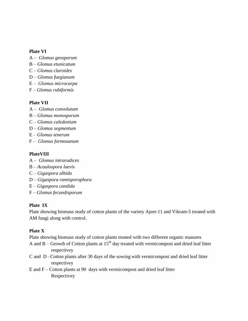
Plate VI
A – Glomus geosporum
B – Glomus etunicatum
C – Glomus claroides
D – Glomus fuegianum
E – Glomus microcarpa
F – Glomus rubiformis
Plate VII
A – Glomus convolutum
B – Glomus monosporum
C – Glomus caledonium
D – Glomus segmentum
E – Glomus tenerum
F – Glomus formosanum
PlateVIII
A – Glomus intraradices
B – Acaulospora laevis
C – Gigaspora albida
D – Gigaspora ramisporophora
E – Gigaspora candida
F – Glomus fecundisporum
Plate IX
Plate showing biomass study of cotton plants of the variety Ajeet-11 and Vikram-5 treated with
AM fungi along with control.
Plate X
Plate showing biomass study of cotton plants treated with two different organic manures
A and B – Growth of Cotton plants at 15th
day treated with vermicompost and dried leaf litter
respectivey
C and D –Cotton plants after 30 days of the sowing with vermicompost and dried leaf litter
respectivey
E and F – Cotton plants at 90 days with vermicompost and dried leaf litter
Respectivey

Plate XI
A to C- Biomass study of cotton variety treated with different fungi viz. A. Aspergillus niger,
B. Gliocladium virens and C. Trichoderma viride
D and E- Root colonization in roots of cotton plants treated with AM fungi
Plate XII
A – Fungal Filterate of different fungi
B and C – Effect of Fusarium oxysporum on seeds of Non Bt and Bt variety
D and E – Effect of Trichoderma viride on seeds of Non Bt and Bt variety
F and G – Effect of Gliocladium virens on seeds of Non Bt and Bt variety
Plate XIII
List of Plants used for the experiment of Biological Control against three pathogenic fungi
A – Annona reticulata L.
B – Balanites roxburghii Planchon
C – Cochlospermum religiosa (L.) Alst
D – Limonia acidissima L.
E – Sapindus emarginatus Vahl.
F – Tephrosia jamnagarensis Sant.
Plate XIV
A – Growth of F.oxysporum in methanolic extract of Annona reticulata
B – Growth of F.oxysporum in methanolic extract of Balanites roxburghii
C – Growth of F.oxysporum in methanolic extract of Cochlospermum religiosa
D – Growth of F.oxysporum in methanolic extract of Feronia acidissima
Plate XV
A – Growth of A.alternaria in methanolic extract of Tephrosia jamnagarensis
B – Growth of F.oxysporum in methanolic extract of Cochlospermum religiosa
C – Growth of F.oxysporum in methanolic extract of Balanites roxburghii
D – Growth of F.oxysporum in methanolic extract of Feronia acidissima
Copyright © 2022 FDOKUMEN