GnRH as a Cell Proliferation Regulator: Mechanism of Action and Evolutionary Implications
9
2004 Zoological Society of Japan ZOOLOGICAL SCIENCE 21 : 1005–1013 (2004) [REVIEW] GnRH as a Cell Proliferation Regulator: Mechanism of Action and Evolutionary Implications Masahiro Enomoto and Min Kyun Park * Department of Biological Sciences, Graduate School of Science, The University of Tokyo, 7-3-1 Hongo, Bunkyo, Tokyo 113-0033, Japan ABSTRACT —Gonadotropin-releasing hormone (GnRH) is well known as the central regulator of the repro- ductive system through its stimulation of gonadotropin release from the pituitary. Studies on GnRH have demonstrated that GnRH has both stimulatory and inhibitory effects on cell proliferation depending on the cell type; however, the mechanisms of these effects remain to be elucidated. Against this background we used four human cell lines, TSU-Pr1, Jurkat, HHUA and DU145, and newly found that GnRH increased or decreased the colony-formation depending on the cell line. Moreover, we demonstrated that the stim- ulatory and inhibitory effects of GnRH exhibit distinct ligand selectivities. In order to investigate the molec- ular basis of these phenomena, analyses of the expression of human GnRH receptors were performed and, moreover, the effects of GnRH were analyzed under conditions in which human GnRH receptors were knocked down by the technique of RNA interference. Consequently, it was found that human type II GnRH receptor, which had been suspected of being nonfunctional because of alterations in its sequence, is involved in the effects of GnRH on cell proliferation. In this article, the influence of the autocrine activities of the cells is also reviewed, focusing on the characteristics of substances secreted from the four cell lines. Based on recent studies of GnRH and its receptors and our up-to-date findings, the evolutionary implica- tions of GnRH action are discussed. Key words: GnRH, GnRH receptor, cell proliferation, colony-formation, reproduction INTRODUCTION GnRH was originally identified as a hypothalamic decapeptide which promotes gonadotropin secretion from pituitary gonadotropes and was named gonadotropin-releas- ing hormone (Matsuo et al ., 1971; Amoss et al ., 1971). Thereafter, multiple GnRH isoforms and multiple types of GnRH receptors were reported in a wide range of organisms in addition to vertebrates (Millar et al ., 2004) and broad dis- tribution of them in extrapituitary tissues and organs has been demonstrated (Klausen et al ., 2002; Millar, 2003; Ike- moto et al ., 2003, 2004). Through these observations the study of extrapituitary GnRH systems has become one of the important topics regarding the physiological significance of GnRH. The concept of extrapituitary GnRH systems is also important in terms of the evolution of the reproductive system. Now that the existence of GnRH has been con- firmed in invertebrates, which have no pituitary gland, it is suggested that in these organisms GnRH acts directly on the gonads (Terakado, 2001; Kusakabe et al ., 2003). In our previous studies (Kogo et al ., 1995, 1999a, b; Park et al ., 1999), GnRH receptor mRNA expression was demonstrated in the rat ovary, which strongly suggests the idea that GnRH is involved in the induction of follicular atresia and ovulation (Table 1). There have also been other studies suggesting that GnRH directly controls gonadal function. (Gobetti et al ., 1992; Fasano et al ., 1995; Takekida et al ., 2003; Sifer et al ., 2003), however clear evidence supporting this suggestion has not been obtained yet. Moreover, in addition to the direct effects on gonads, it has been reported that GnRH may act as a neuromodulator or an immunomodulator. (Eis- then et al ., 2000; Oka, 2002; Ford et al ., 2003; Azad et al ., 1997; Enomoto et al ., 2001; Chen et al ., 2002). Although a number of studies have been performed on these effects of GnRH, little is known about the molecular mechanisms and physiological settings in which GnRH exerts its activities in * Corresponding author: Tel. +81-3-5841-4437; FAX. +81-3-5841-4439. E-mail: [email protected]
-
Upload
independent -
Category
Documents
-
view
5 -
download
0
Transcript of GnRH as a Cell Proliferation Regulator: Mechanism of Action and Evolutionary Implications
2004 Zoological Society of JapanZOOLOGICAL SCIENCE
21
: 1005–1013 (2004)
[REVIEW]
GnRH as a Cell Proliferation Regulator: Mechanism ofAction and Evolutionary Implications
Masahiro Enomoto and Min Kyun Park*
Department of Biological Sciences, Graduate School of Science, The University ofTokyo, 7-3-1 Hongo, Bunkyo, Tokyo 113-0033, Japan
ABSTRACT
—Gonadotropin-releasing hormone (GnRH) is well known as the central regulator of the repro-ductive system through its stimulation of gonadotropin release from the pituitary. Studies on GnRH havedemonstrated that GnRH has both stimulatory and inhibitory effects on cell proliferation depending on thecell type; however, the mechanisms of these effects remain to be elucidated. Against this background weused four human cell lines, TSU-Pr1, Jurkat, HHUA and DU145, and newly found that GnRH increasedor decreased the colony-formation depending on the cell line. Moreover, we demonstrated that the stim-ulatory and inhibitory effects of GnRH exhibit distinct ligand selectivities. In order to investigate the molec-ular basis of these phenomena, analyses of the expression of human GnRH receptors were performedand, moreover, the effects of GnRH were analyzed under conditions in which human GnRH receptors wereknocked down by the technique of RNA interference. Consequently, it was found that human type II GnRHreceptor, which had been suspected of being nonfunctional because of alterations in its sequence, isinvolved in the effects of GnRH on cell proliferation. In this article, the influence of the autocrine activitiesof the cells is also reviewed, focusing on the characteristics of substances secreted from the four cell lines.Based on recent studies of GnRH and its receptors and our up-to-date findings, the evolutionary implica-tions of GnRH action are discussed.
Key words:
GnRH, GnRH receptor, cell proliferation, colony-formation, reproduction
INTRODUCTION
GnRH was originally identified as a hypothalamicdecapeptide which promotes gonadotropin secretion frompituitary gonadotropes and was named gonadotropin-releas-ing hormone (Matsuo
et al
., 1971; Amoss
et al
., 1971).Thereafter, multiple GnRH isoforms and multiple types ofGnRH receptors were reported in a wide range of organismsin addition to vertebrates (Millar
et al
., 2004) and broad dis-tribution of them in extrapituitary tissues and organs hasbeen demonstrated (Klausen
et al
., 2002; Millar, 2003; Ike-moto
et al
., 2003, 2004). Through these observations thestudy of extrapituitary GnRH systems has become one ofthe important topics regarding the physiological significanceof GnRH. The concept of extrapituitary GnRH systems isalso important in terms of the evolution of the reproductive
system. Now that the existence of GnRH has been con-firmed in invertebrates, which have no pituitary gland, it issuggested that in these organisms GnRH acts directly onthe gonads (Terakado, 2001; Kusakabe
et al
., 2003). In ourprevious studies (Kogo
et al
., 1995, 1999a, b; Park
et al
.,1999), GnRH receptor mRNA expression was demonstratedin the rat ovary, which strongly suggests the idea that GnRHis involved in the induction of follicular atresia and ovulation(Table 1). There have also been other studies suggestingthat GnRH directly controls gonadal function. (Gobetti
et al
.,1992; Fasano
et al
., 1995; Takekida
et al
., 2003; Sifer
et al
.,2003), however clear evidence supporting this suggestionhas not been obtained yet. Moreover, in addition to thedirect effects on gonads, it has been reported that GnRHmay act as a neuromodulator or an immunomodulator. (Eis-then
et al
., 2000; Oka, 2002; Ford
et al
., 2003; Azad
et al
.,1997; Enomoto
et al
., 2001; Chen
et al
., 2002). Although anumber of studies have been performed on these effects ofGnRH, little is known about the molecular mechanisms andphysiological settings in which GnRH exerts its activities in
* Corresponding author: Tel. +81-3-5841-4437;FAX. +81-3-5841-4439.E-mail: [email protected]
M. Enomoto and M. K. Park1006
extrapituitary tissues or organs.There are interesting
in vitro
studies about the diversefunctions of GnRH. GnRH promotes or inhibits cell prolifer-ation depending on the cell type. As for the promotiveeffects of GnRH, effects on thymocytes, splenocytes, andlymphocytes have been reported (Marchetti
et al
., 1989;Batticane
et al
., 1991; Azad
et al
., 1997). Regarding inhibi-tory effects, effects against hormone-related tumors are wellknown, and these effects are now applied clinically (Schally,1999; Grundker
et al
., 2002). These opposite effects ofGnRH on cell proliferation are very interesting, but it hasremained unknown whether GnRH has such activities inphysiological settings, mainly because most previous stud-ies were performed using cancer cell lines and pharmaco-logical doses of GnRH. However, recently, the effects ofGnRH on the cell proliferation of non-tumor cells, as well ason cell adhesion, cell migration, and cytoskeletal remodelinghave been reported (Chen
et al
., 2002; Romanelli
et al
.,2004; Davidson
et al
., 2004) and the physiological roles ofthe effects of GnRH on cell proliferation are being reconsid-ered.
In fact, there is evidence for the effects of GnRH ongonadotrope cell proliferation (Sakai
et al
., 1988; Kakar
etal
., 1997; Miles
et al
., 2004), and thus GnRH may originallyact as one of the cell growth regulators of gonadotropes.Moreover, there is a possibility that the activities of GnRHthat alter their direction of effects depending on the cell typeunderlie the diverse physiological functions of GnRH. Wehave focused on the two opposite effects of GnRH on cellproliferation, particularly the relationship between theseeffects and the diverse functions of GnRH, and investigated
the molecular mechanisms of the effects of GnRH on cellproliferation. So far, we have found stimulatory and inhibi-tory activities of the physiological dose of GnRH on thecolony-formation of cells using an originally establishedmethod, the colony-forming efficiency assay (Enomoto
etal
., 2001 and 2004a, b). The experimental models used inour studies are TSU-Pr1 (from human prostatic carcinoma,Iizumi
et al
., 1987), Jurkat (from human mature leukemia,Gills and Watson, 1980), DU145 (from human prostatic car-cinoma, Mickey
et al
., 1977; Stone
et al
., 1978), and HHUA(from human endometrial carcinoma, Ishiwata
et al
., 1984)cell lines. Recently, interesting findings about the mecha-nisms of the two opposite effects of GnRH have beenobtained by adaptation of the technique of RNA interferenceto our original assay system (Enomoto
et al
., 2004b). More-over, we have obtained strong evidence for the influence ofautocrine activities of the cells on the GnRH effects(Enomoto
et al
., 2001, 2004a). In this article, our up-to-datefindings about the mechanisms of the stimulatory and inhib-itory effects of GnRH will be reviewed, focusing on humanGnRH receptors and the influence of autocrine activities ofthe cells on the GnRH effects. The evolutionary implicationof the GnRH activities on cell proliferation will be also dis-cussed.
1. Effects of GnRH on colony-formation1-1. Stimulatory and inhibitory effects of GnRH
A number of studies have demonstrated that variouscarcinomas of breast, ovary, endometrium, prostate, pan-creas and liver origin respond to GnRH and its analogues(Schally, 1999). It has also been reported that treatment of
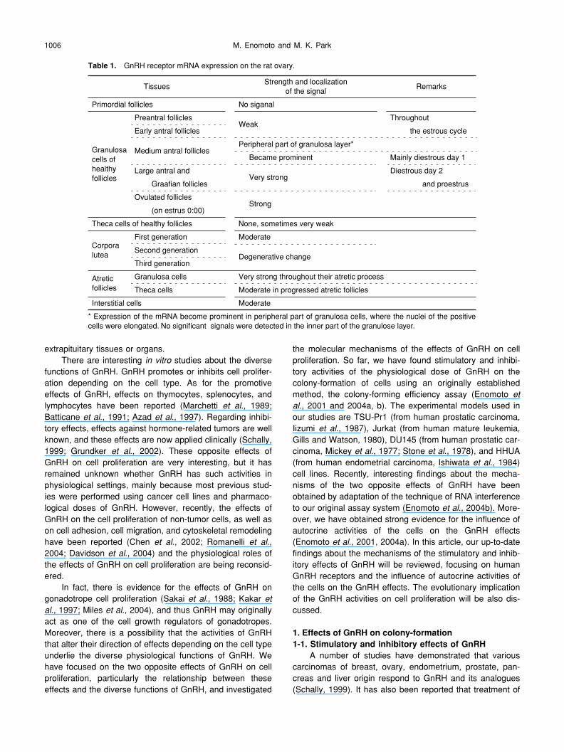
Table 1.
GnRH receptor mRNA expression on the rat ovary.
TissuesStrength and localization
of the signalRemarks
Primordial follicles No siganal
Granulosacells ofhealthyfollicles
Preantral folliclesWeak
Throughout
Early antral follicles the estrous cycle
Medium antral folliclesPeripheral part of granulosa layer*
Became prominent Mainly diestrous day 1
Large antral andVery strong
Diestrous day 2
Graafian follicles and proestrus
Ovulated folliclesStrong
(on estrus 0:00)
Theca cells of healthy follicles None, sometimes very weak
Corporalutea
First generation Moderate
Second generationDegenerative change
Third generation
Atreticfollicles
Granulosa cells Very strong throughout their atretic process
Theca cells Moderate in progressed atretic follicles
Interstitial cells Moderate
* Expression of the mRNA become prominent in peripheral part of granulosa cells, where the nuclei of the positivecells were elongated. No significant signals were detected in the inner part of the granulose layer.
GnRH as a Cell Proliferation Regulator 1007
thymocytes, splenocytes, and lymphocytes with GnRH andits analogues increased their proliferative activity (Marchetti
et al
., 1989; Batticane
et al
., 1991; Azad
et al
., 1997). Inthese studies the effects of GnRH on cell proliferation wereinvestigated by measuring tumor weight and/or volume, therate of cell proliferation, or
3
H-thymidine incorporation (Red-ding and Schally, 1983; Sharoni
et al
., 1989; Pati andHabibi, 1995; Dondi
et al
., 1998). However, to our knowl-edge, there have been no studies that compared stimulatorywith inhibitory effects. One of the reasons for this was thelack of a method that makes it possible to detect both effectssensitively. Thus, we first established an original assay, thecolony-forming efficiency assay (Enomoto
et al
., 2001). Thecolony-forming efficiency assay is based on limiting dilutionanalysis with minor modifications. The detailed procedurewas described in our previous report (Enomoto
et al
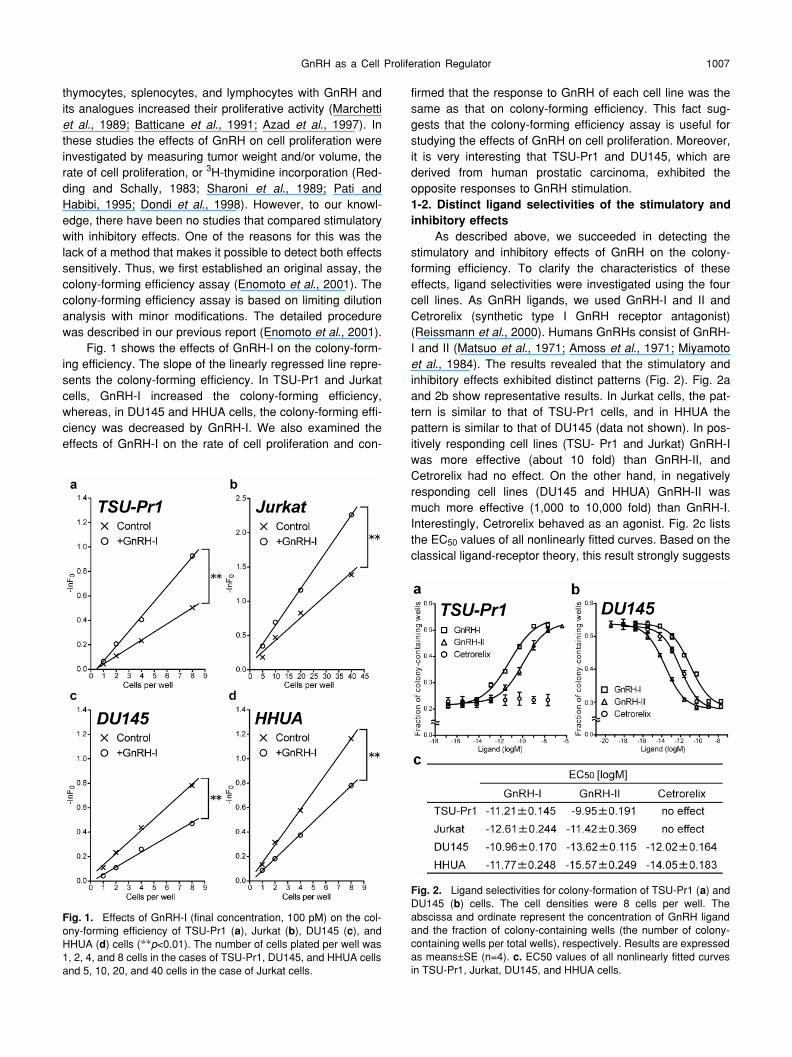
., 2001).Fig. 1 shows the effects of GnRH-I on the colony-form-
ing efficiency. The slope of the linearly regressed line repre-sents the colony-forming efficiency. In TSU-Pr1 and Jurkatcells, GnRH-I increased the colony-forming efficiency,whereas, in DU145 and HHUA cells, the colony-forming effi-ciency was decreased by GnRH-I. We also examined theeffects of GnRH-I on the rate of cell proliferation and con-
firmed that the response to GnRH of each cell line was thesame as that on colony-forming efficiency. This fact sug-gests that the colony-forming efficiency assay is useful forstudying the effects of GnRH on cell proliferation. Moreover,it is very interesting that TSU-Pr1 and DU145, which arederived from human prostatic carcinoma, exhibited theopposite responses to GnRH stimulation.
1-2. Distinct ligand selectivities of the stimulatory andinhibitory effects
As described above, we succeeded in detecting thestimulatory and inhibitory effects of GnRH on the colony-forming efficiency. To clarify the characteristics of theseeffects, ligand selectivities were investigated using the fourcell lines. As GnRH ligands, we used GnRH-I and II andCetrorelix (synthetic type I GnRH receptor antagonist)(Reissmann
et al
., 2000). Humans GnRHs consist of GnRH-I and II (Matsuo
et al
., 1971; Amoss
et al
., 1971; Miyamoto
et al
., 1984). The results revealed that the stimulatory andinhibitory effects exhibited distinct patterns (Fig. 2). Fig. 2aand 2b show representative results. In Jurkat cells, the pat-tern is similar to that of TSU-Pr1 cells, and in HHUA thepattern is similar to that of DU145 (data not shown). In pos-itively responding cell lines (TSU- Pr1 and Jurkat) GnRH-Iwas more effective (about 10 fold) than GnRH-II, andCetrorelix had no effect. On the other hand, in negativelyresponding cell lines (DU145 and HHUA) GnRH-II wasmuch more effective (1,000 to 10,000 fold) than GnRH-I.Interestingly, Cetrorelix behaved as an agonist. Fig. 2c liststhe EC
50
values of all nonlinearly fitted curves. Based on theclassical ligand-receptor theory, this result strongly suggests
Fig. 1.
Effects of GnRH-I (final concentration, 100 pM) on the col-ony-forming efficiency of TSU-Pr1 (
a
), Jurkat (
b
), DU145 (
c
), andHHUA (
d
) cells (
**
p
<0.01). The number of cells plated per well was1, 2, 4, and 8 cells in the cases of TSU-Pr1, DU145, and HHUA cellsand 5, 10, 20, and 40 cells in the case of Jurkat cells.
Fig. 2.
Ligand selectivities for colony-formation of TSU-Pr1 (
a
) andDU145 (
b
) cells. The cell densities were 8 cells per well. Theabscissa and ordinate represent the concentration of GnRH ligandand the fraction of colony-containing wells (the number of colony-containing wells per total wells), respectively. Results are expressedas means
±
SE (n=4).
c.
EC50 values of all nonlinearly fitted curvesin TSU-Pr1, Jurkat, DU145, and HHUA cells.

M. Enomoto and M. K. Park1008
that the stimulatory and inhibitory effects of GnRH are medi-ated by different GnRH receptors. Thus, we investigated thispossibility as described in the following chapter.
2. Two types of human GnRH receptors2-1. Expression of human GnRH receptors
Among the human GnRH receptors, type I (hGnRHR-1)and II (hGnRHR-2) GnRH receptors are known. Type Ireceptor was cloned first and has been well characterized(Kakar
et al
., 1992), and recently the genomic loci encodingthe human type II GnRH receptor has been cloned (Fau-rholm
et al
., 2001). However, previous studies revealed apuzzling fact about the human type II GnRH receptor gene.The protein coding sequences contain a frameshift mutationand a TGA stop codon, compared with other primate type IIGnRH receptor sequences that were demonstrated to befunctional (Faurholm
et al
., 2001; Neill, 2002; Morgan
et al
.,2003). So far, no full-length sequence of a human type IIreceptor that is translated into protein has been discovered.
In 2001, Millar
et al
. demonstrated that human type IGnRH receptor and marmoset type II GnRH receptor, whichhas 90% amino acid identity with the deduced human typeII GnRH receptor amino acid sequence, exhibited distinctlydifferent patterns of ligand selectivity, as shown Fig. 2. Thisresult strongly suggests that human type II GnRH receptoris functional as a receptor, and we first presumed that thedistinctly different patterns of ligand selectivity between thestimulatory and inhibitory effects should be due to theexpression of a different GnRH receptor subtype. Thus weperformed expression analyses of human GnRH receptorsin the four cell lines. For hGnRHR-1, a single PCR productwas amplified with one primer set in all four cell lines (upperpanel in Fig. 3). In contrast, for hGnRHR-2, two PCR prod-ucts were amplified with one primer set in all four cell lines(lower panel in Fig. 3). The results of direct sequencingdemonstrated that the longer PCR product was identicalwith the human type II GnRH receptor (GeneBank acces-sion No. AY077708) (Van Biljon
et al
., 2002), and the lowerone was identical with the human type II GnRH receptorsplice variant (hGnRHR-2v) (GeneBank accession No.AY081843) (Neill, 2002). As shown in Fig. 3, all four celllines expressed hGnRHR-1, 2, and 2v, which suggests that
the opposite responses to GnRH stimulation may not be dueto the differences of expressed GnRH receptor subtypes.
2-2. Involvement of each human GnRH receptor in theeffects of GnRH
In the expression analyses, no clear differencesbetween positively responding cell lines (TSU-Pr1 and Jur-kat) and negatively responding cell line (DU145 and HHUA)were observed. Next we examined the effects of GnRH-Iand II and Cetrorelix on colony-forming efficiency under thecondition that the expression of each of the human GnRHreceptors was suppressed individually by the technique ofRNA interference (RNAi) using p
Silencer
2.0-U6, a shorthairpin interfering RNA (shRNA) expression vector (AmbionInc., Austin, TX). This technique efficiently inducessequence-specific mRNA degradation and thereby sup-presses the target gene expression (Brummelkamp
et al
.,2002; Hannon, 2002; Shi, 2003; Dykxhoor
et al
., 2003), andthus the roles of human type II GnRH receptor and its splicevariant can be investigated even if they don’t function asproteins. Table 2 shows the summary of the completeresults. As a negative control, we used the p
Silencer
nega-tive control vector encoding a shRNA whose sequence isnot found in the mouse, rat, or human genome databases(Ambion Inc., Austin, TX), and the resultant colony-formingefficiencies were the same as those of non-transfected cells(data not shown).
As shown in Table 2, when hGnRHR-1 was knocked-down, all three GnRH ligands failed to show significanteffects and the colony-forming efficiencies were similar tothose of the non-transfected cells. When hGnRHR-2 wasknocked-down, GnRH-II and Cetrorelix had no significanteffects, and only GnRH-I was significantly effective. ForhGnRHR-2v, quite interesting and remarkable results wereobtained.When hGnRHR-2v was knocked-down, the direc-tion of response to the stimulation of GnRH-I and II in TSU-
Fig. 3.
Expression of human GnRH receptor 1 (the upper panel), 2and 2 variant (the lower panel) in TSU-Pr1, Jurkat, DU145, andHHUA cells. RT- are negative controls in which non-reverse tran-scribed mRNA of each cell line was used as the template. The left-hand lane contains the molecular marker.
Table 2.
Summary of the results when each human GnRH receptorwas knocked down.
TSU-Pr1 Jurkat DU145 HHUA
non-transfectedcells
GnRH-I
+ + – –
GnRH-II
+ + – –
Cetrorelix n.e. n.e.
– –
hGnRHR-1knocked down
GnRH-I n.e. n.e. n.e. n.e.
GnRH-II n.e. n.e. n.e. n.e.
Cetrorelix n.e. n.e. n.e. n.e.
hGnRHR-2knocked down
GnRH-I
+ + – –
GnRH-II n.e. n.e. n.e. n.e.
Cetrorelix n.e. n.e. n.e. n.e.
hGnRHR-2vknocked down
GnRH-I
– – – –
GnRH-II
– – – –
Cetrorelix
– – – –
Normal cells means non-transfected cells. +, significant stimulatoryeffect of GnRH (p<0.05); –, significant inhibitory effect of GnRH(p<0.05); n.e., no significant effect of GnRH.

GnRH as a Cell Proliferation Regulator 1009
Pr1 and Jurkat cells was reversed from a positive effect toa negative one, and Cetrorelix behaved as an agonist. Tothe best of our knowledge, this result is the first demonstra-tion that a receptor can regulate the direction of theresponse to the ligand stimulation. As for DU145 and HHUAcells, no such alteration was observed upon the suppressionof hGnRHR-2v. Although hGnRHR-2v mRNA is expressedin DU145 and HHUA cells, it appears to have no function inthese cells. This fact may be due to the balance of theexpression levels of hGnRHR-2 and 2v. In fact, as shown inFig. 3, in DU145 and HHUA cells these levels are quite dif-ferent, whereas in Jurkat and TSU-Pr1 cells they are muchless different; however, further studies will be necessary toclarify this point.
Summarizing our results focusing on each receptor,hGnRHR-1 was indispensable for the effectiveness of allthree GnRH ligands, and hGnRHR-2 was necessary for theeffectiveness of GnRH-II and Cetrorelix. Furthermore, hGn-RHR-2v plays a role in mediating the stimulatory effects ofGnRH-I and II and the effectiveness of Cetrorelix. One pos-sible explanation for these complicated results is that hGn-RHR-2 and 2v directly interact with hGnRHR-1 and formhetero-dimers or oligomers. In fact, many recent studieshave provided evidence that G-protein coupled receptors(GPCRs) can exist as either dimers or higher order oligo-mers (Milligan
et al
., 2004; Pfleger
et al
., 2004). Promotionor inhibition of signal transduction by ligand stimulation andalteration of G-protein selectivity by receptor dimerizationhave been also reported (AbdAlla
et al
., 2001a, b). More-over, there is evidence that co-expression of GPCR pairscan generate distinct ligand-binding sites (Maggio
et al
.,1998). Fig. 4 shows one of the proposed structural modelsfor GPCR receptor dimers, 5,6-domain swapped dimers(Gouldson
et al
., 1998). Each transmembrane alpha helix isrepresented as a circle. The hinge loop represents intracel-lular loop 3. As shown in this figure, another binding pocketis generated by receptor dimerization. This could accountfor our distinct patterns of ligand selectivities. We shouldalso consider the possibility of cross-talk among intracellularsignalings and functions as non-coding RNAs (Storz, 2002)of hGnRHR-2 and 2v mRNAs to modulate the signaling
mediated by hGnRHR-1. However, in any case, furtherinvestigations of the human type II GnRH receptor and thesplice variant will be required to elucidate the detailed mech-anisms by which human type II GnRH receptor and its splicevariant are involved in the effects of GnRH on cell prolifera-tion.
3. Influence of autocrine activities of the cells on theGnRH effects
The colony-forming efficiency assay is a method thatcan examine cell proliferation at the low cell density (10 to
Fig. 4.
Schematic models of the monomer and the 5,6-domainswapped dimer of G-protein coupled receptor. Each transmem-brane alpha helix is represented as a circle. The hinge loop repre-sents intracellular loop 3. The presumed binding pockets are shownas grey areas.
Fig. 5.
Effects of Buserelin (final concentration, 5 pg/ml) on therate of cell proliferation of Jurkat (
a
) and HHUA (
b
) cells when cul-ture medium was exchanged every 24 hours. Results are expressedas means
±
SE (n=4;
**
p
<0.01;
***
p
<0.001). The experimentalschedule is illustrated in
c
.
M. Enomoto and M. K. Park1010
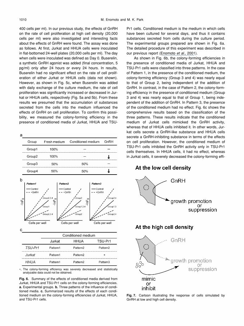
400 cells per ml). In our previous study, the effects of GnRHon the rate of cell proliferation at high cell density (20,000cells per ml) were also investigated and interesting factsabout the effects of GnRH were found. The assay was doneas follows. At first, Jurkat and HHUA cells were inoculatedin flat-bottomed 24-well plates (20,000 cells per ml). The daywhen cells were inoculated was defined as Day 0. Buserelin,a synthetic GnRH agonist was added (final concentration, 5pg/ml) only after 24 hours or every 24 hours. In results,Buserelin had no significant effect on the rate of cell prolif-eration of either Jurkat or HHUA cells (data not shown).However, as shown in Fig. 5c, when Buserelin was addedwith daily exchange of the culture medium, the rate of cellproliferation was significantly increased or decreased in Jur-kat or HHUA cells, respectively (Fig. 5a and 5b). From theseresults we presumed that the accumulation of substancessecreted from the cells into the medium influenced theeffects of GnRH on cell proliferation. To confirm this possi-bility, we measured the colony-forming efficiency in thepresence of conditioned media of Jurkat, HHUA and TSU-
Pr1 cells. Conditioned medium is the medium in which cellshave been cultured for several days, and thus it containssubstances secreted from cells during the culture period.The experimental groups prepared are shown in Fig. 6a.The detailed procedure of this experiment was described inour previous report (Enomoto
et al
., 2001).As shown in Fig. 6b, the colony-forming efficiencies in
the presence of conditioned media of Jurkat, HHUA andTSU-Pr1 cells were classified into three patterns. In the caseof Pattern 1, in the presence of the conditioned medium, thecolony-forming efficiency (Group 3 and 4) was nearly equalto that of Group 2, being independent of the addition ofGnRH. In contrast, in the case of Pattern 2, the colony-form-ing efficiency in the presence of conditioned medium (Group3 and 4) was nearly equal to that of Group 1, being inde-pendent of the addition of GnRH. In Pattern 3, the presenceof the conditioned medium had no effect. Fig. 6c shows thecomprehensive results based on the classification of thethree patterns. These results indicate that the conditionedmedium of Jurkat cells mimicked the GnRH activity,whereas that of HHUA cells inhibited it. In other words, Jur-kat cells secrete a GnRH-like substance and HHUA cellssecrete a GnRH-inhibiting substance in terms of the effectson cell proliferation. However, the conditioned medium ofTSU-Pr1 cells inhibited the GnRH activity only in TSU-Pr1cells themselves. In HHUA cells, it had no effect, whereasin Jurkat cells, it severely decreased the colony-forming effi-
*,
The colony-forming efficiency was severely decreased and statisticallyanalyzable data could not be obtained.
Fig. 6.
Summary of the effects of conditioned media derived fromJurkat, HHUA and TSU-Pr1 cells on the colony-forming efficiencies.
a.
Experimental groups.
b.
Three patterns of the influence of condi-tioned media.
c.
Summarized results of the effects of each condi-tioned medium on the colony-forming efficiencies of Jurkat, HHUA,and TSU-Pr1 cells.
Fig. 7.
Cartoon illustrating the response of cells simulated byGnRH at low and high cell density.

GnRH as a Cell Proliferation Regulator 1011
ciency and statistically analyzable data could not beobtained. Summarizing these results, the conditioned mediaof Jurkat and HHUA cells consistently affected the GnRHactivities regardless of the cell type, whereas that of TSU-Pr1 had cell type-dependent effects. Our results suggestthat each of these three cell lines secrete a distinct sub-stance that affects GnRH activities, and these substanceswill be identified in future studies.
Our interpretation of the results introduced in this chap-ter is schematized in Fig. 7. At low cell density, GnRH pro-motes or suppresses cell proliferation depending on the celltype. In contrast, at high cell density autocrine factors of thecells mimic or inhibit the GnRH activity and the effects ofGnRH are masked. From this point of view, the colony-form-ing efficiency assay is a sensitive method that can limit theeffects of autocrine activity of the cells.
EVOLUTIONARY IMPLICATIONS
In this article, we reviewed our recent findings about themechanisms of the stimulatory and inhibitory effects ofGnRH on cell proliferation, focusing on the involvement ofGnRH receptors. The detailed role of each receptor remainsto be elucidated. In addition, we described the autocrineactivities of the cells that affect the GnRH effects. Identifica-tion of these factors is important to clarify the effects ofGnRH on cell proliferation
in vivo
.Viewing the historical progress on studies of GnRH, we
are reminded of one of the bioactive substances in the hypo-thalamus-pituitary-gonadal axis, namely activin. Activin wasoriginally isolated based on its activity in stimulating follicle-stimulating hormone (FSH) release from the anterior pitu-itary (Ling
et al., 1986; Vale et al., 1986). In addition to itsendocrine function, activin has been found to possess vari-ous activities in different biological systems, e.g., erythroiddifferentiation, nerve cell survival, Xenopus laevis embryonicmesoderm induction, bone growth promotion, and soma-tostatin induction (Mathews, 1994). Subsequently, it wasfound that activin regulates a wide variety of cellular events,including cell proliferation, differentiation, and apoptosis. Forexample, in addition to endocrine function in the pituitary,activin also controls the activity of the hypothalamus andovary, indicating that activin has profound autocrine andparacrine effects on the female reproductive system (Pengand Mukai, 2000). GnRH was originally identified as a hypo-thalamic decapeptide that promotes gonadotropin release,and thereafter a number of studies have strongly suggestedthat it has various physiological activities, such as neuro-modulation, immunomodulation, and regulation of follicularatresia and ovulation. Moreover, it was demonstrated thatGnRH regulates cell proliferation, cell migration, and cellattachment. Based on these similarities between activin andGnRH, the idea that GnRH may have roles in embryogene-sis and morphogenesis occurs to us. In fact, there havebeen several reports suggesting that GnRH may play a sub-stantial autocrine or paracrine role in human fertilization,
early embryonic development, and implantation (Seshagiriet al., 1994; Casan et al., 1999; Raga et al., 1999). Fromthese points of view, our studies of the molecular mecha-nisms of the GnRH actions on cell proliferation will provideimportant clues for clarifying the physiological roles ofGnRH, particularly in embryogenesis and morphogenesis.Moreover, extending our findings about human GnRHreceptor signalings to other species will lead to a compre-hensive understanding of the GnRH system, including theGnRH system in invertebrates, and clarification of the evo-lution of the reproductive system.
ACKNOWLEDGMENTS
We heartily express great gratitude to S. Kawashima (EmeritusProfessor, The University of Tokyo) for helpful advice throughoutour study. We are also grateful to Prof. T. Minegishi (Department ofObstetrics and Gynaecology, School of Medicine, Gunma Univer-sity) for a gift of Cetrorelix and Dr. T. Iizumi (Department of Urology,Teikyo University School of Medicine) and Dr. J. Y. Seong and Ms.D. Y. Oh (Hormone Research Center, Chonnam National Univer-sity) for helpful advice on culturing TSU-Pr1 cells. Lastly, we arethankful to Prof. Y. Oka, Dr. Y. Akazome and Ms. M. Kyokuwa(Department of Biological Sciences, Graduate School of Science,The University of Tokyo) for supports throughout our study. Ourstudies summarized in this review were supported in part byGrants-in-Aid for Scientific Research from the Ministry of Education,Culture, Sports, Science and Technology of Japan.
REFERENCES
AbdAlla S, Lother H, el Massiery A, Quitterer U (2001) IncreasedAT1 receptor heterodimers in preeclampsia mediate enhancedangiotensin II responsiveness. Nat Med 7: 1003–1009
AbdAlla S, Lother H, Abdel-tawab AM, Quitterer U (2001) Theangiotensin II AT2 receptor is an AT1 receptor antagonist. JBiol Chem 276: 39721–39726
Amoss M, Burgus R, Blackwell R, Vale W, Fellows R, Guillemin R(1971) Purification, amino acid composition and N-terminus ofthe hypothalamic luteinizing hormone releasing factor (LRF) ofovine origin. Biochem Biophys Res Commun 44: 205–210
Azad N, LaPaglia N, Kirsteins L, Uddin S, Steiner J, Williams DW,Lawrence AM, Emanuele NV (1997) Jurkat cell proliferativeactivity is increased by luteinizing hormone-releasing hormone.J Endocrinol 153: 241–249
Batticane N, Morale MC, Gallo F, Farinella Z, Marchetti B (1991)Luteinizing hormone-releasing hormone signaling at the lym-phocyte involves stimulation of interleukin-2 receptor expres-sion. Endocrinology 129: 277–286
Brummelkamp TR, Bernards R, Agami R (2002) A system for stableexpression of short interfering RNAs in mammalian cells. Sci-ence 296: 550–553
Casan EM, Raga F, Polan ML (1999) GnRH mRNA and proteinexpression in human preimplantation embryos. Mol HumReprod 5: 234–239
Chen A, Ganor Y, Rahimipour S, Ben-Aroya N, Koch Y, Levite M(2002) The neuropeptides GnRH-II and GnRH-I are producedby human T cells and trigger laminin receptor gene expression,adhesion, chemotaxis and homing to specific organs. Nat Med8: 1421–1426
Davidson L, Pawson AJ, Millar RP, Maudsley S (2004) Cytoskeletalreorganization dependence of signaling by the gonadotropin-releasing hormone receptor. J Biol Chem 279: 1980–1993
M. Enomoto and M. K. Park1012
Dondi D, Moretti RM, Montagnani Marelli M, Pratesi G, Polizzi D,Milani M, Motta M, Limonta P (1998) Growth-inhibitory effectsof luteinizing hormone-releasing hormone (LHRH) agonists onxenografts of the DU 145 human androgen-independent pros-tate cancer cell line in nude mice. Int J Cancer 76: 506–511
Dykxhoorn DM, Novina CD, Sharp PA (2003) Killing the messenger:short RNAs that silence gene expression. Nat Rev Mol Cell Biol4: 457–467
Eisthen HL, Delay RJ, Wirsig-Wiechmann CR, Dionne VE (2000)Neuromodulatory effects of gonadotropin releasing hormone onolfactory receptor neurons. J Neurosci 20: 3947–3955
Enomoto M, Mori T, Park MK (2001) GnRH agonist Buserelinaffects colony-forming efficiency of HHUA and Jurkat cells. Bio-chem Biophys Res Commun 289: 1180–1187
Enomoto M, Seong JY, Kawashima S, Park MK (2004) Proliferationof TSU-Pr1, a human prostatic carcinoma cell line is stimulatedby gonadotropin-releasing hormone. Life Sci 74: 3141–3152
Enomoto M, Endo D, Kawashima S, Park MK (2004) Human type IIGnRH receptor mediates effects of GnRH on cell proliferation.Zool Sci 21: 763–770
Fasano S, D’Antonio M, Chieffi P, Cobellis G, Pierantoni R (1995)Chicken GnRH-II and salmon GnRH effects on plasma and tes-ticular androgen concentrations in the male frog, Rana escu-lenta, during the annual reproductive cycle. Comp BiochemPhysiol C Pharmacol Toxicol Endocrinol 112: 79–86
Faurholm B, Millar RP, Katz AA (2001) The genes encoding thetype II gonadotropin-releasing hormone receptor and the ribo-nucleoprotein RBM8A in humans overlap in two genomic loci.Genomics 78: 15–18
Ford CP, Dryden WF, Smith PA (2003) Neurotrophic regulation ofcalcium channels by the peptide neurotransmitter luteinizinghormone releasing hormone. J Neurosci 23: 7169–7175
Gillis S, Watson J (1980) Biochemical and biological characteriza-tion of lymphocyte regulatory molecules. V. Identification of aninterleukin 2-producing human leukemia T cell line. J Exp Med152: 1709–1719
Gobbetti A, Zerani M (1992) Mammalian GnRH involvement in pros-taglandin F2 alpha and sex steroid hormones testicular releasein two amphibian species: the anuran water frog, Rana escu-lenta, and the urodele crested newt, Triturus carnifex. GenComp Endocrinol 87: 240–248
Gouldson PR, Snell CR, Bywater RP, Higgs C, Reynolds CA (1998)Domain swapping in G-protein coupled receptor dimers. Pro-tein Eng 11: 1181–1193
Grundker C, Gunthert AR, Westphalen S, Emons G (2002) Biologyof the gonadotropin-releasing hormone system in gynecologicalcancers. Eur J Endocrinol 146: 1–14
Hannon GJ (2002) RNA interference. Nature 418: 244–251Iizumi T, Yazaki T, Kanoh S, Kondo I, Koiso K (1987) Establishment
of a new prostatic carcinoma cell line (TSU-Pr1). J Urol 137:1304–1306
Ikemoto T, Park MK (2003) Identification and characterization of thereptilian GnRH-II gene in the leopard gecko, Eublepharis macu-larius, and its evolutionary considerations. Gene 316: 157–165
Ikemoto T, Enomoto M, Park MK (2004) Identification and charac-terization of a reptilian GnRH receptor from the leopard gecko.Mol Cell Endocrinol 214: 137–147
Ishiwata I, Ishiwata C, Soma M, Arai J, Ishikawa H (1984) Establish-ment of human endometrial adenocarcinoma cell line contain-ing estradiol-17 beta and progesterone receptors. GynecologicOncology 17: 281–290
Kakar SS, Musgrove LC, Devor DC, Sellers JC, Neill JD (1992)Cloning, sequencing, and expression of human gonadotropinreleasing hormone (GnRH) receptor. Biochem Biophys ResCommun 189: 289–295
Kakar SS, Nath S, Bunn J, Jennes L (1997) The inhibition of growthand down-regulation of gonadotropin releasing hormone
(GnRH) receptor in alphaT3-1 cells by GnRH agonist. Antican-cer Drugs 8: 369–375
Klausen C, Chang JP, Habibi HR (2002) Multiplicity of gonadotro-pin-releasing hormone signaling: a comparative perspective.Prog Brain Res 141: 111–128
Kogo H, Kudo A, Park MK, Mori T, Kawashima S (1995) In situdetection of gonadotropin-releasing hormone (GnRH) receptormRNA expression in the rat ovarian follicles. J Exp Zool 272:62–68
Kogo H, Fujimoto T, Mori T (1999) Evidence for gonadotropin-releasing hormone receptor mRNA expression by estrogen inrat granulosa cells. Cell Tissue Res 297: 459–465
Kogo H, Fujimoto T, Park MK, Mori T (1999) Gonadotropin-releas-ing hormone receptor mRNA expression in the ovaries of neo-natal and adult rats. Cells Tissues Organs 164: 14–22
Kusakabe T, Mishima S, Shimada I, Kitajima Y, Tsuda M (2003)Structure, expression, and cluster organization of genes encod-ing gonadotropin-releasing hormone receptors found in theneural complex of the ascidian Ciona intestinalis. Gene 322:77–84
Ling N, Ying SY, Ueno N, Shimasaki S, Esch F, Hotta M, GuilleminR (1986) Pituitary FSH is released by a heterodimer of thebeta-subunits from the two forms of inhibin. Nature 321: 779–782
Maggio R, Barbier P, Colelli A, Salvadori F, Demontis G, Corsini GU(1999) G protein-linked receptors: pharmacological evidencefor the formation of heterodimers. J Pharmacol Exp Ther 291:251–257
Marchetti B, Guarcello V, Morale MC, Bartoloni G, Raiti F, PalumboG Jr, Farinella Z, Cordaro S, Scapagnini U (1989) Luteinizinghormone-releasing hormone (LHRH) agonist restoration ofage-associated decline of thymus weight, thymic LHRH recep-tors, and thymocyte proliferative capacity. Endocrinology 125:1037–1045
Mathews LS (1994) Activin receptors and cellular signaling by thereceptor serine kinase family. Endocr Rev 15: 310–325
Matsuo H, Baba Y, Nair RM, Arimura A, Schally AV (1971) Structureof the porcine LH- and FSH-releasing hormone. I. The pro-posed amino acid sequence. Biochem Biophys Res Commun43: 1334–1339
Mickey DD, Stone KR, Wunderli H, Mickey GH, Vollmer RT, Paul-son DF (1977) Heterotransplantation of a human prostaticadenocarcinoma cell line in nude mice. Cancer Res 37: 4049–4058
Miles LE, Hanyaloglu AC, Dromey JR, Pfleger KD, Eidne KA (2004)Gonadotropin-releasing hormone receptor-mediated growthsuppression of immortalized LbetaT2 gonadotrope and stableHEK293 cell lines. Endocrinology 145: 194–204
Millar R, Lowe S, Conklin D, Pawson A, Maudsley S, Troskie B, OttT, Millar M, Lincoln G, Sellar R, Faurholm B, Scobie G, Kuest-ner R, Terasawa E, Katz A (2001) A novel mammalian receptorfor the evolutionarily conserved type II GnRH. Proc Natl AcadSci USA 98: 9636–9641
Millar RP (2003) GnRH II and type II GnRH receptors. Trends Endo-crinol Metab 14: 35–43
Millar RP, Lu ZL, Pawson AJ, Flanagan CA, Morgan K, MaudsleySR (2004) Gonadotropin-releasing hormone receptors. EndocrRev 25: 235–275
Milligan G, Lopez-Gimenez J, Wilson S, Carrillo JJ (2004) Selectiv-ity in the oligomerisation of G protein-coupled receptors. SeminCell Dev Biol 15: 263–268
Miyamoto K, Hasegawa Y, Nomura M, Igarashi M, Kangawa K, Mat-suo H (1984) Identification of the second gonadotropin-releas-ing hormone in chicken hypothalamus: evidence thatgonadotropin secretion is probably controlled by two distinctgonadotropin-releasing hormones in avian species. Proc NatlAcad Sci USA 81: 3874–3878

GnRH as a Cell Proliferation Regulator 1013
Morgan K, Conklin D, Pawson AJ, Sellar R, Ott TR, Millar RP (2003)A transcriptionally active human type II gonadotropin-releasinghormone receptor gene homolog overlaps two genes in theantisense orientation on chromosome 1q.12. Endocrinology144: 423–436
Neill JD, Duck LW, Sellers JC, Musgrove LC (2001) A gonadotro-pin-releasing hormone (GnRH) receptor specific for GnRH II inprimates. Biochem Biophys Res Commun 282: 1012–1018
Neill JD (2002) GnRH and GnRH receptor genes in the humangenome. Endocrinology 143: 737–743
Oka Y (2002) Physiology and release activity of GnRH neurons.Prog Brain Res 141: 259–281
Park MK, Kogo H, Kaito H, Mori T (1999) Characterization of theGnRH receptor mRNA expression in the rat ovary. In “RecentProgress in Molecular and Comparative Endocrinology” Ed byH. B. Kwon, J. M. P. Joss, S. Ishii, Hormone Research Center,Chonnam National University, Kwangju, pp 284–289
Pati D, Habibi HR (1995) Inhibition of human hepatocarcinoma cellproliferation by mammalian and fish gonadotropin-releasinghormones. Endocrinology 136: 75–84
Peng C, Mukai ST (2000) Activins and their receptors in femalereproduction. Biochem Cell Biol 78: 261–279
Pfleger KD, Kroeger KM, Eidne KA (2004) Receptors for hypotha-lamic releasing hormones TRH and GnRH: oligomerization andinteractions with intracellular proteins. Semin Cell Dev Biol 15:269–280
Raga F, Casan EM, Kruessel J, Wen Y, Bonilla-Musoles F, PolanML (1999) The role of gonadotropin-releasing hormone inmurine preimplantation embryonic development. Endocrinology140: 3705–3712
Redding TW, Schally AV (1983) Inhibition of mammary tumorgrowth in rats and mice by administration of agonistic andantagonistic analogs of luteinizing hormone-releasing hormone.Proc Natl Acad Sci USA 80: 1459–1462
Reissmann T, Schally AV, Bouchard P, Riethmiiller H, Engel J(2000) The LHRH antagonist cetrorelix: a review. Hum ReprodUpdate 6: 322–331
Romanelli RG, Barni T, Maggi M, Luconi M, Failli P, Pezzatini A,Pelo E, Torricelli F, Crescioli C, Ferruzzi P, Salerno R, MariniM, Rotella CM, Vannelli GB (2004) Expression and function ofgonadotropin-releasing hormone (GnRH) receptor in humanolfactory GnRH-secreting neurons: an autocrine GnRH loopunderlies neuronal migration. J Biol Chem 279: 117–126
Sakai T, Inoue K, Hasegawa Y, Kurosumi K (1988) Effect of passiveimmunization to gonadotropin-releasing hormone (GnRH)using GnRH antiserum on the mitotic activity of gonadotrophsin castrated male rats. Endocrinology 122: 2803–2808
Schally AV (1999) Luteinizing hormone-releasing hormone analogs:their impact on the control of tumorigenesis. Peptides 20:1247–1262
Seshagiri PB, Terasawa E, Hearn JP (1994) The secretion of gona-dotrophin-releasing hormone by peri-implantation embryos ofthe rhesus monkey: comparison with the secretion of chorionicgonadotrophin. Hum Reprod 9: 1300–1307
Sharoni Y, Bosin E, Miinster A, Levy J, Schally AV (1989) Inhibitionof growth of human mammary tumor cells by potent antagonistsof luteinizing hormone-releasing hormone. Proc Natl Acad SciUSA 86: 1648–1651
Shi Y (2003) Mammalian RNAi for the masses. Trends Genet 19: 9–12
Sifer C, Blanc-Layrac G, Bringuier AF, Porcher R, Madelenat P,Feldmann G, Benifla JL (2003) Effects of a Gonadotropin-Releasing Hormone agonist and Follicle Stimulating Hormoneon the incidence of apoptosis in human luteinized granulosacells. Eur J Obstet Gynecol Reprod Biol 110: 43–48
Stone KR, Mickey DD, Wunderli H, Mickey GH, Paulson DF (1978)Isolation of a human prostate carcinoma cell line (DU 145). Int JCancer 21: 274–281
Storz G (2002) An expanding universe of noncoding RNAs. Science296: 1260–1263
Takekida S, Matsuo H, Maruo T (2003) GnRH agonist action ongranulosa cells at varying follicular stages. Mol Cell Endocrinol202: 155–164
Terakado K (2001) Induction of gamete release by gonadotropin-releasing hormone in a protochordate, Ciona intestinalis. GenComp Endocrinol 124: 277–284
Vale W, Rivier J, Vaughan J, McClintock R, Corrigan A, Woo W,Karr D, Spiess J (1986) Purification and characterization of anFSH releasing protein from porcine ovarian follicular fluid.Nature 321: 776–779
van Biljon W, Wykes S, Scherer S, Krawetz SA, Hapgood J (2002)Type II gonadotropin-releasing hormone receptor transcripts inhuman sperm. Biol Reprod 67: 1741–1749
(Received July 28, 2004 / Invited Review)