Glucose oxidase immobilization on a novel cellulose acetate–polymethylmethacrylate membrane
10
Journal of Biotechnology 121 (2006) 351–360 Glucose oxidase immobilization on a novel cellulose acetate–polymethylmethacrylate membrane S. Rauf, A. Ihsan, K. Akhtar, M.A. Ghauri, M. Rahman, M.A. Anwar, A.M. Khalid ∗ Bioprocess Technology Division, National Institute for Biotechnology and Genetic Engineering (NIBGE), P.O. Box 577, Jhang Road, Faisalabad, Pakistan Received 8 January 2005; received in revised form 1 August 2005; accepted 19 August 2005 Abstract Glucose oxidase (GOD) was immobilized on cellulose acetate–polymethylmethacrylate (CA–PMMA) membrane. The immo- bilized GOD showed better performance as compared to the free enzyme in terms of thermal stability retaining 46% of the original activity at 70 ◦ C where the original activity corresponded to that obtained at 20 ◦ C. FT-IR and SEM were employed to study the membrane morphology and structure after treatment at 70 ◦ C. The pH profile of the immobilized and the free enzyme was found to be similar. A 2.4-fold increase in K m value was observed after immobilization whereas V max value was lower for the immobi- lized GOD. Immobilized glucose oxidase showed improved operational stability by maintaining 33% of the initial activity after 35 cycles of repeated use and was found to retain 94% of activity after 1 month storage period. Improved resistance against urea denaturation was achieved and the immobilized glucose oxidase retained 50% of the activity without urea in the presence of 5 M urea whereas free enzyme retained only 8% activity. © 2005 Elsevier B.V. All rights reserved. Keywords: Cellulose acetate–polymethylmethacrylate (CA–PMMA); Glucose oxidase (GOD); Membrane; Biosensor; Immobilization; FT-IR; SEM 1. Introduction Enzymes are always the research focus because of their unique ability to recognize target molecule quickly and accurately in a complex system. This molecular recognition ability has attracted researchers ∗ Corresponding author. Tel.: +92 41 2651475; fax: +92 41 2651472. E-mail address: [email protected] (A.M. Khalid). to improve their functionality and reusability by using different immobilization matrices and techniques. Enzymes have been used in a large number of practi- cal applications through immobilization on a variety of supports (Hayashi and Ikada, 1990; Bailey, 1983; Kang et al., 1993). The techniques used to immobilize the enzyme is one of the key factors for developing reliable biosensors, thus new immobilized schemes and novel materials that can improve the analytical capacities of sensor devices are highly desired (Yanga et al., 2004). 0168-1656/$ – see front matter © 2005 Elsevier B.V. All rights reserved. doi:10.1016/j.jbiotec.2005.08.019
Transcript of Glucose oxidase immobilization on a novel cellulose acetate–polymethylmethacrylate membrane
Journal of Biotechnology 121 (2006) 351–360
Glucose oxidase immobilization on a novel celluloseacetate–polymethylmethacrylate membrane
S. Rauf, A. Ihsan, K. Akhtar, M.A. Ghauri, M. Rahman, M.A. Anwar, A.M. Khalid∗
Bioprocess Technology Division, National Institute for Biotechnology and Genetic Engineering (NIBGE),P.O. Box 577, Jhang Road, Faisalabad, Pakistan
Received 8 January 2005; received in revised form 1 August 2005; accepted 19 August 2005
Abstract
Glucose oxidase (GOD) was immobilized on cellulose acetate–polymethylmethacrylate (CA–PMMA) membrane. The immo-bilized GOD showed better performance as compared to the free enzyme in terms of thermal stability retaining 46% of the originalactivity at 70◦C where the original activity corresponded to that obtained at 20◦C. FT-IR and SEM were employed to study themembrane morphology and structure after treatment at 70◦C. The pH profile of the immobilized and the free enzyme was foundto be similar. A 2.4-fold increase inKm value was observed after immobilization whereasVmax value was lower for the immobi-lized GOD. Immobilized glucose oxidase showed improved operational stability by maintaining 33% of the initial activity after35 cycles of repeated use and was found to retain 94% of activity after 1 month storage period. Improved resistance against ureadenaturation was achieved and the immobilized glucose oxidase retained 50% of the activity without urea in the presence of 5 Mu©
K ; FT-IR;S
1
oqm
f
ing
racti-ofng
thebleovels of
0
rea whereas free enzyme retained only 8% activity.2005 Elsevier B.V. All rights reserved.
eywords: Cellulose acetate–polymethylmethacrylate (CA–PMMA); Glucose oxidase (GOD); Membrane; Biosensor; ImmobilizationEM
. Introduction
Enzymes are always the research focus becausef their unique ability to recognize target moleculeuickly and accurately in a complex system. Thisolecular recognition ability has attracted researchers
∗ Corresponding author. Tel.: +92 41 2651475;ax: +92 41 2651472.
E-mail address: [email protected] (A.M. Khalid).
to improve their functionality and reusability by usdifferent immobilization matrices and techniques.
Enzymes have been used in a large number of pcal applications through immobilization on a varietysupports (Hayashi and Ikada, 1990; Bailey, 1983; Kaet al., 1993). The techniques used to immobilizeenzyme is one of the key factors for developing reliabiosensors, thus new immobilized schemes and nmaterials that can improve the analytical capacitiesensor devices are highly desired (Yanga et al., 2004).
168-1656/$ – see front matter © 2005 Elsevier B.V. All rights reserved.doi:10.1016/j.jbiotec.2005.08.019
352 S. Rauf et al. / Journal of Biotechnology 121 (2006) 351–360
Glucose oxidase (GOD) (EC 1.1.3.4) is a flavopro-tein which catalyzes the oxidation of�-d-glucose bymolecular oxygen to�-gluconolactone, which subse-quently spontaneously hydrolyzes to gluconic acid andhydrogen peroxide (Zoldak et al., 2004). The enzymeis of considerable commercial importance (Ahmadet al., 2001). Industrially, it is used in the removalof glucose or oxygen from food products and in pro-duction of gluconic acid (Rohr et al., 1983). The mostimportant application of glucose oxidase is as a molec-ular diagnostic tool. The enzyme is used in biosensorsfor the quantitative determination ofd-glucose in sam-ples such as body fluids, foodstuffs, beverages, andfermentation products (Turner et al., 1987; Schmid andKarube, 1988).
In order to enhance the properties such as reusabil-ity, operational stability, recovery and self life etc.glucose oxidase has been immobilized on differentsupports, involving different immobilization methods(Sosnitza et al., 1998; Sarath Babu et al., 2004; Tirkeset al., 2002a,b; Ramanathan et al., 2000; Arica andHasirci, 1993; Szajani et al., 1987). Compared withfree enzyme, most commonly immobilized enzymessuffer increased Michaelis constant, lowered activityand mass transfer limitation mainly related to supportmatrix and its chemical composition used for this pur-pose. The use of synthetic polymers support for enzymeimmobilization has several advantages such as inert-ness to microorganisms, higher chemical resistance andoption to use complex buffer system mostly requiredi rialsw tions( al.,1 m-b tions cuso
bridm yl-m thr iza-t 97T emw sucha uread andk icro-s obi-
lized enzyme on the membrane after treatment at hightemperature.
2. Materials and methods
2.1. Immobilization of GOD on CA–PMMAmembrane
Cellulose acetate–polymethylmethacrylate (CA–PMMA) membrane was prepared by dissolving 0.4 gof cellulose acetate (Sigma) and 0.1 g of polymethyl-methacrylate (Acros) in acetone–chloroform mixture(4 ml acetone + 1 ml chloroform). The solution wasspread on the glass sheet with the help of a spreaderadjusting to 0.5 mm thickness. Solvent was allowedto evaporate allowing the formation of membrane of0.5 mm thickness. Glucose oxidase (E.C.1.1.3.4) fromAspergillus niger was immobilized by glutaraldehydecross linking method (Gouda et al., 2001) with slightmodification. Membranes were activated by treatingwith 2.5% glutaraldehyde solution for 3 h and washedwith de-ionized water to remove excess of glutaralde-hyde. These membranes were then placed in glucoseoxidase solution prepared in 0.1 M phosphate buffer(pH 6) at 25◦C for 24 h and then washed thoroughlywith de-ionized water in order to remove the unboundenzyme. At the end of immobilization procedure, mem-branes were cut into pieces of 1 in.2 (Fig. 1). Differ-e inge
2
ionw ntra-t lyo int erox-i nolt asa
g
H
n biosensor systems. Different membrane mateere developed and used for biosensor applica
Suye et al., 1998; Liu et al., 1995; Jianghong et996). Therefore, the development of low cost merane materials which can be applied as immobilizaupport with improved performance is the main fof this research area.
Current study was aimed to evaluate a new hyaterial of cellulose acetate (CA) and polymethethacrylate (PMMA) for GOD immobilization, bo
eported as excellent carriers for enzyme immobilion (Guoyong-Sheng et al., 2004; Bulmus et al., 19).he catalytic properties of the immobilized systere studied and compared with native enzyme,s stability to heat denaturation and pH extremes,enaturation, stability during storage, repeated useinetic parameters. FT-IR and scanning electron mcopic techniques were employed to study the imm
nt enzyme units (96–432) were tried for optimiznzyme concentration to be loaded.
.2. Assay for enzyme activity measurement
A colorimetric method based on Trinder’s reactas used for the determination of glucose conce
ion (Lott and Turner, 1975). Glucose is enzymaticalxidized to gluconic acid and hydrogen peroxide
he presence of glucose oxidase. The hydrogen pde reacts with 4-aminoantipyrene (4-AAP) and pheo form pink colored quinoneimine dye, which hbsorption maxima at 505 nm.
lucose+H2O+O2glucose oxidase−→ gluconic acid+ H2O2
2O2+4-AAP+phenolperoxidase−→ quinoneimine+ H2O
S. Rauf et al. / Journal of Biotechnology 121 (2006) 351–360 353
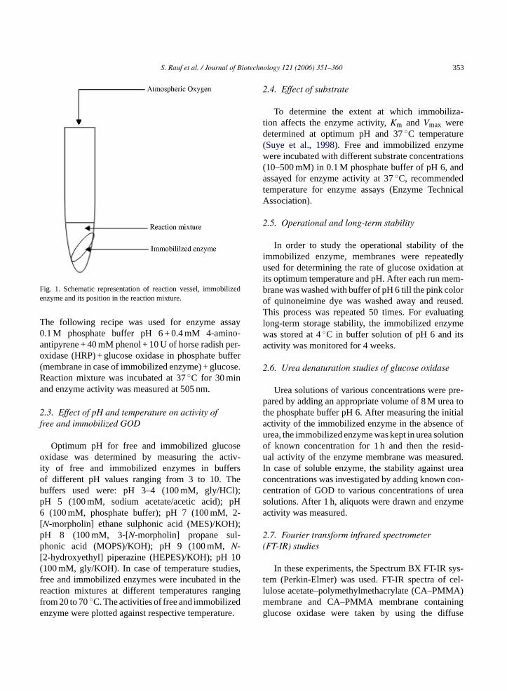
Fig. 1. Schematic representation of reaction vessel, immobilizedenzyme and its position in the reaction mixture.
The following recipe was used for enzyme assay0.1 M phosphate buffer pH 6 + 0.4 mM 4-amino-antipyrene + 40 mM phenol + 10 U of horse radish per-oxidase (HRP) + glucose oxidase in phosphate buffer(membrane in case of immobilized enzyme) + glucose.Reaction mixture was incubated at 37◦C for 30 minand enzyme activity was measured at 505 nm.
2.3. Effect of pH and temperature on activity offree and immobilized GOD
Optimum pH for free and immobilized glucoseoxidase was determined by measuring the activ-ity of free and immobilized enzymes in buffersof different pH values ranging from 3 to 10. Thebuffers used were: pH 3–4 (100 mM, gly/HCl);pH 5 (100 mM, sodium acetate/acetic acid); pH6 (100 mM, phosphate buffer); pH 7 (100 mM, 2-[N-morpholin] ethane sulphonic acid (MES)/KOH);pH 8 (100 mM, 3-[N-morpholin] propane sul-phonic acid (MOPS)/KOH); pH 9 (100 mM,N-[2-hydroxyethyl] piperazine (HEPES)/KOH); pH 10(100 mM, gly/KOH). In case of temperature studies,free and immobilized enzymes were incubated in thereaction mixtures at different temperatures rangingfrom 20 to 70◦C. The activities of free and immobilizedenzyme were plotted against respective temperature.
2.4. Effect of substrate
To determine the extent at which immobiliza-tion affects the enzyme activity,Km and Vmax weredetermined at optimum pH and 37◦C temperature(Suye et al., 1998). Free and immobilized enzymewere incubated with different substrate concentrations(10–500 mM) in 0.1 M phosphate buffer of pH 6, andassayed for enzyme activity at 37◦C, recommendedtemperature for enzyme assays (Enzyme TechnicalAssociation).
2.5. Operational and long-term stability
In order to study the operational stability of theimmobilized enzyme, membranes were repeatedlyused for determining the rate of glucose oxidation atits optimum temperature and pH. After each run mem-brane was washed with buffer of pH 6 till the pink colorof quinoneimine dye was washed away and reused.This process was repeated 50 times. For evaluatinglong-term storage stability, the immobilized enzymewas stored at 4◦C in buffer solution of pH 6 and itsactivity was monitored for 4 weeks.
2.6. Urea denaturation studies of glucose oxidase
Urea solutions of various concentrations were pre-pared by adding an appropriate volume of 8 M urea tothe phosphate buffer pH 6. After measuring the initiala ofu tiono sid-u red.I ureac con-c reas mea
2(
ys-t cel-l A)m ingg fuse
ctivity of the immobilized enzyme in the absencerea, the immobilized enzyme was kept in urea soluf known concentration for 1 h and then the real activity of the enzyme membrane was measu
n case of soluble enzyme, the stability againstoncentrations was investigated by adding knownentration of GOD to various concentrations of uolutions. After 1 h, aliquots were drawn and enzyctivity was measured.
.7. Fourier transform infrared spectrometerFT-IR) studies
In these experiments, the Spectrum BX FT-IR sem (Perkin-Elmer) was used. FT-IR spectra ofulose acetate–polymethylmethacrylate (CA–PMM
embrane and CA–PMMA membrane containlucose oxidase were taken by using the dif

354 S. Rauf et al. / Journal of Biotechnology 121 (2006) 351–360
reflectance accessory. The spectra were taken in therange of 4000–400 cm−1. The FT-IR spectra of cellu-lose acetate–polymethylmethacrylate membrane con-taining immobilized glucose oxidase after treatment at70◦C were also taken. In this experiment, membranewas treated at 70◦C for 15 min in phosphate buffer pH6, then washed with de-ionized water and dried beforetaking the spectra.
2.8. Scanning electron microscopy (SEM) studies
In these experiments, Scanning Electron Micro-scope JEOL Model #JSM 5910 Oxford Instrumentswas used. Scanning electron micrographs of theCA–PMMA membrane with and without glucose oxi-dase were taken after carbon coating because themembrane was not good conductor. Scanning electronmicrograph of the membrane containing glucose oxi-dase after treatment at 70◦C was also taken. In thisexperiment, membrane was treated at 70◦C for 15 minsin phosphate buffer pH 6, then washed with de-ionizedwater and dried. The membrane was subjected to car-bon coating and then electron micrograph was taken.
3. Results and discussion
Glucose oxidase was immobilized on celluloseacetate–polymethylmethacrylate (CA–PMMA) mem-b od.T ont neta Up ent-a ymea ases
(Sharma et al., 2003). Further increase in enzyme con-centration was found to decrease the percent activ-ity of enzyme as aggregation of enzyme moleculeson the membrane surface might cause the blockageof enzyme active sites. The activity of immobilizedenzyme was decreased after immobilization. Suchreduction is highly likely because glutaraldehyde beinga strong bifunctional reagent modifies the enzyme dras-tically leading to conformational changes and loss ofactivity (Broun, 1976). In spite of higherKm value thedeveloped material exhibited superiority in terms ofoperational stability, storage stability to other reportedmaterials (Isik et al., 2003; Yang et al., 2004).
3.1. Morphology of CA–PMMA and CA–PMMAcontaining GOD
The membrane morphology, structure and glucoseoxidase immobilization was characterized by using FT-IR and scanning electron microscopy (SEM). Manyauthors characterized their reported material using bothSEM and FT-IR (Yang et al., 2004; Ying et al., 2002;Jianghong et al., 1996; Godjevargova et al., 1999,2000). As former is among the most extensively usedtools for morphological characterization of materialsand later is molecular characterization technique thatprovides information about the chemical make up ofmaterials, therefore a combination of both provides atrue picture including not only the topographic featuresb st ofo reat-m mea
ne( us.T
TI
E bound
1122334
rane by using glutaraldehyde cross-linking methhe optimum enzyme concentration to be loaded
he membrane was found to be 288 U with 75%ctivity (Table 1). The drastic increase after 240erhaps reflects the better productive collision percge between the substrate and immobilized enzs the enzyme density on the membrane incre
able 1mmobilization of glucose oxidase on CA–PMMA
nzyme added,X (U) Enzyme not bound,Y (U) Enzyme
96 5 9144 10 13492 10 18240 10 23088 15 27336 20 31684 20 36432 25 407
ut also the chemical nature of materials. To the beur knowledge the use of these techniques after tent of the membrane containing immobilized enzyt high temperature is not reported yet.
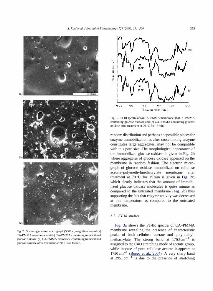
The electron micrograph of CA–PMMA membraFig. 2a) shows that the membrane is slightly porohe pore size ranges from 0.2 to 0.6�m appearing with
,A = X − Y Net activity,B (U) Percent activity,B/A × 100
20 2243 3273 4099 43
205 75205 65186 51167 41
S. Rauf et al. / Journal of Biotechnology 121 (2006) 351–360 355
Fig. 2. Scanning electron micrograph (5000×, magnification) of (a)CA-PMMA membrane and (b) CA-PMMA containing immobilizedglucose oxidase. (c) CA-PMMA membrane containing immobilizedglucose oxidase after treatment at 70◦C for 15 min.
Fig. 3. FT-IR spectra of (a) CA–PMMA membrane, (b) CA–PMMAcontaining glucose oxidase and (c) CA–PMMA containing glucoseoxidase after treatment at 70◦C for 15 min.
random distribution and perhaps not possible places forenzyme immobilization as after cross-linking enzymeconstitutes large aggregates, may not be compatiblewith this pore size. The morphological appearance ofthe immobilized glucose oxidase is given inFig. 2bwhere aggregates of glucose oxidase appeared on themembrane in random fashion. The electron micro-graph of glucose oxidase immobilized on celluloseacetate–polymethylmethacrylate membrane aftertreatment at 70◦C for 15 min is given inFig. 2c,which clearly indicates that the amount of immobi-lized glucose oxidase molecules is quite minute ascompared to the untreated membrane (Fig. 2b) thussupporting the fact that enzyme activity was decreasedat this temperature as compared to the untreatedmembrane.
3.2. FT-IR studies
Fig. 3a shows the FT-IR spectra of CA–PMMAmembrane revealing the presence of characteristicpeaks of both cellulose acetate and polymethyl-methacrylate. The strong band at 1763 cm−1 isassigned to the CO stretching mode of acetate group,while in case of pure cellulose acetate it appears at1750 cm−1 (Borgo et al., 2004). A very sharp bandat 2951 cm−1 is due to the presence of stretching

356 S. Rauf et al. / Journal of Biotechnology 121 (2006) 351–360
vibration of ester group of the polymethylmethacry-late (Balamurugan et al., 2004). The small peakat 1731 cm−1 indicated the presence of ester car-bonyl group stretching vibration of methylmethacry-late. When glucose oxidase was immobilized onCA–PMMA, only one small, sharp peak at 2372 cm−1
was observed (Fig. 3b). This experiment was repeatedthree times in order to confirm this peak. This peakwas assigned to R–N+ group with the help of soft-ware (HaveItAll IR, BIO-RAD) in the instrument.The detailed literature studies of GOD amino acidsequences (Frederick et al., 1990) and acid base charac-teristic titration curves profile of individual amino acidled to the conclusion that R–N+ peak is due to the proto-nated form (imidazolium) of histidine side chain. SinceGOD was immobilized at pH 6, at this pH both thebasic (imidazole) and acidic (imidazolium) forms of thehistidine residues were present in equilibrium (Devlin,1993). The presence of this peak confirms the immo-bilization of glucose oxidase on the membrane. Aftertreatment at 70◦C, the peak at 2372 cm−1 appearedagain, indicating that glucose oxidase remains bondedto the membrane (Fig. 3c).
3.3. Kinetic studies of free and immobilized GOD
The effect of immobilization on kinetic parameterswas studied by measuring the rates of glucose oxidasereaction by free and immobilized GOD at various con-centrations of glucose. The Lineweaver–Burk plot fort n inF mt vely.I as ac Thef 6;S
E
dd hei andp hec rriertA aseo ass
Fig. 4. Lineweaver–Burk plot for the native and immobilizedenzyme. (©) Free enzyme; (�) immobilized enzyme.
The values ofKm, Vmax and ‘EF’ for free andimmobilized GOD are shown inTable 2. Glucoseoxidase immobilized on CA–PMMA showed 2.4-foldincreases in the value ofKm as compared to the freeGOD. The same behavior has been described previ-ously when GOD was immobilized on silk fibroinmembrane (Jianghong et al., 1996). This higherKmvalue can be attributed to the diffusional resistance ofthe carrier against substrate and/or product (Karim andHashinaga, 2002) and less porous structure of the mem-brane.
3.4. Effect of pH and temperature on activity offree and immobilized GOD
The effect of pH on the activity of free and immo-bilized glucose oxidase is given inFig. 5. Althoughimmobilization usually alters the optimum pH ofenzyme (Yang et al., 2004; Godjevargova et al., 2000)but this was not observed in present case (optimumpH was 6 in both free and immobilized enzyme) asreported earlier (Isik et al., 2003; Ying et al., 2002). ThepH profile of free and immobilized enzyme is almostsame, depicting no change due to immobilization in
Table 2Kinetic parameters of the free and immobilized GOD
GOD Km (mM) Vmax (mM/min) Effectiveness factor
Free 17.42 1.34 1Immobilized 41.65 1.17 0.87
he free and immobilized glucose oxidase is giveig. 4. TheKm andVmax values were calculated fro
he slope and intercept of the straight lines, respectin addition, the effectiveness factor (EF) was usedomparison parameter for the immobilized system.ormula for ‘EF’ (Dahodwala and Humphrey, 197huler and Kargi, 2002) is given by
F = Vmax(immbilized enzyme)/Vmax(free enzyme)
If the value of ‘EF’≤ 1, no diffusion is assumeue to immobilization process. If ‘EF’ is >1, then t
mmobilization may have an effect on the substrateroduct diffusion. The ‘EF’ value is usually <1 in tase of immobilization system that provides a bao transport of substrate and product (Taqieddin andmiji, 2004). This value is also less than one in our cf immobilized system depicts that immobilization home effect on substrate and product diffusion.
S. Rauf et al. / Journal of Biotechnology 121 (2006) 351–360 357
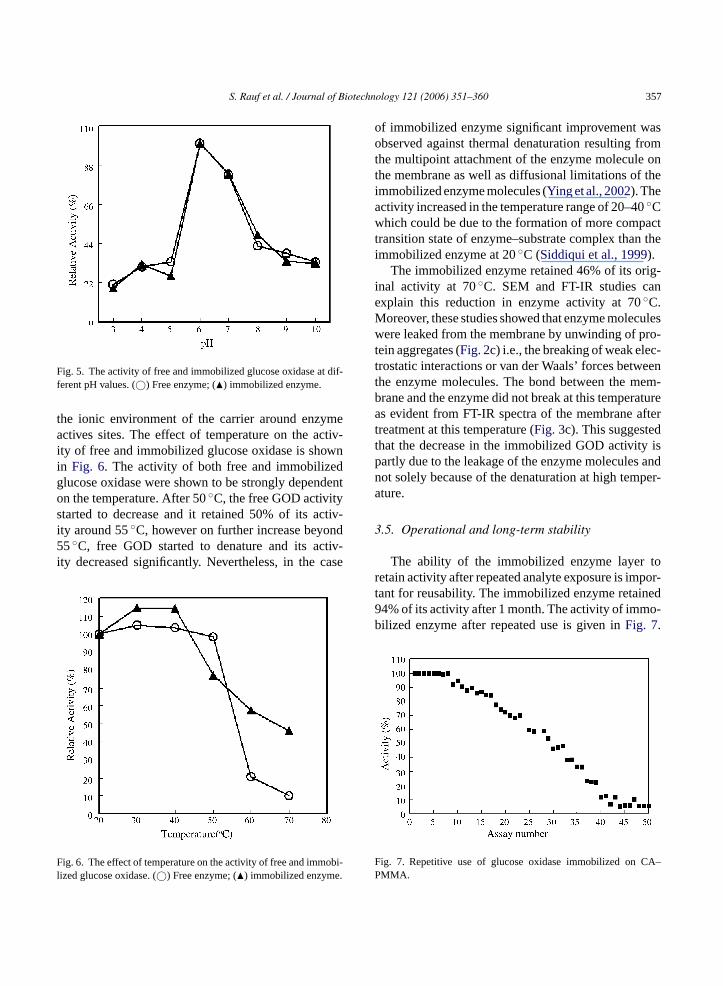
Fig. 5. The activity of free and immobilized glucose oxidase at dif-ferent pH values. (©) Free enzyme; (�) immobilized enzyme.
the ionic environment of the carrier around enzymeactives sites. The effect of temperature on the activ-ity of free and immobilized glucose oxidase is shownin Fig. 6. The activity of both free and immobilizedglucose oxidase were shown to be strongly dependenton the temperature. After 50◦C, the free GOD activitystarted to decrease and it retained 50% of its activ-ity around 55◦C, however on further increase beyond55◦C, free GOD started to denature and its activ-ity decreased significantly. Nevertheless, in the case
Fig. 6. The effect of temperature on the activity of free and immobi-lized glucose oxidase. (©) Free enzyme; (�) immobilized enzyme.
of immobilized enzyme significant improvement wasobserved against thermal denaturation resulting fromthe multipoint attachment of the enzyme molecule onthe membrane as well as diffusional limitations of theimmobilized enzyme molecules (Ying et al., 2002). Theactivity increased in the temperature range of 20–40◦Cwhich could be due to the formation of more compacttransition state of enzyme–substrate complex than theimmobilized enzyme at 20◦C (Siddiqui et al., 1999).
The immobilized enzyme retained 46% of its orig-inal activity at 70◦C. SEM and FT-IR studies canexplain this reduction in enzyme activity at 70◦C.Moreover, these studies showed that enzyme moleculeswere leaked from the membrane by unwinding of pro-tein aggregates (Fig. 2c) i.e., the breaking of weak elec-trostatic interactions or van der Waals’ forces betweenthe enzyme molecules. The bond between the mem-brane and the enzyme did not break at this temperatureas evident from FT-IR spectra of the membrane aftertreatment at this temperature (Fig. 3c). This suggestedthat the decrease in the immobilized GOD activity ispartly due to the leakage of the enzyme molecules andnot solely because of the denaturation at high temper-ature.
3.5. Operational and long-term stability
The ability of the immobilized enzyme layer toretain activity after repeated analyte exposure is impor-t ed9 o-b
F CA–P
ant for reusability. The immobilized enzyme retain4% of its activity after 1 month. The activity of immilized enzyme after repeated use is given inFig. 7.
ig. 7. Repetitive use of glucose oxidase immobilized onMMA.
358 S. Rauf et al. / Journal of Biotechnology 121 (2006) 351–360
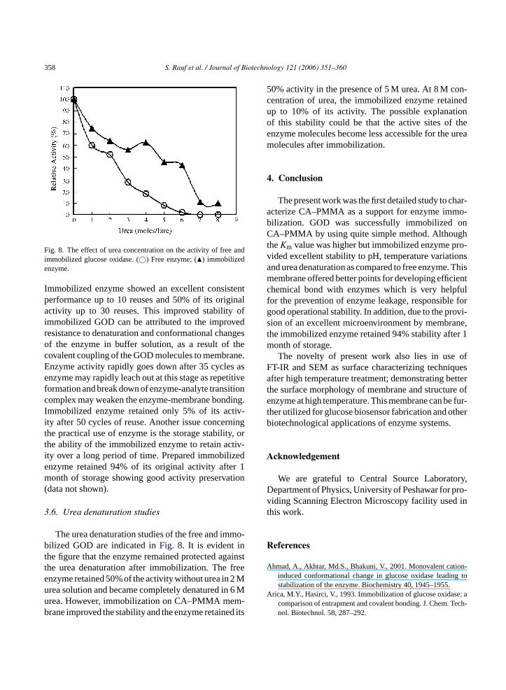
Fig. 8. The effect of urea concentration on the activity of free andimmobilized glucose oxidase. (©) Free enzyme; (�) immobilizedenzyme.
Immobilized enzyme showed an excellent consistentperformance up to 10 reuses and 50% of its originalactivity up to 30 reuses. This improved stability ofimmobilized GOD can be attributed to the improvedresistance to denaturation and conformational changesof the enzyme in buffer solution, as a result of thecovalent coupling of the GOD molecules to membrane.Enzyme activity rapidly goes down after 35 cycles asenzyme may rapidly leach out at this stage as repetitiveformation and break down of enzyme-analyte transitioncomplex may weaken the enzyme-membrane bonding.Immobilized enzyme retained only 5% of its activ-ity after 50 cycles of reuse. Another issue concerningthe practical use of enzyme is the storage stability, orthe ability of the immobilized enzyme to retain activ-ity over a long period of time. Prepared immobilizedenzyme retained 94% of its original activity after 1month of storage showing good activity preservation(data not shown).
3.6. Urea denaturation studies
The urea denaturation studies of the free and immo-bilized GOD are indicated inFig. 8. It is evident inthe figure that the enzyme remained protected againstthe urea denaturation after immobilization. The freeenzyme retained 50% of the activity without urea in 2 Murea solution and became completely denatured in 6 Murea. However, immobilization on CA–PMMA mem-brane improved the stability and the enzyme retained its
50% activity in the presence of 5 M urea. At 8 M con-centration of urea, the immobilized enzyme retainedup to 10% of its activity. The possible explanationof this stability could be that the active sites of theenzyme molecules become less accessible for the ureamolecules after immobilization.
4. Conclusion
The present work was the first detailed study to char-acterize CA–PMMA as a support for enzyme immo-bilization. GOD was successfully immobilized onCA–PMMA by using quite simple method. AlthoughtheKm value was higher but immobilized enzyme pro-vided excellent stability to pH, temperature variationsand urea denaturation as compared to free enzyme. Thismembrane offered better points for developing efficientchemical bond with enzymes which is very helpfulfor the prevention of enzyme leakage, responsible forgood operational stability. In addition, due to the provi-sion of an excellent microenvironment by membrane,the immobilized enzyme retained 94% stability after 1month of storage.
The novelty of present work also lies in use ofFT-IR and SEM as surface characterizing techniquesafter high temperature treatment; demonstrating betterthe surface morphology of membrane and structure ofenzyme at high temperature. This membrane can be fur-ther utilized for glucose biosensor fabrication and otherb
A
ory,D pro-v int
R
A on-g to
A : aTech-
iotechnological applications of enzyme systems.
cknowledgement
We are grateful to Central Source Laboratepartment of Physics, University of Peshawar foriding Scanning Electron Microscopy facility usedhis work.
eferences
hmad, A., Akhtar, Md.S., Bhakuni, V., 2001. Monovalent catiinduced conformational change in glucose oxidase leadinstabilization of the enzyme. Biochemistry 40, 1945–1955.
rica, M.Y., Hasirci, V., 1993. Immobilization of glucose oxidasecomparison of entrapment and covalent bonding. J. Chem.nol. Biotechnol. 58, 287–292.
S. Rauf et al. / Journal of Biotechnology 121 (2006) 351–360 359
Bailey, J.E., 1983. Immobilization of glucoamylase and glucose oxi-dase in activated carbon: effect of particle size and immobiliza-tion conditions on enzyme activity and effectiveness. Biotechnol.Bioeng. 15, 1923–1935.
Balamurugan, A., Kannan, S., Selvaraj, V., Rajeswari, S., 2004.Development and spectral characterization of polymethyl-methacrylate/hydroxyapatite composite for biomedical applica-tions. Trends Biomater. Artif. Organs. 18, 41–45.
Borgo, C.A., Lazarin, A.M., Kholin, Y.V., landers, R., Gushikem, Y.,2004. The ion exchange properties and equilibrium constants ofLi+, Na+ and K+ on zirconium phosphate highly dispersed ona cellulose acetate fibers surface. J. Braz. Chem. Soc. 15, 50–57.
Broun, G.B., 1976. In: Mosbach (Ed.), Methods in Enzymology, vol.44. Academic Press, New York, pp. 268–269.
Bulmus, V., Ayhan, H., Piskin, E., 1997. Modified PMMA monosizemicrobeads for glucose oxidase immobilization. Chem. Eng. J.65, 71–76.
Dahodwala, S.K., Humphrey, A.E., 1976. Pore diffusion model fora two substrate enzymatic reaction: application to galactose oxi-dase immobilized on porous glass particles. Biotechnol. Bioeng.18, 987–1000.
Devlin, T.M., 1993. Text Book of Biochemistry with Clinical Corre-latrions, 3rd ed. Wiley-Liss, Inc., New York, USA, p. 29.
Frederick, K.R., Tung, J., Emerick, R.S., Masiraz, F.R., Chamberlin,S.H., Vasavada, A., Rosenberg, S., 1990. Glucose oxidase fromAspergillus niger. J. Biol. Chem. 256, 3793–3802.
Godjevargova, T., Konsulov, V., Dimov, A., 1999. Prepara-tion of an ultrafiltration membrane from the copolymer ofacrylonitrile–glycidylmethacrylate utilized for immobilizationof glucose oxidase. J. Membrane Sci. 152, 235–240.
Godjevargova, T., Konsulov, V., Dimov, A., Vasileva, N., 2000.Behavior of glucose oxidase immobilized on ultrafiltrationmembranes obtained by copolymerizing acrylonitrile andN-vinylimidazol. J. Membrane Sci. 172, 279–285.
G s onnsor-
13,
G ellu-ZUS
H nto600,
I obi-tyl-39,
J D.,in itsepa-sen.
K ion14,
Karim, M.R., Hashinaga, F., 2002. Preparation and propertiesof immobilized pummelo limonoid glycosyl transferase. Proc.Biochem. 38, 809–814.
Liu, Y., Liu, H.Y., Qian, J.H., Deng, J.Q., Yu, T.Y., 1995. Immo-bilization of glucose oxidase in the regenerated silk fibroinmembrane—characterization of the membrane structure and itsapplication to an amperometric glucose sensor employing fer-rocene as electron shuttle. J. Chem. Technol. Biotechnol. 64,269–276.
Lott, J., Turner, K., 1975. Evaluation of Trinder’s glucose oxidasemethod for measuring glucose in serum and urine. Clin. Chem.21, 1754–1760.
Ramanathan, K., Pandey, S.S., Kumar, R., Gulati, A., Murthy,S.N., Malhotra, D.B., 2000. Covalent immobilization of glu-cose oxidase to poly(O-amino benzoic acid) for applica-tion to glucose biosensor. J. Appl. Polym. Sci. 78, 662–667.
Rohr, M., Kubicek, C.P., Kominek, J., 1983. In: Rehm, H.J., Reed,G. (Eds.), Biotechnology, vol. 3. Verlag Chemie, Weinheim, Ger-many, pp. 455–465.
Sarath Babu, V.R., Kumar, M.A., Karanth, N.G., Thakur, M.S., 2004.Stabilization of immobilized glucose oxidase against thermalinactivation by silanization for biosensor applications. Biosens.Bioelec. 19, 1337–1341.
Schmid, R.D., Karube, I., 1988. In: Rehm, H.J., Reed, G. (Eds.),Biotechnology, vol. 6b. Verlag Chemie, Weinheim, Germany, pp.317–365.
Sharma, S., Kaur, P., Jain, A., Rajeswari, M.R., Gupta, M.N., 2003.A smart bioconjugate of chymotrypsin. Biomacromolecules 4,330–336.
Shuler, M., Kargi, F., 2002. Bioprocess Engineering: Basic Concepts,2nd ed. Prentice-Hall, Upper Sadle River, NJ.
Siddiqui, K.S., Shemsi, A.H., Anwar, M.A., Rashid, M.H., Rajoka,M.I., 1999. Partial and complete alteration surface charges ofcarboxymethylcellulase by chemical modification: thermosta-
ob.
S er, T.,for
S osene.
S tion,glu-
T vel25,
T gci,yr-ro-
T gci,yr-ro-
ouda, M.D., Thakur, M., Karanth, N.G., 2001. Stability studieimmobilized glucose oxidase using an amperometric bioseeffect of protein based stabilizing agents. Electroanalysis849–855.
uoyong-Sheng, Jie, W., Xi-Jin, S., 2004. Characterization of close acetate micropore membrane immobilized acylase I. J5, 1608–1612.
ayashi, T., Ikada, Y., 1990. Lipoprotein lipase immobilization opolyacrolein microspheres. Biotechnol. Bioeng. 36, 593–http://www.enzymetechnicalassoc.org/dietary.html.
sik, S., Alkan, S., Toppare, L., Cianga, I., Yagci, Y., 2003. Immlization of invertase and glucose oxidase in poly 2-methylbu2-(3-thienyl) acetate/polypyrrole matrices. Eur. Polym. J.2375–2381.
ianghong, Q., Yongcheng, L., Haiying, L., Tongyin, Y., Jiaqi,1996. Characteristics of regenerated silk fibroin membraneapplication to the immobilization of glucose oxidase and prration of ap-benzoquinone mediating sensor for glucose. FreJ. Anal. Chem. 354, 173–178.
ang, I.K., Kown, B.K., Lee, J.H., Lee, B.H., 1993. Immobilizatof proteins on polymethylmethacrylate films. Biomaterials787–792.
bilization in water-miscible organic solvents. Enzyme MicrTechnol. 24, 599–608.
osnitza, P., Farooqui, M., Saleemuddin, M., Ulber, R., Schep1998. Application of reversible immobilization techniquesbiosensors. Anal. Chim. Acta 368, 197–203.
uye, S., Kumon, Y., Ishigaki, A., 1998. Immobilization of glucoxidase on poly-(l-lysine)-modified polycarbonate membraBiotechnol. Appl. Biochem. 27, 245–248.
zajani, B., Molnar, A., Klamar, G., Kalman, M., 1987. Preparacharacterization, and potential application of an immobilizedcose oxidase. Appl. Biochem. Biotechnol. 14, 37–47.
aqieddin, E., Amiji, M., 2004. Enzyme immobilization in noalginate-chitosan core shell microcapsules. Biomaterials1937–1945.
irkes, S., Toppare, L., Alkan, S., Bakir, U., Onen, A., YaY., 2002a. Immobilization of glucose oxidase in polyprole/polytetrahydrofuran graft copolymers. Int. J. Biol. Macmol. 30, 81–87.
irkes, S., Toppare, L., Alkan, S., Bakir, U., Onen, A., YaY., 2002b. Immobilization of glucose oxidase in polyprole/polytetrahydrofuran graft copolymers. Int. J. Biol. Macmol. 30, 81–87.
360 S. Rauf et al. / Journal of Biotechnology 121 (2006) 351–360
Turner, A.P.F., Karube, I., Wilson, G.S., 1987. Biosensors-Fundamentals and Applications. Oxford University Press,Oxford, UK.
Yang, Y.M., Wang, J.W., Tan, R.X., 2004. Immobilization of glu-cose oxidase on chitosan-SiO2 gel. Enzyme Microb. Technol.34, 126–131.
Yanga, Y., Yanga, H., Yanga, M., Liua, Y., Shena, G., Yua, R.,2004. Amperometric glucose biosensor based on a surface treatednanoporous ZrO2/chitosan composite film as immobilizationmatrix. Anal. Chim. Acta 525, 213–220.
Ying, L., Kang, E.T., Neoh, K.G., 2002. Covalent immobilization ofglucose oxidase on mocroporous membrane prepared from poly(vinylidene fluoride) with grafted poly (acrylic acid) side chains.J. Membrane Sci. 208, 361–374.
Zoldak, G., Zubrik, A., Musatov, A., Stupak, M., Sedlak, E.,2004. Irreversible thermal denaturation of glucose oxidasefrom Aspergillus niger is the transition to the denaturedstate with residual structure. J. Biol. Chem. 279, 47601–47609.

![Investigation of unique interactions between cellulose acetate and ionic liquid [EMIM]SCN, and their influences on hollow fiber ultrafiltration membranes](https://static.fdokumen.com/doc/165x107/631fd3cad85b325bc2095926/investigation-of-unique-interactions-between-cellulose-acetate-and-ionic-liquid.jpg)