Global gene expression of Poncirus trifoliata, Citrus sunki and their hybrids under infection of...
13
RESEARCH ARTICLE Open Access Global gene expression of Poncirus trifoliata, Citrus sunki and their hybrids under infection of Phytophthora parasitica Leonardo P Boava 1* , Mariângela Cristofani-Yaly 1 , Valéria S Mafra 1 , Karen Kubo 1 , Luciano T Kishi 1 , Marco A Takita 1 , Marcelo Ribeiro-Alves 2 , Marcos A Machado 1 Abstract Background: Gummosis and root rot caused by Phytophthora are among the most economically important diseases in citrus. Four F 1 resistant hybrids (Pool R), and four F 1 susceptible hybrids (Pool S) to P. parasitica, were selected from a cross between susceptible Citrus sunki and resistant Poncirus trifoliata cv. Rubidoux. We investigated gene expression in pools of four resistant and four susceptible hybrids in comparison with their parents 48 hours after P. parasitica inoculation. We proposed that genes differentially expressed between resistant and susceptible parents and between their resistant and susceptible hybrids provide promising candidates for identifying transcripts involved in disease resistance. A microarray containing 62,876 UniGene transcripts selected from the CitEST database and prepared by NimbleGen Systems was used for analyzing global gene expression 48 hours after infection with P. parasitica. Results: Three pairs of data comparisons (P. trifoliata/C. sunki, Pool R/C. sunki and Pool R/Pool S) were performed. With a filter of false-discovery rate less than 0.05 and fold change greater than 3.0, 21 UniGene transcripts common to the three pairwise comparative were found to be up-regulated, and 3 UniGene transcripts were down-regulated. Among them, our results indicated that the selected transcripts were probably involved in the whole process of plant defense responses to pathogen attack, including transcriptional regulation, signaling, activation of defense genes participating in HR, single dominant genes (R gene) such as TIR-NBS-LRR and RPS4 and switch of defense- related metabolism pathway. Differentially expressed genes were validated by RT-qPCR in susceptible and resistant plants and between inoculated and uninoculated control plants Conclusions: Twenty four UniGene transcripts were identified as candidate genes for Citrus response to P. parasitica. UniGene transcripts were likely to be involved in disease resistance, such as genes potentially involved in secondary metabolite synthesis, intracellular osmotic adjustment, signal transduction pathways of cell death, oxidative burst and defense gene expression. Furthermore, our microarray data suggest another type of resistance in Citrus-Phytophthora interaction conferred by single dominant genes (R gene) since we encountered two previously reported R genes (TIR-NBS-LRR and RPS4) upregulated in the resistant genotypes relative to susceptible. We identified 7 transcripts with homology in other plants but yet unclear functional characterization which are an interesting pool for further analyses and 3 transcripts where no significant similarity was found. This is the first microarray study addressing an evaluation of transcriptional changes in response to P. parasitica in Citrus. * Correspondence: [email protected] 1 Centro APTA Citros Sylvio Moreira, CP4, 13490-970, Cordeirópolis-SP, Brazil Full list of author information is available at the end of the article Boava et al. BMC Genomics 2011, 12:39 http://www.biomedcentral.com/1471-2164/12/39 © 2011 Boava et al; licensee BioMed Central Ltd. This is an Open Access article distributed under the terms of the Creative Commons Attribution License (http://creativecommons.org/licenses/by/2.0), which permits unrestricted use, distribution, and reproduction in any medium, provided the original work is properly cited.
-
Upload
independent -
Category
Documents
-
view
0 -
download
0
Transcript of Global gene expression of Poncirus trifoliata, Citrus sunki and their hybrids under infection of...
RESEARCH ARTICLE Open Access
Global gene expression of Poncirus trifoliata,Citrus sunki and their hybrids under infectionof Phytophthora parasiticaLeonardo P Boava1*, Mariângela Cristofani-Yaly1, Valéria S Mafra1, Karen Kubo1, Luciano T Kishi1, Marco A Takita1,Marcelo Ribeiro-Alves2, Marcos A Machado1
Abstract
Background: Gummosis and root rot caused by Phytophthora are among the most economically importantdiseases in citrus. Four F1 resistant hybrids (Pool R), and four F1 susceptible hybrids (Pool S) to P. parasitica, wereselected from a cross between susceptible Citrus sunki and resistant Poncirus trifoliata cv. Rubidoux. We investigatedgene expression in pools of four resistant and four susceptible hybrids in comparison with their parents 48 hoursafter P. parasitica inoculation. We proposed that genes differentially expressed between resistant and susceptibleparents and between their resistant and susceptible hybrids provide promising candidates for identifyingtranscripts involved in disease resistance. A microarray containing 62,876 UniGene transcripts selected from theCitEST database and prepared by NimbleGen Systems was used for analyzing global gene expression 48 hoursafter infection with P. parasitica.
Results: Three pairs of data comparisons (P. trifoliata/C. sunki, Pool R/C. sunki and Pool R/Pool S) were performed.With a filter of false-discovery rate less than 0.05 and fold change greater than 3.0, 21 UniGene transcripts commonto the three pairwise comparative were found to be up-regulated, and 3 UniGene transcripts were down-regulated.Among them, our results indicated that the selected transcripts were probably involved in the whole process ofplant defense responses to pathogen attack, including transcriptional regulation, signaling, activation of defensegenes participating in HR, single dominant genes (R gene) such as TIR-NBS-LRR and RPS4 and switch of defense-related metabolism pathway. Differentially expressed genes were validated by RT-qPCR in susceptible and resistantplants and between inoculated and uninoculated control plants
Conclusions: Twenty four UniGene transcripts were identified as candidate genes for Citrus response to P.parasitica. UniGene transcripts were likely to be involved in disease resistance, such as genes potentially involved insecondary metabolite synthesis, intracellular osmotic adjustment, signal transduction pathways of cell death,oxidative burst and defense gene expression. Furthermore, our microarray data suggest another type of resistancein Citrus-Phytophthora interaction conferred by single dominant genes (R gene) since we encountered twopreviously reported R genes (TIR-NBS-LRR and RPS4) upregulated in the resistant genotypes relative to susceptible.We identified 7 transcripts with homology in other plants but yet unclear functional characterization which are aninteresting pool for further analyses and 3 transcripts where no significant similarity was found. This is the firstmicroarray study addressing an evaluation of transcriptional changes in response to P. parasitica in Citrus.
* Correspondence: [email protected] APTA Citros Sylvio Moreira, CP4, 13490-970, Cordeirópolis-SP, BrazilFull list of author information is available at the end of the article
Boava et al. BMC Genomics 2011, 12:39http://www.biomedcentral.com/1471-2164/12/39
© 2011 Boava et al; licensee BioMed Central Ltd. This is an Open Access article distributed under the terms of the Creative CommonsAttribution License (http://creativecommons.org/licenses/by/2.0), which permits unrestricted use, distribution, and reproduction inany medium, provided the original work is properly cited.
BackgroundPhytophthora nicotianae Breda de Haan (Phytophthoraparasitica Dastur) and Phytophthora citrophthora(Smith & Smith) have caused severe damage in Citrusnurseries and orchards worldwide. In Brazil, P. parasi-tica is the predominant species associated with the dis-ease, found in more than 95% of groves and nurseries[1]. These pathogens infect the main scaffold branchesof the tree, inducing the formation of cankers with gumexudation and the expansion of the lesions upwardsaffects secondary branches, while downward expansionaffects the trunk and roots [2]. Infected trees usuallylack vigor and may die prematurely [3]. P. parasitica isan oomycete, belonging to the kingdom Stremenopiles,which comprises a diverse group of organisms that hasbeen consolidated as a result of analysis of mitochon-drial and ribosomal DNA sequences [4]. These patho-gens establish intimate relations with their hosts byforming haustoria during the infection, which are struc-tures used for obtaining nutrients from the plant, redir-ecting host metabolism and suppressing host defence inbiotrophy [5]. P. parasitica is considered a hemibio-troph, and therefore it first establishes itself in host tis-sues as a biotroph but then switches to a morenecrotrophic type of growth, rapidly invading and killinghost cells.Selection and breeding for resistance to Phytophthora
in citrus species is considered the most efficientapproach to control the disease, since there are varyingdegrees of resistance within the genera Citrus and itsrelatives. In this context, Poncirus trifoliata is an impor-tant genotype because of its agronomic valuable charac-teristics including resistance to Phytophthora, CitrusTristeza Virus (CTV) and citrus nematode (Tylenchulussemipenetrans) [6].In response to pathogen attack, a series of plant
defense responses leading to the hypersensitive reaction,cell wall modifications, production of reactive oxygenspecies (ROS), accumulation of phytoalexins, and synth-esis of pathogenesis related (PR) proteins may be acti-vated [7]. In general, the salicylic acid (SA)-dependentsignaling leads to expression of PR proteins, the produc-tion of ROS, and localized cell death. This leads to adefense that is effective against biotrophs because itrestricts pathogen growth via hypersensitive death of theinfected cells. The same reaction allows growth of anecrotrophic pathogen. Against necrotrophic pathogens,jasmonic acid (JA) and ethylene (ET)-dependentdefenses are successfully employed in plants activating adifferent set of PR proteins [8]. But there is evidence forextensive cross-talk between signaling pathways invol-ving antagonistic and synergistic interactions [9]. Inthese aspects, it is interesting to investigate the plantresponse mechanisms in the presence of hemibiotrophic
pathogens like P. parasitica, since they comprise bothlifestyles during their development.Biotechnology tools associated with conventional
citrus breeding programs have facilitated the develop-ment of cultivars with desirable characteristics [10].Microarray technology has been used to identify geneexpression changes in several responses to stress,enabling comparison of transcript levels for thousandsof genes simultaneously, such as response of sweetorange (Citrus sinensis) to ‘Candidatus Liberibacter asia-ticus’ infection [7] and [11]. In citrus, the first transcriptprofiling data was reported by Shimada et al., [12] whoconstructed a cDNA microarray to monitor expressionof mRNA during fruit development. Subsequently, sev-eral citrus DNA microarray platforms were developed,such as The Spanish Citrus Genomic Consortium. Affy-metrix developed and released a citrus GeneChip con-taining 960,444 total 25- mer oligos in an 11 micronformat (http://www.affymetrix.com/analysis/index.affx),based on the NCBI citrus EST collection [6].A new approach to study hybrid vigor at the molecu-
lar scale is to survey gene expression as a phenotype inF1 hybrids and their parental [13]. Genes in hybrids areinherited from the parents; thus, variation in regulationof the genes often leads to variation in the level of geneexpression in hybrids, which in turn may alter the phe-notype of these hybrids [14]. Due to the sensitivity ofmicroarrays, plant-to-plant variation of gene expressioncould be reduced by bulk harvesting of resistant andsusceptible hybrids. According to Wenger et al., [15]pooling segregants based on their phenotype allows theregion of the genome responsible for the phenotype tobe detected because DNA polymorphisms in regionsunlinked to the responsible locus will segregate ran-domly and be ‘’evened’’ out, while sequences or poly-morphisms either directly responsible for the trait, orvery closely linked to it, will be present in all positivesegregants and absent in all negative segregants. Accord-ing to Meng et al., [16] the majority of DNA microar-rays in use today are created from single genomes thatdo not reflect the genetic diversity of a heterogeneousgroup. One alternative approach is to incorporategenetic information from several species within a singlemicroarray slide. Mixed-DNA microarrays can be usedto quickly assess the distribution of genetic diversityacross multiple species.In previous studies, four resistant and four susceptible
F1 hybrids were selected from the population derivedfrom a cross between Citrus sunki Hort. ex. Tan. and P.trifoliata (L.) Raf cv. Rubidoux, which are respectively,susceptible and resistant to P. parasitica [17,18]. Weproposed that genes differentially expressed betweenresistant and susceptible parents and between theirresistant and susceptible hybrids provide promising
Boava et al. BMC Genomics 2011, 12:39http://www.biomedcentral.com/1471-2164/12/39
Page 2 of 13
candidates for identifying transcripts involved in diseaseresistance. In the present study, we investigated geneexpression in resistant and susceptible hybrids in com-parison with their parents 48 hours after P. parasiticainfection using a microarray chip containing 62,876 Uni-Gene transcripts selected from P. trifoliata, C. sinensisand C. reticulata libraries. Five differentially expressedgenes were validated by reverse transcription quantita-tive real-time PCR (RT-qPCR) in susceptible and resis-tant plants and used to detect differences in expressionbetween inoculated and uninoculated control plants.
MethodsPlant material and inoculationIn a previous study, four resistant and four susceptiblefive-year-old hybrids (F1 population of Citrus sunkiHort. ex. Tan. × Poncirus trifoliata (L.) Raf cv. Rubi-doux) were selected from 314 field-grown plants. Theselected hybrids were the most resistant and susceptibleto P. parasitica infection according to Boava [17] andSiviero et al. [18]. Three buds from each hybrid andparental line were collected and grafted onto 6-month-old Rangpur lime rootstocks. After six months, plantswere inoculated by a mycelia disc; a mycelial block wasplaced onto the center of a cut made in the stem andcovered with Parafilm. After 48 hours, leaves were har-vested individually, separately flash-frozen in liquidnitrogen, and then stored at 80°C prior to RNA isola-tion. The selection of this harvesting time was based inprevious studies on the time course of the type localand systemic defense followed by activation of manygenes involved in the interaction Citrus-Phytophthora,detailed by Teixeira [19]. All inoculated plants werekept in a greenhouse at an average temperature of 25°Cand 90% (± 0.5%) relative humidity until the finalevaluation, measurement of the lesion size, at 40 dayspostinoculation. Experimental design was completelyrandomized including three biological replicates for eachparent and three for each individual hybrid.In addition, in another experiment with compatible
plant-pathogen interactions, we used the P. trifoliatagenotypes to detect differences in gene expression levelbetween inoculated and uninoculated control plants.The plants for these experiments were grown andinoculated independently from microarray experiments.In this new experiment, we collected leaves of P. trifo-liata for RNA isolations at 48 hours after P. parasiticainoculation and uninoculated control plants Experimen-tal design was completely randomized including threebiological replicates.
Isolation of total RNA and sample labelingTotal RNA was isolated with RNeasy Plant Mini Kit (Qia-gen) and treated with RNase-free DNase (Qiagen)
according to the manufacturer’s instructions. The con-centration of total RNA was determined using a Nano-Drop ND-1000 spectrophotometer (NanoDropTechnologies). RNA integrity was verified using a Bioana-lyzer 1000 (Agilent). Following RNA isolation, the sampleswere pooled into resistant (Pool R) and susceptible hybrids(Pool S) to minimize variation between individual RNAsamples. Three biological replicates of each parent andeach resistant and susceptible pooled hybrids were usedfor hybridization with a cDNA microarray. RNA sampleswere sent to Roche NimbleGen Systems, where cDNAsynthesis and Cy3 labeling were performed. Equalamounts of total RNA for each sample were converted todouble-stranded cDNA using the SuperScript II cDNAConversion Kit (Invitrogen). Because this method uses anoligo (dT) primer, RNA strands lacking poly(A) tails arelikely to be underrepresented. Cy3 labeling, hybridizationand data acquisition were performed at the NimbleGenfacility following the manufacturer’s procedures.
Microarray data analysisA total of 62,876 UniGene transcripts (31,583 ofC. sinensis, 18,712 of C. reticulata and 12,581 of P. trifo-liata) selected from the CitEST database, assembledfrom the ESTs submitted to NCBI (GenBank accessionnumbers EY649559 to EY842485) were used to con-struct oligonucleotide microarray chips by Roche Nim-bleGen Systems using a multi-step approach to selectprobes with optimal predicted hybridization characteris-tics. Three probes were selected per UniGene, compris-ing a probe set, and each probe set is represented onthe final array by two replicates. All probes weredesigned as perfect match oligonucleotides. The experi-mental design and all microarray data have been depos-ited in the NCBI Gene Expression Omnibus (GEO,http://www.ncbi.nlm.nih.gov/geo accession numberGSE20412). Arrays were hybridized and processed byRoche NimbleGen Systems as previously described [20].For each resistant and susceptible hybrid pool and eachparent sample, hybridization was performed on indepen-dent microarrays. Arrays were scanned by Roche Nim-bleGen using a GenePix 4000B microarray scanner(Molecular Devices, Sunnyvale, CA) and the data wereextracted using NimbleScan software. For each probeset, an expression measure was calculated using therobust multiarray average (RMA) [21], consisting ofthree preprocessing steps: convolution background cor-rection, quantile normalization [22], and a summariza-tion based on a robust multiarray model fit using themedian polish algorithm. Probe set data with these nor-malized expression values, provided by Roche Nimble-Gen Systems in RMA calls files, were imported toArrayStar software 3.0 version (DNASTAR Inc., Madi-son, WI), where statistical analysis was performed.
Boava et al. BMC Genomics 2011, 12:39http://www.biomedcentral.com/1471-2164/12/39
Page 3 of 13
Three pairs of data comparisons that might reveal anassociation with the resistance of citrus to P. parasitica,were performed: (i) P. trifoliata Rubidoux versusC. sunki; (ii) resistant hybrids (Pool R) versus C. sunki;and (iii) resistant hybrids (Pool R) versus susceptiblehybrids (Pool S). For each comparison, a moderatedt-test value was calculated, and p-values were adjustedfor multiple comparisons by the false-discovery rate cor-rection [23]. Afterwards, UniGene transcripts that wereconsistently differentially expressed (greater than 3.0-fold up or less than 0.33-fold down; p-value ≤ 0.05)between replicate samples were identified as involved indisease resistance. In order to eliminate UniGene tran-scripts selected as a consequence of genotype differencesbut not directly related to disease resistance, Venn dia-grams were used to identify the intersection amongthese three sets of informative transcripts. These tran-scripts were then rechecked by BLASTX searchesagainst the GenBank database, and further classified intocategories according to the Munich Information Centerfor Protein Sequences classification system (MIPS;http://www.helmholtz-muenchen.de/mips).
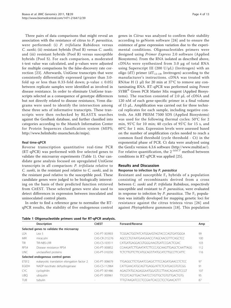
Real time-qPCRReverse transcription quantitative real-time PCR(RT-qPCR) was performed with five selected genes tovalidate the microarray experiments (Table 1). Our can-didate gene analysis focused on upregulated UniGenetranscripts in all comparison: P. trifoliata relative toC. sunki, in the resistant pool relative to C. sunki, and inthe resistant pool relative to the susceptible pool. Thesecandidate genes were judged to be biologically interest-ing on the basis of their predicted function retrievedfrom CitEST. These selected genes were also used todetect differences in expression between inoculated anduninoculated control plants.In order to find a reference gene to normalize the RT-
qPCR results, the stability of five endogenous control
genes in Citrus was analyzed to confirm their stabilityaccording to geNorm software [24] and to ensure theexistence of gene expression variation due to the experi-mental conditions. Oligonucleotides primers weredesigned using Primer Express 2.0 software (AppliedBiosystems). From the RNA isolated as described above,cDNAs were synthesized from 3.0 μg of total RNAusing Superscript III (200 U/μL) (Invitrogen) with anoligo (dT) primer (dT12-18, Invitrogen) according to themanufacturer’s instructions. cDNA was treated withRNAse H (1 μl) for 20 min at 37°C to remove any con-taminating RNA. RT-qPCR was performed using PowerSYBR® Green PCR Master Mix reagent (Applied Biosys-tems). The reaction consisted of 2.0 μL of cDNA and120 nM of each gene-specific primer in a final volumeof 15 μL. Amplification was carried out for three techni-cal replicates for each sample, including negative con-trols. An ABI PRISM 7500 SDS (Applied Biosystems)was used for the following thermal cycles: 50°C for 2min, 95°C for 10 min; 40 cycles of 95°C for 15 s, and60°C for 1 min. Expression levels were assessed basedon the number of amplification cycles needed to reach acommon fixed threshold (cycle threshold - Ct) in theexponential phase of PCR. Ct data were analyzed usingthe GenEx version 4.3.6 software (http://www.multid.se/).For relative quantification, the 2-ΔΔCT method betweenconditions in RT-qPCR was applied [25].
Results and DiscussionResponse to infection by P. parasiticaResistant and susceptible F1 hybrids of a populationconsisting of recombinants derived from a crossbetween C. sunki and P. trifoliata Rubidoux, respectivelysusceptible and resistant to P. parasitica, were evaluatedin response to infection by P. parasitica. The F1 popula-tion was initially developed for mapping genetic loci forresistance against the citrus tristeza virus [26] andagainst Phytophthora gummosis [18]. This population
Table 1 Oligonucleotide primers used for RT-qPCR analysis.
Description CitEST Forward/Reverse Amp
Selected genes to validate the microarray
LEA Lea 5 CAS-PT-303903 TCGGACTGGTATCATGGA/GTAGTACCCAGTGATGGGA 99
MIR miraculin CAS-CR-215276 AGCCCTGTAATGAAGAACC/TAGCAACGTTTCAGCTCC 100
TIR TIR-NBS-LRR CAS-CS-103511 CATGATGAGGACGTGGG/AAGTGATCCGACTCGAC 103
RPS4 Disease resistance RPS4 CAS-PT-300852 CCAAGATCTTGAATATCTTCCC/GCAAGTTGAGCTCAATTAGG 112
UNC unclassified proteins CAS-PT-310250 TCTCTTGTTCTTCATGCAGT/TATGCATCTTGCCTTCATTC 116
Selected endogenous control genes
ETEF2 eukaryotic translation elongation factor 2 CAS-PT-306679 TTGAGGCTTCTGAATCGAG/CTTTCCAGATGAACCTCTCC 97
EGIDH NADP-isocitrate dehydrogenase CAS-CS-112964 CATTGAACATGCAGTTGAGG/ATTCTCATGACGTGTCGG 91
CYC cyclophilin CAS-PT-301486 AGAGTATGCAGAGGAATGG/GTCCTTAACAGAAGTCCGT 107
UBQ ubiquitin CAS-PT-300961 TTCGTCAGTTGACTAATCCT/GTTGCTGTGTTGACTGTG 95
TUB tubulin - TTTGTAAGATCCCTCCGA/TCACCCTCCTGAACATTT 87
Boava et al. BMC Genomics 2011, 12:39http://www.biomedcentral.com/1471-2164/12/39
Page 4 of 13
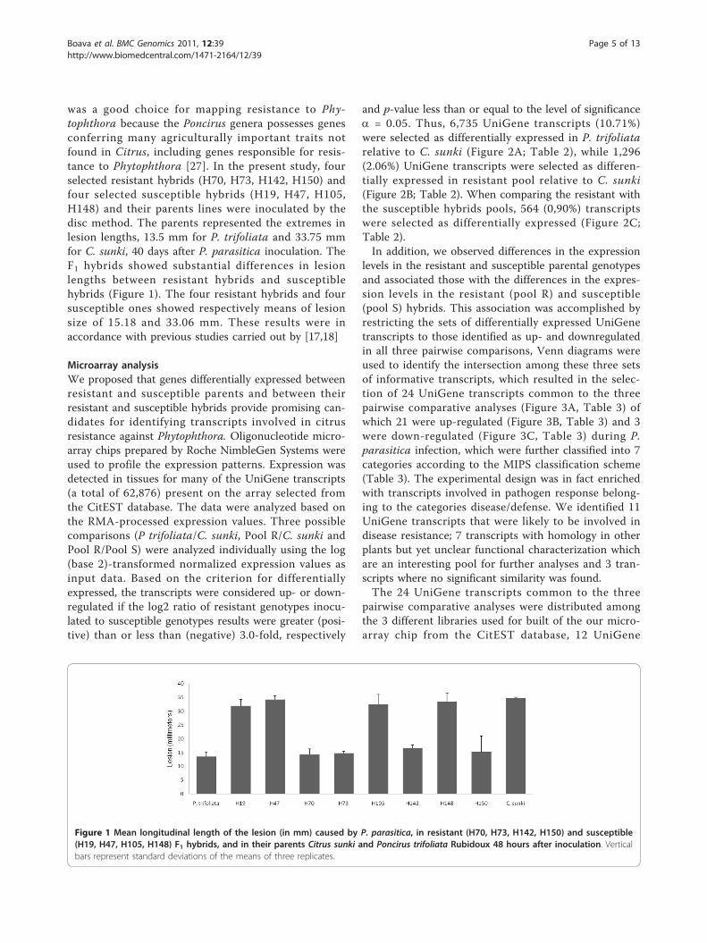
was a good choice for mapping resistance to Phy-tophthora because the Poncirus genera possesses genesconferring many agriculturally important traits notfound in Citrus, including genes responsible for resis-tance to Phytophthora [27]. In the present study, fourselected resistant hybrids (H70, H73, H142, H150) andfour selected susceptible hybrids (H19, H47, H105,H148) and their parents lines were inoculated by thedisc method. The parents represented the extremes inlesion lengths, 13.5 mm for P. trifoliata and 33.75 mmfor C. sunki, 40 days after P. parasitica inoculation. TheF1 hybrids showed substantial differences in lesionlengths between resistant hybrids and susceptiblehybrids (Figure 1). The four resistant hybrids and foursusceptible ones showed respectively means of lesionsize of 15.18 and 33.06 mm. These results were inaccordance with previous studies carried out by [17,18]
Microarray analysisWe proposed that genes differentially expressed betweenresistant and susceptible parents and between theirresistant and susceptible hybrids provide promising can-didates for identifying transcripts involved in citrusresistance against Phytophthora. Oligonucleotide micro-array chips prepared by Roche NimbleGen Systems wereused to profile the expression patterns. Expression wasdetected in tissues for many of the UniGene transcripts(a total of 62,876) present on the array selected fromthe CitEST database. The data were analyzed based onthe RMA-processed expression values. Three possiblecomparisons (P trifoliata/C. sunki, Pool R/C. sunki andPool R/Pool S) were analyzed individually using the log(base 2)-transformed normalized expression values asinput data. Based on the criterion for differentiallyexpressed, the transcripts were considered up- or down-regulated if the log2 ratio of resistant genotypes inocu-lated to susceptible genotypes results were greater (posi-tive) than or less than (negative) 3.0-fold, respectively
and p-value less than or equal to the level of significancea = 0.05. Thus, 6,735 UniGene transcripts (10.71%)were selected as differentially expressed in P. trifoliatarelative to C. sunki (Figure 2A; Table 2), while 1,296(2.06%) UniGene transcripts were selected as differen-tially expressed in resistant pool relative to C. sunki(Figure 2B; Table 2). When comparing the resistant withthe susceptible hybrids pools, 564 (0,90%) transcriptswere selected as differentially expressed (Figure 2C;Table 2).In addition, we observed differences in the expression
levels in the resistant and susceptible parental genotypesand associated those with the differences in the expres-sion levels in the resistant (pool R) and susceptible(pool S) hybrids. This association was accomplished byrestricting the sets of differentially expressed UniGenetranscripts to those identified as up- and downregulatedin all three pairwise comparisons, Venn diagrams wereused to identify the intersection among these three setsof informative transcripts, which resulted in the selec-tion of 24 UniGene transcripts common to the threepairwise comparative analyses (Figure 3A, Table 3) ofwhich 21 were up-regulated (Figure 3B, Table 3) and 3were down-regulated (Figure 3C, Table 3) during P.parasitica infection, which were further classified into 7categories according to the MIPS classification scheme(Table 3). The experimental design was in fact enrichedwith transcripts involved in pathogen response belong-ing to the categories disease/defense. We identified 11UniGene transcripts that were likely to be involved indisease resistance; 7 transcripts with homology in otherplants but yet unclear functional characterization whichare an interesting pool for further analyses and 3 tran-scripts where no significant similarity was found.The 24 UniGene transcripts common to the three
pairwise comparative analyses were distributed amongthe 3 different libraries used for built of the our micro-array chip from the CitEST database, 12 UniGene
Figure 1 Mean longitudinal length of the lesion (in mm) caused by P. parasitica, in resistant (H70, H73, H142, H150) and susceptible(H19, H47, H105, H148) F1 hybrids, and in their parents Citrus sunki and Poncirus trifoliata Rubidoux 48 hours after inoculation. Verticalbars represent standard deviations of the means of three replicates.
Boava et al. BMC Genomics 2011, 12:39http://www.biomedcentral.com/1471-2164/12/39
Page 5 of 13

transcripts were identified in the UniGene set of C.sinensis, 10 in P. trifoliata, and 2 in C. reticulate.According to Wan et al. [28], the use of mixed-DNAmicroarrays is advantageous because much more infor-mation is incorporated into the analysis. In the presentwork, there were 18,712 UniGene transcripts fromP. trifoliata libraries of the CitEST database and wedecided to incorporate UniGene sequences fromC. sinensis and C. reticulata to be represented in thearray and consequently enrich the analyses. The SpanishCitrus Genomic Consortium also developed a mixed-DNA microarrays composed of 24,000- element cDNAarray containing 20,000 unigenes, based on nearly90,000 high-quality sequences generated from 52 differ-ent cDNA libraries [6].
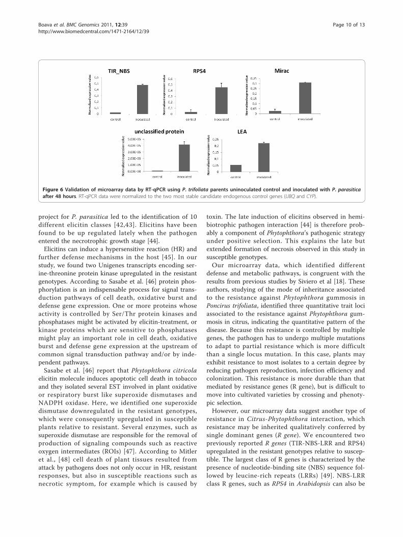
Real time-qPCR validationRT-qPCR is currently the most sensitive method tocompare gene expression at both low and high levels.To avoid distortions, RT-qPCR requires a referencegene (or a few genes) with the most stable expression
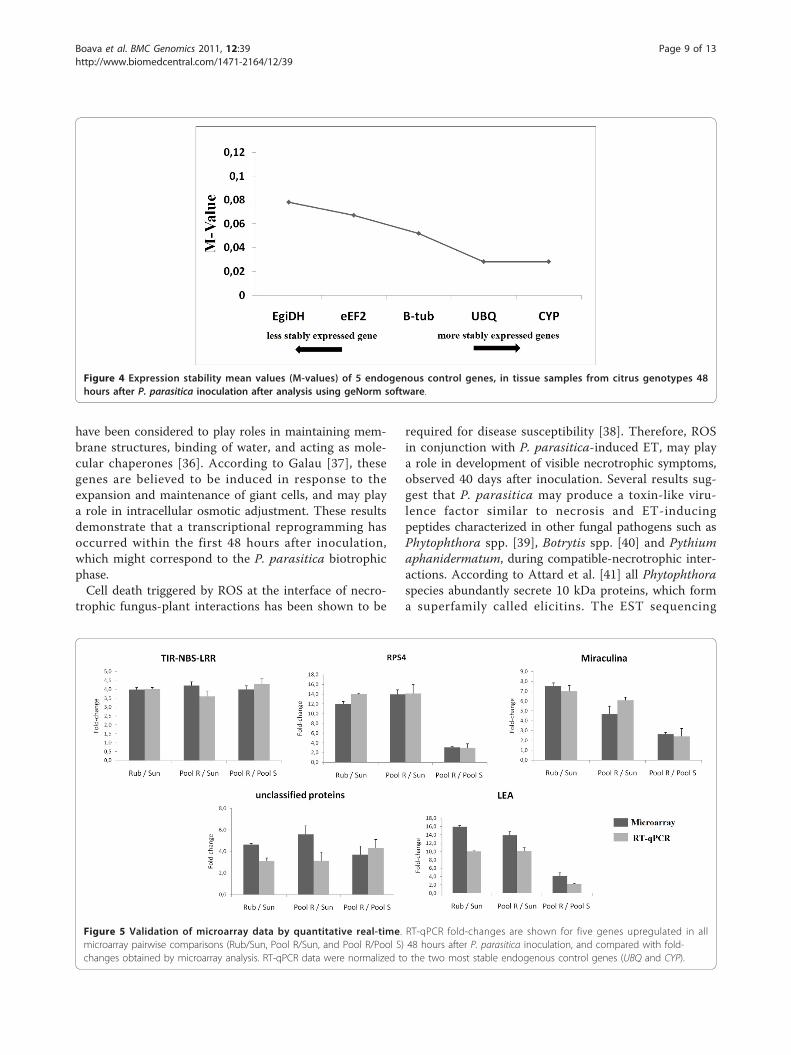
over the experiment, i.e., a gene presenting expressionlevels that is little influenced by the experimental orenvironmental conditions, to correct for sample-to-sam-ple variation in RT-qPCR efficiency and errors in samplequantification. Several reference genes tested presentedvaried expression due to the experimental conditions indifferent genotypes exposed to biotic and abiotic stimuli[29]. To support the choice of the best reference genes,i.e., the most stable genes following pathogen inocula-tion in the different genotypes, the GeNorm algorithm[24], implemented in GenEx version 4.3.6. software(http://www.multid.se/) was used. In the present work,among the five reference genes tested (Table 1), ubiqui-tin (UBQ) and cyclophilin (CYP) had the lowest expres-sion stability mean values (M-value = 0.0257), i.e., thesegenes had more stable expression than other evaluatedgenes (Figure 4), and the data were normalized by thenormalization factor calculated using these most stablereference genes.Five UniGene transcripts that were differentially
expressed in the microarray analysis were selected forRT-qPCR. Our candidate gene analysis focused on upre-gulated genes in the resistant genotypes that werejudged to be biologically interesting on the basis of theirpredicted function retrieved from CitEST. We selectedgenes that are likely involved in disease resistance, suchas one gene potentially involved in secondary metabolitesynthesis and protein activity regulation (miraculina)and genes potentially involved in cell rescue, defenseand virulence (TIR-NBS-LRR and RPS4) and one genehomolog in other plant but yet unclear functional char-acterization which are an interesting for further analyses.Comparison of the results from RT-qPCR with those
Figure 2 Analysis of differential gene expression in P. trifoliata ’Rubidoux’, C. sunki, resistant (Pool R) and susceptible (Pool S) hybrids.Signal correlation plots were used to examine comparisons between: (A) The average signal derived from the three biological replicates ofresistant parent (Rubidoux) (Y-axis) and susceptible parent (Sunki) (X-axis) graphed on a logarithmic (base 2) scale. Note the prevalence of genesshowing distinct expression patterns in the two genotypes; (B) A similar graph was made to compare expression in the resistant hybrids pool(Pool R) (Y-axis) and the susceptible parent (Sunki) (X-axis); (C) A similar graph of expression data of the resistant hybrids pool (Y-axis) plottedagainst the expression data of the susceptible hybrids pool (X-axis).
Table 2 Comparisons between different genotypesinoculated with P. parasitica (P. trifoliata/C. sunki, Pool R/C. sunki, and Pool R/Pool S)
Comparison
Total Up Down
P. trifoliata/C. sunki 6,735 3,392 3,343
Pool R/C. sunki 1,296 870 426
Pool R/Pool S 564 205 359
The comparisons were analyzed individually. Genes greater (positive) than orless than (negative) 3.0-fold, respectively and p-value less than or equal to thelevel of significance a = 0.05 were considered differentially expressed.
Boava et al. BMC Genomics 2011, 12:39http://www.biomedcentral.com/1471-2164/12/39
Page 6 of 13

from microarray analyses revealed roughly similar pat-terns or tendencies of expression in the three compara-tive pairwise analyses (Figure 5). Notably, using highstringency in the analysis of the microarray, data showedonly twenty one genes were upregulated in commonamong the three comparative pairwise analyses. Thevalidation with RT-qPCR showed that all the selectedgenes were in fact upregulated to similar levels in allcomparison (≥ 3.0-fold, p-value ≤ 0.05). All fivetranscripts were upregulated in P. trifoliata relative toC. sunki, in the resistant pool relative to C. sunki, and inthe resistant pool relative to the susceptible pool. Theseresults revealed similar patterns of expression with thosefrom microarray analyses.In addition, besides of estimating and confirm the fold
change at each comparative pairwise analyses on micro-array, we include in our RT-qPCR analysis, five selectedgenes in inoculated and uninoculated plants of P. trifo-liata. (Figure 6). Thus, this confirmation offers anopportunity to determine transcription patterns of thesystemic response of Citrus to infection with the patho-gen P. parasitica providing information on differentdefense and metabolic pathways. With this analysis thedifferences in gene expression probably are involved inthe response to infection to P. parasitica and maybesome of them can be related to the resistance mechan-ism and the changes found are not only due to inherent
constitutive gene expression differences among thegenotypes”.
Functional analyses of differentially-expressed genesUpon recognition of P. parasitica by its host Citrus, aseries of signaling pathways are switched on, which leadto the metabolic reprogramming of the host plant. Phy-tophthora species are usually biotrophic or hemibio-trophic pathogens that keep host cells alive (at leastinitially) to enable nutrient uptake from the plant cells[30]. A more extensive cell death response could causeopposite effects on the outcome of biotrophic versusnecrotrophic plant-pathogen interactions. Hemibio-trophic pathogens adopt a two-step infection style.According to Kanneganti et al. [31], during the phase ofinfection that follows penetration of host tissue, theyrequire living cells, much like biotrophic pathogens. Incontrast, in a later phase of the disease, they causeextensive necrosis of host tissue, resulting in profusecolonization and sporulation. This infection cycle sug-gests that host cell death may impact the disease differ-ently depending on its timing.The time-point of infection was selected for the isola-
tion of defense-related transcripts because, in compati-ble interactions, transcripts involved in pathogenresponse were observed at 48 hours (e.g., [19]) and thisis reflected by the low number of transcripts of
Figure 3 Venn diagrams showing the differentially expressed UniGene transcripts in three different comparisons between resistant(Pool R) and susceptible (Pool S) F1 hybrids and their parents, Citrus sunki (Sunki) and Poncirus trifoliata Rubidoux (Rub), respectivelysusceptible and resistant to P. parasitica, 48 hours after inoculation. Transcripts profile greater (positive) than or less than (negative) 3.0-fold, respectively and p-value less than or equal to the level of significance a = 0.05 were considered differentially expressed. A) differentiallyexpressed UniGene; B) upregulated; C) downregulated
Boava et al. BMC Genomics 2011, 12:39http://www.biomedcentral.com/1471-2164/12/39
Page 7 of 13
secondary metabolism isolated in this study. Accordingto Grenville-Briggs and van West [32]Phytophthora spe-cies are hemibiotrophs being biotrophic for the initialstage of up to 36 h after inoculation. Molecular studiescarried out with hemibiotrophic pathogens infectingplants helped the identification of several putative genesthat are expressed at the stage biotrophy and areinvolved in membrane or cell wall biosynthesis, aminoacid metabolism, osmoregulation, phosphorylation, pro-tein secretion and energy consumption [33].
In our study, genes potentially involved in secondarymetabolite synthesis were identified, including leu-coanthocyanidin dioxygenase, which is the key enzymeleading to the synthesis of anthocyanins. Anthocyaninsare known to be potent agents acting against oxidativestress [34]. The exact role of anthocyanins in defense isnot clear but it could be that these compounds neutra-lize damaging reactive oxygen species (ROS) [35]. A lateembryogenesis-abundant LEA5 protein was also foundupregulated in the resistant genotypes. LEA proteins
Table 3 Differentially expressed UniGene transcripts in three different comparisons
ID CITEST GENE_INFO accessionnumber
Categories (MIPS) Rub × Sun Pool R × Sunk Pool R ×Pool S
Upregulated Fold P_Value Fold P_Value Fold P_Value
CAS-PT-300852 Disease resistance proteinRPS4
BAB11393.1 cell rescue, defense andvirulence
4,0 0,01 5,0 0,00 3,0 0,00
CAS-CS-103511 TIR-NBS-LRR resistanceprotein
XP_002325496.1 cell rescue, defense andvirulence
4,0 0,02 4,2 0,02 4,0 0,01
CAS-PT-303903 Lea5 protein Q39644.1 cell rescue, defense andvirulence
30,9 0,00 22,0 0,00 4,1 0,01
CAS-PT-301521 FNR2 (ferredoxin-NADP(+) NP_001077566.1 cell type localisation 13,5 0,02 13,2 0,03 3,3 0,02
CAS-CS-115011 Leucoanthocyanidindioxygenase
XP_002528475.1 metabolism 5,3 0,03 6,4 0,04 3,2 0,01
CAS-CS-103575 Miraculin-like protein 2 ACL78790.1 protein activity regulation 43,1 0,01 25,1 0,02 10,1 0,01
CAS-CR-209520 AL07-2p ACJ03067.1 protein fate (folding, mod.,destination)
87,5 0,00 106,4 0,02 3,7 0,00
CAS-CS-119563 Serine-threonine proteinkinase
XP_002514954.1 protein fate (folding, mod.,destination)
86,1 0,01 162,3 0,02 7,9 0,00
CAS-CS-122102 Serine-threonine proteinkinase
XP_002512394.1 protein fate (folding, mod.,destination)
25,5 0,01 50,8 0,04 4,6 0,02
CAS-CS-128948 Transducin family protein ABS32230.1 protein with binding function 6,8 0,04 4,0 0,02 3,1 0,05
CAS-CS-118778 Synaptobrevin-relatedfamily
XP_002304221.1 subcellular localisation 5,6 0,02 5,6 0,04 4,4 0,05
CAS-PT-303418 Putative DNA bindingprotein
ABO93454.1 subcellular localisation 10,7 0,00 9,9 0,02 3,1 0,03
CAS-PT-305376 Gag-pol polyprotein AAR13298.1 transposable elements 17,2 0,04 3,7 0,02 3,3 0,02
CAS-PT-309741 Hypothetical protein XP_002609958.1 unclassified proteins 18,7 0,01 12,0 0,05 3,3 0,01
CAS-PT-309581 Hypothetical protein XP_001018346.1 unclassified proteins 24,9 0,00 27,1 0,03 7,0 0,01
CAS-CS-114114 Hypothetical protein XP_002280912.1 unclassified proteins 19,6 0,02 29,4 0,04 3,1 0,01
CAS-PT-310125 Hypothetical protein XP_002275595.1 unclassified proteins 13,1 0,01 28,4 0,01 5,4 0,01
CAS-CS-105219 Hypothetical protein XP_002590734.1 unclassified proteins 6,6 0,03 6,2 0,02 3,1 0,02
CAS-PT-305253 No significant similarityfound
unclassified proteins 25,5 0,01 6,2 0,05 3,2 0,01
CAS-CS-127334 No significant similarityfound
unclassified proteins 10,2 0,00 6,2 0,04 3,6 0,00
CAS-PT-301031 No significant similarityfound
unclassified proteins 70,6 0,01 37,6 0,01 3,4 0,01
Downregulated
CAS-CS-101776 superoxide dismutase CAA03881.1 function or cofactorrequirement
4,0 0,02 4,2 0,02 4,0 0,01
CAS-CS-114557 predicted protein XP_002324594.1 unclassified proteins 30,9 0,00 22,0 0,00 4,1 0,01
CAS-CR-203746 predicted protein XP_002331132.1 unclassified proteins 13,5 0,02 13,2 0,03 3,3 0,02
P. trifoliata Rubidoux versus C. sunki; resistant hybrids (Pool R) versus C. sunki; and resistant hybrids (Pool R) versus susceptible hybrids (Pool S), 48 hours after P.Parasitica inoculation.
Boava et al. BMC Genomics 2011, 12:39http://www.biomedcentral.com/1471-2164/12/39
Page 8 of 13
have been considered to play roles in maintaining mem-brane structures, binding of water, and acting as mole-cular chaperones [36]. According to Galau [37], thesegenes are believed to be induced in response to theexpansion and maintenance of giant cells, and may playa role in intracellular osmotic adjustment. These resultsdemonstrate that a transcriptional reprogramming hasoccurred within the first 48 hours after inoculation,which might correspond to the P. parasitica biotrophicphase.Cell death triggered by ROS at the interface of necro-
trophic fungus-plant interactions has been shown to be
required for disease susceptibility [38]. Therefore, ROSin conjunction with P. parasitica-induced ET, may playa role in development of visible necrotrophic symptoms,observed 40 days after inoculation. Several results sug-gest that P. parasitica may produce a toxin-like viru-lence factor similar to necrosis and ET-inducingpeptides characterized in other fungal pathogens such asPhytophthora spp. [39], Botrytis spp. [40] and Pythiumaphanidermatum, during compatible-necrotrophic inter-actions. According to Attard et al. [41] all Phytophthoraspecies abundantly secrete 10 kDa proteins, which forma superfamily called elicitins. The EST sequencing
Figure 4 Expression stability mean values (M-values) of 5 endogenous control genes, in tissue samples from citrus genotypes 48hours after P. parasitica inoculation after analysis using geNorm software.
Figure 5 Validation of microarray data by quantitative real-time. RT-qPCR fold-changes are shown for five genes upregulated in allmicroarray pairwise comparisons (Rub/Sun, Pool R/Sun, and Pool R/Pool S) 48 hours after P. parasitica inoculation, and compared with fold-changes obtained by microarray analysis. RT-qPCR data were normalized to the two most stable endogenous control genes (UBQ and CYP).
Boava et al. BMC Genomics 2011, 12:39http://www.biomedcentral.com/1471-2164/12/39
Page 9 of 13
project for P. parasitica led to the identification of 10different elicitin classes [42,43]. Elicitins have beenfound to be up regulated lately when the pathogenentered the necrotrophic growth stage [44].Elicitins can induce a hypersensitive reaction (HR) and
further defense mechanisms in the host [45]. In ourstudy, we found two Unigenes transcripts encoding ser-ine-threonine protein kinase upregulated in the resistantgenotypes. According to Sasabe et al. [46] protein phos-phorylation is an indispensable process for signal trans-duction pathways of cell death, oxidative burst anddefense gene expression. One or more proteins whoseactivity is controlled by Ser/Thr protein kinases andphosphatases might be activated by elicitin-treatment, orkinase proteins which are sensitive to phosphatasesmight play an important role in cell death, oxidativeburst and defense gene expression at the upstream ofcommon signal transduction pathway and/or by inde-pendent pathways.Sasabe et al. [46] report that Phytophthora citricola
elicitin molecule induces apoptotic cell death in tobaccoand they isolated several EST involved in plant oxidativeor respiratory burst like superoxide dismutases andNADPH oxidase. Here, we identified one superoxidedismutase downregulated in the resistant genotypes,which were consequently upregulated in susceptibleplants relative to resistant. Several enzymes, such assuperoxide dismutase are responsible for the removal ofproduction of signaling compounds such as reactiveoxygen intermediates (ROIs) [47]. According to Mitleret al., [48] cell death of plant tissues resulted fromattack by pathogens does not only occur in HR, resistantresponses, but also in susceptible reactions such asnecrotic symptom, for example which is caused by
toxin. The late induction of elicitins observed in hemi-biotrophic pathogen interaction [44] is therefore prob-ably a component of Phytophthora’s pathogenic strategyunder positive selection. This explains the late butextended formation of necrosis observed in this study insusceptible genotypes.Our microarray data, which identified different
defense and metabolic pathways, is congruent with theresults from previous studies by Siviero et al [18]. Theseauthors, studying of the mode of inheritance associatedto the resistance against Phytophthora gummosis inPoncirus trifoliata, identified three quantitative trait lociassociated to the resistance against Phytophthora gum-mosis in citrus, indicating the quantitative pattern of thedisease. Because this resistance is controlled by multiplegenes, the pathogen has to undergo multiple mutationsto adapt to partial resistance which is more difficultthan a single locus mutation. In this case, plants mayexhibit resistance to most isolates to a certain degree byreducing pathogen reproduction, infection efficiency andcolonization. This resistance is more durable than thatmediated by resistance genes (R gene), but is difficult tomove into cultivated varieties by crossing and phenoty-pic selection.However, our microarray data suggest another type of
resistance in Citrus-Phytophthora interaction, whichresistance may be inherited qualitatively conferred bysingle dominant genes (R gene). We encountered twopreviously reported R genes (TIR-NBS-LRR and RPS4)upregulated in the resistant genotypes relative to suscep-tible. The largest class of R genes is characterized by thepresence of nucleotide-binding site (NBS) sequence fol-lowed by leucine-rich repeats (LRRs) [49]. NBS-LRRclass R genes, such as RPS4 in Arabidopsis can also be
Figure 6 Validation of microarray data by RT-qPCR using P. trifoliata parents uninoculated control and inoculated with P. parasiticaafter 48 hours. RT-qPCR data were normalized to the two most stable candidate endogenous control genes (UBQ and CYP).
Boava et al. BMC Genomics 2011, 12:39http://www.biomedcentral.com/1471-2164/12/39
Page 10 of 13
characterized by the presence of a leading sequencehomologous to the TIR (Toll-Interleukin-1 receptor)that is responsible for cytoplasmic signaling in animals[49]. Previous data suggest that the TIR domain of RPS4is important for induction of cell death when RPS4 istransiently expressed in tobacco leaves [50]. NBS-LRR-mediated resistance has been identified against numer-ous types of biotrophic or hemibiotrophic pathogens,including fungi, oomycetes, viruses and bacteria, andthese types of resistance genes have been identifiedacross a wide range of plants [51].Qualitative resistance is mediated by R genes that lead
to a race-specific hypersensitive response. These R genesonly provide short-lived resistance in the field as newvirulent races of the pathogen rapidly overcome theresistance encoded by single race-specific resistancegenes [52]. In contrast, quantitative resistance is con-trolled by many interacting genes that do not preventinfection, but slow down the development of the patho-gen at individual infection sites on the plant, and hence,lasts longer [53].Although the presence of R gene does not contradict the
statement that the Poncirus genera used in our experimentspossess quantitative pattern of resistance to P. parasitica ,these genes showed strong induction with an alteration ofmore than 4-fold in resistant genotypes relative to suscepti-ble (Table 3). This finding further support the previouspoint of view that the HR was one of the main defenseresponses. R genes are also thought to encode specificreceptors that recognize elicitors and initiate signal trans-duction cascades resulting in the HR [54]. Although amajor feature of the HR is a rapid and local cell death,many defense-related genes that are not involved in celldeath are also activated and may play a more importantrole in resistance to pathogen. Therefore, it is furtherinferred that the HR in resistant genotypes may result froma broad-spectrum recognition of the pathogen by the pro-duct of unknown R gene(s) or R gene analogues, or that asame or similar defense system, especially the downstreamsignaling components such as kinases and other defensegenes, may exist specific resistances.
ConclusionThis is the first global gene expression study in Citrus ×Phytophthora gummosis interaction. We identified 24 Phy-tophthora gummosis responsive transcripts. The func-tional classification of these genes and their expressionprofile at 48 h after inoculation provide useful informationon resistance of citrus to Phytophthora. The selected tran-scripts were validated by RT-qPCR in susceptible andresistant plants and between inoculated and uninoculatedcontrol plants. Our results indicated that the selected tran-scripts were really upregulated in the resistant genotypesand probably involved in the whole process of plant
defense responses to pathogen attack, including transcrip-tional regulation, signaling, activation of defense genesparticipating in HR, such as TIR-NBS-LRR and RPS4,switch of defense-related metabolism pathway.As we have a linkage map for P. trifoliata and three
QTL for Phytophthora gummosis resistance localized onthis map, the integration of genomics and genetic map-ping using genetic genomics approaches will providenew insights into resistance to this disease and helpwith the development of improved disease managementstrategies. The genes that we have identified as upregu-lated across the resistant genotypes will be valuable forongoing work in eQTL mapping.
AcknowledgementsThe present study was conducted with the financial support of Fundação deAmparo a Pesquisa do estado de São Paulo (FAPESP) (Process n. 2007/08435-5) and Instituto Nacional de Ciência e Tecnologia (INCT) de Genômicapara Melhoramento de Citros (Process n. 573848/2008-4) which provided ascholarship to LPB (process n. 150721/2009-9) from the Conselho Nacionalde Pesquisa de Desenvolvimento (CNPq). The authors thank Marta Tanrikuluof http://ScienceDocs.com for editing of an earlier version of this manuscript.
Author details1Centro APTA Citros Sylvio Moreira, CP4, 13490-970, Cordeirópolis-SP, Brazil.2Centro de Desenvolvimento Tecnológico em Saúde, Fundação OswaldoCruz. Av. Brasil, 4365 - 21040-900 - Rio de Janeiro-RJ, Brasil.
Authors’ contributionsMCY and MAM planned and supervised the study. LPB, MCY, contributed tothe design and execution of the experiments detailed. LPB and MCY carriedout plant growth and inoculation. LPB, VSM and KK participated inextraction of plant RNA. LPB and MRA performed microarray analysis. LPB,VSM and KK contributed to the primer design and RT-qPCR validation. KTLperformed annotations of the UniGene transcripts. LPB and MCY drafted themanuscript. MCY, MAT and MAM provided intellectual input. MAM, MRA,VSM, KK and MAT revised the manuscript. All authors read and approved thefinal manuscript.
Received: 11 August 2010 Accepted: 17 January 2011Published: 17 January 2011
References1. Medina Filho HP, Bordignon R, Siqueira W, Feichtenberger E, Carvalho MRT:
Tolerância de híbridos e de clones de porta-enxertos de citros àinfecção de raízes por Phytophthora nicotianae. Fitopatologia Brasileira2004, 29:169-178.
2. Alvarez LA, Gramaje D, Abad-Campos P, García JJ: Seasonal susceptibilityof citrus scions to Phytophthora citrophthora and P. nicotianae and theinfluence of environmental and host-linked factors on infectiondevelopment. Eur J Plant Pathol 2009, 124:621-635.
3. Queiroz BPV, Melo IS: Antagonism of Serratia marcescens towardsPhytophthora parasitica and its effects in promoting the growth ofcitrus. Braz J Microbiol 2006, 37:448-450.
4. Alexopoulos CJ, Mims CW, Blackwell M: Phylum Oomycota. In IntroductoryMycology. Edited by: Alexopoulos CJ, Mims CW, Blackwell M. John Wiley1996:683-737.
5. O’Connell RJ, Panstruga R: Tete a tete inside a plant cell. Establishingcompatibility between plants and biotrophic fungi and oomycetes. NewPhytologist 2006, 171:699-718.
6. Talon M, Gmitter FG Jr: Citrus genomics. Int J Pl Genomics 2008, Article ID528361, 17 pages.
7. Albrecht U, Bowman KD: Gene expression in Citrus sinensis (L.) Osbeckfollowing infection with the bacterial pathogen CandidatusLiberibacter asiaticus causing Huanglongbing in Florida. Plant Sci 2008,175:291-306.
Boava et al. BMC Genomics 2011, 12:39http://www.biomedcentral.com/1471-2164/12/39
Page 11 of 13
8. Cordelier S, de Ruffray P, Fritig B, Kauffmann S: Biological and molecularcomparison between localized and systemic acquired resistanceinduced in tobacco by a Phytophthora megasperma glycoprotein elicitin.Plant Mol Biol 2003, 51:109-118.
9. De Vos M, Van Oosten VR, Van Poecke RM, Van Pelt JA, Pozo MJ,Mueller MJ, Buchala AJ, Me’traux JP, Van Loon LC, Dicke M, Pieterse CM:Signal signature and transcriptome.
10. Mourão Filho FAA, Pio R, Mendes BMJ, Azevedo FA, Schinor EH,Entelmann FA, Alves ASR, Cantuarias-Avilés TE: Evaluation of citrus somatichybrids for tolerance to Phytophthora nicotianae and citrus tristeza virus.Sci Hortic 2008, 115:301-308.
11. Kim JS, Sagaram US, Burns JK, Li JL, Wang N: Response of sweet orange(Citrus sinensis) to ‘Candidatus Liberibacter asiaticus’ infection:microscopy and microarray analyses. Phytopathology 2009, 99:50-57.
12. Shimada T, Fujii H, Endo T, Yazaki J, Kishimoto N, Shimbo K, Kikuchi S,Omura M: Toward comprehensive expression profiling by microarrayanalysis in Citrus: monitoring the expression profiles of 2213 genesduring fruit development. Plant Sci 2005, 168:1383-1385.
13. Holland JB: Genetic architecture of complex traits in plants. Curr OpinPlant Biol 2007, 10:156-161.
14. Doebley J, Lukens L: Transcriptional regulators and the evolution of plantform. Plant Cell 1998, 10:1075-1082.
15. Wenger JW, Schwartz K, Sherlock G: Bulk Segregant Analysis by High-Through. put Sequencing Reveals a Novel Xylose Utilization Gene fromSaccharomyces cerevisiae. PLoS Genet 2010, 6(5):e1000942.
16. Meng D, Broschat SL: Call DR: A Java-based tool for the design ofclassification microarrays. BMC Bioinformatics 2008, 9:328.
17. Boava LP: Estabilidade de QTLs ligados à resistência dos citros a gomose,causada por Phytophthora parasitica. PhD thesis Universidade EstadualPaulista; 2004.
18. Siviero A, Cristofani M, Furtado EL, Garcia AAF, Coelho ASG, Machado MA:Identification of QTLs associated with citrus resistance to Phytophthoragummosis. J Appl Genet 2006, 47:23-28.
19. Teixeira JEC: Genes de defesa de Citrus sunki e Poncirus trifoliata:expressão constitutiva e induzida por Phytophthora parasítica. PhD thesisUniversidade Federal de Lavras; 2005.
20. Ulijasz AT, Andes DR, Glasner JD, Weisblum B: Regulation of iron transportin Streptococcus pneumoniae by RitR, an orphan response regulator.J Bacteriol 2004, 186:8123-8136.
21. Irizarry RA, Hobbs B, Collin F, Beazer-Barclay YD, Antonellis KJ, Scherf U,Speed TP: Exploration, normalization, and summaries of high densityoligonucleotide array probe level data. Biostatistics 2003, 4:249-264.
22. Bolstad BM, Irizarry RA, Astrand M, Speed TP: A comparison ofnormalization methods for high density oligonucleotide array databased on variance and bias. Bioinformatics 2003, 19:185-193.
23. Benjamini Y, Hochberg Y: Controlling the false discovery rate: a practicaland powerful approach to multiple testing. J Roy Stat Soc B Met 1995,57:289-300.
24. Vandesompele J, De Preter K, Pattyn F: Accurate normalisation of real-time quantitative RT-PCR data by geometric averaging of multipleinternal control genes. Genome Biol 2002, 3:0034.1-0034.
25. Livak KJ, Schmittgen TD: Analysis of relative gene expression data usingreal-time quantitative PCR and the 2-ΔΔCT method. Methods 2001,25:402-408.
26. Cristofani M, Machado MA, Grattapaglia D: Genetic linkage maps of Citrussunki Hort. ex.Tan. and Poncirus trifoliata (L.) Raf. and mapping of citrustristeza virus resistance gene. Euphytica 1999, 109:25-32.
27. Chen C, Bowman KD, Choi Ya, Dang PM, Rao MN, Huang S, Soneji JR,McCollum TG, Gmitter FG: EST-SSR genetic maps for Citrus sinensis andPoncirus trifoliate. Tree Genet Genomes 2008, 4:1-10.
28. Wan Y, Broschat SL, Call DR: Validation of mixed-genome microarrays as amethod for genetic discrimination. Appl Environ Microbiol 2007,73:1425-1432.
29. Boava LP, Laia ML, Jacob TR, Dabbas KM, Gonçalves JF, Ferro JA, Ferro MI,Furtado EL: Selection of endogenous genes for gene expression studiesin Eucalyptus under biotic (Puccinia psidii) and abiotic (acibenzolar-S-methyl) stresses using RT-qPCR. BMC Res Notes 2010, 24:3:43.
30. Takemoto D, Hardham AR, Jones DA: Differences in cell death inductionby Phytophthora elicitins are determined by signal componentsdownstream of MAP Kinase kinase in different species of Nicotiana and
cultivars of Brassica rapa and Raphanus sativus. Plant Physiol 2005,138:1491-1504.
31. Kanneganti TD, Huitema E, Cakir C, Kamoun S: Synergistic interaction ofthe plant cell death pathway induced by Phytophthora infestans Nep1-like protein piNPP1.1 and INF1 elicitin. Mol Plant Microbe Interact 2006,19:854-863.
32. Grenville-Briggs LJ, van West P: The biotrophic stages of oomycete-plantinteractions. Advances in Applied. Microbiology 2005, 57:217-243.
33. Tör M: Tapping into molecular conversation between oomycete plantpathogens and their hosts. Eur J Plant Pathol 2008, 122:57-69.
34. Stintzing FC, Stintzing AS, Carle R, Frei B, Wrolstad RE: Color andantioxidant properties of cyanidin-based anthocyanin pigments. J AgricFood Chem 2002, 50:6172-6181.
35. Calla B, Vuong T, Radwan O, Hartman GL, Clough SJ: Gene ExpressionProfiling Soybean Stem Tissue Early Response to Sclerotinia sclerotiorumand in Silico Mapping in Relation to Resistance Markers. The PlantGenome 2009, 2:149-166.
36. Wise M, Tunnaciliffe A: POPP the question: What do LEA proteins do?Trends Plant Sci 2004, 9:13-17.
37. Galau GA, Wang HYC, Hughes DW: Cotton Lea5 and Lea14 encodeatypical late embryogenesis-abundant proteins. Plant Physiol 1993,101:695-696.
38. Okubara PA, Paulitz TC: Root defenses to fungal pathogens: A molecularprospective. Plant Soil 2005, 274:215-226.
39. Qutob D, Kemmerling B, Brunner F, Küfner I, Engelhardt S, Gust AA,Luberacki B, Seitz HU, Stahl D, Rauhut T, Glawischnig E, Schween G,Lacombe B, Watanabe N, Lam E, Schlichting R, Scheel D, Nau K, Dodt G,Hubert D, Gijzen M, Nürnberger T: Phytotoxicity and innate immuneresponses induced by Nep1-like proteins. Plant Cell 2006, 18:3721-3744.
40. Staats M, van Baarlen P, Schouten A, van Kan JA, Bakker FT: Positiveselection in phytotoxic protein-encoding genes of Botrytis species. FungalGenet Bio 2007, 44:52-63.
41. Attard A, Gourgues M, Galiana E, Panabières F, Ponchet M, et al: Strategiesof attack and defense in plant-oomycete interactions, accentuated forPhytophthora parasitica Dastur (syn. P. Nicotianae Breda de Haan).J Plant Physiol 2008, 165:83-94.
42. Panabieres F, Amselem J, Galiana E, Le Berre JY: Gene identification in theoomycete pathogen Phytophthora parasitica during in vitro vegetativegrowth through expressed sequence tags. Fungal Genet Biol 2005,42:611-23.
43. Le Berre JY, Engler G, Panabieres F: Exploration of the late stages of thetomato-Phytophthora parasitica interactions through histological analysisand generation of expressed sequence tags. New Phytol 2008,177(2):480-492.
44. Moy P, Qutob D, Chapman BP, Atkinson I, Gijzen M: Patterns of geneexpression upon infection of soybean plants by Phytophthora sojae. MolPlant Microbe Interact 2004, 17:1051-1062.
45. Kamoun S: A catalogue of the effector secretome of plant pathogenicoomycetes. Annu Rev Phytopathol 2006, 44:41-60.
46. Sasabe M, Takeuchi K, Kamoun S, Ichinose Y, Govers F, Toyoda K, Shiraishi T,Yamada T: Independent pathways leading to apoptotic cell death,oxidative burst and defense gene expression in response to elicitin intobacco cell suspension culture. Eur J Biochem 2000, 267:5005-5013.
47. Möller IM: Plant mitochondria and oxidative stress: electron transport,NADPH turnover, and metabolism of reactive oxygen species. Annu RevPlant Physiol Plant Mol Biol 2001, 52:561-59.
48. Mittler R, Shulaev V, Seskar M, Lam E: Inhibition of programmed cell deathin tobacco plants during a pathogen-induced hypersensitive response atlow oxygen pressure. Plant Cell 1996, 8:1991-2001.
49. Hammond-Kosack KE, Jones JDG: Plant disease resistance genes. Annu RevPlant Physiol Plant Mol Biol 1997, 48:575-607.
50. Zhang Y, Dorey S, Swiderski M, Jones JDG: Expression of RPS4 in tobaccoinduces an AvrRps4-independent HR that requires EDS1, SGT1 andHSP90. Plant J 2004, 40:213-224.
51. Jones JDG, Dangl JL: The plant immune system. Nature 2006, 444:323-329.52. Colon LT, Turkensteen LJ, Prummel W, Budding DJ, Hoogendoorn J:
Durable resistance to late blight (Phytophthora infestans) in old potatocultivars. Eur J Plant Pathol 1995, 101:387-397.
53. Bormann CA, Rickert AM, Ruiz RAC, Paal J, Strahwald J, Buhr K, Gebhardt C:Tagging quantitative trait loci formaturity-corrected late blight
Boava et al. BMC Genomics 2011, 12:39http://www.biomedcentral.com/1471-2164/12/39
Page 12 of 13
resistance in tetraploid potato with PCR-based candidate gene markers.Mol Plant Microbe Interact 2004, 17:1126-1138.
54. Baker B, Zambryski P, Staskawicz B, Dineshkumar SP: Signaling in plantmicrobe interactions. Science 1997, 276:726-733.
doi:10.1186/1471-2164-12-39Cite this article as: Boava et al.: Global gene expression of Poncirustrifoliata, Citrus sunki and their hybrids under infection of Phytophthoraparasitica. BMC Genomics 2011 12:39.
Submit your next manuscript to BioMed Centraland take full advantage of:
• Convenient online submission
• Thorough peer review
• No space constraints or color figure charges
• Immediate publication on acceptance
• Inclusion in PubMed, CAS, Scopus and Google Scholar
• Research which is freely available for redistribution
Submit your manuscript at www.biomedcentral.com/submit
Boava et al. BMC Genomics 2011, 12:39http://www.biomedcentral.com/1471-2164/12/39
Page 13 of 13