Global Changes in the Rat Heart Proteome Induced by Prolonged Morphine Treatment and Withdrawal
13
Global Changes in the Rat Heart Proteome Induced by Prolonged Morphine Treatment and Withdrawal Zdenka Drastichova 1 , Jitka Skrabalova 1 , Petr Jedelsky 2 , Jan Neckar 3 , Frantisek Kolar 3 , Jiri Novotny 1 * 1 Department of Physiology, Faculty of Science, Charles University in Prague, Prague, Czech Republic, 2 Department of Cell Biology, Faculty of Science, Charles University in Prague, Prague, Czech Republic, 3 Department of Developmental Cardiology, Institute of Physiology, Academy of Sciences of the Czech Republic, Prague, Czech Republic Abstract Morphine belongs among the most commonly used opioids in medical practice due to its strong analgesic effects. However, sustained administration of morphine leads to the development of tolerance and dependence and may cause long-lasting alterations in nervous tissue. Although proteomic approaches enabled to reveal changes in multiple gene expression in the brain as a consequence of morphine treatment, there is lack of information about the effect of this drug on heart tissue. Here we studied the effect of 10-day morphine exposure and subsequent drug withdrawal (3 or 6 days) on the rat heart proteome. Using the iTRAQ technique, we identified 541 proteins in the cytosol, 595 proteins in the plasma membrane-enriched fraction and 538 proteins in the mitochondria-enriched fraction derived from the left ventricles. Altogether, the expression levels of 237 proteins were altered by morphine treatment or withdrawal. The majority of changes (58 proteins) occurred in the cytosol after a 3-day abstinence period. Significant alterations were found in the expression of heat shock proteins (HSP27, a-B crystallin, HSP70, HSP10 and HSP60), whose levels were markedly up- regulated after morphine treatment or withdrawal. Besides that morphine exposure up-regulated MAPK p38 (isoform CRA_b) which is a well-known up-stream mediator of phosphorylation and activation of HSP27 and a-B crystallin. Whereas there were no alterations in the levels of proteins involved in oxidative stress, several changes were determined in the levels of pro- and anti-apoptotic proteins. These data provide a complex view on quantitative changes in the cardiac proteome induced by morphine treatment or withdrawal and demonstrate great sensitivity of this organ to morphine. Citation: Drastichova Z, Skrabalova J, Jedelsky P, Neckar J, Kolar F, et al. (2012) Global Changes in the Rat Heart Proteome Induced by Prolonged Morphine Treatment and Withdrawal. PLoS ONE 7(10): e47167. doi:10.1371/journal.pone.0047167 Editor: Xin-Liang Ma, Thomas Jefferson University, United States of America Received August 6, 2012; Accepted September 10, 2012; Published October 9, 2012 Copyright: ß 2012 Drastichova et al. This is an open-access article distributed under the terms of the Creative Commons Attribution License, which permits unrestricted use, distribution, and reproduction in any medium, provided the original author and source are credited. Funding: This work was supported by the Grant Agency of the Academy of Sciences of the Czech Republic (IAA501110901), the Charles University Grant Agency (429511), the Czech Ministry of Education, Youth and Sport (MSM0021620858) and by the grant SVV-2012-265206. The funders had no role in study design, data collection and analysis, decision to publish, or preparation of the manuscript. Competing Interests: The authors have declared that no competing interests exist. * E-mail: [email protected] Introduction Morphine is one of the most effective drugs known for pain- relieving effects and it has been successfully used in medical practice for a long time as a powerful analgesic to treat many kinds of chronic pain [1–3]. Morphine exerts its physiological effects through opioid receptors (ORs), which belong to the large family of G protein-coupled receptors (GPCRs) [4]. Because ORs are mainly expressed in the central nervous system [5–6], and morphine is a potentially highly addictive substance [7–9], a great deal of attention has been paid to studying the impact of morphine and other opioids on nervous tissue. Chronic administration of morphine was found to induce changes in OR-mediated signaling, which may underlie the development of opioid tolerance and dependence [10–11]. Importantly, neuronal changes induced by opioids have been observed to persist for a long time following cessation of drug exposure [12]. A number of studies indicated that morphine affects the expression of genes involved in processes as diverse as cell signaling [13–16], transcription [17–18], apoptotis [19–20] and cytoskeleton assembly [21]. Nowadays, there is increasing evidence to show that morphine can alter protein expression in different brain areas, even following a single dose. Numerous proteomic studies further expanded the list of brain proteins potentially altered by morphine [22–31]. Protein changes induced by morphine treatment have also been observed in in vitro experiments on the human neuroblastoma SH-SY5Y cell line, CHO (Chinese hamster ovary) epithelial cells [32–33] and primary cortical astrocytes [34]. Whereas the pharmacology and function of opioids in the central nervous system have been quite extensively characterized, the actions of these drugs in peripheral tissues are relatively less understood. It has been well established that morphine may exert significant effects on cardiovascular system [35] and it is used for treatment of some types of heart disease [36]. Morphine has also been studied in connection with its potential cardioprotective effects against ischemia-reperfusion injury [37–40]. Despite its broad therapeutic application, the current knowledge regarding morphine effects on myocardial protein expression is rather limited. In our previous study, we found that prolonged administration of high doses of morphine to rats up-regulated the expression of some cytoprotective proteins in the left ventricular myocardium, such as ORP150 (hypoxia up-regulated protein1), GRP78 (78 kDa glucose-regulated protein), HSP27 (heat shock protein beta-1) and HSC71 (heat shock cognate 71 kDa protein) [41]. However, information about the possible PLOS ONE | www.plosone.org 1 October 2012 | Volume 7 | Issue 10 | e47167
-
Upload
independent -
Category
Documents
-
view
1 -
download
0
Transcript of Global Changes in the Rat Heart Proteome Induced by Prolonged Morphine Treatment and Withdrawal
Global Changes in the Rat Heart Proteome Induced byProlonged Morphine Treatment and WithdrawalZdenka Drastichova1, Jitka Skrabalova1, Petr Jedelsky2, Jan Neckar3, Frantisek Kolar3, Jiri Novotny1*
1Department of Physiology, Faculty of Science, Charles University in Prague, Prague, Czech Republic, 2Department of Cell Biology, Faculty of Science, Charles University
in Prague, Prague, Czech Republic, 3Department of Developmental Cardiology, Institute of Physiology, Academy of Sciences of the Czech Republic, Prague, Czech
Republic
Abstract
Morphine belongs among the most commonly used opioids in medical practice due to its strong analgesic effects.However, sustained administration of morphine leads to the development of tolerance and dependence and may causelong-lasting alterations in nervous tissue. Although proteomic approaches enabled to reveal changes in multiple geneexpression in the brain as a consequence of morphine treatment, there is lack of information about the effect of this drugon heart tissue. Here we studied the effect of 10-day morphine exposure and subsequent drug withdrawal (3 or 6 days) onthe rat heart proteome. Using the iTRAQ technique, we identified 541 proteins in the cytosol, 595 proteins in the plasmamembrane-enriched fraction and 538 proteins in the mitochondria-enriched fraction derived from the left ventricles.Altogether, the expression levels of 237 proteins were altered by morphine treatment or withdrawal. The majority ofchanges (58 proteins) occurred in the cytosol after a 3-day abstinence period. Significant alterations were found in theexpression of heat shock proteins (HSP27, a-B crystallin, HSP70, HSP10 and HSP60), whose levels were markedly up-regulated after morphine treatment or withdrawal. Besides that morphine exposure up-regulated MAPK p38 (isoformCRA_b) which is a well-known up-stream mediator of phosphorylation and activation of HSP27 and a-B crystallin. Whereasthere were no alterations in the levels of proteins involved in oxidative stress, several changes were determined in the levelsof pro- and anti-apoptotic proteins. These data provide a complex view on quantitative changes in the cardiac proteomeinduced by morphine treatment or withdrawal and demonstrate great sensitivity of this organ to morphine.
Citation: Drastichova Z, Skrabalova J, Jedelsky P, Neckar J, Kolar F, et al. (2012) Global Changes in the Rat Heart Proteome Induced by Prolonged MorphineTreatment and Withdrawal. PLoS ONE 7(10): e47167. doi:10.1371/journal.pone.0047167
Editor: Xin-Liang Ma, Thomas Jefferson University, United States of America
Received August 6, 2012; Accepted September 10, 2012; Published October 9, 2012
Copyright: � 2012 Drastichova et al. This is an open-access article distributed under the terms of the Creative Commons Attribution License, which permitsunrestricted use, distribution, and reproduction in any medium, provided the original author and source are credited.
Funding: This work was supported by the Grant Agency of the Academy of Sciences of the Czech Republic (IAA501110901), the Charles University Grant Agency(429511), the Czech Ministry of Education, Youth and Sport (MSM0021620858) and by the grant SVV-2012-265206. The funders had no role in study design, datacollection and analysis, decision to publish, or preparation of the manuscript.
Competing Interests: The authors have declared that no competing interests exist.
* E-mail: [email protected]
Introduction
Morphine is one of the most effective drugs known for pain-
relieving effects and it has been successfully used in medical
practice for a long time as a powerful analgesic to treat many kinds
of chronic pain [1–3]. Morphine exerts its physiological effects
through opioid receptors (ORs), which belong to the large family
of G protein-coupled receptors (GPCRs) [4]. Because ORs are
mainly expressed in the central nervous system [5–6], and
morphine is a potentially highly addictive substance [7–9], a great
deal of attention has been paid to studying the impact of morphine
and other opioids on nervous tissue.
Chronic administration of morphine was found to induce
changes in OR-mediated signaling, which may underlie the
development of opioid tolerance and dependence [10–11].
Importantly, neuronal changes induced by opioids have been
observed to persist for a long time following cessation of drug
exposure [12]. A number of studies indicated that morphine affects
the expression of genes involved in processes as diverse as cell
signaling [13–16], transcription [17–18], apoptotis [19–20] and
cytoskeleton assembly [21]. Nowadays, there is increasing
evidence to show that morphine can alter protein expression in
different brain areas, even following a single dose. Numerous
proteomic studies further expanded the list of brain proteins
potentially altered by morphine [22–31]. Protein changes induced
by morphine treatment have also been observed in in vitro
experiments on the human neuroblastoma SH-SY5Y cell line,
CHO (Chinese hamster ovary) epithelial cells [32–33] and
primary cortical astrocytes [34].
Whereas the pharmacology and function of opioids in the
central nervous system have been quite extensively characterized,
the actions of these drugs in peripheral tissues are relatively less
understood. It has been well established that morphine may exert
significant effects on cardiovascular system [35] and it is used for
treatment of some types of heart disease [36]. Morphine has also
been studied in connection with its potential cardioprotective
effects against ischemia-reperfusion injury [37–40]. Despite its
broad therapeutic application, the current knowledge regarding
morphine effects on myocardial protein expression is rather
limited. In our previous study, we found that prolonged
administration of high doses of morphine to rats up-regulated
the expression of some cytoprotective proteins in the left
ventricular myocardium, such as ORP150 (hypoxia up-regulated
protein1), GRP78 (78 kDa glucose-regulated protein), HSP27
(heat shock protein beta-1) and HSC71 (heat shock cognate
71 kDa protein) [41]. However, information about the possible
PLOS ONE | www.plosone.org 1 October 2012 | Volume 7 | Issue 10 | e47167
effect of morphine withdrawal on the expression of myocardial
proteins is missing.
Our present work dealing with the impact of prolonged
morphine treatment and subsequent withdrawal on the rat heart
proteome has been designed to allow extension of previous
findings which were based on a rather limited number of protein
spots showing alterations in intensity on 2D gels. For this purpose
we used a novel proteomic method called iTRAQ (isobaric tag for
relative and absolute quantitation). Besides two-dimensional
differential in-gel electrophoresis (2D-DIGE) or proteomic meth-
ods based on stable isotope labeling (e.g., isotope-coded affinity tag
(ICAT) and stable isotope labeling with amino acids in cell culture
(SILAC)), the iTRAQ technique has proved to be very suitable
especially for comparative studies in which more than two samples
should be evaluated in parallel [42–43]. This approach was
demonstrated to be more sensitive than 2D-DIGE and ICAT [44].
In contrast to the methods using stable isotope labeling, iTRAQ
enables all samples to be processed simultaneously, which reduces
analysis time [45]. The big advantage of iTRAQ over 2D
electrophoresis, which is the most commonly used method in
proteomics, lies in the possibility of identifying low abundant
proteins [46] as well as integral membrane proteins [47].
Membrane proteins must be solubilized by detergents before 2D
electrophoresis, which can be quite a difficult task [48–49].
iTRAQ is a s powerful proteomic approach based on usage of four
amine specific isobaric reagents which label the primary amines of
peptides from four different biological samples. These isobaric
mass labels are placed at the N-termini and lysine side chains of
peptides and produce abundant MS/MS signature ions at m/z
114.1, 115.1, 116.1, and 117.1. Their relative peak areas are
determined by the relative proportions of the labeled peptides
[43]. Using this proteomic technique we succeeded in identifying
1090 proteins and revealed that both prolonged morphine
treatment and withdrawal is accompanied with wide-ranging
changes in myocardial proteins involved in different functions.
Methods
Animal ModelAll animal experiments complied with the Guide for the Care
and Use of Laboratory Animals (NIH Publication No. 85–23,
revised 1996) and they were performed with the approval of the
Animal Care and Use Committee of the Institute of Physiology,
Academy of Sciences of the Czech Republic (Protocol #52/2008).
Adult male Wistar rats were housed in groups of 3–4 in standard
boxes enriched with saw-dust bedding. They were maintained on
a 12-h light/dark cycle with ad libitum access to food and water.
Control group of rats (C, n= 10) received an intramuscular (i.m.)
injection of sterile normal saline and three groups of animals
(n = 10 each) were treated with morphine (10 mg/kg/day, i.m.
injection for 10 consecutive days). The first group of morphine-
exposed animals was sacrificed 24 h (M), the second group 3 days
(MW-I) and the third group 6 days (MW-II) after the last dose to
assess the presumed impact of drug withdrawal. After terminating
the experiments, hearts were rapidly excised, dissected, snap-
frozen in liquid nitrogen and stored at 280uC until use.
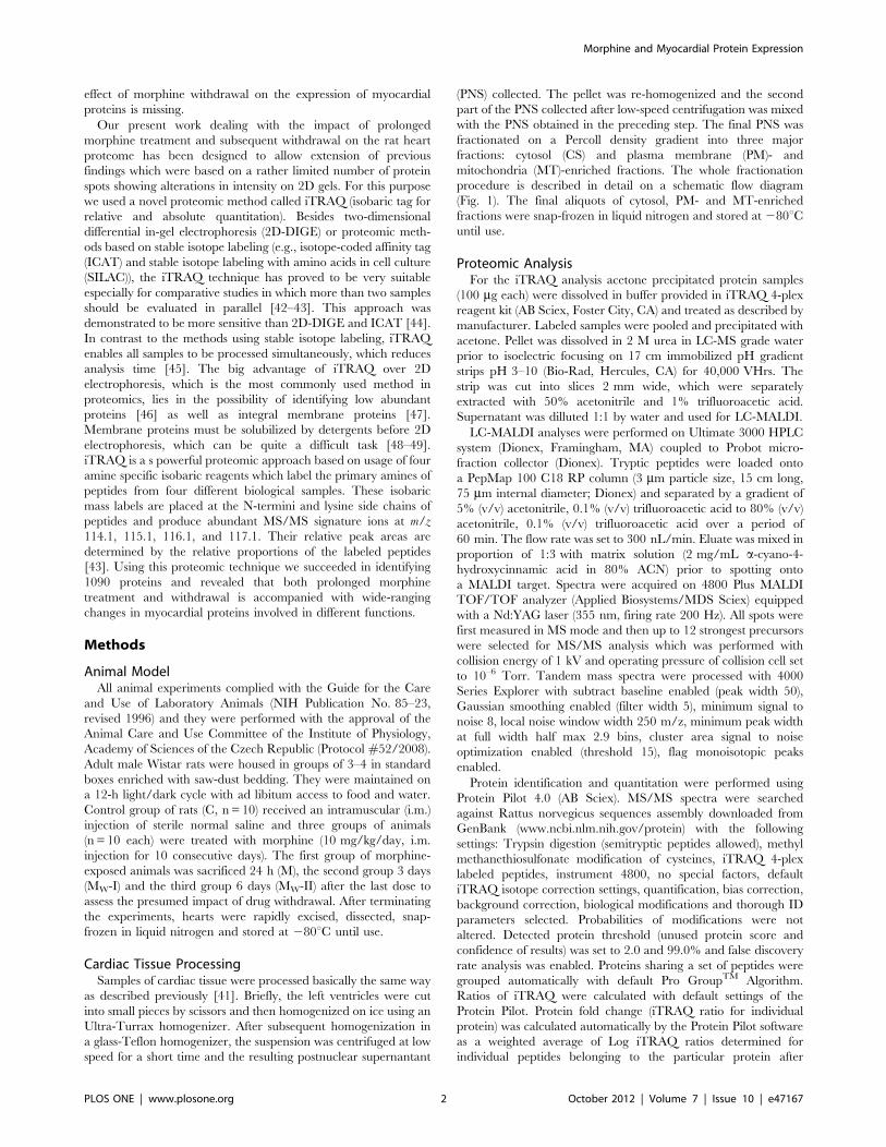
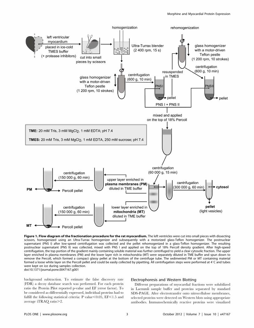
Cardiac Tissue ProcessingSamples of cardiac tissue were processed basically the same way
as described previously [41]. Briefly, the left ventricles were cut
into small pieces by scissors and then homogenized on ice using an
Ultra-Turrax homogenizer. After subsequent homogenization in
a glass-Teflon homogenizer, the suspension was centrifuged at low
speed for a short time and the resulting postnuclear supernantant
(PNS) collected. The pellet was re-homogenized and the second
part of the PNS collected after low-speed centrifugation was mixed
with the PNS obtained in the preceding step. The final PNS was
fractionated on a Percoll density gradient into three major
fractions: cytosol (CS) and plasma membrane (PM)- and
mitochondria (MT)-enriched fractions. The whole fractionation
procedure is described in detail on a schematic flow diagram
(Fig. 1). The final aliquots of cytosol, PM- and MT-enriched
fractions were snap-frozen in liquid nitrogen and stored at 280uCuntil use.
Proteomic AnalysisFor the iTRAQ analysis acetone precipitated protein samples
(100 mg each) were dissolved in buffer provided in iTRAQ 4-plex
reagent kit (AB Sciex, Foster City, CA) and treated as described by
manufacturer. Labeled samples were pooled and precipitated with
acetone. Pellet was dissolved in 2 M urea in LC-MS grade water
prior to isoelectric focusing on 17 cm immobilized pH gradient
strips pH 3–10 (Bio-Rad, Hercules, CA) for 40,000 VHrs. The
strip was cut into slices 2 mm wide, which were separately
extracted with 50% acetonitrile and 1% trifluoroacetic acid.
Supernatant was dilluted 1:1 by water and used for LC-MALDI.
LC-MALDI analyses were performed on Ultimate 3000 HPLC
system (Dionex, Framingham, MA) coupled to Probot micro-
fraction collector (Dionex). Tryptic peptides were loaded onto
a PepMap 100 C18 RP column (3 mm particle size, 15 cm long,
75 mm internal diameter; Dionex) and separated by a gradient of
5% (v/v) acetonitrile, 0.1% (v/v) trifluoroacetic acid to 80% (v/v)
acetonitrile, 0.1% (v/v) trifluoroacetic acid over a period of
60 min. The flow rate was set to 300 nL/min. Eluate was mixed in
proportion of 1:3 with matrix solution (2 mg/mL a-cyano-4-hydroxycinnamic acid in 80% ACN) prior to spotting onto
a MALDI target. Spectra were acquired on 4800 Plus MALDI
TOF/TOF analyzer (Applied Biosystems/MDS Sciex) equipped
with a Nd:YAG laser (355 nm, firing rate 200 Hz). All spots were
first measured in MS mode and then up to 12 strongest precursors
were selected for MS/MS analysis which was performed with
collision energy of 1 kV and operating pressure of collision cell set
to 10–6 Torr. Tandem mass spectra were processed with 4000
Series Explorer with subtract baseline enabled (peak width 50),
Gaussian smoothing enabled (filter width 5), minimum signal to
noise 8, local noise window width 250 m/z, minimum peak width
at full width half max 2.9 bins, cluster area signal to noise
optimization enabled (threshold 15), flag monoisotopic peaks
enabled.
Protein identification and quantitation were performed using
Protein Pilot 4.0 (AB Sciex). MS/MS spectra were searched
against Rattus norvegicus sequences assembly downloaded from
GenBank (www.ncbi.nlm.nih.gov/protein) with the following
settings: Trypsin digestion (semitryptic peptides allowed), methyl
methanethiosulfonate modification of cysteines, iTRAQ 4-plex
labeled peptides, instrument 4800, no special factors, default
iTRAQ isotope correction settings, quantification, bias correction,
background correction, biological modifications and thorough ID
parameters selected. Probabilities of modifications were not
altered. Detected protein threshold (unused protein score and
confidence of results) was set to 2.0 and 99.0% and false discovery
rate analysis was enabled. Proteins sharing a set of peptides were
grouped automatically with default Pro GroupTM Algorithm.
Ratios of iTRAQ were calculated with default settings of the
Protein Pilot. Protein fold change (iTRAQ ratio for individual
protein) was calculated automatically by the Protein Pilot software
as a weighted average of Log iTRAQ ratios determined for
individual peptides belonging to the particular protein after
Morphine and Myocardial Protein Expression
PLOS ONE | www.plosone.org 2 October 2012 | Volume 7 | Issue 10 | e47167
background subtraction. To estimate the false discovery rate
(FDR) a decoy database search was performed. For each protein
ratio the Protein Pilot reported p-value and EF (error factor). To
be considered as differentially expressed, individual proteins had to
fulfill the following statistical criteria: P value,0.05, EF,1.5 and
average iTRAQ ratio.2.
Electrophoresis and Western BlottingDifferent preparations of myocardial fractions were solubilized
in Laemmli sample buffer and proteins separated by standard
SDS-PAGE. After electrotransfer onto nitrocellulose membranes,
selected proteins were detected on Western blots using appropriate
antibodies. Immunochemically reactive proteins were visualized
Figure 1. Flow diagram of the fractionation procedure for the rat myocardium. The left ventricles were cut into small pieces with dissectingscissors, homogenized using an Ultra-Turrax homogenizer and subsequently with a motorized glass-Teflon homogenizer. The postnuclearsupernatant (PNS I) after low-speed centrifugation was collected and the pellet rehomogenized in a glass-Teflon homogenizer. The resultingpostnuclear supernatant (PNS II) was collected, mixed with PNS I and applied on the top of 18% Percoll density gradient. After high-speedcentrifugation, the top portion of the gradient mainly containing soluble material was further centrifuged to yield a clear cytosolic fraction. The upperlayer enriched in plasma membranes (PM) and the lower layer rich in mitochondria (MT) were separately diluted in TME buffer and spun down toremove the Percoll, which formed a compact glassy pellet at the bottom of the centrifuge tube. The sedimented PM or MT containing materialformed a loose white layer on the Percoll pellet and could be easily collected by pipetting. All centrifugation steps were performed at 4uC and tubeswere kept on ice during samples collection.doi:10.1371/journal.pone.0047167.g001
Morphine and Myocardial Protein Expression
PLOS ONE | www.plosone.org 3 October 2012 | Volume 7 | Issue 10 | e47167
by conventional enhanced chemiluminiscence detection (Pierce
Biotechnology, Rockford, IL, USA). The Western blots were
scanned and evaluated quantitatively by means of densitometry
using ImageQuantTM TL software (GE Healthcare, Chalfont St.
Giles, UK).
Results and Discussion
Identification of Proteins in Myocardial FractionsThe left ventricles isolated from control (C), morphine-treated
(M) and morphine-withdrawn (MW-I and MW-II) rats were
subfractionated into cytosolic (CS), plasma membrane (PM)- and
mitochondria (MT)-enriched fractions (Fig. 1). Enrichment of
these fractions with typical cytosolic, plasma membrane-bound or
mitochondrial proteins was demonstrated previously [41]. Proteins
present in each preparation were digested with trypsin and peptide
populations in the individual fractions were labeled with distinct
iTRAQ reagents. The corresponding peptide populations from
samples of all four experimental groups were then combined for
further analyses.
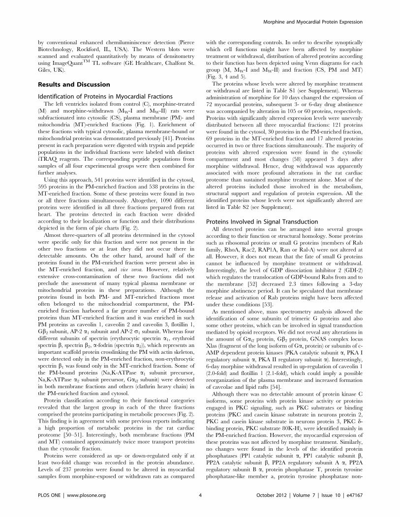
Using this approach, 541 proteins were identified in the cytosol,
595 proteins in the PM-enriched fraction and 538 proteins in the
MT-enriched fraction. Some of these proteins were found in two
or all three fractions simultaneously. Altogether, 1090 different
proteins were identified in all three fractions prepared from rat
heart. The proteins detected in each fraction were divided
according to their localization or function and their distributions
depicted in the form of pie charts (Fig. 2).
Almost three-quarters of all proteins determined in the cytosol
were specific only for this fraction and were not present in the
other two fractions or at least they did not occur there in
detectable amounts. On the other hand, around half of the
proteins found in the PM-enriched fraction were present also in
the MT-enriched fraction, and vice versa. However, relatively
extensive cross-contamination of these two fractions did not
preclude the assessment of many typical plasma membrane or
mitochondrial proteins in these preparations. Although the
proteins found in both PM- and MT-enriched fractions most
often belonged to the mitochondrial compartment, the PM-
enriched fraction harbored a far greater number of PM-bound
proteins than MT-enriched fraction and it was enriched in such
PM proteins as caveolin 1, caveolin 2 and caveolin 3, flotillin 1,
Gb2 subunit, AP-2 a1 subunit and AP-2 s1 subunit. Whereas four
different subunits of spectrin (erythrocytic spectrin a1, erythroidspectrin b, spectrin b2, a-fodrin (spectrin a2)), which represents an
important scaffold protein crosslinking the PM with actin skeleton,
were detected only in the PM-enriched fraction, non-erythrocytic
spectrin b1 was found only in the MT-enriched fraction. Some of
the PM-bound proteins (Na,K-ATPase a1 subunit precursor,
Na,K-ATPase a2 subunit precursor, Gai2 subunit) were detected
in both membrane fractions and others (clathrin heavy chain) in
the PM-enriched fraction and cytosol.
Protein classification according to their functional categories
revealed that the largest group in each of the three fractions
comprised the proteins participating in metabolic processes (Fig. 2).
This finding is in agreement with some previous reports indicating
a high proportion of metabolic proteins in the rat cardiac
proteome [50–51]. Interestingly, both membrane fractions (PM
and MT) contained approximately twice more transport proteins
than the cytosolic fraction.
Proteins were considered as up- or down-regulated only if at
least two-fold change was recorded in the protein abundance.
Levels of 237 proteins were found to be altered in myocardial
samples from morphine-exposed or withdrawn rats as compared
with the corresponding controls. In order to describe synoptically
which cell functions might have been affected by morphine
treatment or withdrawal, distribution of altered proteins according
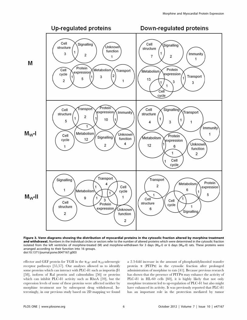
to their function has been depicted using Venn diagrams for each
group (M, MW-I and MW-II) and fraction (CS, PM and MT)
(Fig. 3, 4 and 5).
The proteins whose levels were altered by morphine treatment
or withdrawal are listed in Table S1 (see Supplement). Whereas
administration of morphine for 10 days changed the expression of
72 myocardial proteins, subsequent 3- or 6-day drug abstinence
was accompanied by alteration in 105 or 60 proteins, respectively.
Proteins with significantly altered expression levels were unevenly
distributed between all three myocardial fractions: 121 proteins
were found in the cytosol, 30 proteins in the PM-enriched fraction,
69 proteins in the MT-enriched fraction and 17 altered proteins
occurred in two or three fractions simultaneously. The majority of
proteins with altered expression were found in the cytosolic
compartment and most changes (58) appeared 3 days after
morphine withdrawal. Hence, drug withdrawal was apparently
associated with more profound alterations in the rat cardiac
proteome than sustained morphine treatment alone. Most of the
altered proteins included those involved in the metabolism,
structural support and regulation of protein expression. All the
identified proteins whose levels were not significantly altered are
listed in Table S2 (see Supplement).
Proteins Involved in Signal TransductionAll detected proteins can be arranged into several groups
according to their function or structural homology. Some proteins
such as ribosomal proteins or small G proteins (members of Rab
family, RhoA, Rac2, RAP1A, Ran or Ral-A) were not altered at
all. However, it does not mean that the fate of small G proteins
cannot be influenced by morphine treatment or withdrawal.
Interestingly, the level of GDP dissociation inhibitor 2 (GDI-2)
which regulates the translocation of GDP-bound Rabs from and to
the membrane [52] decreased 2.3 times following a 3-day
morphine abstinence period. It can be speculated that membrane
release and activation of Rab proteins might have been affected
under these conditions [53].
As mentioned above, mass spectrometry analysis allowed the
identification of some subunits of trimeric G proteins and also
some other proteins, which can be involved in signal transduction
mediated by opioid receptors. We did not reveal any alterations in
the amount of Gai2 protein, Gb2 protein, GNAS complex locus
Xlas (fragment of the long isoform of Gas protein) or subunits of c-AMP dependent protein kinases (PKA catalytic subunit a, PKA I
regulatory subunit a, PKA II regulatory subunit a). Interestingly,6-day morphine withdrawal resulted in up-regulation of caveolin 1
(2.0-fold) and flotillin 1 (2.1-fold), which could imply a possible
reorganization of the plasma membrane and increased formation
of caveolae and lipid rafts [54].
Although there was no detectable amount of protein kinase C
isoforms, some proteins with protein kinase activity or proteins
engaged in PKC signaling, such as PKC substrates or binding
proteins (PKC and casein kinase substrate in neurons protein 2,
PKC and casein kinase substrate in neurons protein 3, PKC d-binding protein, PKC substrate 80K-H), were identified mainly in
the PM-enriched fraction. However, the myocardial expression of
these proteins was not affected by morphine treatment. Similarly,
no changes were found in the levels of the identified protein
phosphatases (PP1 catalytic subunit a, PP1 catalytic subunit b,PP2A catalytic subunit b, PP2A regulatory subunit A a, PP2Aregulatory subunit B a, protein phosphatase T, protein tyrosine
phosphatase-like member a, protein tyrosine phosphatase non-
Morphine and Myocardial Protein Expression
PLOS ONE | www.plosone.org 4 October 2012 | Volume 7 | Issue 10 | e47167
receptor type 11, protein-tyrosine phosphatase mitochondrial 1,
low molecular weight protein tyrosine phosphatase isoform A).
Interestingly, phospholipase C d1 (PLC-d1) was found to be up-
regulated (3.2-fold) in the cytosolic fraction after morphine
treatment, and 3 days of abstinence resulted in its marked
down-regulation (10.7-fold). PLC-d isoforms were shown to differ
from the other PLC isoforms because they are not activated by
heterotrimeric G proteins or protein phosphorylation cascades
[55]. It was proposed that this enzyme is activated by a rise in the
concentration of free cytosolic calcium generated by PLC-b and
further amplifies Ca2+ signals initiated by activation of PLC-b, -cand -e isoforms [56]. It also can be activated by transglutaminase
II (TGII or Gah), which is an atypical G protein with GTPase and
transglutaminase activities [57]. PLC-d1 was shown to be an
Figure 2. Classification of proteins identified in the left ventricular myocardium according to their subcellular localization andfunction. The proteins of each fraction (CT, PM and MT) isolated from hearts of control (C), morphine-treated (M), and morphine-withdrawn (MW-Iand MW-II) rats were labeled by isobaric reagents provided in iTRAQ 4-plex reagent kit. After LC-MALDI analyses performed on Ultimate 3000 HPLCsystem and acquisition of spectra on 4800 Plus MALDI TOF/TOF analyzer, proteins were identified and quantified using Protein Pilot 4.0. Localizationand function of the proteins were assigned on the basis of current annotations in the Swiss-Prot database. Sections of the pie charts represent theproportion of proteins found within each functional category.doi:10.1371/journal.pone.0047167.g002
Morphine and Myocardial Protein Expression
PLOS ONE | www.plosone.org 5 October 2012 | Volume 7 | Issue 10 | e47167
effector and GEF protein for TGII in the a1B- and a1D-adrenergicreceptor pathways [55,57]. Our analyses allowed us to identify
some proteins which can interact with PLC-d1 such as importin b1[58], isoform of Ral protein and calmodulins [56] or proteins
which can inhibit PLC-d1 activity such as RhoA [59], but the
expression levels of none of these proteins were affected neither by
morphine treatment nor by subsequent drug withdrawal. In-
terestingly, in our previous study based on 2D mapping we found
a 2.3-fold increase in the amount of phosphatidylinositol transfer
protein a (PITPa) in the cytosolic fraction after prolonged
administration of morphine to rats [41]. Because previous research
has shown that the presence of PITPa may enhance the activity of
PLC-d1 in HL-60 cells [60], it is highly likely that not only
morphine treatment led to up-regulation of PLC-d1 but also might
have enhanced its activity. It was previously reported that PLC-d1has an important role in the protection mediated by tumor
Figure 3. Venn diagrams showing the distribution of myocardial proteins in the cytosolic fraction altered by morphine treatmentand withdrawal. Numbers in the individual circles or sectors refer to the number of altered proteins which were determined in the cytosolic fractionisolated from the left ventricles of morphine-treated (M) and morphine-withdrawn for 3 days (MW-I) or 6 days (MW-II) rats. These proteins werearranged according to their function into 16 groups.doi:10.1371/journal.pone.0047167.g003
Morphine and Myocardial Protein Expression
PLOS ONE | www.plosone.org 6 October 2012 | Volume 7 | Issue 10 | e47167
necrosis factor (TNF) receptor against adriamycin-induced
cardiotoxicity [61]. The present finding of a 3.2-fold increase of
PLC-d1 in the cytosolic fraction of myocardial samples from
morphine-treated rats suggests that this enzyme could also
participate in the molecular mechanism of cardioprotective action
of this drug. However, only future research aimed at untangling
the molecular mechanism of cardioprotection conferred by
morphine can prove or disprove this speculative hypothesis. Apart
from PLC-d1, lysophospholipase 1 and phospholipase A2 activat-
ing protein were also identified by iTRAQ analysis but myocardial
levels of these proteins were not altered by morphine.
It is known that the acute or chronic morphine treatment can
cause the elevation of free cytosolic calcium concentration via
PLC-b [62–64]. One might therefore speculate that the levels of
some calcium binding proteins or proteins involved in calcium
handling, transport and signaling could possibly be influenced by
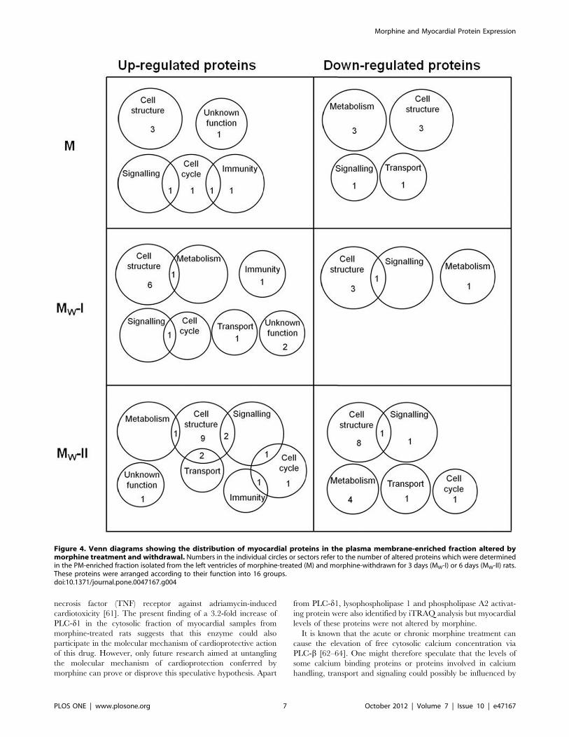
Figure 4. Venn diagrams showing the distribution of myocardial proteins in the plasma membrane-enriched fraction altered bymorphine treatment and withdrawal. Numbers in the individual circles or sectors refer to the number of altered proteins which were determinedin the PM-enriched fraction isolated from the left ventricles of morphine-treated (M) and morphine-withdrawn for 3 days (MW-I) or 6 days (MW-II) rats.These proteins were arranged according to their function into 16 groups.doi:10.1371/journal.pone.0047167.g004
Morphine and Myocardial Protein Expression
PLOS ONE | www.plosone.org 7 October 2012 | Volume 7 | Issue 10 | e47167
morphine treatment. Nevertheless, only histidine-rich calcium
protein identified in the PM-enriched fraction was significantly up-
regulated (3- or 3.9-fold, respectively) after 3- or 6-day drug
withdrawal. Interestingly, its level was not changed in the other
two fractions prepared from samples derived from morphine-
exposed animals (M, MW-I, MW-II), compared to the correspond-
ing controls. The other proteins in this group (calcium binding
protein, calcium regulated heat stable protein 1, calcyclin,
calmodulin 2, calnexin, calsequestrin 1, calsequestrin 2, calpain
small subunit 1, sarcalumenin, sarcoplasmic reticulum Ca2+-
ATPase, vasopressin-activated calcium-binding mobilizing recep-
tor protein) were not altered at all. These results suggest that if
Ca2+ signaling pathways were affected by morphine, this effect
need not necessarily be accompanied by marked quantitative
changes of Ca2+ binding proteins.
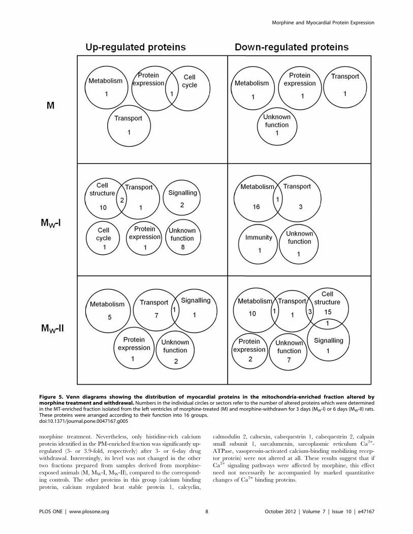
Figure 5. Venn diagrams showing the distribution of myocardial proteins in the mitochondria-enriched fraction altered bymorphine treatment and withdrawal. Numbers in the individual circles or sectors refer to the number of altered proteins which were determinedin the MT-enriched fraction isolated from the left ventricles of morphine-treated (M) and morphine-withdrawn for 3 days (MW-I) or 6 days (MW-II) rats.These proteins were arranged according to their function into 16 groups.doi:10.1371/journal.pone.0047167.g005
Morphine and Myocardial Protein Expression
PLOS ONE | www.plosone.org 8 October 2012 | Volume 7 | Issue 10 | e47167
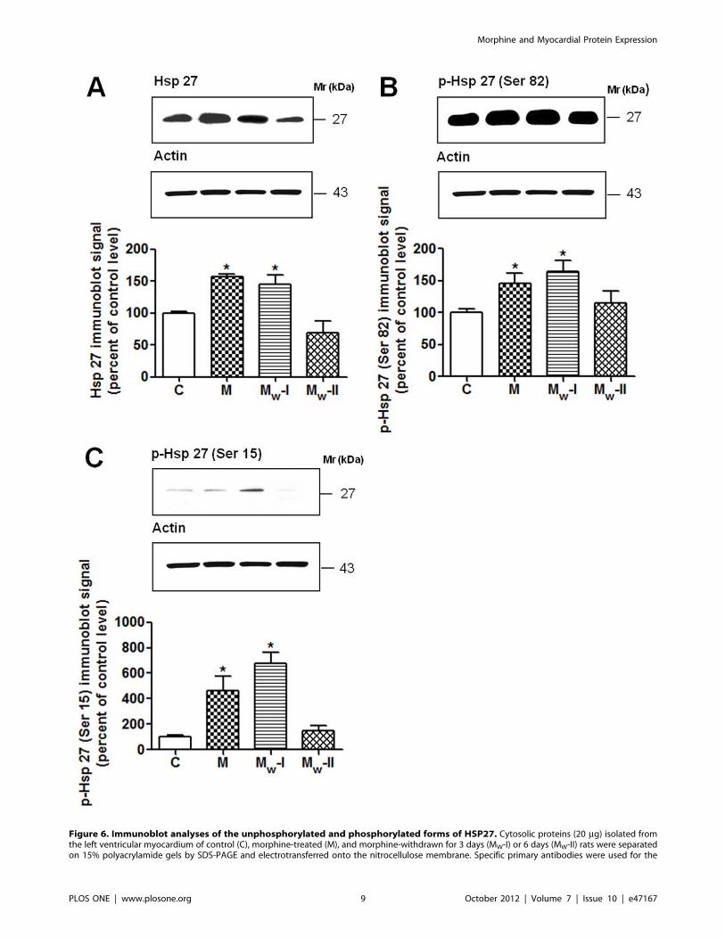
Figure 6. Immunoblot analyses of the unphosphorylated and phosphorylated forms of HSP27. Cytosolic proteins (20 mg) isolated fromthe left ventricular myocardium of control (C), morphine-treated (M), and morphine-withdrawn for 3 days (MW-I) or 6 days (MW-II) rats were separatedon 15% polyacrylamide gels by SDS-PAGE and electrotransferred onto the nitrocellulose membrane. Specific primary antibodies were used for the
Morphine and Myocardial Protein Expression
PLOS ONE | www.plosone.org 9 October 2012 | Volume 7 | Issue 10 | e47167
Proteins Involved in Apoptosis and Oxidative StressA number of studies dealing with morphine effects on cell
viability, apoptosis and oxidative stress have been published during
the past decade. It was reported that morphine treatment resulted
in apoptosis in macrophages [65–66], SH-SY5Y [67] or microglia
and neurons [68–69]. On the other hand, protective effects of
morphine were observed in macrophages [70], astrocytes [69,71]
or heart [37–38,64]. Morphine has also been shown to enhance
the generation of reactive oxygen species (ROS) in macrophages
[65] and SH-SY5Y [67]. Hence, it seems that morphine can cause
either cell apoptosis or protection depending on the cell type and
experimental context. Therefore, it is worth paying attention to
potential changes of proteins involved in apoptosis, cellular
protection and ROS generation.
Many proteins involved in cellular protection/apoptosis, which
were detected in our present study, were not altered after
morphine treatment or withdrawal, e.g. proteins that can induce
apoptosis and/or activate caspases (apoptosis-inducing factor,
diablo, C9 protein, B-cell associated protein 31, BCL2/adenovirus
E1B 19 kDa-interacting protein 3, endonuclease G, Gb2-like 1,
reticulon-3, VDAC1, SP120), proteins that protect cells against
apoptosis (CHIP28k, mitochondrial inner membrane protein,
glutathione peroxidase 4 isoform A precursor, mitochondrial
protein 18 kDa, prohibitin, protein DJ-1, vanin 1), proteins
engaged in release of cytochrome c from mitochondria into the
cytoplasm (Clu protein, growth hormone-inducible transmem-
brane protein, mitochondrial fission 1 protein, presenilin associ-
ated rhomboid like) or proteins conferring protection against
release of cytochrome c (mitochondrial OPA1, optic atrophy 1
homolog, sulphated glycoprotein 2).
We determined four proteins possibly involved in the induction
of apoptosis, expression of which was affected by morphine.
Programmed cell death 5 protein was up-regulated (2.1 fold) in the
cytosolic fraction after morphine treatment, dynamin 1 like was
up-regulated (2.6-fold) in the cytosolic fraction after a 3-day drug
abstinence period, programmed cell death 6 interacting protein
was up-regulated (2.6-fold) in the cytosolic fraction after 6-day
abstinence and lactate dehydrogenase A was down-regulated (3.4-
fold) in the cytosolic fraction after 6-day abstinence. Interestingly,
the level of the latter protein was not changed in the PM- and MT-
enriched fractions. Another four proteins were found to be altered
after morphine exposure or withdrawal that can play a role in
protection of cells against apoptosis. Cystatin B was down-
regulated (2.2-fold) and Bcl2-associated anthanogene 3 up-
regulated (2.4-fold) in the cytosolic fraction after morphine
treatment, prothymosin a was down-regulated (4.9-fold) in the
cytosolic fraction after 3 days of drug abstinence, acetyl-CoA
acyltransferase 2 was down-regulated (3.2-fold) in the MT-
enriched fraction after 6 days of abstinence (its level was
unchanged in the PM-enriched fraction and cytosol). These results
indicate that a certain imbalance between pro- and anti-apoptotic
proteins may arise in the myocardium during morphine treatment
or withdrawal. However, up-regulation of programmed cell death
5 protein in the cytosol does not necessarily mean the induction of
apoptosis because this protein is translocated from the cytosol to
the nucleus during the early stage of apoptosis [72].
It seems that if chronic morphine treatment induced apoptosis,
it would not be mediated by the formation of pores in the
mitochondrial membrane and release of cytochrome c. OPA1
protein and mitochondrial inner membrane protein, which control
the shape of mitochondrial cristae and thus may regulate
cytochrome c redistribution [73–75], were not affected by
morphine treatment or withdrawal. The expression of another
protein involved in mitochondrial fusion, mitofusin-1 [73–74], was
also not changed. Mitochondrial morphology thus did not seem to
be affected by morphine. Moreover, no release of cytochrome c
from mitochondria to the cytoplasm was observed after morphine
treatment or withdrawal; the levels of cytochrome c detected in all
three fractions were not altered.
Calpain-dependent proteolysis can contribute to cell death by
cleavage of pro-apoptotic Bid [76]. There are two different major
isoforms of calpain, m-calpain (calpain 1) and m-calpain (calpain
2), which contain a large 80 kDa catalytic subunit and a common
small 30 kDa regulatory subunit [77]. They are activated by
elevation of intracellular calcium and their proteolytic activity is
regulated by specific inhibitor calpastatin [77]. In our present
study, a small regulatory subunit of calpain, m-calpain and
calpastatin were identified. Nevertheless, the levels of these
proteins were not altered by morphine treatment or withdrawal.
In addition, expression of the majority of caspase and calpain
substrates (a-fodrin, actin, protein phosphatase 2A, vimentin,
lamin A, Ca2+-ATPase, Ga protein, PKA, ryanodine receptors,
talin, tropomyosin, tubulins, vimentin, vinculin) [78–79] identified
in our study were also not altered. Only tau and troponin T were
down-regulated (17.0-fold and 2.8-fold, respectively) after 3-day
drug abstinence. These results indicate that calpain and caspase
pathways were not significantly activated by morphine treatment
or withdrawal.
Similarly, there were no significant changes in the expression
levels of proteins involved in modulation of oxidative stress, such as
superoxide dismutase 1, superoxide dismutase 2, glutathione
peroxidase, glutathione peroxidase 4 isoform A precursor,
glutaredoxin 3, glutaredoxin 5, peroxiredoxin 2, peroxiredoxin
3, peroxiredoxin 5, peroxiredoxin 6, antioxidant enzyme B166,
catalase, protein DJ-1, thioredoxin 1, thioredoxin like protein 1,
thioredoxin domain containing 17, thioredoxin reductase 2,
protein disulfide isomerise A6, CDGSH iron sulfur domain
containing protein 1, dihydrolipoyl dehydrogenase mitochondrial.
HSPsSome members of the family of heat shock proteins (HSPs) were
markedly affected by morphine treatment or withdrawal. Altera-
tions in HSPs expression were observed mainly in the cytosolic
fraction and less in the MT-enriched fraction. No changes were
found in the PM-enriched fraction. Heat shock 10 kDa protein 1
(HSP10) was up-regulated (2.1-fold) in the cytosolic fraction after
morphine treatment. Interestingly, this protein was up-regulated
(2.3-fold) in the cytosol and down-regulated in the MT-enriched
fraction (2.1-fold) after 6-day drug abstinence. These results
suggest that HSP10 is affected by morphine and that subsequent
withdrawal can cause its redistribution from the cytoplasm to
membrane compartments.
Heat shock 60 kDa protein (HSP60) was significantly up-
regulated only in the cytosolic fraction (and not in both membrane
fractions) after morphine treatment (2.0-fold) and a 6-day
morphine abstinence (2.0-fold). Similarly to HSP10, a slight
redistribution of HSP60 to the MT-enriched fraction (1.8-fold
increase) was found after 6-day morphine abstinence, which may
detection of unphosphorylated HSP27 (A) and HSP27 phosphorylated at Ser82 (B) and Ser15 (C). Actin was used as a loading control and the relativelevels of individual forms of HSP27 (HSP27, p-HSP27-Ser82 or p-HSP27-Ser15) after normalization were expressed as a percentage of thecorresponding control level. Data represent the mean6S.E.M. of three separate experiments; *p,0.05 vs control.doi:10.1371/journal.pone.0047167.g006
Morphine and Myocardial Protein Expression
PLOS ONE | www.plosone.org 10 October 2012 | Volume 7 | Issue 10 | e47167
imply that both proteins may undergo a common fate. The anti-
apoptotic effect of both HSP10 and HSP60 has been well
established in several studies in which over-expression of these
proteins protected myocytes and H9C2 cells against ischemic
injury [80–81]. In addition, HSP60 have been shown to interact
with Bax/Bak proteins, thus providing protection against apopto-
sis [82].
Heat shock 70 kDa protein 1A/1B (HSP70) was markedly up-
regulated in the cytosolic fraction (15.9-fold) as well as MT-
enriched fraction (2.3-fold) after morphine treatment. This protein
was also up-regulated (6.8-fold) in the cytosolic fraction after 6
days of morphine abstinence. Importantly, it has been demon-
strated that over-expression of HSP70 protects the myocardium
from ischemic injury [83].
Two members of the family of small heat shock proteins with
cell protective effects, heat shock protein 27 kDa protein 1
(HSP27, HSPB1) and a-B crystallin [84–85], were also found to
be altered. Both HSP27 and a-B crystallin were up-regulated (2.6-
and 2.1-fold, respectively) in the cytosolic fraction after morphine
treatment. a-B crystallin was up-regulated (2.1-fold) in the
cytosolic fraction also after a 6-day drug abstinence. It was shown
that these proteins can be phosphorylated and activated via the
MAPK p38 (MAPK14)/MAPKAP-2 pathway [86–87]. Interest-
ingly, in our study MAPK14 (isoform CRA_b) was found to be up-
regulated (2.5-fold) in the cytosolic fraction after morphine
treatment. Therefore, it is plausible to assume that cardioprotec-
tion induced by morphine may occur at least partly via the MAPK
p38/MAPKAP-2/HSP27 or a-B crystalline pathway.
In order to determine whether HSP27 could be activated by
morphine treatment or withdrawal, its phosphorylation state was
assessed by Western blotting using specific antibodies against
phosphorylated amino acids Ser82 and Ser15 of HSP27 (Fig. 6).
Both these sites can be phosphorylated by MAPKAP-2 and the
major site of phosphorylation of this protein is Ser82 [88]. The
increased expression levels of HSP27 in samples from morphine
treated animals were verified by Western blotting (Fig. 6A). The
amount of HSP27 phosphorylated on Ser15 and, to a lesser extent,
on Ser82 was also increased (Fig. 6B,C). Although HSP27 was
phosphorylated at both phosphorylation sites after morphine
treatment, 3-day drug abstinence further increased its phosphor-
ylation. By contrast, after 6 days of abstinence HSP27 phosphor-
ylation returned to normal values. These results partly support
some recently published findings. Two recent studies have
reported that naloxone-precipitated morphine withdrawal induced
activation of HSP27 by phosphorylation at Ser15 but not at Ser82
[89–90]. Moreover, although chronic morphine treatment led to
increased expression of HSP27, enhanced phosphorylation and
activation of this protein was not observed [89–90]. The partial
discrepancy between these and our present observations may be
explained by different experimental conditions. Nevertheless, it
can be concluded that morphine withdrawal can strongly enhance
phosphorylation of HSP27 at Ser15, irrespective whether it is
spontaneous (in the case of morphine abstinence) or elicited by
treatment with the opioid inverse agonist naloxone.
In order to determine whether HSP27 could be activated after
morphine treatment or withdrawal, its phosphorylation was
assessed by immunoblotting with specific antibodies against
phosphorylated amino acids Ser82 and Ser15 of HSP27 (Fig. 2).
Both sites can be phosphorylated by MAPKAP-2 and the major
site of phosphorylation of this protein is Ser82 [88]. It was verified
using antibodies against HSP27 that morphine treatment led to
up-regulation of HSP27. Nevertheless, it resulted also in
phosphorylation of Ser15 and in less extent in phosphorylation
of Ser82 (Fig. 2).
Non-phosporylated HSP27 was shown to exist in the form
of large oligomers and its phosphorylation leads to dissociation of
these oligomers [91]. Phosphorylation and oligomerization of
HSP27 is connected with modulation of the interaction between
HSP27 and actin [91–92]. While the non-phosphorylated HSP27
can block actin polymerization, the phosphorylation of HSP27 is
related to re-organization of actin-based cytoskeletal structures
[88]. It has been suggested that this re-organization of the actin
cytoskeleton induced by phosphorylation of HSP27 could lead to
cytoprotection due to stabilization of actin filaments [93].
The phosphorylation and activation of HSP27 might be also
related to the observed down-regulation of tau after 3 days of
morphine abstinence. HSP27 and a-B crystallin can interact with
microtubules and neurofilaments and protect against protein
aggregation [94]. Both proteins can interact with pathological
hyperphosphorylated tau and thus facilitate its dephosphorylation
and degradation. This process was observed mainly in studies
dealing with Alzheimer disease [94–96].
Conclusions and Future DirectionsThis work follows up our previous research focused on the
consequences of prolonged morphine administration on the rat
heart [41] and further elaborates this issue. Results of our present
proteomic study clearly indicate that iTRAQ approach may yield
a wealth of information regarding the effect of morphine on the
cardiac proteome. Quantitative analysis of proteomics data
obtained by this method revealed a number of significant changes
induced by both morphine exposure and withdrawal. An
important outcome from this study is the realization that the
protein and hence gene expression responses to morphine in the
heart are quite complex. A similarly wide-ranging response to
morphine has been previously observed in brain tissue.
A number of significant changes found in the expression of
different myocardial proteins due to morphine treatment suggest
that the potential cardiac effects of this drug should be carefully
taken into account when using it for medical purposes. Impor-
tantly, as can be seen from this study, morphine withdrawal may
apparently have even a greater impact on the heart proteome than
the use of this compound itself. Hence, morphine should be
considered as a drug with potentially profound effects on cardiac
protein expression profiling, which may have been difficult to
appreciate so far. Future studies should elucidate whether and to
what extent morphine-induced changes in protein expression may
play a specific role in the regulation of heart function.
Supporting Information
Table S1 Complete list of the myocardial proteinsaltered after morphine treatment or withdrawal. The
proteins whose expression levels were altered at least twice after
morphine treatment (M) or drug withdrawal for 3 days (MW-I) or 6
days (MW-II) compared to controls were arranged according to
their function into several groups. Expression values of up-
regulated (q) or down-regulated (Q) proteins are expressed as fold
change from untreated controls. Number of accession (gi numbers
from GenBank/EMBL/DDBJ databases) and fraction in which
protein alteration was detected are quoted for each protein (CS,
cytosol; PM, plasma membrane-enriched fraction; MT, mito-
chondria-enriched fraction). %Cov, the percentage of matching
amino acids from the identified peptides divided by the total
number of amino acids in the sequence. Peptides, number of
unique peptides per identified protein. The occurrence of
individual proteins in other fraction(s) without alterations after
morphine treatment or withdrawal is mentioned in Notes noted
Morphine and Myocardial Protein Expression
PLOS ONE | www.plosone.org 11 October 2012 | Volume 7 | Issue 10 | e47167
using the following markings: (1) CS, no change; (2) PM, no
change; (3) MT, no change; (4) CS+PM, no change; (5) PM+MT,
no change.
(PDF)
Table S2 Complete list of the myocardial proteinswhose levels were not significantly altered after mor-phine treatment or withdrawal. The proteins whose
expression levels were not significantly altered after morphine
treatment (M) or withdrawal for 3 days (MW-I) or 6 days (MW-II)
compared to controls were arranged according to their function
into several groups. Number of accession (gi numbers from
GenBank/EMBL/DDBJ databases) and fraction in which the
protein was detected are quoted for each protein (CS, cytosol; PM,
plasma membrane-enriched fraction; MT, mitochondria-enriched
fraction). %Cov, the percentage of matching amino acids from
identified peptides divided by the total number of amino acids in
the sequence. Peptides, number of unique peptides per identified
protein.
(PDF)
Author Contributions
Conceived and designed the experiments: J. Novotny ZD. Performed the
experiments: ZD JS J. Neckar. Analyzed the data: ZD JS PJ. Wrote the
paper: ZD FK J. Novotny.
References
1. Flemming K (2010) The use of morphine to treat cancer-related pain: a synthesis
of quantitative and qualitative research. J Pain Symptom Manage 39: 139–154.
2. Vallejo R, Barkin RL, Wang VC (2011) Pharmacology of opioids in the
treatment of chronic pain syndromes. Pain Physician 14: E343–360.
3. Gregori SD, Gregori MD, Ranzani GN, Allegri M, Minella C, et al. (2012)
Morphine metabolism, transport and brain disposition. Metab Brain Dis 27: 1–
5.
4. Kieffer BL, Evans CJ (2009) Opioid receptors: from binding sites to visible
molecules in vivo. Neuropharmacology 56 Suppl 1: 205–212.
5. Mansour A, Fox CA, Burke S, Meng F, Thompson RC, et al. (1994) Mu, delta,
and kappa opioid receptor mRNA expression in the rat CNS: an in situ
hybridization study. J Comp Neurol 350: 412–438.
6. Mansour A, Fox CA, Akil H, Watson SJ (1995) Opioid-receptor mRNA
expression in the rat CNS: anatomical and functional implications. Trends
Neurosci 18: 22–29.
7. Narita M, Funada M, Suzuki T (2001) Regulations of opioid dependence by
opioid receptor types. Pharmacol Ther 89: 1–15.
8. Manchikanti L, Atluri S, Trescot AM, Giordano J (2008) Monitoring opioid
adherence in chronic pain patients: tools, techniques, and utility. Pain Physician
11: S155–180.
9. Wang J, Yuan W, Li MD (2011) Genes and pathways co-associated with the
exposure to multiple drugs of abuse, including alcohol, amphetamine/
methamphetamine, cocaine, marijuana, morphine, and/or nicotine: a review
of proteomics analyses. Mol Neurobiol 44: 269–286.
10. Gintzler AR, Chakrabarti S (2006) Post-opioid receptor adaptations to chronic
morphine; altered functionality and associations of signaling molecules. Life Sci
79: 717–722.
11. Christie MJ (2008) Cellular neuroadaptations to chronic opioids: tolerance,
withdrawal and addiction. Br J Pharmacol 154: 384–396.
12. Nestler EJ (2004) Molecular mechanisms of drug addiction. Neuropharmacology
47 Suppl 1: 24–32.
13. Fan LW, Tien LT, Tanaka S, Ma T, Chudapongse N, et al. (2003) Changes in
the brain kappa-opioid receptor levels of rats in withdrawal from physical
dependence upon butorphanol. Neuroscience 121: 1063–1074.
14. Murray F, Harrison NJ, Grimwood S, Bristow LJ, Hutson PH (2007) Nucleus
accumbens NMDA receptor subunit expression and function is enhanced in
morphine-dependent rats. Eur J Pharmacol 562: 191–197.
15. Chen Y, Jiang Y, Yue W, Zhou Y, Lu L, et al. (2008) Chronic, but not acute
morphine treatment, up-regulates alpha-Ca2+/calmodulin dependent protein
kinase II gene expression in rat brain. Neurochem Res 33: 2092–2098.
16. Mickiewicz AL, Napier TC (2011) Repeated exposure to morphine alters surface
expression of AMPA receptors in the rat medial prefrontal cortex. Eur J Neurosci
33: 259–265.
17. Hamlin AS, McNally GP, Osborne PB (2007) Induction of c-Fos and zif268 in
the nociceptive amygdala parallel abstinence hyperalgesia in rats briefly exposed
to morphine. Neuropharmacology 53: 330–343.
18. Kaplan GB, Leite-Morris KA, Fan W, Young AJ, Guy MD (2011) Opiate
sensitization induces FosB/DeltaFosB expression in prefrontal cortical, striatal
and amygdala brain regions. PLoS One 6: e23574.
19. Boronat MA, Garcia-Fuster MJ, Garcia-Sevilla JA (2001) Chronic morphine
induces up-regulation of the pro-apoptotic Fas receptor and down-regulation of
the anti-apoptotic Bcl-2 oncoprotein in rat brain. Br J Pharmacol 134: 1263–
1270.
20. Emeterio EP, Tramullas M, Hurle MA (2006) Modulation of apoptosis in the
mouse brain after morphine treatments and morphine withdrawal. J Neurosci
Res 83: 1352–1361.
21. Marie-Claire C, Courtin C, Roques BP, Noble F (2004) Cytoskeletal genes
regulation by chronic morphine treatment in rat striatum. Neuropsychophar-
macology 29: 2208–2215.
22. Ammon-Treiber S, Hollt V (2005) Morphine-induced changes of gene
expression in the brain. Addict Biol 10: 81–89.
23. Prokai L, Zharikova AD, Stevens SM Jr (2005) Effect of chronic morphine
exposure on the synaptic plasma-membrane subproteome of rats: a quantitative
protein profiling study based on isotope-coded affinity tags and liquid
chromatography/mass spectrometry. J Mass Spectrom 40: 169–175.
24. Bierczynska-Krzysik A, Bonar E, Drabik A, Noga M, Suder P, et al. (2006) Rat
brain proteome in morphine dependence. Neurochem Int 49: 401–406.
25. Bierczynska-Krzysik A, Pradeep John JP, Silberring J, Kotlinska J, Dylag T, et
al. (2006) Proteomic analysis of rat cerebral cortex, hippocampus and striatum
after exposure to morphine. Int J Mol Med 18: 775–784.
26. Li KW, Jimenez CR, van der Schors RC, Hornshaw MP, Schoffelmeer AN, et
al. (2006) Intermittent administration of morphine alters protein expression in
rat nucleus accumbens. Proteomics 6: 2003–2008.
27. Moron JA, Abul-Husn NS, Rozenfeld R, Dolios G, Wang R, et al. (2007)
Morphine administration alters the profile of hippocampal postsynaptic density-
associated proteins: a proteomics study focusing on endocytic proteins. Mol Cell
Proteomics 6: 29–42.
28. Yang L, Sun ZS, Zhu YP (2007) Proteomic analysis of rat prefrontal cortex in
three phases of morphine-induced conditioned place preference. J Proteome Res
6: 2239–2247.
29. Bodzon-Kulakowska A, Suder P, Mak P, Bierczynska-Krzysik A, Lubec G, et al.
(2009) Proteomic analysis of striatal neuronal cell cultures after morphine
administration. J Sep Sci 32: 1200–1210.
30. Abul-Husn NS, Annangudi SP, Ma’ayan A, Ramos-Ortolaza DL, Stockton SD
Jr, et al. (2011) Chronic morphine alters the presynaptic protein profile:
identification of novel molecular targets using proteomics and network analysis.
PLoS One 6: e25535.
31. Bu Q, Yang Y, Yan G, Hu Z, Hu C, et al. (2012) Proteomic analysis of the
nucleus accumbens in rhesus monkeys of morphine dependence and withdrawal
intervention. J Proteomics 75: 1330–1342.
32. Neasta J, Uttenweiler-Joseph S, Chaoui K, Monsarrat B, Meunier JC, et al.
(2006) Effect of long-term exposure of SH-SY5Y cells to morphine: a whole cell
proteomic analysis. Proteome Sci 4: 23.
33. Xu H, Wang X, Zimmerman D, Boja ES, Wang J, et al. (2005) Chronic
morphine up-regulates G alpha12 and cytoskeletal proteins in Chinese hamster
ovary cells expressing the cloned mu opioid receptor. J Pharmacol Exp Ther
315: 248–255.
34. Suder P, Bodzon-Kulakowska A, Mak P, Bierczynska-Krzysik A, Daszykowski
M, et al. (2009) The proteomic analysis of primary cortical astrocyte cell culture
after morphine administration. J Proteome Res 8: 4633–4640.
35. Shanazari AA, Aslani Z, Ramshini E, Alaei H (2011) Acute and chronic effects
of morphine on cardiovascular system and the baroreflexes sensitivity during
severe increase in blood pressure in rats. ARYA Atheroscler 7: 111–117.
36. Pang PS, Komajda M, Gheorghiade M (2010) The current and future
management of acute heart failure syndromes. Eur Heart J 31: 784–793.
37. Murphy GS, Szokol JW, Marymont JH, Avram MJ, Vender JS (2006) Opioids
and cardioprotection: the impact of morphine and fentanyl on recovery of
ventricular function after cardiopulmonary bypass. J Cardiothorac Vasc Anesth
20: 493–502.
38. Li R, Wong GT, Wong TM, Zhang Y, Xia Z, et al. (2009) Intrathecal morphine
preconditioning induces cardioprotection via activation of delta, kappa, and mu
opioid receptors in rats. Anesth Analg 108: 23–29.
39. Peart JN, Hoe LE, Gross GJ, Headrick JP (2011) Sustained ligand-activated
preconditioning via delta-opioid receptors. J Pharmacol Exp Ther 336: 274–281.
40. Skrabalova J, Neckar J, Hejnova L, Bartonova I, Kolar F, et al. (2012)
Antiarrhythmic effect of prolonged morphine exposure is accompanied by
altered myocardial adenylyl cyclase signaling in rats. Pharmacol Rep 64: 351–
359.
41. Drastichova Z, Skrabalova J, Neckar J, Kolar F, Novotny J (2011) Prolonged
morphine administration alters protein expression in the rat myocardium.
J Biomed Sci 18: 89.
42. Yan W, Chen SS (2005) Mass spectrometry-based quantitative proteomic
profiling. Brief Funct Genomic Proteomic 4: 27–38.
43. Ross PL, Huang YN, Marchese JN, Williamson B, Parker K, et al. (2004)
Multiplexed protein quantitation in Saccharomyces cerevisiae using amine-
reactive isobaric tagging reagents. Mol Cell Proteomics 3: 1154–1169.
Morphine and Myocardial Protein Expression
PLOS ONE | www.plosone.org 12 October 2012 | Volume 7 | Issue 10 | e47167
44. Wu WW, Wang G, Baek SJ, Shen RF (2006) Comparative study of three
proteomic quantitative methods, DIGE, cICAT, and iTRAQ, using 2D gel- orLC-MALDI TOF/TOF. J Proteome Res 5: 651–658.
45. Shadforth IP, Dunkley TP, Lilley KS, Bessant C (2005) i-Tracker: for
quantitative proteomics using iTRAQ. BMC Genomics 6: 145.46. Ernoult E, Gamelin E, Guette C (2008) Improved proteome coverage by using
iTRAQ labelling and peptide OFFGEL fractionation. Proteome Sci 6: 27.47. Polisetty RV, Gautam P, Sharma R, Harsha HC, Nair SC, et al. (2012) LC-MS/
MS analysis of differentially expressed glioblastoma membrane proteome reveals
altered calcium signaling and other protein groups of regulatory functions. MolCell Proteomics 11: M111 013565.
48. Molloy MP (2000) Two-dimensional electrophoresis of membrane proteins usingimmobilized pH gradients. Anal Biochem 280: 1–10.
49. Rabilloud T (2009) Membrane proteins and proteomics: love is possible, but sodifficult. Electrophoresis 30 Suppl 1: S174–180.
50. Chung JH, Choi HJ, Kim SY, Hong KS, Min SK, et al. (2011) Proteomic and
biochemical analyses reveal the activation of unfolded protein response, ERK-1/2 and ribosomal protein S6 signaling in experimental autoimmune myocarditis
rat model. BMC Genomics 12: 520.51. Huang QY, Fang CW, Huang HQ (2011) Alteration of heart tissue protein
profiles in acute cadmium-treated scallops Patinopecten yessoensis. Arch
Environ Contam Toxicol 60: 90–98.52. Seabra MC, Wasmeier C (2004) Controlling the location and activation of Rab
GTPases. Curr Opin Cell Biol 16: 451–457.53. Shisheva A, Chinni SR, DeMarco C (1999) General role of GDP dissociation
inhibitor 2 in membrane release of Rab proteins: modulations of its functionalinteractions by in vitro and in vivo structural modifications. Biochemistry 38:
11711–11721.
54. Quest AF, Leyton L, Parraga M (2004) Caveolins, caveolae, and lipid rafts incellular transport, signaling, and disease. Biochem Cell Biol 82: 129–144.
55. Rebecchi MJ, Pentyala SN (2000) Structure, function, and control ofphosphoinositide-specific phospholipase C. Physiol Rev 80: 1291–1335.
56. Sidhu RS, Clough RR, Bhullar RP (2005) Regulation of phospholipase C-delta1
through direct interactions with the small GTPase Ral and calmodulin. J BiolChem 280: 21933–21941.
57. Baek KJ, Kang S, Damron D, Im M (2001) Phospholipase Cdelta1 is a guaninenucleotide exchanging factor for transglutaminase II (Galpha h) and promotes
alpha 1B-adrenoreceptor-mediated GTP binding and intracellular calciumrelease. J Biol Chem 276: 5591–5597.
58. Yagisawa H (2006) Nucleocytoplasmic shuttling of phospholipase C-delta1: a link
to Ca2+. J Cell Biochem 97: 233–243.59. Hodson EA, Ashley CC, Hughes AD, Lymn JS (1998) Regulation of
phospholipase C-delta by GTP-binding proteins-rhoA as an inhibitorymodulator. Biochim Biophys Acta 1403: 97–101.
60. Allen V, Swigart P, Cheung R, Cockcroft S, Katan M (1997) Regulation of
inositol lipid-specific phospholipase cdelta by changes in Ca2+ ion concentra-tions. Biochem J 327 (Pt 2): 545–552.
61. Lien YC, Noel T, Liu H, Stromberg AJ, Chen KC, et al. (2006) PhospholipaseC-delta1 is a critical target for tumor necrosis factor receptor-mediated
protection against adriamycin-induced cardiac injury. Cancer Res 66: 4329–4338.
62. Quillan JM, Carlson KW, Song C, Wang D, Sadee W (2002) Differential effects
of mu-opioid receptor ligands on Ca(2+) signaling. J Pharmacol Exp Ther 302:1002–1012.
63. Chakrabarti S, Liu NJ, Gintzler AR (2003) Reciprocal modulation ofphospholipase Cbeta isoforms: adaptation to chronic morphine. Proc Natl
Acad Sci U S A 100: 13686–13691.
64. Barrere-Lemaire S, Combes N, Sportouch-Dukhan C, Richard S, Nargeot J, etal. (2005) Morphine mimics the antiapoptotic effect of preconditioning via an
Ins(1,4,5)P3 signaling pathway in rat ventricular myocytes. Am J Physiol HeartCirc Physiol 288: H83–88.
65. Bhat RS, Bhaskaran M, Mongia A, Hitosugi N, Singhal PC (2004) Morphine-
induced macrophage apoptosis: oxidative stress and strategies for modulation.J Leukoc Biol 75: 1131–1138.
66. Singhal PC, Bhaskaran M, Patel J, Patel K, Kasinath BS, et al. (2002) Role ofp38 mitogen-activated protein kinase phosphorylation and Fas-Fas ligand
interaction in morphine-induced macrophage apoptosis. J Immunol 168: 4025–4033.
67. Lin X, Wang YJ, Li Q, Hou YY, Hong MH, et al. (2009) Chronic high-dose
morphine treatment promotes SH-SY5Y cell apoptosis via c-Jun N-terminalkinase-mediated activation of mitochondria-dependent pathway. FEBS J 276:
2022–2036.68. Mao J, Sung B, Ji RR, Lim G (2002) Neuronal apoptosis associated with
morphine tolerance: evidence for an opioid-induced neurotoxic mechanism.
J Neurosci 22: 7650–7661.69. Hu S, Sheng WS, Lokensgard JR, Peterson PK (2002) Morphine induces
apoptosis of human microglia and neurons. Neuropharmacology 42: 829–836.70. Ohara T, Itoh T, Takahashi M (2005) Immunosuppression by morphine-
induced lymphocyte apoptosis: is it a real issue? Anesth Analg 101: 1117–1122,table of contents.
71. Kim MS, Cheong YP, So HS, Lee KM, Kim TY, et al. (2001) Protective effects
of morphine in peroxynitrite-induced apoptosis of primary rat neonatalastrocytes: potential involvement of G protein and phosphatidylinositol 3-kinase
(PI3 kinase). Biochem Pharmacol 61: 779–786.
72. Chen Y, Sun R, Han W, Zhang Y, Song Q, et al. (2001) Nuclear translocation ofPDCD5 (TFAR19): an early signal for apoptosis? FEBS Lett 509: 191–196.
73. Frezza C, Cipolat S, Martins de Brito O, Micaroni M, Beznoussenko GV, et al.(2006) OPA1 controls apoptotic cristae remodeling independently from
mitochondrial fusion. Cell 126: 177–189.
74. Chen L, Gong Q, Stice JP, Knowlton AA (2009) Mitochondrial OPA1,apoptosis, and heart failure. Cardiovasc Res 84: 91–99.
75. John GB, Shang Y, Li L, Renken C, Mannella CA, et al. (2005) Themitochondrial inner membrane protein mitofilin controls cristae morphology.
Mol Biol Cell 16: 1543–1554.76. Gill C, Mestril R, Samali A (2002) Losing heart: the role of apoptosis in heart
disease–a novel therapeutic target? FASEB J 16: 135–146.
77. Wu HY, Lynch DR (2006) Calpain and synaptic function. Mol Neurobiol 33:215–236.
78. Chan SL, Mattson MP (1999) Caspase and calpain substrates: roles in synapticplasticity and cell death. J Neurosci Res 58: 167–190.
79. Goll DE, Thompson VF, Li H, Wei W, Cong J (2003) The calpain system.
Physiol Rev 83: 731–801.80. Lin KM, Lin B, Lian IY, Mestril R, Scheffler IE, et al. (2001) Combined and
individual mitochondrial HSP60 and HSP10 expression in cardiac myocytesprotects mitochondrial function and prevents apoptotic cell deaths induced by
simulated ischemia-reoxygenation. Circulation 103: 1787–1792.81. Lau S, Patnaik N, Sayen MR, Mestril R (1997) Simultaneous overexpression of
two stress proteins in rat cardiomyocytes and myogenic cells confers protection
against ischemia-induced injury. Circulation 96: 2287–2294.82. Kirchhoff SR, Gupta S, Knowlton AA (2002) Cytosolic heat shock protein 60,
apoptosis, and myocardial injury. Circulation 105: 2899–2904.83. Gray CC, Amrani M, Yacoub MH (1999) Heat stress proteins and myocardial
protection: experimental model or potential clinical tool? Int J Biochem Cell Biol
31: 559–573.84. Martin JL, Mestril R, Hilal-Dandan R, Brunton LL, Dillmann WH (1997) Small
heat shock proteins and protection against ischemic injury in cardiac myocytes.Circulation 96: 4343–4348.
85. Ray PS, Martin JL, Swanson EA, Otani H, Dillmann WH, et al. (2001)Transgene overexpression of alphaB crystallin confers simultaneous protection
against cardiomyocyte apoptosis and necrosis during myocardial ischemia and
reperfusion. FASEB J 15: 393–402.86. Dokas LA, Malone AM, Williams FE, Nauli SM, Messer WS Jr (2011) Multiple
protein kinases determine the phosphorylated state of the small heat shockprotein, HSP27, in SH-SY5Y neuroblastoma cells. Neuropharmacology 61: 12–
24.
87. Hoover HE, Thuerauf DJ, Martindale JJ, Glembotski CC (2000) alpha B-crystallin gene induction and phosphorylation by MKK6-activated p38. A
potential role for alpha B-crystallin as a target of the p38 branch of the cardiacstress response. J Biol Chem 275: 23825–23833.
88. Bitar KN (2002) HSP27 phosphorylation and interaction with actin-myosin insmooth muscle contraction. Am J Physiol Gastrointest Liver Physiol 282: G894–
903.
89. Almela P, Martinez-Laorden E, Atucha NM, Milanes MV, Laorden ML (2011)Naloxone-precipitated morphine withdrawal evokes phosphorylation of heat
shock protein 27 in rat heart through extracellular signal-regulated kinase. J MolCell Cardiol 51: 129–139.
90. Martinez-Laorden E, Hurle MA, Milanes MV, Laorden ML, Almela P (2012)
Morphine withdrawal activates hypothalamic-pituitary adrenal axis and heatshock protein 27 in the left ventricle: Role of extracellular signal-regulated
kinase. J Pharmacol Exp Ther.91. Lambert H, Charette SJ, Bernier AF, Guimond A, Landry J (1999) HSP27
multimerization mediated by phosphorylation-sensitive intermolecular interac-
tions at the amino terminus. J Biol Chem 274: 9378–9385.92. Benndorf R, Hayess K, Ryazantsev S, Wieske M, Behlke J, et al. (1994)
Phosphorylation and supramolecular organization of murine small heat shockprotein HSP25 abolish its actin polymerization-inhibiting activity. J Biol Chem
269: 20780–20784.93. Robinson AA, Dunn MJ, McCormack A, dos Remedios C, Rose ML (2010)
Protective effect of phosphorylated Hsp27 in coronary arteries through actin
stabilization. J Mol Cell Cardiol 49: 370–379.94. Shimura H, Miura-Shimura Y, Kosik KS (2004) Binding of tau to heat shock
protein 27 leads to decreased concentration of hyperphosphorylated tau andenhanced cell survival. J Biol Chem 279: 17957–17962.
95. Sahara N, Maeda S, Yoshiike Y, Mizoroki T, Yamashita S, et al. (2007)
Molecular chaperone-mediated tau protein metabolism counteracts theformation of granular tau oligomers in human brain. J Neurosci Res 85:
3098–3108.96. Bjorkdahl C, Sjogren MJ, Zhou X, Concha H, Avila J, et al. (2008) Small heat
shock proteins Hsp27 or alphaB-crystallin and the protein components ofneurofibrillary tangles: tau and neurofilaments. J Neurosci Res 86: 1343–1352.
Morphine and Myocardial Protein Expression
PLOS ONE | www.plosone.org 13 October 2012 | Volume 7 | Issue 10 | e47167