Expression of a receptor protein tyrosine phosphatase in human glial tumors
Upload
independentCategory
view
3download
0
Glial Innate Immunity Generated by Non-AggregatedAlpha-Synuclein in Mouse: Differences betweenWild-type and Parkinson’s Disease-Linked MutantsCintia Roodveldt1, Adahir Labrador-Garrido1, Elena Gonzalez-Rey2, Rafael Fernandez-Montesinos1,
Marta Caro2, Christian C. Lachaud1, Christopher A. Waudby3,4, Mario Delgado2, Christopher M. Dobson3,
David Pozo1*
1 CABIMER-Andalusian Center for Molecular Biology and Regenerative Medicine, Consejo Superior de Investigaciones Cientı́ficos, University of Seville-UPO-Junta de
Andalucia, Seville, Spain, 2 Institute of Parasitology and Biomedicine Lopez-Neyra, Consejo Superior de Investigaciones Cientı́ficos, Granada, Spain, 3 Department of
Chemistry, University of Cambridge, Cambridge, United Kingdom, 4 Department of Structural and Molecular Biology, University College, London, United Kingdom
Abstract
Background: Parkinson’s disease (PD) is a progressive neurodegenerative disorder characterized pathologically by thepresence in the brain of intracellular protein inclusions highly enriched in aggregated alpha-synuclein (a-Syn). Although ithas been established that progression of the disease is accompanied by sustained activation of microglia, the underlyingmolecules and factors involved in these immune-triggered mechanisms remain largely unexplored. Lately, accumulatingevidence has shown the presence of extracellular a-Syn both in its aggregated and monomeric forms in cerebrospinal fluidand blood plasma. However, the effect of extracellular a-Syn on cellular activation and immune mediators, as well as theimpact of familial PD-linked a-Syn mutants on this stimulation, are still largely unknown.
Methods and Findings: In this work, we have compared the activation profiles of non-aggregated, extracellular wild-typeand PD-linked mutant a-Syn variants on primary glial and microglial cell cultures. After stimulation of cells with a-Syn, wemeasured the release of Th1- and Th2- type cytokines as well as IP-10/CXCL10, RANTES/CCL5, MCP-1/CCL2 and MIP-1a/CCL3chemokines. Contrary to what had been observed using cell lines or for the case of aggregated a-Syn, we found strongdifferences in the immune response generated by wild-type a-Syn and the familial PD mutants (A30P, E46K and A53T).
Conclusions: These findings might contribute to explain the differences in the onset and progression of this highlydebilitating disease, which could be of value in the development of rational approaches towards effective control ofimmune responses that are associated with PD.
Citation: Roodveldt C, Labrador-Garrido A, Gonzalez-Rey E, Fernandez-Montesinos R, Caro M, et al. (2010) Glial Innate Immunity Generated by Non-AggregatedAlpha-Synuclein in Mouse: Differences between Wild-type and Parkinson’s Disease-Linked Mutants. PLoS ONE 5(10): e13481. doi:10.1371/journal.pone.0013481
Editor: Joseph Najbauer, City of Hope National Medical Center, United States of America
Received February 24, 2010; Accepted September 24, 2010; Published October 26, 2010
Copyright: � 2010 Roodveldt et al. This is an open-access article distributed under the terms of the Creative Commons Attribution License, which permitsunrestricted use, distribution, and reproduction in any medium, provided the original author and source are credited.
Funding: CR held a Federation of the Societies of Biochemistry and Molecular Biology Long-Term Fellowship during part of this work, and currently holds apostdoctoral fellowship by the Spanish Ministry of Science and Innovation (Juan de la Cierva Programme). RFM holds an FPI fellowship at the University of SevilleDepartment of Normal and Pathological Cytology and Histology. This work was supported by Instituto de Salud Carlos III - Fund for Health of Spain (to MD andDP); Junta de Andalucia BIO-323 (DP) and CTS-541 (MD); Alicia Kloplowitz Foundation (to MD and DP); and Wellcome Trust and Leverhulme Trust grants (CMD).The funders had no role in study design, data collection and analysis, decision to publish, or preparation of the manuscript.
Competing Interests: The authors have declared that no competing interests exist.
* E-mail: [email protected]
Introduction
Parkinson’s disease (PD) is the second most common neurode-
generative disorder, after Alzheimer’s disease. It is characterized
pathologically by the presence of deposits of aggregated a-
synuclein (a-Syn) in intracellular inclusions, known as Lewy
bodies, in the substantia nigra pars compacta (SN) of the brain [1,2],
and by the loss of dopaminergic neurons [3,4]. There is
considerable evidence indicating a role of a-Syn in the etiology
of PD, in which the conversion of a-Syn from soluble monomers to
aggregated amyloid-like insoluble forms is a key event in PD
pathogenesis [5]. However, the cellular and molecular mecha-
nisms underlying the pathological actions of a-Syn are still not
completely understood. Traditionally, a-Syn has been viewed as
an exclusively intracellular, cytoplasmic protein which is highly
expressed in dopaminergic neuronal cells. Lately, accumulating
evidence showing the uptake of extracellular a-Syn by glia and
neurons via endocytosis [6,7], the release and exocytosis of a-Syn
to the medium [8,9], and the presence of a-Syn in cerebrospinal
fluid [10,11] and blood [11] both in its aggregated and non-
aggregated forms has pointed at the importance of studying the
effects of extracellular a-Syn on surrounding cells in the brain.
Alpha-Syn is a 140-amino acid protein that is highly enriched in
presynaptic neuronal terminals, in particular in the neocortex,
hippocampus, and SN [12], as well as within astrocytes and
oligodendroglia [13,14]. The physiological role of a-Syn is still
being established, but its interaction with pre-synaptic membranes
suggests that one function may be the regulation of synaptic vesicle
pools, including control of dopamine levels [15]. Alpha-Syn
belongs to the group of proteins described as natively unfolded
PLoS ONE | www.plosone.org 1 October 2010 | Volume 5 | Issue 10 | e13481

[16], meaning that it does not adopt a well-defined globular
structure, but instead a broad ensemble of dynamically interacting
and largely disordered conformations [17,18]. Three missense
mutations, A53T, A30P and E46K, as well as multiple copies of
wild-type (Wt) a-Syn, are linked to hereditary, early-onset PD [19–
22]. In vitro studies have shown that the ensemble of a-Syn
conformers is perturbed by the mutations [23], at least in the cases
of A30P and A53T studied. Presumably as a result of the differences
in their structural, biophysical and biochemical characteristics, the
various mutants have been reported to have different cytotoxic
effects, and this cytotoxicity to be mediated by different pathways
(reviewed in [24]). Nevertheless, the factors contributing to both
familial and sporadic cases of PD are not understood in any detail.
Even though the central nervous system (CNS) has been
traditionally seen as an immune-privileged organ, it has become
increasingly evident that inflammation is actively involved in the
pathogenesis of various degenerative diseases including multiple
sclerosis, Alzheimer’s disease and PD (reviewed in [25]). Indeed,
accumulating evidence indicates that the onset and progression of
PD is accompanied by a robust and highly localized inflammatory
response mediated by reactive astrocytes and activated microglia
in affected areas in the brain of PD patients [26–30]. Whether
microglial activation protects or exacerbates neuronal loss is
currently the subject of debate [31–34]. Significantly, a link was
established a few years ago between extracellular, aggregated a-
Syn and activation of microglia [35] leading to dopaminergic
neurotoxicity, and a few recent in vivo studies have shown that
microglial activation and neurodegeneration can be directly
caused by a-Syn overexpression [36–40]. Even though evidence
has accumulated pointing at the importance of the immunological
features of a-Syn related to the pathogenesis of Parkinson’s disease
(reviewed in [2,41]), the effects of extracellular a-Syn on the
cellular and molecular components of the immune system linked
to PD pathology [42], remain largely unexplored.
Up to this point, research on a-Syn-mediated cell response has
focused primarily on the effects of aggregated a-Syn on
neuroinflammation [43] or on activation of microglia [24,35,44–
46]. In turn, most of these studies have focused on nitrated a-Syn
[43,45,46], assuming that extracellular a-Syn has been modified in
a similar manner to a-Syn found in Lewy bodies [47,48] 2a
typically pro-oxidative environment2 an assumption that is still
uncertain [42] and might not be valid for secreted a-Syn.
Moreover, it has been recently shown that non-aggregated,
exogenous a-Syn can regulate the key brain cytoactive molecules
matrix metalloproteinase-9 and tissue plasminogen activator in
glial cells [49,50], and induces higher TNF-a, IL-1b and ROS
release levels than aggregated a-Syn in microglia [50]. Further-
more, it has also been observed that, in contrast to the aggregated
form, monomeric a-Syn enhances microglial phagocytosis [51].
These results and other recent findings point at the importance of
exploring the effects on the immune response of non-aggregated/
monomeric as well as aggregated extracellular a-Syn. Despite the
fact that some investigations in this direction have been done using
monocytic cell lines [52], human astrocytes [53], or microglia [37],
nothing has been reported about the cytokine expression profile of
primary microglial cells induced by non-aggregated a-Syn, under
conditions where the aggregation state of the protein has been
characterized. Likewise, apart from a recent article focusing on the
pro-inflammatory effects of an a-Syn double mutant which does
not exist in nature [54], a comparative study of wild-type a-Syn
and pathologically relevant a-Syn mutants is still lacking.
Moreover, so far there are no data available on key chemokines
that might control the differential homing and activation of T cell
subsets, monocytes and glial cells in this context.
In this work, we have compared the activation profile of non-
aggregated, extracellular Wt a-Syn and its PD-linked variants, by
measuring the release of key interleukins and chemokines in glial
cells. Our findings demonstrate significant differences in the
immune response profiling of Wt a-Syn and PD-related mutants
that might indicate the existence of different pathways towards PD
onset and progression.
Results and Discussion
Characterization of a-Synuclein preparationsIt is known that a-Syn has a tendency to self-assemble under
certain conditions to form dimers [55,56] and higher order
oligomeric species in addition to amyloid-like fibrils [57]. In order
to assess the purity and oligomerization state of the a-Syn
preparations used in this study, we subjected the purified Wt and
mutant a-Syn protein variants to electrophoretic analysis
(Figure 1). As expected from additional analysis by mass
spectrometry (not shown), each of the four a-Syn preparations
migrated as a single, well defined band corresponding to ca.
14.5 kDa as analysed by SDS-PAGE (Figure 1A). However,
when the samples were subjected to native PAGE (Figure 1B),
they migrated as less defined bands of ca. 110–120 kDa, as found
previously for monomeric a-Syn a natively unfolded protein with a
large charge/mass ratio under these conditions [58]. Finally, the
rather smeared bands corresponding to ca. 50–60 kDa observed
by Blue Native PAGE (BN-PAGE) (Figure 1C), in which proteins
migrate solely as a function of their apparent mass [59], indicates
that the a-Syn preparations are monomeric. Taken together, our
data demonstrate a well defined monomeric state for the
functional characterization of immune-elicited reponses by the
a-Syn protein variants. In order to analyse the oligomeric state of
a-Syn present in the cell cultures after 20 hours, a specific and
sensitive ELISA assay [60] was used. As can be observed, a low
amount of a-Syn oligomers were formed by the end of the
incubation step with cells, with #4.3% of oligomers relative to the
initially added a-Syn (Figure 1D). In addition, in order to assess
the total amount of a-Syn still present in the medium by the end of
the incubation step with cells, a time-course quantification of a-
Syn in the culture supernatants was performed after 1, 6 and
20 hours (Figure S1). We found that after 20 hours, the a-Syn
content was still ca. 60% of the exogenously added amount.
In order to assess the level of cytotoxicity induced by a-Syn at
the concentrations used in this study, we next performed lactate
dehydrogenase (LDH) release assays with microglial cells. After
incubating primary mixed cultures for 20 hours with the a-Syn
variants at 5 mg/ml (the highest concentrations used in this work),
the cytotoxicity levels displayed by all four a-Syn variants were
found to be very low (#8%) and similar to basal control levels
(Figure 1E). Therefore, it can be concluded that the parameters
measured in this study after a-Syn treatment of cell cultures are
not linked to alterations in cellular viability and therefore represent
a specific a-Syn immune mediated-response.
Characterization of primary microglial culturesMicroglial cells were purified from long-term cultures of
neonatal mouse brains as described in the Materials and Methods
section. The purity of the isolated microglial fraction was
evaluated by two independent approaches. First, immunofluores-
cence procedures were used as shown in Figure 2, where the
absence of contaminating macroglial cells after purification was
confirmed since less than 1% of the total cells stained positively for
GFAP, an intermediate filament specifically expressed in macro-
glial cells (Figure 2A). As a positive control, most of the cells
a-Syn and Innate Immunity
PLoS ONE | www.plosone.org 2 October 2010 | Volume 5 | Issue 10 | e13481
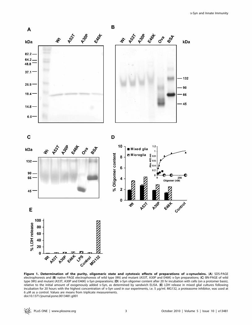
Figure 1. Determination of the purity, oligomeric state and cytotoxic effects of preparations of a-synucleins. (A) SDS-PAGEelectrophoresis and (B) native PAGE electrophoresis of wild type (Wt) and mutant (A53T, A30P and E46K) a-Syn preparations; (C) BN-PAGE of wildtype (Wt) and mutant (A53T, A30P and E46K) a-Syn preparations; (D) a-Syn oligomer content after 20 hr incubation with cells (on a protomer basis),relative to the initial amount of exogenously added a-Syn, as determined by sandwich ELISA. (E) LDH release in mixed glial cultures followingincubation for 20 hours with the highest concentration of a-Syn used in our experiments, i.e. 5 mg/ml. MG132, a proteasome inhibitor, was used at6 mM as a control. Values are means from triplicate measurements.doi:10.1371/journal.pone.0013481.g001
a-Syn and Innate Immunity
PLoS ONE | www.plosone.org 3 October 2010 | Volume 5 | Issue 10 | e13481

detached by trypsinization of the long-term cultures from neonatal
mouse brains –expected to be macroglia– were found to be
strongly immunostained for GFAP (not shown). As a second
measure of the purity of microglial cell cultures, the majority of
cells were found to express the pan haematopoietic lineage marker
CD45 (Figure 2B), the monocyte-macrophage marker CD11b
(Figure 2C), and the mature macrophage markers CD68 and F4/
80 (Figures 2D and 2E, respectively). In addition, quantitative
RT-PCR was employed to amplify specifically the GFAP and
CD11b genes (Figure 2F), and the results indicate no detectable
mRNA expression of the GFAP macroglial marker, and a clear
up-regulation of the microglia-specific CD11b gene expression
after stimulation by lipopolyssacharide (LPS). Taken together, the
cell marker profiles observed confirms the high purity of the
microglial cultures and hence to the reliability of the results
obtained using our purified microglial cell culture model.
Pro-inflammatory response of a-Syn-stimulated glia andmicroglia
In the normal CNS, brain tissue provides an immunosuppres-
sive environment, which seems to be important for the proper
function of the CNS. Under circumstances that cause disruption of
this environment, including chronic inflammatory conditions in
neurodegenerative diseases, a variety of immune regulatory and
inflammatory mediators can be activated [25]. Although various
types of cells have been identified as sources of cytokines in the
CNS, microglia appear to be a principal source of pro-
inflammatory and immune regulatory cytokines [25,61]. In order
to explore the immunological properties of extracellular, non-
aggregated a-Syn, as well as to evaluate the importance of the
cellular context in this process, we measured by ELISA the release
of a series of key cytokines after incubation for 20 hours of mouse
primary cultures of mixed glia and isolated microglia, with
exogenously added Wt a-Syn or the early onset PD-linked a-Syn
variants A30P, E46K, and A53T (at 0.2, 1 or 5 mg/ml).
First, we assayed the release of IL-6, TNF-a, IFN-c and IL-1b,
four key pro-inflammatory cytokines (Figure 3 and S2). Wild-type
a-Syn moderately stimulated the release of IL-6 in mixed glial
cultures (Figure 3A, left panel), but contrary to findings for
aggregated Wt a-Syn [62], this effect was hardly detectable on
isolated microglia (Figure 3A, right panel). And in contrast to the
comparable IL-6 response observed for the four a-Syn variants
reported in a study using the human U-373 MG astrocytoma cell
line [53], we found remarkable differences in their behaviour,
notably a very strong IL-6-mediated pro-inflammatory response
induced by the A30P and E46K variants after stimulation of both
mixed glial and microglial cultures. These differences could be
explained by the nature of the cell line used by Klegeris and
coworkers. Alternatively, given that astrocytes are the most
abundant glial cell population of the CNS participating in local
innate immune responses; this result could be consistent with our
finding of less marked differences (Figure 3A) between the a-Syn
variants in mixed glial cultures. The A53T variant, however,
caused a weak but significant increase in IL-6 levels on total glia
but not on isolated microglia, similar to Wt a-Syn but even less
prominent. In the case of TNF-a and IFN-c levels measured in
microglial cultures, only stimulation with A30P produced a
significant increase (Figure S2), partially coinciding with the IL-
6 observed profile. Therefore, A30P and E46K appear to drive IL-
6, TNF-a, and IFN-c cytokine secretion in the context of PD-
affected glia.
When we assayed the levels of IL-1b from mixed glial cultures
(Figure 3B, left panel), only the A30P and E46K a-Syn variants
Figure 2. Evaluation of the purity of isolated microglial cell fractions. Immunofluorescence characterization of purified microglial cellcultures for the specific macroglial lineage marker GFAP (A), the pan haematopoietic lineage marker CD45 (B), and the mature macrophages markersCD11b (C), CD68 (D), and F4/80 (E). Nuclei are counterstained in blue with Hoechst 33342. Scale bar: 100 mm. mRNA expression (qRT-PCR) fromisolated microglial preparations after no stimulation (control) or after stimulation with 1 mg/ml LPS for 20 hours (F). Specific primers for amplifyingthe GFAP and CD11b genes were used.doi:10.1371/journal.pone.0013481.g002
a-Syn and Innate Immunity
PLoS ONE | www.plosone.org 4 October 2010 | Volume 5 | Issue 10 | e13481

showed a significant stimulatory effect. Interestingly, when tested
on isolated microglial cells, while A30P did not cause a significant
rise in IL-1b levels relative to the control, Wt a-Syn 2in
agreement with a previous report of mRNA levels in a comparable
experiment design [37]2 and especially E46K, showed a very
significant increase in cytokine release (Figure 3B, right panel).
Taken together, these results suggest that A30P and E46K, as
opposed to either Wt a-Syn or the A53T variant, induce a pro-
inflammatory response in glia. Remarkably, only two of the three
PD-linked variants of a-synuclein were IL-1b inducers in primary
glia, and contrary to expectations from previous studies performed
using cell line cultures and other studies involving modified a-
synuclein species, our results show that the A53T a-Syn variant
has instead a very modest activity in regulating the innate immune
response by primary glia and microglia. These findings indicate
the need for further detailed studies on the role of the glia in early
PD onset before detailed conclusions as to the role of the latter in
PD can be drawn.
Previously, the available data had shown that the A30P and
A53T a-Syn variants, but not the Wt and the E46K forms,
efficiently induced the release of IL-1b when added to THP-1
macrophage cell line cultures [52] and that all the a-Syn variants
were able to increase IL-1b secretion in THP-1 cells only when co-
treatments with INF-c were included, suggesting a pro-inflamma-
tory response in already immune-primed THP-1 cells [52]. Our
data, obtained with primary microglial cultures, indicate a very
different behaviour. Thus, Wt and E46K a-Syn in our study
appear to be pro-inflammatory in primary microglia, while the
A30P and A53T variants seem to be unable to produce a
significant response. These differential responses of these two
studies point at the importance of the differentiation/maturation
status of the primary microglia, which suggests that there could be
multiple subtle but important differences in immune responses
during the establishment of PD. It is interesting that these
differences were only revealed in primary settings that were not
over-primed, as was the case of IFN-c treated THP-1 cells.
Figure 3. Pro-inflammatory interleukin profile of a-Syn-stimulated primary mixed glial and isolated microglial cultures. IL-6 (A) andIL-1b (B) release was measured by ELISA in culture supernantants of mixed glia (left) and microglia (right) after a 20-hour treatment with monomericWt or mutant a-Syn variants or lipopolysaccharide (LPS). Values are mean 6 S.E.M. (n = 4). * P,0.05, ** P,0.01, *** P,0.001. The results shown arerepresentative of two or three independent experiments with microglia and mixed glial cultures, respectively.doi:10.1371/journal.pone.0013481.g003
a-Syn and Innate Immunity
PLoS ONE | www.plosone.org 5 October 2010 | Volume 5 | Issue 10 | e13481

The observed effects on IL-1b secretion by primary glia and
microglia are of particular interest considering the emerging role
of an adaptive immune response in PD, in particular by CD4+ T
cells [63]. Given IL-1’s known effects to promote T cells responses,
our findings on IL-1b regulation by a-Syn variants in innate
immunocompetent cells require further attention in view of the
potential effects of the latter in mediating immune tolerance and T
effector responses.
IL-10 regulation by a-Syn-stimulated glia and microgliaAlthough most studies in the past have focused on microglial
production of pro-inflammatory cytokines, a large body of
evidence has supported the notion that microglia also produce
cytokines with anti-inflammatory or regulatory activities [25].
Indeed, a strong induction of IL-10 2recognized as an anti-
inflammatory cytokine2 had been observed for microglial cells
stimulated with nitrated, aggregated Wt a-Syn [62]. We therefore
investigated the effects of non-aggregated and unmodified a-Syn
on glial secretion of IL-10 2a Th2 immunoregulator2 which
reduces cytokine production by Th1 cells (Figure 4). Our results
show that only the A30P variant produced a significant increase in
IL-10 levels in mixed glial cells (top panel) whilst, on the contrary,
the A53T variant caused a significant reduction of IL-10 basal
levels, also observed in microglia, likely suggesting a lack of
microglial response, a differential uptake by microglia, or an effect
of the uptaken a-Syn on the endogenous IL-10 when A53T is
present. In this sense, it has been reported a link between a-Syn
and the microglial activation features [64], including phagocytic
ability.
In microglial cells, on the other hand, only the E46K variant
produced an increase in IL-10 levels as compared to the control
(bottom panel). These results might suggest that microglial cells, in
order to produce a-Syn-driven endogenous IL-10, require both
IL-6 and IL-1b secretion. In this sense, while Wt a-Syn increased
only IL-1b production in microglial cells, A30P increased only the
IL-6 production. These facts could reflect the requirement of a
doubly activated state of the microglia for IL-10 production.
Although the general mechanism that generate IL-10 production
within the CNS during neuroinflammation is still not well enough
understood, our results support a role for a-Syn in the modulation
of the microglial phenotype as suggested by Austin and
collaborators [64].
Chemokine release profiles by a-Syn-stimulated glia andmicroglia
Chemokines are involved in a wide variety of disorders in the
CNS and their actions contribute to reactive glial changes and
neuronal injury in neuroinflammatory conditions [65–67]. We
therefore sought to determine the chemokine release profiles of
glial and microglial cells induced by non-aggregated Wt and PD-
linked a-Syn variants. In particular, we assayed the release of IP-
10/CXCL10, RANTES/CCL5, MCP-1/CCL2 and MIP-1a/
CCL3 chemokines (Figure 5).
The regulation by a-Syn of IP-10/CXCL10 in CNS cells has
not been previously reported. In addition, the only data available
regarding RANTES/CCL5 levels in PD are from a transgenic rat
model for A53T a-Syn [38], or from sera and thus relate to
peripheral dysregulation in the cytokine network associated with
PD patients [68–70], and although no information is given on the
genetic background of these PD patients, these studies reported
increased levels of circulating RANTES/CCL5. Our results show
large and comparable effects for A30P and E46K variants on IP-
10/CXCL10 and RANTES/CCL5 release, both by mixed glial
cultures and by microglia (Figures 5A and 5B). Remarkably,
neither Wt nor A53T in both glial cultures and microglia
produced a relevant increase in IP-10/CXCL10 or RANTES/
CCL5 secretion, the latter result in agreement with the one
reported for the striatum and SN in the (A53T) rat model [38].
These data suggest a specific role for A30P and E46K a-Syn
variants associated with enhancement of Th1-cell recruitment,
activation, and effector potential.
Besides their role as chemoattractants, IP-10/CXCL10 and
RANTES/CCL5 are known to induce T-cell proliferation and
cytokine production [71], suggesting a role for A30P and E46K a-
Figure 4. Immunoregulatory effect of a-Syn-stimulation inprimary mixed glial and isolated microglial cultures. IL-10release was measured by ELISA in supernatants of a-Syn-stimulatedmixed-glial cultures (top) and microglia (bottom) after a 20-hourtreatment with monomeric Wt or mutant a-Syn variants, or lipopoly-saccharide (LPS). Values are mean 6 S.E.M. (n = 4). * P,0.05, ** P,0.01,*** P,0.001. The results shown are representative of two or threeindependent experiments with microglia and mixed glial cultures,respectively.doi:10.1371/journal.pone.0013481.g004
a-Syn and Innate Immunity
PLoS ONE | www.plosone.org 6 October 2010 | Volume 5 | Issue 10 | e13481

Syn variants beyond purely innate pro-inflammatory responses. In
this sense, IP-10/CXCL10 is pivotal in generating antigen-specific
T-cells [71]. As the possible roles of adaptive immune responses in
PD is gaining increasing attention [72,73], our data should
contribute to a better understanding of the differential immune
responses exerted by the different a-Syn variants in terms of
augmented microglia and macrophage recruitment and/or
activation. The strong pro-inflammatory species (IL-1b, IL-6, IP-
10/CXCL10, and RANTES/CCL5) whose release the A30P and
E46K variants seem to promote in the CNS could lead to
augmented macrophage recruitment and/or activation. This view
is supported by the observation of upregulation of MCP-1/CCL2
and MIP-1a/CCL3 by A30P and E46K (Figures 5C and 5D).
Interestingly, the large effects observed for A30P and E46K were
much more pronounced in microglia, indicating a context-
dependent stimulation mechanism. Further studies to asses the
Figure 5. Chemokine release profile of a-Syn-stimulated primary mixed glial cultures and isolated microglia cultures. IP-10 (A),RANTES (B), MCP-1 (C), and MIP-1a (D) were measured in supernantants of mixed-glial cultures (left) and microglia (right) after a 20-hour treatmentwith monomeric Wt or mutant a-Syn variants, or lipopolysaccharide (LPS). All chemokines were assayed by ELISA as described in Materials andMethods. Values are mean 6 S.E.M. (n = 2). * P,0.05, ** P,0.01. The results shown are representative of two and three independent experimentswith microglia and mixed glial cultures, respectively.doi:10.1371/journal.pone.0013481.g005
a-Syn and Innate Immunity
PLoS ONE | www.plosone.org 7 October 2010 | Volume 5 | Issue 10 | e13481

extent to which peripheral inflammation may amplify the
neuroinflammation contributing to PD are needed, but our results
raise the possibility of the need for a more personalised
manipulation of the different PD situations in terms of immuno-
supressory treatments and/or immunomodulatory therapeutic
approaches.
Previously, in addition to increased levels of TNF-a, IL-6, and
INF-c, stimulation of microglia with nitrated, aggregated a-Syn
had been shown to enhance the secretion of MCP-1/CCL2
[46,62]. In this work, we also found significantly higher levels of
MCP-1/CCL2 induced by non-aggregated Wt a-Syn, as well as a
similar MCP-1 release profile in mixed glial cultures for Wt a-Syn
and its A30P and E46K variants, while A53T caused no detectable
chemokine release (Figure 5C).
In summary, stimulation of total glia and microglia with A30P
and E46K variants show large increases of IP-10/CXCL10,
RANTES/CCL5, MCP-1/CCL2 and MIP-1a/CCL3 levels. Wt
a-Syn, however, only induced the release of MCP-1/CCL2 and
MIP-1a/CCL3, but not in isolated microglia, suggesting potential
differential effects of Wt a-Syn and its variants on Th1/Th2
activation and/or recruitment, and a more prominent effect for
A30P and E46K a-Syn variants in adaptive immune responses in
PD.
Effect of non-aggregated a-Syn stimulation on microglialphagocytosis
Phagocytosis is believed to be involved in steady-state tissue
homeostasis, via the clearance of apoptotic cells, and the
promotion of tissue repair and resolution of the wound [66,74].
These aspects are related to the ‘alternative activation’ and
acquired deactivation of the microglia, as a counter phenotype to
the ‘classically activated’, pro-inflammatory, microglia [74].
Therefore, in order to assess the role of Wt a-Syn and the PD-
linked variants on phagocytosis, we used fluorescein-conjugated
tracker microparticles for measuring the phagocytosis capacity of
differentially activated primary microglial cells (Figure 6).
It has been previously reported that monomeric Wt a-Syn
enhances phagocytosis of a microglial cell line [51], and indeed,
our results show that Wt and A53T a-Syn promote phagocytosis of
microglial cells (Figure 6B), while the opposite effect was
observed for cell cultures stimulated by the A30P and E46K a-
Syn variants (Figures 6A and 6B). Interestingly, the phagocytic
capacity measured for the stimulated microglia was observed to be
inversely correlated to the IL-6 release levels induced by the
different a-Syn variants (Figure 3A). Our findings point to
induction of differential microglial phenotypes by a-Syn variants.
Thus, A53T a-Syn, which was not associated with a robust pro-
inflammatory activity, produced an increase in the phagocytic
capacity that could reflect an alternative activation state of the
microglia, as observed with CNS damage [61]. However, Wt a-
Syn, which was associated with a moderate proinflammatory
response in our model, promoted phagocytosis in microglia,
indicating a combination of alternative and classical activation
states, a scenario that has been related to chronic inflammatory
processes such as those observed in Alzheimer’s disease [74]. On
the other hand, the A30P and E46K variants of a-Syn induced a
strong pro-inflammatory response, combined with reduced
phagocytic capacity, reflecting a classical activation state, which
is clearly associated with the most cytotoxic situation.
Taken together, our results suggest that extracellular, non-
aggregated Wt a-Syn produces a moderate to low pro-inflamma-
tory response in glia, together with a reduction of the
immunoregulatory response, and a moderate stimulation of Th1
chemokine secretion. The A30P and E46K pathological variants,
on the other hand, can induce strong pro-inflammatory and
immunoregulatory responses, together with marked increases in
chemokine release levels, both in total glia and microglia. This
exacerbated native immune response generated by these two a-
Syn variants might explain the earlier onset and more rapid
evolution of these two genetic forms of PD as compared to the
sporadic variety. Intriguingly, our results from the pathologically-
linked A53T variant, apart from a weak effect on IL-6 levels, did
not provoke a significant native immune response. This finding
suggests that there are other mechanisms of neurodegeneration
that can contribute to the pathogenesis of PD, perhaps involving
adaptive immune responses that might be promoted specifically by
the A53T variant.
In comparison to the classical sporadic form of PD, the clinical
phenotypes associated with mutations in a-Syn are characterized
by an earlier disease onset but a reduced prevalence of tremor
[75–79]. Studies that help to correlate the different a-Syn variants
with the mechanism of neurodegeneration, and ultimately with
disease progression, are therefore of considerable importance.
However, we should be cautious about preclinical studies in
animal models relating to translational research, as immune
responses might vary between mice and human systems [80].
Equally important is to mention the lack of physiologically
oriented studies employing human primary microglia, rather than
immortalised cell lines. In this context, our results on the effects on
glial native immunity exerted by extracellular, non-aggregated Wt
a-Syn and the various familial PD-linked variants could be of
value in the development of rational approaches towards effective
control of immune responses that are associated with PD.
Materials and Methods
a-synuclein overexpression, purification, and preparationHuman wild-type a-Syn and the A30P, E46K and A53T
mutants were overexpressed in E. coli BL21(DE3) cells using
plasmid pT7-7 and purified as described previously [81] with
minor modifications, as follows. After cell transformation,
BL21(DE3)-competent cells were grown in LB in the presence of
ampicillin (100 mg/ml). Protein expression was induced with
1 mM IPTG, and cells were harvested by centrifugation at
3,500 g after shaking at 37uCfor 4 hours. The cell pellet was
resuspended in 10 mM Tris–HCl (pH 8.0), 1 mM EDTA, and
EDTA-free protein inhibitor cocktail (Roche Diagnostics, Burgess
Hill, UK), and lysed by multiple freeze–thaw cycles and
sonication. The cell suspension was boiled for 20 min and
centrifuged at 20,000 g. Streptomycin sulphate was added to the
supernatant to a final concentration of 10 mg/ml and the mixture
was stirred for 15 min at 4uC. After centrifugation at 13,500 rpm,
the supernatant was collected and ammonium sulphate was added
(to 0.36 g/ml). The solution was stirred for 30 min at 4uC and
centrifuged again at 13,500 rpm. The pellet was resuspended in
25 mM Tris–HCl (pH 7.7), and loaded onto an HQ/M-column
on a BioCAD (Applied Biosystems, Foster City, USA) workstation.
The synuclein proteins were eluted at ca. 300 mM NaCl with a
salt gradient from 0 mM to 600 mM NaCl. The pure a-Syn
fractions were pooled together and dialyzed extensively at 4uCagainst water. Protein purities were .95% as determined by SDS-
PAGE, and molecular masses were confirmed by electrospray
mass spectrometry.
In order to get rid of any possible bound contaminants, the
purified proteins were subjected to denaturation with 6 M urea in
20 mM Tris/HCl (pH 8.0) for 1 hour at room temperature and
subsequently dialyzed at 4uC against 20 mM Tris/HCl (pH 8.0)
containing 3 M, 1.5 M, 0.75 M, and 0.375 M urea, and finally
a-Syn and Innate Immunity
PLoS ONE | www.plosone.org 8 October 2010 | Volume 5 | Issue 10 | e13481

against water. Afterwards, the protein preparations were concen-
trated by centrifugation using a 10 k Amicon Ultra-Centrifugal
Filter Device (Millipore Iberica, Madrid, Spain), and passed under
sterile conditions through a 0.22-mm filter, and stored at 280uC.
PAGE electrophoresis analyses of the a-Syn preparationsTen mg of protein were loaded onto a 15% SDS-PAGE gel (the
loading buffer containing 0.1 M DTT), or 25 mg onto a 4–12%
gradient Tris-glycine native PAGE gel (Lonza Group, Basel,
Switzerland), and subjected to electrophoresis at 200 V. Gels were
stained with Blue Silver [82], and destained with water. For Blue-
Native PAGE (BN-PAGE), samples were prepared according to
the protocol described previously for purified samples [83] with
some modifications. Briefly, 12 mg of protein for experimental
samples and 20 mg for protein markers were loaded on a
continuous 15% poly-acrylamide gel. Ovalbumin and bovine
serum albumin (Sigma-Aldrich, St. Louis, USA) were used as
protein markers for native PAGE and BN-PAGE assays. After
Figure 6. Effect of a-Syn-stimulation on microglial phagocytosis. (A) After treatment of the primary microglial cell cultures with a-Syn (1 mg/ml) for 20 hours, cells were incubated with fluorescent microspheres for 1 hour. After fixing the cells, phagocytosis was assessed by fluorescencemicroscopy analysis. The phagocytic index was calculated by dividing the fluorescence from the phagocytosed microspheres by the total number ofcells in the images. Four images were analysed for each sample in each experiment, and the results shown are representative of three independentexperiments. A.U.: arbitrary units. (Representative microscopy images used to determine the phagocityc activity of microglial cultures stimulated withWt (top) an A30P (centre) a-Syn, or non stimulated microglial cells (bottom). From left to right, green fluorescent microspheres, Hoechst-stained cells,and cells as observed in the absence of fluorescence. Scale bar: 50 mM.doi:10.1371/journal.pone.0013481.g006
a-Syn and Innate Immunity
PLoS ONE | www.plosone.org 9 October 2010 | Volume 5 | Issue 10 | e13481

electrophoresis, gels were destained in a solution of 50% methanol
and 10% acetic acid until the bands were clearly visible.
Time-course determination of total a-Syn and oligomercontent in the medium after incubation with cells
For a quantitative assessment of the oligomeric content of
culture supernatants, a specific sandwich ELISA assay [60] and a
calibration curve with oligomers of an a-Syn variant carrying six
point mutations (V63A, T64S, N65H, V66L, V71F, T72S), were
used. Such oligomers were prepared as follows: a 700 mM solution
in PBS of the mutant a-Syn was incubated overnight at RT, and
subjected to centrifugation with a 100 kDa cut-off centrifugal filter
(Millipore #UFC510024). The resulting retentate was rinsed with
150 ml of cold PBS and subjected to centrifugation as before. This
procedure was repeated three more times, in order to eliminate the
non-oligomerized protein. The oligomeric nature of the prepared
a-Syn fraction was confirmed by native PAGE and Western blot
(not shown), and the a-synuclein protomer concentration was
determined with the BCA assay kit (Pierce, Rockford, USA.).
Quantification of total a-Syn species in culture supernatants
after 0, 1, 6 and 20 hours of incubation with cells at 37uC was
performed by ELISA. Briefly: individual wells of 96-well ELISA
plates were coated with 100 ml of cell-free culture supernatants.
Plates were incubated at 37uC for 2 hours and washed with 0.5%
Tween20/PBS, pH 7.2 (PBST). Plates were blocked with 200 ml of
1% BSA in PBS and incubation at 37uC for 1 hour. After washing
with PBST, 100 ml/well of a 1 mg/ml biotinylated anti-a-Syn 211
mouse mAb [Santa Cruz Biotechnology, Santa Cruz, USA;
biotinylated as previously described [60] diluted in 1% BSA in
PBS were added and plates were incubated at 37uC for 1 hour.
After washing with PBST, 100 ml/well of 1:5000 dilution of
ExtrAvidin-alkaline phosphatase (Sigma-Aldrich, St. Louis, USA)
in 1% BSA in PBS were added, and plates were incubated at 37uCfor 30 min. After washing with PST, 100 ml/well of pNPP
substrate (Sigma, St. Louis, USA) were added, and absorbance
at 405 nm was measured within 30 minutes.
Cell culturesMixed glial cultures were prepared from the cerebral cortices of
1–3 days-old C57BL/6 mice (University of Seville Animal Core
Facility, Seville, Spain) according to previously described methods
[33] with some modifications. After mechanical, trypsin-mediated
(BioWhittaker, Verviers, Belgium) dissociation, followed by filtra-
tion in DMEM-F12 with 10% inactivated FBS (BioWhittaker,
Verviers, Belgium), cells were cultured at 37uC onto 12-well plates
treated with poly-D-lysine (Sigma-Aldrich, St. Louis, USA). After 2
days, half of the volume of culture medium was carefully changed,
and completely changed after 4 days of culture. Cells were used for
stimulation or microglial isolation at day 18–22 of culture.
Microglial isolation was carried out according to previously
described methods [84] with some modifications. The supernatant
was removed and the wells were washed with DMEM-F12 without
inactivated FBS; this conditioned medium was stored to be used
later. Cells were incubated for 30–45 min with DMEM-F12/
trypsin 0.25% solution at 37uC and complete medium was added to
inactivate trypsin and the supernatant containing microglial cells
collected. Conditioned medium was added to attached microglia;
the following day the medium was changed for normal medium,
and microglial cultures were stimulated after 5 days post isolation.
Immunofluorescence analysisPurified microglia were characterized by immunocytochemistry
on the basis of their expression of the pan haematopoietic marker
CD45 and of the monocyte/macrophage markers CD11b, F4/80
and CD68. Additionally, the absence of Glial Fibrillary Acidic
Protein (GFAP)-positive astrocytes in purified microglial cell
cultures was also established. By contrast, trypsin-detached cells,
largely astrocytes but containing some microglial cells, were plated
back into 12-multiwell tissue culture plates and immunocyto-
chemically processed as mentioned above.
For detection of CD45, CD11b and CD68 cell-surface antigens,
cells were fixed for 10 min at room temperature (RT) in PBS
containing 4% paraformaldehyde, washed twice in PBS and finally
blocked overnight at 4uC in PBS containing 3% bovine serum
albumin (BSA). The cells were then incubated for 1 hour at 4uCwith a fluorescein isothiocyanate (FITC)-conjugated rat anti-
mouse CD45 monoclonal antibody (Leukocyte Common Antigen,
Ly-5, clone 30-F11, BD Pharmingen, Franklin Lakes, USA) and a
FITC-conjugated rat anti-mouse CD11b monoclonal antibody
(clone M1/70, BD Pharmingen, Franklin Lakes, USA). Cells were
exposed to primary rat anti-mouse F4/80 monoclonal antibodies
(clone Cl:A3-1, Serotec, Oxford, UK) for 1 hour at RT at a final
dilution of 1:100 in PBS. For detection of CD68 and GFAP
intracellular antigens, 4% PFA-fixed cells were permeabilized and
blocked overnight at 4uC in PBS containing 3% BSA and 0.5% of
the permeabilizing detergent Triton-X-100. The following day,
cells were incubated for 1 hour at 4uC in PBS containing a
dilution of 1:100 rat anti-mouse monoclonal CD68 antibodies
(clone FA-11, Serotec, Oxford, UK) and a 1:300 dilution of anti-
GFAP mouse monoclonal antibodies (clone G-A-5, Sigma-
Aldrich, Saint Louis, USA). Labelling with F4/80 and CD68
primary antibodies was detected from fluorescence measurements
after incubating cells for 30 min at RT in PBS containing 1:100
diluted FITC-conjugated goat anti-rat IgG secondary antibodies
(Jackson ImmunoResearch, West Grove, USA). For GFAP
detection, an Alexa Fluor 488 nm goat anti-mouse IgG secondary
antibody solution (Invitrogen, Paisley, UK) was added for 30 min
at RT at a final dilution of 1:300 in PBS. Immunofluorescence
images were captured with an inverted fluorescence microscope
Olympus IX71 using the digital image processing softwares DP
Controller and DP Manager (Olympus Europa, Hamburg,
Germany).
Phagocytosis assaysFluoresbriteTM carboxylate 0.75 m microspheres (2.64% Solid-
Latex; Polysciences Inc, Warrington, USA) were used as
fluorescein-conjugated tracker microparticles for measuring the
phagocytosis capacity of differentially activated microglial cells.
One hour before starting the phagocytosis assay, fluorescent
microspheres (1.0861011 particles/ml) were mixed at a ratio of
1 ml/20 ml inactivated FBS (BioWhittaker, Verviers, Belgium) and
incubated for 1 hour at 37uC in order to opsonise fully the
carboxylate groups. The mixture of microspheres and FBS was
then resuspended in fresh DMEM-F12 medium (BioWhittaker,
Verviers, Belgium), with L-glutamine and P/S antibiotics
supplements to obtain normal 10% FBS-supplemented media
containing 5.46108 microspheres/ml.
After removal of 400 ml of supernantant from the a-Syn-
stimulated microglial cell cultures, a volume of 150 ml of
resuspended microspheres was added to each well to obtain a
final concentration of 1.086108 particles/ml. The particles were
then homogenously distributed throughout each well by gentle
movements of the plate and incubated for 1 hour at 37uC. The
medium containing non-phagocytosed microspheres was then
removed and the cells were washed with PBS prior to their fixation
with 4% paraformaldehyde in PBS for 30 min at 4uC. One ml of
PBS containing the nuclear fluorescent dye Hoechst 33342 (1 mg/
a-Syn and Innate Immunity
PLoS ONE | www.plosone.org 10 October 2010 | Volume 5 | Issue 10 | e13481

ml) was then added to the cells, and the plates were stored at 4uCfor a minimum of 24 hours before being analyzed.
Determination of the phagocytic indexFor each cell culture condition, the phagocytic capacity of
microglial cells was determined by analysing fluorescent images of
phagocytosed FITC-labelled microspheres and by staining cell
nuclei with Hoechst 33342. An Olympus IX71 fluorescence
microscope equipped with the digital image processing softwares
DPController and DPManager (Olympus Europa, Hamburg,
Germany) was used.
For each random field, a mean phagocytic index was calculated
by determining the intensity of specific green and blue fluores-
cence emissions with digital imaging analysis software MetaMorph
(MDS Analytical Technologies, Toronto, Canada). Specific green
fluorescence emitted by FITC-microspheres was determined by
subtracting the mean background fluorescence calculated from
three different areas of the image where no cells were present,
from the overall mean green fluorescence of the entire image.
Specific blue emitted nuclear fluorescence was then calculated in a
similar manner. The phagocytic index corresponds to the specific
green/blue ratio. The mean phagocytic index was calculated from
4 random fields of cells (.100 cells) and was considered as a
representative value of the phagocytic capacity of the microglial
cells, as previously determined for mouse peritoneal macrophages
within normal media for 24 hours with 1 ml/ml of lipopolysachar-
ide (LPS).
qRT-PCRExpression of GFAP and CD11b was determined by using a
two-step quantitative real-time PCR. Total RNA from two
microglial culture wells, one without treatment and one treated
with LPS for 16 hours, was extracted using the RNeasy Micro Kit
(Qiagen GmbH, Hilden, Germany) according to the manufactur-
er’s protocol. One mg of RNA was reverse-transcribed using the
Quantitect Reverse Transcription kit (Qiagen GmbH, Hilden,
Germany) according to the manufacturer’s protocol. qPCR was
performed with SYBRH Premix EX TaqTM (Perfect Real Time)
(Takara Bio Inc., Otsu, Shiga, Japan) on an ABI Prism 7500 Real
Time PCR System. Primers used were: HPRT_For: 59-GTAAT-
GATCAGTCAACGGGGGAC-39, HPRT_Rev: 59-CCAGCAA-
GCTTGCAACCTTAACCA-39; GFAP_For: 59-ATCGAGATC-
GCCACCTACAG-39, GFAP_Rev: 59-CTCACATCACCACG-
TCCTTG-39; CD11b_For: 59-CAGATCAACAATGTGACCG-
TATGG-39, CD11b_Rev: 59-CATCATGTCCTTGTACTGC-
CGC-39). Multiple transcripts were analyzed simultaneously for 40
cycles using an optimized qRT-PCR thermal profile. Changes in
gene expression were determined using the 22DDCt method with
normalization to endogenous hypoxanthinephophoribosyltransfer-
ase (HPRT) control.
Cytotoxicity assaysThe cytotoxic effect of the a-Syn variants under study was
evaluated from the extent of LDH release with by using the LDH
Cytoxicity Detection Kit (Roche, Basel, Switzerland) in MGC,
following stimulation with the maximum concentration of wild-
type a-Syn, its mutants or LPS (Sigma-Aldrich, St. Louis, USA),
used in the present experiments (5 mg/ml for a-synuclein samples
and 1 mg/ml for LPS). The limiting value in each case was
determined using 6 mM MG132 (Sigma-Aldrich, St. Louis, USA),
which is known to be lethal for cells at the concentration used,
again using the manufacturer’s protocol.
Cytokine release measurementsGlial mixed cultures and isolated microglial cultures were
stimulated with Wt a-Syn and its mutational variants at different
concentrations (5 mg/ml and 1 mg/ml for MGC and 1 mg/ml and
0.2 mg/ml for MiG) for 20 hours. LPS at a concentration of 1 mg/
ml, and culture medium alone were used as positive and negative
controls, respectively. Culture supernatants were harvested and
centrifuged at 700 g for 5 min. and cell-cleared supernatants were
recovered and stored at 280uC before cytokine measurement. IL-
6, IL-1b, TNF-a, IFN-c and IL-10 levels were assayed using
Mouse IL-6/IL-1b/TNF-a/IFN-c/IL-10 BD OptEIA ELISA set
(BD Biosciences, Madrid, Spain) according to the manufacturer’s
protocol. Chemokine levels in the culture supernatants were
determined by a specific sandwich ELISA by using capture/
biotinylated detection antibodies obtained from Peprotech (Lon-
don, UK) according to the manufacturer’s recommendations.
Cytokine profiles shown are representative of three independent
experiments.
Ethics statementAll animals were handled in strict accordance with good animal
practice as defined by the relevant national/EU guidelines and the
CEA-CABIMER Experimental Animal Committee, and all
animal work was approved by the appropriate committee (file
CEA-2010-14).
Data analysisAll values are expressed as mean 6 S.E.M. Statistical
significance (Student’s test, two-tailed) was evaluated using SPSS
Statistics 17.0 (IBM Company, Chicago, USA).
Supporting Information
Figure S1. Time-course quantitation of total a-Syn in cell
culture supernatants. Total a-Syn content measured by direct
ELISA in culture supernatants recovered after addition of wild-
type a-Syn to mixed glial cultures and incubation for 0, 1, 6 and
20 hours at 37uC.
Found at: doi:10.1371/journal.pone.0013481.s001 (0.06 MB TIF)
Figure S2. TNF-a and IFN-c release profile of a-Syn-
stimulated primary microglial cultures. TNF-a (A) and
IFN-c (B) levels were measured by ELISA in culture supernantants
of microglia after a 20-hour treatment with exogeneously added a-
Syn variants, or lipopolysaccharide (LPS). Values are mean 6
S.E.M. (n = 3). The results shown are representative of two
independent experiments.
Found at: doi:10.1371/journal.pone.0013481.s002 (0.13 MB TIF)
Author Contributions
Conceived and designed the experiments: CR CMD DP. Performed the
experiments: CR ALG EGR RFM MC CCL CAW MD DP. Analyzed the
data: CR ALG EGR RFM MC MD CMD DP. Contributed reagents/
materials/analysis tools: CAW. Wrote the paper: CR DP.
References
1. Spillantini MG, Crowther RA, Jakes R, Hasegawa M, Goedert M (1998) alpha-
Synuclein in filamentous inclusions of Lewy bodies from Parkinson’s disease and
dementia with Lewy bodies. Proc Natl Acad Sci USA 95: 6469–6473.
2. Croisier E, Moran LB, Dexter DT, Pearce RK, Graeber MB (2005) Microglial
inflammation in the parkinsonian substantia nigra: relationship to alpha-
synuclein deposition. J Neuroinflammation 2: 14.
a-Syn and Innate Immunity
PLoS ONE | www.plosone.org 11 October 2010 | Volume 5 | Issue 10 | e13481

3. Eriksen JL, Wszolek Z, Petrucelli L (2005) Molecular pathogenesis of Parkinson
disease. Arch Neurol 62: 353–357.
4. Moore DJ, West AB, Dawson VL, Dawson TM (2005) Molecular pathophys-
iology of Parkinson’s disease. Annu Rev Neurosci 28: 57–87.
5. Chiti F, Dobson CM (2006) Protein misfolding, functional amyloid, and human
disease. Annu Rev Biochem 75: 333–366.
6. Sung JY, Kim J, Paik SR, Park JH, Ahn YS, et al. (2001) Induction of neuronal
cell death by Rab5A-dependent endocytosis of alpha-synuclein. J Biol Chem
276: 27441–27448.
7. Liu J, Zhou Y, Wang Y, Fong H, Murray TM, et al. (2007) Identification of
proteins involved in microglial endocytosis of alpha-synuclein. J Proteome Res 6:
3614–3627.
8. Lee HJ, Patel S, Lee SJ (2005) Intravesicular localization and exocytosis of
alpha-synuclein and its aggregates. J Neurosci 25: 6016–6024.
9. Sung JY, Park SM, Lee CH, Um JW, Lee HJ, et al. (2005) Proteolytic cleavage
of extracellular secreted a-synuclein via matrix metalloproteinases. J Biol Chem
280: 25216–25224.
10. Borghi R, Marchese R, Negro A, Marinelli L, Forloni G, et al. (2000) Full lengthalpha-synuclein is present in cerebrospinal fluid from Parkinson’s disease and
normal subjects. Neurosci Lett 287: 65–67.
11. El-Agnaf OM, Salem SA, Paleologou KE, Cooper LJ, Fullwood NJ, et al. (2003)
Alpha-synuclein implicated in Parkinson’s disease is present in extracellular
biological fluids, including human plasma. FASEB J 17: 1945–1947.
12. Kim S, Seo JH, Suh YH (2004) Alpha-synuclein, Parkinson’s disease, and
Alzheimer’s disease. Parkinsonism Relat Disord 10: S9–13.
13. Richter-Landsberg C, Gorath M, Trojanowski JQ, Lee VM (2000) alpha-
synuclein is developmentally expressed in cultured rat brain oligodendrocytes.J Neurosci Res 62: 9–14.
14. Mori F, Tanji K, Yoshimoto M, Takahashi H, Wakabayashi K (2002)
Demonstration of alpha-synuclein immunoreactivity in neuronal and glial
cytoplasm in normal human brain tissue using proteinase K and formic acid
pretreatment. Exp Neurol 176: 98–104.
15. Perez RG, Hastings TG (2004) Could a loss of alpha-synuclein function put
dopaminergic neurons at risk? J Neurochem 89: 1318–1324.
16. Weinreb PH, Zhen W, Poon AW, Conway KA, Lansbury PT, Jr. (1996) NACP,a protein implicated in Alzheimer’s disease and learning, is natively unfolded.
Biochemistry 35: 13709–13715.
17. Dedmon MM, Lindorff-Larsen K, Christodoulou J, Vendruscolo M,
Dobson CM (2005) Mapping long-range interactions in alpha-synuclein using
spin-label NMR and ensemble molecular dynamics simulations. J Am ChemSoc 127: 476–477.
18. Bertoncini CW, Jung YS, Fernandez CO, Hoyer W, Griesinger C, et al. (2005)
Release of long-range tertiary interactions potentiates aggregation of natively
unstructured alpha-synuclein. Proc Natl Acad Sci USA 102: 1430–1435.
19. Polymeropoulos MH, Lavedan C, Leroy E, Ide SE, Dehejia A, et al. (1997)
Mutation in the alpha-synuclein gene identified in families with Parkinson’s
disease. Science 276: 2045–2047.
20. Kruger R, Kuhn W, Muller T, Woitalla D, Graeber M, et al. (1998) Ala30Pro
mutation in the gene encoding alpha-synuclein in Parkinson’s disease. Nat Genet18: 106–108.
21. Zarranz JJ, Alegre J, Gomez-Esteban JC, Lezcano E, Ros R, et al. (2004) The
new mutation, E46K, of alpha-synuclein causes Parkinson and Lewy body
dementia. Ann Neurol 55: 164–173.
22. Gasser T (2005) Genetics of Parkinson’s disease. Curr Opin Neurol 18: 363–369.
23. Bertoncini CW, Fernandez CO, Griesinger C, Jovin TM, Zweckstetter M (2005)
Familial mutants of alpha-synuclein with increased neurotoxicity have a
destabilized conformation. J Biol Chem 280: 30649–30652.
24. Cookson MR (2009) alpha-Synuclein and neuronal cell death. Mol Neurode-
gener 4: 9.
25. Kim YS, Joh TH (2006) Microglia, major player in the brain inflammation: their
roles in the pathogenesis of Parkinson’s disease. Exp Mol Med 38: 333–347.
26. McGeer PL, Itagaki S, Boyes BE, McGeer EG (1988) Reactive microglia are
positive for HLA-DR in the substantia nigra of Parkinson’s and Alzheimer’s
disease brains. Neurology 38: 1285–1291.
27. Forno LS (1996) Neuropathology of Parkinson’s disease. J Neuropathol ExpNeurol 55: 259–272.
28. Banati RB, Daniel SE, Blunt SB (1998) Glial pathology but absence of apoptotic
nigral neurons in long-standing Parkinson’s disease. Mov Disord 13: 221–227.
29. Knott C, Stern G, Wilkin GP (2000) Inflammatory regulators in Parkinson’s
disease: iNOS, lipocortin-1, and cyclooxygenases-1 and -2. Mol Cell Neurosci
16: 724–739.
30. Mirza B, Hadberg H, Thomsen P, Moos T (2000) The absence of reactive
astrocytosis is indicative of a unique inflammatory process in Parkinson’s disease.Neuroscience 95: 425–432.
31. Vila M, Jackson-Lewis V, Guegan C, Wu DC, Teismann P, et al. (2001) The
role of glial cells in Parkinson’s disease. Curr Opin Neurol 14: 483–489.
32. Wu DC, Jackson-Lewis V, Vila M, Tieu K, Teismann P, et al. (2002) Blockadeof microglial activation is neuroprotective in the 1-methyl-4-phenyl-1,2,3,6-
tetrahydropyridine mouse model of Parkinson disease. J Neurosci 22:
1763–1771.
33. Delgado M, Ganea D (2003) Neuroprotective effect of vasoactive intestinal
peptide (VIP) in a mouse model of Parkinson’s disease by blocking microglialactivation. FASEB J 17: 944–946.
34. Sanchez-Pernaute R, Ferree A, Cooper O, Yu M, Brownell AL, et al. (2004)
Selective COX-2 inhibition prevents progressive dopamine neuron degeneration
in a rat model of Parkinson’s disease. J Neuroinflammation 1: 6.
35. Zhang W, Wang T, Pei Z, Miller DS, Wu X, et al. (2005) Aggregated alpha-
synuclein activates microglia: a process leading to disease progression in
Parkinson’s disease. FASEB J 19: 533–542.
36. Theodore S, Cao S, McLean PJ, Standaert DG (2008) Targeted overexpression
of human alpha-synuclein triggers microglial activation and an adaptive immune
response in a mouse model of Parkinson disease. J Neuropathol Exp Neurol 67:
1149–1158.
37. Su X, Maguire-Zeiss KA, Giuliano R, Prifti L, Venkatesh K, et al. (2008)
Synuclein activates microglia in a model of Parkinson’s disease. Neurobiol Aging
29: 1690–1701.
38. Chung CY, Koprich JB, Siddiqi H, Isacson O (2009) Dynamic changes in
presynaptic and axonal transport proteins combined with striatal neuroin-
flammation precede dopaminergic neuronal loss in a rat model of AAV alpha-
synucleinopathy. J Neurosci 29: 3365–3373.
39. Sanchez-Guajardo V, Febbraro F, Kirik D, Romero-Ramos M (2010) Microglia
acquire distinct activation profiles depending on the degree of alpha-synuclein
neuropathology in a rAAV based model of Parkinson’s disease. PLoS One 5:
e8784.
40. Gu XL, Long CX, Sun L, Xie C, Lin X, et al. (2010) Astrocytic expression of
Parkinson’s disease-related A53T alpha-synuclein causes neurodegeneration in
mice. Mol Brain 3: 12.
41. Roodveldt C, Christodoulou J, Dobson CM (2008) Immunological features of
alpha-synuclein in Parkinson’s disease. J Cell Mol Med 12: 1820–1829.
42. Lee SJ (2008) Origins and effects of extracellular alpha-synuclein: implications in
Parkinson’s disease. J Mol Neurosci 34: 17–22.
43. Benner EJ, Banerjee R, Reynolds AD, Sherman S, Pisarev VM, et al. (2008)
Nitrated alpha-Synuclein Immunity Accelerates Degeneration of Nigral
Dopaminergic Neurons. PLoS One 3: e1376.
44. Zhang W, Dallas S, Zhang D, Guo JP, Pang H, et al. (2007) Microglial PHOX
and Mac-1 are essential to the enhanced dopaminergic neurodegeneration
elicited by A30P and A53T mutant alpha-synuclein. Glia 55: 1178–1188.
45. Thomas MP, Chartrand K, Reynolds A, Vitvitsky V, Banerjee R, et al. (2007)
Ion channel blockade attenuates aggregated alpha-synuclein induction of
microglial reactive oxygen species: relevance for the pathogenesis of Parkinson’s
disease. J Neurochem 100: 503–519.
46. Reynolds AD, Glanzer JG, Kadiu I, Ricardo-Dukelow M, Chaudhuri A, et al.
(2008) Nitrated alpha-synuclein-activated microglial profiling for Parkinson’s
disease. J Neurochem 104: 1504–1525.
47. Giasson BI, Duda JE, Murray IV, Chen Q, Souza JM, et al. (2000) Oxidative
damage linked to neurodegeneration by selective alpha-synuclein nitration in
synucleinopathy lesions. Science 290: 985–989.
48. Souza JM, Giasson BI, Chen Q, Lee VM, Ischiropoulos H (2000) Dityrosine
cross-linking promotes formation of stable alpha-synuclein polymers. Implication
of nitrative and oxidative stress in the pathogenesis of neurodegenerative
synucleinopathies. J Biol Chem 275: 18344–18349.
49. Joo SH, Kwon KJ, Kim JW, Hasan MR, Lee HJ, et al. (2010) Regulation of
matrix metalloproteinase-9 and tissue plasminogen activator activity by alpha-
synuclein in rat primary glial cells. Neurosci Lett 469: 352–356.
50. Lee EJ, Woo MS, Moon PG, Baek MC, Choi IY, et al. (2010) Alpha-synuclein
activates microglia by inducing the expressions of matrix metalloproteinases and
the subsequent activation of protease-activated receptor-1. J Immunol 185:
615–623.
51. Park JY, Paik SR, Jou I, Park SM (2008) Microglial phagocytosis is enhanced by
monomeric alpha-synuclein, not aggregated alpha-synuclein: implications for
Parkinson’s disease. Glia 56: 1215–1223.
52. Klegeris A, Pelech S, Giasson BI, Maguire J, Zhang H, et al. (2008) Alpha-
synuclein activates stress signaling protein kinases in THP-1 cells and microglia.
Neurobiol Aging 29: 739–752.
53. Klegeris A, Giasson BI, Zhang H, Maguire J, Pelech S, et al. (2006) Alpha-
synuclein and its disease-causing mutants induce ICAM-1 and IL-6 in human
astrocytes and astrocytoma cells. FASEB J 20: 2000–2008.
54. Su X, Federoff HJ, Maguire-Zeiss KA (2009) Mutant alpha-Synuclein
overexpression mediates early proinflammatory activity. Neurotox Res 16:
238–254.
55. Jensen PH, Hojrup P, Hager H, Nielsen MS, Jacobsen L, et al. (1997) Binding of
Abeta to alpha- and beta-synucleins: identification of segments in alpha-
synuclein/NAC precursor that bind Abeta and NAC. Biochem J 323: 539–546.
56. Cole NB, Murphy DD, Grider T, Rueter S, Brasaemle D, et al. (2002) Lipid
droplet binding and oligomerization properties of the Parkinson’s disease protein
alpha-synuclein. J Biol Chem 277: 6344–6352.
57. Lashuel HA, Hartley D, Petre BM, Walz T, Lansbury PT, Jr. (2002)
Neurodegenerative disease: amyloid pores from pathogenic mutations. Nature
418: 291.
58. Moussa CE, Wersinger C, Rusnak M, Tomita Y, Sidhu A (2004) Abnormal
migration of human wild-type alpha-synuclein upon gel electrophoresis.
Neurosci Lett 371: 239–243.
59. Schagger H, von Jagow G (1991) Blue native electrophoresis for isolation of
membrane protein complexes in enzymatically active form. Anal Biochem 199:
223–231.
a-Syn and Innate Immunity
PLoS ONE | www.plosone.org 12 October 2010 | Volume 5 | Issue 10 | e13481

60. El-Agnaf OM, Salem SA, Paleologou KE, Curran MD, Gibson MJ, et al. (2006)
Detection of oligomeric forms of alpha-synuclein protein in human plasma as apotential biomarker for Parkinson’s disease. FASEB J 20: 419–425.
61. Long-Smith CM, Sullivan AM, Nolan YM (2009) The influence of microglia on
the pathogenesis of Parkinson’s disease. Prog Neurobiol 89: 277–287.62. Reynolds AD, Stone DK, Mosley RL, Gendelman HE (2009) Nitrated a-
synuclein-induced alterations in microglial immunity are regulated by CD4+ Tcell subsets. J Immunol 182: 4137–4149.
63. Reynolds AD, Banerjee R, Liu J, Gendelman HE, Mosley RL (2007)
Neuroprotective activities of CD4+CD25+ regulatory T cells in an animalmodel of Parkinson’s disease. J Leukoc Biol 82: 1083–1094.
64. Austin SA, Floden AM, Murphy EJ, Combs CK (2006) Alpha-synucleinexpression modulates microglial activation phenotype. J Neurosci 26:
10558–10563.65. Barreiro O, Martin P, Gonzalez-Amaro R, Sanchez-Madrid F (2010) Molecular
cues guiding inflammatory responses. Cardiovasc Res 86: 174–182.
66. Schwartz M (2010) ‘‘Tissue-repairing’’ blood-derived macrophages are essentialfor healing of the injured spinal cord: From skin-activated macrophages to
infiltrating blood-derived cells? Brain Behav Immun 24: 1054–1057.67. Rich RR, Fleisher TA, Shearer WT, Shroeder HW, Jr., Frew AJ, Weyand CM
(2008) Clinical Immunology. Principles and Practice TA F, ed. Elsevier Inc.
68. Rentzos M, Nikolaou C, Andreadou E, Paraskevas GP, Rombos A, et al. (2007)Circulating interleukin-15 and RANTES chemokine in Parkinson’s disease. Acta
Neurol Scand 116: 374–379.69. Gangemi S, Basile G, Merendino RA, Epifanio A, Di Pasquale G, et al. (2003)
Effect of levodopa on interleukin-15 and RANTES circulating levels in patientsaffected by Parkinson’s disease. Mediators Inflamm 12: 251–253.
70. Reale M, Iarlori C, Thomas A, Gambi D, Perfetti B, et al. (2009) Peripheral
cytokines profile in Parkinson’s disease. Brain Behav Immun 23: 55–63.71. Dufour JH, Dziejman M, Liu MT, Leung JH, Lane TE, et al. (2002) IFN-
gamma-inducible protein 10 (IP-10; CXCL10)-deficient mice reveal a role forIP-10 in effector T cell generation and trafficking. J Immunol 168: 3195–3204.
72. Stone DK, Reynolds AD, Mosley RL, Gendelman HE (2009) Innate and
adaptive immunity for the pathobiology of Parkinson’s disease. Antioxid RedoxSignal 11: 2151–2166.
73. Hirsch EC, Hunot S (2009) Neuroinflammation in Parkinson’s disease: a target
for neuroprotection? Lancet Neurol 8: 382–397.
74. Colton CA, Wilcock DM (2010) Assessing Activation States in Microglia. CNS
Neurol Disord Drug Targets 9: 174–191.
75. Bonifati V, Oostra BA, Heutink P (2004) Unraveling the pathogenesis of
Parkinson’s disease: the contribution of monogenic forms. Cell Mol Life Sci 61:
1729–1750.
76. Spira PJ, Sharpe DM, Halliday G, Cavanagh J, Nicholson GA (2001) Clinical
and pathological features of a Parkinsonian syndrome in a family with an
Ala53Thr alpha-synuclein mutation. Ann Neurol 49: 313–319.
77. Golbe LI, Di Iorio G, Sanges G, Lazzarini AM, La Sala S, et al. (1996) Clinical
genetic analysis of Parkinson’s disease in the Contursi kindred. Ann Neurol 40:
767–775.
78. Papapetropoulos S, Paschalis C, Athanassiadou A, Papadimitriou A, Ellul J,
et al. (2001) Clinical phenotype in patients with alpha-synuclein Parkinson’s
disease living in Greece in comparison with patients with sporadic Parkinson’s
disease. J Neurol Neurosurg Psychiatry 70: 662–665.
79. Bostantjopoulou S, Katsarou Z, Papadimitriou A, Veletza V, Hatzigeorgiou G,
et al. (2001) Clinical features of parkinsonian patients with the alpha-synuclein
(G209A) mutation. Mov Disord 16: 1007–1013.
80. Mestas J, Hughes CC (2004) Of mice and not men: differences between mouse
and human immunology. J Immunol 172: 2731–2738.
81. Hoyer W, Antony T, Cherny D, Heim G, Jovin TM, et al. (2002) Dependence
of alpha-synuclein aggregate morphology on solution conditions. J Mol Biol 322:
383–393.
82. Candiano G, Bruschi M, Musante L, Santucci L, Ghiggeri GM, et al. (2004)
Blue silver: a very sensitive colloidal Coomassie G-250 staining for proteome
analysis. Electrophoresis 25: 1327–1333.
83. Wittig I, Braun HP, Schagger H (2006) Blue native PAGE. Nat Protoc 1:
418–428.
84. Saura J, Tusell JM, Serratosa J (2003) High-yield isolation of murine microglia
by mild trypsinization. Glia 44: 183–189.
a-Syn and Innate Immunity
PLoS ONE | www.plosone.org 13 October 2010 | Volume 5 | Issue 10 | e13481
Copyright © 2022 FDOKUMEN