Genetic and Biochemical Evidence for the Involvement of a-1,4 Glucanotransferases in Amylopectin...
11
Genetic and Biochemical Evidence for the Involvement of a-1,4 Glucanotransferases in Amylopectin Synthesis 1 Christophe Colleoni, David Dauville ´e, Gregory Mouille, Alain Bule ´on, Daniel Gallant, Brigitte Bouchet, Matthew Morell, Michael Samuel, Brigitte Delrue, Christophe d’Hulst, Christophe Bliard, Jean-Marc Nuzillard, and Steven Ball* Laboratoire de Chimie Biologique, Unite ´ Mixte de Recherche du Centre National de la Recherche Scientifique no. 8576, Universite ´ des Sciences et Technologies de Lille, 59655 Villeneuve D’Ascq cedex France (C.C., D.D., G.M., B.D., C.d.H., S.B.); Institut National de la Recherche Agronomique, Centre de Recherches Agroalimentaires, Rue de la Ge ´raudie `re, B.P. 71627, 44316 Nantes cedex 03, France (A.B., D.G., B.B.); Commonwealth Scientific and Industrial Research Organization, Division of Plant Industry, G.P.O. Box 1600, Canberra, Australian Capital Territory 2601, Australia (M.M., M.S.); and Laboratoire de Pharmacognosie, Groupe de Glycotechnie, Universite ´ de Reims, Moulin de la Housse–BP 1039, 51097 Reims cedex 2, France (C.B., J.-M.N.) We describe a novel mutation in the Chlamydomonas reinhardtii STA11 gene, which results in significantly reduced granular starch deposition and major modifications in amylopectin structure and granule shape. This defect simultaneously leads to the accumulation of linear malto-oligosaccharides. The sta11-1 mutation causes the absence of an a-1,4 glucanotransferase known as disproportionat- ing enzyme (D-enzyme). D-enzyme activity was found to be corre- lated with the amount of wild-type allele doses in gene dosage experiments. All other enzymes involved in starch biosynthesis, including ADP-glucose pyrophosphorylase, debranching enzymes, soluble and granule-bound starch synthases, branching enzymes, phosphorylases, a-glucosidases (maltases), and amylases, were un- affected by the mutation. These data indicate that the D-enzyme is required for normal starch granule biogenesis in the monocellular alga C. reinhardtii. Starch is one of the most abundant biological polymers present in the earth’s biosphere and remains the major supply of calories in both human and animal diets. As is the case for animal, fungal, and bacterial glycogen, starch is made solely of Glc residues linked in a-1,4 positions and branched in a-1,6 positions. Unlike glycogen, starch gran- ules consist of complex, semicrytalline structures of unlim- ited size (for review, see Bule ´on et al., 1998). Amylopectin, the major polysaccharide fraction of starch, is considered to be the only molecular fraction required to generate normal granules. The building of the polymer structure depends on the transfer by SS of Glc in the a-1,4 position from ADP-Glc to the nonreducing end of growing chains. Intro- duction of the a-1,6 branch proceeds through the cleavage (by branching enzyme) of a pre-existing a-1,4-linked glu- can and the transfer of the cleaved glucan in the a-1,6 position. It was previously thought that the specific fea- tures of starch structure depended solely on the concerted action of multiple forms of SS and branching enzyme; however, other enzymes are likely to be involved in the process. Our strategy consisted of isolating mutants defective in amylopectin synthesis, thereby defining the functions in- volved in starch granule biogenesis. From the analysis of mutants of the monocellular green alga Chlamydomonas reinhardtii (Mouille et al., 1996), maize (James et al., 1995), rice (Nakamura et al., 1996), and Arabidopsis (Zeeman et al., 1998b), it was determined that debranching enzymes were required to trim a-1,6 linkages from a precursor (pre-amylopectin) into a mature amylopectin molecule (Ball et al., 1996) or that they were required to prevent glycogen production by the starch synthesis machinery (Zeeman et al., 1998b). Continued genetic analysis is likely to identify additional enzymes needed for starch biosyn- thesis that would not have been foreseen from the basic biosynthetic steps (Mouille et al., 1996). Eukaryotic algae are of particular relevance for studies dealing with starch synthesis. Starch polysaccharides are not found in bacteria or fungi. Because C. reinhardtii is the only starch-storing unicellular organism intensively stud- ied by geneticists, it offers a unique opportunity to under- stand the basic mechanisms of starch granule biogenesis. Indeed, growth-arrested C. reinhardtii cells accumulate glu- copolysaccharides that are very similar to cereal en- dosperm storage starch, so this organism serves as a highly useful model for starch synthesis in crop plants. C. rein- 1 This work was supported by grants from the Ministe `re de l’Education Nationale, by the Centre National de la Recherche Scientifique (Unite ´ Mixte de Recherche du Centre National de la Recherche Scientifique no. 8576, Director Andre ´ Verbert), by the University of Lille, by the University of Reims, and by a grant from Biogemma (Cambridge, UK). * Corresponding author; e-mail [email protected]; fax 33–3–20 – 43– 65–55. Abbreviations: APTS, 8-amino-1,3,6-pyrenetrisulfonic acid; D-enzyme, disproportionating enzyme; DP, degree of polymeriza- tion; GBSS, granule-bound starch synthase; SEM, scanning elec- tron microscopy; SS, soluble starch synthase; TEM, transmission electron microscopy. Plant Physiology, August 1999, Vol. 120, pp. 993–1003, www.plantphysiol.org © 1999 American Society of Plant Physiologists 993 www.plant.org on October 16, 2014 - Published by www.plantphysiol.org Downloaded from Copyright © 1999 American Society of Plant Biologists. All rights reserved.
-
Upload
independent -
Category
Documents
-
view
1 -
download
0
Transcript of Genetic and Biochemical Evidence for the Involvement of a-1,4 Glucanotransferases in Amylopectin...
Genetic and Biochemical Evidence for the Involvement ofa-1,4 Glucanotransferases in Amylopectin Synthesis1
Christophe Colleoni, David Dauvillee, Gregory Mouille, Alain Buleon, Daniel Gallant, Brigitte Bouchet,Matthew Morell, Michael Samuel, Brigitte Delrue, Christophe d’Hulst, Christophe Bliard,
Jean-Marc Nuzillard, and Steven Ball*
Laboratoire de Chimie Biologique, Unite Mixte de Recherche du Centre National de la Recherche Scientifiqueno. 8576, Universite des Sciences et Technologies de Lille, 59655 Villeneuve D’Ascq cedex France (C.C., D.D.,
G.M., B.D., C.d.H., S.B.); Institut National de la Recherche Agronomique, Centre de RecherchesAgroalimentaires, Rue de la Geraudiere, B.P. 71627, 44316 Nantes cedex 03, France (A.B., D.G., B.B.);
Commonwealth Scientific and Industrial Research Organization, Division of Plant Industry, G.P.O. Box 1600,Canberra, Australian Capital Territory 2601, Australia (M.M., M.S.); and Laboratoire de Pharmacognosie,
Groupe de Glycotechnie, Universite de Reims, Moulin de la Housse–BP 1039,51097 Reims cedex 2, France (C.B., J.-M.N.)
We describe a novel mutation in the Chlamydomonas reinhardtiiSTA11 gene, which results in significantly reduced granular starchdeposition and major modifications in amylopectin structure andgranule shape. This defect simultaneously leads to the accumulationof linear malto-oligosaccharides. The sta11-1 mutation causes theabsence of an a-1,4 glucanotransferase known as disproportionat-ing enzyme (D-enzyme). D-enzyme activity was found to be corre-lated with the amount of wild-type allele doses in gene dosageexperiments. All other enzymes involved in starch biosynthesis,including ADP-glucose pyrophosphorylase, debranching enzymes,soluble and granule-bound starch synthases, branching enzymes,phosphorylases, a-glucosidases (maltases), and amylases, were un-affected by the mutation. These data indicate that the D-enzyme isrequired for normal starch granule biogenesis in the monocellularalga C. reinhardtii.
Starch is one of the most abundant biological polymerspresent in the earth’s biosphere and remains the majorsupply of calories in both human and animal diets. As isthe case for animal, fungal, and bacterial glycogen, starch ismade solely of Glc residues linked in a-1,4 positions andbranched in a-1,6 positions. Unlike glycogen, starch gran-ules consist of complex, semicrytalline structures of unlim-ited size (for review, see Buleon et al., 1998). Amylopectin,the major polysaccharide fraction of starch, is considered tobe the only molecular fraction required to generate normalgranules. The building of the polymer structure dependson the transfer by SS of Glc in the a-1,4 position fromADP-Glc to the nonreducing end of growing chains. Intro-
duction of the a-1,6 branch proceeds through the cleavage(by branching enzyme) of a pre-existing a-1,4-linked glu-can and the transfer of the cleaved glucan in the a-1,6position. It was previously thought that the specific fea-tures of starch structure depended solely on the concertedaction of multiple forms of SS and branching enzyme;however, other enzymes are likely to be involved in theprocess.
Our strategy consisted of isolating mutants defective inamylopectin synthesis, thereby defining the functions in-volved in starch granule biogenesis. From the analysis ofmutants of the monocellular green alga Chlamydomonasreinhardtii (Mouille et al., 1996), maize (James et al., 1995),rice (Nakamura et al., 1996), and Arabidopsis (Zeeman etal., 1998b), it was determined that debranching enzymeswere required to trim a-1,6 linkages from a precursor(pre-amylopectin) into a mature amylopectin molecule(Ball et al., 1996) or that they were required to preventglycogen production by the starch synthesis machinery(Zeeman et al., 1998b). Continued genetic analysis is likelyto identify additional enzymes needed for starch biosyn-thesis that would not have been foreseen from the basicbiosynthetic steps (Mouille et al., 1996).
Eukaryotic algae are of particular relevance for studiesdealing with starch synthesis. Starch polysaccharides arenot found in bacteria or fungi. Because C. reinhardtii is theonly starch-storing unicellular organism intensively stud-ied by geneticists, it offers a unique opportunity to under-stand the basic mechanisms of starch granule biogenesis.Indeed, growth-arrested C. reinhardtii cells accumulate glu-copolysaccharides that are very similar to cereal en-dosperm storage starch, so this organism serves as a highlyuseful model for starch synthesis in crop plants. C. rein-
1 This work was supported by grants from the Ministere del’Education Nationale, by the Centre National de la RechercheScientifique (Unite Mixte de Recherche du Centre National de laRecherche Scientifique no. 8576, Director Andre Verbert), by theUniversity of Lille, by the University of Reims, and by a grant fromBiogemma (Cambridge, UK).
* Corresponding author; e-mail [email protected]; fax33–3–20 – 43– 65–55.
Abbreviations: APTS, 8-amino-1,3,6-pyrenetrisulfonic acid;D-enzyme, disproportionating enzyme; DP, degree of polymeriza-tion; GBSS, granule-bound starch synthase; SEM, scanning elec-tron microscopy; SS, soluble starch synthase; TEM, transmissionelectron microscopy.
Plant Physiology, August 1999, Vol. 120, pp. 993–1003, www.plantphysiol.org © 1999 American Society of Plant Physiologists
993 www.plant.org on October 16, 2014 - Published by www.plantphysiol.orgDownloaded from
Copyright © 1999 American Society of Plant Biologists. All rights reserved.
hardtii cells contain the same set of starch biosynthesisenzymes and, most importantly, respond in an analogousfashion to mutations affecting these activities (for reviewsee Ball, 1998).
D-enzymes define a-glucanotransferases that transferunbranched malto-oligosaccharide groups from a donora-1,4 glucan of at least three Glc residues (maltotriose) to arecipient oligosaccharide at the final expense of Glc forma-tion (Peat et al., 1956). It is thought that this activity dis-proportionates short glucans into longer oligosaccharidesto facilitate their degradation through phosphorylases orhydrolases (Boos and Schuman, 1998).
This study characterized a novel C. reinhardtii mutation,sta11-1, which causes decreased levels of starch, modifica-tion of amylopectin structure, increased amylose content,modification of granule size and shape, and accumulationof unbranched oligosaccharides. All enzymes previouslysuspected to be involved in starch biosynthesis are unaf-fected by the presence of the sta11-1 mutation; however,sta11-1 mutants lack an enzyme required for maltotriosehydrolysis. Zymogram analysis established that the miss-ing maltotriose-metabolizing enzyme, as described forhigher plants, is a D-enzyme.
These data indicate that a-1,4 glucanotransferases areimportant components of the amylopectin synthesis ma-chinery in C. reinhardtii. The results are generally similar tothose regarding debranching enzyme function, because inboth instances enzymes thought to be involved in thebreakdown of glucopolysaccharides are apparently in-volved in starch biosynthesis.
MATERIALS AND METHODS
Materials
U-14C Glc-1-P and [d-Glc-U-14C]ADP-Glc were pur-chased from Amersham. ADP-Glc and maize amylopectinwere from Sigma. Pseudomonas amyloderamosa isoamylasewas from Megazyme (Sydney, Australia). Glc-1-P, rabbitmuscle glycogen, yeast hexokinase, yeast Glc-6-P dehydro-genase, and rabbit muscle phosphorylase were obtainedfrom Boehringer Mannheim.
Chlamydomonas reinhardtii Strains, UV Mutagenesis,Growth Conditions, Cytological Observations, and Media
The wild-type reference Chlamydomonas reinhardtiistrains used in this study were 330 (mt1 arg7 cw15 nit1 nit2)and 137C (mt2 nit1 nit2). According to the genotypes of themutants or segregants tested, diploids were selected bycomplementation on minimal medium after crossing eitherwith strain A35 (mt1 pab2 ac14), 37 (mt2 pab2 ac14), NV314(mt2 pab2 ac14 sta1-1), strain 37E-17 (mt2 pab2 ac14 sta3-1),GST2 (mt2 nit1 nit2 sta5-1), BAFR1 (mt1 nit1 nit2 cw15arg7-7 sta2–29::ARG7), or 18B (mt2 nit1 nit2 sta2-1). Thesta11-1 strains most commonly used for complementationwere CO 214 (mt1 nit1 nit2 sta11-1), CO 29 (mt1 pab2 ac14sta11-1), and JV45J (mt2 nit1 nit2 sta11-1).
UV mutagenesis was performed by irradiating cells at20% survival using a 254-nm transilluminator (model TS-
15, Ultra-Violet Products, San Gabriel, CA) displaying apeak intensity of 7.0 mW cm22. Irradiation was followedby overnight incubation in high-salt medium in darkness.
All experiments were carried out in continuous light (80mE/m2 s21) in the presence of acetate at 24°C in liquidcultures that were shaken vigorously without air or CO2
bubbling. Late-log-phase cultures were inoculated at 105
cells mL21 and harvested at 2 3 106 cells mL21. Nitrogen-starved cultures were inoculated at 5.105 cells mL21 andharvested after 4 d at a final density of 1 to 2 3 106 cellsmL21. Genetic techniques were as described by Harris(1989a). Standard TAP (Tris acetate phosphate) mediumwas fully detailed in Harris (1989b), while nitrogen-starvedmedium (TAP-N) and diploid clone selection were de-scribed in Ball et al. (1990, 1991) and Delrue et al. (1992).Fixation and embedding protocols were as described inDauvillee et al. (1999).
Structural Analysis of Polysaccharides
Wide-angle x-ray diffraction and TEM and SEM analyseswere as detailed in Buleon et al. (1997). Gel permeationchromatography of delipidated starch fractions dispersedin 10 mm NaOH was as described in Delrue et al. (1992)and Libessart et al. (1995), and was performed on Sepha-rose CL2B (Pharmacia). Gel permeation chromatography-purified amylose and amylopectin were debranched withisoamylase. 1H-NMR of gel permeation chromatography-purified starch fractions were as described previously(Fontaine et al., 1993). The APTS-tagged chains producedby isoamylase-mediated debranching were separated byhigh-resolution slab gel electrophoresis on a DNA se-quencer (O’Shea and Morell, 1996). The results obtainedwere confirmed by capillary electrophoresis analysis ofdebranched amylopectins obtained from two mutant andtwo wild-type meiotic offspring. Capillary electrophoresiswas carried out as previously described (O’Shea et al.,1998).
Measures of Starch Levels, Starch Purification, andSpectral Properties of the Iodine-Starch Complex
A full account of amyloglucosidase assays, starch puri-fication on Percoll gradients, and lmax, the wavelength ofthe maximal absorbance of the iodine-polysaccharide com-plex, can be found in Delrue et al. (1992).
Crude Extract Preparation, Enzyme Assays, PartialPurification of Enzyme Activities, and Zymograms
Soluble crude extracts were always prepared from late-log-phase cells (2 3 106 cells mL21) grown in high-saltacetate medium under continuous light (80 mE m22 s21).All assays were conducted in conditions of linearity withrespect to time and amount of crude extract. Phosphoglu-comutase, ADP-Glc pyrophosphorylase, and phosphory-lase activities were monitored by using the standard assaysdescribed in Ball et al. (1991) and Van den Koornhuyse etal. (1996). For SS and branching enzymes, the assays usedwere those described by Fontaine et al. (1993) and Libessart
994 Colleoni et al. Plant Physiol. Vol. 120, 1999
www.plant.org on October 16, 2014 - Published by www.plantphysiol.orgDownloaded from Copyright © 1999 American Society of Plant Biologists. All rights reserved.
et al. (1995). GBSS I was monitored as previously described(Delrue et al., 1992) from the starch purified from nitrogen-supplied cultures. a-Glucosidase was monitored by measur-ing the Glc produced from maltose hydrolysis in sodiumacetate, pH 6.5, by the standard Glc-6-P dehydrogenaseassay described in Ball et al. (1991). The analysis was com-pleted by zymograms as detailed in Buleon et al. (1997) andin Mouille et al. (1996). For partial purification, a 10% (w/v)protamine sulfate precipitation was applied to 5 3109 cellsin 4 mL of 50 mm sodium acetate, pH 6.0, and 1 mm DTT,loaded on a FPLC anion-exchange column (1.6 3 10 cm, 1mL min21, 0–0.5 m NaCl gradient; DEAE Mono-Q, Pharma-cia), and 1-mL fractions were collected. Aliquots (300 mL)corresponding to each fraction were stored at 280°C, whilethe different enzyme activities were detected by our stan-dard enzyme assays or zymogram procedures.
D-enzyme activity was first monitored by measuring theproduction of Glc from maltotriose as follows. Amounts ofcrude extracts corresponding to 20 to 200 mg of total pro-tein buffered in sodium acetate, pH 6.5, in a 1-mL finalvolume were incubated from 5 to 30 min at 30°C with 50mm maltotriose. Glc was monitored by the Glc-6-P dehy-drogenase assay as previously described (Ball et al., 1991).
The activity was followed on zymograms under eitherdenaturing or native migration conditions. Denaturation ofthe extract was as previously described (Mouille et al.,1996). The equivalent of 50 to 200 mg of undenatured ordenatured crude extract protein was loaded on a 30:1 (acry:bis), 7.5% (w/v) acrylamide, 1.5-mm-thick polyacrylamidegel (native conditions) or in a similar gel containing 0.1%(w/v) SDS (denaturing conditions). Migration at 15 V cM21
was performed for 90 min at 4°C. At the end of the run, thedenaturing gels were first subjected to renaturation as de-scribed in Mouille et al. (1996). The renatured and nativegels were incubated overnight in the dark at room temper-ature in 30 mL of 3 mg mL21 maltotriose; 200 mm Tris/HClpH 8.0, 1 mm EDTA, 42 mm MgCl2, 0.014% (w/v) NADP,0.027% (w/v) NAD, 0.027% (w/v) MTT, 0.015% (w/v)PMS, 1.5 mm ATP, 1 unit mL21 hexokinase, and 0.5 unitmL21 Glc-6-PDH. The gel was subsequently rinsed withdistilled water and photographed.
To ascertain that the 62-kD Glc-producing activity dis-played D-enzyme activity, we cut off 5-mm-wide strips ofgels containing the renatured activity and incubated themin 0.5 mL of buffer containing 2 mm Glc, maltose, malto-triose, maltotetraose, maltopentaose, maltohexaose, andmaltoheptaose. After overnight incubation, the gel stripwas discarded and the buffer was lyophylized and redis-solved in 50 mL of distilled water to be spotted immediatelyon a TLC plate (Silica Gel 60, Merck) in butanol:ethanol:water (5:5:4), and revealed by orcinol sulfuric staining.
Oligosaccharide Purification
Malto-oligosaccharides were prepared from 3 L ofnitrogen-starved culture, inoculated at 105 cells mL21, andharvested after 7 d of growth under continuous light (80 mEm22 s21) on high-salt acetate medium (Harris, 1989b) withgentle shaking. Algae were ruptured by passage in aFrench press cell (10,000 p.s.i.) at a density of 108 cells
mL21 in water. The crude extract was immediately frozenat 280°C. After thawing, cell debris were discarded bycentrifugation at 10,000g for 15 min at 4°C. The supernatantwas boiled for 5 min to inactivate enzymatic activities, andcentrifuged at 10,000g for 15 min. The supernatant wasthen lyophilized, and the lyophilized material was resus-pended in 1 mL of distilled water and loaded onto a Dowex50:2 (1- 3 6-cm) column immediately coupled to a Dowex1:2 (1- 3 6-cm) column equilibrated with water. Four mi-croliters of each fraction (1 mL) was subjected to TLC(Silica Gel 60 column). Malto-oligosaccharides were re-vealed by spraying with orcinol (2 g L21) dissolved in 20%(v/v) sulfuric acid. The TLC plate was subsequently incu-bated at 80°C for 10 min. After pooling and neutralizationwith one drop of 30% ammonium hydroxide, the pool wasconcentrated by rotary evaporation and desalted by gelfiltration chromatography on a column (TSK HW-40,Merck) equilibrated in 0.5% (w/v) acetic acid. The desaltedmaterial was concentrated by rotary evaporation and ly-ophilized. The sample was kept at room temperature untilit was subjected to NMR analysis.
Gene Dosage Experiments
Diploid and triploid strains were constructed as follows.To obtain the homozygous mutant diploid, we crossed CO216 (mt2 ac14 nit1 nit2 sta11-1) and CO 27 (mt1 pab2 nit1nit2 sta11-1) and selected the diploid after 4 d of growth onminimal medium supplied with ammonium. Vegetativediploid strains heterozygous for mating type display anmt2 mating type. After checking the phenotype, cellularvolume, protein content, and mating type, we crossed thehomozygous mutant either with CO 29 to obtain the ho-mozygous triploid mutant or with strain 37 to obtain thesta11-1/sta11-1/1 triploid. To obtain the homozygous wild-type diploid, we crossed CO 218 (mt2 ac14 nit1 nit2) andCO 42 (mt1 pab2 ac14). The colonies were selected on
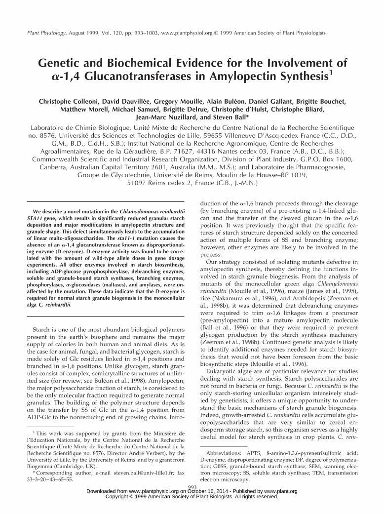
Figure 1. Wild-type and mutant iodine-staining phenotype. Iodinestain of cell patches incubated for 7 d on solid nitrogen-deprivedmedium. Genotypes with respect to starch are indicated for ourreference strains. sta2–27::ARG7, sta3–1, and sta7–5::ARG7 corre-spond to highly specific defects respectively in GBSS, SS, and de-branching enzyme (Delrue et al., 1992; Fontaine et al., 1993,Mouille et al., 1996). 1, Wild type. The original mutant strain JV45J(sta11-1) and two recombinants, CO199 and CO180, display thetypical yellow stain of low-starch mutants, while the wild-type re-combinant CO23 shows the typical dark-blue color of the wild-typereference strain.
The Function of a-1,4 Glucanotransferases in Starch Synthesis 995
www.plant.org on October 16, 2014 - Published by www.plantphysiol.orgDownloaded from Copyright © 1999 American Society of Plant Biologists. All rights reserved.
minimal medium supplied with acetate using nitrate as thenitrogen source (Ball et al., 1991).
The homozygous wild-type diploid was crossed with CO35 (mt1pab2 nit1 nit2) to obtain the triploid wild-type (1/1/1) and with CO 26 (mt1pab2 nit1 nit2 sta11-1) to obtain1/1/sta11-1. The triploid strains were selected on minimalmedium with nitrate as the nitrogen source. The heterozy-gous diploid strain was obtained by crossing JV45J and 37,and was selected on minimal medium with nitrate as the
nitrogen source. After selection on the appropriate me-dium, the haploid, diploid, or triploid nature of the cloneswas confirmed by retesting the phenotypes and measuringboth the average cell volume distribution and the cellprotein content from unsynchronized cultures. We alsoconfirmed the mating types of the clones. For each con-struct we selected three independent clones. Gene dosagesare thus averages from three separate colonies for eachconstruct. Phosphoglucomutase activity was assayed (Ballet al., 1991) and used as an internal standard during theseexperiments.
RESULTS
Isolation and Characterization of Low-Starch Mutants
Among 5 3 104 colonies screened by the standard iodine-staining technique after UV mutagenesis of a wild-type C.reinhardtii strain (137C), we isolated a low-starch mutant(JV45J) that accumulated 8% of the normal amount of
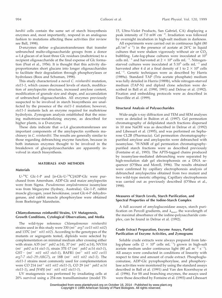
Figure 2. Growth curves of wild-type and mutant sta11-1 strains.Unsynchronized precultures of three mutant and three wild-typeprogeny from a cross involving the mutant JV45J and the wild-typestrain 37 were grown in TAP medium (Harris, 1989b) to late logphase at 2 3 106 cells mL21. The cultures were inoculated at 5 3 104
cells mL21 and subjected to a 12-h d (80 mE m22 s21)/12-h nightcycle of culture at 20°C with vigorous shaking. A, TAP medium (withacetate). Average mutant cell counts (f) and wild-type (l) cellcounts are displayed as log of cell number versus time. B, TP medium(without acetate). Average mutant cell counts (f) and wild-type (l)cell counts are displayed as log of cell number versus time. Thecultures are severely CO2 limited under these conditions and there-fore grew at a very slow but comparable rate.
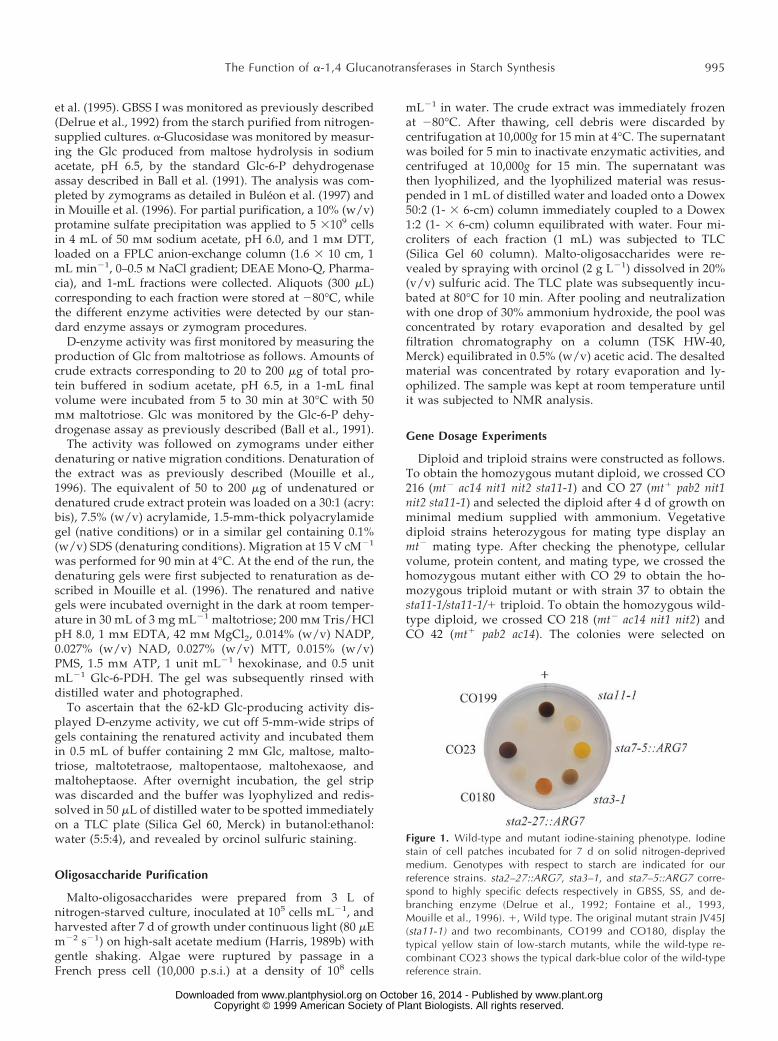
Figure 3. Relative frequency distributions of oligosaccharides. Un-debranched malto-oligosaccharides accumulated by the sta11-1 ref-erence strain JV45J were separated according to length after APTSfluorescence labeling and separation on a DNA sequencer. Percent-ages of chains ranging between a DP of 1 to 16 (chains containing1–16 Glc residues) are scaled on the y axis.
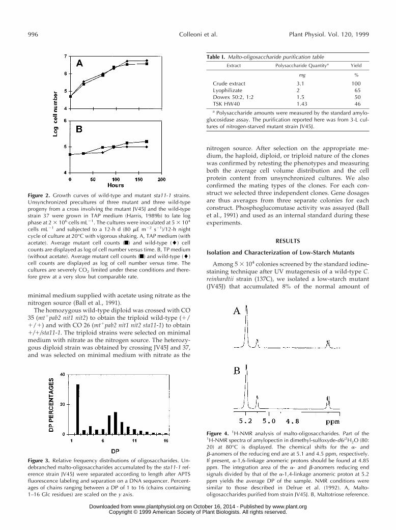
Figure 4. 1H-NMR analysis of malto-oligosaccharides. Part of the1H-NMR spectra of amylopectin in dimethyl-sulfoxyde-d6/2H2O (80:20) at 80°C is displayed. The chemical shifts for the a- andb-anomers of the reducing end are at 5.1 and 4.5 ppm, respectively.If present, a-1,6-linkage anomeric protons should be found at 4.85ppm. The integration area of the a- and b-anomers reducing endsignals divided by that of the a-1,4-linkage anomeric proton at 5.2ppm yields the average DP of the sample. NMR conditions weresimilar to those described in Delrue et al. (1992). A, Malto-oligosaccharides purified from strain JV45J. B, Maltotriose reference.
Table I. Malto-oligosaccharide purification table
Extract Polysaccharide Quantitya Yield
mg %
Crude extract 3.1 100Lyophilizate 2 65Dowex 50:2, 1:2 1.5 50TSK HW40 1.43 46a Polysaccharide amounts were measured by the standard amylo-
glucosidase assay. The purification reported here was from 3-L cul-tures of nitrogen-starved mutant strain JV45J.
996 Colleoni et al. Plant Physiol. Vol. 120, 1999
www.plant.org on October 16, 2014 - Published by www.plantphysiol.orgDownloaded from Copyright © 1999 American Society of Plant Biologists. All rights reserved.
starch under conditions of maximum synthesis (Fig. 1). Thedefect behaved as a standard single-recessive Mendeliantrait upon crossing. In addition, all diploids generatedby crossing with reference mutant strains carrying a defectin the STA1, STA2, STA3, STA4, STA5, STA6, STA7, orSTA8 genes proved to be wild type for starch amount andstructure. Therefore, we defined a novel genetic locus,which we named STA11. In addition to starch, the sta11-1mutants accumulated a significant amount (2% of theamount of starch in a wild-type strain) of water-soluble,amyloglucosidase-digestible material. No growth defectscould be detected upon growing the mutants for over a
week in continuous light with or without acetate or undera 12-h day/12-h night cycle with or without acetate (Fig. 2).
Characterization of the Water-Soluble,Amyloglucosidase-Digestible Material
We found no evidence for the presence of high-mass,water-soluble polysaccharides. Indeed, the amount ofwater-soluble amyloglucosidase-digestible material ex-cluded from the gel permeation chromatography columnwas less than 1% of the total water-soluble polysaccharidefound in crude extracts. The water-soluble polysaccharide
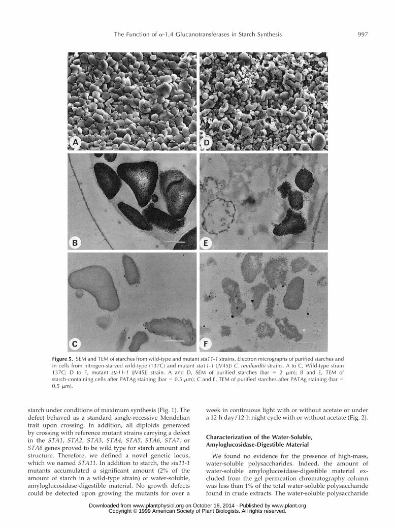
Figure 5. SEM and TEM of starches from wild-type and mutant sta11-1 strains. Electron micrographs of purified starches andin cells from nitrogen-starved wild-type (137C) and mutant sta11-1 (JV45J) C. reinhardtii strains. A to C, Wild-type strain137C; D to F, mutant sta11-1 (JV45J) strain. A and D, SEM of purified starches (bar 5 2 mm); B and E, TEM ofstarch-containing cells after PATAg staining (bar 5 0.5 mm); C and F, TEM of purified starches after PATAg staining (bar 50.5 mm).
The Function of a-1,4 Glucanotransferases in Starch Synthesis 997
www.plant.org on October 16, 2014 - Published by www.plantphysiol.orgDownloaded from Copyright © 1999 American Society of Plant Biologists. All rights reserved.
thus consisted solely of a-1,4-linked glucans, the size dis-tribution of which is displayed in Figure 3. These glucanswere further purified (Table I) and subjected to a detailedstructural characterization. The size distribution before andafter purification was identical. The mix of oligosaccha-rides generated an averaged 1H-NMR spectrum very sim-ilar to that of maltotriose standards (Fig. 4). As demon-strated by 1H-NMR analysis, branches, if present, fellbelow our detection level (1%). Co-segregation between theoligosaccharide fraction and the low-starch phenotype wasfound among all sta11-1-carrying strains tested (n 5 50).
Characterization of the Residual Mutant Starch
The residual starch structure was monitored through avariety of techniques, including wide-angle x-ray diffrac-tion analysis (not shown), TEM and SEM (Fig. 5), separa-tion of amylose and amylopectin through gel permeationchromatography (Fig. 6), and enzymatic debranching of thepurified amylose and amylopectin (Fig. 7). The starch gran-ules showed a significant relative increase in amylose (Ta-ble II). The distributions displayed in Figure 6, A and B,suggested a decrease in the mass of the amylose fraction.This was confirmed by mixing a low amount of labeledmutant starch with a high amount of wild-type referencepolysaccharide on the same column. The x-ray diffracto-grams switched from the wild-type A-type lattice with high crystallinity to a mix of A- and B-type lattices with
very low crystallinities. The shape of the granules wasparticularly affected, as illustrated in Figure 5.
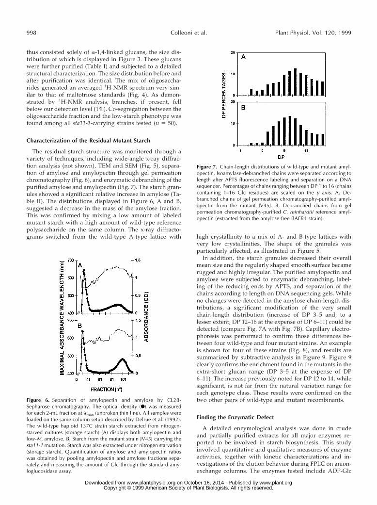
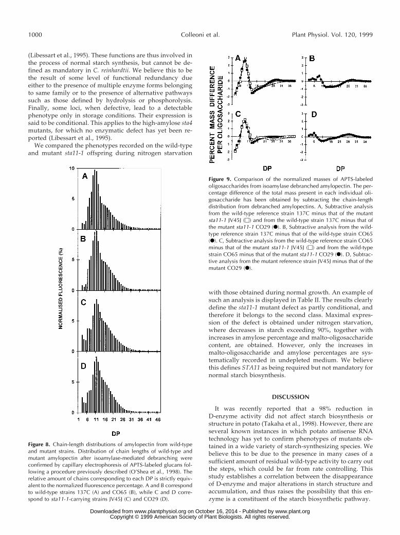
In addition, the starch granules decreased their overallmean size and the regularly shaped smooth surface becamerugged and highly irregular. The purified amylopectin andamylose were subjected to enzymatic debranching, label-ing of the reducing ends by APTS, and separation of thechains according to length on DNA sequencing gels. Whileno changes were detected in the amylose chain-length dis-tributions, a significant modification of the very smallchain-length distribution (increase of DP 3–5 and, to alesser extent, DP 12–16 at the expense of DP 6–11) could bedetected (compare Fig. 7A with Fig. 7B). Capillary electro-phoresis was performed to confirm those differences be-tween four wild-type and four mutant strains. An exampleis shown for four of these strains (Fig. 8), and results aresummarized by subtractive analysis in Figure 9. Figure 9clearly confirms the enrichment found in the mutants in theextra-short glucan range (DP 3–5 at the expense of DP6–11). The increase previously noted for DP 12 to 14, whilesignificant, is not far from the natural variation range foreach genotype class. These results were confirmed on thetwo other pairs of wild-type and mutant recombinants.
Finding the Enzymatic Defect
A detailed enzymological analysis was done in crudeand partially purified extracts for all major enzymes re-ported to be involved in starch biosynthesis. This studyinvolved quantitative and qualitative measures of enzymeactivities, together with kinetic characterizations and in-vestigations of the elution behavior during FPLC on anion-exchange columns. The enzymes tested include ADP-Glc
Figure 6. Separation of amylopectin and amylose by CL2B-Sepharose chromatography. The optical density (F) was measuredfor each 2-mL fraction at lmax (unbroken thin line). All samples wereloaded on the same column setup described by Delrue et al. (1992).The wild-type haploid 137C strain starch extracted from nitrogen-starved cultures (storage starch) (A) displays both amylopectin andlow-Mr amylose. B, Starch from the mutant strain JV45J carrying thesta11-1 mutation. Starch was also extracted under nitrogen starvation(storage starch). Quantification of amylose and amylopectin ratioswas obtained by pooling amylopectin and amylose fractions sepa-rately and measuring the amount of Glc through the standard amy-loglucosidase assay.
Figure 7. Chain-length distributions of wild-type and mutant amyl-opectin. Isoamylase-debranched chains were separated according tolength after APTS fluorescence labeling and separation on a DNAsequencer. Percentages of chains ranging between DP 1 to 16 (chainscontaining 1–16 Glc residues) are scaled on the y axis. A, De-branched chains of gel permeation chromatography-purified amyl-opectin from the mutant JV45J. B, Debranched chains from gelpermeation chromatography-purified C. reinhardtii reference amyl-opectin (extracted from the amylose-free BAFR1 strain).
998 Colleoni et al. Plant Physiol. Vol. 120, 1999
www.plant.org on October 16, 2014 - Published by www.plantphysiol.orgDownloaded from Copyright © 1999 American Society of Plant Biologists. All rights reserved.
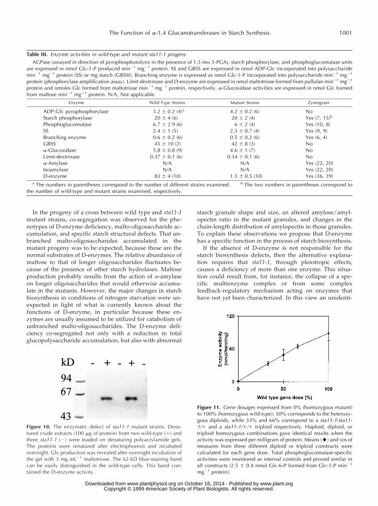
pyrophosphorylase, phosphoglucomutase, SS I, SS II,GBSS, both types of branching enzymes, debranching en-zymes (limit-dextrinase and isoamylase), phosphorylases,maltase (a-glucosidase), and all starch hydrolases thatcould be detected in starch-containing zymogram gels (Ta-ble III).
No qualitative nor quantitative differences in these en-zyme activities were found to co-segregate with the mutantgene. However, when we applied the standard assay usedin plants to measure D-enzyme activity, we were surprisedto find a 99% decrease in the rate of Glc production frommaltotriose (Table III). The mutants were also clearly de-fective in the production of Glc from maltotetraose, malto-pentaose, maltohexaose, and maltoheptaose. However, theproduction of Glc from maltose was comparable in mutantand wild-type strains. This production amounted to lessthan 5% of that measured with maltotriose in the wild-typeprogeny. We therefore assume that the production of Glcfrom maltoriose by a-glucosidase or other hydrolases isnegligible in face of the amount of D-enzyme activitypresent in our extracts. This result is similar to that re-ported for higher plant leaf extracts (Zeeman et al., 1998a).
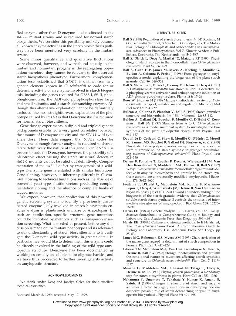
Glc production from maltotriose thus defines a quanti-tative and specific assay for D-enzyme activity in C. rein-hardtii extracts. We adapted the zymogram techniques usedto detect Glc production to assay the action of D-enzyme onmaltotriose (Fig. 10). We were able to detect the activityafter denaturation and renaturation, which allowed us toestimate the mass of the enzyme. This 62-kD band wasabsent in all meiotic progeny bearing the mutation (n 5 75)and was systematically present in wild-type strains. Tomake sure that the 62-kD band contained oligosaccharide-disproportionating activity, we incubated gel slices inbuffer containing Glc, maltose, maltotriose, maltotetraose,maltopentaose, maltohexaose, or maltoheptaose (notshown). The lyophilized incubation buffers were then spot-ted onto TLC plates and the oligosaccharides revealed bythe orcinol-sulfuric acid method (Mouille et al., 1996). The62-kD band obeyed the rules set years ago by Peat et al.(1956) to define D-enzymes: it could not use Glc or maltoseas sole substrates, but could disproportionate a-1,4-linked
oligosaccharides of at least three Glc residues into longeroligosaccharides.
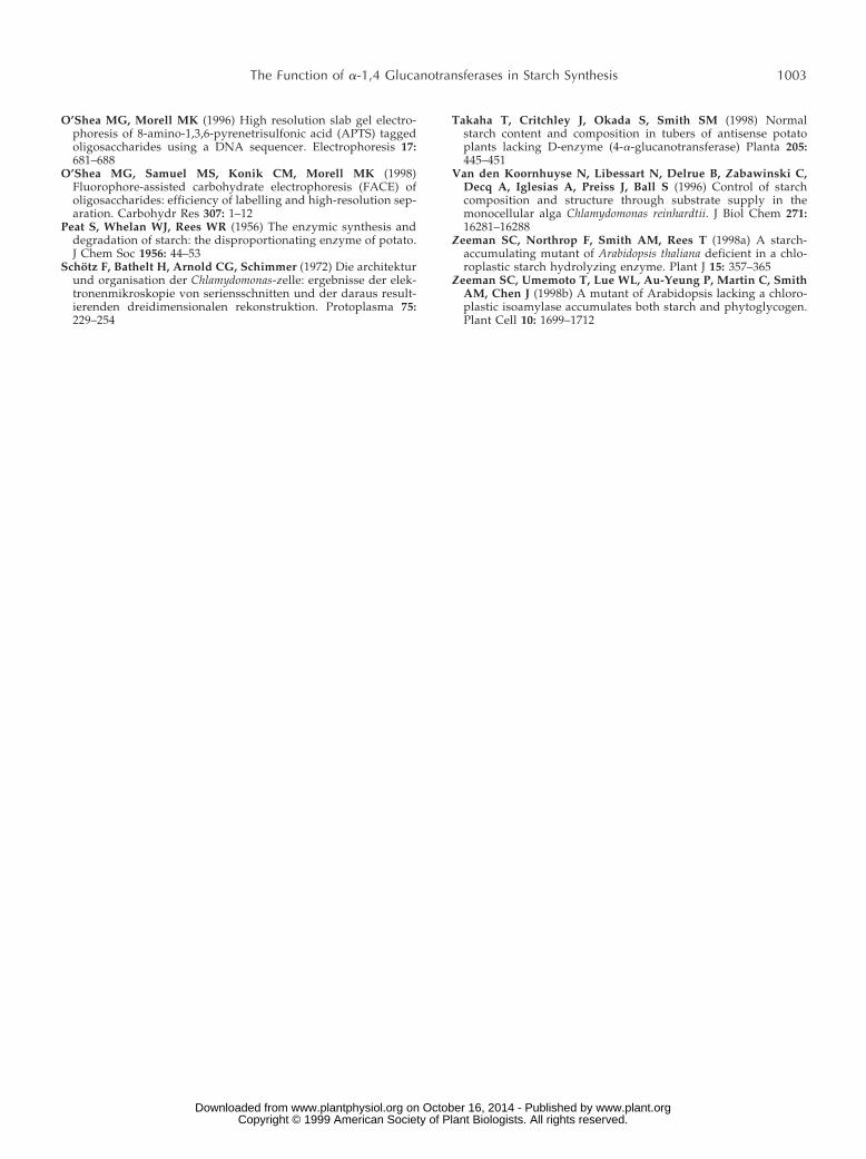
Gene Dosage Experiments
We were able to monitor the amount of enzyme activitypresent within the plastids (8.7 mmol of Glc formed frommaltotriose min21 mL21) or to perform gene dosage exper-iments in diploid and triploid zygotes. The enzyme activitycorrelated with the relative amount of wild-type alleles, aswould be expected if STA11 encoded D-enzyme (Fig. 11).Knowing the cell and organelle volumes of the strains usedin this work, we were able to calculate the physiologicalenzyme concentrations (Schotz et al., 1972). We were alsoable to estimate the amount of malto-oligosaccharides inwild-type (50–200 mg mL21 of chloroplast) and mutant(0.5–1 mg mL21) strains.
The Expression of the Mutant Phenotype IsPartly Conditional
We have previously noted that expressivity of mutantphenotypes can vary when transitory starch is comparedwith the more classical storage form of the polysaccharide(Libessart et al., 1995). In C. reinhardtii, storage starch syn-thesis is obtained by using nutrient starvation conditions,leading to the arrest of cell division and to the accumula-tion of starch. On the other hand, transitory starch synthe-sis is obtained under conditions of active photosynthesisand cell division. Mutant phenotypes of C. reinhardtii fallinto three classes. The first class concerns mutations whosephenotypes express themselves equally in both physiolog-ical conditions. Mutations affecting the small and largesubunits of ADP-Glc pyrophosphorylase, GBSS, and de-branching enzyme fall within this class (Ball et al., 1991;Delrue et al., 1992; Libessart et al., 1995; Mouille et al., 1996;Dauvillee et al., 1999). The functions thereby defined aresaid to be mandatory for either amylose or starch synthesis.
The second class contains mutations expressed in bothconditions but with lesser expressivity upon transitorystarch. These include mutations affecting the major SS
Table II. Phenotype of wild-type and mutant strains during storage or transitory starch synthesisValues listed are average of three separate measures in a single experiment.
Strain Genotypelmax
a Starchb MOSc Amd
1N 2N 1N 2N 1N 2N 1N 2N
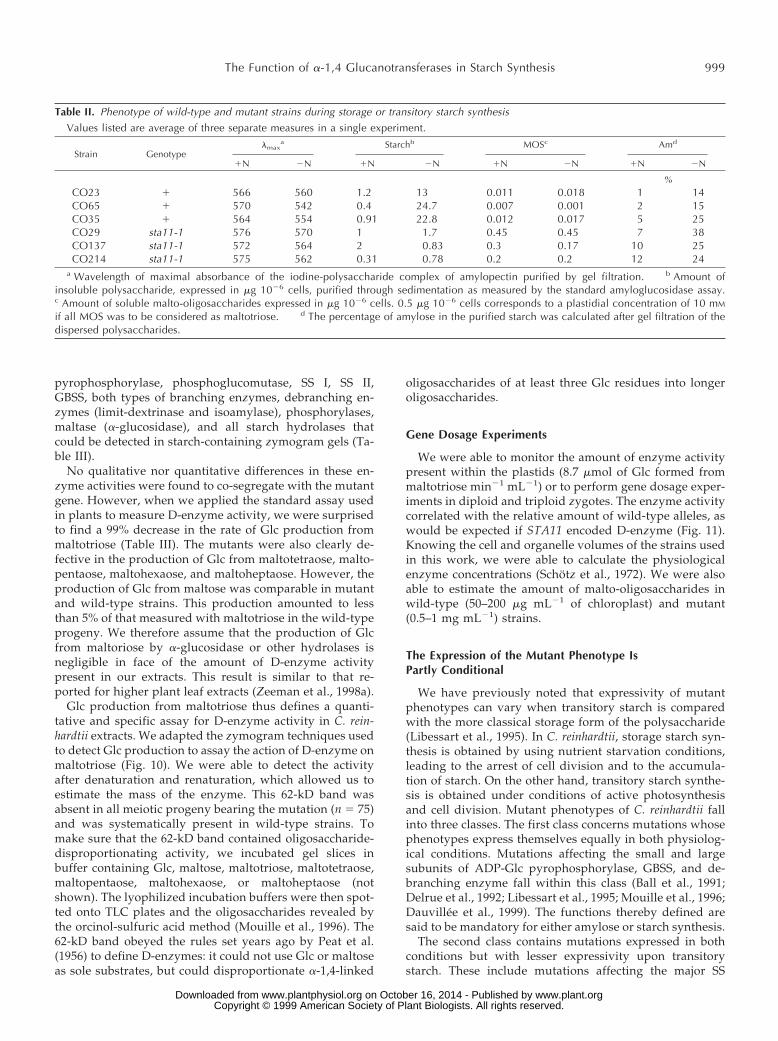
%CO23 1 566 560 1.2 13 0.011 0.018 1 14CO65 1 570 542 0.4 24.7 0.007 0.001 2 15CO35 1 564 554 0.91 22.8 0.012 0.017 5 25CO29 sta11-1 576 570 1 1.7 0.45 0.45 7 38CO137 sta11-1 572 564 2 0.83 0.3 0.17 10 25CO214 sta11-1 575 562 0.31 0.78 0.2 0.2 12 24
a Wavelength of maximal absorbance of the iodine-polysaccharide complex of amylopectin purified by gel filtration. b Amount ofinsoluble polysaccharide, expressed in mg 1026 cells, purified through sedimentation as measured by the standard amyloglucosidase assay.c Amount of soluble malto-oligosaccharides expressed in mg 1026 cells. 0.5 mg 1026 cells corresponds to a plastidial concentration of 10 mM
if all MOS was to be considered as maltotriose. d The percentage of amylose in the purified starch was calculated after gel filtration of thedispersed polysaccharides.
The Function of a-1,4 Glucanotransferases in Starch Synthesis 999
www.plant.org on October 16, 2014 - Published by www.plantphysiol.orgDownloaded from Copyright © 1999 American Society of Plant Biologists. All rights reserved.
(Libessart et al., 1995). These functions are thus involved inthe process of normal starch synthesis, but cannot be de-fined as mandatory in C. reinhardtii. We believe this to bethe result of some level of functional redundancy dueeither to the presence of multiple enzyme forms belongingto same family or to the presence of alternative pathwayssuch as those defined by hydrolysis or phosphorolysis.Finally, some loci, when defective, lead to a detectablephenotype only in storage conditions. Their expression issaid to be conditional. This applies to the high-amylose sta4mutants, for which no enzymatic defect has yet been re-ported (Libessart et al., 1995).
We compared the phenotypes recorded on the wild-typeand mutant sta11-1 offspring during nitrogen starvation
with those obtained during normal growth. An example ofsuch an analysis is displayed in Table II. The results clearlydefine the sta11-1 mutant defect as partly conditional, andtherefore it belongs to the second class. Maximal expres-sion of the defect is obtained under nitrogen starvation,where decreases in starch exceeding 90%, together withincreases in amylose percentage and malto-oligosaccharidecontent, are obtained. However, only the increases inmalto-oligosaccharide and amylose percentages are sys-tematically recorded in undepleted medium. We believethis defines STA11 as being required but not mandatory fornormal starch biosynthesis.
DISCUSSION
It was recently reported that a 98% reduction inD-enzyme activity did not affect starch biosynthesis orstructure in potato (Takaha et al., 1998). However, there areseveral known instances in which potato antisense RNAtechnology has yet to confirm phenotypes of mutants ob-tained in a wide variety of starch-synthesizing species. Webelieve this to be due to the presence in many cases of asufficient amount of residual wild-type activity to carry outthe steps, which could be far from rate controlling. Thisstudy establishes a correlation between the disappearanceof D-enzyme and major alterations in starch structure andaccumulation, and thus raises the possibility that this en-zyme is a constituent of the starch biosynthetic pathway.
Figure 8. Chain-length distributions of amylopectin from wild-typeand mutant strains. Distribution of chain lengths of wild-type andmutant amylopectin after isoamylase-mediated debranching wereconfirmed by capillary electrophoresis of APTS-labeled glucans fol-lowing a procedure previously described (O’Shea et al., 1998). Therelative amount of chains corresponding to each DP is strictly equiv-alent to the normalized fluorescence percentage. A and B correspondto wild-type strains 137C (A) and CO65 (B), while C and D corre-spond to sta11-1-carrying strains JV45J (C) and CO29 (D).
Figure 9. Comparison of the normalized masses of APTS-labeledoligosaccharides from isoamylase debranched amylopectin. The per-centage difference of the total mass present in each individual oli-gosaccharide has been obtained by subtracting the chain-lengthdistribution from debranched amylopectins. A, Subtractive analysisfrom the wild-type reference strain 137C minus that of the mutantsta11-1 JV45J (M) and from the wild-type strain 137C minus that ofthe mutant sta11-1 CO29 (F). B, Subtractive analysis from the wild-type reference strain 137C minus that of the wild-type strain CO65(F). C, Subtractive analysis from the wild-type reference strain CO65minus that of the mutant sta11-1 JV45J (M) and from the wild-typestrain CO65 minus that of the mutant sta11-1 CO29 (F). D, Subtrac-tive analysis from the mutant reference strain JV45J minus that of themutant CO29 (F).
1000 Colleoni et al. Plant Physiol. Vol. 120, 1999
www.plant.org on October 16, 2014 - Published by www.plantphysiol.orgDownloaded from Copyright © 1999 American Society of Plant Biologists. All rights reserved.
In the progeny of a cross between wild type and sta11-1mutant strains, co-segregation was observed for the phe-notypes of D-enzyme deficiency, malto-oligosaccharide ac-cumulation, and specific starch structural defects. That un-branched malto-oligosaccharides accumulated in themutant progeny was to be expected, because these are thenormal substrates of D-enzymes. The relative abundance ofmaltose to that of longer oligosaccharides fluctuates be-cause of the presence of other starch hydrolases. Maltoseproduction probably results from the action of a-amylaseon longer oligosaccharides that would otherwise accumu-late in the mutants. However, the major changes in starchbiosynthesis in conditions of nitrogen starvation were un-expected in light of what is currently known about thefunctions of D-enzyme, in particular because these en-zymes are usually assumed to be utilized for catabolism ofunbranched malto-oligosaccharides. The D-enzyme defi-ciency co-segregated not only with a reduction in totalglucopolysaccharide accumulation, but also with abnormal
starch granule shape and size, an altered amylose/amyl-opectin ratio in the mutant granules, and changes in thechain-length distribution of amylopectin in those granules.To explain these observations we propose that D-enzymehas a specific function in the process of starch biosynthesis.
If the absence of D-enzyme is not responsible for thestarch biosynthesis defects, then the alternative explana-tion requires that sta11-1, through pleiotropic effects,causes a deficiency of more than one enzyme. This situa-tion could result from, for instance, the collapse of a spe-cific multienzyme complex or from some complexfeedback-regulatory mechanism acting on enzymes thathave not yet been characterized. In this view an unidenti-
Figure 10. The enzymatic defect of sta11-1 mutant strains. Dena-tured crude extracts (100 mg of protein) from two wild-type (1) andthree sta11-1 (2) were loaded on denaturing polyacrylamide gels.The proteins were renatured after electrophoresis and incubatedovernight. Glc production was revealed after overnight incubation ofthe gel with 3 mg mL21 maltotriose. The 62-kD blue-staining bandcan be easily distinguished in the wild-type cells. This band con-tained the D-enzyme activity.
Figure 11. Gene dosages expressed from 0% (homozygous mutant)to 100% (homozygous wild-type); 50% corresponds to the heterozy-gous diploids, while 33% and 66% correspond to a sta11-1/sta11-1/1 and a sta11-1/1/1 triploid respectively. Haploid, diploid, ortriploid homozygous combinations gave identical results when theactivity was expressed per milligram of protein. Means (l) and SDs ofmeasures from three different diploid or triploid constructs werecalculated for each gene dose. Total phosphoglucomutase-specificactivities were monitored as internal controls and proved similar inall constructs (2.5 6 0.4 nmol Glc-6-P formed from Glc-1-P min21
mg21 protein).
Table III. Enzyme activities in wild-type and mutant sta11-1 progenyAGPase (assayed in direction of pyrophosphorolysis in the presence of 1.5 mM 3-PGA), starch phosphorylase, and phosphoglucomutase units
are expressed in nmol Glc-1-P produced min21 mg21 protein. SS and GBSS are expressed in nmol ADP-Glc incorporated into polysaccharidemin21 mg21 protein (SS) or mg starch (GBSSI). Branching enzyme is expressed as nmol Glc-1-P incorporated into polysaccharide min21 mg21
protein (phosphorylase amplification assay). Limit-dextrinase and D-enzyme are expressed in nmol maltotriose formed from pullulan min21 mg21
protein and nmoles Glc formed from maltotriose min21 mg21 protein, respectively. a-Glucosidase activities are expressed in nmol Glc formedfrom maltose min21 mg21 protein. N/A, Not applicable.
Enzyme Wild-Type Strains Mutant Strains Zymogram
ADP-Glc pyrophosphorylase 3.2 6 0.2 (4)a 4.2 6 0.2 (6) NoStarch phosphorylase 20 6 4 (6) 20 6 2 (4) Yes (7, 15)b
Phosphoglucomutase 6.7 6 2.9 (6) 6 6 2 (4) Yes (10, 8)SS 2.4 6 1 (5) 2.3 6 0.7 (4) Yes (9, 9)Branching enzyme 0.6 6 0.2 (6) 0.5 6 0.2 (6) Yes (6, 4)GBSS 45 6 10 (2) 42 6 8 (3) Noa-Glucosidase 5.8 6 0.8 (9) 4.6 6 1 (7) NoLimit-dextrinase 0.37 6 0.1 (6) 0.34 6 0.1 (6) Noa-Amylase N/A N/A Yes (22, 20)Isoamylase N/A N/A Yes (22, 20)D-enzyme 83 6 4 (10) 1.3 6 0.5 (10) Yes (36, 39)
a The numbers in parentheses correspond to the number of different strains examined. b The two numbers in parentheses correspond tothe number of wild-type and mutant strains examined, respectively.
The Function of a-1,4 Glucanotransferases in Starch Synthesis 1001
www.plant.org on October 16, 2014 - Published by www.plantphysiol.orgDownloaded from Copyright © 1999 American Society of Plant Biologists. All rights reserved.
fied enzyme other than D-enzyme is also affected in thesta11-1 mutant strains, and is required for normal starchbiosynthesis. We consider this possibility unlikely becauseall known enzyme activities in the starch biosynthesis path-way have been monitored very carefully in the mutantstrains.
Some minor quantitative and qualitative fluctuationswere observed, however, and were found equally in themutant and nonmutant progeny in the segregating popu-lation; therefore, they cannot be relevant to the observedstarch biosynthesis phenotype. Furthermore, complemen-tation tests established that STA11 is distinct from anygenetic element known in C. reinhardtii to code for ordetermine activity of an enzyme involved in starch biogen-esis, including the genes required for GBSS I, SS II, phos-phoglucomutase, the ADP-Glc pyrophosphorylase largeand small subunits, and a starch-debranching enzyme. Al-though this alternative explanation cannot be definitivelyexcluded, the most straightforward explanation of the phe-notype caused by sta11-1 is that D-enzyme itself is requiredfor normal starch biosynthesis.
Gene dosage experiments in diploid and triploid geneticbackgrounds established a very good correlation betweenthe amount of D-enzyme activity and the STA11 wild-typeallele dose. These data suggest that STA11 codes forD-enzyme, although further analysis is required to charac-terize definitively the nature of this gene. Even if STA11 isproven to code for D-enzyme, however, the possibility of apleiotropic effect causing the starch structural defects insta11-1 mutants cannot be ruled out definitively. Comple-mentation of the sta11-1 defect by transgenesis of a wild-type D-enzyme gene is entailed with similar limitations.Gene cloning, however, is inherently difficult in C. rein-hardtii owing to technical limitations such as the absence ofpowerful yeast-type shuttle vectors precluding comple-mentation cloning and the absence of complete banks oftagged mutants.
We expect, however, that application of this powerfulgenetic screening system to identify a previously unsus-pected enzyme likely involved in starch biosynthesis en-ables analysis in plants such as maize or Arabidopsis. Insuch an application, specific structural gene mutationscould be identified by methods such as transposon inser-tion screening. What is needed at present, before any dis-cussion is made on the mutant phenotype and its relevanceto our understanding of starch biosynthesis, is to investi-gate the D-enzyme wild-type activity in greater detail. Inparticular, we would like to determine if this enzyme couldbe directly involved in the building of the wild-type amy-lopectin structure. D-enzyme has been documented asworking essentially on soluble malto-oligosaccharides, andwe have thus proceeded to further investigate its activityon amylopectin structure.
ACKNOWLEDGMENTS
We thank Andre Decq and Jocelyn Celen for their excellenttechnical assistance.
Received March 8, 1999; accepted May 17, 1999.
LITERATURE CITED
Ball S (1998) Regulation of starch biosynthesis. In J-D Rochaix, MGoldschmidt-Clermont, S Merchant, Govindjee, eds, The Molec-ular Biology of Chloroplasts and Mitochondria in Chlamydomo-nas. Advances in Photosynthesis, Vol 7. Kluwer Academic Pub-lishers, Dordrecht, The Netherlands, pp 549–567
Ball S, Dirick L, Decq A, Martiat JC, Matagne RF (1990) Physi-ology of starch storage in the monocellular alga Chlamydomonasreinhardtii. Plant Sci 66: 1–9
Ball S, Guan H-P, James M, Myers A, Keeling P, Mouille G,Buleon A, Colonna P, Preiss J (1996) From glycogen to amyl-opectin: a model explaining the biogenesis of the plant starchgranule. Cell 86: 349–352
Ball S, Marianne T, Dirick L, Fresnoy M, Delrue B, Decq A (1991)A Chlamydomonas reinhardtii low-starch mutant is defective for3-phosphoglycerate activation and orthophosphate inhibition ofADP-glucose pyrophosphorylase. Planta 185: 17–26
Boos W, Shuman H (1998) Maltose/maltodextrin system of Esch-erichia coli: transport, metabolism and regulation. Microbiol MolBiol Rev 62: 204–229
Buleon A, Colonna P, Planchot V, Ball, S (1998) Starch granules:structure and biosynthesis. Int J Biol Macromol 23: 85–112
Buleon A, Gallant DJ, Bouchet B, Mouille G, D’Hulst C, Koss-man J, Ball SG (1997) Starches from A to C: Chlamydomonasreinhardtii as a model microbial system to investigate the bio-synthesis of the plant amylopectin crystal. Plant Physiol 115:949–957
Dauvillee D, Colleoni C, Shaw E, Mouille G, D’Hulst C, MorellM, Samuel MS, Bouchet B, Gallant DJ, Sinskey A, et al. (1999)Novel starch-like polysaccharides are synthesized by a solubleform of granule-bound starch synthase in glycogen accumulat-ing mutants of Chlamydomonas reinhardtii. Plant Physiol 119:321–330
Delrue B, Fontaine T, Routier F, Decq A, Wieruszeski JM, VanDen Koornhuyse N, Maddelein M-L, Fournet B, Ball S (1992)Waxy Chlamydomonas reinhardtii: monocellular algal mutants de-fective in amylose biosynthesis and granule-bound starch syn-thase accumulate a structurally modified amylopectin. J Bacte-riol 174: 3612–3620
Fontaine T, D’Hulst C, Maddelein M-L, Routier F, Marianne-Pepin T, Decq A, Wieruszeski JM, Delrue B, Van Den Koorn-huyse N, Bossu JP, et al. (1993) Toward an understanding of thebiogenesis of the starch granule: evidence that Chlamydomonassoluble starch starch synthase II controls the synthesis of inter-mediate size glucans of amylopectin. J Biol Chem 268: 16223–16230
Harris EH (1989a) Genetic analysis. In E Harris, ed, The Chlamy-domonas Sourcebook. A Comprehensive Guide to Biology andLaboratory Use. Academic Press, San Diego, pp 399–446
Harris EH (1989b) Culture and storage methods. In E Harris, ed,The Chlamydomonas Sourcebook. A Comprehensive Guide toBiology and Laboratory Use. Academic Press, San Diego, pp25–63
James MG, Robertson DS, Myers AM (1995) Characterization ofthe maize gene sugary1, a determinant of starch composition inkernels. Plant Cell 7: 417–429
Libessart N, Maddelein M-L, Van Den Koornhuyse N, Decq A,Delrue B, Ball SG (1995) Storage, photosynthesis and growth:the conditional nature of mutations affecting starch synthesisand structure in Chlamydomonas reinhardtii. Plant Cell 7: 1117–1127
Mouille G, Maddelein M-L, Libessart N, Talaga P, Decq A,Delrue B, Ball S (1996) Phytoglycogen processing: a mandatorystep for starch biosynthesis in plants. Plant Cell 8: 1353–1366
Nakamura Y, Umemoto T, Takahata Y, Komae K, Amano E,Satoh, H (1996) Changes in structure of starch and enzymeactivities affected by sugary mutations in developing rice en-dosperm: possible role of starch debranching enzyme in amyl-opectin biosynthesis. Physiol Plant 97: 491–498
1002 Colleoni et al. Plant Physiol. Vol. 120, 1999
www.plant.org on October 16, 2014 - Published by www.plantphysiol.orgDownloaded from Copyright © 1999 American Society of Plant Biologists. All rights reserved.
O’Shea MG, Morell MK (1996) High resolution slab gel electro-phoresis of 8-amino-1,3,6-pyrenetrisulfonic acid (APTS) taggedoligosaccharides using a DNA sequencer. Electrophoresis 17:681–688
O’Shea MG, Samuel MS, Konik CM, Morell MK (1998)Fluorophore-assisted carbohydrate electrophoresis (FACE) ofoligosaccharides: efficiency of labelling and high-resolution sep-aration. Carbohydr Res 307: 1–12
Peat S, Whelan WJ, Rees WR (1956) The enzymic synthesis anddegradation of starch: the disproportionating enzyme of potato.J Chem Soc 1956: 44–53
Schotz F, Bathelt H, Arnold CG, Schimmer (1972) Die architekturund organisation der Chlamydomonas-zelle: ergebnisse der elek-tronenmikroskopie von seriensschnitten und der daraus result-ierenden dreidimensionalen rekonstruktion. Protoplasma 75:229–254
Takaha T, Critchley J, Okada S, Smith SM (1998) Normalstarch content and composition in tubers of antisense potatoplants lacking D-enzyme (4-a-glucanotransferase) Planta 205:445–451
Van den Koornhuyse N, Libessart N, Delrue B, Zabawinski C,Decq A, Iglesias A, Preiss J, Ball S (1996) Control of starchcomposition and structure through substrate supply in themonocellular alga Chlamydomonas reinhardtii. J Biol Chem 271:16281–16288
Zeeman SC, Northrop F, Smith AM, Rees T (1998a) A starch-accumulating mutant of Arabidopsis thaliana deficient in a chlo-roplastic starch hydrolyzing enzyme. Plant J 15: 357–365
Zeeman SC, Umemoto T, Lue WL, Au-Yeung P, Martin C, SmithAM, Chen J (1998b) A mutant of Arabidopsis lacking a chloro-plastic isoamylase accumulates both starch and phytoglycogen.Plant Cell 10: 1699–1712
The Function of a-1,4 Glucanotransferases in Starch Synthesis 1003
www.plant.org on October 16, 2014 - Published by www.plantphysiol.orgDownloaded from Copyright © 1999 American Society of Plant Biologists. All rights reserved.