Scytalidium thermophilum. Biochemical and regulatory properties
10
J. Basic Microbiol. 40 (2000) 2, 83 – 92 (Departamento de Biologia – Faculdade de Filosofia Ciências e Letras de Ribeirão Preto – Uni- versidade de São Paulo; 1 Centro de Química de Proteínas and Departamento de Ginecologia e Obstetrícia – Faculdade de Medicina de Ribeirão Preto – Universidade de São Paulo) Glucoamylase activity from the thermophilic fungus Scytalidium thermophilum. Biochemical and regulatory properties MARIANA CEREIA, HÉCTOR F. TERENZI, JOÃO A. JORGE, LEWIS J. GREENE 1 , JOSE C. ROSA 1 and MARIA DE LOURDES T. M. POLIZELI (Received 01 September 1999/Accepted 08 November 1999) A glucoamylase activity produced by the thermophilic fungus Scytalidium thermophilum was purified 6.8-fold by ion exchange chromatography. The protein exhibited a molecular mass of about 86 kDa (7% PAGE and SDS-PAGE), or 68.5 kDa (Bio Sil SEC-400 FPLC). The pI of the enzyme was 8.4. Optima of pH and temperature, with starch or maltose as substrates were, 6.5/60 qC and 5.0/55 qC, respectively. The enzyme had a half-life of 22 min at 55 qC with starch as substrate, and it was fully stable with maltose. Maltase activity was activated by 10 mM Ba ++ (7.8%); Mn ++ (12.4%) or Mg ++ (28%). The enzyme contained approximately 25.5% carbohydrate. K m and V max values for starch and maltose were 0.28 mg/ml, 67.2 U/mg protein and 1.40 mg/ml, 5.61 U/mg protein, respectively. The products of hydrolysis of starch, detected by thin layer chromatography, showed only glucose after 30 min, indicating a glucoamylase activity. The amino terminal sequence of the purified protein showed 93% homology with a glucoamylase activity purified from Humicola grisea var. thermoidea. Biotechnological industries show increasing interest in the use of microbial sources of amylolytic activity (VIHINEN and MÄNTSÄLÄ 1989, GUZMÁN-MALDONADO and PAREDEZ- LÓPEZ 1995, CRABB and MITCHINSON 1997, JAMES and LEE 1997). For instance, glucoa- mylase [,(1,4)-D-glucan glucohydrolase, EC 3.2.1.3] activity, which attacks ,-1,4 link- ages from the nonreducing end of starch, releasing D-glucose in the --configuration, is widely used for the production of high-glucose (96 to 98% glucose) and high-fructose (55% fructose) syrups. Thermophilic microorganisms are regarded as an attractive source of thermostable amy- lolytic enzymes, for industrial use (TOSI et al. 1993, ALMEIDA et al. 1995, KADOWAKI et al. 1996, MISHRA and MAHESHWARI 1996, POLIZELI et al. 1996, BUSCH et al. 1997, BUAINAIN et al. 1998, VAN DEN BURG et al. 1998). In the present study we describe an extracellular glucoamylase activity produced by Scytalidium thermophilum, a fungus isolated from phase II of mushroom compost soil (STRAATSMA and SAMSON 1993). We describe an optimised medium for the enzyme production, and some of its biochemical properties. This enzyme was partially sequenced at the amino terminus, which showed homology with a glucoamy- lase produced by the thermophilic fungus Humicola grisea var. thermoidea. Materials and methods Organism and growth conditions: Scytalidium thermophilum (15.8) [= CBS 671.88 = ATCC 66938] was maintained at 45 qC, in slants of solid 4% oatmeal baby food (QUAKER) medium. Conidia from 10-day-old cultures were inoculated into 125 ml ERLENMEYER flasks containing 25 ml liquid medium: 0.1% calcium carbonate; 0.6% yeast extract; 0.1% peptone; 1.0% sodium chloride; 0.6% ammonium
Transcript of Scytalidium thermophilum. Biochemical and regulatory properties

J. Basic Microbiol. 40 (2000) 2, 83–92
(Departamento de Biologia – Faculdade de Filosofia Ciências e Letras de Ribeirão Preto – Uni-versidade de São Paulo; 1Centro de Química de Proteínas and Departamento de Ginecologia eObstetrícia – Faculdade de Medicina de Ribeirão Preto – Universidade de São Paulo)
Glucoamylase activity from the thermophilic fungusScytalidium thermophilum.Biochemical and regulatory properties
MARIANA CEREIA, HÉCTOR F. TERENZI, JOÃO A. JORGE, LEWIS J. GREENE1, JOSE C. ROSA1
and MARIA DE LOURDES T. M. POLIZELI
(Received 01 September 1999/Accepted 08 November 1999)
A glucoamylase activity produced by the thermophilic fungus Scytalidium thermophilum was purified6.8-fold by ion exchange chromatography. The protein exhibited a molecular mass of about 86 kDa(7% PAGE and SDS-PAGE), or 68.5 kDa (Bio Sil SEC-400 FPLC). The pI of the enzyme was 8.4.Optima of pH and temperature, with starch or maltose as substrates were, 6.5/60 �C and 5.0/55 �C,respectively. The enzyme had a half-life of 22 min at 55 �C with starch as substrate, and it was fullystable with maltose. Maltase activity was activated by 10 mM Ba++ (7.8%); Mn++ (12.4%) or Mg++
(28%). The enzyme contained approximately 25.5% carbohydrate. Km and Vmax values for starch andmaltose were 0.28 mg/ml, 67.2 U/mg protein and 1.40 mg/ml, 5.61 U/mg protein, respectively. Theproducts of hydrolysis of starch, detected by thin layer chromatography, showed only glucose after30 min, indicating a glucoamylase activity. The amino terminal sequence of the purified proteinshowed 93% homology with a glucoamylase activity purified from Humicola grisea var. thermoidea.
Biotechnological industries show increasing interest in the use of microbial sources ofamylolytic activity (VIHINEN and MÄNTSÄLÄ 1989, GUZMÁN-MALDONADO and PAREDEZ-LÓPEZ 1995, CRABB and MITCHINSON 1997, JAMES and LEE 1997). For instance, glucoa-mylase [�(1,4)-D-glucan glucohydrolase, EC 3.2.1.3] activity, which attacks �-1,4 link-ages from the nonreducing end of starch, releasing D-glucose in the �-configuration, iswidely used for the production of high-glucose (96 to 98% glucose) and high-fructose (55%fructose) syrups.
Thermophilic microorganisms are regarded as an attractive source of thermostable amy-lolytic enzymes, for industrial use (TOSI et al. 1993, ALMEIDA et al. 1995, KADOWAKI et al.1996, MISHRA and MAHESHWARI 1996, POLIZELI et al. 1996, BUSCH et al. 1997, BUAINAINet al. 1998, VAN DEN BURG et al. 1998). In the present study we describe an extracellularglucoamylase activity produced by Scytalidium thermophilum, a fungus isolated from phaseII of mushroom compost soil (STRAATSMA and SAMSON 1993). We describe an optimisedmedium for the enzyme production, and some of its biochemical properties. This enzymewas partially sequenced at the amino terminus, which showed homology with a glucoamy-lase produced by the thermophilic fungus Humicola grisea var. thermoidea.
Materials and methods
Organism and growth conditions: Scytalidium thermophilum (15.8) [= CBS 671.88 = ATCC 66938]was maintained at 45 �C, in slants of solid 4% oatmeal baby food (QUAKER) medium. Conidia from10-day-old cultures were inoculated into 125 ml ERLENMEYER flasks containing 25 ml liquid medium:0.1% calcium carbonate; 0.6% yeast extract; 0.1% peptone; 1.0% sodium chloride; 0.6% ammonium

84 M. CEREIA
acetate and 1.0% cassava flour, pH 6.0. The cultures were incubated at 45 �C without agitation. Afterseven days of incubation, when glucoamylase activity reached a maximum, the cultures wereharvested by filtration and the filtrate was saved as a source of crude extracellular glucoamylase.Mycelial pads were ground with sea sand, at 0 �C with ten vol. of cold 100 mM sodium acetate buffer,pH 5.5 (buffer A). After centrifugation (20,000 � g, 20 min, 4 �C) the supernatant fraction was thesource of intracellular enzyme.
Enzymatic assays, determination of protein and neutral carbohydrate: Glucoamylase activitywas assayed at 60 �C, in a reaction mixture containing 0.2 ml of diluted enzyme and 0.2 ml of 1.0%starch in 100 mM sodium acetate buffer, pH 5.5. The amount of glucose released was estimated by theglucose oxidase procedure (BERGMEYER and BERNT 1974). Maltase activity was assayed as describedabove, except that 1.7% maltose was used as substrate. An enzyme unit is the amount that produces1 �mol of glucose per minute. Protein was determined by the method of LOWRY et al. (1951). Totalneutral carbohydrate was quantified by the phenol sulphuric acid method of DUBOIS et al. (1956),using D-mannose as standard.
Gel filtration: The molecular weight of the purified enzyme was estimated by FPLC using a Bio Sil-SEC-400 filtration column (0.78 � 30.0 cm) equilibrated and eluted with 50 mM Tris-HCl, pH 7.5 plus100 mM KCl. The flow rate was 1 ml/min and 1.0 ml fractions were collected and assayed. Voidvolume (Vo) was determined with Blue Dextran, using as MW markers �-amylase (200 kDa); alcoholdehydrogenase (150 kDa); bovine serum albumin (66 kDa) and ovalbumin (45 kDa).
Electrophoresis and amino acid sequencing: Non-denaturating PAGE (7.0%) electrophoresis wascarried out by the method of REISFIELD et al. (1962) and SDS-PAGE (7.0%) according to LAEMMLI(1970). MW markers were: �-galactosidase (116 kDa); phosphorylase-b (97 kDa); bovine serumalbumin (66 kDa); ovalbumin (45 kDa), and carbonic anydrase (29 kDa). Protein was stained withsilver (BLUM et al. 1987). Glucoamylase activity was visualised on polyacrylamide gels as describedby BASAVESWARA RAO et al. (1981). Isoelectric focusing was carried out according to O’FARRELLet al. (1977) using Pharmalite pH 8.0–10.5. For amino acid sequencing the protein was electroblottedonto a PVDF membrane after SDS PAGE. The blotted protein band was submitted to automaticEDMAN degradation. The sequencing was carried out in a Procise model 491 protein sequencer(PE-Applied Biosystem, Foster city) using gas-phase chemistry with on-line identification ofphenylthiohydantoin derivative. A 10 picomol standard was used to quantify PTH-amino acids.
Chromatography of hydrolysis products: The hydrolysis products of glucoamylase activity onsoluble starch or maltose as substrates were analysed by thin-layer chromatography on silica gel(DC-Alufolien Kieselgel 60, MERCK). The mobile phase was: butanol/ethanol/water (5 :3 :2, by vol.).Sugars were detected with orcinol (FONTANA et al. 1988).
Chemicals: DEAE-cellulose, CM-cellulose, acrylamide and molecular weight standards werepurchased from SIGMA Chemicals Co. Maltose was purchased from MERCK. Cassava flour wasmanufactured in Maceio, State of Alagoas, Brazil. All other reagents were of analytical grade.
Results
The composition of the culture medium (M-5; TOSI et al. 1993) was optimised for maximalproduction and secretion of glucoamylase activity, by modifying the original concentration,or substituting some of its constituents. The enzyme levels were not significantly affectedby altering the concentrations of calcium carbonate, peptone or yeast extract (not shown).However, glucoamylase production, and principally its secretion, were markedly stimulatedby increasing the concentration of sodium chloride above 0.4% (Table 1). In the absence ofthis salt, the amount of glucoamylase released into the medium was approximately one-fourth of total activity, in contrast with cultures grown with 1% of sodium chloride, whichsecreted about 80% of the enzyme. Higher concentrations of sodium chloride diminishedgrowth and did not improve further the enzyme production.

Glucoamylase from a thermophilic fungus 85
Table 1Effect of NaCl on the production of glucoamylase activity and on the growth of Scytalidium thermo-philum
NaCl*%
Extra(Total U)
Intra(Total U)
Protein(Total mg)
0.0 1.78 5.42 7.750.2 2.66 6.76 11.470.4 3.39 6.34 11.320.5 6.51 5.74 10.980.8 10.25 2.71 10.051.0 10.88 2.40 10.01
* NaCl was added to 25 ml of M-5 medium containing 0.1% CaCO3; 0.1% peptone; 0.6% yeastextract; 0.2% gelatine and 1.0% starch. The pH was adjusted to 6.0.
Other nitrogen-containing compounds were tested as substitutes for gelatine (Table 2).Ammonium acetate, ammonium sulphate and ammonium nitrate increased glucoamylasesecretion from about 87% to 93%–96%. A very high stimulus of glucoamylase productionwas elicited by ammonium acetate, which increased five-fold specific activity. When othercarbohydrate sources were used as substitutes for starch, the best result was obtained withcassava flour, which doubled the specific enzyme production. Altogether, the modificationsintroduced in the composition of the M-5 medium increased by 36-fold (from about 1.8 to65 U/mg cell protein) the specific activity of amylase, and the enzyme was almost totally(96%) released into the culture medium.
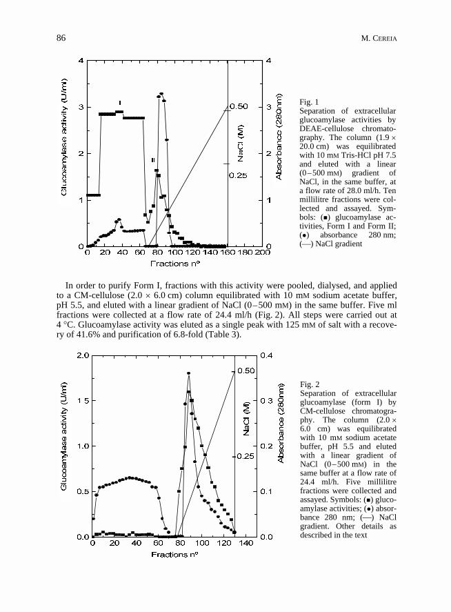
In order to purify the extracellular glucoamylase, a dialysed crude filtrate was applied toa DEAE-cellulose column (1.9 � 20.0 cm) equilibrated and eluted with 10 mM Tris-HClpH 7.5, at the flow rate of 28 ml/h. Ten ml fractions were collected and assayed for enzymeactivity. Glucoamylase activity was eluted into two fractions. Form I (76% of total activity)did not bind to the resin and eluted with a bulk of protein in the void volume, whereas formII (24% of total activity) was retained and eluted at 52 mM concentration of a linear(0–500 mM) NaCl gradient, prepared in the same buffer (Fig. 1). We concentrated in puri-fying and characterising glucoamylase form I only, considering that it represented the largerenzymatic component, in comparison with form II. Nevertheless, preliminary results (notshown) had revealed that glucoamylase form I and form II exhibited quite similar enzyma-tic properties.
Table 2Effect of nitrogen sources on glucoamylase production
Compoundsadded *
Extracellular(total U)
Intracellular(total U)
Protein(total U)
none 2.66 0.50 1.71gelatine 9.19 1.33 2.71CH3COONH4 83.88 3.05 4.13(NH4)H2PO3 1.85 1.27 2.49NH4NO3 9.67 0.30 1.56(NH4)2SO4 19.80 1.53 3.35Urea 8.59 1.57 3.16
* The compounds were added (1% w/v) to 25 ml of M-5 medium containing by 0.1% CaCO3; 0.1%peptone; 0.5% NaCl; 0.6% yeast extract and 1.0% starch. The pH was adjusted to 6.0.
86 M. CEREIA
In order to purify Form I, fractions with this activity were pooled, dialysed, and appliedto a CM-cellulose (2.0 � 6.0 cm) column equilibrated with 10 mM sodium acetate buffer,pH 5.5, and eluted with a linear gradient of NaCl (0–500 mM) in the same buffer. Five mlfractions were collected at a flow rate of 24.4 ml/h (Fig. 2). All steps were carried out at4 �C. Glucoamylase activity was eluted as a single peak with 125 mM of salt with a recove-ry of 41.6% and purification of 6.8-fold (Table 3).
�
�
Fig. 1Separation of extracellularglucoamylase activities byDEAE-cellulose chromato-graphy. The column (1.9 �20.0 cm) was equilibratedwith 10 mM Tris-HCl pH 7.5and eluted with a linear(0–500 mM) gradient ofNaCl, in the same buffer, ata flow rate of 28.0 ml/h. Tenmillilitre fractions were col-lected and assayed. Sym-bols: (�) glucoamylase ac-tivities, Form I and Form II;(�) absorbance 280 nm;(�) NaCl gradient
Fig. 2Separation of extracellularglucoamylase (form I) byCM-cellulose chromatogra-phy. The column (2.0 �6.0 cm) was equilibratedwith 10 mM sodium acetatebuffer, pH 5.5 and elutedwith a linear gradient ofNaCl (0–500 mM) in thesame buffer at a flow rate of24.4 ml/h. Five millilitrefractions were collected andassayed. Symbols: (�) gluco-amylase activities; (�) absor-bance 280 nm; (�) NaClgradient. Other details asdescribed in the text

Glucoamylase from a thermophilic fungus 87
Table 3Summary of the purification steps of extracellular glucoamylase form I
Step Totalvolume(ml)
Totalprotein(mg)
Totalactivity
Specificactivity(U/mg)
Yield(%)
Purifi-cation(-fold)
Filtrate 780 303.4 2828 9.3 100 –DEAE-cellulose Form I Form II
710164
50.4 56.6
2146 682
42.612.1
75.9 24.1
4.61.3
CM-cellulose 175 18.7 1176 62.9 41.6 6.8
The purified enzyme showed a single protein band in PAGE (Fig. 3A) and SDS-PAGE(Fig. 3B) coincident with glucoamylase activity against starch (Fig. 3C) or maltose(Fig. 3D), as substrates. The molecular mass of the enzyme was estimated to be 86.0 kDa(SDS-PAGE), or 68.5 kDa (Bio Sil-SEC-400 filtration). This difference in apparent mole-cular mass could be due to the fact that the enzyme was a glycoprotein (25.5% carbohydratecontent). According to HAMES and RICKWOOD (1985) many glycoproteins migrate anoma-lously, even in the presence of excess SDS and thiol reagents, probably because they bindSDS only to the protein part of the molecule. The reduced net charge resulting from reduc-ed SDS binding lowers the polypeptide mobility during electrophoresis, yielding artifactu-ally high molecular weight estimates.
Thin layer chromatography (TLC) of the products of hydrolysis of starch (Fig. 4A) ormaltose (Fig. 4B) revealed only glucose as the hydrolysis product, after 30 min of reaction,indicating that the purified enzyme was a glucoamylase [�(1,4)-D-glucan glucohydrolase,EC 3.2.1.3].
Table 4 summarises some biochemical properties of S. thermophilum glucoamylase acti-vity using either starch or maltose as substrates. Activation energy values (Ea) for the hy-drolysis of starch and maltose, calculated from ARRHENIUS plots were, 2.64 and 1.97 Kcalmol–1 respectively. The isoelectric point was 8.4. Optima of pH and temperature, usingstarch or maltose as substrates, were 6.5/60 �C and 5.0/55 �C, respectively. Glucoamylasehad a half-life of 22 minutes at 55 �C using starch as substrate, and it was completely stablewith maltose. The thermostability of the enzyme was improved (100% after 60 min at60 �C) when it was incubated at 60 �C in the presence of either 1% (w/v) starch, or 10%sorbitol, and then assayed for glucoamylase activity using starch or maltose as substrates.Similar results were reported by others (TOSI et al. 1993, SILVA and PERALTA 1998).
The preferred substrates for S. thermophilum glucoamylase activity were starch, maltose,glycogen, amylopectin, amylose, and isomaltose, in that order (data not shown). These dataare compatible with the values of Km and Vmax calculated from HANES plots (HANES 1932),determined at the optimum temperature for glucoamylase and maltase activities, usingstarch or maltose as substrates at concentrations from 0.05 to 4.5 mg/ml and 0.2 to10.0 mg/ml, respectively (Table 4). Glucoamylase activity was inhibited by several metalions used at 1 mM concentration, such as Ba++ (20%), Ca++ (22.7%), Cu++ (17.2%), EDTA(23%), Hg+ (24.4%), Mg++ (20.8%), Mn++ (21.1%), Na+ (22.8%), NH4
+ (13.7%), Zn++
(24.1%). On the other hand, maltase activity was activated by 10 mM Ba++ (7.8%), 10 mMMn++ (12.4%), and 10 mM Mg++ (28%). Other reagents inhibited 20 to 87% the enzyme.�-mercaptoethanol completely inhibited the hydrolysis of both substrates. This result mightsuggest the participation of protein disulphide bonds in the catalytic activity.
Sequence data of the first fifteen amino terminal residues of the purified protein(Table 5) revealed that S. thermophilum glucoamylase shared 93% homology with thatof the thermophilic fungus Humicola grisea var. thermoidea. No signal peptide wasfound.

88 M. CEREIA
Discussion
The production of glucoamylase by S. thermophilum was largely increased by replacingstarch by cassava flour, and gelatine by ammonium acetate, in the original M5 culture me-dium. Production of microbial glucoamylase in cassava flour was reported by others(KILIKIAN 1996, REDDY and BASAPPA 1996). Glucoamylase activity from Aspergillus flavuscultivated on cassava peel was 170-times higher than that obtained with soluble starch(SANI et al. 1992).
An interesting result was observed when S. thermophilum was cultivated in the presenceof different concentrations of sodium chloride (Table 1). This salt increased the secretionglucoamylase activity. The influence of osmotic potential on enzyme levels has been stu-died in only a few microbial systems, and different effects have been reported. Osmoticstress favours the production and secretion of a periplasmic �-galactosidase from Penicilli-um notatum (FIEDUREK 1998). Similar results were obtained for cellulase activity in Tri-choderma reesei and Sporotrichum cellulophilum growing on wheat bran (KIM et al. 1985).However, the possibility that the increased glucoamylase secretion in S. thermophilum isdue to changes in external osmolarity, is still a speculation.
Fig. 3Gel electrophoresis of purifiedglucoamylase activity. (A) silver-stained 7% PAGE. Lanes 1 and 2,0.62 �g and 1.0 �g, respectively;(B) 7% SDS-PAGE. Lane 3, puri-fied glucoamylase (2.5 �g). MM,molecular mass markers. (C) glu-coamylase activity gels of samplesfrom the purification steps usingstarch and (D) maltose as substrate

Glucoamylase from a thermophilic fungus 89
Table 4Kinetic constants and some intrinsic properties of S. thermophilum glucoamylase activities usingstarch and maltose as substrate
Parameters Starch Maltose
Activation energy (Ea Kcal mol–1) 2.64 1.97Carbohydrate content (%) 25.50Isoelectric point (pI) 8.40Molecular mass (Filtration Gel, kDa) 68.50Molecular mass (SDS-PAGE, kDa) 86.00Optimum pH 6.50 5.00Optimum temperature (�C) 60.00 55.00Stability 55 �C (T50 – min) 22.00 60.00Km (mg ml–1) 0.28 1.40Vmax (U mg–1 protein) 67.20 5.61
S. thermophilum glucoamylase activity was resolved into two components by ion ex-change chromatography. Form I (76% of total activity) was purified 6.8-fold. Some proper-ties of this glucoamylase, such as glycoproteic nature, molecular mass, specific activity andability to hydrolyse starch and maltose, resembled those of other fungal glucoamylases(reviewed by VIHINEN and MÄNTSÄLÄ 1989, GUZMÁN-MALDONADO and PAREDEZ-LÓPEZ1995, JAMES and LEE 1997). This enzyme hydrolysed preferentially starch, but also exhibit-ed a significant maltase activity. Maltase and glucoamylase activities co-migrated in PAGE(Fig. 3 A–D). These activities differed in optimum pH, effect of temperature and also acti-vation energy. Apparently, the activity against starch was protected at higher pH and tempe-
Fig. 4Analysis of the hydrolysis productsreleased by the purified S. thermophi-lum glucoamylase acting on starch (A)or maltose (B). Glucoamylase (3.6 mU)was incubated with the substrates(10 mg/ml) in 100 mM sodium acetatebuffer, pH 5.5, at 60 �C, for differenttimes (0; 0.5; 1; 2; 24 hours). Abbre-viations: G, glucose; M, maltose

90 M. CEREIA
Table 5N-terminal amino acid sequence of the protein produced by the thermophilic fungi Scytalidiumthermophilum
Edman cycle Amino acid pmol H. grisea
01 Ile 2.27 Ile02 Asn 1.81 Asn03 Thr 1.28 Thr04 Glu 1.58 Glu05 Lys 1.10 Lys06 Ser 0.11 Pro07 Ile 0.98 Ile08 Ala 1.09 Ala09 Trp 0.45 Trp10 Asn 0.78 Asn11 Lys 0.53 Lys12 Leu 0.81 Leu13 Leu 0.99 Leu14 Ala 1.03 Ala15 Asn 0.72 Asn
Sequence data of Humicola grisea var. thermoidea homologous enzyme was take from Gen Bankdatabase, (accession number M89475). The recovery of PTH – amino acids is given in picomol,which corresponds 80% of the product and is reported without any corrections
ratures. This observation agrees with H. grisea glucoamylase (TOSI et al. 1993) and sug-gested a common catalytic site for starch and maltose hydrolysis, but specialised subsitesfor each of these substrates (SIVAKAMI and RADHAKRISHNAN 1976, KISS et al. 1981).
The purified glucoamylase presented 93% homology with 15 amino terminal residues ofthe same enzyme from the thermophilic fungus Humicola grisea var. thermoidea. Glucoa-mylase structural, functional, and evolutionary relationships had been revised recently(COUTINHO and REILLY 1997).
The present study may contribute to the knowledge of the evolutionary tree of glucoa-mylase sequences, and to the basic knowledge of a glucoamylase that could be usedindustrially.
Acknowledgements
This work was supported by grants from Fundação de Amparo à Pesquisa do Estado de São Paulo(FAPESP) and Conselho de Desenvolvimento Científico e Tecnológico (CNPq). H. F. T.; J. A. J.;L. J. G.; J. C. R. and M. L. T. M. P. are Research Fellows of CNPq. M. C. is a recipient of FAPESPFellowship. This work is part of a Master Dissertation submitted to the Departamento de Biologia daFaculdade de Filosofia, Ciências e Letras de Ribeirão Preto – Universidade de São Paulo. We thankAMAURI PINHAL, MAURICIO DE OLIVEIRA and RICARDO F. ALARCON for technical assistance.
References
ALMEIDA, E. M., POLIZELI, M. L. T. M., TERENZI, H. F. and JORGE, J. A., 1995. Purification and bio-chemical characterisation of �-xylosidase from Humicola grisea var. thermoidea. FEMS Microbiol.Lett., 130, 171–176.
BASAVEWARA RAO, V., SASTRI, N. V. S. and SUBBA RAO, P. V., 1981. Purification and characteriza-tion of a thermostable glucoamylase from the thermophilic fungus Thermomyces lanuginosus. Bio-chem. J., 193, 379–387.

Glucoamylase from a thermophilic fungus 91
BERGMEYER, H. V. and BERNT, E., 1974. In: Methods of Enzymatic Analysis. Vol. 3. H. U. BERG-MEYER (Editors). pp. 1205–1215. Verlag-Chimie Academic Press, New York.
BLUM, H., BEIER, H. and GROSS, H. J., 1987. Improved silver staining of plant protein, RNA andDNA in polyacrylamide gels. Electrophoresis, 81, 93–99.
BUAINAIN, L. B., KADOWAKI, M. K., POLIZELI, M. L. T. M., TERENZI, H. F. and JORGE, J. A., 1998.Characterization of a conidial alkaline phosphatase from the thermophilic fungus Humicola griseavar. thermoidea. J. Basic. Microbiol., 38, 85–94.
BUSCH, J. E., PORTER, E. G. and STUTZENBERGER, F. J., 1997. Introduction of �-amylase by malto-oligosaccharides in Thermonospora curvata. J. Appl. Microbiol., 82, 669–676.
COUTINHO, P. M. and REILLY, P. J., 1997. Glucoamylase structural, functional and evolutionaryrelationships. Proteins: structure, function and genetics. (Biosis Vol. 1988), 29, 334–347.
CRABB, W. D. and MITCHINSON, C., 1997. Enzymes involved in the processing of starch to sugars.Trends Biotechnol., 15, 349–352.
DUBOIS, M., GILLES, K. A., HAMILTON, J. K., REBERS, P. A. and SMITH, F., 1956. Colorimetricmethod for determination of sugar and related substances. Anal. Chem., 28, 350–356.
FIEDUREK, J., 1998. Enhancement of �-galactosidase production and secretion by high osmotic stressin Penicillium notatum. Microbiol. Res., 153, 65–69.
FONTANA, J. D., GEBARA, M., BLUMEL, M., SCHINEIDER, H., MACKENZIE C. R. and FOHNSON, K. G.,1988. �-4-O-methyl-D-glucuronidase component of xylanolytic complexes. Methods Enzymol.,160, 560–571.
GUZMÁN-MALDONADO, H. and PAREDES-LÓPEZ, O., 1995. Amylolytic enzymes and products derivedfrom starch: A review. Crit. Ver. Food Sci. Nutrit., 35, 373–403.
HAMES, B. D. and RICKWOOD, D., 1985. Gel electrophoresis of proteins – A practical approach.pp. 14–15. IRL Press. Oxford – Washington D.C.
HANES, C. S., 1932. The effect of starch concentration upon the velocity of hydrolysis by the amylaseof germinated barley. Biochem. J., 26, 1406–1421.
JAMES, J. A. and LEE, B. H., 1997. Glucoamylases: Microbial sources, industrial applications andmolecular biology – A Review. J. Food Biochem., 21, 1–52.
KADOWAKI, M. K., POLIZELI, M. L. T. M., TERENZI, H. F. and JORGE, J. A., 1996. Characterization ofa threalase activities from the thermophilic fungus Scytalidium thermophilum. FEMS Microbiol.Lett., 1291, 199–205.
KILIKIAN, B. V., 1996. Production of glucoamylase by fed-batch culture of Aspergillus awamoriNRRL3112. Ver. Microbiol., 27, 137–141.
KIM, H. K., MOSOBUCHI, M. M., SEKI, T. and RYU, D. D. Y., 1985. Cellulase production by a solidstate culture system. Biotechnol. Bioeng., 27, 1445–1450.
KISS, L., BERKI, L. K. and NANASI, P., 1981. Evidence for a single catalytic and two bindingsites in the almond emulsin �-D-glucosidase molecule. Biochem. Biophys. Res. Commun., 98,792–799.
LAEMMLI, U. K., 1970. Cleavage of structural proteins during the assembly of the head of bacterio-phage T4. Nature, 227, 680–685.
LOWRY, O. H., ROSEBROUGH, N. J., FAN, A. L. and RANDALL, R. J., 1951. Protein measurement withthe folin phenol reagent. J. Biol. Chem., 193, 267–275.
MISHRA, R. S. and MAHESHWARI, R., 1996. Amylases of the thermophilic fungus Thermomyces lanu-ginosus: Their purification, properties, action on starch and response to heat. J. Biosci., 21,653–672.
O’FARRELL, P. Z., GOODMAN, H. M. and O’FARRELL, P. H., 1977. High resolution two-dimensionalelectrophoresis of basic as well as acidic proteins. Cell., 12, 1133–1142.
POLIZELI, M. L. T. M., JORGE, J. A. and TERENZI, H. F., 1996. Effect of carbon source on the�-glucosidase system of the thermophilic fungus Humicola grisea. World J. Microbiol. Biotech-nol., 12, 297–299.
REDDY, O. V. S. and BASAPPA, S. C., 1996. Direct fermentation of cassava starch to ethanol by mixedcultures of Endomycopsis fibuligera and Zymomonas mobilis: synergism and limitations. Biotech-nol. Lett., 18, 1315–1318.
REISFIELD, R. A., SEWIS, V. J. and WILLIANS, D. C., 1962. Disc electrophoresis of basis proteins andpeptides on polyacrilamide gels. Nature, 195, 281–283.
SANI, A., AWE, F. A. and AKINYANJU, A. J., 1992. Amylase synthesis in Aspergillus flavus andAspergillus niger grown on cassava peel. J. Ind. Microbiol., 10, 55–59.

92 M. CEREIA
SILVA, W. B. and PERALTA, R. M., 1998. Purification and characterization of a thermostable glucoa-mylase from Aspergillus fumigatus. Can. J. Microbiol., 44, 493–497.
SIVAKAMI, S. and RADHAKRISHNAN, N. A., 1976. Kinetic studies on glucoamylase of rabbit smallintestine. Biochem. J., 153, 321–327.
STRAATSMA, G. and SAMSOM, R. A., 1993. Taxonomy of Scytalidium thermophilum, an importantfungus in mushroom compost. Mycol. Res., 97, 32kkk328.
TOSI, L. R. O., TERENZI, H. F. and JORGE, J. A., 1993. Purification and characterization of an extra-cellular glucoamylase from the thermophilic fungus Humicola grisea var. thermoidea. Can. J. Mi-crobiol. 39, 846–852.
VAN DER BURG, B., VRIEND, G., VELTMAN, O. R., GERARD, V. and EIJSINK, V. G. H., 1998. Enginee-ring an enzyme to resist boiling. Biochem., 95, 2056–2060.
VIHINEN, M. and MÄNTSÄLÄ, P., 1989. Microbial amylolitic enzymes (Review). Crit. Rev. Biochem.Mol. Biol., 24, 329–418.
Mailing address: MARIA DE LOURDES TEIXERA DE MORAES POLIZELI, Departamento de Biologia,Faculdade de Filosofia, Ciências e Letras de Ribeirão Preto, Universidade de São Paulo, Av.Bandeirantes, 3900, 14.040-901 – Ribeirão Preto – SP, Brazile-mail: [email protected]