General signs and symptoms of abdominal diseases
64
General signs and symptoms of abdominal diseases Dr. Förhécz Zsolt Semmelweis University 3rd Department of Internal Medicine Faculty of Medicine, 3rd Year 2018/2019 1st Semester
-
Upload
khangminh22 -
Category
Documents
-
view
1 -
download
0
Transcript of General signs and symptoms of abdominal diseases

General signs and symptoms of abdominal diseases
Dr. Förhécz ZsoltSemmelweis University 3rd Department of Internal Medicine
Faculty of Medicine, 3rd Year2018/2019 1st Semester

• For descriptive purposes, the abdomen is divided by imaginary lines crossing at the umbilicus, forming the right upper, right lower, left upper, and left lower quadrants.
• Another system divides the abdomen into nine sections. Terms for three of them are commonly used: epigastric, umbilical, and hypogastric, or suprapubic
Common or Concerning Symptoms• Indigestion or anorexia
• Nausea, vomiting, or hematemesis
• Abdominal pain
• Dysphagia and/or odynophagia
• Change in bowel function
• Constipation or diarrhea
• Jaundice

“How is your appetite?”• Anorexia, nausea, vomiting in many gastrointestinal disorders; and
– also in pregnancy,
– diabetic ketoacidosis,
– adrenal insufficiency,
– hypercalcemia,
– uremia,
– liver disease,
– emotional states,
– adverse drug reactions
– Induced but without nausea in anorexia/ bulimia.

• Anorexia is a loss or lack of appetite.
• Some patients may not actually vomit but raise esophageal or gastric contents in the absence of nausea or retching, called regurgitation.– in esophageal narrowing from stricture or cancer; also with incompetent gastroesophageal
sphincter
• Ask about any vomitus or regurgitated material and inspect it yourself if possible!!!!– What color is it? – What does the vomitus smell like?– How much has there been? – Ask specifically if it contains any blood and try to determine how much?

• Fecal odor – in small bowel obstruction
– or gastrocolic fistula
• Gastric juice is clear or mucoid. Small amounts of yellowish or greenish bile are common and have no special significance.
• Brownish or blackish vomitus with a “coffee-grounds” appearance suggests blood altered by gastric acid. Coffee-grounds emesis or red blood are termed hematemesis.– in duodenal or peptic ulcer, esophageal or gastric varices,
gastritis, tumors

Do the patient’s symptoms suggest any complications of vomiting???
• such as aspiration into the lungs, seen in elderly, debilitated, or obtunded patients?
• Is there dehydration or electrolyte imbalance from prolonged vomiting,
• or significant loss of blood?– light-headedness or syncope depend on the rate
and volume of bleeding, and rarely appear until blood loss ≥ 500 ml.

Dysphagia
• difficulty swallowing, or dysphagia, the sense that food or liquid is sticking, hesitating, or “won’t go down right.”– Dysphagia may result from
esophageal disorders or from difficulty transferring food from the mouth to the esophagus.
– The sensation of a lump in the throat or in the retrosternal area, unassociated with swallowing, is not true dysphagia.
• Ask the patient to point to where the dysphagia occurs ?? and describe with what types of food???
• Pointing to the throat suggests a transfer or esophageal disorder; pointing to the chest suggests an esophageal disorder
• Does it occur with relatively solid foods such as meat, with softer foods such as ground meat and mashed potatoes, or with hot or cold liquids? Has the pattern changed?
• with solid food in mechanical narrowing of the esophagus;
• related to both solids and liquids suggests a disorder of esophageal motility.
• Establish the timing????. • When did it start? Is it intermittent or persistent? Is it
progressing? If so, over what period of time? What are the associated symptoms and medical conditions?

• Odynophagia, or pain on swallowing, mayoccur in two forms. – sharp, burning pain suggests mucosal
inflammation
– squeezing, cramping pain suggests a muscular cause.
– Odynophagia may accompany dysphagia, but either symptom may occurindependently
Mucosalinflammation in reflux esophagitisor infection fromCandida, herpesvirus, orcytomegalovirus.

DYSPHAGIA

Heartburn• or a sense of burning or warmth that is retrosternal and may radiate from the epigastrium to
the neck
• suggests gastric acid reflux into the esophagus; often precipitated by a heavy meal, lying down, or bending forward, also by ingested alcohol, citrus juices, or aspirin.– If chronic, consider reflux esophagitis
• If persistent, especially in the epigastric area, it may DD:raise the question of heart disease.– Some patients with coronary artery disease describe their pain as burning, “like indigestion.”
– Pay special attention to what brings on the discomfort and what relieves it. – Is it precipitated by exertion and relieved by rest suggesting angina, – or is it related to meals and made worse during or after eating suggesting gastroesophageal
reflux?
• Excessive gas, especially with frequent belching, abdominal bloating or distention, or flatus, the passage of gas by rectum, normally about 600 ml per day.– Find out if these symptoms are associated with eating specific
foods.
– Ask if symptoms are related to ingestion of milk or milk products.
– Belching, but not bloating or excess flatus, normally seen in aerophagia, or swallowing air.
– Also consider legumes and other gas-producing foods, intestinal lactase deficiency, irritable bowel syndrome.

• Unpleasant abdominal fullness after meals of normal size, or early satiety, the inability to eat a full meal.– diabetic gastroparesis,
– anticholinergic drugs,
– gastric outlet obstruction,
– gastric cancer;
– early satiety in hepatitis.

Abdominal pain
• Visceral pain occurs when hollow abdominal organs such as the intestine or biliary tree contract unusually forcefully or when they are distended or stretched. – Solid organs such as the liver can also become
painful when their capsules are stretched. – Visceral pain may be difficult to localize.– It is typically, though not necessarily, palpable near
the midline, at levels that vary according to the structure involved
– Visceral pain varies in quality and may be gnawing, burning, cramping, or aching.
– When it becomes severe, it may be associated with sweating, pallor, nausea, vomiting, and restlessness.
• Visceral pain in the right upper quadrant from liver distention against its capsule in alcoholic hepatitis
• Visceral periumbilical pain in early acute appendicitis from distention of inflamed appendix, gradually changing to parietal pain in the right lower quadrant from inflammation of the adjacent parietal peritoneum

• Parietal pain originates in the parietal peritoneum and is caused by inflammation. – It is a steady aching pain that is usually more severe than
visceral pain and more precisely localized over the involved structure
– It is typically aggravated by movement or coughing.
– Patients with this type of pain usually prefer to lie still.
• Referred pain is felt in more distant sites, which are innervated at approximately the same spinal levels as the disordered structure.– Referred pain often develops as the initial pain becomes
more intense and thus seems to radiate or travel from the initial site.
– It may be felt superficially or deeply but is usually well localized.
• Pain may also be referred to the abdomen from the chest, spine, or pelvis, thus complicating the assessment of abdominal pain.
• Pain of duodenal or pancreatic origin may be referred to the back;
• pain from the biliary tree, to the right shoulder or the right posterior chest.
• Pain from pleurisy or acute myocardial infarction may be referred to the upper abdomen.

Ask patients • to describe the abdominal pain in their own words
• ask them to point to the pain.
• “Where does the pain start?”
• “Does it radiate or travel anywhere?”
• “What is the pain like?” “Is it aching, burning, gnawing, or what?”
• “How severe is the pain?”
• “How about on a scale of 1 to 10?” Find out if it is bearable and if it interferes with the patient’s usual activities. Does it make the patient lie down?

Timing of the pain• Did it start suddenly or gradually?
• When did the pain begin?
• How long does it last?
• What is its pattern over a 24-hour period?
• Over weeks and months?
• Are you dealing with an acute illness or a chronic or recurring one?

• Determine what factors aggravate or relieve the pain,
• with special reference to meals,
• antacids, alcohol, medications (including aspirin and aspirinlike drugs and any over-the-counter drugs),
• emotional factors,
• and body position.
• is the pain related to defecation, urination, or menstruation?
• any symptoms that are associated with the pain, such as fever or chills, and ask in what sequence they occur.


Assess bowel function• Start with open-ended questions: “How are
your bowel movements?”
• “How frequent are they?”
• “Do you have any difficulties?”
• “Have you noticed any change in your bowel habits?”
• Frequency of bowel movements normally ranges from about three times a day to twice a week. A change in pattern within these limits, however, may be significant for an individual patient.

Patients vary widely in their views of constipation and diarrhea. • Be sure to clarify what the patient means by these terms.
• For example, is constipation . . .– a decrease in frequency of bowel movements? – . . . The passage of hard and perhaps painful stools?– . . . The need to strain unusually hard?– . . . A sense of incomplete defecation or pressure in the rectum? s
• Ask if the patient actually looks at the stool. If yes, what does the stool look like in terms of color and bulk?
• What remedies has the patient tried? Do medications, stress, unrealistic idea about normal bowel habits, or time and setting allotted for defecation play a role?
• Occasionally there is complete constipation with no passage of either feces or gas, or obstipation.

Constipation

about the color of the stools • ask about any black tarry stools,
suggesting melena,
• or red blood in the stools, known as hematochezia
• If either condition is present, find out how long and how often.
• If the blood is red, how much is there?
• Is it pure blood mixed in with stool or on the surface of the stool?
• Is there blood on the toilet paper? - in hemorrhoids

Diarrhea is an excessive frequency in the passage of stools that are usually unformed or watery.
• Ask about size, frequency, and volume.
• Are the stools bulky or small?
• How many episodes of diarrhea occur each day?– consistently large diarrheal stools often in small bowel or
proximal colon disorders;
– small frequent stools with urgency of defecation in left colon or rectal disorders
• Assess the course of diarrhea over time. – Is it acute, chronic, or recurrent?
– Or is your patient experiencing the first acute episode of a chronic or recurrent illness?

• Does the diarrhea awaken the patient at night? - Nocturnal diarrhea suggests a pathophysiologic cause.
• What seem to be the aggravating or relieving factors? - Relief after passing feces or gas suggests left colon or rectal disorders;
• Does the patient get relief from a bowel movement, or is there an intense urge with straining but little or no result, known as tenesmus. - in rectal conditions near the anal sphincter.
• What is the setting? Does it entail travel, stress, or a new medication?
• Do family members or companions have similar symptoms?
• Are there associated symptoms?

• Are the stools greasy or oily? Frothy? Foul smelling? Floating on the surface because of excessive gas, making them difficult to flush? Accompanied by mucus, pus, or blood?
– Large yellowish or gray greasy foul smelling, sometimes frothy or floating stools in steatorrhea, or fatty stools—seen in malabsorption


Jaundice or icterus
• yellowish discoloration of the skin and sclerae from increased levels of bilirubin
• Normally the hepatocytes conjugate, or combine, unconjugated bilirubin with other substances, making the bile water soluble, and then excrete it into the bile.
• The bile passes through the cystic duct into the common bile duct, which also drains the extrahepatic ducts from the liver.
• More distally the common bile duct and the pancreatic ducts empty into the duodenum at the ampulla of Vater. Mechanisms of jaundice include:– Increased production of bilirubin –haemolytic anemia– Decreased uptake of bilirubin by the hepatocytes– Decreased ability of the liver to conjugate bilirubin –Gilbert’s Sy– Decreased excretion of bilirubin into the bile, resulting in absorption of
conjugated bilirubin back into the blood -• in viral hepatitis, cirrhosis, primary biliary cirrhosis, druginduced cholestasis,
as with oral contraceptives, methyl testosterone, chlorpromazine

• Intrahepatic jaundice can be hepatocellular, from damage to the hepatocytes, or cholestatic, from impaired excretion due to damaged hepatocytes or intrahepatic bile ducts.
• Extrahepatic jaundice arises from obstruction of the extrahepatic bile ducts, most commonly the cystic and common bile ducts.– Obstruction of the common bile duct by
gallstones or pancreatic carcinoma

Assess the jaundiced patient
• What was the color of the urineas the patient became ill? – When the level of conjugated bilirubin increases in the blood, it may be excreted into the
urine, turning the urine a dark yellowish brown or tea color. - indicates impaired excretion of bilirubin into the gastrointestinal tract.
– Unconjugated bilirubin is not watersoluble so is not excreted into urine.
• Ask also about the color of the stools– When excretion of bile into the intestine is completely obstructed, the stools become gray or
light-colored, or acholic, without bile. - briefly in viral hepatitis, common in obstructive jaundice
• Does the skin itch without other obvious explanation? Is there associated pain? What is its pattern? Has it been recurrent in the past? – itching in cholestatic or obstructive jaundice;– pain from a distended liver capsule, biliary cholic, pancreatic cancer

Inspection
• Scars. Describe or diagram their location.
• Striae. Old silver striae or stretch marks are normal.
• Dilated veins. A few small veins may be visible normally.
• Rashes and lesions

• The umbilicus. Observe its contour and location, and any signs of inflammation or hernia
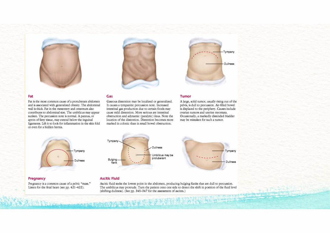
• The contour of the abdomen– Is it flat, rounded, protuberant, or scaphoid
(markedly concave or hollowed)?
• Do the flanks bulge or are there any local bulges? Include in this survey the inguinal and femoral areas.
• Is the abdomen symmetric?
• Are there visible organs or masses? Look for an enlarged liver or spleen that has descended below the rib cage.
• Peristalsis.Observe for several minutes if you suspect intestinal obstruction. Peristalsis may be visible normally in very thin people.
• Pulsations. The normal aortic pulsation is frequently visible in the epigastrium.

AUSCULTATION
• Auscultation provides important information about bowel motility.
• Listen to the abdomen before performing percussion or palpation, since these maneuvers may alter the frequency of bowel sounds
• Auscultation may also reveal bruits, vascular sounds resembling heart murmurs, over the aorta or other arteries in the abdomen, which suggest vascular occlusive disease.
• A bruit in one of these areas that has both systolic and diastolic components strongly suggests renal artery stenosis as the cause of hypertension
• Bruits with both systolic and diastolic components suggest the turbulent blood flow of partial arterial occlusion

PERCUSSION

PERCUSSION
• Percussion helps you to assess the amount and distribution of gas in the abdomen and to identify possible masses that are solid or fluid filled.
• Its use in estimating the size of the liver and spleen will be described in later sections.
• Percuss the abdomen lightly in all four quadrants to assess the distribution of tympany and dullness.
• Tympany usually predominates because of gas in the gastrointestinal tract, but scattered areas of dullness due to fluid and feces there are also typical.

• large dull areas that might indicate an underlying mass or enlarged organ- Pregnant uterus, ovarian tumor, distended bladder, large liver or spleen
• protuberant abdomen, note where abdominal tympany changes to the dullness of solid posterior structures -Dullness in both flanks indicates further assessment for ascites
• Briefly percuss the lower anterior chest, between lungs above and costal margins below.
• On the right, you will usually find the dullness of liver; on the left, the tympany that overlies the gastric air bubble and the splenic flexure of the colon.

PALPATION
• Light Palpation.
• Feeling the abdomen gently– is especially helpful in identifying abdominal
tenderness, muscular resistance, and some superficial organs and masses. It also serves to reassure and relax the patient.
– Involuntary rigidity (muscular spasm) typically persists despite these maneuvers. It indicates peritoneal inflammation.

• Deep Palpation.
• This is usually required to delineate abdominal masses. Again using the palmar surfaces of your fingers, feel in all four quadrants.
• Identify any masses and note their location, size, shape, consistency, tenderness, pulsations, and any mobility with respiration or with the examining hand.
• Correlate your palpable findings with their percussion notes.
Abdominal masses may be categorized in several ways: • physiologic (pregnant uterus), • inflammatory (diverticulitis of the colon),• vascular (an aneurysm of the abdominal aorta),• neoplastic (carcinoma of the colon), • or obstructive (a distended bladder or dilated
loop of bowel).

Assessment for Peritoneal Inflammation• Abdominal pain and tenderness, especially when
associated with muscular spasm, suggest inflammation of the parietal peritoneum.
• Localize the pain as accurately as possible.
• First, even before palpation, ask the patient to coughand determine where the cough produced pain.
• Then, palpate gently with one finger to map the tender area.
• Pain produced by light percussion has similar localizing value.
• These gentle maneuvers may be all you need to establish an area of peritoneal inflammation.

• Look for rebound tenderness.
• Press your fingers in firmly and slowly, and then quickly withdraw them.
• Watch and listen to the patient for signs of pain.
• Ask the patient – to compare which hurt more, the pressing or the
letting go – and to show you exactly where it hurt.
• Pain induced or increased by quick withdrawal constitutes rebound tenderness.
• It results from the rapid movement of an inflamed peritoneum.


ASSESSING POSSIBLE ASCITES • A protuberant abdomen with bulging flanks
suggests the possibility of ascitic fluid.
• Because ascitic fluid characteristically sinks with gravity, while gas-filled loops of bowel float to the top, percussion gives a dull note in dependent areas of the abdomen.
• Look for such a pattern by percussing outward in several directions from the central area of tympany.
• Map the border between tympany and dullness

• Test for shifting dullness.
• After mapping the borders of tympany and dullness, ask the patient to turn onto one side.
• Percuss and mark the borders again.
• In a person without ascites, the borders between tympany and dullness usually stay relatively constant.

• Test for a fluid wave.
• Ask the patient or an assistant to press the edges of both hands firmly down the midline of the abdomen.
• This pressure helps to stop the transmission of a wave through fat.
• While you tap one flank sharply with your fingertips, feel on the opposite flank for an impulse transmitted through the fluid.
• Unfortunately, this sign is often negative until ascites is obvious, and it is sometimes positive in people without ascites.

• Identifying an Organ or a Mass in an Ascitic Abdomen.
• Try to ballottethe organ or mass, exemplified here by an enlarged liver.
• Straighten and stiffen the fingers of one hand together, place them on the abdominal surface, and make a brief jabbing movement directly toward the anticipated structure.
• This quick movement often displaces the fluid so that your fingertips can briefly touch the surface of the structure through the abdominal wall.

ASSESSING POSSIBLE APPENDICITIS
• Ask the patient to point to where the pain began and where it is now. Ask the patient to cough.– Determine whether and where pain results– The pain of appendicitis classically begins
near the umbilicus, then shifts to the right lower quadrant, where coughing increases it. Elderly patients report this pattern less frequently than younger ones.
• Search carefully for an area of local tenderness– Localized tenderness anywhere in the right
lower quadrant, even in the right flank, may indicate appendicitis.
• Feel for muscular rigidity - Early voluntary guarding may be replaced by involuntary muscular rigidity.

• Perform a rectal examination and, in women, a pelvic examination.– These maneuvers may not help you to discriminate well
between a normal and an inflamed appendix, but they may help to identify an inflamed appendix atypically located within the pelvic cavity.
– They may also suggest other causes of the abdominal pain.
– Right-sided rectal tenderness may be caused by, for example, inflamed adnexa or an inflamed seminal vesicle, as well as by an inflamed appendix.

• Check the tender area for rebound tenderness. – Rebound tenderness suggests peritoneal inflammation, as from appendicitis.
• Check for Rovsing’s signand for referred rebound tenderness. Press deeply and evenly in the leftlower quadrant. Then quickly withdraw your fingers– Pain in the right lower quadrant during left-sided pressure suggests appendicitis (a
positive Rovsing’s sign). So does right lower quadrant pain on quick withdrawal (referred rebound tenderness).
• Look for a psoas sign. Place your hand just above the patient’s right knee and ask the patient to raise that thigh against your hand– Then extend the patient’s right leg at the hip. Flexion of the leg at the hip makes the
psoas muscle contract; extension stretches it.• suggesting irritation of the psoas muscle by an inflamed appendix.

• Look for an obturator sign. Flex the patient’s right thigh at the hip, with the knee bent, and rotate the leg internally at the hip. This maneuver stretches the internal obturator muscle– Right hypogastric pain constitutes a positive obturator sign, suggesting irritation of the
obturator muscle by an inflamed appendix.
• Test for cutaneous hyperesthesia. At a series of points down the abdominal wall, gently pick up a fold of skin between your thumb and index finger, without pinching it. This maneuver should not normally be painful– Localized pain with this maneuver, in all or part of the right lower quadrant, may
accompany appendicitis
ASSESSING POSSIBLE ACUTE CHOLECYSTITIS • When right upper quadrant pain and tenderness suggest
acute cholecystitis, look for Murphy’s sign.
• Hook your left thumb or the fingers of your right hand under the costal margin at the point where the lateral border of the rectus muscle intersects with the costal margin.
• Alternatively, if the liver is enlarged, hook your thumb or fingers under the liver edge at a comparable point below.
• Ask the patient to take a deep breath.
• Watch the patient’s breathing and note the degree of tenderness.
• A sharp increase in tenderness with a sudden stop in inspiratory effort constitutes a positive Murphy’s sign of acute cholecystitis. Hepatic tenderness may also increase with this maneuver, but is usually less well localized
ASSESSING VENTRAL HERNIAS • Ventral hernias are hernias in the abdominal
wall exclusive of groin hernias.
• If you suspect but do not see an umbilical or incisional hernia, ask the patient to raise both head and shoulders off the table.– The bulge of a hernia will usually appear with this
action

MASS IN THE ABDOMINAL WALL • To Distinguish an Abdominal Mass From a Mass
in the Abdominal Wall.
• An occasional mass is in the abdominal wall rather than inside the abdominal cavity.
• Ask the patient either to raise the head and shoulders or to strain down, thus tightening the abdominal muscles.
• Feel for the mass again.– A mass in the abdominal wall remains palpable; an
intra-abdominal mass is obscured by muscular contraction.


Cím és tartalom diagrammal
0
1
2
3
4
5
6
1. KATEGÓRIA 2. KATEGÓRIA 3. KATEGÓRIA 4. KATEGÓRIA
DIAGRAMCÍM1. adatsor 2. adatsor 3. adatsor

Két tartalomrész elrendezés táblázattal• Első pont szövege
• Második pont szövege
• Harmadik pont szövege
Osztály A csoport B csoport
1. osztály 82 95
2. osztály 76 88
3. osztály 84 90

Két tartalomrész elrendezés SmartArt-ábrával
1. tevékenység
2. tevékenység
3. tevékenység
4. tevékenység
5. tevékenység
• Első pont szövege
• Második pont szövege
• Harmadik pont szövege

Diacím hozzáadása – 1

Diacím hozzáadása – 2

Diacím hozzáadása – 3


Diacím hozzáadása – 4