
Abdominal tuberculosis-a disease revived - NCBI
7
Annals of the Royal College of Surgeons of England (1983) vol. 65 Abdominal tuberculosis-a disease revived N V ADDISON FRCS (onsultant Surgeon, The Royal Infirmary, Bradford Key words: ABDONII.NAL TUBERCULOSIS; TUBERCULOSIS PERITONITIS; TUBERCULOUS ENTERITIS; CROHN'S DISEASE Summary Abdominal tuberculosis was common in the United Kingdom in the 18th and 19th centuries and in thefirst half of the 20th century. During the 1950's the recognition of Crohn's disease, the use of streptomycin and other drugs, and the pasteurisation of milk led to the virtual disappearance of abdominal tuberculosis in the western world. During the last two decades a new type, mycobacterium tuberculosis hominis, has appeared mainly in the immigrant population, especially in those from the Indian subcontinent. A retrospective review of 68 patients with abdominal tuberculosis is presented. The pathology, diagnosis and management of these cases is discussed, together with the differential diagnosis of Crohn's disease. It is suggested that the immigrant brings the disease into the United Kingdom in his mesenteric glands and that the disease is reactivated or 'revived' at some later date due to some modification of the immune process. Introduction and historical review It was almost 100 years after the death ofJohn Hunter that Robert Koch, in 1882, discovered the tubercle bacillus when working in the Imperial Health Office in Berlin. He thus established the identity of a number of conditions previously regarded as distinct clinical entities such as lupus, scrofula, phthisis and Pott's disease. Whilst working at his brother William's anatomy school in London, John Hunter had worked with Percival Pott at St. Bartholomew's Hospital between 1748 and 1759, although it was 1779 before Pott first described tuberculous disease of the spine which still bears his name. Like others at that time, John Hunter was unaware of the infective nature of Pott's disease and also scrofula, although during the same period his observations and research into the mode of transmission of both syphilis and gonorrhoea are very well known. John Hunter probably had his suspicions regarding the cause of phthisis, as about November 1759 he is said to have contracted somne infection affecting his lungs, and believed that he might be threatened with tuberculosis. There is no doubt that he suffered a severe illness and on advice took a complete rest from his active life during the second half of 1760. Historical records reveal that tuberculosis had been re- cognised as a disease from the earliest time, and Hippocrates in the 4th century BC bestowed the name phthisis, meaning wasting, upon the disease as it affected the lungs. It is also interesting to note that one of the aphorisms of Hippocrates warned that 'phthisical persons die if diarrhoea sets in'. In other words, he had made the observation that patients with open pulmonary tuberculosis could develop intestinal com- plications. The association of intestinal ulceration, per- foration and peritonitis, was vividly described in 1643 in the Based on a Hunterian lecture delivered at the University of Leeds on 5 May 1982 autopsy report of Louis XIII (1). This is considered one of the earliest reports of secondary intestinal tuberculosis. Tuberculous glands of the neck, which were likened in appearance to a sow's bulging neck, were given the name scrofula, from the Latin scrofa meaning sow. This was also known as King's Evill from the superstition that a touch of the royal hand of French and English monarchs conveyed a cure to the affected person. Since the 5th century, the kings of France were believed to receive this healing power from God at the time of their anointment; Edward the Confessor in the 11th century had also claimed this healing power for the English kings. Charles II is said to have touched some 90 000 patients suffering from scrofula, with reputedly good results, and this practice continued well into the 19th century. In 1696 Richard Wiseman in his Fourth Treatise (2) described scrofula, and from his post mortem examinations he re- cognised the same pathology in the mesentery, lungs, testes and pericardium, as well as lymph nodes, bones and joints. In other words, he depicted surgical tuberculosis. Careful reading of John Hunter's words reveal observations that were very relevant to surgical tuberculosis. In 1786 (3) he wrote in his Lectures on the Principles of Surgery, that scrofula was 'a specific disease but without a morbid poison'. He also observed that other animals besides the human subject were liable to it and probably from the same causes. He then writes 'the true scrofula seems attended with little or no inflammation; or if it is so, its sedative quality prevents it from producing pain; [presumably he was describing a cold abscess] but when there is less of the true scrofula, it may give pain. However, it is attended with many of the effects of inflammation, as extravasation of coaguable lymph produc- ing swelling, and also adhesion of the cellular membrane or of the sides of circumscribed cavities'. John Hunter then goes on to describe the clinical features of abdominal tuberculosis. 'I have seen the whole of the intestine adhering to the peritoneum seemingly from a scrofulous cause, and having scrofulous tumours and suppurations in them also; the symptoms were tightness of the belly without pain, and costiveness; at other times, purging'. He also stated that adhesions in the lungs of consumptive people were probably of this kind, and like Richard Wiseman, he recognised that the disease process of scrofula could affect not only lymph glands but the lungs, uterus, testicle and bones. During the 18th and 19th centuries, abdominal tubercu- losis was probably as common as pulmonary tuberculosis and other types of the disease, and in his monograph in 1828 (4) John Abercrombie from Edinburgh gives a very lucid description of chronic peritonitis which is clearly identifiable with tuberculous peritonitis. He records the disease as 'occurring in young persons as an insidious affection of utmost danger yet extremely obscure in its symptoms'. He observed that chronic peritonitis was generally combined with disease of mesenteric glands and tubercular disease of
-
Upload
khangminh22 -
Category
Documents
-
view
1 -
download
0
Transcript of Abdominal tuberculosis-a disease revived - NCBI

Annals of the Royal College of Surgeons of England (1983) vol. 65
Abdominal tuberculosis-a disease revived
N V ADDISON FRCS(onsultant Surgeon, The Royal Infirmary, Bradford
Key words: ABDONII.NAL TUBERCULOSIS; TUBERCULOSIS PERITONITIS; TUBERCULOUS ENTERITIS; CROHN'S DISEASE
SummaryAbdominal tuberculosis was common in the United Kingdom in the18th and 19th centuries and in thefirst halfofthe 20th century. Duringthe 1950's the recognition of Crohn's disease, the use of streptomycinand other drugs, and the pasteurisation of milk led to the virtualdisappearance of abdominal tuberculosis in the western world.
During the last two decades a new type, mycobacterium tuberculosishominis, has appeared mainly in the immigrant population, especiallyin those from the Indian subcontinent.A retrospective review of68 patients with abdominal tuberculosis is
presented. The pathology, diagnosis and management of these cases isdiscussed, together with the differential diagnosis of Crohn's disease.
It is suggested that the immigrant brings the disease into the UnitedKingdom in his mesenteric glands and that the disease is reactivated or'revived' at some later date due to some modification of the immuneprocess.
Introduction and historical reviewIt was almost 100 years after the death ofJohn Hunter thatRobert Koch, in 1882, discovered the tubercle bacillus whenworking in the Imperial Health Office in Berlin. He thusestablished the identity of a number of conditions previouslyregarded as distinct clinical entities such as lupus, scrofula,phthisis and Pott's disease. Whilst working at his brotherWilliam's anatomy school in London, John Hunter hadworked with Percival Pott at St. Bartholomew's Hospitalbetween 1748 and 1759, although it was 1779 before Pottfirst described tuberculous disease of the spine which stillbears his name. Like others at that time, John Hunter wasunaware of the infective nature of Pott's disease and alsoscrofula, although during the same period his observationsand research into the mode of transmission of both syphilisand gonorrhoea are very well known. John Hunter probablyhad his suspicions regarding the cause of phthisis, as aboutNovember 1759 he is said to have contracted somne infectionaffecting his lungs, and believed that he might be threatenedwith tuberculosis. There is no doubt that he suffered a severeillness and on advice took a complete rest from his active lifeduring the second half of 1760.
Historical records reveal that tuberculosis had been re-cognised as a disease from the earliest time, and Hippocratesin the 4th century BC bestowed the name phthisis, meaningwasting, upon the disease as it affected the lungs. It is alsointeresting to note that one of the aphorisms of Hippocrateswarned that 'phthisical persons die if diarrhoea sets in'. Inother words, he had made the observation that patients withopen pulmonary tuberculosis could develop intestinal com-plications. The association of intestinal ulceration, per-foration and peritonitis, was vividly described in 1643 in the
Based on a Hunterian lecture delivered at the University of Leedson 5 May 1982
autopsy report of Louis XIII (1). This is considered one ofthe earliest reports of secondary intestinal tuberculosis.
Tuberculous glands of the neck, which were likened inappearance to a sow's bulging neck, were given the namescrofula, from the Latin scrofa meaning sow. This was alsoknown as King's Evill from the superstition that a touch ofthe royal hand of French and English monarchs conveyed acure to the affected person. Since the 5th century, the kings ofFrance were believed to receive this healing power from Godat the time of their anointment; Edward the Confessor in the11th century had also claimed this healing power for theEnglish kings. Charles II is said to have touched some 90 000patients suffering from scrofula, with reputedly good results,and this practice continued well into the 19th century. In1696 Richard Wiseman in his Fourth Treatise (2) describedscrofula, and from his post mortem examinations he re-cognised the same pathology in the mesentery, lungs, testesand pericardium, as well as lymph nodes, bones and joints.In other words, he depicted surgical tuberculosis. Carefulreading of John Hunter's words reveal observations thatwere very relevant to surgical tuberculosis. In 1786 (3) hewrote in his Lectures on the Principles of Surgery, thatscrofula was 'a specific disease but without a morbid poison'.He also observed that other animals besides the humansubject were liable to it and probably from the same causes.He then writes 'the true scrofula seems attended with little orno inflammation; or if it is so, its sedative quality prevents itfrom producing pain; [presumably he was describing a coldabscess] but when there is less of the true scrofula, it may givepain. However, it is attended with many of the effects ofinflammation, as extravasation of coaguable lymph produc-ing swelling, and also adhesion of the cellular membrane orof the sides of circumscribed cavities'. John Hunter then goeson to describe the clinical features ofabdominal tuberculosis.'I have seen the whole of the intestine adhering to theperitoneum seemingly from a scrofulous cause, and havingscrofulous tumours and suppurations in them also; thesymptoms were tightness of the belly without pain, andcostiveness; at other times, purging'. He also stated thatadhesions in the lungs of consumptive people were probablyof this kind, and like Richard Wiseman, he recognised thatthe disease process of scrofula could affect not only lymphglands but the lungs, uterus, testicle and bones.
During the 18th and 19th centuries, abdominal tubercu-losis was probably as common as pulmonary tuberculosisand other types of the disease, and in his monograph in 1828(4) John Abercrombie from Edinburgh gives a very luciddescription of chronic peritonitis which is clearly identifiablewith tuberculous peritonitis. He records the disease as'occurring in young persons as an insidious affection ofutmost danger yet extremely obscure in its symptoms'. Heobserved that chronic peritonitis was generally combinedwith disease of mesenteric glands and tubercular disease of

106 N 1' Addison
the lung. Then follows a description of an interestingcondition called tympanites which he attributed to chronicperitonitis (5). This disease occurred in females, who pre-sented with a firm, tense abdomen, usually associated withamenorrhoea and often mistaken for pregnancy, especially inthose ladies who had married late in life. If treated at anearly period the condition generally disappeared in a shorttime under a course of mild purgatives such as 'Harrowgatewater', a therapeutic agent located not very far from Leeds.
During the second half of the 19th century, surgeons werebeginning to operate on the abdomen and abdominaltuberculosis is reported to have been most commonly foundafter malignant and acute inflammatory conditions. At thebeginning of the 20th century it had become customary todescribe three forms of abdominal tuberculosis:
1) ulcerative tuberculosis or tuberculous enteritis;2) tuberculous peritonitis;3) hyperplastic tuberculosis.
William Boyd (6) in 1925, described the hyperplastic groupas being a separate type occurring in young women under 40years of age. There was great formation of tuberculousgranulation tissue in the ileocaecal region, with abundantgiant cells, few tubercles and absent caseation. The affectedpart of the bowel was thickened, narrow and stiff and formeda tumour-like mass resembling a carcinoma. The neighbour-ing glands were usually enlarged and in these cases thetubercle bacilli were rarely demonstrated and there wasusually no evidence of tuberculosis in the rest of the body. In1928, in his paper on chronic intestinal tuberculosis,Matthew Stewart (7) Professor of Pathology at Leeds Uni-versity, stated that tuberculous ulceration of the intestinewas present in half the cases of pulmonary tuberculosis whichcame to autopsy. He offered the hypothesis that the hyper-plastic type was caused by a bovine s!train of mild virulencein a patient with a high resistance to tubercle, which resultedin marked fibrosis in the ileocaecal region. Because of thesefeatures in hyperplastic tuberculosis, some observers at thattime doubted the specific nature of the disease, and there isno doubt that many cases of hyperplastic ileocaecal tuberclewere in fact Crohn's disease. In some instances, text bookdescriptions of the lesions were word for word perfectdescriptions of Crohn's lesions which had yet to be described.In 1902 Mayo Robson, a surgeon at Leeds General Infir-mary, presented a paper at the Clinical Society of London onthe surgical treatment of chronic intestinal tuberculosis.From his reports and descriptions, 3 of his 7 patients had infact Crohn's disease. Even at the beginning of this century,reports of ileocaecal tuberculosis without lung involvementwere common, and in the 1920's surgeons were beginning torealise that there were lesions other than abdominal tubercu-losis that could cause thickening and narrowing of the smallintestine. Matthew Stewart commented that 'whether thereis such a thing as a hyperplastic enterocolitis of non-tuberculous origin still remains to be seen. I am inclined tothink there is, in spite of arguments already put forward'.And so, in 1932 Crohn, Ginsberg and Oppenheimer (8)published their classical paper on the disease of the smallintestine which was to become known as Crohn's disease.The classification of abdominal tuberculosis remained, how-ever, as before.
During the 1950's the recognition of Crohn's disease, theuse of streptomycin and other drugs, and the pasteurisationof milk led to the virtual disappearance of abdominaltuberculosis caused by human and bovine strains in thewestern world. It is still a common disease on the continent ofAfrica and the subcontinent of India. In the 1960's and1 970's a new type, mycobacterium tuberculosis hominis,emerged with many reports in the British literature describ-ing the disease as it is seen in Britain today where the vastmajority of tuberculosis occurs in immigrants, particularlythose from the Indian subcontinent. As the immigrantpopulation has increased, so has the incidence of abdominal
tuberculosis. Between 1967 and 1980 I reviewed 68 patientswith abdominal tuberculosis, in a city of 400 000 people with50 000 immigrants. The interval occurring between im-migration and clinical presentation of the disease varied from6 months to 16 years and on average was just a little over 5years. The precise reason for such an interval is unknown. Itis more than likely that the immigrant brings the disease intothis country and it is reactivated at some later date. There isexperimental evidence to suggest that there is some alter-ation in immune patterns that may occur as a result of thestress of immigration. Whilst it is\ true that the majority ofpatients who now present with abdominal tuberculosis arefrom the Indian subcontinent originally, it is not always thecase, as 140 0 of the patients in our 1979 series (9) were of theindigenous population. In other series, white subjects werealso reported to suffer from this disease (Table 1). Of 68patients reviewed, 41 were male and 27 were female. The agerange of the patients was from 8 to 52 years. The mean age ofthe males was 30 years and the females 32 years.
TABLE I
No. of whileSeries subjects
Howell & Knapton (10) (1964) 5 (420o)Anscombe el al (11) (1967) 8 (88O )Mandal & Schofield (12) (1976) 3 (200%o)Shukla & Hughes (13) (1978) 5 (62° ,)Findlay & Addison (9) (1979) 7 (I4 ())Lambrianides el al (14) (1980) 2 (8°O)
Pathological featuresFrom a clinicopathological view, I would propose two maingroups, (a) tuberculous peritonities, (b) gastrointestinal tub-erculosis, and there may be a combination of both types inthe same patient.
In tuberculous peritonitis there is usually a straw colouredascites, more marked in the acute type. The peritoneum isstudded with fine white tubercles and there is thickening ofthe walls of the intestine and other organs and the greateromentum is greatly infiltrated and thickened. There may becaseous masses of varying size in the peritoneal cavity.
Histological appearances reveal classical granulomas withepithelioid cells, Langhan's giant cells, central necrosis andan outer rim of lymphocytes scattered through the omentumand mesenteric fat. Acid fast bacilli are not alwaysdemonstrable.
In the gastrointestinal group, gastro-duodenal tubercu-losis is rare and in this series there have been 4 patients, 1with a lesion in the pylorus and 3 with lesions in theduodenum. Mandal and Schofield (12) reported 3 patients in1976, and in 1979 Mukerjee and Singal (15) had anincidence of 2.800, in 500 patierfts operated on for abdominaltuberculosis.
Tuberculous enteritis is commonest in the ileocaecalregion, seen in my series (680°) and others, but may involveany part of the small or large intestine. The wall of theintestine is thickened due to granulomatous infiltration andfibrosis, and forms a mass in the right iliac fossa (Fig. 1).There are enlarged lymph nodes and tiny miliary tuberclesmay be found over the peritoneal surface of the bowel andmesentery. There are no classical features of tuberculosis onnaked eye appearances and the lesion may be indistin-guishable from Crohn's disease as the mesenteric fat isincreased and there is 'fat wrapping' around small bowelstrictures as in Crohn's disease. Tuberculous lesions in thesmall intestine not only mimic Crohn's disease, but lym-phoma and ischaemic strictures, and there can be multiple

Abdominal Tuberculosis a disease revived 107
FIG. 1 -Nakcd eve appearances of ileocaccal tuberculosis withthickenied wall of inltestine due to granulomatous infiltration andfibrosis and associated cenlarged lymph nodes.
FIG. 2 Ilcocaccal tuberculosis with cxcess amount of fibrosis andmucosal ulcerationi simulating malignant disease.
skip lesions. There is always superficial ulceration of themucosa with irregular and undermined edges, associatedwith pseudopolyps giving rise to the appearance of a cobble-stone mucosa like Crohn's and this leads to narrowing of thelumen.On histological examination there is little difference
between ulcerative lesions in the small intestine and thehypertrophic mass found in the ileocaecal region. In thisregion there is an excess amount of fibrosis in the thickenedbowel wall and the hard white tissue associated withulceration mimics malignant disease (Fig. 2). It has beensuggested that secondary infection of tuberculous ulcers withpyogenic organisms may contribute to this appearance.The mucosal ulcers do not usually penetrate the muscu-
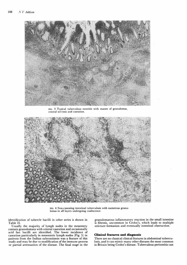
laris mucosa, but deep to this there are masses ofgranulomaswhich may coalesce and often show central necrosis withcaseation (Fig. 3). The granulomata are scattered in alllayers of the intestine and may involve the serosa. Caseationtends to be more common in the indigenous populationespecially in this series. Sometimes caseation is not seen inthose patients from the Indian subcontinent and so thehistological distinction between tuberculosis and Crohn'smay be difficult. In non-caseating tuberculosis the granu-lomas are more numerous, larger and well defined, and maycoalesce (Fig. 4). In Crohn's disease of the small intestinegranulomas are certainly fewer in number, and the sub-mucosa is widened due to oedema, whereas it is reduced orobliterated in tuberculosis. Inflammation in both Crohn'sand tuberculosis has a transmural spread. In Crohn's it is anonspecific inflammatory exudate, whereas in tuberculosis aswe have seen, there are characteristic granulomas in alllayers. Abscess and fistula formation are uncommon intuberculosis, unlike Crohn's disease. The only certain way ofdistinguishing between Crohn's and tuberculosis is the actualdemonstration of the tuberele bacillus within the sectionunder examination, but this is not always possible eventhough the lesion histologically is typically tuberculous (16).The bacilli are more likely to be found when there iscaseation necrosis. In my series, 58°o were found to havetuberele bacilli of the hominis type. The incidence of
108 A' V Addison
FIG. 3 Typical tuberculous enteritis with masses of granulomas,central necrosis and cascation.
FIG. 4 Non-caseating intestinal tuberculosis with numerous granu-lomas in all layers undergoing coalescence.
identification of tubercle bacilli in other series is shown inTable II.
Usually the majority of lymph nodes in the mesenterycontain granulomata with central caseation and occasionallyacid fast bacilli are identified. The lower incidence ofcaseation particularly in mesenteric lymph nodes (Fig. 5) inpatients from the Indian subcontinent was a feature of thisstudy and may be due to modification of the immune processor partial attenuation of the disease. The final stage in the
granulomatous inflammatory reaction in the small intestineis fibrosis, uncommon in Crohn's, which leads to multiplestricture formation and eventually intestinal obstruction.
Clinical features and diagnosisThere are no classical clinical features in abdominal tubercu-losis, and it can mimic many other diseases the most commonin Britain being Crohn's disease. Tuberculous peritonitis can

Abdominal Tuberculosis a disease revived 109
TABLE II
tlubercle bacilliSeries identified
Howell & Knapton (10) (1964) 58%/OAnscombe et al (11) 1967) 22%Prakash (16) (1975) 6%(Y(Shukla & Hughes (13) (1978) 75/,)Lambrianides el al (14) (1980) 4°/OPresent series 58 /,,
is said to occur in up to 850)) of patients with intestinaltuberculosis alone.
Diarrhoea occurs in 10°o0 of patients with abdominaltuberculosis and is due to small bowel ulceration. There isoccasionally blood and mucus in the stools suggesting othertypes of inflammatory bowel disease and acute tuberculouscolitis can occur (Fig. 6). In my experience ano-rectal lesionsare rare in tuberculosis and if present are in fact due toCrohn's disease.
In the nonacute case, there are no pathognomonic clinicalfeatures of abdominal tuberculosis and the accuracy of
FIG. 5 Classical tuberculous granulomata in mesenteric lymphnodes.
be extremely obscure in its symptoms, as John Abercrombiepointed out in 1828.Abdominal pain is the commonest symptom and was seen
in 8lo of patients in this series. In my experience, tubercu-lous peritonitis usually presents as an acute abdomen mainlyin young children and adolescents of both sexes and mimicsacute appendicitis. In 2 female Asian patients chronictuberculous peritonitis was only discovered at the time ofroutine cholecystectomy for gallstones.
Ascites with abdominal pain and distension may be thefirst indication of tuberculous peritonitis either in the acuteor chronic phase. As well as ascites there may be anassociated palpable mass in the right iliac fossa and thusmalignancy is considered. In my experience, carcinoma ofthe colon, like diverticulitis, is extremely rare in Asians, evenin those who have lived in the United Kingdom for manyyears.
Acute or chronic obstruction with vomiting is probablythe most frequent clinical presentation encountered and thiswould certainly appear to be the case in India (15,17).
Following abdominal pain the next most common groupofsymptoms was pyrexia, night sweats, abdominal distensionand weight loss. Pulmonary tuberculosis was present, eitheropen or healed, in 320o in this series and there wasgynaecological involvement in 110O of the females.
Ileocaecal tuberculosis usually presents as a mass in theright iliac fossa which may simulate either Crohn's disease,an appendix mass, a malignant lesion of the right colon, oran amoeboma in Africa and India. This clinical presentation
clinical diagnosis is only 50O"0 in India where the disease isendemic (18). Abdominal tuberculosis should, therefore, beconsidered in any patient, especially of Asian origin, who hasobscure abdominal symptoms. The presentation can simplybe that of vague ill health, lethargy and loss of weight.
InvestigationA plain X-ray of the abdomen may show small bowel fluidlevels with or without ascites and there may be diffusecalcification or evidence of a localised abscess. In ileocaecaltuberculosis there are characteristic radiological ap-pearances on barium enema examination. The caecumdisappears and the ascending colon shortens; the ileum mayretain its normal calibre but passes vertically upwards intothe colon (Fig. 7).
In the colon a typical tuberculous lesion may simulatecarcinoma with characteristic shouldering. Occasionally thelesion may be in the descending colon and may be multiple.Rarely disease of the oesophagus, stomach and duodenummay be demonstrable on barium meal examination. Patientswith gastrointestinal tuberculosis may present with evidenceof an iron deficiency anaemia. The ESR may be elevatedand was in 560)) of patients in this series. On the other hand,the ESR is frequently within normal limits. Heaf andMantoux tests are not particularly helpful and in this serieswere only positive in 31O/. The identification of the tuberclebacillus is extremely important and was actually detected onculture of stools, ascitic fluid or tissue in 58%o of patients, butas previously mentioned, this varies in different series in the

110 A' Vr Addison
FIG. 6 Barium enema demonstrating acute tuberculous colitisaffiecting the pelvic colon and confirmed by biopsy.
United Kingdom. Abdominal laparoscopy is now becominga diagnostic procedure of choice and in 1979 Wolfe, Behnand Jackson (19) reported a positive biopsy in 8 of 11
patients and thus diagnostic laparotomy was avoided. In1978 Udwadia in Bombay (20) found 29 tuberculous lesionsin 384 laparoscopies in patients with abdominal symptomsand signs but in whom all the standard investigations fortuberculosis were unhelpful. In a recent series (personalcommunication) of 321 laparoscopies he confirmed thediagnosis in 218 and stated that peritoneal tubercles wereseen in 206 patients. Colonoscopy is now becoming a veryuseful diagnostic procedure even in ileocaecal tuberculosis.Finally, laparotomy may be the only means of making adefinite diagnosis ofabdominal tuberculosis. Like the clinicalsymptomatology we have seen that there are no diagnosticfeatures' of tuberculosis on naked eye examination. Anileocaecal mass, small bowel strictures, enlarged mesentericlymph nodes, thickened bowel wall and mesentery are allcommon findings in both tuberculosis and Crohn's disease,and similar appearances may be seen in lymphomata. Ahard tuberculous caecal mass is commonly mistaken forcarcinoma.
Surgical managementThe surgical management of abdominal tuberculosis de-pends on whether the condition is acute or nonacute. Youngchildren and adolescents may present as an acute ap-pendicitis and the abdomen is explored. If there is nomechanical obstruction of the bowel present, then an om-ental or gland biopsy should be carried out and the abdomenclosed. Ascitic fluid should be sent for culture and animalinoculation for tubercle bacilli. Once the diagnosis has beenestablished a course of antituberculous drugs should begiven. In the case of acute intestinal obstruction, localisedresection of the solitary stenotic nodule or multiple lesions ina short segment of bowel is the treatment of choice althoughileoplasty for strictures has been advocated (21).
For ileocaecal lesions, a limited right hemicolectomy is the
FI(;. 7 Classical radiological appearances of ileocaecal tuberculosis.The ileum passes vertically upwards into the ascending colon whichhas shortened.
operation of choice in my experience and good results havebeen reported (22). Seventeen patients underwent this pro-cedure in my series, and again, this should be followed by acourse of antituberculous drugs. In addition six patients weretreated by small bowel resection and three had a large bowelresection. By-pass of the lesion with an ileotransversecolostomy should be avoided, as patients tend to fare badlyafter this procedure just as they do in the treatment ofCrohn's disease. Short circuiting procedures like gastro-jejunostomy can be used in duodenal tuberculosis. A stenoticlesion in the colon should be treated by local resection if apreoperative diagnosis of tuberculosis is made, but if thelesion is thought -to be a carcinoma then a more radicalpartial colectomy should be carried out. The results ofcolonic resection are equal to those following right hemi-colectomy, but should always be followed by a course ofadjuvant antituberculous drugs. Specific antituberculousdrugs should include Rimactazid and ethambutol.
Crohn's disease and gastrointestinal tuberculosisCrohn's disease and gastrointestinal tuberculosis occur bothin the immigrant and in the indigenous population and bothdiseases may present in a very similar way. Tuberculosisshould always be considered in the differential diagnosis of amass in the right iliac fossa. Similarly, Crohn's disease andulcerative colitis must be considered in the differentialdiagnosis of gastrointestinal tuberculosis in immigrants.Since the recognition of Crohn's disease in 1932, there is nodoubt that many cases of so-called primary hyperplasticileocaecal tuberculosis were in fact Crohn's and many ofthese cases without recognisable caseation proved on furtherhistological examination to be cases of Crohn's disease.Caseating granulomas are considered an essential criterionin the diagnosis of tuberculosis but may be difficult todemonstrate as we have seen in the Asians in this study. The

Abdominal Tuberculosis-a disease revived 111
only absolutely certain way of distinguishing betweenCrohn's disease and tuberculosis is the actual demonstrationof tubercle bacilli within the section under examination.However, we have seen in Prakash's series (16) of 92 patientswith confirmed ileocaecal tuberculosis, that positive bac-terial evidence was onIly found in 6 patients. Granulomas arepresent in 1)oth Crohn's disease and tuberculosis. In tubercu-losis it is not unusual to find them in the mesenteric lymphnodes while they are absent in the intestine, whether or notthe patient has received any form of chemotherapy. InCrohn's disease, if the lymph nodes do not show granulomasthen none are seen in the intestine. Histologically, in Crohn'sthe transmural disease is a nonspecific inflammatory exudatewhereas in tuberculosis there are characteristic granulomasin all lavers.
ConclusionsAbdominal tuberculosis is no longer a very rare disease inparts of the United Kingdom and in fact is now 'a diseaserevived'. This is largely due to the arrival of the Asianimmigranit, but the disease still occurs in the indigenouspopulation. In the past, abdominal tuberculosis developedafter swallowing the bovine bacillus in infected milk, and thehuman bacillus in infected sputum. Bovine tuberculosis hasnow disappeared in the United Kingdom and most patientswith abdominal tuberculosis did have evidence of the diseaseelsewNhere in the body, usually the lungs. The exception tothis was the group described as 'hyperplastic ileocaecaltuberculosis' where true caseation was usually absent and thetubercle bacillus was never isolated. As there is now littleopen pulmonary tuberculosis one speculates as to how theintestine and peritoneum become involved in the absence ofovert disease elsewhere in the body. In my series 680( had noevidence of pulmonary tuberculosis.
It is doubtful whether true primary lesions occur in thesmall intestine but they certainly do occur in mesentericlymph nodes. The human bacilli pass through the intestinalmucosa of Peyer's patches without producing a local lesionand, transported by macrophages, enter the intestinallymphatics and settle in the mesenteric lymph nodes wherethey may multiply or remain dormant for long periods oftime, sometimes for life.
It is a distinct possibility that ulcerative lesions in thebowel are the result of retrograde lymphatic spread frommesenteric nodes. Matthew Stewart in 1928 said 'localretrograde extension from tuberculous mesenteric glands isperhaps more common than we suppose'. The more ad-vanced tuberculous lesions with caseation in the lymphnodes compared with the bowel lends some support to thissuggestion, although in the Asians in my series there was alow incidence of caseation even in the lymph nodes. Liketyphoid, lymphoid tissue in the ileum develops hypersensi-tivity rapidly to the tubercle bacillus, resulting in ulcerationwhich is secondary to primary infection in lymph nodes.Tuberculous peritonitis may develop in a similar manner tothat of tuberculous enteritis.
It is more than likely the immigrant brings the tubercu-losis into the United Kingdom in his mesenteric lymphglands, and that the disease is reactivated at some later datedue to some modification in the immune process. With thesefacts in mind, tuberculosis must be considered in the dif-ferential diagnosis of acute or chronic abdominal pain in
patients of Asian origin, remembering that it can mimicmany diseases, the commonest in Britain being Crohn'sdisease. When the diagnosis is in doubt, surgical explorationis indicated but even at laparotomy the ultimate diagnosismay only be achieved after histological and bacteriologicalstudies.
I would like to thank Dr. G. I. Horsfield, Consultant Pathologist,for his help, and MIr. Peter Harrison for the photographicillustrationis.
ReferencesI Goldfisclher S, Janis MI. A 42 year old king with a cavitarypulmonary lesion and intestinal perforation. Bull N Y AcadM\ed 1981;57:139-43.
2 Kirkup JR. Xicarv Lecture. The Tercentenary of RichardWiseman's 'Several Chirurgicall Treatises'. Ann R Coll SurgEngl 1976;59:277.
3 Hunter J. lectures on the Principles of Surgery' in CollectedWorks ofJolhin Hunter. PalmerJF, ed. Vol 1, page 593. LondonLongmani ('1835).
4 Abercrombie J. Pathological and practical researches ondiseases of the stomach, the intestinal canal, the liver and otherviscera of the abdomen. Edinburgh. XVaugh and Innes.1928; 191-4.
5 Abercrombie J. Pathological and practical researches ondiseases of the stomach, the intestinal canal, the liver and otherXviscera of the abdomen. Edinburgh. WVaugh and Innes.1928;307-1 1.
6 Boyd WV. Surgical Pathology p. 306. London. Saunders Co.(1925).
7 Stewart MIJ. The pathology of intestinal tuberculosis. Tubercle1928;409-413.
8 Crohn BB, Ginzburg L, Oppenheimer GD. Regional ileitis:pathologic and clinical entity. JAMA 1932;99:1323.
9 FindlayJM, Addison NV, Stevenson DK, Mirza ZA. Tubercu-losis of the gastrointestinal tract in Bradford 1967-77. J R SocMed 1979;72:587-90.
10 Howell JS, Knapton PJ. Ileocaecal tuberculosis. Gut.1964;5:524-29.
11 Anscombe AR, Keddie NC, Schofield PF. Caccal tuberculosis.Gut 1967;8:337-43.
12 Mandal BK, Schofield PF. Abdominal tuberculosis in Britain.Practitioner 1976;216:683-689.
13 Shukla HS, Hughes LE. Abdominal tuberculosis in the 1970's-a continuing problem. BrJ Surg 1978;65:403-5.
14 Lambrianides AL, Ackroyd N, Shorey BA. Abdominal tubercu-losis. BrJ Surg 1980; 67:887-9.
15 Mukerjee P, Singal AK. Intestinal tuberculosis: 500 operatedcases. Proceedings of the Association of Surgeons of East Africa.1979;2:70-5.
16 Prakash A, Sharma LK, Koshal A, Poddar PK. Ileocaccaltuberculosis. Aust NZ J Surg 1975;45:4.
17 Bhansali SK, Desai AN. Abdominal tuberculosis. Clinical ana-lysis of 135 cases. Indian Journal of Surgery 1968;30:218-32.
18 Das P, Shukla HS. Clinical diagnosis of abdominal tuberculosisBrJ Surg 1976;63:941-6.
19 WNolfeJHN, Behn AR, Jackson BT. Tuberculous peritonitis androle of diagnostic laparoscopy. Lancet 1979; 1:852-3.
20 Udwadia TE. Peritoneoscopy in the diagnosis of abdominaltuberculosis. Indian Journal of Surgery 1978;40:91-5.
21 Katariya RN, Sood S, Rao PG, Rao PLNG. Stricture-plasty fortubercular strictures of the gastrointestinal tract. Br J Surg1977;64:469-8.
22 Byrom HB, Mann CV. Clinical features and surgical manage-ment of ileocaecal tuberculosis. Proc R Soc Med 1969;62:1230-1233.


