Gender differences in hemispheric asymmetry for face processing
10
BioMed Central Page 1 of 10 (page number not for citation purposes) BMC Neuroscience Open Access Research article Gender differences in hemispheric asymmetry for face processing Alice M Proverbio* 1,2 , Valentina Brignone †1 , Silvia Matarazzo †1 , Marzia Del Zotto 1,2 and Alberto Zani 2 Address: 1 Department of Psychology, University of Milano-Bicocca, Viale dell'Innovazione 10, 20126, Milan, Italy and 2 Institute of Bioimaging and Molecular Physiology (CNR), Via Fratelli Cervi 93, 20090, Milano-Segrate, Italy Email: Alice M Proverbio* - [email protected]; Valentina Brignone - [email protected]; Silvia Matarazzo - [email protected]; Marzia Del Zotto - [email protected]; Alberto Zani - [email protected] * Corresponding author †Equal contributors Abstract Background: Current cognitive neuroscience models predict a right-hemispheric dominance for face processing in humans. However, neuroimaging and electromagnetic data in the literature provide conflicting evidence of a right-sided brain asymmetry for decoding the structural properties of faces. The purpose of this study was to investigate whether this inconsistency might be due to gender differences in hemispheric asymmetry. Results: In this study, event-related brain potentials (ERPs) were recorded in 40 healthy, strictly right-handed individuals (20 women and 20 men) while they observed infants' faces expressing a variety of emotions. Early face-sensitive P1 and N1 responses to neutral vs. affective expressions were measured over the occipital/temporal cortices, and the responses were analyzed according to viewer gender. Along with a strong right hemispheric dominance for men, the results showed a lack of asymmetry for face processing in the amplitude of the occipito-temporal N1 response in women to both neutral and affective faces. Conclusion: Men showed an asymmetric functioning of visual cortex while decoding faces and expressions, whereas women showed a more bilateral functioning. These results indicate the importance of gender effects in the lateralization of the occipito-temporal response during the processing of face identity, structure, familiarity, or affective content. Background Using functional magnetic resonance imaging (fMRI), Kanwisher and coworkers [1] found an area in the fusi- form gyrus that was significantly more active when the subjects viewed faces than when they viewed assorted common objects. The authors concluded that this area, hereafter called the fusiform face area (FFA), might be spe- cifically involved in the perception of faces, not ruling out that other structures might be play a role in this process. Indeed, Haxby et al. (2000) provided evidence that face perception involves a distributed and hierarchically organized network of the occipito-temporal regions. In this model, the core system consists of the extrastriate vis- ual cortex (FFA), which mediates the analysis of face struc- ture, while the superior temporal sulcus (STS) mediates the analysis of changeable aspects of the face, such as eye gaze, facial expression, and lip movements. Published: 08 June 2006 BMC Neuroscience 2006, 7:44 doi:10.1186/1471-2202-7-44 Received: 23 January 2006 Accepted: 08 June 2006 This article is available from: http://www.biomedcentral.com/1471-2202/7/44 © 2006 Proverbio et al; licensee BioMed Central Ltd. This is an Open Access article distributed under the terms of the Creative Commons Attribution License (http://creativecommons.org/licenses/by/2.0 ), which permits unrestricted use, distribution, and reproduction in any medium, provided the original work is properly cited.
Transcript of Gender differences in hemispheric asymmetry for face processing
BioMed CentralBMC Neuroscience
ss
Open AcceResearch articleGender differences in hemispheric asymmetry for face processingAlice M Proverbio*1,2, Valentina Brignone†1, Silvia Matarazzo†1, Marzia Del Zotto1,2 and Alberto Zani2Address: 1Department of Psychology, University of Milano-Bicocca, Viale dell'Innovazione 10, 20126, Milan, Italy and 2Institute of Bioimaging and Molecular Physiology (CNR), Via Fratelli Cervi 93, 20090, Milano-Segrate, Italy
Email: Alice M Proverbio* - [email protected]; Valentina Brignone - [email protected]; Silvia Matarazzo - [email protected]; Marzia Del Zotto - [email protected]; Alberto Zani - [email protected]
* Corresponding author †Equal contributors
AbstractBackground: Current cognitive neuroscience models predict a right-hemispheric dominance forface processing in humans. However, neuroimaging and electromagnetic data in the literatureprovide conflicting evidence of a right-sided brain asymmetry for decoding the structural propertiesof faces. The purpose of this study was to investigate whether this inconsistency might be due togender differences in hemispheric asymmetry.
Results: In this study, event-related brain potentials (ERPs) were recorded in 40 healthy, strictlyright-handed individuals (20 women and 20 men) while they observed infants' faces expressing avariety of emotions. Early face-sensitive P1 and N1 responses to neutral vs. affective expressionswere measured over the occipital/temporal cortices, and the responses were analyzed accordingto viewer gender. Along with a strong right hemispheric dominance for men, the results showed alack of asymmetry for face processing in the amplitude of the occipito-temporal N1 response inwomen to both neutral and affective faces.
Conclusion: Men showed an asymmetric functioning of visual cortex while decoding faces andexpressions, whereas women showed a more bilateral functioning. These results indicate theimportance of gender effects in the lateralization of the occipito-temporal response during theprocessing of face identity, structure, familiarity, or affective content.
BackgroundUsing functional magnetic resonance imaging (fMRI),Kanwisher and coworkers [1] found an area in the fusi-form gyrus that was significantly more active when thesubjects viewed faces than when they viewed assortedcommon objects. The authors concluded that this area,hereafter called the fusiform face area (FFA), might be spe-cifically involved in the perception of faces, not ruling outthat other structures might be play a role in this process.
Indeed, Haxby et al. (2000) provided evidence that faceperception involves a distributed and hierarchicallyorganized network of the occipito-temporal regions. Inthis model, the core system consists of the extrastriate vis-ual cortex (FFA), which mediates the analysis of face struc-ture, while the superior temporal sulcus (STS) mediatesthe analysis of changeable aspects of the face, such as eyegaze, facial expression, and lip movements.
Published: 08 June 2006
BMC Neuroscience 2006, 7:44 doi:10.1186/1471-2202-7-44
Received: 23 January 2006Accepted: 08 June 2006
This article is available from: http://www.biomedcentral.com/1471-2202/7/44
© 2006 Proverbio et al; licensee BioMed Central Ltd.This is an Open Access article distributed under the terms of the Creative Commons Attribution License (http://creativecommons.org/licenses/by/2.0), which permits unrestricted use, distribution, and reproduction in any medium, provided the original work is properly cited.
Page 1 of 10(page number not for citation purposes)
BMC Neuroscience 2006, 7:44 http://www.biomedcentral.com/1471-2202/7/44
Interestingly, the Kanwisher et al. study [1] showed anactivation of FFA only in the right hemisphere in abouthalf the subjects (both men and women), whereas theother subjects showed bilateral activation. These resultsraised the possibility of functional hemispheric asymme-try in the FFA. Studies addressing this possibility have pro-vided conflicting evidence: several human [2-6] andanimal studies [7] found stronger activity in the righthemisphere, while other studies failed to support thenotion of a strict right-lateralization (e.g. [8] performed in5 men and 7 women).
Closer examination of several studies offers more details,but no consensus, on hemispheric asymmetry in areasdevoted to face processing. Yovel and Kanwisher [9]found significantly higher fMRI responses to faces than toobjects in both the left and right mid-fusiform gyrusregions, although this effect was slightly greater in theright than the left FFA. In another fMRI study [10], aregion that responded more strongly to faces than toobjects was found within the right fusiform gyrus in 8 sub-jects (both women and men); however, in 6 of these sub-jects the same significant pattern was also found in the leftfusiform gyrus. Recently, Pourtois and coworkers [11] per-formed an fMRI study on face identity processing on agroup of 8 men and 6 women. Results revealed a reducedresponse in the lateral occipital and fusiform cortex withface repetition. Specifically, view-sensitive repetitioneffects were found in both the left and right fusiform cor-tices, while the left (but not right) fusiform cortex showedviewpoint-independent repetition effects. These findingswere interpreted as a sign of left hemisphere dominancein terms of the ability to link visual facial appearance withspecific identity knowledge. In line with this, a case hasbeen reported of hyperfamiliarity for unknown faces afterleft lateral temporo-occipital damage in a female patient[12], suggesting a possible role of the left hemisphere inidentity processing. Again, a recent fMRI study [13] car-ried out on 8 women and 7 men provided evidence of asignificant activation of right fusiform and occipital gyrus(2260 voxels), left fusiform gyrus, left inferior, and mid-dle temporal gyrus (3022 voxels) for a face familiarityeffect during gender classification, thus providing a com-plex lateralization pattern for processing face structuresand properties.
Event-related potential (ERP) and magnetoencephalogra-phy (MEG) recordings of brain activity have provided cru-cial information about the temporal unfolding of neuralmechanisms involved in face processing (see a list ofrecent papers in Table 1). In particular, these recordingshave identified a posterior-lateral negative peak at alatency of approximately 170 ms (referred to as "N170").This peak has a larger amplitude in response to faces thanto other control stimuli (such as houses, objects, trees, or
words), and is sensitive to face inversion (upright vs.inverted). N170 is thought to reflect processes involved inthe structural encoding of faces. In addition, several stud-ies have found that affective information modulates brainresponse to human faces as early as 120–150 ms [14-17].The combination of electromagnetic and functional neu-roimaging data identified the possible generator of N1 inthe ventral occipito-temporal cortex (FFA and superiortemporal sulcus or STS) [16,18,19,53], suggesting that N1might be the electromagnetic manifestation of a face-processing area activity. An analysis of the relevant litera-ture shows that the topographic distribution of the face-specific N170 is not always right-sided in right-handedindividuals. Based on a thorough review of methods andsubject samples used in the relevant literature (see Table1), we hypothesized that this topographic distributionmight depend on marked inter-individual differences,possibly related to viewer gender.
It is of great interest to note that face-specific N170responses were found to be bilateral or even left-sided instudies involving a sample in which women were themajority [20-24]. Equally interesting, in a recent paper onprosopagnosia in which both male and female patientswere considered [20], 2 out of the 3 male patients showedan M170 response which was not sensitive to faces (asopposed to houses) while the third patient showed aright-sided sensitivity to faces. As for the two femalepatients, one of them showed a lack of sensitivity to facesat M170 level, while the second one showed a left-sidedsensitivity.
Many face processing studies using MEG, ERP, neuroim-aging, or neuropsychological data do not take subject gen-der into account as a variable that might affect asymmetryin brain activation. The specific goal of this study was toinvestigate the timing and topography of brain activity inmen and women during processing of neutral and affec-tive faces in order to detect whether there are gender dif-ferences in lateralization. To address this question, earlyface-sensitive P1 and N1 responses over the occipital/tem-poral cortices to neutral and affective expressions weremeasured in strictly right-handed men and women.
ResultsBehavioral dataA repeated-measures ANOVA was performed on meanresponse times (RTs), but showed no significant gendereffect on response speed (F[1,38] = 0.617; p = 0.44;females = 658 ms, males = 672 ms). Men and women sub-jects also did not significantly differ in accuracy; however,error percentages were too few to be statistically analyzed.The emotional valence of faces affected RTs (F[1,38] =191; p < 0.000001), which were faster to negative expres-
Page 2 of 10(page number not for citation purposes)
BMC Neuroscience 2006, 7:44 http://www.biomedcentral.com/1471-2202/7/44
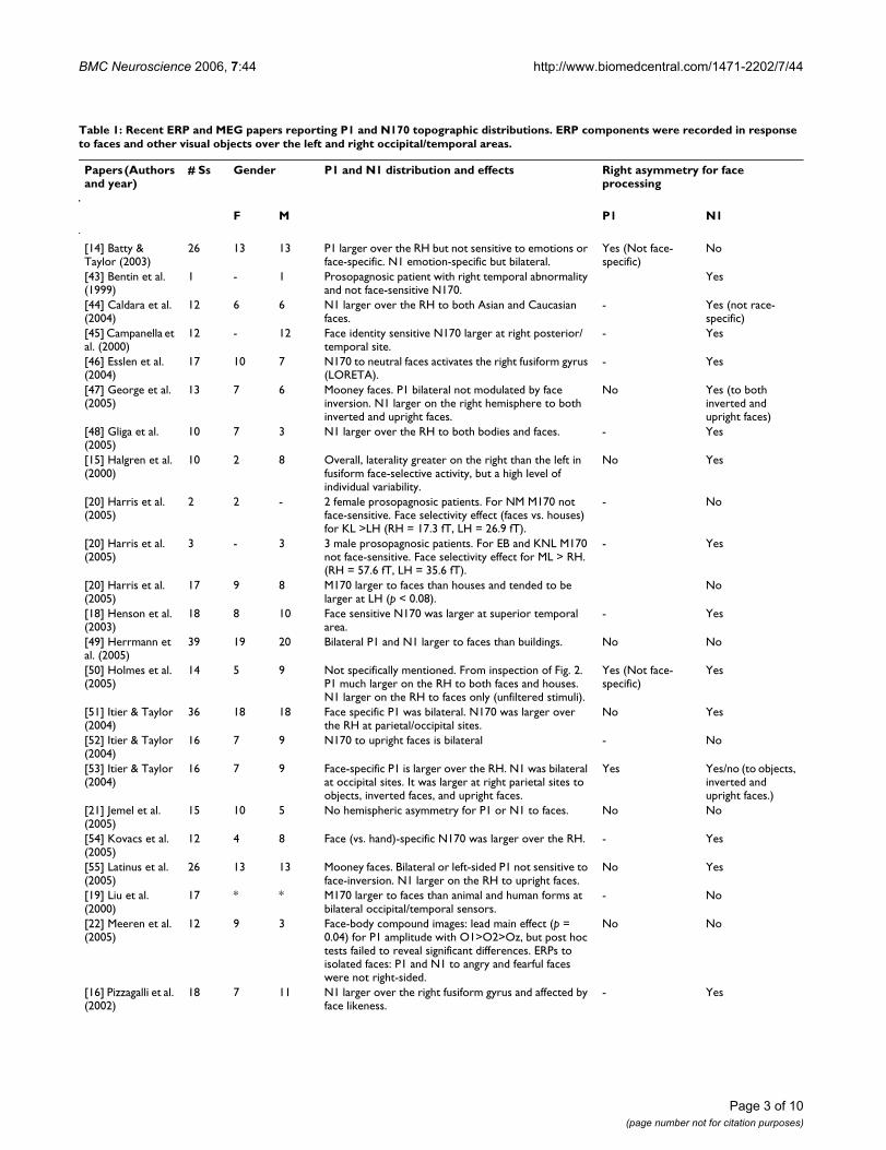
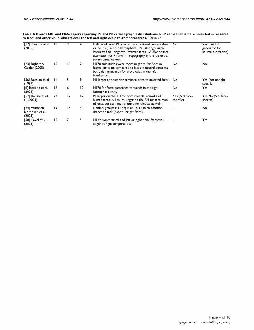
Table 1: Recent ERP and MEG papers reporting P1 and N170 topographic distributions. ERP components were recorded in response to faces and other visual objects over the left and right occipital/temporal areas.
Papers (Authors and year)
# Ss Gender P1 and N1 distribution and effects Right asymmetry for face processing
F M P1 N1
[14] Batty & Taylor (2003)
26 13 13 P1 larger over the RH but not sensitive to emotions or face-specific. N1 emotion-specific but bilateral.
Yes (Not face- specific)
No
[43] Bentin et al. (1999)
1 - 1 Prosopagnosic patient with right temporal abnormality and not face-sensitive N170.
Yes
[44] Caldara et al. (2004)
12 6 6 N1 larger over the RH to both Asian and Caucasian faces.
- Yes (not race-specific)
[45] Campanella et al. (2000)
12 - 12 Face identity sensitive N170 larger at right posterior/temporal site.
- Yes
[46] Esslen et al. (2004)
17 10 7 N170 to neutral faces activates the right fusiform gyrus (LORETA).
- Yes
[47] George et al. (2005)
13 7 6 Mooney faces. P1 bilateral not modulated by face inversion. N1 larger on the right hemisphere to both inverted and upright faces.
No Yes (to both inverted and upright faces)
[48] Gliga et al. (2005)
10 7 3 N1 larger over the RH to both bodies and faces. - Yes
[15] Halgren et al. (2000)
10 2 8 Overall, laterality greater on the right than the left in fusiform face-selective activity, but a high level of individual variability.
No Yes
[20] Harris et al. (2005)
2 2 - 2 female prosopagnosic patients. For NM M170 not face-sensitive. Face selectivity effect (faces vs. houses) for KL >LH (RH = 17.3 fT, LH = 26.9 fT).
- No
[20] Harris et al. (2005)
3 - 3 3 male prosopagnosic patients. For EB and KNL M170 not face-sensitive. Face selectivity effect for ML > RH. (RH = 57.6 fT, LH = 35.6 fT).
- Yes
[20] Harris et al. (2005)
17 9 8 M170 larger to faces than houses and tended to be larger at LH (p < 0.08).
No
[18] Henson et al. (2003)
18 8 10 Face sensitive N170 was larger at superior temporal area.
- Yes
[49] Herrmann et al. (2005)
39 19 20 Bilateral P1 and N1 larger to faces than buildings. No No
[50] Holmes et al. (2005)
14 5 9 Not specifically mentioned. From inspection of Fig. 2. P1 much larger on the RH to both faces and houses. N1 larger on the RH to faces only (unfiltered stimuli).
Yes (Not face- specific)
Yes
[51] Itier & Taylor (2004)
36 18 18 Face specific P1 was bilateral. N170 was larger over the RH at parietal/occipital sites.
No Yes
[52] Itier & Taylor (2004)
16 7 9 N170 to upright faces is bilateral - No
[53] Itier & Taylor (2004)
16 7 9 Face-specific P1 is larger over the RH. N1 was bilateral at occipital sites. It was larger at right parietal sites to objects, inverted faces, and upright faces.
Yes Yes/no (to objects, inverted and upright faces.)
[21] Jemel et al. (2005)
15 10 5 No hemispheric asymmetry for P1 or N1 to faces. No No
[54] Kovacs et al. (2005)
12 4 8 Face (vs. hand)-specific N170 was larger over the RH. - Yes
[55] Latinus et al. (2005)
26 13 13 Mooney faces. Bilateral or left-sided P1 not sensitive to face-inversion. N1 larger on the RH to upright faces.
No Yes
[19] Liu et al. (2000)
17 * * M170 larger to faces than animal and human forms at bilateral occipital/temporal sensors.
- No
[22] Meeren et al. (2005)
12 9 3 Face-body compound images: lead main effect (p = 0.04) for P1 amplitude with O1>O2>Oz, but post hoc tests failed to reveal significant differences. ERPs to isolated faces: P1 and N1 to angry and fearful faces were not right-sided.
No No
[16] Pizzagalli et al. (2002)
18 7 11 N1 larger over the right fusiform gyrus and affected by face likeness.
- Yes
Page 3 of 10(page number not for citation purposes)
BMC Neuroscience 2006, 7:44 http://www.biomedcentral.com/1471-2202/7/44
[17] Pourtois et al. (2005)
13 9 4 Unfiltered faces: P1 affected by emotional content (fear vs. neutral) in both hemispheres. N1 strongly right-lateralized to upright vs. inverted faces. LAURA source estimation for P1 and N1 topography in the left extra-striate visual cortex
No Yes (but LH generator for source estimation)
[23] Righart & Gelder (2005)
12 10 2 N170 amplitudes were more negative for faces in fearful contexts compared to faces in neutral contexts, but only significantly for electrodes in the left hemisphere.
No No
[56] Rossion et al. (1999)
14 5 9 N1 larger at posterior temporal sites to inverted faces. No Yes (not upright specific)
[6] Rossion et al. (2003)
16 6 10 N170 for faces compared to words in the right hemisphere only.
No Yes
[57] Rousselet et al. (2004)
24 12 12 P1 larger on the RH for both objects, animal and human faces. N1 much larger on the RH for face than objects, but asymmetry found for objects as well.
Yes (Not face- specific)
Yes/No (Not face- specific)
[24] Valkonen-Korhonen et al. (2005)
19 15 4 Control group: N1 Larger at T5/T6 in an emotion detection task (happy upright faces).
- No
[58] Yovel et al. (2003)
12 7 5 N1 to symmetrical and left or right hemi-faces was larger at right temporal site.
- Yes
Table 1: Recent ERP and MEG papers reporting P1 and N170 topographic distributions. ERP components were recorded in response to faces and other visual objects over the left and right occipital/temporal areas. (Continued)
Page 4 of 10(page number not for citation purposes)
BMC Neuroscience 2006, 7:44 http://www.biomedcentral.com/1471-2202/7/44
sions (613 ms) than to neutral expressions (717 ms) forall viewers.
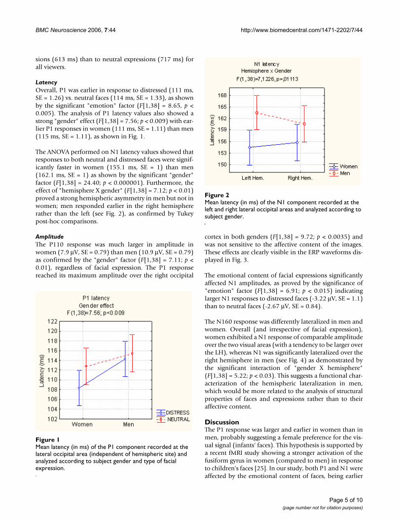
LatencyOverall, P1 was earlier in response to distressed (111 ms,SE = 1.26) vs. neutral faces (114 ms, SE = 1.33), as shownby the significant "emotion" factor (F[1,38] = 8.65, p <0.005). The analysis of P1 latency values also showed astrong "gender" effect (F[1,38] = 7.56; p < 0.009) with ear-lier P1 responses in women (111 ms, SE = 1.11) than men(115 ms, SE = 1.11), as shown in Fig. 1.
The ANOVA performed on N1 latency values showed thatresponses to both neutral and distressed faces were signif-icantly faster in women (155.1 ms, SE = 1) than men(162.1 ms, SE = 1) as shown by the significant "gender"factor (F[1,38] = 24.40; p < 0.000001). Furthermore, theeffect of "hemisphere X gender" (F[1,38] = 7.12; p < 0.01)proved a strong hemispheric asymmetry in men but not inwomen; men responded earlier in the right hemisphererather than the left (see Fig. 2), as confirmed by Tukeypost-hoc comparisons.
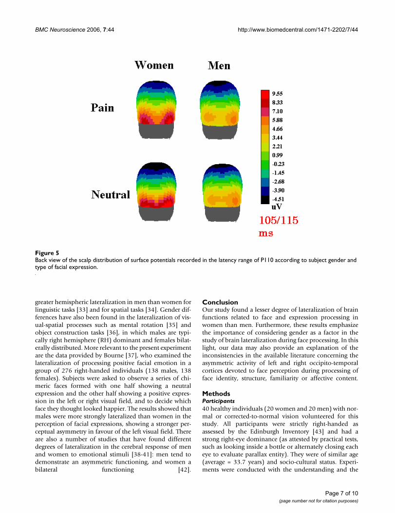
AmplitudeThe P110 response was much larger in amplitude inwomen (7.9 μV, SE = 0.79) than men (10.9 μV, SE = 0.79)as confirmed by the "gender" factor (F[1,38] = 7.11; p <0.01), regardless of facial expression. The P1 responsereached its maximum amplitude over the right occipital
cortex in both genders (F[1,38] = 9.72; p < 0.0035) andwas not sensitive to the affective content of the images.These effects are clearly visible in the ERP waveforms dis-played in Fig. 3.
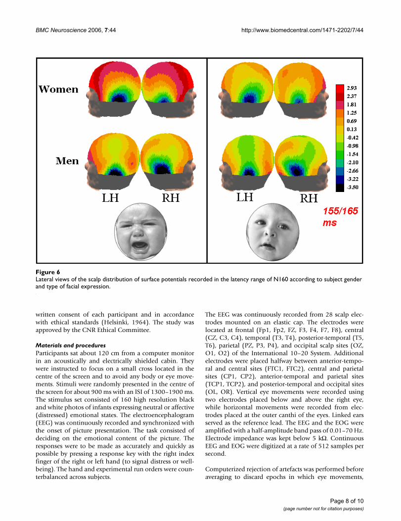
The emotional content of facial expressions significantlyaffected N1 amplitudes, as proved by the significance of"emotion" factor (F[1,38] = 6.91; p < 0.015) indicatinglarger N1 responses to distressed faces (-3.22 μV, SE = 1.1)than to neutral faces (-2.67 μV, SE = 0.84).
The N160 response was differently lateralized in men andwomen. Overall (and irrespective of facial expression),women exhibited a N1 response of comparable amplitudeover the two visual areas (with a tendency to be larger overthe LH), whereas N1 was significantly lateralized over theright hemisphere in men (see Fig. 4) as demonstrated bythe significant interaction of "gender X hemisphere"(F[1,38] = 5.22; p < 0.03). This suggests a functional char-acterization of the hemispheric lateralization in men,which would be more related to the analysis of structuralproperties of faces and expressions rather than to theiraffective content.
DiscussionThe P1 response was larger and earlier in women than inmen, probably suggesting a female preference for the vis-ual signal (infants' faces). This hypothesis is supported bya recent fMRI study showing a stronger activation of thefusiform gyrus in women (compared to men) in responseto children's faces [25]. In our study, both P1 and N1 wereaffected by the emotional content of faces, being earlier
Mean latency (in ms) of the N1 component recorded at the left and right lateral occipital areas and analyzed according to subject genderFigure 2Mean latency (in ms) of the N1 component recorded at the left and right lateral occipital areas and analyzed according to subject gender.
Mean latency (in ms) of the P1 component recorded at the lateral occipital area (independent of hemispheric site) and analyzed according to subject gender and type of facial expressionFigure 1Mean latency (in ms) of the P1 component recorded at the lateral occipital area (independent of hemispheric site) and analyzed according to subject gender and type of facial expression.
Page 5 of 10(page number not for citation purposes)

BMC Neuroscience 2006, 7:44 http://www.biomedcentral.com/1471-2202/7/44
(P110) and larger (N160) in response to distressed facesas opposed to neutral faces. These data fit with the availa-ble literature which supports the notion of early effects ofemotional [14-17] and attentional factors [26-28] in thefirst stages of visual cortical processing. Overall, the P1component was larger over the right occipital area in allindividuals, and to all stimuli, as clearly visible from thetopographic maps in Fig. 5.
This effect might be due either to sensory or cognitive fac-tors. Since all stimuli were faces, in this experiment theasymmetry cannot be ascribed to a generic effect of faceprocessing. Indeed, the literature on face recognition doesnot support the evidence of a right lateralization for theP1 response, but, rather, a bilateral distribution is oftenreported (when P1 is considered, see Table 1). Further-more, in studies involving visual-spatial or selective atten-tion tasks, the P1 component is often described as largerat the right than the left occipital lateral sites both forspace orienting (e.g., [29]) and processing of global con-figurations (e.g., [30]). In addition, P1 is always right-lat-eralized in response to low spatial frequency patterns evenin passive viewing conditions [31,32]. For these reasons,
we cannot discuss the P1 right lateralization as an index ofa hemispheric dominance for face processing.
On the other hand, the face-specific N160 component wasclearly lateralized differently in the men and women inour study. Indeed, a strong gender effect in the hemi-spheric lateralization of N1 component was observed,both in the latency and amplitude of cerebral response.This hemispheric asymmetry in men was not restricted tothe processing of affective faces, and was significant inresponse to both neutral and distressed faces (see topo-graphic maps in Fig. 6, displaying N1 scalp voltage distri-bution). Thus, a right hemispheric dominance issuggested for face processing in men but not in women.This may explain the many inconsistencies present in therelevant ERP and neuroimaging literature, which some-times predicts a bilateral effect and other times a strongright-sided activity in regions devoted to face processing.These conclusions often rely on a mixed gender popula-tion, in which men and women are not necessarily equallyrepresented (see Table 1).
Our results are also in line with many studies that showgender differences in the degree of lateralization of cogni-tive and affective processes. Considerable data support
Mean amplitude (in μV) of the N1 component recorded at left and right lateral occipital areas and analyzed according to subject genderFigure 4Mean amplitude (in μV) of the N1 component recorded at left and right lateral occipital areas and analyzed according to subject gender.Grand-average ERPs recorded at left and right occipital sites in response to neutral and affective faces according to sub-ject gender (women = solid line, men = dashed line)Figure 3
Grand-average ERPs recorded at left and right occipital sites in response to neutral and affective faces according to sub-ject gender (women = solid line, men = dashed line).
Page 6 of 10(page number not for citation purposes)
BMC Neuroscience 2006, 7:44 http://www.biomedcentral.com/1471-2202/7/44
greater hemispheric lateralization in men than women forlinguistic tasks [33] and for spatial tasks [34]. Gender dif-ferences have also been found in the lateralization of vis-ual-spatial processes such as mental rotation [35] andobject construction tasks [36], in which males are typi-cally right hemisphere (RH) dominant and females bilat-erally distributed. More relevant to the present experimentare the data provided by Bourne [37], who examined thelateralization of processing positive facial emotion in agroup of 276 right-handed individuals (138 males, 138females). Subjects were asked to observe a series of chi-meric faces formed with one half showing a neutralexpression and the other half showing a positive expres-sion in the left or right visual field, and to decide whichface they thought looked happier. The results showed thatmales were more strongly lateralized than women in theperception of facial expressions, showing a stronger per-ceptual asymmetry in favour of the left visual field. Thereare also a number of studies that have found differentdegrees of lateralization in the cerebral response of menand women to emotional stimuli [38-41]: men tend todemonstrate an asymmetric functioning, and women abilateral functioning [42].
ConclusionOur study found a lesser degree of lateralization of brainfunctions related to face and expression processing inwomen than men. Furthermore, these results emphasizethe importance of considering gender as a factor in thestudy of brain lateralization during face processing. In thislight, our data may also provide an explanation of theinconsistencies in the available literature concerning theasymmetric activity of left and right occipito-temporalcortices devoted to face perception during processing offace identity, structure, familiarity or affective content.
MethodsParticipants40 healthy individuals (20 women and 20 men) with nor-mal or corrected-to-normal vision volunteered for thisstudy. All participants were strictly right-handed asassessed by the Edinburgh Inventory [43] and had astrong right-eye dominance (as attested by practical tests,such as looking inside a bottle or alternately closing eacheye to evaluate parallax entity). They were of similar age(average = 33.7 years) and socio-cultural status. Experi-ments were conducted with the understanding and the
Back view of the scalp distribution of surface potentials recorded in the latency range of P110 according to subject gender and type of facial expressionFigure 5Back view of the scalp distribution of surface potentials recorded in the latency range of P110 according to subject gender and type of facial expression.
Page 7 of 10(page number not for citation purposes)
BMC Neuroscience 2006, 7:44 http://www.biomedcentral.com/1471-2202/7/44
written consent of each participant and in accordancewith ethical standards (Helsinki, 1964). The study wasapproved by the CNR Ethical Committee.
Materials and proceduresParticipants sat about 120 cm from a computer monitorin an acoustically and electrically shielded cabin. Theywere instructed to focus on a small cross located in thecentre of the screen and to avoid any body or eye move-ments. Stimuli were randomly presented in the centre ofthe screen for about 900 ms with an ISI of 1300–1900 ms.The stimulus set consisted of 160 high resolution blackand white photos of infants expressing neutral or affective(distressed) emotional states. The electroencephalogram(EEG) was continuously recorded and synchronized withthe onset of picture presentation. The task consisted ofdeciding on the emotional content of the picture. Theresponses were to be made as accurately and quickly aspossible by pressing a response key with the right indexfinger of the right or left hand (to signal distress or well-being). The hand and experimental run orders were coun-terbalanced across subjects.
The EEG was continuously recorded from 28 scalp elec-trodes mounted on an elastic cap. The electrodes werelocated at frontal (Fp1, Fp2, FZ, F3, F4, F7, F8), central(CZ, C3, C4), temporal (T3, T4), posterior-temporal (T5,T6), parietal (PZ, P3, P4), and occipital scalp sites (OZ,O1, O2) of the International 10–20 System. Additionalelectrodes were placed halfway between anterior-tempo-ral and central sites (FTC1, FTC2), central and parietalsites (CP1, CP2), anterior-temporal and parietal sites(TCP1, TCP2), and posterior-temporal and occipital sites(OL, OR). Vertical eye movements were recorded usingtwo electrodes placed below and above the right eye,while horizontal movements were recorded from elec-trodes placed at the outer canthi of the eyes. Linked earsserved as the reference lead. The EEG and the EOG wereamplified with a half-amplitude band pass of 0.01–70 Hz.Electrode impedance was kept below 5 kΩ. ContinuousEEG and EOG were digitized at a rate of 512 samples persecond.
Computerized rejection of artefacts was performed beforeaveraging to discard epochs in which eye movements,
Lateral views of the scalp distribution of surface potentials recorded in the latency range of N160 according to subject gender and type of facial expressionFigure 6Lateral views of the scalp distribution of surface potentials recorded in the latency range of N160 according to subject gender and type of facial expression.
Page 8 of 10(page number not for citation purposes)
BMC Neuroscience 2006, 7:44 http://www.biomedcentral.com/1471-2202/7/44
blinks, excessive muscle potentials, or amplifier blockingoccurred. The artefact's rejection criterion was a peak-to-peak amplitude exceeding ± 50 μV and the rejection ratewas about 5%. ERPs were averaged offline from 100 msbefore until 1000 ms after presentation of the final word.ERP trials associated with an incorrect behaviouralresponse were excluded from further analysis. For eachsubject, distinct ERP averages were obtained according toinfant's facial expression. ERP components were identi-fied and measured with reference to the baseline voltageaverages over the interval from -100 ms to 0 ms. P1 andN1 peak amplitude and latency values were measured atlateral occipital sites (OL, OR), where both componentsreached their maximum amplitude, in the time windowbetween 90–140 ms and 145–175 ms. The McCarthy-Wood correction, sometimes used to normalize ERPamplitudes, was not applied to our data, in line withrecent findings in the literature [60].
ERP data of amplitude and latency were analyzed bymeans of 3- and 4-way repeated measure ANOVAs; the P1and N1 component were analyzed separately. For P1,there was one between-group factor, "gender" (womenand men) and two within-group factors, "emotion" (neu-tral, distress) and "cerebral hemisphere" (left and right).For N1 there was the extra within-group factor "electrodesite" (lateral/occipital, posterior/temporal). Behaviouraldata were analyzed by means of a 2 way repeated-meas-ures ANOVA performed on mean RTs according to "gen-der" of viewers and emotional valence of stimuli("emotion" factor).
Authors' contributionsAMP conceived of the study, coordinated data acquisitionand analysis, interpreted the data and drafted the manu-script. SM, VB and MDZ (funded by Fondazione San Raf-faele del Monte Tabor) participated in the design of thestudy, carried out data collection and performed statisticalanalyses. AZ participated in the study design and coordi-nation and helped to draft the manuscript. All authorsread and approved the final manuscript.
AcknowledgementsWe thank Roberta Adorni for her kind support. The study was supported by MIUR 2003119330_003 and CNR grants to AMP and AZ which were used for data collection and analysis and writing of the manuscript.
References1. Kanwisher N, McDermott J, Chun MM: The fusiform face area: a
module in human extrastriate cortex specialized for faceperception. J Neurosci 1997, 17(11):4302-11.
2. Hsiao FJ, Hsieh JC, Lin YY, Chang Y: The effects of face spatial fre-quencies on cortical processing revealed by magnetoen-cephalography. Neuroscience Letters 2005, 380:54-9.
3. Haxby JV, Hoffman EA, Gobbini MI: The distributed human neu-ral system for face perception. Trends in Cogn Scie 2000,4(6):223-233.
4. Pegna AJ, Khateb A, Michel CM, Landis T: Visual recognition offaces, objects, and words using degraded stimuli: where andwhen it occurs. Hum Brain Map 2004, 22:300-11.
5. Puce A, Allison T, Asgari M, Gore JC, McCarthy G: Differential sen-sitivity of human visual cortex to faces, letterstrings, and tex-tures: A functional magnetic resonance imaging study. JNeurosci 1996, 16:5205-15.
6. Rossion B, Joyce CA, Cottrell GW, Tarr MJ: Early lateralizationand orientation tuning for face, word, and object processingin the visual cortex. Neuroimage 2003, 20(3):1609-24.
7. Pinsk MA, DeSimone K, Moore T, Gross CG, Kastner S: Represen-tations of faces and body parts in macaque temporal cortex:a functional MRI study. PNAS 2005, 102(19):6996-7001.
8. Loffler G, Yourganov G, Wilkinson F, Wilson HR: fMRI evidencefor the neural representation of faces. Nat Neurosci 2005,8:1386-91.
9. Yovel G, Kanwisher N: Face Perception. Domain Specific, NotProcess Specific. Neuron 2004, 44:889-898.
10. Wojciulik E, Kanwisher N, Driver J: Covert visual attention mod-ulates face-specific activity in the human fusiform gyrus:fMRI study. J Neurophysiol 1998, 79(3):1574-8.
11. Pourtois G, Schwartz S, Seghier ML, Lazeyras F, Vuilleumier P: Por-traits or people? Distinct representations of face identity inthe human visual cortex. J Cogn Neurosci 2005, 17(7):1043-57.
12. Vuilleumier P, Mohr C, Valenza N, Wetzel C, Landis T: Hyperfamil-iarity for unknown faces after left lateral temporo-occipitalvenous infarction: a double dissociation with prosopagnosia.Brain 2003, 126(4):889-907.
13. Elfgren C, van Westen D, Passant U, Larsson EM, Mannfolk P, Frans-son P: fMRI activity in the medial temporal lobe duringfamous face processing. Neuroimage 2006, 30(2):609-16.
14. Batty M, Taylor MJ: Early processing of the six basic facial emo-tional expressions. Cogn Brain Res 2003, 17(3):613-20.
15. Halgren E, Raij T, Marinkovic K, Jousmaki V, Hari R: Cognitiveresponse profile of the human fusiform face area as deter-mined by MEG. Cereb Cortex 2000, 10(1):69-81.
16. Pizzagalli DA, Lehmann D, Hendrick AM, Regard M, Pascual-MarquiRD, Davidson RJ: Affective judgments of faces modulate earlyactivity (approximately 160 ms) within the fusiform gyri.Neuroimage 2002, 16(3):663-77.
17. Pourtois G, Dan ES, Grandjean D, Sander D, Vuilleumier P:Enhanced extrastriate visual response to bandpass spatialfrequency filtered fearful faces: time course and topographicevoked-potentials mapping. Hum Brain Mapp 2005, 26(1):65-79.
18. Henson RN, Goshen-Gottstein Y, Ganel T, Otten LJ, Quayle A, RuggMD: Electrophysiological and Haemodynamic Correlates ofFace Perception, Recognition and Priming. Cereb Cortex 2003,7:793-805.
19. Liu J, Higuchi M, Marantz A, Kanwisher N: The selectivity of theoccipitotemporal M170 for faces. NeuroRep 2000, 11:337-341.
20. Harris AM, Duchaine BC, Nakayama K: Normal and abnormalface selectivity of the M170 response in developmental pro-sopagnosics. Neuropsychologia 2005, 43(14):2125-36.
21. Jemel B, Pisani M, Rousselle L, Crommelinck M, Bruyer R: Exploringthe functional architecture of person recognition systemwith event-related potentials in a within- and cross-domainself-priming of faces. Neuropsychologia 2005, 43(14):2024-40.
22. Meeren HKM, van Heijnsbergen CCRJ, de Gelder B: Rapid percep-tual integration of facial expression and emotional body lan-guage. PNAS 2005, 102(45):16518-23.
23. Righart R, de Gelder B: Context Influences Early PerceptualAnalysis of Faces – An Electrophysiological Study. Cereb Cor-tex 2005.
24. Valkonen-Korhonen M, Tarkka IM, Paakkonen A, Kremlacek J, Lehto-nen J, Partanen J, Karhu J: Electrical brain responses evoked byhuman faces in acute psychosis. Cogn Brain Res 2005, 23(2–3):277-86.
25. Platek SM, Keenan JP, Mohamed FB: Sex differences in the neuralcorrelates of child facial resemblance: an event-related fMRIstudy. Neuroimage 2005, 25(4):1336-44.
26. Zani A, Proverbio AM: ERP signs of early selective attentioneffects to check size. EEG Clin Neurophysiol 1995, 95:277-292.
27. Zani A, Proverbio AM: Attention modulation of short latencyERPs by selective attention to conjunction of spatial fre-quency and location. J Psychophysiol 1997, 11:21-32.
Page 9 of 10(page number not for citation purposes)
http://www.ncbi.nlm.nih.gov/entrez/query.fcgi?cmd=Retrieve&db=PubMed&dopt=Abstract&list_uids=9151747
http://www.ncbi.nlm.nih.gov/entrez/query.fcgi?cmd=Retrieve&db=PubMed&dopt=Abstract&list_uids=9151747
http://www.ncbi.nlm.nih.gov/entrez/query.fcgi?cmd=Retrieve&db=PubMed&dopt=Abstract&list_uids=9151747
http://www.ncbi.nlm.nih.gov/entrez/query.fcgi?cmd=Retrieve&db=PubMed&dopt=Abstract&list_uids=8756449
http://www.ncbi.nlm.nih.gov/entrez/query.fcgi?cmd=Retrieve&db=PubMed&dopt=Abstract&list_uids=8756449
http://www.ncbi.nlm.nih.gov/entrez/query.fcgi?cmd=Retrieve&db=PubMed&dopt=Abstract&list_uids=8756449
http://www.ncbi.nlm.nih.gov/entrez/query.fcgi?cmd=Retrieve&db=PubMed&dopt=Abstract&list_uids=9497433
http://www.ncbi.nlm.nih.gov/entrez/query.fcgi?cmd=Retrieve&db=PubMed&dopt=Abstract&list_uids=9497433
BMC Neuroscience 2006, 7:44 http://www.biomedcentral.com/1471-2202/7/44
Publish with BioMed Central and every scientist can read your work free of charge
"BioMed Central will be the most significant development for disseminating the results of biomedical research in our lifetime."
Sir Paul Nurse, Cancer Research UK
Your research papers will be:
available free of charge to the entire biomedical community
peer reviewed and published immediately upon acceptance
cited in PubMed and archived on PubMed Central
yours — you keep the copyright
Submit your manuscript here:http://www.biomedcentral.com/info/publishing_adv.asp
BioMedcentral
28. Zani A, Proverbio AM: The timing of attentional modulation ofvisual processing as indexed by ERPs. In Encyclopedic Handbookof Neurobiology of Attention Edited by: Itti L, Rees G, Tsotsos J. SanDiego, Elsevier; 2005:514-519.
29. Fu S, Greenwood PM, Parasuraman R: Brain mechanisms of invol-untary visuospatial attention: an event-related potentialstudy. Hum Brain Mapp 2005, 25(4):378-90.
30. Proverbio AM, Zani A: Electrophysiological indexes of illusorycontours perception in humans. Neuropsychologia 2002,40(5):479-91.
31. Proverbio AM, Zani A, Avella C: Differential activation of multi-ple current sources of foveal VEPs as a function of spatial fre-quency. Brain Topography 1996, 9:59-69.
32. Kenemans JL, Baas JM, Mangun GR, Lijffijt M, Verbaten MN: On theprocessing of spatial frequencies as revealed by evoked-potential source modeling. Clin Neurophysiol 2000,111(6):1113-23.
33. Shaywitz BA, Shaywitz SE, Pugh KR, Constable RT, Skudlarski P, Ful-bright RK, Bronen RA, Fletcher JM, Shankweiler DP, Katz L, et al.: Sexdifferences in the functional organization of the brain for lan-guage. Nature 1995, 373:607-9.
34. Rilea SL, Roskos-Ewoldsen B, Boles D: Sex differences in spatialability: a lateralization of function approach. Brain Cogn 2004,56(3):332-43.
35. Johnson BW, McKenzie KJ, Hamm JP: Cerebral asymmetry formental rotation: effects of response hand, handedness andgender. Neurorep 2002, 13(15):1929-32.
36. Rasmjou S, Hausmann M, Gunturkun O: Hemispheric dominanceand gender in the perception of an illusion. Neuropsychologia1999, 37(9):1041-7.
37. Bourne VJ: Lateralized processing of positive facial emotion:sex differences in strength of hemispheric dominance. Neu-ropsychologia 2005, 43(6):953-6.
38. Lee TM, Liu HL, Hoosain R, Liao WT, Wu CT, Yuen KS, Chan CC,Fox PT, Gao JH: Gender differences in neural correlates of rec-ognition of happy and sad faces in humans assessed by func-tional magnetic resonance imaging. Neuroscience Letter 2002,333(1):13-6.
39. Kemp AH, Silberstein RB, Armstrong SM, Nathan PJ: Gender differ-ences in the cortical electrophysiological processing of visualemotional stimuli. Neuroimage 2004, 21(2):632-646.
40. Killgore WD, Yurgelun-Todd DA: Sex differences in amygdalaactivation during the perception of facial affect. Neurorep2001, 12(11):2543-7.
41. Wager TD, Phan KL, Liberzon I, Taylor SF: Valence, gender, andlateralization of functional brain anatomy in emotion: ameta-analysis of findings from neuroimaging. Neuroimage2003, 19(3):513-31.
42. Pardo JV, Pardo PJ, Raichle ME: Neural correlates of self-induceddysphoria. American Journal of Psychiatry 1993, 150:713-719.
43. Oldfield RC: The assessment and analysis of handedness: theEdinburgh inventory. Neuropsychologia 1971, 9:97-113.
44. Bentin S, Deouell LY, Soroker N: Selective visual streaming inface recognition: evidence from developmental prosopagno-sia. Neurorep 1999, 10(4):823-7.
45. Caldara R, Rossion B, Bovet P, Hauert CA: Event-related poten-tials and time course of the "other-race" face classificationadvantage. Neurorep 2004, 15(5):905-10.
46. Campanella S, Hanoteau C, Depy D, Rossion B, Bruyer R, Crommel-inck M, Guerit JM: Right N170 modulation in a face discrimina-tion task: an account for categorical perception of familiarfaces. Psychophysiol 2000, 37(6):796-806.
47. Esslen M, Pascual-Marqui RD, Hell D, Kochi K, Lehmann D: Brainareas and time course of emotional processing. Neuroimage2004, 21(4):1189-203.
48. George N, Jemel B, Fiori N, Chaby L, Renault B: Electrophysiolog-ical correlates of facial decision: Insights from upright andupside-down Mooney-face perception. Cogn Brain Res 2005,310:663-673.
49. Gliga T, Dehaene-Lambertz G: Structural encoding of body andface in human infants and adults. J Cogn Neurosci 2005,17(8):1328-40.
50. Herrmann MJ, Ehlis AC, Ellgring H, Fallgatter AJ: Early stages(P100) of face perception in humans as measured withevent-related potentials (ERPs). J Neural Transm 2005,112(8):1073-81.
51. Holmes A, Winston JS, Eimer M: The role of spatial frequencyinformation for ERP components sensitive to faces and emo-tional facial expression. Brain Res Cogn 2005, 25(2):508-20.
52. Itier RJ, Taylor MJ: Effects of repetition learning on upright,inverted and contrast-reversed face processing using ERPs.NeuroImage 2004, 21(4):1518-32.
53. Itier RJ, Taylor MJ: Source analysis of the N170 to faces andobjects. Neurorep 2004, 15(8):1261-5.
54. Itier RJ, Taylor MJ: N170 or N1? Spatiotemporal differencesbetween object and face processing using ERPs. Cereb Cortex2004, 14(2):132-42.
55. Kovacs G, Zimmer M, Banko E, Harza I, Antal A, Vidnyanszky Z: Elec-trophysiological Correlates of Visual Adaptation to Facesand Body Parts in Humans. Cereb Cortex 2005.
56. Latinus M, Taylor MJ: Holistic processing of faces: learningeffects with Mooney faces. Cogn Neurosci 2005, 17(8):1316-27.
57. Rossion B, Delvenne JF, Debatisse D, Goffaux V, Bruyer R, Crommel-inck M, Guerit JM: Spatio-temporal localization of the faceinversion effect: an event-related potentials study. Biol Psychol1999, 50(3):173-89.
58. Rousselet GA, Mace MJ, Fabre-Thorpe M: Animal and humanfaces in natural scenes: How specific to human faces is theN170 ERP component? Journal of Vision 2004, 4:13-21.
59. Yovel G, Levy J, Grabowecky M, Paller KA: Neural correlates ofthe left-visual-field superiority in face perception appear atmultiple stages of face processing. J Cogn Neurosci 2003,15(3):462-74.
60. Urbach TP, Kutas M: The intractability of scaling scalp distribu-tions to infer neuroelectric sources. Psychophysiol 2002,39:791-808.
Page 10 of 10(page number not for citation purposes)
http://www.ncbi.nlm.nih.gov/entrez/query.fcgi?cmd=Retrieve&db=PubMed&dopt=Abstract&list_uids=7854416
http://www.ncbi.nlm.nih.gov/entrez/query.fcgi?cmd=Retrieve&db=PubMed&dopt=Abstract&list_uids=7854416
http://www.ncbi.nlm.nih.gov/entrez/query.fcgi?cmd=Retrieve&db=PubMed&dopt=Abstract&list_uids=7854416
http://www.ncbi.nlm.nih.gov/entrez/query.fcgi?cmd=Retrieve&db=PubMed&dopt=Abstract&list_uids=8480815
http://www.ncbi.nlm.nih.gov/entrez/query.fcgi?cmd=Retrieve&db=PubMed&dopt=Abstract&list_uids=8480815
http://www.ncbi.nlm.nih.gov/entrez/query.fcgi?cmd=Retrieve&db=PubMed&dopt=Abstract&list_uids=5146491