Gap junction inhibition prevents drug-induced liver toxicity and fulminant hepatic failure
12
Gap Junction Inhibition Prevents Drug-induced Liver Toxicity and Fulminant Hepatic Failure Suraj J Patel a,b , Jack M Milwid a,b , Kevin R King a , Stefan Bohr a , Arvin Iracheta a , Matthew Li a , Antonia Vitalo a , Biju Parekkadan a , Rohit Jindal a,c , and Martin L Yarmush +,a,b,c a Center for Engineering in Medicine and the Department of Surgery, Massachusetts General Hospital, and the Shriners Burns Hospital, Boston, MA 02114, USA b Harvard-MIT Division of Health Science and Technology, Harvard Medical School, Massachusetts Institute of Technology, Cambridge, MA 02139, USA c Department of Biomedical Engineering, Rutgers University, Piscataway NJ 08854, USA Abstract Drug-induced liver injury (DILI) limits the development and utilization of numerous therapeutic compounds, and consequently presents major challenges to the pharmaceutical industry and clinical medicine 1, 2 . Acetaminophen (APAP) containing compounds are among the most frequently prescribed drugs, and also the most common cause of DILI 3 . Here we describe a pharmacological strategy that targets gap junction communication to prevent amplification of fulminant hepatic failure and APAP-induced hepatotoxicity. We report that connexin 32 (Cx32), a key hepatic gap junction protein, is an essential mediator of DILI by showing that mice deficient in Cx32 are protected against liver damage, acute inflammation, and death. We identified a small molecule inhibitor of Cx32 as a novel hepatoprotectant that achieves the same result in wildtype mice when coadministered with known hepatotoxic drugs. These findings demonstrate that gap junction inhibition is an effective therapy for limiting DILI, and suggest a novel pharmaceutical strategy to improve drug safety. Drug safety is an important public health concern affecting the pharmaceutical industry, regulatory agencies, and physicians. The decision to develop, approve, and prescribe a drug requires that the therapeutic benefits of medications be continuously weighed against their potential toxicities 1, 4 . The liver represents an important target of drug toxicity because it metabolizes exogenous compounds into reactive intermediates, which can then cause progressive hepatocyte damage, fulminant hepatic failure, and death if liver transplantation is not performed 5 . Due to its association with significant patient morbidity and mortality, drug-induced liver injury (DILI) is the most frequently cited reason for abandoning compounds early in development or withdrawing them from the market after approval 1, 6 . Moreover, many clinically available drugs, most notably acetaminophen, are dose limited because of their potential to induce significant liver injury 7 . + Correspondence should be addressed to ([email protected]) Massachusetts General Hospital Shriners Burns Hospital 51 Blossom Street Research Boston, MA 02114 Tel: 617-726-3474 Fax: 617-573-9471. Author Contributions. S.J.P. initiated the project, performed the experiments, analyzed data, and wrote the manuscript. J.M.M. and K.R.K. performed the experiments, provided experimental advice, and wrote the manuscript. S.B., M.L., A.I., and A.V. performed the experiments and analyzed data. R.J. and B.P. provided conceptual and editorial support. M.L.Y. supervised the project. Competing Interests Statement. The authors have competing interests as defined by Nature Publishing Group, or other interests that might be perceived to influence the results and/or discussion reported in this article. NIH Public Access Author Manuscript Nat Biotechnol. Author manuscript; available in PMC 2013 March 27. Published in final edited form as: Nat Biotechnol. ; 30(2): 179–183. doi:10.1038/nbt.2089. NIH-PA Author Manuscript NIH-PA Author Manuscript NIH-PA Author Manuscript
-
Upload
independent -
Category
Documents
-
view
4 -
download
0
Transcript of Gap junction inhibition prevents drug-induced liver toxicity and fulminant hepatic failure
Gap Junction Inhibition Prevents Drug-induced Liver Toxicityand Fulminant Hepatic Failure
Suraj J Patela,b, Jack M Milwida,b, Kevin R Kinga, Stefan Bohra, Arvin Irachetaa, MatthewLia, Antonia Vitaloa, Biju Parekkadana, Rohit Jindala,c, and Martin L Yarmush+,a,b,c
aCenter for Engineering in Medicine and the Department of Surgery, Massachusetts GeneralHospital, and the Shriners Burns Hospital, Boston, MA 02114, USAbHarvard-MIT Division of Health Science and Technology, Harvard Medical School,Massachusetts Institute of Technology, Cambridge, MA 02139, USAcDepartment of Biomedical Engineering, Rutgers University, Piscataway NJ 08854, USA
AbstractDrug-induced liver injury (DILI) limits the development and utilization of numerous therapeuticcompounds, and consequently presents major challenges to the pharmaceutical industry andclinical medicine1, 2. Acetaminophen (APAP) containing compounds are among the mostfrequently prescribed drugs, and also the most common cause of DILI3. Here we describe apharmacological strategy that targets gap junction communication to prevent amplification offulminant hepatic failure and APAP-induced hepatotoxicity. We report that connexin 32 (Cx32), akey hepatic gap junction protein, is an essential mediator of DILI by showing that mice deficientin Cx32 are protected against liver damage, acute inflammation, and death. We identified a smallmolecule inhibitor of Cx32 as a novel hepatoprotectant that achieves the same result in wildtypemice when coadministered with known hepatotoxic drugs. These findings demonstrate that gapjunction inhibition is an effective therapy for limiting DILI, and suggest a novel pharmaceuticalstrategy to improve drug safety.
Drug safety is an important public health concern affecting the pharmaceutical industry,regulatory agencies, and physicians. The decision to develop, approve, and prescribe a drugrequires that the therapeutic benefits of medications be continuously weighed against theirpotential toxicities1, 4. The liver represents an important target of drug toxicity because itmetabolizes exogenous compounds into reactive intermediates, which can then causeprogressive hepatocyte damage, fulminant hepatic failure, and death if liver transplantationis not performed5. Due to its association with significant patient morbidity and mortality,drug-induced liver injury (DILI) is the most frequently cited reason for abandoningcompounds early in development or withdrawing them from the market after approval1, 6.Moreover, many clinically available drugs, most notably acetaminophen, are dose limitedbecause of their potential to induce significant liver injury7.
+Correspondence should be addressed to ([email protected]) Massachusetts General Hospital Shriners Burns Hospital 51 Blossom StreetResearch Boston, MA 02114 Tel: 617-726-3474 Fax: 617-573-9471.
Author Contributions. S.J.P. initiated the project, performed the experiments, analyzed data, and wrote the manuscript. J.M.M. andK.R.K. performed the experiments, provided experimental advice, and wrote the manuscript. S.B., M.L., A.I., and A.V. performed theexperiments and analyzed data. R.J. and B.P. provided conceptual and editorial support. M.L.Y. supervised the project.
Competing Interests Statement. The authors have competing interests as defined by Nature Publishing Group, or other interests thatmight be perceived to influence the results and/or discussion reported in this article.
NIH Public AccessAuthor ManuscriptNat Biotechnol. Author manuscript; available in PMC 2013 March 27.
Published in final edited form as:Nat Biotechnol. ; 30(2): 179–183. doi:10.1038/nbt.2089.
NIH
-PA Author Manuscript
NIH
-PA Author Manuscript
NIH
-PA Author Manuscript
The pathogenesis of DILI involves a multiphase process that exhibits marked spatialheterogeneity2, 8-10. The liver is functionally heterogeneous at baseline, with the majority ofP450 enzyme expression and drug metabolism occurring in hepatocytes near the centralveins11. In the initial phase of acute DILI, the perivenular cells are preferentially injured byreactive drug metabolites while the rest of the parenchyma remains viable11. The initialdirect injury, which can be dose-dependent or idiosyncratic, subsequently progresses toinvolve the entire hepatic lobule through host immune mechanisms that remain poorlydefined12. Interestingly, acetaminophen (APAP), the most common cause of DILI, results inhepatocellular damage that continues to occur even after the concentration of circulatinghepatotoxin has declined to unmeasurable levels, suggesting an important role forpropagation of preexisting injury rather than de novo direct injury13. Targeting propagationof direct hepatocyte injury represents a generalizable strategy for limiting the severity ofdose-dependent and potentially idiosyncratic DILI. However, current efforts have beenlimited by inadequate knowledge of the molecular details underlying DILI propagation.
Recent work by our group and others have demonstrated a link between hepatic gapjunctions, molecular channels composed of connexin proteins (Cx) that enable directintercellular communication between coupled cells14, and the amplification of liverinflammation15, 16. Based on these findings, we hypothesized that liver-specific gap junctioninhibitors function as a novel class of ‘hepatoprotectants’ that can be coformulated withhepatotoxic drugs to prevent progression of liver injury. We first examined the dependencyof chemical-induced hepatotoxicity on connexin 32 (Cx32), the predominant gap junctionprotein expressed in the liver. Cx32-deficient (Cx32−/−) and wildtype (Cx32+/+) mice weretreated with a single dose of thioacetamide (TAA), a classic hepatotoxin known to causefulminant hepatic failure. After 24 hours, Cx32+/+ mice had significantly elevated serumALT/AST levels, indicative of severe hepatocellular damage, whereas Cx32−/− miceexhibited near normal levels of ALT/AST and significantly reduced histological evidence ofparenchymal damage in the liver (Fig. 1a-c). The livers of Cx32+/+ mice displayed a classicperivenular pattern of injury that extended into the hepatic lobule, while Cx32−/− miceexhibited only a small, localized perivenular ring of injury and necrosis that did notsignificantly radiate throughout the hepatic lobule (Fig. 1c). The overall inflammatoryresponse of the liver and the associated recruitment of neutrophils were also attenuated inCx32−/− mice (Fig. 1d, e). This decreased hepatotoxicity in Cx32−/− mice lead to a dramaticdifference in TAA-induced mortality. Treatment of Cx32+/+ mice with a lethal dose of TAAresulted in 100% mortality, compared to a 100% survival in Cx32−/− mice (Fig. 1f). Weconfirmed that the protective effects of gap junction deficiency were not simply a result ofdefective drug metabolism by showing that serum concentrations of TAA and its toxicmetabolite were the same in Cx32+/+ and Cx32−/− mice, as was phase I cytochrome P450and phase II GST activity (Supplementary Fig. 1, 2). Together, these results demonstratethat Cx32 is an essential mediator of chemical-induced liver injury, and suggest that Cx32inhibition might be a viable strategy for preventing hepatotoxic effects of drugs.
Given the unique spatial distribution of both the injury and protection observed in the liversof Cx32+/+ and Cx32−/− mice, respectively, we hypothesized that gap junctioncommunication might promote the propagation and amplification of an injury signal afterTAA treatment. Since oxidative stress is a well-established consequence of hepatotoxinexposure17, 18 and free radicals are known to propagate through gap junctions, weconsidered the possibility that gap junctions amplify liver injury by promoting thepropagation of an oxidative stress signal throughout the parenchyma. We first demonstratedthe dependence of DILI on oxidative stress (Supplementary Fig. 3), and then compared theburden of intracellular free radicals in the livers of Cx32+/+ and Cx32−/− mice. We foundthat livers of TAA-treated Cx32+/+ mice exhibited intense focal regions of ROS activity,while those of Cx32−/− mice showed comparatively little activity (Supplementary Fig. 4a).
Patel et al. Page 2
Nat Biotechnol. Author manuscript; available in PMC 2013 March 27.
NIH
-PA Author Manuscript
NIH
-PA Author Manuscript
NIH
-PA Author Manuscript
We next devised an in vitro co-culture system to test whether hepatotoxin-exposed cellsrequire Cx32 to propagate an oxidative stress signal to surrounding unexposed neighbors(Supplementary Fig. 4b,c). Connexin-deficient HeLa cells (HeLa WT) and Cx32-expressingHeLa cells (HeLa Cx32) were stimulated with the reactive metabolite of TAA(Thioacetamide sulfoxide; TASO) in the presence or absence of free radical scavengers18,and plated onto the Cx32-expressing hepatocyte-derived H35 cells preloaded with afluorescent ROS probe (Supplementary Fig. 4c). Using this co-culture system, we detectedROS in unexposed neighbors only when they came into contact with TASO-exposed cellsexpressing Cx32, and found that this effect is prevented by pretreating the TASO-exposedcells with free radical scavengers (Supplementary Fig. 4d). Together these results suggestthat Cx32 gap junctions propagate hepatotoxin-induced oxidative stress betweenhepatocytes.
To investigate the therapeutic potential of Cx32 inhibition for preventing hepatotoxicity, wefirst screened chemical libraries for small molecule inhibitors of Cx32 that could beadministered to wildtype mice to achieve the same hepatoprotection observed in the Cx32deficient mice. We identified 2-aminoethoxydipenyl-borate (2APB), a compound previouslyshown to transiently inhibit Cx32 gap junctions in vitro19. We determined the specificity of2APB for Cx32 gap junctions by performing a calcein-AM dye-coupling “parachute” assayusing Cx32- and Cx43-expressing HeLa cells (HeLa Cx32, HeLa Cx43). Treatment with2APB blocked the spread of calcein via Cx32 gap junctions, but had a modest effect onCx43 gap junctions (Fig. 2a,b). We then adapted the classic “scrape and load” gap junctionintercellular communication assay to explanted liver slices from Cx32+/+ and Cx32−/− mice,and demonstrated that 2APB effectively blocks hepatic gap junction communication in vivo(Fig. 2c). We next examined the effect of 2APB pretreatment of TAA- and APAP-inducedliver injury. APAP is the active ingredient in several common over-the-counter andprescription pain medications. When wildtype mice were pretreated with a single dose of2APB 60 minutes prior to challenge with TAA or APAP, the hepatoprotective response wasstrikingly similar to that observed in Cx32−/− mice. Compared to DMSO vehicle-treatedmice, those that received 2APB exhibited significantly reduced serum ALT and dramaticallyreduced histological evidence of hepatocellular injury and necrosis (Fig. 2d-g). Weconfirmed that the hepatoprotective effect of 2APB was not a result of defective drugmetabolism by showing that serum concentrations of TAA and TASO were similar in micetreated with DMSO vehicle or 2APB (Supplementary Fig. 1). These findings demonstratethat 2APB is an effective hepatoprotectant, and support the hypothesis that targeting theCx32 gap junction pathway with small molecule inhibitors represents a viable strategy forlimiting DILI.
Unfortunately, pretreatment strategies for reducing drug hepatotoxicity have limitedpractical utility in both drug development and the clinic. Therefore, we considered acoadministration strategy, in which efficacious but potentially hepatotoxic drugs arecoformulated with hepatoprotectants such as 2APB to improve their safety profiles. Todevelop this strategy, we used APAP, the most common cause of death due to acute liverfailure20. We coadministered known hepatotoxic drugs (APAP or TAA) with thehepatoprotectant 2APB to wildtype mice, and compared their responses to mice receivingAPAP or TAA with the appropriate DMSO vehicle. Coadministration of 2APB with eitherAPAP or TAA dramatically reduced serum ALT, total hepatic free radicals levels,histological evidence of hepatic necrosis and hemorrhage, liver inflammation, andneutrophil infiltration to near normal levels (Fig. 3a-d and Supplementary Fig. 5).Remarkably, coadministration of 2APB reduced APAP-induced mortality from 80 to 30%,and TAA-induced mortality from 100 to 0% (Fig. 3e). Furthermore, we confirmed that thishepatoprotective effect of 2APB was not a result of its vehicle, DMSO, by changing thevehicle to ethanol (Supplementary Fig. 6). Together, these results suggest a novel strategy in
Patel et al. Page 3
Nat Biotechnol. Author manuscript; available in PMC 2013 March 27.
NIH
-PA Author Manuscript
NIH
-PA Author Manuscript
NIH
-PA Author Manuscript
which potential hepatotoxic drugs are coformulated with hepatoprotectants, such as Cx32inhibitors, to improve their overall safety profile and reduce the clinical incidence of DILI.
While coformulation of hepatotoxic compounds with gap junction inhibitors is an attractivestrategy for future drug development, it is not directly applicable to the treatment of patientspresenting after ingestion of compounds such as APAP at hepatotoxic doses. Therefore, weexamined whether Cx32 inhibition could rescue from hepatotoxicity after drug ingestion.Single injections of 2APB or a DMSO vehicle were given at various times after challengewith either APAP or TAA. Mice treated with 2APB 1.5 hours after either APAP or TAAadministration showed a nearly complete absence of hepatotoxicity (Fig. 3f, g). Even whenadministered 6 hours after hepatotoxin exposure, when hepatic necrosis is already evidentand toxin metabolism is completed, 2APB rescue therapy reduced serum ALT levels andlimited hepatocellular damage and necrosis (Fig. 3f, g). These findings demonstrate thathepatic gap junction inhibition with potent small compounds such as 2APB, successfullyrescues from DILI and may provide a clinically useful means to treat liver injury associatedwith dose dependent hepatotoxic drugs such as APAP. However, the specificity of 2APBneeds further investigation, as hepatoprotection might be occurring via various mechanisms,including but not limited to gap junction inhibition.
Drug-induced hepatotoxicity is classically divided into dose-dependent and idiosyncraticliver injury. The prototypical dose-dependent hepatotoxin, APAP, is the most commoncause of DILI and is responsible for approximately half of the cases of acute liverfailure3, 20. In contrast, idiosyncratic DILI affects over 900 clinically available drugs, butdoes so at a low incidence and in an unpredictable fashion2. Consequently, there are noprototypical idiopathic hepatotoxins and no experimental animal models of idiopathic DILIwith which to study its pathogenesis or develop new therapies21. The strategy described herewas developed in the context of dose-dependent hepatotoxins such as APAP, because oftheir clinical importance as well as their experimental accessibility. Given that the strategytargets the propagation phase of DILI, the possibly exists that the findings have applicabilityin cases of idiosyncratic DILI. Such generalizability was previously demonstrated for N-acetylcysteine (NAC), which was originally developed as an antidote for dose-dependentAPAP hepatotoxicity, but has also shown benefit in the treatment of patients with non-APAP-induced acute liver failure 22. Additionally, recent evidence suggests that dose-dependent and idiosyncratic DILI may share some common modes of action and thereforepotentially benefit from similar therapies 23.
Applications for hepatoprotectants such as 2APB range from early drug development toclinical medicine. In the pharmaceutical industry, coformulation and coadministration haspreviously been used to reduce gastrointestinal24 and renal toxicity25 but it has yet to beapplied to hepatotoxicity, the most common reason for abandoning efficacious compoundsduring preclinical and clinical trials. Coformulation of hepatoprotectants such as 2APB withpotential hepatotoxins represents a promising strategy for rescuing compounds in the drugdevelopment pipeline by improving their safety profiles. In clinical medicine, thehepatoprotectant coadministration strategy might be used to allow continuation ofmedications such as statins, antibiotics, anti-epileptics, or anti-TB drugs when they provokesignificant elevations in liver enzymes or when they must be given to patients withpreexisting liver disease. Finally, the most immediate clinical application ofhepatoprotectants such as 2APB is in the acute management of acetaminophen overdose.Currently, the only available therapies are NAC, supportive care, and liver transplantation.We demonstrated the that 2APB can prevent and even rescue mice from death due to APAPtoxicity, adding to the therapeutic armamentarium for treatment of this frequently fatalcondition. It remains unclear to what extent 2APB will be helpful in idiosyncratic DILI sincethere are no animals models, however early success treating non-APAP DILI with NAC
Patel et al. Page 4
Nat Biotechnol. Author manuscript; available in PMC 2013 March 27.
NIH
-PA Author Manuscript
NIH
-PA Author Manuscript
NIH
-PA Author Manuscript

suggests the potential for generalization from dose-dependent to idiosyncratic DILI 22.These applications represent promising areas for further research.
Gap junctions represent an elegant mechanism for enabling direct communication betweenneighboring cells. In some organs such as the heart, gap junctions have a clearly identifiedphysiological role26 and clinical trials of modulators are currently in progress such as aPhase II trial of a gap junction inhibitor intended to prevent life threatening arrhythmiasfollowing myocardial infarction27. In the liver however, the role of gap junctions remainspoorly understood. Here we show that progression of DILI is gap junction-dependent andthat Cx32 plays an essential role in amplifying injury, making it an ideal therapeutic targetfor hepatoprotection.
MethodsAnimals and cell lines
C57BL/6 mice were purchased from Jackson Laboratory. Cx32−/− mice were a generous gift(see Acknowledgements). All animal protocols were approved by Massachusetts GeneralHospital Subcommittee on Research Animal Care. For survival experiments, animals wereeuthanized when they became moribund according to the criteria of lack of response tostimuli or lack of righting reflex. H35 hepatocyte-derived cells were maintained aspreviously described28. Connexin 26, 32, and 43 expressing HeLa cells were gifts (seeAcknowledgements).
TAA-induced hepatotoxicityTAA (Sigma Aldrich) solution was made fresh for each experiment in 0.9% saline at 20 mg/ml. TAA was dosed at 200, 500 or 1000 mg/kg, depending on the experiment, and injectedintraperitoneally. Control mice received the appropriate volume of 0.9% saline. Animalswere euthanized by ketamine/xylazine injection at 24 hours for collection of serum and livertissue for qPCR, GSH/GST assay, MPO activity assay, and histology. For survivalexperiments, animals were observed every 24 hours for 30 days.
APAP-induced hepatotoxicityAPAP (Sigma Aldrich) solution was made fresh for each experiment in 0.9% saline at 20mg/ml and heated until dissolved. APAP was dosed at 500 or 750 mg/kg, and injectedintraperitoneally after 15 hours of starvation. Animals were euthanized by ketamine/xylazineinjection at 16 hours for collection of serum and liver tissue for MPO activity assay andhistology.
2-Aminoethoxydiphenyl Borate treatment2APB (Sigma Aldrich) was made fresh for each experiment in DMSO (200 mg/ml) or 99%ethanol (100 mg/ml) as a vehicle. 2APB was dosed at 1 or 20 mg/kg, and was administeredbefore (60 minutes), with, or after (1.5, 3 or 6 hours) the appropriate dose of TAA or APAP.All vehicle control mice received the same volume of DMSO (.1 mL/kg) or ethanol (.2 mL/kg) used to dissolve 2APB, mixed with the appropriate volume of the hepatotoxin (TAA orAPAP) dissolved in saline.
DMSO treatmentFresh anhydrous DMSO (Sigma Aldrich) was used for each experiment. DMSO was dosedat 0.1 or 1 ml/kg, and coadministered with 200 mg/kg TAA or saline.
Patel et al. Page 5
Nat Biotechnol. Author manuscript; available in PMC 2013 March 27.
NIH
-PA Author Manuscript
NIH
-PA Author Manuscript
NIH
-PA Author Manuscript

N-Acetylcysteine treatmentNAC (Sigma Aldrich) was made fresh for each experiment in 0.9% saline at 20 mg/ml.NAC was doses at 200 mg/kg, and injected intravenously in the tail vein of mice after 15hours of starvation.
Myeloperoxidase (MPO) activity assayMouse liver tissues were homogenized in MPO buffer (0.5% hexadecyl trimethylammonium bromide, 10 mM EDTA, 50 mM Na2HPO4, pH 5.4) using a Polytronhomogenizer. Liver homogenates were then subject to three freeze-thaw cycles and clearedby centrifugation. MPO reaction was carried out using the Invitrogen EnzChekMyeloperoxidase Activity Assay Kit according to the manufacturer’s protocol.
Quantitative RT-PCRMouse liver tissues were crushed to a powder in liquid nitrogen, and total RNA wasextracted using the Invitrogen Trizol RNA extraction kit, and then purified using the RT2
qPCR-Grade RNA Isolation Kit (SA Biosciences), according to the manufacturer’s protocol.Total RNA (500 ng) was converted into cDNA using the RT2 First Strand Kit (SABiosciences). Quantitative RT-PCR was performed using the Stratagene Mx3000P QPCRSystem and the RT2 qPCR Master Mix Kit (SA Biosciences). Quantitative RT-PCR wasperformed for mRNA expression of Gapdh, TNFα, pro-IL-1β, IL6, CCL5, Cyp1a1, Cyp1a2,Cyp2e1, Cyp2b10, and Cyp3a using primers designed by SA Biosciences. Expression ofGapdh was used to standardize the samples, and the results were expressed as a ratio relativeto control.
Dye-coupling Parachute Assay for Gap junction communicationHeLa Cx32 and HeLa Cx43 cells were grown to confluence, and then double-labeled with10 μM CM-DiI, a membrane dye that does not spread via gap junctions, and 10 μM calcein-AM, which is converted intracellularly into the gap junction-permeable dye calcein. Thelabeled cells were then trypsinized, washed, and seeded onto confluent unlabeled recipientHeLa Cx32 or Cx43 cells, respectively, at a 1:200 ratio, in the presence or absence of 2APB(25 μM). The labeled cells were allowed to attach to the monolayer of unlabeled cells andform gap junctions for 4 hours at 37°C and then examined by fluorescence microscopy. Foreach 2APB experimental condition, the number of unlabeled recipient cells positive forcalcein and negative for DiI was determined and normalized to no 2APB control conditions.
Tissue scrape and load assay for Gap junction communicationMice were treated i.p. with saline or 2-APB (20 mg/kg), and 3 hours later livers wereexcised and freshly sliced. A 27-gauge needle was dipped into a solution containing 0.5%Lucifer Yellow (Invitrogen) and 0.5% 10kDa dextran-Texas Red (Invitrogen), and theneedle was used to both mechanically damage a small area of each slice and apply the dyes.The liver slices were incubated with the dye solution for 5 minutes, rinsed in saline, fixed in4% paraformaldehyde for 30 minutes, frozen in OCT compound, cyro-sectioned into 7 μmsections, rinsed in saline, mounted, and imaged by fluorescence microscopy.
H2DCFH-DA and dihydroethidine hydrochloride (DEH) stainingFreshly cut frozen liver sections (7 μm) were stained with 10 μM H2DCFH-DA(Invitrogen) or 2 μM DEH (Invitrogen) for 30 minutes at 37°C, and imaged by fluorescencemicroscopy as previously described29.
Patel et al. Page 6
Nat Biotechnol. Author manuscript; available in PMC 2013 March 27.
NIH
-PA Author Manuscript
NIH
-PA Author Manuscript
NIH
-PA Author Manuscript
Fluorescence microscopyFluorescence images were captured on a Zeiss 200 Axiovert microscope at a fixed exposureand gain. Images for the tissue scrape load assay were quantified using custom imageanalysis routines written in MATLAB15. Briefly, images were median filtered, auto-thresholded, and segregated to identify discreet closed regions representing the spread ofLucifer Yellow dye and dextran-Texas Red. Regional outlines were plotted as contour mapsby displaying iso-intensity lines at the determined threshold level.
Flow cytometryCultured H35 hepatocyte-derived cells were loaded with 10 μM H2DCFH-DA for 30minutes at 37°C. This cell-permeable compound is converted into a non-fluorescent product(H2DCF), and oxidized by free radicals to the highly fluorescent dichlorofluoresceine(DCF). Cells were washed in PBS three times, and then treated with saline, TAA (25 μM),TASO (5 μM), or H2O2 (100 μM) for 2 hours, or subject to the transplant co-culture assay.After treatment, cells were trypsinized, washed in PBS, and analyzed by flow cytometry.
Transplant co-culture assayConnexin 32 expressing HeLa (HeLa Cx32) and connexin 43 expressing HeLa (HeLa Cx43)cells were stimulated with saline, TASO (5 μM), or H2O2 (100 μM) in suspension for 2hours, in the presence or absence of cell permeable anti-oxidant MnTMPyP (Calbiochem).Two hours after treatment, HeLa cells were washed 3 times in PBS, counted, and platedonto a sub-confluent layer of H35 cells, preloaded with H2DCFH-DA, at a cell ratio of 2:1.After 4 hours of co-culture, cells were trypsinized and H35 cells were analyzed for ROSactivity, as indicated by H2DCFH-DA fluorescence, by flow cytometry.
HPLC-based quantification of TAA and TASOTo quantify TAA and TASO in serum of mice, a reverse-phase HPLC assay was used, aspreviously described. Briefly, 7% acetonitrile, 50 mM sodium sulfate, and 50 mM potassiumphosphate buffer was used as the mobile phase. An SPS-ODS column (5 μm; RegisTechnologies) was used to separate the components at 1 ml/min. TAA was detected by UVabsorption at 212 nm, and TASO at 290 nm, using a photodiode array detector. Retentiontimes for TAA and TASO were approximately 4.1 and 3 min, respectively. Standards wereprepared by including known amounts of TAA and TASO in serum from untreated mice.
Synthesis of TASOThioacetamide S-Oxide (TASO) was synthesized as previously described30. Briefly,thioacetamide (TAA) was dissolved in acetone and chilled to -5°C. Then 30% H2O2 wasadded rapidly, the mixture was agitated thoroughly, and stored at 4°C for 24 hours until theproduct crystallized. The product, TASO, was recovered by filtration and washed with 5portions of cold acetone. The purity was examined by HPLC, as previously described30.
Analysis of GST activity and total GSH contentMouse liver tissues were lysed in 100 mM potassium phosphate, containing 2 mM EDTA,and total protein content was determined. Enzymatic activity toward 1-chloro-2,4-dinitrobenzene (CNDB) (Sigma Aldrich) was assayed in a buffer containing 100 mMpotassium phosphate, 0.1% Triton X-100, 1 mM glutathione and 1mM CNDB. Formation ofglutathione/CNDB conjugate was measured in a spectrophotometer at 340 nm, as anindicator of GST activity. Total GSH content was measured using the Glutathione Assay Kit(Sigma Aldrich), as per the manufacturer’s protocol. Briefly, mouse liver tissues were lysedand total protein content was determined. Samples were deproteinized with 5% 5-sulfosalicylic acid, and glutathione content of the samples was assayed using a kinetic assay
Patel et al. Page 7
Nat Biotechnol. Author manuscript; available in PMC 2013 March 27.
NIH
-PA Author Manuscript
NIH
-PA Author Manuscript
NIH
-PA Author Manuscript

in which catalytic amounts of glutathione cause a continuous reduction of 5,5′-dithiobis-(2-nitrobenzoic) acid (DTNB) to TNB. TNB was measured colorimetrically at 412 nm, as anindicator of total GSH content.
Statistical AnalysisResults are reported as mean plus/minus standard deviation. Statistical analysis wasperformed using the Student’s t-test, with P<.05 considered significant.
Supplementary MaterialRefer to Web version on PubMed Central for supplementary material.
AcknowledgmentsThe authors thank K. Willecke (University of Bonn) and D. Paul (Harvard University) for the generous gift ofCx32−/− mice, K. Willecke (University of Bonn) for connexin expressing HeLa cells, H. Duffy (Harvard MedicalSchool) for development of the tissue scrape and load assay for GJIC, and M. Izamis (Harvard Medical School) forHPLC assistance. S.J.P. was supported in part by a Department of Defense CDMRP Prostate Cancer PredoctoralTraining Award and a Shriner Hospital for Children Postdoctoral Fellowship Award. The work was supported bygrants from the US National Institutes of Health (DK059766 and P41 EB-002503) and from the Shriners Hospitalfor Children.
Abbreviations
ALT alanine aminotransferase
AST aspartate aminotransferase
Cx32 connexin 32
TASO thioacetamide S-oxide
ROS reactive oxygen species
DMSO dimethyl sulfoxide
DILI drug-induced liver injury
2APB 2-aminoethyoxydiphenyl-borate
TAA thioacetamide
APAP acetaminophen
NAC N-Acetylcysteine
References1. Lee WM. Drug-induced hepatotoxicity. N Engl J Med. 2003; 349:474–485. [PubMed: 12890847]
2. Kaplowitz N. Idiosyncratic drug hepatotoxicity. Nat Rev Drug Discov. 2005; 4:489–499. [PubMed:15931258]
3. Hutson S. Painkiller concerns grow ahead of new guidelines. Nat Med. 16:10. [PubMed: 20057401]
4. Platt R, Madre L, Reynolds R, Tilson H. Active drug safety surveillance: a tool to improve publichealth. Pharmacoepidemiol Drug Saf. 2008; 17:1175–1182. [PubMed: 18823068]
5. Navarro VJ, Senior JR. Drug-related hepatotoxicity. N Engl J Med. 2006; 354:731–739. [PubMed:16481640]
6. Wysowski DK, Swartz L. Adverse drug event surveillance and drug withdrawals in the UnitedStates, 1969-2002: the importance of reporting suspected reactions. Arch Intern Med. 2005;165:1363–1369. [PubMed: 15983284]
Patel et al. Page 8
Nat Biotechnol. Author manuscript; available in PMC 2013 March 27.
NIH
-PA Author Manuscript
NIH
-PA Author Manuscript
NIH
-PA Author Manuscript
7. Lee WM. Acetaminophen toxicity: changing perceptions on a social/medical issue. Hepatology.2007; 46:966–970. [PubMed: 17894320]
8. Liu ZX, Han D, Gunawan B, Kaplowitz N. Neutrophil depletion protects against murineacetaminophen hepatotoxicity. Hepatology. 2006; 43:1220–1230. [PubMed: 16729305]
9. Chen CJ, et al. Identification of a key pathway required for the sterile inflammatory responsetriggered by dying cells. Nat Med. 2007; 13:851–856. [PubMed: 17572686]
10. Imaeda AB, et al. Acetaminophen-induced hepatotoxicity in mice is dependent on Tlr9 and theNalp3 inflammasome. J Clin Invest. 2009; 119:305–314. [PubMed: 19164858]
11. Lindros KO. Zonation of cytochrome P450 expression, drug metabolism and toxicity in liver. GenPharmacol. 1997; 28:191–196. [PubMed: 9013193]
12. Tujios S, Fontana RJ. Mechanisms of drug-induced liver injury: from bedside to bench. Nat RevGastroenterol Hepatol. 8:202–211. [PubMed: 21386809]
13. Bartolone JB, Cohen SD, Khairallah EA. Immunohistochemical localization of acetaminophen-bound liver proteins. Fundam Appl Toxicol. 1989; 13:859–862. [PubMed: 2695380]
14. Segretain D, Falk MM. Regulation of connexin biosynthesis, assembly, gap junction formation,and removal. Biochim Biophys Acta. 2004; 1662:3–21. [PubMed: 15033576]
15. Patel SJ, King KR, Casali M, Yarmush ML. DNA-triggered innate immune responses arepropagated by gap junction communication. Proc Natl Acad Sci U S A. 2009; 106:12867–12872.[PubMed: 19617563]
16. Naiki-Ito A, et al. Gap junction dysfunction reduces acetaminophen hepatotoxicity with impact onapoptotic signaling and connexin 43 protein induction in rat. Toxicol Pathol. 38:280–286.[PubMed: 20097795]
17. Jaeschke H, et al. Mechanisms of hepatotoxicity. Toxicol Sci. 2002; 65:166–176. [PubMed:11812920]
18. Ferret PJ, et al. Detoxification of reactive oxygen species by a nonpeptidyl mimic of superoxidedismutase cures acetaminophen-induced acute liver failure in the mouse. Hepatology. 2001;33:1173–1180. [PubMed: 11343246]
19. Tao L, Harris AL. 2-aminoethoxydiphenyl borate directly inhibits channels composed ofconnexin26 and/or connexin32. Mol Pharmacol. 2007; 71:570–579. [PubMed: 17095584]
20. Chun LJ, Tong MJ, Busuttil RW, Hiatt JR. Acetaminophen hepatotoxicity and acute liver failure. JClin Gastroenterol. 2009; 43:342–349. [PubMed: 19169150]
21. Shenton JM, Chen J, Uetrecht JP. Animal models of idiosyncratic drug reactions. Chem BiolInteract. 2004; 150:53–70. [PubMed: 15522261]
22. Lee WM, et al. Intravenous N-acetylcysteine improves transplant-free survival in early stage non-acetaminophen acute liver failure. Gastroenterology. 2009; 137:856–864. 864, e851. [PubMed:19524577]
23. Roth RA, Ganey PE. Intrinsic versus idiosyncratic drug-induced hepatotoxicity--two villains orone? J Pharmacol Exp Ther. 332:692–697. [PubMed: 20019161]
24. Taha AS, et al. Famotidine for the prevention of gastric and duodenal ulcers caused bynonsteroidal antiinflammatory drugs. N Engl J Med. 1996; 334:1435–1439. [PubMed: 8618582]
25. Birnbaum J, Kahan FM, Kropp H, MacDonald JS. Carbapenems, a new class of beta-lactamantibiotics. Discovery and development of imipenem/cilastatin. Am J Med. 1985; 78:3–21.
26. Rohr S. Role of gap junctions in the propagation of the cardiac action potential. Cardiovasc Res.2004; 62:309–322. [PubMed: 15094351]
27. Wit AL, Duffy HS. Drug development for treatment of cardiac arrhythmias: targeting the gapjunctions. Am J Physiol Heart Circ Physiol. 2008; 294:H16–18. [PubMed: 17890421]
28. King KR, et al. A high-throughput microfluidic real-time gene expression living cell array. LabChip. 2007; 7:77–85. [PubMed: 17180208]
29. Owusu-Ansah E, Yavari A, Mandal S, Banerjee U. Distinct mitochondrial retrograde signalscontrol the G1-S cell cycle checkpoint. Nat Genet. 2008; 40:356–361. [PubMed: 18246068]
30. Porter WR, Neal RA. Metabolism of thioacetamide and thioacetamide S-oxide by rat livermicrosomes. Drug Metab Dispos. 1978; 6:379–388. [PubMed: 28917]
Patel et al. Page 9
Nat Biotechnol. Author manuscript; available in PMC 2013 March 27.
NIH
-PA Author Manuscript
NIH
-PA Author Manuscript
NIH
-PA Author Manuscript
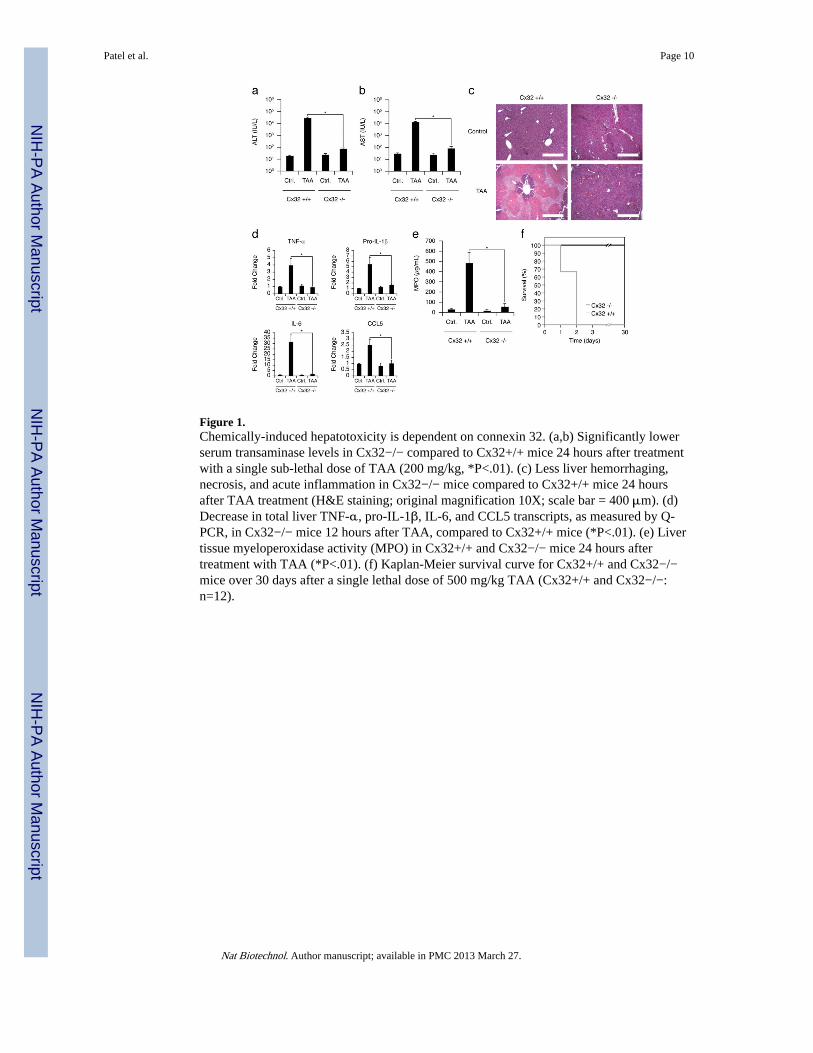
Figure 1.Chemically-induced hepatotoxicity is dependent on connexin 32. (a,b) Significantly lowerserum transaminase levels in Cx32−/− compared to Cx32+/+ mice 24 hours after treatmentwith a single sub-lethal dose of TAA (200 mg/kg, *P<.01). (c) Less liver hemorrhaging,necrosis, and acute inflammation in Cx32−/− mice compared to Cx32+/+ mice 24 hoursafter TAA treatment (H&E staining; original magnification 10X; scale bar = 400 μm). (d)Decrease in total liver TNF-α, pro-IL-1β, IL-6, and CCL5 transcripts, as measured by Q-PCR, in Cx32−/− mice 12 hours after TAA, compared to Cx32+/+ mice (*P<.01). (e) Livertissue myeloperoxidase activity (MPO) in Cx32+/+ and Cx32−/− mice 24 hours aftertreatment with TAA (*P<.01). (f) Kaplan-Meier survival curve for Cx32+/+ and Cx32−/−mice over 30 days after a single lethal dose of 500 mg/kg TAA (Cx32+/+ and Cx32−/−:n=12).
Patel et al. Page 10
Nat Biotechnol. Author manuscript; available in PMC 2013 March 27.
NIH
-PA Author Manuscript
NIH
-PA Author Manuscript
NIH
-PA Author Manuscript

Figure 2.Small molecule inhibitors of Cx32 selectively block hepatic gap junction communicationand prevent drug-induced hepatotoxicity. (a, b) A dye-coupling parachute assay was used todetermine the specificity of 2APB for Cx32 gap junctions compared to Cx43 gap junctions.HeLa Cx32 or Cx43 cells were loaded with gap junction permeable calcein-AM (10 μM)and impermeable CM-DiI (10 μM), and seeded onto unlabeled recipient HeLa Cx32 orCx43 cells, respectively, in the presence or absence of 2APB (25 μM). (a, b) After 4 hours,the co-culture was analyzed by fluorescence microscopy (n>5), and gap junctioncommunication was assessed by calculating the average number of calcein positive cells perCM-Dil positive cell, normalized to control conditions without 2APB treatment (*P<.01).(c) A tissue version of the scrape and load test was developed to evaluate functional gapjunction intercellular communication in liver tissue. Cx32+/+ and Cx32−/− mice weretreated with saline or 2APB (20 mg/kg) for 3 hours. Livers were excised, cut into 2-3 mmslices, and a small area of each slice was mechanically damaged with the insertion of a 27gauge needle coated with 0.5% Lucifer yellow (gap junction permeable) and 0.5% Texas redlabeled dextran (gap junction impermeable). Slices were washed, fixed, cryosectioned (7μm), and analyzed by fluorescence microscopy (n>5) and automated image analysissoftware to produce iso-intensity contour maps outlining spread of Lucifer yellow (green)and dextran-Texas red (red). Hepatic gap junction connectivity is demonstrated by thespread of gap junction permeable Lucifer yellow compared to gap junction impermeabledextran-Texas red. (d, e) Serum ALT levels and (f, g) H&E liver histology (originalmagnification 10X; scale bar = 400 μm) from wildtype mice pretreated with 2APB (20 mg/kg) or vehicle (DMSO, .1 mL/kg) 60 minutes prior to challenge with either TAA (200 mg/kg) or APAP (500 mg/kg), and then sacrificed 24 hours after TAA or 16 hours after APAPchallenge (*P<.01).
Patel et al. Page 11
Nat Biotechnol. Author manuscript; available in PMC 2013 March 27.
NIH
-PA Author Manuscript
NIH
-PA Author Manuscript
NIH
-PA Author Manuscript

Figure 3.Coadministration of hepatotoxic compounds with Cx32 inhibitors reduces liver injury, limitsinflammation, and enhances survival. (a) Serum ALT levels, (b) liver tissue MPO activity,and (c) H&E liver histology (original magnification 10X; scale bar = 400 μm) fromwildtype mice after treatment with 2APB alone (20 mg/kg), TAA (200 mg/kg) plus vehicle(DMSO, .1 mL/kg), TAA (200 mg/kg) plus 2APB (1 or 20 mg/kg), APAP (500 mg/kg) plusvehicle (DMSO, .1 mL/kg), or APAP (500 mg/kg) plus 2APB (20 mg/kg). Livers and serawere collected 24 hours after TAA challenge, or 16 hours after APAP challenge (*P<.05,**P<.01). (d) Q-PCR for TNF-α and pro-IL-1β from whole livers of wildtype mice treatedwith APAP and 2APB as described above (*P<.01). (e) Kaplan-Meier survival curve forwildtype mice over 30 days after a single lethal dose of TAA (500 mg/kg) plus vehicle(DMSO, .1 mL/kg), TAA (500 mg/kg) plus 2APB (20 mg/kg), APAP (750 mg/kg) plusvehicle (DMSO, .1 mL/kg), or APAP (750 mg/kg) plus 2APB (20 mg/kg) (n=10). (f) SerumALT levels and (j) H&E liver histology (original magnification 10X; scale bar = 400 μm)from wildtype mice after TAA (200 mg/kg) or APAP (500 mg/kg), with or without a rescueinjection of 2APB (20 mg/kg) 1.5, 3 or 6 hours after hepatotoxin challenge. Livers and serawere collected 24 hours after TAA challenge, or 16 hours after APAP challenge (*P<.01).2APB was dissolved in DMSO (200 mg/mL) for all experiments. All DMSO vehiclecontrols received this same amount of DMSO (.1 mL/kg) as the 2APB groups, with theappropriate volume of the hepatotoxin (TAA or APAP) dissolved in saline.
Patel et al. Page 12
Nat Biotechnol. Author manuscript; available in PMC 2013 March 27.
NIH
-PA Author Manuscript
NIH
-PA Author Manuscript
NIH
-PA Author Manuscript