
Functional studies indicate amantadine binds to the pore of the influenza A virus M2...
6
Functional studies indicate amantadine binds to the pore of the influenza A virus M2 proton-selective ion channel Xianghong Jing* † , Chunlong Ma †‡ , Yuki Ohigashi ‡ , Fernando A. Oliveira ‡ , Theodore S. Jardetzky § , Lawrence H. Pinto ‡ , and Robert A. Lamb* ¶ *Department of Biochemistry, Molecular Biology, and Cell Biology, ‡ Department of Neurobiology and Physiology, Northwestern University, Evanston, IL 60208-3500; § Department of Structural Biology, Stanford University, Palo Alto, CA 94305-5126; and ¶ Howard Hughes Medical Institute, Northwestern University, Evanston, IL 60208-3500 Contributed by Robert A. Lamb, May 22, 2008 (sent for review May 16, 2008) Influenza A and B viruses contain proton-selective ion channels, A/M2 and BM2, respectively, and the A/M2 channel activity is inhibited by the drugs amantadine and its methyl derivative rimantadine. The structure of the pore-transmembrane domain has been determined by both x-ray crystallography [Stouffer et al. (2008) Nature 451:596 –599] and by NMR methods [Schnell and Chou (2008) Nature 451:591–595]. Whereas the crystal structure indicates a single amantadine molecule in the pore of the channel, the NMR data show four rimantadine molecules bound on the outside of the helices toward the cytoplasmic side of the mem- brane. Drug binding includes interactions with residues 40 – 45 with a polar hydrogen bond between rimantadine and aspartic acid residue 44 (D44) that appears to be important. These two distinct drug-binding sites led to two incompatible drug inhibition mechanisms. We mutagenized D44 and R45 to alanine as these mutations are likely to interfere with rimantadine binding and lead to a drug insensitive channel. However, the D44A channel was found to be sensitive to amantadine when measured by electro- physiological recordings in oocytes of Xenopus laevis and in mammalian cells, and when the D44 and R45 mutations were introduced into the influenza virus genome. Furthermore, trans- planting A/M2 pore residues 24 –36 into BM2, yielded a pH- activated chimeric ion channel that was partially inhibited by amantadine. Thus, taken together our functional data suggest that amantadine/rimantadine binding outside of the channel pore is not the primary site associated with the pharmacological inhibition of the A/M2 ion channel. amantadine inhibition of influenza virus amantadine resistance drug-binding site influenza reverse genetics I nfluenza A and B viruses are enveloped negative-strand segmented RNA viruses that cause epidemics, and for influ- enza A virus, pandemics. At the biochemical and cellular biological level the viruses have some common properties. These include virus particles using host cell surface expressed sialic acid as their receptor and entry into cells by endocytosis. Virus uncoating takes place in the lumen of the endosome and is mediated by the hemagglutinin glycoprotein which is triggered by the acidic environment to undergo a protein refolding event that causes the fusion of the viral envelope with the endosomal membrane (reviewed in ref. 1). Inf luenza A and B viruses encode the A/M2 and BM2 integral membrane proteins (2, 3), respectively. A/M2 and BM2 are proton selective ion channels (4 – 6) that permit protons to enter virus particles during uncoating in endosomes. Acidification of the virus interior causes dissociation of the membrane (M1) protein from the ribonucleoprotein core (3, 7) a necessary step for import of the ribonucleoproteins into the nucleus (8). A/M2 and BM2 ion channel activity also modulates the pH of the trans-Golgi network in virus-infected cells equilibrating the acidic pH of the lumen of the Golgi with the cytoplasm (9, 10) (reviewed in ref. 11). The mature A/M2 protein consists of 96 residues with a 23-residue N-terminal extracellular domain, a single internal hydrophobic domain of 19 residues that acts as a TM domain and forms the pore of the channel, and a 54-residue cytoplasmic tail (2). The BM2 protein consists of a 7-residue ectodomain, a 19-residue TM domain, and an 82-residue cyto- plasmic tail (3). It has been shown for both A/M2 and BM2 that the proteins form homotetramers (3, 12, 13). The ectodomain of A/M2 contains two cysteine residues that form disulfide bonds but neither of these disulfide bonds is needed for ion channel activity (14) nor for replication of the virus in tissue culture or mice (15). The functionally active A/M2 and BM2 channels are the homotetramers and each channel has a centrally located pore for proton conduction (16–21). The A/M2 and BM2 proton channel proteins of influenza A and B viruses are among the smallest bona fide ion channel proteins (with the properties of ion selectivity and activation), and they succeed in accomplishing the same function with only a meager similarity in primary amino acid sequence. The only homology between the amino acid sequences of these two proteins is found in the TM domain HXXXW motif of the inner membrane-spanning residues. His- tidine 37 confers the high proton selectivity of the A/M2 channel (22–24) and tryptophan 41 acts as the channel gate, opening and closing the pore of the A/M2 channel (25). It is thought that His 19 and Trp 23 in BM2 have the same roles in BM2 channel function as they do in the A/M2 channel (6). The activity of A/M2 ion channel is inhibited by the antiviral drug amantadine and its methyl derivative rimantadine (26, 27). The drug binds at both neutral (closed state) and acidic pH (open channel). Influenza A viruses that mutate and become drug resistant contain mutations in the A/M2 TM domain and naturally arising mutations mostly occur at TM domain residues Val 27, Ala 30, Ser 31, and Gly 34 (26). Neither inf luenza B virus growth nor BM2 ion channel activity are inhibited by the drugs. Recently, Stouffer et al. (28) using X-ray crystallography published the atomic structure of the A/M2 TM domain with and without bound amantadine and Schnell and Chou (29), using NMR methods, copublished the structure of a somewhat larger A/M2 peptide (M2 residues 18–60) that included the TM Author contributions: X.J., C.M., Y.O., T.S.J., L.H.P., and R.A.L. designed research; X.J., C.M., Y.O., F.A.O., and T.S.J. performed research; X.J., C.M., Y.O., F.A.O., T.S.J., L.H.P., and R.A.L. analyzed data; and X.J., T.S.J., L.H.P., and R.A.L. wrote the paper. The authors declare no conflict of interest. Freely available online through the PNAS open access option. † X.J. and C.M. contributed equally to this work. To whom correspondence should be addressed at: Department of Biochemistry, Molecular Biology and Cell Biology, Northwestern University, 2205 Tech Drive, Evanston, IL 60208- 3500. E-mail: [email protected]. This article contains supporting information online at www.pnas.org/cgi/content/full/ 0804958105/DCSupplemental. © 2008 by The National Academy of Sciences of the USA www.pnas.orgcgidoi10.1073pnas.0804958105 PNAS August 5, 2008 vol. 105 no. 31 10967–10972 MICROBIOLOGY
Transcript of Functional studies indicate amantadine binds to the pore of the influenza A virus M2...
Functional studies indicate amantadine binds to thepore of the influenza A virus M2 proton-selectiveion channelXianghong Jing*†, Chunlong Ma†‡, Yuki Ohigashi‡, Fernando A. Oliveira‡, Theodore S. Jardetzky§, Lawrence H. Pinto‡,and Robert A. Lamb*¶�
*Department of Biochemistry, Molecular Biology, and Cell Biology, ‡Department of Neurobiology and Physiology, Northwestern University,Evanston, IL 60208-3500; §Department of Structural Biology, Stanford University, Palo Alto, CA 94305-5126; and ¶Howard HughesMedical Institute, Northwestern University, Evanston, IL 60208-3500
Contributed by Robert A. Lamb, May 22, 2008 (sent for review May 16, 2008)
Influenza A and B viruses contain proton-selective ion channels,A/M2 and BM2, respectively, and the A/M2 channel activity isinhibited by the drugs amantadine and its methyl derivativerimantadine. The structure of the pore-transmembrane domain hasbeen determined by both x-ray crystallography [Stouffer et al.(2008) Nature 451:596–599] and by NMR methods [Schnell andChou (2008) Nature 451:591–595]. Whereas the crystal structureindicates a single amantadine molecule in the pore of the channel,the NMR data show four rimantadine molecules bound on theoutside of the helices toward the cytoplasmic side of the mem-brane. Drug binding includes interactions with residues 40–45with a polar hydrogen bond between rimantadine and asparticacid residue 44 (D44) that appears to be important. These twodistinct drug-binding sites led to two incompatible drug inhibitionmechanisms. We mutagenized D44 and R45 to alanine as thesemutations are likely to interfere with rimantadine binding and leadto a drug insensitive channel. However, the D44A channel wasfound to be sensitive to amantadine when measured by electro-physiological recordings in oocytes of Xenopus laevis and inmammalian cells, and when the D44 and R45 mutations wereintroduced into the influenza virus genome. Furthermore, trans-planting A/M2 pore residues 24–36 into BM2, yielded a pH-activated chimeric ion channel that was partially inhibited byamantadine. Thus, taken together our functional data suggest thatamantadine/rimantadine binding outside of the channel pore isnot the primary site associated with the pharmacological inhibitionof the A/M2 ion channel.
amantadine inhibition of influenza virus � amantadine resistance �drug-binding site � influenza reverse genetics
Influenza A and B viruses are enveloped negative-strandsegmented RNA viruses that cause epidemics, and for influ-
enza A virus, pandemics. At the biochemical and cellularbiological level the viruses have some common properties. Theseinclude virus particles using host cell surface expressed sialic acidas their receptor and entry into cells by endocytosis. Virusuncoating takes place in the lumen of the endosome and ismediated by the hemagglutinin glycoprotein which is triggeredby the acidic environment to undergo a protein refolding eventthat causes the fusion of the viral envelope with the endosomalmembrane (reviewed in ref. 1).
Influenza A and B viruses encode the A/M2 and BM2 integralmembrane proteins (2, 3), respectively. A/M2 and BM2 areproton selective ion channels (4–6) that permit protons to entervirus particles during uncoating in endosomes. Acidification ofthe virus interior causes dissociation of the membrane (M1)protein from the ribonucleoprotein core (3, 7) a necessary stepfor import of the ribonucleoproteins into the nucleus (8). A/M2and BM2 ion channel activity also modulates the pH of thetrans-Golgi network in virus-infected cells equilibrating theacidic pH of the lumen of the Golgi with the cytoplasm (9, 10)
(reviewed in ref. 11). The mature A/M2 protein consists of 96residues with a 23-residue N-terminal extracellular domain, asingle internal hydrophobic domain of 19 residues that acts as aTM domain and forms the pore of the channel, and a 54-residuecytoplasmic tail (2). The BM2 protein consists of a 7-residueectodomain, a 19-residue TM domain, and an 82-residue cyto-plasmic tail (3). It has been shown for both A/M2 and BM2 thatthe proteins form homotetramers (3, 12, 13). The ectodomain ofA/M2 contains two cysteine residues that form disulfide bondsbut neither of these disulfide bonds is needed for ion channelactivity (14) nor for replication of the virus in tissue culture ormice (15). The functionally active A/M2 and BM2 channels arethe homotetramers and each channel has a centrally located porefor proton conduction (16–21). The A/M2 and BM2 protonchannel proteins of influenza A and B viruses are among thesmallest bona fide ion channel proteins (with the properties ofion selectivity and activation), and they succeed in accomplishingthe same function with only a meager similarity in primary aminoacid sequence. The only homology between the amino acidsequences of these two proteins is found in the TM domainHXXXW motif of the inner membrane-spanning residues. His-tidine 37 confers the high proton selectivity of the A/M2 channel(22–24) and tryptophan 41 acts as the channel gate, opening andclosing the pore of the A/M2 channel (25). It is thought that His19 and Trp 23 in BM2 have the same roles in BM2 channelfunction as they do in the A/M2 channel (6).
The activity of A/M2 ion channel is inhibited by the antiviraldrug amantadine and its methyl derivative rimantadine (26, 27).The drug binds at both neutral (closed state) and acidic pH(open channel). Influenza A viruses that mutate and becomedrug resistant contain mutations in the A/M2 TM domain andnaturally arising mutations mostly occur at TM domain residuesVal 27, Ala 30, Ser 31, and Gly 34 (26). Neither influenza B virusgrowth nor BM2 ion channel activity are inhibited by the drugs.
Recently, Stouffer et al. (28) using X-ray crystallographypublished the atomic structure of the A/M2 TM domain with andwithout bound amantadine and Schnell and Chou (29), usingNMR methods, copublished the structure of a somewhat largerA/M2 peptide (M2 residues 18–60) that included the TM
Author contributions: X.J., C.M., Y.O., T.S.J., L.H.P., and R.A.L. designed research; X.J., C.M.,Y.O., F.A.O., and T.S.J. performed research; X.J., C.M., Y.O., F.A.O., T.S.J., L.H.P., and R.A.L.analyzed data; and X.J., T.S.J., L.H.P., and R.A.L. wrote the paper.
The authors declare no conflict of interest.
Freely available online through the PNAS open access option.
†X.J. and C.M. contributed equally to this work.
�To whom correspondence should be addressed at: Department of Biochemistry, MolecularBiology and Cell Biology, Northwestern University, 2205 Tech Drive, Evanston, IL 60208-3500. E-mail: [email protected].
This article contains supporting information online at www.pnas.org/cgi/content/full/0804958105/DCSupplemental.
© 2008 by The National Academy of Sciences of the USA
www.pnas.org�cgi�doi�10.1073�pnas.0804958105 PNAS � August 5, 2008 � vol. 105 � no. 31 � 10967–10972
MIC
ROBI
OLO
GY

domain and 17 residues of the cytoplasmic tail. As predictedfrom earlier biochemical and NMR studies (17, 30) the recenthigh-resolution structural data show the overall architecture ofthe TM domain is a four-helix bundle and the data confirmpredictions that His 37 forms the pH sensor and Trp 41 thechannel gate (25, 31). The Trp 41 indole rings are at van derWaals distance from each other, prohibiting passage of water orions and the gate is further stabilized in the closed state byinter-subunit hydrogen bonds with Asp 44 (28, 29).
The crystal structures were solved at lower pH than the NMRstructure, and the former structures are more open near theC-terminal His 37 than the NMR structure. However, thestructures have a major difference in that the crystal structuredata indicate a single amantadine molecule in the pore of thechannel that is surrounded by Val 27, Ala 30, Ser 31, and Gly 34(28). In contrast, from the NMR data obtained with the peptidein a solution of 40 mM rimantadine and �300 mM dihexanoyl-phosphatidyl-choline, four rimantadine molecules were foundbound on the outside of the protein helix facing the lipid bilayerand located in the membrane environment at the end of the helixtoward the cytoplasmic face of the channel. Drug bindingincludes interactions with residues 40–45 with a polar hydrogenbond between rimantadine and aspartic acid residue 44 (D44)that appears to be important (29) (Fig. 1). However, although theNMR structure is very well defined in the TM region, the
drug-binding site around D44 is not well defined and rimanta-dine adopts many different conformations. Even within a singlesnapshot, each of the four rimantadine molecules within atetramer display significant variability in their interactions. Fig.1A illustrates the variability in geometries of rimantadine-N toAsp-carboxylate interactions (distances in angstroms) using datafrom frame 7 of 2RLF. Fig. 1B shows a structure in which theTM regions from the ensemble of NMR structures are alignedon the well-structured TM region (including the residues ex-pected to form the binding site). The protein is well ordered inthe TM region, except in the vicinity of the drug where both theside chains and the structure of the drug are variable.
These two distinct drug-binding sites led to two incompatibledrug inhibition mechanisms (32). Stouffer et al. (28) proposedthat amantadine physically occludes the pore of the channelrestricting passage of protons and the naturally arising drug-resistant mutations (TM domain residues 27, 30, 31, and 34) areclose to the proposed channel pore drug-binding site. The Hillcoefficient of 1 for amantadine inhibition is consistent with oneamantadine molecule blocking the channel (33) although otherdrug-binding models are possible. The finding of electron den-sity in the pore of the channel in the presence of amantadine andthe absence of density without amantadine is consistent with thedrug being present in the pore of the channel (28), nonethelessthe caveat has to be added that at 3.5 Å resolution it cannot beproven the density represents amantadine. Schnell and Chou(29) proposed channel inhibition occurred by an allostericmechanism with external drug binding stabilizing the closedstate making it more difficult to move the four helices and openthe channel. Important in this regard is the notion of long-rangehelical effects because of the observation that an amantadine-resistant mutation maps to Leu 38 (L38F) (33) and it wassuggested the mutation probably disturbs helix–helix packingand lowers the energetic cost of opening the channel (29).
We mutagenized D44 and R45 to alanine as these mutationswould be expected to interfere with the observed rimantadineinteraction and may lead to a drug insensitive channel, but thechannel remained drug sensitive. We also substituted A/M2 poreresidues 24–36 into BM2 (in place of BM2 pore residues 6–18)and this chimeric protein yielded a pH-activated ion channel thatwas partially inhibited by amantadine. Thus, our functional datasuggest that amantadine/rimantadine binding outside of thechannel pore is not the primary site associated with the phar-macological inhibition of the A/M2 ion channel.
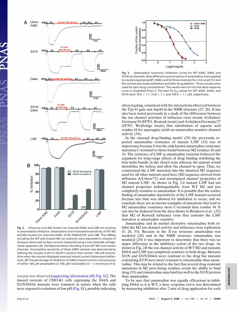
Results and DiscussionIon Channel Activity and Sensitivity to Amantadine Inhibition of M2Mutants. To investigate the interaction between A/M2 ion chan-nel pore residue Asp 44 and amantadine/rimantadine, we mu-tated Asp 44 to alanine and examined the ion channel activityand its sensitivity to amantadine in oocytes of Xenopus laevis(Fig. 2A). We also examined the ion channel activity of a knownamantadine-resistant mutant, S31N and a double mutant S31N/D44A. The levels of cell-surface expression of the WT andmutant channels in oocytes were equivalent (data not shown).The oocytes were initially bathed at pH 8.5 to keep the M2channel in the closed state and then the bathing solution waschanged to pH 5.5 to open the channel (4). Whereas, WT M2 andD44A ion channel currents were sensitive to amantadine, S31Nand S31N/D44A channel currents were largely resistant to thedrug (Fig. 2 A). For WT A/M2 and D44A the amantadineinhibition of current remains even after the oocytes were re-turned to pH 8.5 and then bathed in pH 5.5 because amantadineinhibition is effectively irreversible on this time scale: the reversereaction rate constant of amantadine block is 3 � 10�4s�1 (33).The expression and amantadine sensitivity of WT M2, S31N,D44A, and S31/D44A was also examined in mammalian cells(CHO-K1 cells) and very similar drug sensitivity to that found in
Fig. 1. Structure of the rimantadine binding site at the boundary of the A/M2TM domain with the cytoplasmic tail. (A). A ribbon diagram showing four TMdomain �-helices and the interactions between rimantadine (purple) and D44.Drawn from frame 7 of Protein Data Bank accession no. 2RLF. The variabilityin rimantadine-N to Asp-carboxylate geometries (distances in angstroms) areindicated. (B) A ribbon diagram showing a structure in which the TM regionsfrom the ensemble of NMR structures are aligned on the well-structured TMregion (including the residues expected to form the drug-binding site). Theprotein is well ordered in the TM region, except in the vicinity of the drugwhere both the side-chains and the drug are variable. Drawn from ProteinData Bank accession no. 2RLF from the data of Schnell and Chou (29).
10968 � www.pnas.org�cgi�doi�10.1073�pnas.0804958105 Jing et al.
oocytes was observed [supporting information (SI) Fig. S1]. Theinward currents of CHO-K1 cells expressing the D44A andS31N/D44A mutants were transient in nature when the cellswere exposed to solutions of low pH (Fig. S1), possibly indicating
altered gating, consistent with the interactions observed betweenthe Trp-41 gate and Asp44 in the NMR structure (27, 28). It hasalso been noted previously in a study of the differences betweenthe ion channel activities of influenza virus strains A/chicken/Germany/34 (H7N1, Rostock strain) and A/chicken/Germany/27(H7N7, Weybridge strain) that substitution of aspartic acidresidue 44 for asparagine yields an amantadine sensitive channelactivity (34).
In the external drug-binding model (29) the previously re-ported amantadine resistance of mutant L38F (33) was ofimportance because it was the only known amantadine resistancemutation C-terminal to those found between M2 residues 26 and34. The existence of L38F as amantadine resistant bolstered theargument for long-range effects of drug binding stabilizing thefour-helix bundle in the closed state whereas the mutant woulddestabilize the helices and allow the channel to open. Thus, weconstructed the L38F mutation into the identical M2 sequenceused for all other mutants used here (M2 sequence derived frominfluenza A/Udorn/72) and investigated channel properties ofM2 mutant L38F. As shown in Fig. 2 A mutant L38F had ionchannel properties indistinguishable from WT M2 and wascompletely sensitive to amantadine. It is possible that the earlierfinding of amantadine insensitivity of the L38F mutant occurredbecause less time was allowed for inhibition to occur, and weconclude there are no known examples of mutations that lead toM2 amantadine resistance more C-terminal than residue 34. Itcan also be deduced from the data shown in Betakova et al., (32)that M2 of Rostock influenza virus that contains the L38Fmutation is amantadine sensitive.
Amantadine and its methyl derivative rimantadine both in-hibit the M2 ion channel activity and influenza virus replication(5, 26, 33). Because in the X-ray structure amantadine wasmodeled (28) and in the NMR structure rimantadine wasmodeled (29) it was important to determine that there was nomajor difference in the inhibitory action of the two drugs. Asshown in Fig. 2B the ion channel activity of WT M2 and mutantsD44A and L38F was completely sensitive to both drugs. MutantsS31N and S31N/D44A were resistant to the drug but mutantscontaining S31N were more resistant to rimantadine than aman-tadine. This may be related to the fact that several drug-resistantmutations in M2 pore-lining residues retain the ability to binddrug (35) and rimantadine may bind less well to the S31N proteinthan amantadine.
To be sure that amantadine was equally efficacious in inhib-iting D44A as it is WT, a dose–response curve was determinedby measuring inhibition after 2 min of drug application for each
Fig. 3. Amantadine isochronic inhibition curves for WT A/M2, D44A andS31N ion channels. Nine different concentrations of amantadine were appliedto oocytes expressing WT, D44A, and S31N ion channels for 2 min at pH 5.5 andthe current was measured before and after drug addition. Three oocytes wereused for each drug concentration. The results were fit into the dose-responsecurve in GraphPad Prism 5. The best fit IC50 values for WT A/M2, D44A, andS31N were 16.0 � 1.1, 15.8 � 1.1, and 199.9 � 1.1 �M, respectively.
Fig. 2. Influenza virus M2 mutant ion channels D44A and L38F are sensitiveto amantadine inhibition. Amantadine and rimantadine sensitivity of WT M2and M2 mutant ion channels D44A, S31N, D44A/S31N, and L38F. The mRNAsencoding the WT and mutant M2 ion channels were expressed in oocytes ofXenopus laevis and surface currents measured using a two-electode voltage-clamp apparatus (4). (A) Representative recording traces WT M2 and mutantchannels. Amantadine sensitivity of these A/M2 variants was determined bybathing the oocytes in pH 5.5 Barth’s solution that contain 100 �M amanta-dine when the oocytes displayed maximal inward current (downward deflec-tion). (B) The percentage of inhibition of A/M2 inward current in the presenceof either 100 �M amantadine or 100 �M rimantadine (n � a minimum of 5).
Jing et al. PNAS � August 5, 2008 � vol. 105 � no. 31 � 10969
MIC
ROBI
OLO
GY
of many concentrations of the drug. It was found that WT A/M2and D44A had virtually identical amantadine sensitivities (Fig. 3).
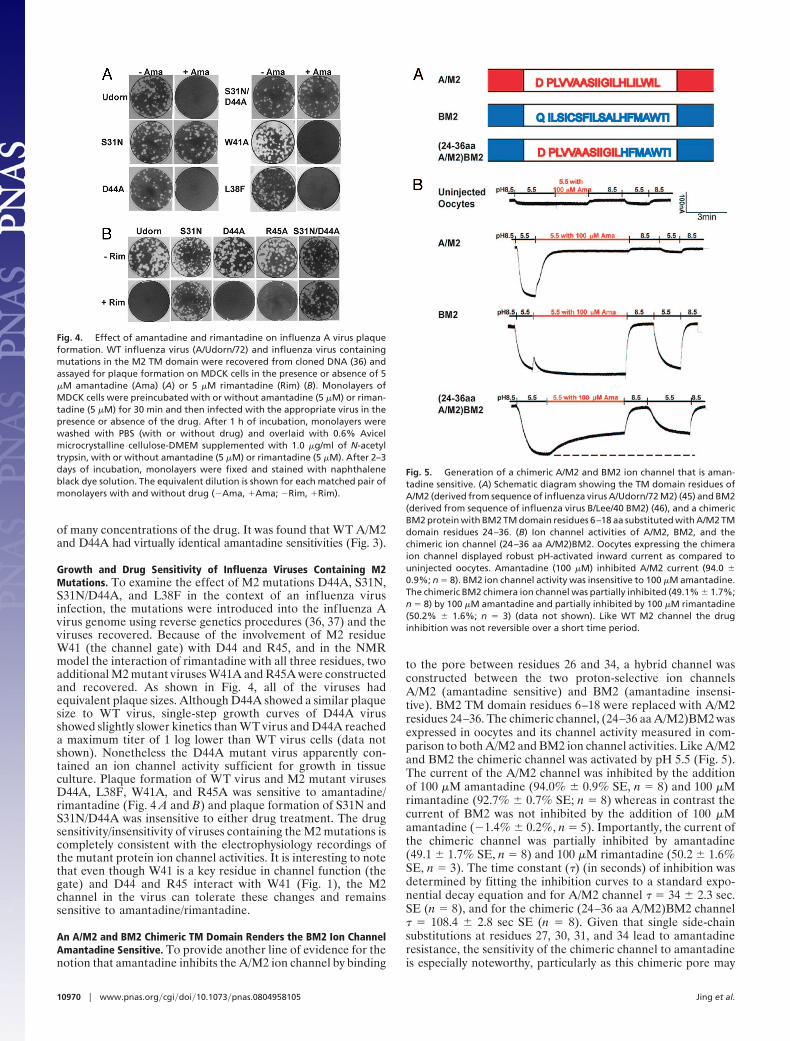
Growth and Drug Sensitivity of Influenza Viruses Containing M2Mutations. To examine the effect of M2 mutations D44A, S31N,S31N/D44A, and L38F in the context of an influenza virusinfection, the mutations were introduced into the influenza Avirus genome using reverse genetics procedures (36, 37) and theviruses recovered. Because of the involvement of M2 residueW41 (the channel gate) with D44 and R45, and in the NMRmodel the interaction of rimantadine with all three residues, twoadditional M2 mutant viruses W41A and R45A were constructedand recovered. As shown in Fig. 4, all of the viruses hadequivalent plaque sizes. Although D44A showed a similar plaquesize to WT virus, single-step growth curves of D44A virusshowed slightly slower kinetics than WT virus and D44A reacheda maximum titer of 1 log lower than WT virus cells (data notshown). Nonetheless the D44A mutant virus apparently con-tained an ion channel activity sufficient for growth in tissueculture. Plaque formation of WT virus and M2 mutant virusesD44A, L38F, W41A, and R45A was sensitive to amantadine/rimantadine (Fig. 4 A and B) and plaque formation of S31N andS31N/D44A was insensitive to either drug treatment. The drugsensitivity/insensitivity of viruses containing the M2 mutations iscompletely consistent with the electrophysiology recordings ofthe mutant protein ion channel activities. It is interesting to notethat even though W41 is a key residue in channel function (thegate) and D44 and R45 interact with W41 (Fig. 1), the M2channel in the virus can tolerate these changes and remainssensitive to amantadine/rimantadine.
An A/M2 and BM2 Chimeric TM Domain Renders the BM2 Ion ChannelAmantadine Sensitive. To provide another line of evidence for thenotion that amantadine inhibits the A/M2 ion channel by binding
to the pore between residues 26 and 34, a hybrid channel wasconstructed between the two proton-selective ion channelsA/M2 (amantadine sensitive) and BM2 (amantadine insensi-tive). BM2 TM domain residues 6–18 were replaced with A/M2residues 24–36. The chimeric channel, (24–36 aa A/M2)BM2 wasexpressed in oocytes and its channel activity measured in com-parison to both A/M2 and BM2 ion channel activities. Like A/M2and BM2 the chimeric channel was activated by pH 5.5 (Fig. 5).The current of the A/M2 channel was inhibited by the additionof 100 �M amantadine (94.0% � 0.9% SE, n � 8) and 100 �Mrimantadine (92.7% � 0.7% SE; n � 8) whereas in contrast thecurrent of BM2 was not inhibited by the addition of 100 �Mamantadine (�1.4% � 0.2%, n � 5). Importantly, the current ofthe chimeric channel was partially inhibited by amantadine(49.1 � 1.7% SE, n � 8) and 100 �M rimantadine (50.2 � 1.6%SE, n � 3). The time constant (�) (in seconds) of inhibition wasdetermined by fitting the inhibition curves to a standard expo-nential decay equation and for A/M2 channel � � 34 � 2.3 sec.SE (n � 8), and for the chimeric (24–36 aa A/M2)BM2 channel� � 108.4 � 2.8 sec SE (n � 8). Given that single side-chainsubstitutions at residues 27, 30, 31, and 34 lead to amantadineresistance, the sensitivity of the chimeric channel to amantadineis especially noteworthy, particularly as this chimeric pore may
Fig. 4. Effect of amantadine and rimantadine on influenza A virus plaqueformation. WT influenza virus (A/Udorn/72) and influenza virus containingmutations in the M2 TM domain were recovered from cloned DNA (36) andassayed for plaque formation on MDCK cells in the presence or absence of 5�M amantadine (Ama) (A) or 5 �M rimantadine (Rim) (B). Monolayers ofMDCK cells were preincubated with or without amantadine (5 �M) or riman-tadine (5 �M) for 30 min and then infected with the appropriate virus in thepresence or absence of the drug. After 1 h of incubation, monolayers werewashed with PBS (with or without drug) and overlaid with 0.6% Avicelmicrocrystalline cellulose-DMEM supplemented with 1.0 �g/ml of N-acetyltrypsin, with or without amantadine (5 �M) or rimantadine (5 �M). After 2–3days of incubation, monolayers were fixed and stained with naphthaleneblack dye solution. The equivalent dilution is shown for each matched pair ofmonolayers with and without drug (�Ama, �Ama; �Rim, �Rim).
Fig. 5. Generation of a chimeric A/M2 and BM2 ion channel that is aman-tadine sensitive. (A) Schematic diagram showing the TM domain residues ofA/M2 (derived from sequence of influenza virus A/Udorn/72 M2) (45) and BM2(derived from sequence of influenza virus B/Lee/40 BM2) (46), and a chimericBM2 protein with BM2 TM domain residues 6–18 aa substituted with A/M2 TMdomain residues 24–36. (B) Ion channel activities of A/M2, BM2, and thechimeric ion channel (24–36 aa A/M2)BM2. Oocytes expressing the chimeraion channel displayed robust pH-activated inward current as compared touninjected oocytes. Amantadine (100 �M) inhibited A/M2 current (94.0 �0.9%; n � 8). BM2 ion channel activity was insensitive to 100 �M amantadine.The chimeric BM2 chimera ion channel was partially inhibited (49.1% � 1.7%;n � 8) by 100 �M amantadine and partially inhibited by 100 �M rimantadine(50.2% � 1.6%; n � 3) (data not shown). Like WT M2 channel the druginhibition was not reversible over a short time period.
10970 � www.pnas.org�cgi�doi�10.1073�pnas.0804958105 Jing et al.
well be altered in structure in subtle ways from the WT M2 pore.Thus, the simplest interpretation of the data is that amantadine/rimantadine interacts with A/M2 residues 24–36 to occlude thechannel pore and that these residues can be transferred into theBM2 pore to render BM2 partially amantadine sensitive.
Summary. Our functional studies on the influenza A virus A/M2proton-selective ion channel and its inhibition by amantadine orrimantadine suggest that amantadine/rimantadine binding out-side of the channel pore is not the primary site associated withthe pharmacological inhibition of the A/M2 ion channel. First,mutation of D44 and R45 to alanine did not alter the sensitivityof the channels to the drug, suggesting that the interactions ofrimantadine with D44 and R45 are not important for druginhibition. Second, the original finding that mutating L38 tophenylalanine caused amantadine resistance appears to be in-correct and thus there is no evidence for long-range effects ofamantadine resistance, which was proposed in the allostericmechanism for rimantadine inhibition from the cytoplasmic sideof the four-helix bundle. Third, the chimeric channel (24–36 aaA/M2)BM2 channel is activated by low pH and partially inhibitedby amantadine. The interpretation of these data that we preferis that we transferred the drug site from A/M2 to the normallydrug-insensitive BM2 ion channel. It seems unlikely amantadinestabilizes the closed state of the chimeric channel by an allostericmechanism and involving M2 residues 24–36, when this chimericprotein lacks the D44 and R45 residues that interact withrimantadine in the NMR model.
There are many examples of second substrate or ligand-binding sites for proteins but the biological significance of thesesecond sites is often unclear. For influenza virus, a second sialicacid binding site was identified on the hemagglutinin (38) andthe avian N9 neuraminidase contains a second sialic acid bindingsite (39, 40). However, neither of these second sites appears tobe required for virus replication. Amantadine partitions intolipid bilayers (41–43) giving the drug access to the cytoplasmicside of the four-helix bundle. Whether the external rimantadinebinding site, that we consider to be a second drug-binding site,has another role in properties of A/M2 function that are notdirectly associated with inhibition of channel activity remains tobe determined.
Materials and MethodsCells, Viruses, and Plasmids. 293-T and Madin–Darby canine kidney (MDCK)cells were maintained in Dulbecco’s modified Eagle’s medium (DMEM) (In-
vitrogen, Carlsbad, CA) supplemented with 10% FBS. Influenza A/Udorn/72virus (WT) and mutant viruses were propagated in MDCK cells overlaid withserum-free DMEM containing 3.0 �g/ml N-acetyl trypsin (NAT; Sigma-Aldrich,St. Louis, MO) at 37°C. WT and mutant viruses were generated by using reversegenetics from cDNAs essentially as described previously (36). The eight ge-nome-sense (pHH21) plasmids and four protein-expressing (pcDNA3.1) plas-mids used to generate influenza virus by reverse genetics have been describedpreviously (36). Mutations into the M2 gene in pHH21 and pGEM3 vectorswere generated using Quick Change mutagenesis (Stratagene, La Jolla, CA).293-T cells were transfected using TransIT-LT1 (Mirus, Madison, WI) accordingto the manufacturer protocols. Virus stocks were propagated in MDCK cells,and the virus titers were determined by plaque assay on MDCK cells. Fordetermination of viral genome sequences viral RNA was extracted by using theQIAamp viral RNA kit (Qiagen, Valencia, CA), followed by Super ReverseTranscriptase (Molecular Genetic Resources, Tampa, FL), using genome-specific primers, and amplified with AmpliTaq DNA polymerase (AppliedBiosystems, Foster City, CA). The complete nucleotide sequences of theM genes were determined using a 3100-Avant genetic analyzer (AppliedBiosystems).
For additional information, see SI Materials and Methods.
Plaque Assays. Confluent monolayers of MDCK cells were incubated with10-fold serially diluted virus samples in DMEM 1% BSA for 1 h at 37°C. Theinoculums were removed, and the cells were washed with phosphate-buffered saline (PBS). The cells were then overlaid with DMEM containing0.6% Avicel microcrystalline cellulose (FMC BioPolymer, Philadelphia, PA) (44)and NAT (1.0 �g/ml). To examine the effect of the drug (amantadine orrimantadine) on plaque formation, monolayers were preincubated withDMEM supplemented with the drug (5 �M) at 37°C for 30 min, and virussamples were preincubated with DMEM 1% BSA with the drug (5 �M) at 4°Cfor 30 min before infection. At 2–3 days after infection, the monolayers werefixed and stained with naphthalene black dye solution (0.1% naphthaleneblack, 6% glacial acetic acid, 1.36% anhydrous sodium acetate).
mRNA Synthesis, Culture, and Microinjection of Oocytes. WT A/M2-pGEM3cDNA and mutant DNAs (D44A/S31N/D44A,S31N/L38F) were linearized usingHindIII and in vitro transcription reactions were performed on the linearizedcDNA using a T7 mMESSAGE mMACHINE transcription kit (Ambion, Austin,TX) and injected into oocytes as described previously (4).
Two-Electrode Voltage-Clamp Analysis. Whole-cell two-electrode voltage-clamp currents were measured 48–72 h after injection of oocytes as describedpreviously (4). Currents were acquired and analyzed using the pCLAMP 10.0software package (Axon Instruments, Sunnyvale, CA).
ACKNOWLEDGMENTS. The research was supported by the National Institutesof Health research grants R01 AI-20201 (R.A.L.) and R01 Al-57363 (L.H.P.).R.A.L. is an investigator of the Howard Hughes Medical Institute.
1. Lamb RA, Krug RM (2001) in Fields Virology eds Knipe D M, Howley PM (Lippincott,Williams and Wilkins, Philadelphia), 4th Ed, pp. 1487–1531.
2. Lamb RA, Zebedee SL, Richardson CD (1985) Influenza virus M2 protein is an integralmembrane protein expressed on the infected-cell surface. Cell 40:627–633.
3. Paterson RG, Takeda M, Ohigashi Y, Pinto LH, Lamb RA (2003) Influenza B virus BM2protein is an oligomeric integral membrane protein expressed at the cell surface.Virology 306:7–17.
4. Pinto LH, Holsinger LJ, Lamb RA (1992) Influenza virus M2 protein has ion channelactivity. Cell 69:517–528.
5. Chizhmakov IV, et al. (1996) Selective proton permeability and pH regulation of theinfluenza virus M2 channel expressed in mouse erythroleukaemia cells. J Physiol494:329–336.
6. Mould JA, et al. (2003) Influenza B virus BM2 protein has ion channel activity thatconducts protons across membranes. Dev Cell 5:175–184.
7. Zhirnov OP (1990) Solubilization of matrix protein M1/M from virions occurs at differ-ent pH for orthomyxo- and paramyxoviruses. Virology 176:274–279.
8. Martin K, Helenius A (1991) Nuclear transport of influenza virus ribonucleopro-teins: The viral matrix protein (M1) promotes export and inhibits import. Cell67:117–130.
9. Ciampor F, et al. (1992) Evidence that the amantadine-induced, M2-mediated conver-sion of influenza A virus hemagglutinin to the low pH conformation occurs in an acidictrans Golgi compartment. Virology 188:14–24.
10. Grambas S, Hay AJ (1992) Maturation of influenza A virus hemagglutinin–estimates ofthe pH encountered during transport and its regulation by the M2 protein. Virology190:11–18.
11. Lamb RA, Pinto LH (2005) in Contemporary Topics in Influenza Virology, ed KawaokaY (Caister Academic, Wymondham, Norfolk, UK), pp. 65–93.
12. Holsinger LJ, Lamb RA (1991) Influenza virus M2 integral membrane protein is ahomotetramer stabilized by formation of disulfide bonds. Virology 183:32–43.
13. Sugrue RJ, Hay AJ (1991) Structural characteristics of the M2 protein of the influenzaA viruses: Evidence that it forms a tetrameric channel. Virology 180:617–624.
14. Holsinger LJ, Shaughnessy MA, Micko A, Pinto LH, Lamb RA (1995) Analysis of theposttranslational modifications of the influenza virus M2 protein. J Virol 69:1219–1225.
15. Castrucci MR, et al. (1997) The cysteine residues of the M2 protein are not required forinfluenza A virus replication. Virology 238:128–134.
16. Sakaguchi T, Tu Q, Pinto LH, Lamb RA (1997) The active oligomeric state of theminimalistic influenza virus M2 ion channel is a tetramer. Proc Natl Acad Sci USA94:5000–5004.
17. Pinto LH, et al. (1997) A functionally defined model for the M2 proton channel ofinfluenza A virus suggests a mechanism for its ion selectivity. Proc Natl Acad Sci USA94:11301–11306.
18. Bauer CM, Pinto LH, Cross TA, Lamb RA (1999) The influenza virus M2 ion channelprotein: Probing the structure of the transmembrane domain in intact cells by usingengineered disulfide cross-linking. Virology 254:196–209.
19. Shuck K, Lamb RA, Pinto LH (2000) Analysis of the pore structure of the influenzaA virus M2 ion channel by the substituted-cysteine accessibility method. J Virol74:7755–7761.
20. Balannik V, Lamb RA, Pinto LH (2008) The oligomeric state of the active BM2 ionchannel protein of influenza B virus. J Biol Chem 283:4895–4904.
21. Ma C, et al. (2008) Identification of the pore-lining residues of the BM2 ion channelprotein of influenza B virus. J Biol Chem, doi: 10.1074/jbc.M710302200.
22. Mould JA, et al. (2000) Permeation and activation of the M2 ion channel of influenzaA virus. J Biol Chem 275:31038–31050.
Jing et al. PNAS � August 5, 2008 � vol. 105 � no. 31 � 10971
MIC
ROBI
OLO
GY
23. Hu J, et al. (2006) Histidines, heart of the hydrogen ion channel from influenza A virus:Toward an understanding of conductance and proton selectivity. Proc Natl Acad SciUSA 103:6865–6870.
24. Venkataraman P, Lamb RA, Pinto LH (2005) Chemical rescue of histidine selectivity filtermutants of the M2 ion channel of influenza A virus. J Biol Chem 280:21463–21472.
25. Tang Y, Zaitseva F, Lamb RA, Pinto LH (2002) The gate of the influenza virus M2 protonchannel is formed by a single tryptophan residue. J Biol Chem 277:39880–39886.
26. Hay AJ, Wolstenholme AJ, Skehel JJ, Smith MH (1985) The molecular basis of thespecific anti-influenza action of amantadine. EMBO J 4:3021–3024.
27. Lamb RA, Holsinger LJ, Pinto LH (1994) in Receptor-Mediated Virus Entry into Cells, edWimmer E (Cold Spring Harbor Laboratory Press, Cold Spring Harbor, NY), pp. 303–321.
28. Stouffer AL, et al. (2008) Structural basis for the function and inhibition of an influenzavirus proton channel. Nature 451:596–599.
29. Schnell JR, Chou JJ (2008) Structure and mechanism of the M2 proton channel ofinfluenza A virus. Nature 451:591–595.
30. Kovacs FA, Cross TA (1997) Transmembrane four-helix bundle of influenza A M2protein channel: Structural implications from helix tilt and orientation. Biophys J73:2511–2517.
31. Pinto LH, Lamb RA (2006) The M2 proton channels of influenza A and B viruses. J BiolChem 281:8997–9000.
32. Miller M (2008) Coughing up flu’s proton channels (News and Views). Nature 451:532–533.
33. Wang C, Takeuchi K, Pinto LH, Lamb RA (1993) Ion channel activity of influenza A virusM2 protein: Characterization of the amantadine block. J Virol 67:5585–5594.
34. Betakova T, Ciampor F, Hay AJ (2005) Influence of residue 44 on the activity of the M2proton channel of influenza A virus. J Gen Virol 86:181–184.
35. Astrahan P, Kass I, Cooper MA, Arkin IT (2004) A novel method of resistance forinfluenza against a channel-blocking antiviral drug. Proteins 55:251–257.
36. Takeda M, Pekosz A, Shuck K, Pinto LH, Lamb RA (2002) Influenza A virus M2 ionchannel activity is essential for efficient replication in tissue culture. J Virol 76:1391–1399.
37. Neumann G, et al. (1999) Generation of influenza A viruses entirely from cloned cDNAs.Proc Natl Acad Sci USA 96:9345–9350.
38. Sauter NK, et al. (1992) Crystallographic detection of a second ligand binding site ininfluenza virus hemagglutinin. Proc Natl Acad Sci USA 89:324–328.
39. Air GM, Ritchie LR, Laver WG, Colman PM (1985) Gene and protein sequence of aninfluenza neuraminidase with hemagglutinin activity. Virology 145:1–7.
40. Varghese JN, et al. (1997) Structural evidence for a second sialic acid binding site inavian influenza virus neuraminidases. Proc Natl Acad Sci USA 94:11808–11812.
41. Epand RM, Epand RF, McKenzie RC (1987) Effects of viral chemotherapeutic agents onmembrane properties: Studies of cyclosporin A, benzlooxycarbonyl-D-Phe-L-Phe-Glyand amantadine. J Biol Chem 262:1526–1529.
42. Subczynski WK, Wojas J, Pezeshk V, Pezeshk A (2000) Partitioning and localization ofspin-labeled amantadine in lipid bilayers: An epr study. J Pharmaceut Sci 87:1249–1254.
43. Wang J, Schnell JR, Chou JJ (2004) Amantadine partition and localization in phospho-lipid membrane: A solution NMR study. Biochem Biophys Res Commun 324:212–217.
44. Matrosovich M, Matrosovich T, Garten W, Klenk HD (2006) New low-viscosity overlaymedium for viral plaque assays. Virol. J. 3.63, doi: 10.1186/1743–422X-3–63.
45. Lamb RA, Lai C-J (1981) Conservation of the influenza virus membrane protein (M1)amino acid sequence and an open reading frame of RNA segment 7 encoding a secondprotein (M2) in H1N1 and H3N2 strains. Virology 112:746–751.
46. Briedis DJ, Lamb RA, Choppin PW (1982) Sequence of RNA segment 7 of the influenzaB virus genome: Partial amino acid homology between the membrane proteins (M1) ofinfluenza A and B viruses and conservation of a second open reading frame. Virology116:581–588.
10972 � www.pnas.org�cgi�doi�10.1073�pnas.0804958105 Jing et al.