
Role of CBP in regulating HIF-1-mediated activation of transcription
7370–7382 Nucleic Acids Research, 2014, Vol. 42, No. 11 Published online 20 March 2014doi: 10.1093/nar/gku206
Functional redundancy between the transcriptionalactivation domains of E2A is mediated by binding tothe KIX domain of CBP/p300Christopher M. Denis1,*, David N. Langelaan1,*, Alyssa C. Kirlin1, Seth Chitayat1,Kim Munro2, Holly L. Spencer1, David P. LeBrun2,3,4,† and Steven P. Smith1,2,†
1Department of Biomedical and Molecular Sciences, Queen’s University, Kingston, Ontario, K7L 3N6, Canada,2Protein Function Discovery Group, Queen’s University, Kingston, Ontario, K7L 3N6, Canada, 3Department ofPathology and Molecular Medicine, Queen’s University, Kingston, Ontario, K7L 3N6, Canada and 4Division of CancerBiology and Genetics, Cancer Research Institute, Queen’s University, Kingston, Ontario, K7L 3N6, Canada
Received September 19, 2013; Revised January 29, 2014; Accepted February 25, 2014
ABSTRACT
The E-protein transcription factors play essentialroles in lymphopoiesis, with E12 and E47 (hereaftercalled E2A) being particularly important in B cellspecification and maturation. The E2A gene is alsoinvolved in a chromosomal translocation that resultsin the leukemogenic oncoprotein E2A-PBX1. The twoactivation domains of E2A, AD1 and AD2, display re-dundant, independent, and cooperative functions ina cell-dependent manner. AD1 of E2A functions bybinding the transcriptional co-activator CBP/p300;this interaction is required in oncogenesis and oc-curs between the conserved �-x-x-�-� motif in AD1and the KIX domain of CBP/p300. However, co-activator recruitment by AD2 has not been charac-terized. Here, we demonstrate that the first of twoconserved �-x-x-�-� motifs within AD2 of E2A inter-acts at the same binding site on KIX as AD1. Muta-genesis uncovered a correspondence between theKIX-binding affinity of AD2 and transcriptional ac-tivation. Although AD2 is dispensable for oncoge-nesis, experimentally increasing the affinity of AD2for KIX uncovered a latent potential to mediate im-mortalization of primary hematopoietic progenitorsby E2A-PBX1. Our findings suggest that redundancybetween the two E2A activation domains with respectto transcriptional activation and oncogenic functionis mediated by binding to the same surface of the KIXdomain of CBP/p300.
INTRODUCTION
Coordinated transcriptional regulation is a critical aspectof hematopoiesis, including the lineage specification of B-and T-lymphocytes, natural killer cells and plasmacytoiddendritic cells from pluripotent hematopoietic stem cells(1–7). The precise cellular programming of this processdictates progressive lineage-specific differentiation, whichcomprises several intermediates and ultimately the mature,functional cell, through specific gene expression systemsthat are tightly regulated by a series of transcription fac-tors. The E-protein family of class I basic helix-loop-helix(bHLH) transcription factors represents one such set oftranscription factors that play essential roles in the devel-opment and speciation of B- and T-lymphocytes (8–12).
The E-protein family comprises the proteins E12 andE47 (hereafter referred to as E2A), which are alternativelyspliced products of the E2A gene (also called TCF3), aswell as HEB and E2–2 (9,13–15). Each family member con-tains a C-terminal bHLH domain that mediates E-proteindimerization and binding to DNA at the E-box CANNTGconsensus sequence in transcriptional promoters and en-hancers (16–19). Two transcriptional activation domains(ADs), AD1 and AD2, are found, respectively, at the N-terminus and in the central region of the proteins (10,13,15)(Figure 1). AD1 and AD2 have been shown to display celltype-specific, redundant, cooperative or independent tran-scriptional regulatory functions (20–26). For example, AD1or AD2 can induce transcription individually, their con-tributions to reporter gene induction are greater than ad-ditive, and deletion of either AD1 or AD2 abrogates theability of E2A to induce B-lymphoid differentiation in amouse-derived pre-B cell line, suggesting functionally re-dundant and cooperative roles in B cell development (20–
*These authors contributed equally to the work.The authors wish it to be known that, in their opinion, the first two authors should be regarded as Joint First Authors.†To whom correspondence should be addressd. Tel: +1 613 533 3188; Fax: +1 613 533 2497; Email: [email protected] may also be addressed to David P. LeBrun. Tel: +1 613 533 3209; Fax: +1 613 533 2907; Email: [email protected]
C© The Author(s) 2014. Published by Oxford University Press on behalf of Nucleic Acids Research.This is an Open Access article distributed under the terms of the Creative Commons Attribution License (http://creativecommons.org/licenses/by-nc/3.0/), whichpermits non-commercial re-use, distribution, and reproduction in any medium, provided the original work is properly cited. For commercial re-use, please [email protected]

Nucleic Acids Research, 2014, Vol. 42, No. 11 7371
1 99
AD1
11GTDKELSDLLDFSMMF26
AD2 bHLH
456945034013
392KIEDHLDEAIHVLRSH407
410GTAGDMHTLLPGHGAL425
(a)
(b)
i)φ x x φ φ
E2A AD2-1 392 K I E D H L D E A I H V L R S H 407
HEB AD2-1 426 D R L D R L D D A I H V L R N H 441
E2-2 AD2-1 388 D R L E R L D D A I H V L R N H 403
E2A AD2-2 410 G T A G D M H T L L P G H G A L 425
HEB AD2-2 451 A G H S D I H S L L G P S H N A 466
E2-2 AD2-2 413 G G H G D M H G I I G P S H N G 428
E2A AD1-PCET 11 G T D K E L S D L L D F S M M F 26
c-Myb 293 K R I K E L E L L L M S T E N E 308
MLL 2844 I L P S D I M D F V L K N T P S 2859
ii)
iii)
Figure 1. Conserved �-x-x-�-� sequences in E2A. (a) Domain architecture of E2A illustrating the activation domains AD1 and AD2 as well as the C-terminal dimerization and DNA-binding bHLH domain. The �-x-x-�-� containing sequences within these domains are illustrated. (b) Sequence alignmentof the �-x-x-�-� containing sequences of (i) AD2–1 and (ii) AD2–2 from E2A, HEB and E2–2 and (iii) the KIX-interacting E2A AD1-PCET motif (31,32)and the ADs of c-Myb and MLL (31). The �-x-x-�-� sequence common to KIX-interacting ADs is indicated and boxed, with � representing a hydrophobicamino acid and x any amino acid. Two sequences within AD2 of E2A, HEB and E2–2 possessing this common sequence are indicated as AD2–1 and AD2–2. Numbering is in accordance with the native protein sequence.
22,26). Their transcriptional function appears to involvethe recruitment of transcriptional coactivators includingCBP/p300, or corepressors including ETO (eight twentyone encoded by RUNK1T1) to transcriptional regulatorycomplexes associated with DNA (27). Indeed, CBP/p300and ETO have been shown to compete for binding to a re-gion of AD1 domain referred to as the ‘p300/CBP and ETOtarget in E-proteins’ or PCET motif, thereby providing amechanism for E-protein mediated transcriptional silencing(28). A functional role for AD2 is supported by the existenceof N-terminal truncated forms of HEB and E2–2 that lackAD1, termed HEBAlt and E2–2Alt, and result from the useof alternative transcription start sites (29). HEBAlt plays aprominent transcriptional regulatory role in the genesis ofearly T cell precursors.
The PCET motif of AD1 is highly conserved amongE-proteins and comprises the overlapping LxxLL andLDFS sequences, which participate in transcriptional reg-ulation by mediating protein–protein interactions (24,30).The LxxLL sequence conforms to the generic �-x-x-�-�motif (where � represents a bulky hydrophobic residue andx corresponds to any amino acid), which mediates numer-ous interactions between transcription factors and tran-scriptional co-regulators. For example, the KIX domainof CBP/p300 interacts directly with �-x-x-�-� sequenceswithin the PCET motif of E2A and in the ADs of p53,mixed lineage leukemia (MLL) and FOXO3a (28,31–35).Direct binding of E2A to the KIX domain of CBP/p300 re-sults in acetylation of E2A and enhanced acetyltransferaseactivity of CBP/p300 (20,22,32,36–38).
The two ADs of E2A are also retained in the onco-genic transcription factor E2A-PBX1, which is producedas a consequence of the recurring chromosomal translo-cation t(1;19) in acute lymphoblastic leukemia. In gener-ating E2A-PBX1, translocation t(1;19) effectively replacesthe bHLH domain of E2A with the DNA-binding home-odomain of PBX1 (39–41). Despite considerable study, themechanistic contribution of E2A-PBX1 to leukemic trans-formation has not been fully elucidated. However, as in B-lymphopoiesis, the recruitment of transcriptional coactiva-tors by E2A-PBX1 appears essential. Our group and othershave recently shown that the E2A PCET:KIX interaction isrequired in E2A-PBX1-mediated oncogenesis (32,36,42).
Similar to AD1, AD2 of E2A has been shown to in-teract with CBP/p300 either independently of, or coop-eratively with, AD1 to promote transcriptional induction,E2A acetylation, E2A nuclear retention and lymphoid de-velopment (20–22). Although existing evidence implicatesthe KIX domain of CBP/p300 in mediating binding toAD2 (20), the structural and biophysical properties of E2AAD2:CBP/p300 binding have not been characterized.
In this study, we characterize a direct interaction betweenE2A AD2 and the KIX domain of CBP/p300, identify thestructural determinants of this interaction using nuclearmagnetic resonance (NMR) spectroscopy and demonstratetheir functional importance for binding, transcriptional ac-tivation and E2A-PBX1-driven oncogenesis. Our findingshelp to explain previous functional observations, includingthe redundant functional roles of the two ADs of E2A andthe other E-protein family members.

7372 Nucleic Acids Research, 2014, Vol. 42, No. 11
MATERIALS AND METHODS
Peptides and recombinant protein constructs
The following synthetic peptides, each with an N-terminal fluorescein isothiocyanate (FITC) tag, weregenerated by the Sheldon Biotechnology Centreor BioBasic Inc.: E2A AD1 (residues 11–24; Ac-GTDKELSDLLDFSM-NH2), E2A AD2–1 (residues394–407; Ac-EDHLDEAIHVLRSH-NH2 and Ac-EDHLDEAIHVLRSHY-NH2 in which a C-terminaltyrosine was incorporated for direct comparison with thesynthetic E2A AD2–1 construct used in NMR studies),AD2–2 (residues 408–420; Ac-AVGTAGDMHTLLP-NH2), E2A AD2–1/2 (residues 394–420 Ac-EDHLDEAIHVLRSHAVGTAGDMHTLLP-NH2)and E2A AD2–1 Leu397Ala, Ala400Leu, Ile401Ala,His402Ala or Leu404Ala mutant peptides. FITC-labeledpeptides were quantified by absorbance of the fluoresceinmoiety at 496 nm using a molar extinction co-efficient of 68000 l.mol−1 cm−1. A non-fluorescent E2A AD2–1 peptidecontaining an additional C-terminal tyrosine residue forquantification by absorbance at 280 nm was generated forNMR-based chemical shift mapping studies.
A pET21a(+) derived plasmid encoding the KIX do-main from mouse CBP 586–672 was kindly provided byDr Peter Wright (Scripps Research Institute, La Jolla,CA). A Phe612Ala/Asp622Ala/Arg624Ala/Lys667Gluquadruple KIX mutant (KIX�PCETsite) andTyr650Ala/Ala654Gln/Tyr658Val triple KIX mutant(KIX�Mybsite) were generated with the QuikChange sitedirected mutagenesis kit (Stratagene). Upon transfor-mation of the KIX-encoding plasmids into Escherichiacoli BL21 (DE3) cells, recombinant protein expression,uniform 13C- and 15N-isotopic labeling and purification ofthe KIX constructs were performed in a similar manner tothat described previously (32), with the exception that theprotein constructs were further purified by size exclusionchromatography on a S75 column in 20 mM MES pH 6.0,1 mM �-mercaptoethanol. Pooled protein solutions werequantified by absorbance at 280 nm with molar extinctioncoefficients of 12 950 l.mol−1cm−1 for KIX / KIX�PCETsite
and 9970 l.mol−1cm−1 for KIX�Mybsite, concentrated to2–3 mM and stored at −20◦C.
DNA for the first 37 residues of E2A, which comprisesthe PCET KIX-interacting region of E2A AD1, and forE2A AD2–1/2 (E2A residues 394–420) was cloned into apET21a(+) derived plasmid containing upstream sequencesencoding a hexahistidine affinity tag, the B1 domain of pro-tein G and a tobacco etch virus (TEV) protease recogni-tion sequence. The plasmids coding for these His-GB1-E2Afusion proteins were electroporated into the BL21 (DE3)strain of E. coli. Cultures were grown in either LB or 15N-or 13C/15N-enriched M9 media and expression was inducedat an optical density of 0.6 by addition of isopropyl �-D-1thiogalactopyranoside (IPTG) to final concentration of 0.5mM. Growth was continued for 4 h at 37◦C or overnight at23◦C for the His-GB1-E2A AD1(1–37) and His-GB1-E2AAD2–1/2 constructs, respectively. Cell pellets from 1 l ofcultures were resuspended in 20 ml of lysis buffer (25 mMTris-HCl pH 8.0, 250 mM NaCl) and heated to 80◦C for 15
min. The resulting lysate was clarified by centrifugation, pu-rified by Ni2+-affinity chromatography and dialyzed exten-sively into 20 mM MES pH 6.0, 1 mM �-mercaptoethanol.To generate the unfused E2A AD2–1/2 construct, His-GB1-E2A AD2–1/2 was dialyzed into cleavage buffer (20mM Tris-HCl pH 8.0, 5 mM �-mercaptoethanol), cleavedwith TEV protease (1:200 molar ratio with His-GB1-E2AAD2–1/2) overnight at 4◦C, applied to Ni2+-charged resinto remove the His-GB1 fragment, and subsequently to a Vy-dac C18 reversed phase high performance liquid chromato-graphic column. Peptide-containing fractions were pooled,lyophilized and stored at −20◦C.
Fluorescence anisotropy titration experiments
Fluorescence anisotropy titrations were performed in fil-tered 20 mM MES pH 6.0, 1 mM �-mercaptoethanol.Wild-type and mutant KIX solutions spiked with 100 nMFITC-labeled E2A peptide were titrated into the sameFITC-labeled peptide. Fluorescence anisotropy readingswere measured on a Spex Fluorolog Tau-3 spectrofluorome-ter (Horiba Jobin Yvon Inc.). The excitation (�ex) and emis-sion (�em) wavelengths used were 492 nm (bandpass of 2nm) and 523 nm (bandpass of 5 nm), respectively, with aphotomultiplier voltage of 950 V and integration time of 10s for each reading. Three readings were taken after each in-jection of titrant and averaged. The data were fitted to thequadratic solution to a one-site binding model with an ad-ditional linear term added to account for non-specific bind-ing (34). Experiments were performed in duplicate with themean ± standard error reported.
NMR spectroscopy
All NMR experiments were performed on a Varian INOVA500 MHz NMR spectrometer equipped with triple reso-nance cryoprobe at 25◦C on samples prepared in 20 mMMES pH 6.8, 1 mM �-mercaptoethanol, 90% H2O/10%D2O. A titration of 200 �M 13C/15N-labeled KIX with0–5 equivalents of unlabeled E2A AD2–1 was monitoredby collection of a 2D 1H-15N HSQC spectrum after eachincremental addition. NMR samples comprising 600 �M13C/15N-labeled KIX in the absence and presence of 2 mME2A AD2–1 were prepared and the following experimentscollected to allow assignment of backbone chemical shiftsof the free KIX and KIX in complex with E2A AD2–1: 1H-15N HSQC, HNCACB, CBCA(CO)NH and 15N-NOESY-HSQC (100 ms mixing time). Backbone amide group chem-ical shift perturbations were calculated as previously de-scribed (43).
The NMR-based competition experiment assessing thedisplacement of E2A AD2–1/2 by E2A AD1-PCET in-volved a sample of 100 �M 15N-labeled E2A AD2–1/2and 300 �M unlabeled KIX in 20 mM MES pH 6.8, 1mM �-mercaptoethanol, 90% H2O/10% D2O. This sam-ple was subsequently titrated with the PCET-containingunlabeled His-GB1-E2A AD1(1–37) construct to a finalconcentration of 1.3 mM. Backbone 1H, 15N, C� and C�
chemical shifts for recombinant E2A AD2–1/2 obtainedby collection and analysis of 1H-15N HSQC, HNCACB,CBCA(CO)NH, HNCACO and HNCO experiments. The

Nucleic Acids Research, 2014, Vol. 42, No. 11 7373
data was processed and analyzed with NMRPipe and NM-RView, respectively (44,45).
Molecular modeling
A sequence alignment of ADs known to bind KIX, includ-ing E2A AD1-PCET, E2A AD2–1, MLL and p53, was per-formed using Praline (46). The alignment was used as in-put in Modeller 9.9 (47) along with the minimized averagestructure of the E2A AD1-PCET:KIX complex structure(PDB: 2KWF; (32)) to generate a E2A AD2–1:KIX com-plex structural model. Figures of the model were generatedwith PyMOL (48).
Transfections and transcriptional activity assays
Plasmids conferring mammalian expression of wild-typeand mutant GAL4-E2A(1–483) fusion proteins were as-sembled as previously described (20). E2A mutations weregenerated using the QuikChange site directed mutagenesiskit (Stratagene). Transfections were performed by the cal-cium phosphate precipitation method using SV293T cellsseeded at 8 × 10⁴ cells/well in a 12-well tissue culture plateas previously described (36) except for the quantities ofplasmid DNA used (0.2 �g/well pCMV-GAL4 construct,0.7 �g/well p5xGAL luciferase reporter and 0.1 �g/wellpCMV-Renilla internal control). For each Gal4-E2A(1–483) fusion construct, at least three independent transfec-tions were performed in 12-well tissue culture plates and theaverage of the Renilla normalized luminescence values wasreported; error bars indicate the standard deviation. Sta-tistical significance was measured using one-way ANOVAwith Games-Howell post hoc test and a significance thresh-old of a one-tailed P value < 0.05.
Retroviral transduction and bone marrow immortalization as-says
The cDNA encoding E2A-PBX1b was ligated into aGFP (green fluorescence protein)-expressing pMIEV retro-viral backbone plasmid using NotI and SalI restric-tion sites. Generation of the E2A-PBX1b Leu20Ala,Leu20Ala/Ala400Leu and Leu20Ala/His402Ala mutantswas performed using a polymerase chain reaction-basedmethod with Pfu Ultra DNA polymerase from Stratagene,as described previously (49). Virus was generated as pre-viously described (32) and the viral titre measured by in-fecting NIH 3T3 fibroblasts with serial dilutions of viralsupernatant and assessing GFP and E2A-PBX1b expres-sion by flow cytometry of live cells. Relative expression ofrecombinant proteins was determined by immunoblottingcell lysates using a 1/1000 dilution of anti-E2A mouse mon-oclonal antibody (Yae, Santa Cruz Biotechnology) and a1/2000 dilution of mouse monoclonal anti-GFP (Roche).Immortalization assays were performed in a similar man-ner to that described previously (32). At 45 days post-transduction the cells were stained with anti-CD11b-PE,anti-F4/80-PECy7 and anti-Gr1-PECy5 (BioLegend) andanalyzed on a Cytomics FC 500 flow cytometer (Beck-man Coulter). Data analysis was performed with FlowJo7 (Treestar).
Figure 2. E2A AD2 binding to KIX is unaffected by disruption ofthe cMyb-binding site. Representative fluorescence anisotropy titrationbinding curves of FITC-labeled E2A AD2–1 peptides with wild-typeKIX or the KIX�Mybsite and KIX�PCETsite mutants in which the c-Myb binding site (Tyr650Ala/Ala654Gln/Tyr658Val) or MLL bindingsite (Phe612Ala/Asp622Ala/Arg624Ala/Lys667Glu) are disrupted, re-spectively.
RESULTS
E2A AD2 binds KIX through a single �-x-x-�-� sequence
Since AD2 can interact with the KIX domain of CBP, andKIX binding by several proteins is mediated by �-x-x-�-�sequences (20,21,32,50), we inspected the peptide sequenceof AD2 for �-x-x-�-� sequences (20,21,32,50). Two such se-quences were identified at positions 397–401 (Leu-Asp-Glu-Ala-Ile) and 415–419 (Met-His-Thr-Leu-Leu) of E2A; sim-ilar �-x-x-�-� containing sequences were observed in AD2of related HEB and E2–2 proteins (Figure 1). The presenceof these sequences in tandem suggested that the correspond-ing regions in the E-proteins could bind to the KIX do-main simultaneously and perhaps in a cooperative mannersimilar to that previously observed for p53 and FOXO3a(35,50).
Fluorescence anisotropy-based titrations indicated thatsynthetic peptides corresponding to residues 394–407 and408–420 of E2A, termed AD2–1 and AD2–2, respectively,bound to the KIX domain with affinities of 17 �M and 283�M (Figure 2; Table 1). A peptide encompassing both �-x-x-�-� containing sequences from E2A AD2 (AD2–1/2;residues 394–420 of E2A) displayed an affinity for the KIXdomain (21 �M) very similar to that observed for E2AAD2–1 (Table 1), indicating that the E2A AD2–1 sequencewas the region responsible for KIX recognition and that theadjacent �-x-x-�-� containing E2A AD2–2 sequence didnot contribute to a cooperative mode of binding to the KIXdomain of CBP/p300. The presence of a C-terminal tyro-sine residue on the E2A AD2–1 peptide for quantitation inthe NMR studies described below had no significant effecton KIX-binding affinity (21 ± 5 �M; data not shown).

7374 Nucleic Acids Research, 2014, Vol. 42, No. 11
Table 1. Affinity of E2A AD1 and AD2 for KIX
Ligand Kd (�M)
(i) E2A peptides titrated with the KIX domain in 20 mM MES, pH 6.0. 1 mM �-mercaptoethanolE2A AD1 (residues 11–24) 5.2 ± 0.9E2A AD2–1 (residues 394–407) 17 ± 4E2A AD2–2 (residues 408–420) 283 ± 5E2A AD2–1/2 (residues 394–420) 21 ± 3E2A AD2–1 Leu397Ala 102 ± 10E2A AD2–1 Ala400Leu 7.2 ± 0.8E2A AD2–1 Ile401Ala 106 ± 13E2A AD2–1 His402Ala 13.5 ± 0.1E2A AD2–1 Leu404Ala 86 ± 5
(ii) Titration of KIX�Mybsite into E2AE2A AD1 (residues 11–24) 3.7 ± 0.2E2A AD2–1 (residues 394–407) 13.9 ± 0.8
(iii) Titration of KIX�PCETsite into E2AE2A AD1 (residues 11–24) 77 ± 4E2A AD2–1 (residues 394–407) 250 ± 70
E2A AD2–1 binds KIX specifically at a site similar to E2AAD1-PCET and MLL
Two binding surfaces have been characterized on the KIXdomain. Whereas most KIX-interactive partners, includingAD1-PCET of E-proteins and the AD of MLL, recognize adeep hydrophobic cleft between helices H2 and H3 of KIX,the ADs of c-Myb and phospho-cAMP response elementbinding protein (CREB) interact with a shallow hydropho-bic groove on the opposite face of KIX (31,32,51,52). Fluo-rescence anisotropy and NMR spectroscopy were employedto delineate whether E2A AD2–1 was engaging the E2AAD1-PCET/MLL site (31,32) or the c-Myb/CREB site(31,51,52) on the KIX domain. A KIX�Mybsite mutant and aKIX�PCETsite mutant were generated to disrupt binding se-lectively at the c-Myb and the E2A AD1-PCET/MLL bind-ing sites, respectively (53,54). In fluorescence anisotropytitrations, the KIX�Mybsite mutant bound E2A AD2–1 withthe same affinity to that of the native KIX domain whereasthe KIX�PCETsite mutant displayed a 15-fold decrease inaffinity for E2A AD2–1 (250 �M versus 17 �M; Figure 2,Supplementary Figure S1, Table 1).
NMR-based chemical shift perturbation mapping stud-ies were performed to more clearly define the E2A AD2–1binding site on the KIX domain and complement the KIXmutagenesis studies. Comparison of the backbone amideproton and 15N chemical shifts of the KIX domain in theabsence and presence of E2A AD2–1 on a per-residue basisrevealed those residues displaying significant chemical shiftchanges (greater than 1 standard deviation above the meanchemical shift change; Figure 3, Supplementary Figure S2).
Mapping of the significantly affected residues onto theKIX domain from our recently reported NMR solutionstructure of the E2A AD1-PCET:KIX complex (32) showedthat the E2A AD2–1 binding surface localized to a hy-drophobic cleft between helices H2 and H3 and buttressedby the intervening L12 loop and G2 310-helix (Figure 4aand b). This region corresponded to the binding site ofE2A AD1-PCET as well as to that of the ADs of MLL,p53, FOXO3a, c-Jun and HTLV-Tax (Figure 4d) (31–33,35,50,55,56). No significant chemical shift perturbations
were observed on the opposite face of the KIX domain cor-responding to the c-Myb/CREB binding site (Figure 4c ande), even in the presence of a significant excess of the E2AAD2–1 peptide (7-fold greater than KIX).
To directly assess whether E2A AD1-PCET and E2AAD2–1 competed for the same binding on the KIX domain,an NMR-based in vitro displacement experiment was per-formed. Backbone 1H-15N resonances corresponding to theAD2–1 region (residues 394–407) of the 15N-E2A AD2–2/1 construct displayed significant chemical shifts in thepresence of unlabeled KIX domain consistent with com-plex formation (Figure 5a). Upon sequential additions ofthe unlabeled PCET-containing E2A AD1(1–37) fragmentto this sample, those backbone 1H-15N resonances corre-sponding to the AD2–1 region of the 15N-E2A AD2–2/1,which displayed KIX-induced changes, returned to chemi-cal shift values consistent with the free form of for the E2AAD2–2/1 peptide (Figure 5b).
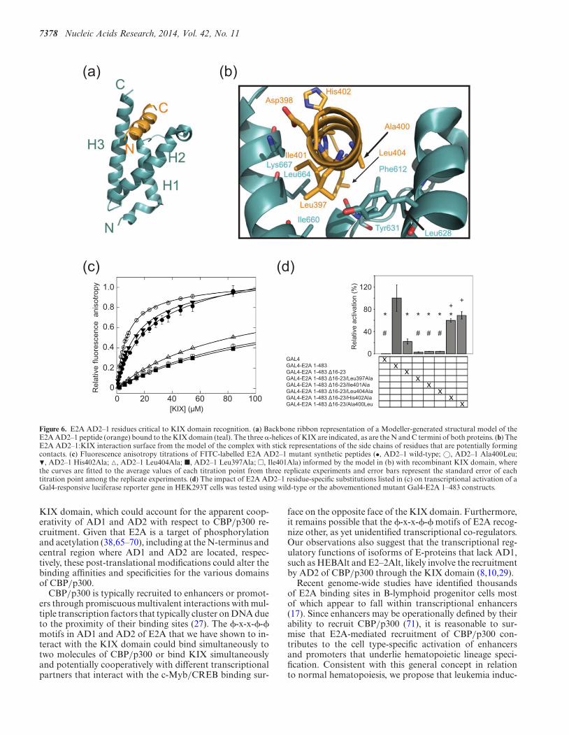
Modeling of the E2A AD2:KIX complex
A structural model of the E2A AD2–1:KIX complex wasgenerated using the E2A AD1-PCET:KIX complex struc-ture (PDB: 2KWF; (32)) as a template in Modeller (47) toaid in deciphering the molecular basis of this interaction(Figure 6a and b). The model suggested that the E2A AD2–1:KIX interaction involved numerous hydrophobic interac-tions, particularly along one face of the helical E2A AD2–1peptide that is buried in the hydrophobic cleft on the KIXdomain (Figure 6b). The hydrophobic residues Leu397,Ala400 and Ile401 within the �-x-x-�-� sequence of E2AAD2–1 were predicted to participate in extensive contactswith the KIX domain. These include interactions of Leu397of E2A AD2–1 with non-polar side chains on the �2 and�3 helices of the KIX domain, Ala400 with non-polar sidechains located on the �1 and �2 helices and Ile401 with non-polar side chains on the �3 helix of the KIX domain. Ad-ditionally, Leu404 was positioned to form hydrophobic in-teractions with non-polar residues within the L12 loop andthe �3 helix. Overall, these results are consistent with thefluorescence anisotropy data, in which the KIX�Mybsite mu-

Nucleic Acids Research, 2014, Vol. 42, No. 11 7375
Figure 3. NMR analysis of the E2A AD2–1:KIX interaction. Plot of average backbone amide chemical shift changes (��) versus the KIX sequence inducedby the binding of the E2A AD2–1 peptide. The backbone amide resonances for Arg623, Asn627 and Lys667 broadened out upon E2A AD2–1 binding andcould not be assigned in the bound spectrum. Chemical shifts were calculated using the formula �� = [(0.17��N)2 + (��HN)2]1/2, as previously described(43). The dashed line indicates the mean average chemical shift change.
tant bound E2A AD2–1 with wild-type affinity while theKIX�PCETsite mutant had a significantly lower affinity forE2A AD2–1, and indicate that E2A AD2–1 binds to thesurface on the KIX domain that is also bound by E2A AD1-PCET.
Affinity of the E2A AD2–1:KIX interaction correlates withE2A transcriptional activity
To investigate the functional implications of the E2A AD2–1:KIX interaction, the effect of alanine substitution ofLeu397 (Leu397Ala) and Ile401 (Ile401Ala) at the N- andC-terminus of the E2A AD2–1 �-x-x-�-� sequence andHis402 and Leu404, which lie C-terminal to this sequence,was tested in in vitro binding and transcriptional activa-tion assays (Figure 6c and d). In fluorescence anisotropytitration experiments, the Leu397Ala and Ile401Ala E2AAD2–1 mutants each displayed a 6-fold decrease in affin-ity for the KIX domain while the Leu404Ala mutation de-creased the affinity of E2A AD2–1 for the KIX domain byapproximately 5-fold (Figure 6c, Table 1). The His402AlaE2A AD2–1 mutant, which was generated as a negativecontrol based on its largely exposed position in the struc-tural model, did not significantly alter the affinity of theE2A AD2–1:KIX interaction (Figure 6c, Table 1).
The presence of a small hydrophobic residue at the fourthposition of the �-x-x-�-� motif in E2A AD2–1 (Ala400),our previous observation that an alanine substitution at thisposition in the E2A AD1-PCET motif (Leu19Ala) impairedthe ability of E2A (1–483) to pull down CBP/p300 (36), andthe structural model of the E2A AD2–1:KIX complex to-gether suggested that the affinity of this interaction couldbe enhanced by increasing the hydrophobicity at this po-sition in E2A AD2–1. Consistent with this hypothesis, anAla400Leu E2A AD2–1 mutant peptide bound to the KIXdomain with an affinity greater than twice that of the nativeE2A AD2–1 sequence (7 �M versus 17 �M; Table 1).
The consequences of these amino acid substitutions onthe transcriptional activation of E2A (1–483) were assessedin a cell-based reporter gene assay (Figure 6d). A mam-malian expression plasmid was engineered to express E2A(1–483) fused to the DNA-binding domain of GAL4. Thisplasmid was transiently co-transfected into SV293T cellswith a plasmid containing a firefly luciferase reporter generegulated by multiple GAL4 binding sites. Deletion of theAD1-PCET motif (�16–23) reduced E2A-driven expres-sion to 22% of wild-type E2A (1–483) (Figure 6d). Muta-tions affecting E2A AD2–1 were evaluated in the context�16–23 in order to determine their effects in an E2A con-struct whose transcriptional function relies predominantlyon AD2. E2A AD2 Leu397Ala, Ile401Ala or Leu404Alasubstitutions further reduced activity to less than 5% ofthat seen with wild-type E2A (1–483) (Figure 6d). In con-trast, the Ala400Leu or His402Ala substitutions partiallyrestored activity to 69% and 60%, respectively, of thatobserved with wild-type E2A(1–483). These substitutionslargely compensated for the impairment of AD1 functioncaused by �16–23, with the Ala400Leu mutation restor-ing activity to levels that were not significantly less thanthat observed for wild-type E2A(1–483) (P = 0.09) whilethe His402Ala mutation restored activity to levels onlymarginally less than wild-type E2A(1–483) (P = 0.04). Theobserved effects on E2A transcriptional activity are ap-proximately commensurate with the effects on KIX-bindingaffinity produced by the same mutations, which supportsthe functional relevance of the contact points predicted bythe E2A AD2–1:KIX model. These findings also suggestthat affinity for the KIX domain is a major determinant oftranscriptional activation by E2A.
Affinity of the E2A AD2–1:KIX interaction correlates withimmortalization of primary bone marrow cells by E2A-PBX1
Several studies have shown that E2A AD1 is required forE2A-PBX1-mediated oncogenesis whereas E2A AD2 is dis-

7376 Nucleic Acids Research, 2014, Vol. 42, No. 11
Figure 4. E2A AD2–1 binds to the KIX domain at the E2A PCET/MLL site. (a) Ribbon representation of the KIX domain from the E2A AD1-PCET:KIXcomplex (PDB: 2KWF; (32)) with those residues perturbed by more than 1 standard deviation above average chemical shift change upon saturation withE2A AD2–1 colored dark blue and resonances of residues that could not be assigned upon saturation due to peak broadening colored light purple. TheE2A AD1-PCET peptide was removed for clarity. (b) Surface representation of the KIX domain illustrated in panel (a). (c) E2A AD1-PCET:KIX complex(PDB: 2KWF; (32)) in which the KIX surface and a cartoon representation of E2A AD1-PCET (in green) are shown to illustrate its location on the KIXdomain. (d) Surface representation of the KIX domain depicted in panel (b) rotated 180◦. (e) Cartoon representation of the c-Myb peptide (pink) depictedon the surface of the KIX domain from the MLL:KIX:c-Myb complex (PDB: 2AGH; (31)).
pensable (20,36,57). The differential contribution by thesetwo ADs could be attributable to uncharacterized quali-tative differences in their functions, as reflected, for exam-ple, in their differential activity across different cell types(13,21). Alternatively, our current results raise the possibil-ity that the lesser oncogenic role of E2A AD2 may be ex-plained more simply by its lower affinity for the KIX do-main in CBP/p300. To investigate the possibility of a latentrelationship between E2A AD2-mediated recruitment ofCBP/p300 and E2A-PBX1-driven oncogenesis, amino acidsubstitutions that increased the affinity of E2A AD2 for the
KIX domain were engineered into full-length E2A-PBX1in the context of a substitution (namely Leu20Ala) that dis-rupts KIX binding to E2A AD1 and abrogates immortal-ization of primary bone marrow cells by E2A-PBX1 (36).Primary mouse bone marrow cells infected with a retro-virus that confers expression of E2A-PBX1 and then main-tained ex vivo in the presence of the cytokine granulocyte-macrophage colony-stimulating factor (GM-CSF) prolifer-ated exponentially and continuously after a brief latencyperiod, whereas cells infected with either the vector con-trol or the Leu20Ala-substituted E2A-PBX1 mutant de-

Nucleic Acids Research, 2014, Vol. 42, No. 11 7377
1H (ppm) 1H (ppm)
1H (ppm)1H (ppm)
15N
(p
pm)
15N
(p
pm)
(a)
(b)
A412
A412
E394
E394
Figure 5. E2A AD1-PCET and E2A AD2–1 compete for binding to thesame site on the KIX domain in vitro. (a) Overlay of two selected regionsfrom 2D 1H-15N HSQC spectra of 100 �M 15N-labeled E2A AD2–1/2 inthe absence (black) and presence of 300 �M unlabeled KIX (red). (b) Over-lay of same two selected regions depicted in (a) from 2D 1H-15N HSQCspectra of 100 �M 15N-labeled E2A AD2–1/2 in the absence (black) andpresence of 300 �M unlabeled KIX (red) and in the presence of 300 �Munlabeled KIX after addition of 1.3 mM E2A AD1(1–37) (green). Someresidues of E2A AD2–1/2 whose backbone amide resonances undergo sig-nificant 1H and 15N chemical shifts in the presence of KIX and are sub-sequently displaced by the addition of E2A AD1(1–37) are indicated byblack arrows, slngle-letter amino acid code and their respective position infull-length E2A, respectively.
clined rapidly after 2 weeks (Figure 7a). Cells infectedwith the Leu20Ala/Ala400Leu E2A-PBX1 mutant prolif-erated persistently for over 5 weeks, albeit at a lower ratethan those infected with wild-type E2A-PBX1, and ap-peared established in culture (Figure 7a). Unlike cells thathad been immortalized with wild-type E2A-PBX1, whichwere predominantly non-adherent, the cells expressing theLeu20Ala/Ala400Leu E2A-PBX1 mutant rapidly segre-gated after each passage into non-adherent and adherentsub-populations with the latter forming a confluent mono-layer of fusiform cells. Characterization by flow cytometryindicated that the cells immortalized with wild-type E2A-PBX1 manifested an immunophenotype (CD11b+ F4/80+
Gr-1−) characteristic of granulocyte/macrophage progeni-tors (Figure 7b). The immunophenotype of cells expressingthe Leu20Ala/Ala400Leu E2A-PBX1 mutant depended ontheir adherence status. Whereas the non-adherent popula-tion included many cells with evidence of granulocytic dif-ferentiation (F4/80−Gr-1+), the adherent cells included adistinct and prevalent population of cells with macrophagefeatures (F4/80+Gr-1+/−). This experiment was carriedout twice with equivalent results. These results suggestthat Leu20Ala/Ala400Leu E2A-PBX1 mutant is less ef-fective than the wild-type oncoprotein in blocking GM-CSF-induced differentiation of hematopoietic progenitorsto granulocytes and macrophages. More generally, thesefunctional results based on primary mouse bone marrowcells show that increasing the affinity of E2A AD2 for theKIX domain of CBP/p300 to one approaching the affin-
ity of the E2A AD1-PCET:KIX interaction uncovered a la-tent potential of E2A AD2 to mediate potentially oncogenictranscriptional effects of E2A-PBX1.
DISCUSSION
The second AD of E2A, AD2, has previously been impli-cated in the regulation E2A-mediated transcription induc-tion and lymphoid development; roles consistent with itsability to interact with CBP/p300 (20–22). The presenceof two tandem �-x-x-�-� motifs within AD2 of E2A, aswell as HEB and E2–2, suggested the hypothetical possi-bility of cooperative binding to the two well-characterizedbinding surfaces on the KIX domain. However, our find-ings demonstrate that a region comprising only the moreN-terminal of these motifs in E2A AD2 (397-Leu-Asp-Glu-Ala-Ile-401) binds KIX with micromolar affinity (Table 1),and that this motif binds to the same hydrophobic cleft onthe KIX domain as that targeted by the �-x-x-�-� contain-ing PCET motif of E2A AD1 (Figure 4). Furthermore, theobservation that disruption of the c-Myb/CREB bindingsite did not impair E2A binding and that this surface ison the opposite face of the KIX domain relative to thatbound by the E2A domains (Figure 2; (51,52)) and AD1and AD2 directly compete for the same binding site on theKIX domain (Figure 5) rules out any potential participa-tion of the c-Myb/CREB site in simultaneous binding toKIX by multiple �-x-x-�-� motifs on E2A. Finally, struc-tures of the AD1-PCET:KIX (32), MLL:KIX:c-Myb (31),FOXO3a:KIX (35) and MLL:KIX:pKID (58) complexeshave a deep binding cleft, which we have shown here to rec-ognize E2A AD2, with a contiguous hydrophobic surfaceand narrow apex formed by helices 2 and 3 and interveningL12 loop and G2 310-helix of the KIX domain. The extensivecontacts between the KIX domain and helical ADs at thissite in the complex structures suggest that, without a signif-icant conformation change yet to be observed for the KIXdomain, this binding site could only occupy a single binding�-x-x-�-� motif thereby ruling out cooperative binding ofthe E2A ADs at the same site; a hypothesis support by ourNMR-based competition experiments (Figure 5). Thus, ourfindings provide support and a structural basis for the func-tional redundancy observed for E2A AD1 and AD2 (20–22).
Our findings are inconsistent with demonstrated abilityof E2A AD1 and AD2–1 to induce reporter gene expressionand bind to CBP/p300 in a greater-than-additive manner(20,21). CBP/p300 comprises several domains in additionto KIX, including the TAZ1, TAZ2/CH3 and NCBD/IBiDdomains, which have been shown to participate protein–protein interactions, most notably with ADs of varioustranscription factors (27). For example, the two N-terminaltranscriptional ADs of p53 can bind the KIX, TAZ1, TAZ2and NCBD domains of CBP/p300 (34,50,59–61); interac-tions which are modulated phosphorylation (50,60,62–64).In the case of E2A, we have shown that the KIX domainis absolutely required for binding of full-length CBP toE2A 1–483, as determined using purified proteins in a pull-down experiment (37). However, it remains possible that el-ements within AD1 or AD2 of E2A can also interact withthese other domains of CBP/p300 in conjunction with the
7378 Nucleic Acids Research, 2014, Vol. 42, No. 11
(a) (b)
(c) (d)
0 20 40 60 80 100[KIX] ( M)
0
0.2
0.4
0.6
0.8
1.0
Rel
ativ
e flu
ores
cenc
e an
isot
ropy
0
40
80
120
Rel
ativ
e ac
tivat
ion
(%)
GAL4GAL4-E2A 1-483GAL4-E2A 1-483 16-23GAL4-E2A 1-483 16-23/Leu397AlaGAL4-E2A 1-483 16-23/Ile401AlaGAL4-E2A 1-483 16-23/Leu404AlaGAL4-E2A 1-483 16-23/His402AlaGAL4-E2A 1-483 16-23/Ala400Leu
Figure 6. E2A AD2–1 residues critical to KIX domain recognition. (a) Backbone ribbon representation of a Modeller-generated structural model of theE2A AD2–1 peptide (orange) bound to the KIX domain (teal). The three �-helices of KIX are indicated, as are the N and C termini of both proteins. (b) TheE2A AD2–1:KIX interaction surface from the model of the complex with stick representations of the side chains of residues that are potentially formingcontacts. (c) Fluorescence anisotropy titrations of FITC-labelled E2A AD2–1 mutant synthetic peptides (•, AD2–1 wild-type; ©, AD2–1 Ala400Leu;�, AD2–1 His402Ala; �, AD2–1 Leu404Ala; �, AD2–1 Leu397Ala; �, Ile401Ala) informed by the model in (b) with recombinant KIX domain, wherethe curves are fitted to the average values of each titration point from three replicate experiments and error bars represent the standard error of eachtitration point among the replicate experiments. (d) The impact of E2A AD2–1 residue-specific substitutions listed in (c) on transcriptional activation of aGal4-responsive luciferase reporter gene in HEK293T cells was tested using wild-type or the abovementioned mutant Gal4-E2A 1–483 constructs.
KIX domain, which could account for the apparent coop-erativity of AD1 and AD2 with respect to CBP/p300 re-cruitment. Given that E2A is a target of phosphorylationand acetylation (38,65–70), including at the N-terminus andcentral region where AD1 and AD2 are located, respec-tively, these post-translational modifications could alter thebinding affinities and specificities for the various domainsof CBP/p300.
CBP/p300 is typically recruited to enhancers or promot-ers through promiscuous multivalent interactions with mul-tiple transcription factors that typically cluster on DNA dueto the proximity of their binding sites (27). The �-x-x-�-�motifs in AD1 and AD2 of E2A that we have shown to in-teract with the KIX domain could bind simultaneously totwo molecules of CBP/p300 or bind KIX simultaneouslyand potentially cooperatively with different transcriptionalpartners that interact with the c-Myb/CREB binding sur-
face on the opposite face of the KIX domain. Furthermore,it remains possible that the �-x-x-�-� motifs of E2A recog-nize other, as yet unidentified transcriptional co-regulators.Our observations also suggest that the transcriptional reg-ulatory functions of isoforms of E-proteins that lack AD1,such as HEBAlt and E2–2Alt, likely involve the recruitmentby AD2 of CBP/p300 through the KIX domain (8,10,29).
Recent genome-wide studies have identified thousandsof E2A binding sites in B-lymphoid progenitor cells mostof which appear to fall within transcriptional enhancers(17). Since enhancers may be operationally defined by theirability to recruit CBP/p300 (71), it is reasonable to sur-mise that E2A-mediated recruitment of CBP/p300 con-tributes to the cell type-specific activation of enhancersand promoters that underlie hematopoietic lineage speci-fication. Consistent with this general concept in relationto normal hematopoiesis, we propose that leukemia induc-

Nucleic Acids Research, 2014, Vol. 42, No. 11 7379
Figure 7. An E2A AD2 Ala400Leu substitution restores immortalization of primary bone marrow cells by AD1-defective E2A-PBX1. (a) Proliferation ofhematopoietic progenitors expressing E2A-PBX1 or engineered variant. For each construct, 1.25 × 10⁶ bone marrow cells were seeded initially in GM-CSF-containing medium immediately after retroviral infection and counted for 40 days. (Inset) Western blot of lysates from NIH 3T3 fibroblasts infectedwith retroviruses conferring expression of the indicated E2A-PBX1b constructs, confirming equivalent viral transduction and expression of recombinantproteins. (b) Flow cytometry-based immunophenotyping of myeloid cells expressing E2A-PBX1 (wild-type or Leu20Ala/Ala400Leu) after 40 days ofculture. The cells immortalized with E2A-PBX1 (Leu20Ala/Ala400Leu) were separated into adherent and non-adherent layers for separate analysis andall samples were stained for CD11b, F4/80 and Gr-1.

7380 Nucleic Acids Research, 2014, Vol. 42, No. 11
tion by E2A-PBX1 involves the aberrant redistribution ofCBP/p300 and other transcriptional co-regulators acrossthe genome at one or more critical phases of hematopoi-etic development. This results in the establishment of an ab-normal, self-sustaining transcriptional gene regulatory net-work and a leukemic cellular phenotype characterized byaberrant retention of cellular immaturity and self-renewalpotential.
The oncogenic importance of aberrant CBP/p300 re-cruitment mediated by E2A-PBX1 is supported by ourdemonstration in the current and previous studies that theoncogenic potency of engineered E2A-PBX1 mutants ispredictable based on their affinity for CBP/p300. Whereaswe showed previously that amino acid substitutions withinthe E2A AD1-PCET motif that reduce its affinity forCBP/p300 impair oncogenesis (20,36), we show in the cur-rent study that a different substitution (namely Ala400Leu)that increases the affinity of E2A AD2 for CBP/p300 un-covers a latent ability of E2A AD2 to mediate E2A-PBX1oncogenesis. Indeed, the ability of the individual E2A ADsto recruit CBP/p300 seems to be a better predictor oftheir oncogenic function than their transcriptional poten-cies as measured using reporter gene assays. In particular,while deletions within E2A AD2 are more deleterious fortrans-activation than deletions within E2A AD1, they haveno measurable effect on E2A-PBX1-mediated oncogenesis(20,21,36). We speculate that this discordance reflects thefailure of simple reporter assays to accurately model the po-tentially complex and subtle gene regulatory consequencesof aberrantly recruiting CBP/p300 to potentially numer-ous cis regulatory elements and perhaps away from othersacross the genome.
The binding affinities of the E2A AD1 (Kd 5.2 �M),Ala400Leu-substituted E2A AD2 (Kd 7.2 �M) andHis402Ala-substituted E2A AD2 (Kd 13.5 �M) for the KIXdomain correlate well with the ability of full-length E2A-PBX1 to immortalize primary myeloid cells (Table 1 andFigure 6). The relatively modest growth rate, the emer-gence of adherent macrophages and the presence of im-munophenotypic features indicating more advanced granu-locytic and macrophage differentiation manifested by cellsexpressing Ala400Leu-substituted E2A-PBX1 suggest at-tenuated oncogenic potency attributable to the marginalaffinity of this mutant for the KIX domain and CBP/p300.These results also tend to validate the use of biophysicalstudies with E2A fragments to model and predict function-ally important interactions between full-length E2A-PBX1and CBP/p300.
The data presented here confirm that AD1 and AD2 ofE2A both bind the KIX domain at the same site, a relativelysmall hydrophobic cleft distinct from where transcriptionfactors including c-Myb and CREB bind (51,52). Giventhe importance of this interaction in oncogenesis by E2A-PBX1, inhibition binding to this cleft on the KIX domaincould prove a useful therapeutic approach in E2A-PBX1-associated leukemia.
SUPPLEMENTARY DATA
Supplementary Data are available at NAR Online.
ACKNOWLEDGEMENTS
The authors would like to thank Dr Peter Wright (ScrippsInstitute, La Jolla, CA) for the KIX expression plasmidand Dr Tara Sprules for assistance with NMR data collec-tion. A.C.K. was a Canadian Institutes of Health ResearchCGS-M Scholarship recipient. C.M.D. was a Natural Sci-ences and Engineering Research Council of Canada CGS-D Scholarship recipient.Conflict of interest statement None declared.
FUNDING
Canadian Institutes of Health Research [MOP 106471to S.P.S., MOP 81333 to D.P.L.]; Terry Fox Foundation-Canadian Institutes of Health Research Training Programin Transdisciplinary Cancer Research [D.N.L.]. Fundingfor open access charge: Canadian Institutes of Health Re-search.
REFERENCES1. Bryder,D. and Sigvardsson,M. (2010) Shaping up a lineage–lessons
from B lymphopoesis. Curr. Opin. Immunol., 22, 148–153.2. Hesslein,D.G. and Lanier,L.L. (2011) Transcriptional control of
natural killer cell development and function. Adv. Immunol., 109,45–85.
3. Koch,U. and Radtke,F. (2011) Mechanisms of T cell developmentand transformation. Annu. Rev. Cell Dev. Biol., 27, 539–562.
4. Naito,T., Tanaka,H., Naoe,Y. and Taniuchi,I. (2011) Transcriptionalcontrol of T-cell development. Int. Immunol., 23, 661–668.
5. Nutt,S.L. and Kee,B.L. (2007) The transcriptional regulation of B celllineage commitment. Immunity, 26, 715–725.
6. Ramirez,K. and Kee,B.L. (2010) Multiple hats for natural killers.Curr. Opin. Immunol., 22, 193–198.
7. Santos,P., Arumemi,F., Park,K.S., Borghesi,L. and Milcarek,C.(2011) Transcriptional and epigenetic regulation of B celldevelopment. Immunol. Res., 50, 105–112.
8. Braunstein,M. and Anderson,M.K. (2012) HEB in the spotlight:Transcriptional regulation of T-cell specification, commitment, anddevelopmental plasticity. Clin. Dev. Immunol., 2012, 678705–678720.
9. de Pooter,R.F. and Kee,B.L. (2010) E proteins and the regulation ofearly lymphocyte development. Immunol. Rev., 238, 93–109.
10. Kee,B.L. (2009) E and ID proteins branch out. Nat. Rev. Immunol., 9,175–184.
11. Xu,W., Carr,T., Ramirez,K., McGregor,S., Sigvardsson,M. andKee,B.L. (2013) E2A transcription factors limit expression of Gata3to facilitate T lymphocyte lineage commitment. Blood, 121,1534–1542.
12. Zhuang,Y., Soriano,P. and Weintraub,H. (1994) The helix-loop-helixgene E2A is required for B cell formation. Cell, 79, 875–884.
13. Aronheim,A., Shiran,R., Rosen,A. and Walker,M.D. (1993) The E2Agene product contains two separable and functionally distincttranscription activation domains. Proc. Natl. Acad. Sci. U.S.A., 90,8063–8067.
14. Henthorn,P., Kiledjian,M. and Kadesch,T. (1990) Two distincttranscription factors that bind the immunoglobulin enhancermicroE5/kappa 2 motif. Science, 247, 467–470.
15. Hu,J.S., Olson,E.N. and Kingston,R.E. (1992) HEB, ahelix-loop-helix protein related to E2A and ITF2 that can modulatethe DNA-binding ability of myogenic regulatory factors. Mol. Cell.Biol., 12, 1031–1042.
16. Greenbaum,S. and Zhuang,Y. (2002) Identification of E2A targetgenes in B lymphocyte development by using a gene tagging-basedchromatin immunoprecipitation system. Proc. Natl. Acad. Sci.U.S.A., 99, 15030–15035.
17. Lin,Y.C., Jhunjhunwala,S., Benner,C., Heinz,S., Welinder,E.,Mansson,R., Sigvardsson,M., Hagman,J., Espinoza,C.A.,Dutkowski,J. et al. (2010) A global network of transcription factors,involving E2A, EBF1 and Foxo1, that orchestrates B cell fate. Nat.Immunol., 11, 635–643.

Nucleic Acids Research, 2014, Vol. 42, No. 11 7381
18. Massari,M.E. and Murre,C. (2000) Helix-loop-helix proteins:regulators of transcription in eucaryotic organisms. Mol. Cell. Biol.,20, 429–440.
19. Murre,C., McCaw,P.S., Vaessin,H., Caudy,M., Jan,L.Y., Jan,Y.N.,Cabrera,C.V., Buskin,J.N., Hauschka,S.D., Lassar,A.B. et al. (1989)Interactions between heterologous helix-loop-helix proteins generatecomplexes that bind specifically to a common DNA sequence. Cell,58, 537–544.
20. Bayly,R., Chuen,L., Currie,R.A., Hyndman,B.D., Casselman,R.,Blobel,G.A. and LeBrun,D.P. (2004) E2A-PBX1 interacts directlywith the KIX domain of CBP/p300 in the induction of proliferationin primary hematopoietic cells. J. Biol. Chem., 279, 55362–55371.
21. Bhalla,S., Spaulding,C., Brumbaugh,R.L., Zagort,D.E.,Massari,M.E., Murre,C. and Kee,B.L. (2008) Differential roles forthe E2A activation domains in B lymphocytes and macrophages. J.Immunol., 180, 1694–1703.
22. Bradney,C., Hjelmeland,M., Komatsu,Y., Yoshida,M., Yao,T.P. andZhuang,Y. (2003) Regulation of E2A activities by histoneacetyltransferases in B lymphocyte development. J. Biol. Chem., 278,2370–2376.
23. Massari,M.E., Grant,P.A., Pray-Grant,M.G., Berger,S.L.,Workman,J.L. and Murre,C. (1999) A conserved motif present in aclass of helix-loop-helix proteins activates transcription by directrecruitment of the SAGA complex. Mol. Cell, 4, 63–73.
24. Massari,M.E., Jennings,P.A. and Murre,C. (1996) The AD1transactivation domain of E2A contains a highly conserved helixwhich is required for its activity in both Saccharomyces cerevisiae andmammalian cells. Mol. Cell. Biol., 16, 121–129.
25. Quong,M.W., Massari,M.E., Zwart,R. and Murre,C. (1993) A newtranscriptional-activation motif restricted to a class ofhelix-loop-helix proteins is functionally conserved in both yeast andmammalian cells. Mol. Cell. Biol., 13, 792–800.
26. Sepp,M., Kannike,K., Eesmaa,A., Urb,M. and Timmusk,T. (2011)Functional diversity of human basic helix-loop-helix transcriptionfactor TCF4 isoforms generated by alternative 5’ exon usage andsplicing. PLoS One, 6, e22138.
27. Wang,F., Marshall,C.B. and Ikura,M. (2013)Transcriptional/epigenetic regulator CBP/p300 in tumorigenesis:structural and functional versatility in target recognition. Cell. Mol.Life Sci., 70, 3989–4008.
28. Zhang,J., Kalkum,M., Yamamura,S., Chait,B.T. and Roeder,R.G.(2004) E protein silencing by the leukemogenic AML1-ETO fusionprotein. Science, 305, 1286–1289.
29. Wang,D., Claus,C.L., Vaccarelli,G., Braunstein,M., Schmitt,T.M.,Zuniga-Pflucker,J.C., Rothenberg,E.V. and Anderson,M.K. (2006)The basic helix-loop-helix transcription factor HEBAlt is expressedin pro-T cells and enhances the generation of T cell precursors. J.Immunol., 177, 109–119.
30. Plevin,M.J., Mills,M.M. and Ikura,M. (2005) The LxxLL motif: amultifunctional binding sequence in transcriptional regulation.Trends Biochem. Sci., 30, 66–69.
31. De Guzman,R.N., Goto,N.K., Dyson,H.J. and Wright,P.E. (2006)Structural basis for cooperative transcription factor binding to theCBP coactivator. J. Mol. Biol., 355, 1005–1013.
32. Denis,C.M., Chitayat,S., Plevin,M.J., Wang,F., Thompson,P., Liu,S.,Spencer,H.L., Ikura,M., LeBrun,D.P. and Smith,S.P. (2012)Structural basis of CBP/p300 recruitment in leukemia induction byE2A-PBX1. Blood, 120, 3968–3977.
33. Goto,N.K., Zor,T., Martinez-Yamout,M., Dyson,H.J. andWright,P.E. (2002) Cooperativity in transcription factor binding tothe coactivator CREB-binding protein (CBP). The mixed lineageleukemia protein (MLL) activation domain binds to an allosteric siteon the KIX domain. J. Biol. Chem., 277, 43168–43174.
34. Teufel,D.P., Freund,S.M., Bycroft,M. and Fersht,A.R. (2007) Fourdomains of p300 each bind tightly to a sequence spanning bothtransactivation subdomains of p53. Proc. Natl. Acad. Sci. U.S.A.,104, 7009–7014.
35. Wang,F., Marshall,C.B., Yamamoto,K., Li,G.Y.,Gasmi-Seabrook,G.M., Okada,H., Mak,T.W. and Ikura,M. (2012)Structures of KIX domain of CBP in complex with two FOXO3atransactivation domains reveal promiscuity and plasticity incoactivator recruitment. Proc. Natl. Acad. Sci. U.S.A., 109,6078–6083.
36. Bayly,R., Murase,T., Hyndman,B.D., Savage,R., Nurmohamed,S.,Munro,K., Casselman,R., Smith,S.P. and LeBrun,D.P. (2006) Criticalrole for a single leucine residue in leukemia induction by E2A-PBX1.Mol. Cell. Biol., 26, 6442–6452.
37. Hyndman,B.D., Thompson,P., Bayly,R., Cote,G.P. and LeBrun,D.P.(2012) E2A proteins enhance the histone acetyltransferase activity ofthe transcriptional co-activators CBP and p300. Biochim. Biophys.Acta, 1819, 446–453.
38. Hyndman,B.D., Thompson,P., Denis,C.M., Chitayat,S., Bayly,R.,Smith,S.P. and LeBrun,D.P. (2012) Mapping acetylation sites in E2Aidentifies a conserved lysine residue in activation domain 1 thatpromotes CBP/p300 recruitment and transcriptional activation.Biochim. Biophys. Acta, 1819, 375–381.
39. Kamps,M.P. (1997) E2A-Pbx1 induces growth, blocks differentiation,and interacts with other homeodomain proteins regulating normaldifferentiation. Curr. Topics Microbiol. Immunol., 220, 25–43.
40. Kamps,M.P. and Baltimore,D. (1993) E2A-Pbx1, the t(1;19)translocation protein of human pre-B-cell acute lymphocyticleukemia, causes acute myeloid leukemia in mice. Mol. Cell. Biol., 13,351–357.
41. LeBrun,D.P. (2003) E2A basic helix-loop-helix transcription factorsin human leukemia. Front. Biosci., 8, s206–s222.
42. Monica,K., LeBrun,D.P., Dedera,D.A., Brown,R. and Cleary,M.L.(1994) Transformation properties of the E2a-Pbx1 chimericoncoprotein: fusion with E2a is essential, but the Pbx1 homeodomainis dispensable. Mol. Cell. Biol., 14, 8304–8314.
43. Farmer,B.T. II, Constantine,K.L., Goldfarb,V., Friedrichs,M.S.,Wittekind,M., Yanchunas,J. Jr, Robertson,J.G. and Mueller,L. (1996)Localizing the NADP+ binding site on the MurB enzyme by NMR.Nat. Struct. Biol., 3, 995–997.
44. Delaglio,F., Grzesiek,S., Vuister,G.W., Zhu,G., Pfeifer,J. and Bax,A.(1995) NMRPipe: a multidimensional spectral processing systembased on UNIX pipes. J. Biomol. NMR, 6, 277–293.
45. Johnson,B.A. (2004) Using NMRView to visualize and analyze theNMR spectra of macromolecules. Methods Mol. Biol., 278, 313–352.
46. Simossis,V.A. and Heringa,J. (2005) PRALINE: a multiple sequencealignment toolbox that integrates homology-extended and secondarystructure information. Nucleic Acids Res., 33, W289–294.
47. Sali,A. and Blundell,T.L. (1993) Comparative protein modelling bysatisfaction of spatial restraints. J. Mol. Biol., 234, 779–815.
48. DeLano,W.L. (2009). DeLano Scientific, San Carlos, CA.www.pymol.org (8 March 2014, date last accessed)
49. Scott,S.P., Teh,A., Peng,C. and Lavin,M.F. (2002) One-stepsite-directed mutagenesis of ATM cDNA in large (20kb) plasmidconstructs. Hum. Mutat., 20, 323-327.
50. Lee,C.W., Arai,M., Martinez-Yamout,M.A., Dyson,H.J. andWright,P.E. (2009) Mapping the interactions of the p53transactivation domain with the KIX domain of CBP. Biochemistry,48, 2115–2124.
51. Radhakrishnan,I., Perez-Alvarado,G.C., Parker,D., Dyson,H.J.,Montminy,M.R. and Wright,P.E. (1997) Solution structure of theKIX domain of CBP bound to the transactivation domain of CREB:a model for activator:coactivator interactions. Cell, 91, 741–752.
52. Zor,T., De Guzman,R.N., Dyson,H.J. and Wright,P.E. (2004)Solution structure of the KIX domain of CBP bound to thetransactivation domain of c-Myb. J. Mol. Biol., 337, 521–534.
53. Kasper,L.H., Boussouar,F., Ney,P.A., Jackson,C.W., Rehg,J., vanDeursen,J.M. and Brindle,P.K. (2002) A transcription-factor-bindingsurface of coactivator p300 is required for haematopoiesis. Nature,419, 738–743.
54. Ramirez,J.A. and Nyborg,J.K. (2007) Molecular characterization ofHTLV-1 Tax interaction with the KIX domain of CBP/p300. J. Mol.Biol., 372, 958–969.
55. Campbell,K.M. and Lumb,K.J. (2002) Structurally distinct modes ofrecognition of the KIX domain of CBP by Jun and CREB.Biochemistry, 41, 13956–13964.
56. Vendel,A.C., McBryant,S.J. and Lumb,K.J. (2003) KIX-mediatedassembly of the CBP-CREB-HTLV-1 tax coactivator-activatorcomplex. Biochemistry, 42, 12481–12487.
57. Kamps,M.P., Wright,D.D. and Lu,Q. (1996) DNA-binding byoncoprotein E2a-Pbx1 is important for blocking differentiation butdispensable for fibroblast transformation. Oncogene, 12, 19–30.

7382 Nucleic Acids Research, 2014, Vol. 42, No. 11
58. Bruschweiler,S., Konrat,R. and Tollinger,M. (2013) AllostericCommunication in the KIX Domain Proceeds through DynamicRepacking of the Hydrophobic Core. ACS Chem. Biol., 8, 1600–1610.
59. Feng,H., Jenkins,L.M., Durell,S.R., Hayashi,R., Mazur,S.J.,Cherry,S., Tropea,J.E., Miller,M., Wlodawer,A., Appella,E. et al.(2009) Structural basis for p300 Taz2-p53 TAD1 binding andmodulation by phosphorylation. Structure, 17, 202–210.
60. Jenkins,L.M., Yamaguchi,H., Hayashi,R., Cherry,S., Tropea,J.E.,Miller,M., Wlodawer,A., Appella,E. and Mazur,S.J. (2009) Twodistinct motifs within the p53 transactivation domain bind to theTaz2 domain of p300 and are differentially affected byphosphorylation. Biochemistry, 48, 1244–1255.
61. Wells,M., Tidow,H., Rutherford,T.J., Markwick,P., Jensen,M.R.,Mylonas,E., Svergun,D.I., Blackledge,M. and Fersht,A.R. (2008)Structure of tumor suppressor p53 and its intrinsically disorderedN-terminal transactivation domain. Proc. Natl. Acad. Sci. U.S.A.,105, 5762–5767.
62. Polley,S., Guha,S., Roy,N.S., Kar,S., Sakaguchi,K., Chuman,Y.,Swaminathan,V., Kundu,T. and Roy,S. (2008) Differential recognitionof phosphorylated transactivation domains of p53 by different p300domains. J. Mol. Biol., 376, 8–12.
63. Ferreon,J.C., Lee,C.W., Arai,M., Martinez-Yamout,M.A.,Dyson,H.J. and Wright,P.E. (2009) Cooperative regulation of p53 bymodulation of ternary complex formation with CBP/p300 andHDM2. Proc. Natl. Acad. Sci. U.S.A., 106, 6591–6596.
64. Teufel,D.P., Bycroft,M. and Fersht,A.R. (2009) Regulation byphosphorylation of the relative affinities of the N-terminal
transactivation domains of p53 for p300 domains and Mdm2.Oncogene, 28, 2112–2118.
65. King,A.M., Van der Put,E., Blomberg,B.B. and Riley,R.L. (2007)Accelerated Notch-dependent degradation of E47 proteins in aged Bcell precursors is associated with increased ERK MAPK activation. J.Immunol., 178, 3521–3529.
66. Lluis,F., Ballestar,E., Suelves,M., Esteller,M. and Munoz-Canoves,P.(2005) E47 phosphorylation by p38 MAPK promotes MyoD/E47association and muscle-specific gene transcription. EMBO J., 24,974–984.
67. Neufeld,B., Grosse-Wilde,A., Hoffmeyer,A., Jordan,B.W., Chen,P.,Dinev,D., Ludwig,S. and Rapp,U.R. (2000) Serine/Threonine kinases3pK and MAPK-activated protein kinase 2 interact with the basichelix-loop-helix transcription factor E47 and repress itstranscriptional activity. J. Biol. Chem., 275, 20239–20242.
68. Pedraza,N., Rafel,M., Navarro,I., Encinas,M., Aldea,M. andGallego,C. (2009) Mixed lineage kinase phosphorylates transcriptionfactor E47 and inhibits TrkB expression to link neuronal death andsurvival pathways. J. Biol. Chem., 284, 32980–32988.
69. Sloan,S.R., Shen,C.P., McCarrick-Walmsley,R. and Kadesch,T.(1996) Phosphorylation of E47 as a potential determinant ofB-cell-specific activity. Mol. Cell. Biol., 16, 6900–6908.
70. Teachenor,R., Beck,K., Wright,L.Y., Shen,Z., Briggs,S.P. andMurre,C. (2012) Biochemical and phosphoproteomic analysis of thehelix-loop-helix protein E47. Mol. Cell. Biol., 32, 1671–1682.
71. Visel,A., Rubin,E.M. and Pennacchio,L.A. (2009) Genomic views ofdistant-acting enhancers. Nature, 461, 199–205.
Copyright © 2022 FDOKUMEN